Embed Size (px)
Citation preview
BIOLOGY OF REPRODUCTION 81, 46–55 (2009)Published online before print 4 March 2009.DOI 10.1095/biolreprod.108.075390
Disruption of Poly(ADP-Ribose) Homeostasis Affects Spermiogenesis and SpermChromatin Integrity in Mice1
Mirella L. Meyer-Ficca,3 Julia Lonchar,3 Christine Credidio,3 Motomasa Ihara,3 Yun Li,3 Zhao-Qi Wang,4,5
and Ralph G. Meyer2,3
Department of Animal Biology and Mari Lowe Center for Comparative Oncology,3 University of Pennsylvania School ofVeterinary Medicine, New Bolton Center for Animal Transgenesis and Germ Cell Research,Kennett Square, PennsylvaniaGenome Stability Group,4 Leibnitz Institute of Age Research-Fritz Lipmann Institute (FLI), Jena, GermanyFaculty of Biology and Pharmacy,5 Friedrich-Schiller-University, Jena, Germany
ABSTRACT
The major function of sperm is the delivery of the paternalgenome to the metaphase II oocyte, ensuring transmission of thegenetic information to the next generation. For successfulfertilization and healthy offspring, sperm DNA must be protectedfrom exogenous insults. This is achieved by packaging the spermDNA into a condensed protamine-bound form, preceded by theprecisely orchestrated removal of histones and intermittentinsertion and removal of transition proteins. This remodelingprocess requires relaxation of supercoiled DNA by transientformation of physiological strand breaks that spermatids, beinghaploid, cannot repair by homologous recombination. In somaticcells, the presence of DNA strand breaks rapidly induces theformation of poly(ADP-ribose) by nuclear poly(ADP-ribose)polymerases, which in turn facilitates DNA strand breaksignaling and assembly of DNA repair complexes. We reportedearlier that chromatin remodeling steps during spermiogenesistrigger poly(ADP-ribose) (PAR) formation. Here, we show thatknockout mice deficient in PARP1, PARG (110-kDa isoform), orboth display morphological and functional sperm abnormalitiesthat are dependent on the individual genotypes, includingresidual DNA strand breaks associated with varying degrees ofsubfertility. The data presented highlight the importance of PARmetabolism, particularly PARG function, as a prerequisite ofproper sperm chromatin quality.
chromatin condensation, chromatin remodeling, condensation,gametogenesis, PARG, PARP, PARP1, PARP2, poly(ADP-ribose),poly(ADP-ribose) glycohydrolase, poly(ADP-ribose) polymerase,residual DNA strand breaks, sonication-resistant spermatid nuclei,sperm, spermatid, spermatogenesis, spermiogenesis
INTRODUCTION
In humans, incomplete chromatin condensation and thepresence of persistent DNA strand breaks indicate a severe
reduction of the fertilization potential in mature spermatozoa[1]. Sperm DNA is normally shielded from potentiallycompromising environmental effects by extremely tightpackaging into transcriptionally inactive chromatin associatedwith protamines linked by covalent disulfite bonds. Thechromatin structure of sperm cells is therefore completelydifferent from that of somatic cells [2, 3].
The complex chromatin reorganization during spermiogen-esis is dramatic, and the steps that lead to packaging of thepaternal genome into the compact transport form are notentirely understood. Histones are first replaced by the transitionproteins TNP1 and TNP2, which are subsequently replaced byprotamines. Along with the exchange of proteins, the DNAstructure changes from the supercoiled nucleosomal structureto a relaxed state [4–6]. This requires the occurrence ofcontrolled transient DNA strand breaks [7–9], particularlyduring the exchange of histones by transition proteins inelongating spermatids such as spermatids of steps 9–12 in mice[10]. Such DNA strand breaks are apparently mostly affordedby the activity of enzymes such as topoisomerase II b (TOP2B)[11, 12] and are therefore presumably mostly protein bound.However, at a certain frequency, open DNA double-strandbreaks appear to be also formed in this process, posing a threatto the developing spermatid cell.
Compared with somatic cells, the presence of DNA strandbreaks carries a greater risk for haploid spermatid cells, as theyare not able to undergo homologous recombination, the favoredand most accurate way of repairing DNA double-strand breaksin diploid cells. Cellular DNA damage repair is facilitated bytwo members of the poly(ADP-ribose) polymerase family,poly(ADP-ribose) polymerase 1 (PARP1) and poly(ADP-ribose) polymerase 2 (PARP2). DNA strand breaks directlyand immediately recruit the ubiquitous PARP1 and PARP2. Onactivation, PARP1 forms a large branched biopolymerpoly(ADP-ribose) (PAR) by cleaving NADþ into nicotinamideand ADP-ribose, which becomes polymerized and covalentlyattached to acceptor proteins such as PARP1 itself in anintermolecular automodification reaction. Examples of othertarget proteins for this posttranslational modification arehistones, p53, and DNA ligase III. Breakdown of the uniquepolymer is facilitated by poly(ADP-ribose) glycohydrolase(PARG), with PAR turnover being a rapid and dynamicprocess mediated by the interplay of PARP1 and PARG [13–18]. Automodified PARP1 is catalytically inactive and isreleased from the DNA strand break. PARP2, another memberof the PARP protein family, has been shown to respondsimilarly and as a backup. The ability of PARP1 and PARP2 tobind to DNA strand breaks and to become activated again isrestored on removal of PAR by PARG, allowing for repeated
1Supported by grants from the National Institutes of Health (NIH R01HD048837 to R.G.M.) and the Mari Lowe Center for ComparativeOncology at the University of Pennsylvania. Z.-Q.W. was supported byAssociation pour la Recherche sur le Cancer (ARC) France.2Correspondence: Ralph G. Meyer, Department of Animal Biology andMari Lowe Center for Comparative Oncology, University of Pennsylva-nia School of Veterinary Medicine, New Bolton Center, Kennett Square,PA 19348. FAX: 610 925 8121; e-mail: [email protected]
Received: 10 December 2008.First decision: 13 January 2009.Accepted: 4 February 2009.� 2009 by the Society for the Study of Reproduction, Inc.eISSN: 1259-7268 http://www.biolreprod.orgISSN: 0006-3363
46
Dow
nloaded from w
ww
.biolreprod.org.
cycles of PAR turnover until the DNA strand break is repaired.Digestion of PAR by PARG is essential to cellular survival,and a complete knockout of Parg leads to an early embryoniclethal phenotype [19].
In humans, incomplete chromatin condensation and thepresence of persistent DNA strand breaks in mature sperma-tozoa indicate a severe reduction of the fertilization potential[1]. Lack of sperm nuclear integrity often results fromincomplete execution of preceding chromatin remodeling stepsand from faulty DNA strand break management in early stepsof spermatid development. We showed previously that thenuclear elongation phase of spermatid development involvesformation of PAR concomitantly with the presence of DNAstrand breaks [20].
In the present study, we tested the hypothesis that PARmetabolism may be required for efficient DNA strand break-mediated chromatin reorganization during postmeiotic germcell maturation using Parp1�/� and Parg(110)�/� mousemodels, as well as a novel double gene-disrupted Parp1�/�/Parg(110)�/� strain. Our studies revealed that mice with aperturbation in PAR metabolism display sperm nuclearabnormalities such as alterations in shaping of the nucleusand in the degree and course of nuclear condensation, as wellas an increase in the amount of residual DNA strand breaks inepididymal sperm.
MATERIALS AND METHODS
Mouse Strains and Tissue Harvesting
Parp1 gene-disrupted mice (Parp1tm1Zqw [Parp1�/�] [21]) and Parg gene-disrupted mice (Pargtm1Zqw [Parg(110)�/�] [22]) were maintained according tothe guidelines of the University of Pennsylvania Institutional Animal Care andUse Committee. Parg(110)�/� mice carry a targeted deletion of exons 2 and 3of the Parg gene, which leads to abrogation of the three large PARG proteinisoforms of 110 kDa, 102 kDa, and 98 kDa, but the two smaller ones of 63 kDa(ubiquitous) and 58 kDa (mitochondrial) [23, 24] are still expressed. Theseanimals exhibit increased PAR formation with increased NADþ depletion aftergenotoxic insults, but at the same time steady-state levels of the biopolymer arealtered, with an overall depression of cellular PARP1 function [25]. The novelParp1�/�/Parg(110)�/� double gene-disrupted mice were generated byconventional crossbreeding of the Parg(110)�/� with Parp1�/� mice that wereoriginally in a 129/S1 background (The Jackson Laboratory). Parp1�/�/Parg(110)�/� double-knockout mice were then crossbred with 129/S6/SvEvTacmice (Taconics) to obtain Parp1�/� and Parg(110)�/� mice and wild-typecontrol animals with a comparable background. All animals used in theinvestigations were routinely genotyped by PCR using primer sequences forParp1 published by Wang et al. [21] and according to The Jackson Laboratoryprotocol and using primer sequences for Parg(110) published by Cortes et al.[22]. Wherever possible, sibling wild-type control animals were used in theexperiments; in all other cases, 129/S6/SvEvTac (Taconics) mice were utilizedas appropriate controls. Experimental procedures were performed by simulta-neously analyzing tissue from age-matched animals of all four genotypes.
Computer-Assisted Sperm Analyses
Computer-assisted sperm analyses (CASAs) were performed using anautomated system (Hamilton-Thorne Research Systems). For comparison ofmouse strains, siblings or age-matched (within 2–3 days) sets of 1–5 animalsamong 60- to 120-day-old mice were simultaneously used in each experiment.Cauda epididymes were quickly prepared from euthanized mice, and sperm werecollected by gently swirling sliced epididymes for 5 min in TYH medium (120mM NaCl, 4.7 mM KCl, 1.7 mM CaCl
2, 1.2 mM KH
2PO
4, 1.2 mM MgSO
4, 25
mM NaHCO3, 5.5 mM glucose, 0.5 mM sodium pyruvate, and 4 mg/ml bovine
serum albumin [BSA]) (Sigma Aldrich). Measurements were repeated six timesper sample, and all experiments were performed at least in triplicates.
Morphometric Analyses of Sperm Nuclei
Morphometric analyses of cauda epididymal sperm were performed on air-dried sperm smears that were fixed in methanol:glacial acetic acid (3:1) andthen embedded in Vectashield mounting medium (Vector Laboratories) with 1
lg/ml 40,6-diamidino-2-phenylindole (DAPI) (Sigma Aldrich). Fluorescencemicroscopy was performed on an inverted fluorescence microscope (Nikon).Random photographs were taken in the DAPI channel using identical exposureconditions, and data were collected using automated and manual data collectionmodes provided by Image-Pro plus 5.1 software (Media Cybernetics). Minorand major axes, area, perimeter, and mean density of at least 750 sperm nucleiper data set per mouse strain were measured using densitometric functions ofthis software. First, a polygonic line following the exact perimeter of eachnucleus, including the hooked tip, was drawn automatically based onstandardized background correction and signal threshold parameters that werekept constant throughout the experiments. Then, minor and major axes weredefined as the longest and shortest possible diagonal lines (Axis
minand Axis
maj,
respectively) through each marked nucleus. The elongation of nuclei wascomputed as the quotient of minor axis / major axis (Axis
min/ Axis
maj), where
Axismin
/ Axismaj¼ 1 describes a perfectly round nucleus. Wild-type mouse
sperm were typically measured as having an elongation of ;0.5. To provide anadditional control, roundness of nuclei was also determined by dividing theexpected perimeter (P
expected) of a nucleus with a given area by the actual
perimeter (Pactual
). If Pexpected
/ Pactual¼ 1, a nucleus would be perfectly round.
Typical mean values in wild-type mice were ;1.32. The mean density, whichdescribes the overall brightness of fluorescence of a given nucleus, was alsodetermined. The result of this measurement reflects the absolute value of opticalextinction over a defined area. The mean density was measured as an additionalestimate of nuclear compaction. Investigations were designed so that samplesfrom all mouse strains were collected, processed, and evaluated in parallelusing age-matched animals (as in all other investigations presented herein) tominimize experimental error. Statistical data analyses were calculated usingExcel (Microsoft).
Immunoblot Analyses
Protein extracts used for SDS-PAGE were obtained by collecting testesfrom several mice each per strain, where all mice of all strains used for a givenexperiment were of the same age. For SDS protein extraction of whole testes,decapsulated testes were weighed and then homogenized in an equal volume of23 Laemmli sample buffer. Protein extracts from sonication-resistant nuclei forSDS-PAGE were prepared by homogenization of four testes in 5 ml SMTTPbuffer (0.31 M sucrose, 5 mM MgCl2, 10 mM Tris/HCl [pH 7.5], 0.05% TritonX-100, and 0.5 mM PMSF) in a tight-fitting 15-ml glass homogenizer. Afterfiltration through a 0.1-mm nylon mesh and centrifugation at 3000 3 g, pelletswere resuspended in 10 ml MTMP buffer (5 mM MgCl
2, 10 mM Tris/HCl [pH
7.5], 0.2% beta-mercaptoethanol, and 0.5 mM PMSF) and centrifuged. Nucleiwere resuspended in 8 ml water and were sonicated twice for 1 min atamplitude 100, with intermittent cooling on ice for 1 min using an ultrasonicprocessor (Cole-Parmer). They were centrifuged, resuspended in 2 ml water,layered over 2 ml SMP (5% sucrose, 5 mM MgCl2, and 10 mM Tris/HCl [pH6.5]), and centrifuged at 740 3 g for 5 min as previously described [26]. Afterresuspending the pellet in 2 ml water, this purification step was repeated, andthen nuclei resistant to ultrasonic shear were diluted and counted using ahemacytometer. Samples were lysed in an appropriate volume of 23 Laemmlisample buffer to give a final concentration of 6.7 3 104 nuclei/ll. Caudaepididymes were obtained by necropsy, and sperm cells were allowed to swimout of the sliced tissue in 0.75 ml/epididymis prewarmed PBS. Spermatozoawere washed by centrifugation (700 3 g), resuspended in PBS, and thenpelleted and lysed in 23 SDS Laemmli sample buffer at a final concentration of2 3 104 cells/ll. An equivalent of 2 3 105 cells/lane was separated by 8% or10% SDS-PAGE, electrotransferred to polyvinylidene fluoride membranes(Immobilon-P; Millipore), and subjected to antibody detection. Primaryantibodies used for immunoblot detection were rabbit anti-PARP1 (SA-253,1:500; Biomol), rabbit anti-PAR (LP96–10, 1:2000; BD Bioscience), rabbitanti-PARG (C-terminal antigen, 1:500) [23], rabbit anti-PARP2 (Yucunpurified, 1:2000; Alexis), mouse anti-phospho-H2AFX (1:500; Upstate),mouse anti-H2AFX (1:1000; R&D Systems), and mouse anti-beta-actin(1:5000; Sigma Aldrich). For Western blot detection, horseradish peroxidase-coupled donkey anti-rabbit, donkey anti-mouse, or donkey anti-goat secondaryantibodies (Jackson Immunolabs) were used and were detected in a standardenhanced chemiluminescence reaction. Signals were quantified using ImageJsoftware (Wayne Rasband, National Institutes of Health; http://rsb.info.nih.gov/ij/). In general, quantification was performed by normalizing the signalintensities observed for samples of different genotypes to the signal intensityobserved for the respective wild-type sample. In Figure 3, PAR signalintensities were compared between wild-type and Parg(110)�/� animals, aswell as between Parp1�/� and Parp1�/�/Parg(110)�/� animals. This procedureadjusts for the different composition in nuclear PARP enzymes in these animals(being PARP1 and PARP2 in the first data set and only PARP2 in the seconddata set) and allows for comparison of PAR accumulation that is dependent onPARG activity only.
POLY(ADP-RIBOSE) METABOLISM AFFECTS SPERM CHROMATIN 47
Dow
nloaded from w
ww
.biolreprod.org.
Flow Cytometry
Sperm chromatin integrity assays [27] were used as an estimate of spermnuclear integrity in direct comparisons of the investigated mouse genotypes. Thistest is based on the metachromatic features of acridine orange (AO), wherefluorescence is green after intercalation into double-stranded DNA chromatin butis red in a complex with single-stranded DNA or RNA. If DNA integrity iscompromised (e.g., by the presence of strand breaks), sperm nuclei becomesensitive to acid denaturation, causing red fluorescence. A total of 20 000 spermcells per sample was analyzed using a FACScan flow cytometer (BD Biosciences)in combination with Cell Quest Pro software 5.2 for raw data analysis. Resultswere expressed as DNA fragmentation index (i.e., the ratio of red:[red þ green]fluorescence) and as the percentage of cells with intense green fluorescence orhigh DNA stainability as an indication of low sperm DNA compaction [27].
RESULTS
Parg(110)�/� Mice Have Reduced Fertility
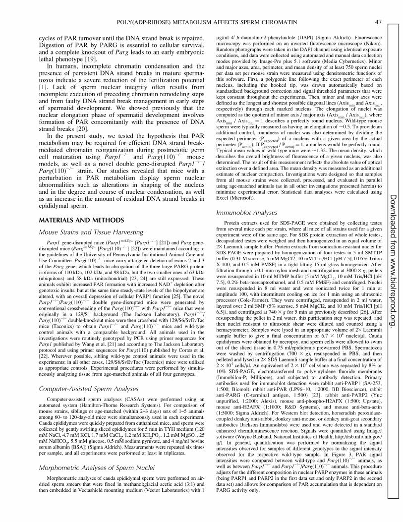
Parg(110)�/�, but not Parp1�/�, mice exhibited a mild buthighly significant (P , 0.0001) subfertility phenotype, withmean 6 SD reduced litter sizes of 4.2 6 2.1 compared with5.7 6 2.1 in 129SVE wild-type animals of (Table 1). Allanimals were routinely removed from the breeding program atage 6 mo, after which fertility may decline naturally in mice,and were replaced by 7- to 8-wk-old animals. Unexpectedly, inParp1�/�/Parg(110)�/� double gene-disrupted mice, reducedfecundity was not observed in the F1 and F2 generations, andbreeding data averages were statistically no different fromthose of the wild type.
Parg(110)�/� Animals Exhibit No Reductionin Sperm Motility
To investigate the reason for reduced fertility of Parg(110)�/�
mice, physiological sperm parameters of mouse strains weretested using a CASA system (Fig. 1). While the mean 6 SEMpercentages of motility spermatozoa and progressive motilityspermatozoa isolated from the cauda epididymis were compa-rable to those of the wild type (with typical motile spermfractions being ;55%–60% in individual experimental days) inParg(110)�/� mice (101.2% 6 6.9% relative to the wild type),Parp1�/� mice had consistently and significantly (P , 0.001)lower percentages of motile sperm (81.5% 6 3.8% relative tothe wild type) and fewer progressively motile sperm amongthose (73.4% 6 11.9% relative to the wild type). Sperm fromParp1�/�/Parg(110)�/� double gene-disrupted animals hadalmost normal mean 6 SEM fractions of motile cells (94.2%6 5.5% relative to the wild type), but progressiveness of motilesperm was impaired at a level that was statistically comparableto that of Parp1�/� mice (74.8% 6 11.4%). This result ispuzzling and indicates that PARP1 may also have a previouslyunrecognized role in sperm cell progressive motility.
Expression of Short PARG Isoforms and Cleavage of PARP2and PARP1 Are Hallmarks of Late Spermiogenesis
The expression of three major enzymes involved in PARmetabolism in spermatogenesis (PARP1, PARP2, and PARG)
was compared in lysates of whole testes, lysates of sonication-resistant nuclei (equivalent to the fraction of spermatids in latesteps of spermiogenesis), or lysates of cauda epididymal sperm(Fig. 2). Immunoblotting of SDS-soluble proteins confirmedthe absence of PARP1 expression in Parp1�/� and Parp1�/�/Parg(110)�/� double-knockout mice (Fig. 2, upper panel).(Note that the upper band is an unspecific signal alwaysproduced by the primary antibody recognizing an unidentifiedprotein that is lost during spermiogenesis.) It also revealed thatin wild-type control animals PARP1 is still present in thesonication-resistant nuclei (SRN). The SRN contains testicularspermatids that should have completed nuclear elongation butare not yet fully condensed. At this stage, a large portion of thePARP1 enzyme is present in a cleaved form, reminiscent of thecaspase 3-dependent PARP1 cleavage during apoptosis [28](Fig. 2, top middle panel). However, there was no indication ofongoing apoptosis in spermatid cells as analyzed by TUNELassays (data not shown). Apoptotic PARP1 cleavage yieldsfragments of 89 kDa and 24 kDa, and only the 89-kDa fragmentis recognized by the antibody used. Epididymal sperm seemedto be devoid of PARP1 enzyme (Fig. 2, top right panel).
PARP2 was detected at low levels in whole-testis lysates butseemed to be highly expressed in SRN (Fig. 2, middle panel).A double band (arrows) indicates that PARP2 is modified orcleaved in a fashion similar to PARP1. The observed sizedifferences resulting in a double band could be due toacetylation, as has been recently described for PARP2 [29].Acetylation leads to a slight upward shift of the PARP2 proteinband. This indicates that the majority of PARP2 protein in thewhole-testis lysates, as well as the PARP2 comprising theupper part of the double band in SRN, is modified, while thelower band represents unmodified protein. Alternatively, theupper band could consist of unmodified PARP2 protein, whilethe lower band represents an as yet undescribed cleavageproduct. Again, spermatozoa seem to possess either non-detectable amounts or no PARP2 (Fig. 2, middle right panel).
As expected, expression of at least one large PARG isoform(110 kDa, 101 kDa, or 98 kDa) was detected in wild-type
FIG. 1. Reduction of sperm motility in Parp1�/�mice. Cauda epididymalspermatozoa from age-matched sets of animals were measured by CASA.Motile sperm characterized as progressive were each recorded aspercentage of the motile population, and overall motility and progres-siveness were expressed relative to the wild type (rel. to wt) in eachexperiment. Data represent five independent experiments involving 3–5animals each. Error bars represent the SEM of three independentexperiments. ** P , 0.001; wt, wild type.
TABLE 1. Fecundity of wild-type (Wt), Parp1�/�, Parg(110)�/�, andParp1�/�/Parg(110)�/� mice.
Parameter Wt 129SVE Parp1�/� Parg(110)�/�Parp1�/�/
Parg(110)�/�
No. pups (litters) 572 (101) 419 (78) 287 (68) 328 (62)Pups/litter 6 SD 5.7 6 2.14 5.4 6 1.95 4.2 6 2.1 5.3 6 2.9t-test vs. Wt NA* P ¼ 0.34 P , 0.0001 P ¼ 0.35
* NA, not applicable.
48 MEYER-FICCA ET AL.
Dow
nloaded from w
ww
.biolreprod.org.
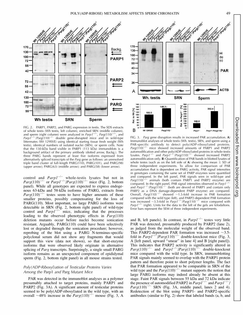
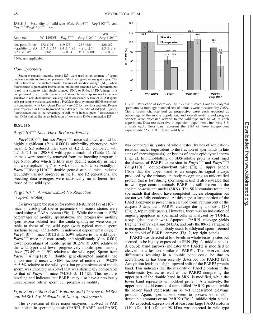
control and Parp1�/� whole-testis lysates but not inParg(110)�/� or Parp1�/�/Parg(110)�/� mice (Fig. 2, bottompanel). While all genotypes are expected to express endoge-nous 63-kDa and 58-kDa isoforms of PARG, extracts fromParg(110)�/� testes seem to have higher amounts of thesesmaller proteins, possibly compensating for the loss ofPARG(110). Most important, no large PARG isoforms weredetectable in SRN (Fig. 2, bottom right panel) of wild-typecontrol and Parp1�/� mice, indicating that the processesleading to the observed phenotypic effects in Parg(110)deletion mutants occur before nuclei become sonicationresistant. Arguably, PARG(110) could have been selectivelylost or degraded through the sonication procedure; however,reprobing of the blot using a PARG N-terminus-specificpolyclonal serum did not show any fragments that wouldsupport this view (data not shown), so that short-enzymeisoforms that were observed likely originate in alternativesplicing of Parg transcripts. Surprisingly, a single small PARGisoform remains as an unexpected component of epididymalsperm (Fig. 2, bottom right panel) in all mouse strains tested.
Poly(ADP-Ribosyl)ation of Testicular Proteins VariesAmong the Parp1 and Parg Mutant Mice
PAR was detected in the immunoblot analyses as a polymerpresumably attached to target proteins, mainly PARP1 andPARP2 (Fig. 3A). A significant amount of testicular proteinsseemed to be poly(ADP-ribosyl)ated in the wild type, with anoverall ;48% increase in the Parg(110)�/� mouse (Fig. 3, A
and B, left panels). In contrast, in Parp1�/� testes very littlePAR was detected, presumably produced by PARP2 (lane 2),as judged from the molecular weight of the observed band.This PARP2-dependent PAR formation was increased ;3.5-fold in Parp1�/�/Parg(110)�/� double-knockout mice (Fig. 3,A [left panel, upward ‘‘smear’’ in lane 4] and B [right panel]).This indicates that PARP2 activity is significantly altered inParg(110)�/� and Parp1�/�/Parg(110)�/� double-knockoutmice compared with the wild type. In SRN, immunoblottingPAR signals mainly seemed to overlap with the PARP1 proteinpattern and therefore point to short polymer lengths. The factthat PAR formation appeared to be comparable in SRN of thewild type and the Parg(110)�/� mutant supports the notion thatlarge PARG isoforms may indeed already be absent at thisstage. Faint PAR signals between 55 kDa and 72 kDa indicatethe presence of automodified PARP2 in Parp1�/� and Parp1�/�/Parg(110)�/� SRN (Fig. 3A, middle panel, lanes 2 and 4).Redetection of the blot using PARP1- and PARP2-specificantibodies (similar to Fig. 2) show that labeled bands (a, b, and
FIG. 2. PARP1, PARP2, and PARG expression in testis. The SDS extractsof whole testis (Wh testis, left column), enriched SRN (middle column),and sperm (right column) were analyzed in Parp1�/�, Parg(110)�/�, andParp1�/�/Parg(110)�/� double gene-disrupted mice and in wild-typelittermates (Wt 129SVE) using identical starting tissue fresh weight (Whtestis), identical numbers of isolated nuclei (SRN), or sperm cells. Notethat the 130-kDa band visible in PARP1 (113 kDa) immunoblots is abackground artifact of the primary antibody (dotted arrow, Backg.). Thethree PARG bands represent at least five isoforms expressed fromalternatively spliced transcripts of the Parg gene as follows: an unresolvedtriple band cluster of full-length PARG(110), PARG(101), and PARG(98)(upper arrow); PARG(63) (middle arrow); and PARG(58) (lower arrow).
FIG. 3. Parg gene disruption results in increased PAR accumulation. A)Immunoblot analyses of whole testis (Wh. testis), SRN, and sperm using aPAR-specific antibody to detect poly(ADP-ribosyl)ated proteins.Parg(110)�/� mice showed increased amounts of PARP1 and PARP2automodification and other poly(ADP-ribosyl)ated proteins in whole-testislysates. Parp1�/� and Parp1�/�/Parg(110)�/� showed increased PARP2automodification only. B) Quantification of PAR bands in blotted lysates ofwhole testes (such as on the left side of A) showing the mean 6 SD ofthree independent experiments. To allow for comparison of PARaccumulation that is dependent on PARG activity, PAR signal intensitiesin genotypes containing the same set of PARP enzymes were quantifiedand compared. In the left panel, PAR signals seen in wild-type andParg(110)�/� animals (both contain PARP1 and PARP2 enzyme) arecompared. In the right panel, PAR signal intensities observed in Parp1�/�
and Parp1�/�/Parg(110)�/� (both are devoid of PARP1 and contain onlyPARP2 as a DNA damage-dependent PARP enzyme) are compared.Overall, Parg(110)�/� showed ;1.5-fold increase in PAR formationcompared with the wild type (left), and PARP2-dependent PAR formationwas increased ;3.5-fold in Parp1�/�/Parg(110)�/� mice compared withParp1�/� (right). Units for the data to the left of the gels are kilodaltons.Error bars represent the SEM of three independent experiments.
POLY(ADP-RIBOSE) METABOLISM AFFECTS SPERM CHROMATIN 49
Dow
nloaded from w
ww
.biolreprod.org.
c) all correspond to PARP1 isoforms absent in the Parp1�/�
mouse. Band c comprises both PARP1 and PARP2 proteins inwild-type and Parg(110)�/� mice but PARP2 only in Parp1�/�
and Parp1�/�/Parg(110)�/� mice (data not shown). PAR wasnot detectable in epididymal sperm samples of either genotype(Fig. 3A, right panel).
Abnormally Reduced Nuclear Elongation and ChromatinCondensation in Parp1�/�, Parg(110)�/�, and Parp1�/�/Parg(110)�/� Epididymal Spermatozoa
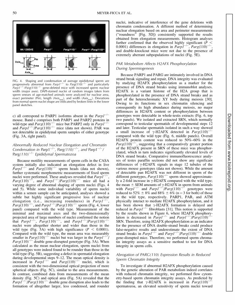
Because motility measurements of sperm cells in the CASAsystem initially also indicated an elongation defect in liveParp1�/� and Parg(110)�/� sperm heads (data not shown),further systematic morphometric measurements of fixed spermnuclei were performed. These analyses revealed that Parp1�/�,Parg(110)�/�, and Parp1�/�/Parg(110)�/� mice all had avarying degree of abnormal shaping of sperm nuclei (Figs. 4and 5). While some individual variability of sperm nucleiwithin a semen sample can be anticipated, analyses showed asubtle but highly significant progressive loss of nuclearelongation (i.e., increasing roundness) in Parp1�/�,Parg(110)�/�, and Parp1�/�/Parg(110)�/� sperm (Fig. 4, lowerpanel) compared with the wild type. Measurement of theminimal and maximal axes and the two-dimensionallyprojected area of large numbers of nuclei confirmed the notionthat Parp1�/�, Parg(110)�/�, and Parp1�/�/Parg(110)�/�
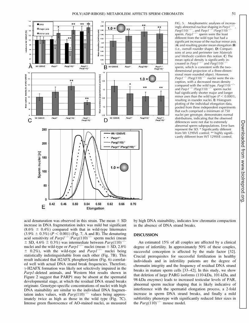
nuclei were altogether shorter and wider than those in thewild type (Fig. 5A) with high significance (P , 0.0001).Compared with the wild type, the mean area was measurablysmaller in Parg(110)�/� nuclei but was larger in the Parp1�/�/Parg(110)�/� double gene-disrupted genotype (Fig. 5A). Whencalculated as the mean nuclear elongation, sperm nuclei fromall genotypes were indeed found to be rounder than those in thewild type (Fig. 5B), suggesting a defect in spermatid elongationduring developmental steps 9–12. The mean optical density isincreased in Parp1�/� and Parg(110)�/� nuclei, which isconsistent with the two-dimensional optical projection of morespherical objects (Fig. 5C), similar to the area measurements.In contrast, combined data from measurements of the meandensity (Fig. 5C) and optical area (Fig. 5A) revealed that theParp1�/�/Parg(110)�/� double gene disruption also leads to theformation of altogether larger, less condensed, and rounder
nuclei, indicative of interference of the gene deletions withchromatin condensation. A different method of determiningnuclear elongation based on area and perimeter measurements(‘‘roundness’’ [Fig. 5D]) consistently supported the resultsobtained from elongation measurements. Histogram analysesfurther confirmed that the observed highly significant (P ,0.0001) differences in elongation in Parp1�/�, Parg(110)�/�,and double-knockout mice were not due to the presence ofextremely aberrant subpopulations of nuclei (Fig. 5E).
PAR Metabolism Affects H2AFX PhosphorylationDuring Spermiogenesis
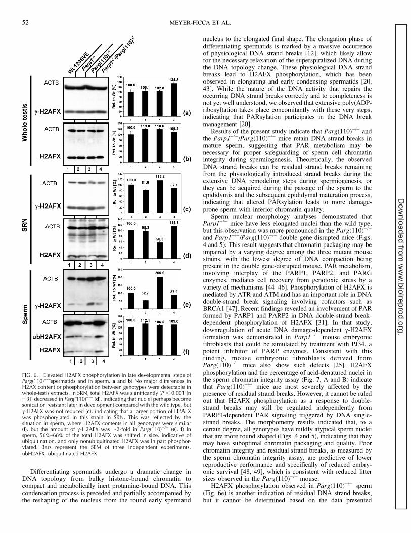
Because PARP1 and PARG are intimately involved in DNAstrand break signaling and repair, DNA integrity was evaluatedby studying H2AFX phosphorylation as a marker for thepresence of DNA strand breaks using immunoblot analyses.H2AFX is a variant histone of the H2A group that isphosphorylated in the presence of DNA strand breaks and aspart of the heterochromatic XY body during meiosis [30].Owing to its functions in sex chromatin silencing andconsequently its high abundance during meiosis, no majordifferences in H2AFX content or phosphorylation betweengenotypes were detectable in whole-testis extracts (Fig. 6, toptwo panels). We isolated and extracted SRN, which normallycorrespond to testicular spermatids of developmental steps 12and later. Testicular spermatids isolated in such a way showeda small increase of c-H2AFX detected in Parg(110)�/�
compared with the wild type (Fig. 6, middle panels). OverallH2AFX protein content was reduced to 50%–60% in theParg(110)�/�, suggesting that a comparatively greater portionof the H2AFX present in SRN of these mice was phosphor-ylated, which in turn indicates significantly elevated levels ofDNA strand breaks. Comparative immunofluorescence analy-ses of testes paraffin sections did not show any significantdifferences of c-H2AFX signals in stage 12 tubules of thevarious mouse genotypes (data not shown). While the amountof detectable pan H2AFX was not different in sperm of thedifferent genotypes, Parg(110)�/� sperm showed approximate-ly a 2-fold increase in c-H2AFX compared with the wild type;the mean 6 SEM amounts of c-H2AFX in sperm from animalswith Parp1�/� and Parp1�/�/Parg(110)�/� genotypes werereduced to 52% 6 8% and 88% 6 6% (n¼ 3 for both) of thosein the wild type, respectively. PARP1, PAR, and ATMphysically interact to mediate H2AFX phosphorylation, and ithas been shown that c-H2AFX formation is delayed andreduced in Parp1�/� fibroblasts [31]. This notion is supportedby the results shown in Figure 6, where H2AFX phosphory-lation is decreased in Parp1�/� and Parp1�/�/Parg(110)�/�
SRN. Therefore, using H2AFX phosphorylation as an indicatorfor the presence of DNA double-strand breaks could producefalse-negative results and underestimate the extent of DNAstrand breaks in Parp1�/� and Parp1�/�/Parg(110)�/� doublegene-disrupted mice. Therefore, we performed sperm chroma-tin integrity assays as a sensitive method to test for DNAintegrity in sperm cells.
Abrogation of PARG(110) Expression Results in ReducedSperm Chromatin Integrity
To investigate if abnormal H2AFX phosphorylation causedby the genetic alteration of PAR metabolism indeed correlateswith reduced chromatin integrity, we performed flow cytom-etry-based sperm chromatin integrity assays. As predicted bythe finding that c-H2AFX is increased in Parg(110)�/�
spermatozoa, an elevated sensitivity of sperm nuclei toward
FIG. 4. Shaping and condensation of average epididymal sperm areprogressively abnormal from Parp1�/� to Parg(110)�/� and particularlyParp1�/�/Parg(110)�/� gene-deleted mice with increased sperm nuclearwidth (major axes). DAPI-stained nuclei of random images taken fromsperm smears of age-matched animals were analyzed for nuclear area,exact perimeter (Pm), length (Axis
maj), and width (Axis
min). Deviations
from normal sperm nuclei shape are indicated by broken lines in the lowerpanel sketches.
50 MEYER-FICCA ET AL.
Dow
nloaded from w
ww
.biolreprod.org.
acid denaturation was observed in this strain. The mean 6 SDincrease in DNA fragmentation index was mild but significant(8.6% 6 0.4%) compared with that in wild-type littermates(3.9% 6 0.3%) (P , 0.001) (Fig. 7, A and B). The denaturingacid sensitivity of Parp1�/�/Parg(110)�/� sperm nuclei (mean6 SD, 4.6% 6 0.3%) was intermediate between Parg(110)�/�
nuclei and the wild-type or Parp1�/� nuclei (mean 6 SD, 2.8%6 0.2%), with the wild-type and Parp1�/� nuclei beingstatistically indistinguishable from each other (Fig. 7B). Thisresult indicated that H2AFX phosphorylation (Fig. 6) correlat-ed well with actual DNA strand break frequencies. Therefore,c-H2AFX formation was likely not selectively impaired in theParp1-deleted animals, and Western blot results shown inFigure 2 suggest that PARP1 may be absent at the spermatiddevelopmental stage, at which the residual DNA strand breaksoriginate. Genotype-specific concentrations of nuclei with highDNA stainability are similar to the individual DNA fragmen-tation index values, with Parg(110)�/� values being approx-imately twice as high as those in the wild type (Fig. 7C).Intense green fluorescence of AO-stained nuclei, as measured
by high DNA stainability, indicates low chromatin compactionin the absence of DNA strand breaks.
DISCUSSION
An estimated 15% of all couples are affected by a clinicaldegree of infertility. In approximately 50% of these couples,successful conception is affected by the male factor [32].Crucial prerequisites for successful fertilization in healthyindividuals and in infertility patients are the degree ofchromatin integrity and the frequency of residual DNA strandbreaks in mature sperm cells [33–42]. In this study, we showthat deletion of large PARG isoforms (110-kDa, 101-kDa, and98-kDa enzymes) leads to increased testicular levels of PAR,abnormal sperm nuclear shaping that is likely indicative ofinterference with the spermatid elongation process, a 2-foldincrease in sperm DNA strand breaks, and finally a mildsubfertility phenotype with significantly reduced litter sizes inthe Parg(110)�/� mouse model.
FIG. 5. Morphometric analyses of increas-ingly abnormal nuclear shaping in Parp1�/�,Parg(110)�/�, and Parp1�/�/Parg(110)�/�
sperm. Parp1�/� sperm were the leastdifferent from the wild type but had asignificant increase of the nuclear minor axis(A) and resulting greater mean elongation (B)(i.e., overall rounder shape). D) Compari-sons of area and perimeter (see Materialsand Methods) confirm this notion. C) Themean optical density is significantly in-creased in Parp1�/� and Parg(110)�/�
sperm, which is consistent with the two-dimensional projection of a three-dimen-sional more rounded object. However,Parp1�/�/Parg(110)�/� nuclei were the ex-ception, with a decreased mean densitycompared with the wild type. Parg(110)�/�
and Parp1�/�/Parg(110)�/� sperm nucleihad significantly shorter major and longerminor axes than the wild type (P , 0.0001),resulting in rounder nuclei. E) Histogramplotting of the individual elongation data,pooled from three independent experimentsthat each comprised a minimum of 750nuclei per genotype, demonstrates normaldistributions, indicating that the observeddifferences were not due to extremelyabnormal sperm subpopulations. Error barsrepresent the SD. * Significantly differentfrom Wt 129SVE control; ** highly signifi-cantly different from WT 129SVE control.
POLY(ADP-RIBOSE) METABOLISM AFFECTS SPERM CHROMATIN 51
Dow
nloaded from w
ww
.biolreprod.org.
Differentiating spermatids undergo a dramatic change inDNA topology from bulky histone-bound chromatin tocompact and metabolically inert protamine-bound DNA. Thiscondensation process is preceded and partially accompanied bythe reshaping of the nucleus from the round early spermatid
nucleus to the elongated final shape. The elongation phase ofdifferentiating spermatids is marked by a massive occurrenceof physiological DNA strand breaks [12], which likely allowfor the necessary relaxation of the superspiralized DNA duringthe DNA topology change. These physiological DNA strandbreaks lead to H2AFX phosphorylation, which has beenobserved in elongating and early condensing spermatids [20,43]. While the nature of the DNA activity that repairs theoccurring DNA strand breaks correctly and to completeness isnot yet well understood, we observed that extensive poly(ADP-ribosyl)ation takes place concomitantly with these very steps,indicating that PARsylation participates in the DNA breakmanagement [20].
Results of the present study indicate that Parg(110)�/� andthe Parp1�/�/Parg(110)�/� mice retain DNA strand breaks inmature sperm, suggesting that PAR metabolism may benecessary for proper safeguarding of sperm cell chromatinintegrity during spermiogenesis. Theoretically, the observedDNA strand breaks can be residual strand breaks remainingfrom the physiologically introduced strand breaks during theextensive DNA remodeling steps during spermiogenesis, orthey can be acquired during the passage of the sperm to theepididymis and the subsequent epididymal maturation process,indicating that altered PARsylation leads to more damage-prone sperm with inferior chromatin quality.
Sperm nuclear morphology analyses demonstrated thatParp1�/� mice have less elongated nuclei than the wild type,but this observation was more pronounced in the Parg(110)�/�
and Parp1�/�/Parg(110)�/� double gene-disrupted mice (Figs.4 and 5). This result suggests that chromatin packaging may beimpaired by a varying degree among the three mutant mousestrains, with the lowest degree of DNA compaction beingpresent in the double gene-disrupted mouse. PAR metabolism,involving interplay of the PARP1, PARP2, and PARGenzymes, mediates cell recovery from genotoxic stress by avariety of mechanisms [44–46]. Phosphorylation of H2AFX ismediated by ATR and ATM and has an important role in DNAdouble-strand break signaling involving cofactors such asBRCA1 [47]. Recent findings revealed an involvement of PARformed by PARP1 and PARP2 in DNA double-strand break-dependent phosphorylation of H2AFX [31]. In that study,downregulation of acute DNA damage-dependent c-H2AFXformation was demonstrated in Parp1�/� mouse embryonicfibroblasts that could be simulated by treatment with PJ34, apotent inhibitor of PARP enzymes. Consistent with thisfinding, mouse embryonic fibroblasts derived fromParg(110)�/� mice also show such defects [25]. H2AFXphosphorylation and the percentage of acid-denatured nuclei inthe sperm chromatin integrity assay (Fig. 7, A and B) indicatethat Parg(110)�/� mice are most severely affected by thepresence of residual strand breaks. However, it cannot be ruledout that H2AFX phosphorylation as a response to double-strand breaks may still be regulated independently fromPARP1-dependent PAR signaling triggered by DNA single-strand breaks. The morphometry results indicated that, to acertain degree, all genotypes have mildly atypical sperm nucleithat are more round shaped (Figs. 4 and 5), indicating that theymay have suboptimal chromatin packaging and quality. Poorchromatin integrity and residual strand breaks, as measured bythe sperm chromatin integrity assay, are predictive of lowerreproductive performance and specifically of reduced embry-onic survival [48, 49], which is consistent with reduced littersizes observed in the Parg(110)�/� mouse.
H2AFX phosphorylation observed in Parg(110)�/� sperm(Fig. 6e) is another indication of residual DNA strand breaks,but it cannot be determined based on the data presented
FIG. 6. Elevated H2AFX phosphorylation in late developmental steps ofParg(110)�/�spermatids and in sperm. a and b) No major differences inH2AX content or phosphorylation between genotypes were detectable inwhole-testis extracts. In SRN, total H2AFX was significantly (P , 0.001 [n¼3]) decreased in Parg(110)�/� (d), indicating that nuclei perhaps becomesonication resistant later in development compared with the wild type, butc-H2AFX was not reduced (c), indicating that a larger portion of H2AFXwas phosphorylated in this strain in SRN. This was reflected by thesituation in sperm, where H2AFX contents in all genotypes were similar(f), but the amount of c-H2AFX was ;2-fold in Parg(110)�/� (e). f) Insperm, 56%–68% of the total H2AFX was shifted in size, indicative ofubiquitination, and only nonubiquitinated H2AFX was in part phosphor-ylated. Bars represent the SEM of three independent experiments.ubH2AFX, ubiquitinated H2AFX.
52 MEYER-FICCA ET AL.
Dow
nloaded from w
ww
.biolreprod.org.

whether these were newly acquired after the sperm had left thetestis or represent unrepaired DNA lesions that stem from DNAremodeling during spermatid elongation. These chromatinreorganization events are accompanied by controlled physio-logical DNA strand breaks mediated by DNA topoisomerases,with a probable predominant role of TOP2B [12, 43]. Putativelesions caused by stalled topoisomerases comprise DNAdouble- and single-strand breaks. Most of the DNA single-strand breaks are repaired using the base excision pathway,which has been commonly thought to involve PARP1;however, its exact function there is still under debate [45,50]. DNA double-strand breaks are repaired by homologousrecombination repair or by nonhomologous end joining insomatic cells. However, because spermatids, being haploid, areunable to perform homologous recombination repair, doublestrands must be repaired by nonhomologous end joining. Thelatter consists of two alternatively used variant pathways, oneinvolving DNA-dependent protein kinase (default pathway)and another involving the homolog of the yeast protein RAD18and PARP1 (backup pathway) [51, 52]. Therefore, it isconceivable that DNA damage found in mature sperm maybe in the form of residual DNA strand breaks that remainedunrepaired owing to functional PARP1 inhibition caused by
abnormal automodification of the enzyme in the PARG(110)deletion mutant (Fig. 3A, left panel). The increased levels ofpoly(ADP-ribosyl)ated PARP are likely caused by the reducedPARG activity in Parg(110)�/� mice [22] in combination withaltered control of PARG activity [25] due to the loss of theregulatory A-domain in the mutant. Western blot analyses inthe present study showed that elevated levels of poly(ADP-ribosyl)ated proteins in whole-testis extracts of Parg(110)�/�
mice (Fig. 3) were not due to increased apoptosis, as indicatedby in situ TUNEL assays (data not shown) and by the absenceof caspase-dependent PARP1 cleavage. The suggestion that theobserved DNA strand breaks are unrepaired residual lesionsrather than newly acquired lesions is supported by the findingthat SRN and sperm from wild-type mice do not containPARG(110). This suggests that the chromatin defects resultingfrom abnormal PARP1/PARG protein composition in spermfrom Parg(110)�/� and Parp1�/�/Parg(110)�/� animals mustarise at earlier time points (e.g., during or before the elongationphase before chromatin condensation). Because c-H2AFXformation was only moderately changed in SRN ofParg(110)�/� mice, it seems reasonable to suggest that DNAstrand breaks first exist as unrepaired single-strand lesions that,depending on their frequency and spacing along the DNA, later
FIG. 7. Sperm chromatin integrity assaysindicate poor sperm chromatin quality inParg(110)�/� mice. A) Acridine orange-based sperm chromatin integrity assays [27]indicate residual DNA strand breaks (redfluorescence after acid denaturation) inParg(110)�/� but not in Parp1�/�, Parp1�/�/Parg(110)�/�, or wild-type 129SVE siblings(R3 region). B) Data were compared asDNA fragmentation index (DFI). C) A regionR2 subpopulation with extremely highgreen fluorescent stainability (high DNAstainability [HDS]) was computed from theraw data and compared. This subpopulationis characterized by overall low or immaturechromatin condensation, allowing for in-tercalation of the dye. Representative dataare shown (n ¼ 4). Bars represent the SD oftriplicates in one representative experiment.** Highly significantly different from Wt129SVE control.
POLY(ADP-RIBOSE) METABOLISM AFFECTS SPERM CHROMATIN 53
Dow
nloaded from w
ww
.biolreprod.org.
develop into double-strand breaks during further condensationof spermatid nuclei, causing H2AFX phosphorylation inmature spermatozoa.
Arguably, the deletion of PARP1 may have a similarlydetrimental effect on DNA repair as its inhibition, but recentevidence shows that inhibition of PARP enzymes (such as byusing a small-molecule inhibitor) results in a delay or defect inDNA single-strand break repair that is different from, and oftenmore severe than, the effect caused by genetic ablation ofPARP1 enzyme [50]. In addition, PARP2, which hassubstantially overlapping functions with PARP1, was stillexpressed in all genetic strains tested in this study, which mayalso account for the mild phenotypes observed in theinvestigations presented herein. In a similarly mild fashion,residual DNA strand breaks, compromised chromatin integrity,and decreased embryonic survival were reported after singledeletion of either transition protein 1 (TNP1) or TNP2 [26, 53],but if deleted together, the effect was much more dramatic [54].However, deletion of both Parp1 and Parp2, which areubiquitously expressed in the mammalian body, was shown tobe inconsistent with embryonic survival [55].
In summary, the results of this study strongly indicate thatPAR metabolism is involved in proper execution of spermatidmaturation. The study demonstrates for the first time (to ourknowledge) that partial deficiency in PAR metabolismresulting from deletion of the Parp1 and Parg genes willnegatively affect chromatin integrity in spermatid nuclei, asmeasured by abnormal sperm nuclear shaping, altered H2AFXphosphorylation, and reduced resistance to acid denaturation,particularly in Parg gene-disrupted mice.
REFERENCES
1. Leduc F, Nkoma GB, Boissonneault G. Spermiogenesis and DNA repair: apossible etiology of human infertility and genetic disorders. Syst BiolReprod Med 2008; 54:3–10.
2. Braun RE. Packaging paternal chromosomes with protamine. Nat Genet2001; 28:10–12.
3. Rousseaux S, Faure AK, Caron C, Lestrat C, Govin J, Hennebicq S, SeleB, Khochbin S. Organizing the sperm nucleus. Gynecol Obstet Fertil2004; 32:785–791.
4. Caron C, Govin J, Rousseaux S, Khochbin S. How to pack the genome fora safe trip. Prog Mol Subcell Biol 2005; 38:65–89.
5. Sassone-Corsi P. Unique chromatin remodeling and transcriptionalregulation in spermatogenesis. Science 2002; 296:2176–2178.
6. Lewis JD, Abbott DW, Ausio J. A haploid affair: core histone transitionsduring spermatogenesis. Biochem Cell Biol 2003; 81:131–140.
7. Boissonneault G. Chromatin remodeling during spermiogenesis: a possiblerole for the transition proteins in DNA strand break repair. FEBS Lett2002; 514:111–114.
8. Risley MS, Einheber S, Bumcrot DA. Changes in DNA topology duringspermatogenesis. Chromosoma 1986; 94:217–227.
9. McPherson S, Longo FJ. Chromatin structure-function alterations duringmammalian spermatogenesis: DNA nicking and repair in elongatingspermatids. Eur J Histochem 1993; 37:109–128.
10. Marcon L, Boissonneault G. Transient DNA strand breaks during mouseand human spermiogenesis: new insights in stage specificity and link tochromatin remodeling. Biol Reprod 2004; 70:910–918.
11. Laberge RM, Boissonneault G. Chromatin remodeling in spermatids: asensitive step for the genetic integrity of the male gamete. Arch Androl2005; 51:125–133.
12. Laberge RM, Boissonneault G. On the nature and origin of DNA strandbreaks in elongating spermatids. Biol Reprod 2005; 73:289–296.
13. Meyer-Ficca ML, Meyer RG, Jacobson EL, Jacobson MK. Poly(ADP-ribose) polymerases: managing genome stability. Int J Biochem Cell Biol2005; 37:920–926.
14. Woodhouse BC, Dianov GL. Poly ADP-ribose polymerase-1: aninternational molecule of mystery. DNA Repair (Amst) 2008; 7:1077–1086.
15. Gagne JP, Hendzel MJ, Droit A, Poirier GG. The expanding role ofpoly(ADP-ribose) metabolism: current challenges and new perspectives.Curr Opin Cell Biol 2006; 18:145–151.
16. Schreiber V, Dantzer F, Ame JC, de Murcia G. Poly(ADP-ribose): novelfunctions for an old molecule. Nat Rev Mol Cell Biol 2006; 7:517–528.
17. Burkle A. Poly(ADP-ribose): the most elaborate metabolite of NADþ.FEBS J 2005; 272:4576–4589.
18. Meyer RG, Meyer-Ficca ML, Jacobson EL, Jacobson MK. Enzymes inpoly(ADP-ribose) metabolism. In: Burkle A (ed.), Poly(ADP-ribosyl)a-tion. Austin, TX: Landes Bioscience; 2004.
19. Koh DW, Lawler AM, Poitras MF, Sasaki M, Wattler S, Nehls MC,Stoger T, Poirier GG, Dawson VL, Dawson TM. Failure to degradepoly(ADP-ribose) causes increased sensitivity to cytotoxicity and earlyembryonic lethality. Proc Natl Acad Sci U S A 2004; 101:17699–17704.
20. Meyer-Ficca ML, Scherthan H, Burkle A, Meyer RG. Poly(ADP-ribosyl)ation during chromatin remodeling steps in rat spermiogenesis.Chromosoma 2005; 114:67–74.
21. Wang ZQ, Auer B, Stingl L, Berghammer H, Haidacher D, Schweiger M,Wagner EF. Mice lacking ADPRT and poly(ADP-ribosyl)ation developnormally but are susceptible to skin disease. Genes Dev 1995; 9:509–520.
22. Cortes U, Tong WM, Coyle DL, Meyer-Ficca ML, Meyer RG, Petrilli V,Herceg Z, Jacobson EL, Jacobson MK, Wang ZQ. Depletion of the 110-kilodalton isoform of poly(ADP-ribose) glycohydrolase increases sensi-tivity to genotoxic and endotoxic stress in mice. Mol Cell Biol 2004; 24:7163–7178.
23. Meyer-Ficca ML, Meyer RG, Coyle DL, Jacobson EL, Jacobson MK.Human poly(ADP-ribose) glycohydrolase is expressed in alternative splicevariants yielding isoforms that localize to different cell compartments. ExpCell Res 2004; 297:521–532.
24. Meyer RG, Meyer-Ficca ML, Whatcott CJ, Jacobson EL, Jacobson MK.Two small enzyme isoforms mediate mammalian mitochondrial poly(ADP-ribose) glycohydrolase (PARG) activity. Exp Cell Res 2007; 313:2920–2936.
25. Gao H, Coyle DL, Meyer-Ficca ML, Meyer R, Jacobson EL, Wang ZQ,Jacobson MK. Altered poly(ADP-ribose) metabolism impairs cellularresponses to genotoxic stress in a hypomorphic mutant of poly(ADP-ribose) glycohydrolase. Exp Cell Res 2007; 313:984–996.
26. Yu YE, Zhang Y, Unni E, Shirley CR, Deng JM, Russell LD, Weil MM,Behringer RR, Meistrich ML. Abnormal spermatogenesis and reducedfertility in transition nuclear protein 1-deficient mice. Proc Natl Acad SciU S A 2000; 97:4683–4688.
27. Evenson D, Jost L. Sperm chromatin structure assay is useful for fertilityassessment. Methods Cell Sci 2000; 22:169–189.
28. Lazebnik YA, Kaufmann SH, Desnoyers S, Poirier GG, Earnshaw WC.Cleavage of poly(ADP-ribose) polymerase by a proteinase with propertieslike ICE. Nature 1994; 371:346–347.
29. Haenni SS, Hassa PO, Altmeyer M, Fey M, Imhof R, Hottiger MO.Identification of lysines 36 and 37 of PARP-2 as targets for acetylation andauto-ADP-ribosylation. Int J Biochem Cell Biol 2008; 40:2274–2283.
30. Fernandez-Capetillo O, Mahadevaiah SK, Celeste A, Romanienko PJ,Camerini-Otero RD, Bonner WM, Manova K, Burgoyne P, NussenzweigA. H2AX is required for chromatin remodeling and inactivation of sexchromosomes in male mouse meiosis. Dev Cell 2003; 4:497–508.
31. Haince JF, Kozlov S, Dawson VL, Dawson TM, Hendzel MJ, Lavin MF,Poirier GG. Ataxia telangiectasia mutated (ATM) signaling network ismodulated by a novel poly(ADP-ribose)-dependent pathway in the earlyresponse to DNA-damaging agents. J Biol Chem 2007; 282:16441–16453.
32. Oehninger S. Strategies for the infertile man. Semin Reprod Med 2001;19:231–237.
33. Agarwal A, Said TM. Role of sperm chromatin abnormalities and DNAdamage in male infertility. Hum Reprod Update 2003; 9:331–345.
34. Sakkas D, Mariethoz E, Manicardi G, Bizzaro D, Bianchi PG, Bianchi U.Origin of DNA damage in ejaculated human spermatozoa. Rev Reprod1999; 4:31–37.
35. Sakkas D, Urner F, Bizzaro D, Manicardi G, Bianchi PG, Shoukir Y,Campana A. Sperm nuclear DNA damage and altered chromatin structure:effect on fertilization and embryo development. Hum Reprod 1998;13(suppl 4):11–19.
36. Sharma RK, Said T, Agarwal A. Sperm DNA damage and its clinicalrelevance in assessing reproductive outcome. Asian J Androl 2004; 6:139–148.
37. Avendano C, Franchi A, Taylor S, Morshedi M, Bocca S, Oehninger S.Fragmentation of DNA in morphologically normal human spermatozoa.Fertil Steril 2009; 91:1077–1084.
38. Carrell DT, De Jonge C, Lamb DJ. The genetics of male infertility: a fieldof study whose time is now. Arch Androl 2006; 52:269–274.
39. Aitken RJ, De Iuliis GN. Value of DNA integrity assays for fertilityevaluation. Soc Reprod Fertil Suppl 2007; 65:81–92.
40. Olsen AK, Lindeman B, Wiger R, Duale N, Brunborg G. How do male
54 MEYER-FICCA ET AL.
Dow
nloaded from w
ww
.biolreprod.org.
germ cells handle DNA damage? Toxicol Appl Pharmacol 2005; 207:521–531.
41. Spano M, Seli E, Bizzaro D, Manicardi GC, Sakkas D. The significance ofsperm nuclear DNA strand breaks on reproductive outcome. Curr OpinObstet Gynecol 2005; 17:255–260.
42. Sakkas D, Moffatt O, Manicardi GC, Mariethoz E, Tarozzi N, Bizzaro D.Nature of DNA damage in ejaculated human spermatozoa and the possibleinvolvement of apoptosis. Biol Reprod 2002; 66:1061–1067.
43. Leduc F, Maquennehan V, Nkoma GB, Boissonneault G. DNA damageresponse during chromatin remodeling in elongating spermatids of mice.Biol Reprod 2008; 78:324–332.
44. Oei SL, Keil C, Ziegler M. Poly(ADP-ribosylation) and genomic stability.Biochem Cell Biol 2005; 83:263–269.
45. Hassa PO, Hottiger MO. The diverse biological roles of mammalianPARPS, a small but powerful family of poly-ADP-ribose polymerases.Front Biosci 2008; 13:3046–3082.
46. Beneke S, Burkle A. Poly(ADP-ribosyl)ation in mammalian ageing.Nucleic Acids Res 2007; 35:7456–7465.
47. Kinner A, Wu W, Staudt C, Iliakis G. Gamma-H2AX in recognition andsignaling of DNA double-strand breaks in the context of chromatin.Nucleic Acids Res 2008; 36:5678–5694.
48. D’Occhio MJ, Hengstberger KJ, Johnston SD. Biology of spermchromatin structure and relationship to male fertility and embryonicsurvival. Anim Reprod Sci 2007; 101:1–17.
49. Lin MH, Kuo-Kuang Lee R, Li SH, Lu CH, Sun FJ, Hwu YM. Spermchromatin structure assay parameters are not related to fertilization rates,
embryo quality, and pregnancy rates in in vitro fertilization andintracytoplasmic sperm injection, but might be related to spontaneousabortion rates. Fertil Steril 2008; 90:352–359.
50. Godon C, Cordelieres FP, Biard D, Giocanti N, Megnin-Chanet F, Hall J,Favaudon V. PARP inhibition versus PARP-1 silencing: differentoutcomes in terms of single-strand break repair and radiation susceptibil-ity. Nucleic Acids Res 2008; 36:4454–4464.
51. Wang M, Wu W, Wu W, Rosidi B, Zhang L, Wang H, Iliakis G. PARP-1and Ku compete for repair of DNA double strand breaks by distinct NHEJpathways. Nucleic Acids Res 2006; 34:6170–6182.
52. Shrivastav M, De Haro LP, Nickoloff JA. Regulation of DNA double-strand break repair pathway choice. Cell Res 2008; 18:134–147.
53. Zhao M, Shirley CR, Yu YE, Mohapatra B, Zhang Y, Unni E, Deng JM,Arango NA, Terry NH, Weil MM, Russell LD, Behringer RR, et al.Targeted disruption of the transition protein 2 gene affects spermchromatin structure and reduces fertility in mice. Mol Cell Biol 2001;21:7243–7255.
54. Shirley CR, Hayashi S, Mounsey S, Yanagimachi R, Meistrich ML.Abnormalities and reduced reproductive potential of sperm from Tnp1-and Tnp2-null double mutant mice. Biol Reprod 2004; 71:1220–1229.
55. Menissier de Murcia J, Ricoul M, Tartier L, Niedergang C, Huber A,Dantzer F, Schreiber V, Ame JC, Dierich A, LeMeur M, Sabatier L,Chambon P, et al. Functional interaction between PARP-1 and PARP-2 inchromosome stability and embryonic development in mouse. EMBO J2003; 22:2255–2263.
POLY(ADP-RIBOSE) METABOLISM AFFECTS SPERM CHROMATIN 55
Dow
nloaded from w
ww
.biolreprod.org.


