Embed Size (px)
Citation preview

Edge et al. BMC Molecular Biology 2013, 14:29http://www.biomedcentral.com/1471-2199/14/29
RESEARCH ARTICLE Open Access
Dissecting domains necessary for activation andrepression of splicing by muscleblind-like protein 1Christopher Edge, Clare Gooding and Christopher WJ Smith*
Abstract
Background: Alternative splicing contributes to the diversity of the proteome, and provides the cell with animportant additional layer of regulation of gene expression. Among the many RNA binding proteins that regulatealternative splicing pathways are the Muscleblind-like (MBNL) proteins. MBNL proteins bind YGCY motifs in RNA viafour CCCH zinc fingers arranged in two tandem arrays, and play a crucial role in the transition from embryonic toadult muscle splicing patterns, deregulation of which leads to Myotonic Dystrophy. Like many other RNA bindingproteins, MBNL proteins can act as both activators or repressors of different splicing events.
Results: We used targeted point mutations to interfere with the RNA binding of MBNL1 zinc fingers individuallyand in combination. The effects of the mutations were tested in assays for splicing repression and activation,including overexpression, complementation of siRNA-mediated knockdown, and artificial tethering using MS2 coatprotein. Mutations were tested in the context of both full length MBNL1 as well as a series of truncation mutants.Individual mutations within full length MBNL1 had little effect, but mutations in ZF1 and 2 combined were moredetrimental than those in ZF 3 and 4, upon splicing activation, repression and RNA binding. Activation and repressionboth required linker sequences between ZF2 and 3, but activation was more sensitive to loss of linker sequences.
Conclusions: Our results highlight the importance of RNA binding by MBNL ZF domains 1 and 2 for splicingregulatory activity, even when the protein is artificially recruited to its regulatory location on target RNAs. However,RNA binding is not sufficient for activity; additional regions between ZF 2 and 3 are also essential. Activation andrepression show differential sensitivity to truncation of this linker region, suggesting interactions with different sets ofcofactors for the two types of activity.
BackgroundPre-mRNA splicing is a critical part of mRNA matur-ation, and alternative splicing is a well establishedmethod of generating diversity and exerting control overthe proteome. It is now recognised that the vast majorityof transcripts are alternatively spliced, allowing produc-tion of many protein isoforms from a single gene (for re-view see [1]). The process is controlled so that certainisoforms are restricted to specific cell types, develop-mental stages, or conditions [2,3]. Alternative splicing iscontrolled in large part by a variety of a protein factorswhich can positively or negatively influence splicing atadjacent splice sites. Early investigations suggested thatproteins of the SR family generally act as splicing activa-tors, while proteins of the hnRNP family typically act as
* Correspondence: [email protected] of Biochemistry, University of Cambridge, Tennis Court Road,Cambridge CB2 1QW, UK
© 2013 Edge et al.; licensee BioMed Central LtCommons Attribution License (http://creativecreproduction in any medium, provided the orwaiver (http://creativecommons.org/publicdomstated.
repressors. More recent global analyses of the activities ofRNA binding proteins has indicated that many of themshow both activator or repressor activity, depending onthe site at which they bind to the target pre-mRNA [4].Loss of regulation of alternative splicing can lead to a
variety of diseases, including Myotonic Dystrophy (DM1),which is caused by expansions of CUG repeats, whichbind and sequester muscleblind like (MBNL) proteins [5].MBNL proteins normally control the transition from em-bryonic to adult isoforms of a sub-set of muscle-specificproteins in heart and skeletal muscle cells [6-8]. In DM1,embryonic isoforms of important muscle proteins areexpressed, which causes the various clinical symptoms[9,10]. For example, myotonia is casued by deregulation ofa MBNL-controlled splicing event in the skeletal musclechloride channel (CLCN1) [11].MBNL is a four zinc-finger (ZF) containing protein (of
the type CX7CX4-6CX3H). The ZF domains are arranged
d. This is an Open Access article distributed under the terms of the Creativeommons.org/licenses/by/2.0), which permits unrestricted use, distribution, andiginal work is properly cited. The Creative Commons Public Domain Dedicationain/zero/1.0/) applies to the data made available in this article, unless otherwise

Edge et al. BMC Molecular Biology 2013, 14:29 Page 2 of 16http://www.biomedcentral.com/1471-2199/14/29
in two tandem arrays in the N-terminal part of the pro-tein (Figure 1A). The RNA binding faces in each dido-main are arranged back-to-back, creating a predictedanti-parallel alignment of RNA binding to adjacent ZFs[12,13]. SELEX experiments have determined the opti-mal MBNL binding sequence to consist of multipleYGCY motifs [14], explaining the binding to CUG ex-pansions. By using U-tracts with two GC steps and ma-nipulating the spacing between them, it has been shownthat MBNL can bind the two sites with as little as a 1 ntspacer separating them, or in a second binding conform-ation with a spacer of around 17 nt [15], suggesting mul-tiple modes of RNA-protein interaction. The publishedcrystal structures of MBNL1 ZF domains [13] showshow the two domains in the ZF34 tandem array interactwith the RNA. Key aromatic residues in ZF3 and 4(F202 and Y236) intercalate between the bases of theGC step, while specific hydrogen bonds from the GCbases to side chains in the protein partly explain thebinding specificity of MBNL-1.The MBNL1 gene is comprised of 12 exons, 10 of
them protein coding, with the ZFs encoded by exons 2–6.Extensive alternative splicing of exons encoding thelinker between ZFs 2 and 3, and the C-terminal end ofthe protein leads to multiple functionally distinct proteinisoforms [16]. Structure-function analyses of MBNL1and 3 have been performed by generating N- and C–terminal truncations and analysing the effect on splicingregulation. In this analysis, the regions of MBNL re-quired for splicing repression and activation differed.Activation required the entire linker sequence betweenZF 2 and 3, while repression required only a smallN-terminal portion of the linker [17]. A second structure-function analysis involved targeted mutations to impairRNA binding by the different ZF domains, and analysis ofthe consequences upon MBNL-repressed and MBNL-activated events [18]. Although activity is usually linked toRNA binding, there is a subset of events where the affinityof MBNL for the RNA is not correlated with activity.Artificial recruitment systems have been used to great
effect to analyze the function of splicing factors andother RNA binding proteins. This method involves ex-pressing the protein of interest as a fusion with a heter-ologous RNA binding protein, such as MS2 coat-protein,and replacing the normal binding site on the target RNAwith an MS2 binding site. This circumvents the normalmode of RNA binding and allows the dissection of spli-cing activator or repressor domains. This approach hasbeen used to investigate SR proteins [19] hnRNP andother RNA binding proteins including hnRNP A1 [20],PTB [21], MBNL1 [22], RbFOX [23] and hnRNPL [24].Here we use targeted mutations to disrupt RNA bind-
ing by individual ZF domains of MBNL1 combined withlarger deletions to analyse the splicing activation and
repression function of MBNL1 in both MS2-tethered andnon-tethered splicing assays. We find that full lengthMBNL1 is remarkably tolerant of mutation to individualZFs or pairs of ZFs in a simple cotransfection assay. How-ever, in MS2 tethering assays of the N-terminal part ofMBNL1 containing the four ZF domains, mutation of ZF3and 4 has no effect on splicing repression, but mutation ofZF1 and 2 is highly deleterious. In contrast, for activationno mutations or pairs of mutations drastically reduce ac-tivity. When artificially recruited, only the first two zincfingers plus a small N-terminal portion of the linker se-quence between ZF 2 and 3 is required for repression,whereas for activation the whole linker sequence is neededeven though this region plays no part in RNA binding. Forboth activation or repression, disruption of RNA bindingby ZFs 1 and 2 is highly deleterious for activity. Ourresults further highlight the distinct requirements ofdifferent regions of MBNL1 for splicing repression andactivation.
ResultsEffect of MBNL RNA binding mutations on MBNL-regulatedsplicing eventsBased on high resolution structures of the TIS11d [25]and MBNL proteins [12,13] we designed point mutationsin each MBNL zinc finger that would disrupt RNA bind-ing, without severely altering the overall fold and struc-ture of the domain. We targeted conserved aromaticresidues F36, Y68, F202 and Y236 in ZF 1–4 respect-ively, and mutated them to alanine (Figure 1A). The mu-tations were introduced individually, in combinations inthe two di-domains (MBNL-FL-M12 and -M34) and intoall four ZF domains simultaneously. Similar mutationshave since been reported by others [18,26]. In order toconfirm that the mutations disrupt RNA binding, re-combinant MBNL1 aa 2–253 was produced with all fourzinc fingers mutated and compared to wildtype proteinin UV crosslinking assays. While the wild-type cross-linked to the RNA the mutant did not (Figure 1B, lowerpanel).We next tested the effects of the MBNL ZF mutants
in assays for splicing repression and splicing activationby MBNL1 in HeLa cells. To test splicing repressor ac-tivity we used a Tpm1 minigene with a point mutationof the branch point of exon 3, which increases exon 3skipping in HeLa cells [22,27]. This minigene respondsmodestly to simple overexpression of MBNL1. However,upon knockdown of MBNL1 (Figure 1E) exon skipping isreduced substantially (from 35 to 13%, Figure 1C, lanes 1,2); complementation with overexpressed MBNL1 restoresexon skipping to 53% (Figure 1C, lane 10). As a modelMBNL-activated exon we used a minigene constructcontaining a Vldlr exon flanked by globin exons [10],which responds to MBNL1 overexpression by increasing

A
ZF1 M1 = F36AZF2 M2 = Y68AZF3 M3 = F202AZF4 M4 = Y236A
MBNL1-FL
C D
FL-M1FL-M
2FL-M
3FL-M
4FL-M
1234
FLFL-M12
FL-M34
3 4 5 6 7 821 9
FL-M1FL-M
2FL-M
3FL-M
4FLM1234
FLFL-M12
FL-M34
Inclusion:
Skipping:
C2 MBNL1siRNA:Reporter: TPM1-ΔBP
Lane: 1 2 3 4 5 6 7 8 9 10
Vldlr
B
665543
675755
43
UV crosslink
rMBNL-N-M1234
*
% e
xon
incl
usio
n
% e
xon
skip
ping
MBNL-FL-M1
MBNL-FL-M2
MBNL-FL-M3
MBNL-FL-M4
MBNL-FL-M1234
MBNL-FL
MBNL-FL-M12
MBNL-FL-M34
F
α-FLAG
MBNL effector:
α-GST
α-MBNL
UV IP
Coomassie
rMBNL-N rM12
34
Lane: 1 2 3 4 5 6 7 8 9 10
rMBNL-N
- - -
***
3 4 5 6 7 821Lane: 9
60
Lane: 1 2 3 4 5 6 7 8 9 10
********
***
***
***
ns
nsns
*****
ns
ns
E
siRNA: MOCK
C2 MBNL1
α-MBNL
1 3 4P3
U D
DYβ βV
72239
183 382253
11691 102
2
40
20
0
60
40
20
0
80
Figure 1 Effects of RNA binding mutations on MBNL1 splicing activity. A. Schematic representation of MBNL1. Zinc fingers are shown in black,the C-terminus in purple. Amino acid positions of deletion boundaries mutants and ZF domain inactivating mutations are indicated. The 382 aa MBNL1isoform lacks sequences corresponding to exons 7 and 9. B. Comparison of wild type MBNL-N and MBNL-N-M1234 UV crosslinking to RNA. Upper panel,Coomassie blue stained gel; lower panel UV crosslinking. RNA used for crosslinking encompassed Tpm1 exon 3 and both upstream and downstreamMBNL elements. The identity of crosslinked MBNL (lanes 1-3) was established by immunoprecipitation with anti-MBNL1 (lane 9) but not anti-GST anti-bodies (lane 8). The asterisked band is a contaminant that was not immunoprecipitated by anti-MBNL1; it does not correspond to the higher molecularweight contaminant in the Coomassie stained samples of mutant protein (lanes 4–6). C. Effects of MBNL1 knockdown and overexpression upon Tpm1splicing in HeLa cells. The cartoon depicts the ΔBP minigene; the U and D MBNL binding elements and P3 and DY pyrimidine tracts are indicated. Theminigene was co-transfected with control (C2, lane 1) or MBNL1 siRNAs (lanes 2–10), and with wild-type MBNL1 (lane 10) or MBNL1 mutants in theindicated ZF domains (lanes 3–9). Values significantly different from FL wild type MBNL1: **, P < 0.01; *** P < 0.001; ns, not significant. Values for theM1234 mutant are statistically significant, but lack of protein expression (panel F) prevents meaningful conclusions from being drawn. D. Effects ofMBNL1 overexpression upon Vldlr splicing in HeLa cells. The cartoon depicts the Vldlr minigene; the MBNL1 binding site is indicated by the yellowdiamond. The minigene was transfected alone (lane 1), with wild-type MBNL1 (lane 9) or mutants in ZF domains (lanes 2–8). E. Western blot of MBNL1in mock transfected, control(C2) or MBNL1 siRNA treated cells. F. Anti-FLAG western blot showing expression of MBNL1 constructs from panels C and D.
Edge et al. BMC Molecular Biology 2013, 14:29 Page 3 of 16http://www.biomedcentral.com/1471-2199/14/29
exon inclusion from 14 to 39% (Figure 1D, lanes 1, 9).Note that in order to facilitate comparison of the repres-sor and activator activities of MBNL1 mutants, we refer
throughout to percentage exon skipping of the repressedTpm1 exon but percentage exon inclusion of the acti-vated Vldlr exon.

Edge et al. BMC Molecular Biology 2013, 14:29 Page 4 of 16http://www.biomedcentral.com/1471-2199/14/29
Compared to wild type MBNL1, all of the ZF domainsingle point-mutants had moderately reduced repressoractivity, producing exon skipping levels of 32-44%(Figure 1C, lanes 3–6), as did the combined ZNF 3 and4 mutant (lane 8). Surprisingly, the mutant with com-bined mutations in ZF 1 and 2 was as active as wild typeMBNL1 (lane 7), despite being expressed at similarlevels to the other constructs (Figure 1F). The mutantwith all four ZF domains impaired showed no activity(Figure 1C, lanes 2 and 9). However, this mutant wasconsistently expressed at lower levels than the otherconstructs (Figure 1F), preventing strong conclusionsabout its activity. We noted that the MBNL proteinswith mutations in ZF1 (MBNL-FL-M1 and MBNL-FL-M12) consistently showed the presence of additionalslower migrating bands that were detected with FLAGantibodies (Figure 1F). We do not know the explanationfor these additional bands, or whether they represent anactive fraction of protein. It is therefore possible that highertotal levels of active proteins with the M1 mutation mightpartially mask loss of activity induced by the mutation.Mutations in ZF1 or 2 had no significant effect upon the
ability of MBNL1 to activate Vldlr splicing (Figure 1D,lanes 2,3,9), while mutations in ZF 3 or 4 individuallycaused a small but significant increase in activity (lanes4,5). Double mutations of ZF 3 and 4 or 1 and 2 combinedwere also without significant effect (lanes 6,7), althoughthe 12 mutant was significantly less active than 34 (P <0.05). Only the quadruple ZF1-4 mutant showed signifi-cantly lower activity than WT MBNL1 (lane 8), but againno firm conclusions could be drawn due to the muchlower expression levels of this mutant (Figure 1F).Taken together, the preceding data indicated that both
repressor and activator activities of MBNL1 are remark-ably tolerant of mutations that impair RNA binding ofindividual ZF domains, and even mutations of both ZFswithin a didomain have limited effects.
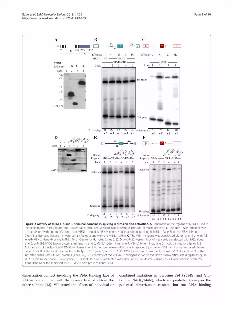
MS2 tethering of MBNL1 activation and repression domainsWe next compared the activities of deletion mutants ofMBNL1 in simple cotransfection and tethered functionassays (Figure 2). Consistent with previous data [22] inthe knockdown/complementation assay with the Tpm1reporter, the N-terminal region of MBNL1 (aa 2–253)had similar repressor activity to the full length protein(Figure 2B, lanes 3,5). In contrast, a C-terminal fragmentof MBNL1 (aa 239–382) had no activity (Figure 2B, lane 4).Similar effects were seen with the Vldlr reporter; the N-terminal fragment had indistinguishable activity to fulllength MBNL1 (Figure 2C, lanes 2,4), while the C-terminalfragment was devoid of activator activity (lane 3), despitebeing expressed to similar levels (Figure 2A).As reported previously [22] replacement of the down-
stream MBNL1 binding element in Tpm1 with a binding
site for MS2 coat protein led to a ~3-fold decrease inexon skipping (Figure 2E, lanes 1,2). Addition of MS2coat protein had little effect (lane 6), while fusionproteins of MS2 with full length MBNL1 or just theN-terminal region led to high levels of exon skipping(lanes 3,4). In contrast, the C-terminal region of MBNL1fused to MS2 had a significant, but much more modesteffect than full length MBNL1-MS2 (lane 5). Replace-ment of the reported MBNL1 binding site containingtwo GC motifs downstream of the Vldlr exon with a sin-gle MS2 site reduced exon inclusion from 14% to 2%(Figure 2F, lanes 1,2), consistent with the activity of thiselement as an MBNL-dependent splicing enhancer inmouse embryonic fibroblasts [10]. Co-transfection withMS2 protein had no effect (lane 6), while full lengthMBNL1-MS2 restored exon inclusion levels (lane 3). Asin the repression assay, the N-terminal of MBNL1-MS2had full activity, while the C-terminal region had partialactivity (lanes 4,5). These data indicate that the N-terminal region of MBNL1 has full activity in simpleco-transfection and artificial tethering repression and en-hancing assays, while the C-terminal region was inactivein simple cotransfections and had partial activity in teth-ered assays.In the artificial tethering assay, the MS2 domain serves
to recruit the fusion protein to the regulated RNA, pre-sumably bypassing the RNA-binding function of at leastsome of the ZF domains. To explore this issue we intro-duced the RNA binding mutations into the ZF domains ofthe MBNL-N-MS2 construct (Figure 3). Tethering of theWT MBNL-N-MS2 downstream of Tpm1 exon 3increased exon skipping from 20% (lanes 1,10) to 71%(lane 9). Individual mutations in ZF1-4 or combined mu-tations in ZF3 and 4 had no effect on activity (lanes 2–5, 7).However, combined mutations in ZF1 and 2 drastically re-duced activity (lane 6), even though the protein wasexpressed (Figure 3B). Indeed, exon skipping levels in thepresence of the ZF12 mutant were not significantly differ-ent from MS2 alone or no cotransfection (lane 6, com-pared to lanes 1 or 10). The quadruple mutant in ZF1-4was also inactive, but again the protein was expressed atvery low levels (lane 8 and Figure 3B). The complete lossof activity upon ZF12 mutation in the tethered repressorassay is in stark contrast to the more modest effects in thesimple cotransfection assay (Figures 1C and 3A). In thetethered activation assay the single mutations in ZF1, 3and 4, and the combined mutation of ZF3 and 4 led to amodest but significant increase in activity while the ZF2mutation was without effect (Figure 3C, lanes 2–5,7 com-pared to 9). Only the dual ZF12 mutant showed decreasedactivity (lane 6) but the effect was modest compared tothe loss of repressor activity.MBNL1 is thought to dimerize through its C terminus
[16,28]. However, the crystal structure of ZF34 revealed a
A
D E
B
Lane: 1 2 3 4
α-FLAG
5755
43
35
28
239 3822532
FL
NC
MBNLEffector: N C FL-
siRNA: C2 MBNL1
Lane: 1 2 3 4 5TPM1-ΔBP
Lane: 1 2 3 4Vldlr
N C FL-N C FL-
Inclusion
Skipping
C
% skipping: % inclusion:
Effector:Effector: -
F- - N-M
S2
C-MS2
FL-MS2
MS2
α-MS2
Reporter: TPM1-ΔBP-DMS2- - MS2
Lane: 1 2 3 4 5 6
C-MS2
N-MS2
FL-MS2
Inclusion
Skipping
Effector:ΔBP Reporter: Vldlr
- - N-MS2
C-MS2
FL-MS2
MS2
Lane: 1 2 3 4 5 6
Vldlr-MS2
Inclusion
Skipping
Effector:
% skipping: % inclusion:
Lane: 1 2 3 4 5 6
β βV1 3 4P3
U D
DY
β βV1 3 4P3 DY
U
48± 5
29± 3
52± 10
27± 4
64± 4
9± 2
39± 8
14± 3
40± 5
51± 5
16± 2
76± 3
78± 2
37± 5
24± 5
14± 3
2± 1
23± 1
24± 2
10± 2
3± 1
Figure 2 Activity of MBNL1 N and C-terminal domains in splicing repression and activation. A. Schematic of the regions of MBNL1 used inthe experiments in this figure (top). Lower panel, anti-FLAG western blot showing expression of MBNL proteins. B. The Tpm1 ΔBP minigene wasco-transfected with control (C2, lane 1) or MBNL1 targeting siRNAs (lanes 2–5). In addition, full-length MBNL1 (lane 5) or the MBNL1 N- orC-terminal domains (lanes 3–4) were cotransfected along with the MBNL1 siRNA. C. The Vldlr minigene was transfected alone (lane 1) or with full-length MBNL1 (lane 4) or the MBNL1 N- or C-terminal domains (lanes 2, 3). D. Anti-MS2 western blot of HeLa cells transfected with MS2 alone,lane 6, or MBNL1-MS2 fusion proteins: full length, lane 5; MBNL1 C-terminus, lane 4, MBNL1 N-terminus, lane 3; mock transfections lanes 1, 2.E. Schematic of the Tpm1 ΔBP DMS2 minigene in which the downstream MBNL site is replaced by a pair of MS2 hairpins (upper panel). Lowerpanel, RT-PCR of HeLa cells transfected with Tpm1 ΔBP (lane 1) or Tpm1 ΔBP DMS2 (lanes 2–6). Cotransfections with MS2 alone (lane 6) or theindicated MBNL1-MS2 fusion proteins (lanes 3–5). F. Schematic of the Vldlr-MS2 minigene in which the downstream MBNL site is replaced by anMS2 hairpin (upper panel). Lower panel, RT-PCR of HeLa cells transfected with Vldlr (lane 1) or Vldlr-MS2 (lanes 2–6). Cotransfections with MS2alone (lane 6) or the indicated MBNL1-MS2 fusion proteins (lanes 3–5).
Edge et al. BMC Molecular Biology 2013, 14:29 Page 5 of 16http://www.biomedcentral.com/1471-2199/14/29
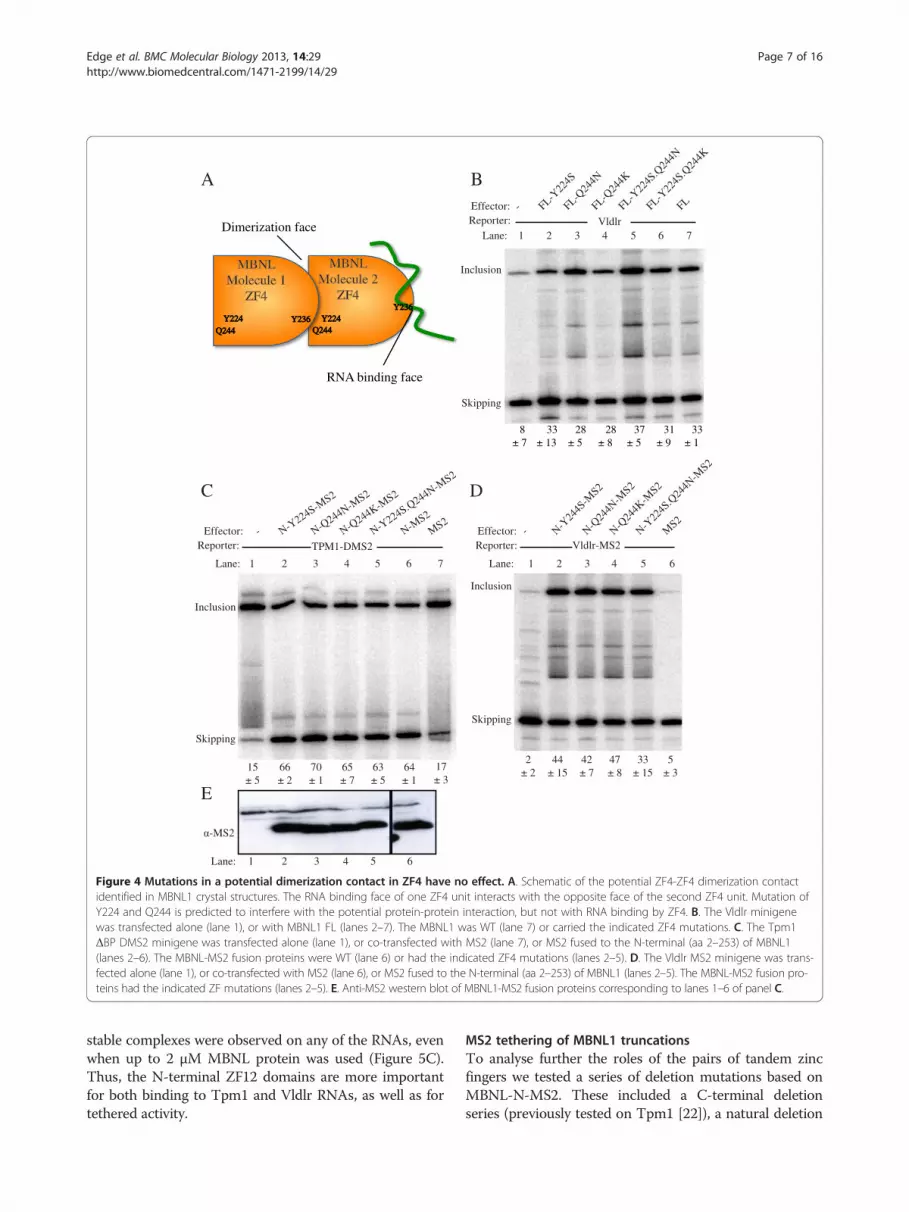
dimerization contact involving the RNA binding face ofZF4 in one subunit, with the reverse face of ZF4 in theother subunit [13]. We tested the effects of individual or
combined mutations in Tyrosine 224 (Y224S) and Glu-tamine 244 (Q244N), which are predicted to impair thepotential dimerization contact, but not RNA binding

Lane: 1 2 3 4 5 6 7 8 9 10
Inclusion
TPM1-ΔBP-DMS2MS2
N-M1-M
S2
N-MS2
N-M2-M
S2
N-M3-M
S2
N-M4-M
S2
N-M12-M
S2
N-M34-M
S2
N-M1234-M
S2A
B
Lane:
1 2 3 4 5 6 7 8 9 10Lane:Reporter:
N-M1-M
S2
N-M2-M
S2
N-M3-M
S2
N-M4-M
S2
N-M12-M
S2
N-M34-M
S2
N-M1234-M
S2
N-MS2
MS2
Vldlr-MS2
C
1 2 3 4 5 6 7 8 9 10Lane:
*****
***
ns
**
**
0
20
40
60
80
100
1 2 3 4 5 6 7 8 9 10
***
nsns nsnsns
MBNLeffector:
Skipping
Inclusion
% E
xon
Skip
ping
α-MS2
Lane: 1 2 3 4 5 6 7 8 9 10
MS2N-M
1-MS2
N-MS2
N-M2-M
S2
N-M3-M
S2
N-M4-M
S2
N-M12-M
S2
N-M34-M
S2
N-M1234-M
S2
***
***
Reporter:
MBNLeffector:
Skipping
% E
xon
Incl
usio
n
0
20
40
60
- -
Figure 3 Effects of RNA binding mutations on tethered MBNL1 repressor and activator function. A. The Tpm1 ΔBP DMS2 minigene wastransfected alone (lane 1), or co-transfected with MS2 (lane 10), or MS2 fused to the N-terminal (aa 2–253) of MBNL1 (lanes 2–9). The MBNL-MS2fusion proteins were WT (lane 9) or had the indicated ZF mutations (lanes 2–8). The horizontal dashed lines indicate the activity of the wild typeN-MS2 (lane 9). Values significantly different from wild type N-MS2 in panels A and C: **, P < 0.01; *** P < 0.001; ns, not significant. Note thatalthough the values for the M1234 mutant in lane 8 of panels A and C are statistically significant, the lack of protein expression of the M1234mutant (panel B) means that meaningful conclusions cannot be drawn. B. Anti-MS2 western blot of MBNL-MS2 fusion proteins used in panels Aand C. C. The Vldlr MS2 minigene was transfected alone (lane 1), or co-transfected with MS2 (lane 10), or MS2 fused to the N-terminal (aa 2–253)of MBNL1 (lanes 2–9). The MBNL-MS2 fusion proteins were WT (lane 9) or had the indicated ZF mutations (lanes 2–8). The horizontal dashed linesindicate the activity of the wild type N-MS2 (lane 9).
Edge et al. BMC Molecular Biology 2013, 14:29 Page 6 of 16http://www.biomedcentral.com/1471-2199/14/29
(Figure 4A). These mutations had no effect upon the teth-ered repressor (4C) or activator (Figure 4D) activities ofMBNL-N-MS2, or on the direct activation of Vldlr byfull length MBNL1 (Figure 4B). These results suggestthat the observed crystal contact between MBNL1subunits is not important for MBNL1 function.
MBNL1 binding to RNA species from MBNL-regulated exonsHaving investigated the role of the MBNL1 ZF domainsin splicing repression and activation, we next tested thebinding of MBNL1-N to RNAs containing the MBNLbinding elements of Vldlr and Tpm1 by electrophoreticmobility shift assay (Figure 5). We compared binding of
WT MBNL1-N with the mutants in ZF12 (M12) andZF34 (M34). WT MBNL1 bound to the Vldlr and up-stream Tpm1 elements, Tpm1 URE, with Kd in the 0.5 –1 nM range, while binding to the downstream Tpm1element Tpm1 Dugc, was approximately 10-fold lower af-finity (Figure 5A, Kd 25–50 nM). With the Vldlr RNA asecond binding event was also observed with a muchlower affinity; we observed no additional binding events toeither of the Tpm1 elements, even though their length issufficient to accommodate multiple binding sites [15].Mutation of ZF34 reduced the affinity of binding to allthree RNAs by about ~20-fold (Figure 5B). In contrast,the effects of mutations in ZF12 were far more drastic; no
B
D
Lane:
Reporter:
1 2 3 4 5 6 7
TPM1-DMS2- N-Y
224S-MS2
N-Q244N-M
S2
N-Q244K-M
S2
N-Y224S.Q244N-M
S2
N-MS2
MS2
-MS2
Inclusion
Skipping
C
15± 5
66± 2
70± 1
65± 7
63± 5
64± 1
17± 3
E
Lane: 1 2 3 4 5 6
Reporter:- M
S2
Vldlr-MS2
Inclusion
Skipping
N-Y24
4S-M
S2N-Q
244N
-MS2
N-Q24
4K-M
S2
N-Y22
4S.Q
244N
-MS2
2± 2
44± 15
42± 7
47± 8
33± 15
5± 3
Effector:Effector:
1 2 3 4 5 6 7Vldlr
- FL-Y22
4S
FL-Q24
4N
FL-Q24
4K
FL-Y22
4S.Q
244N
FL-Y22
4S.Q
244K
FL
Lane:Reporter:
Inclusion
Skipping
Effector:
A
RNA binding face
Dimerization face
MBNLMBNLMolecule 1Molecule 1
ZF4ZF4
MBNLMBNLMolecule 2Molecule 2
ZF4ZF4
Y224Y224 Y224Y224Q244Q244
Y236Y236Q244Q244
Y236Y236
8± 7
33± 13
28± 5
28± 8
37± 5
31± 9
33± 1
Lane: 1 2 3 4 5 6
Figure 4 Mutations in a potential dimerization contact in ZF4 have no effect. A. Schematic of the potential ZF4-ZF4 dimerization contactidentified in MBNL1 crystal structures. The RNA binding face of one ZF4 unit interacts with the opposite face of the second ZF4 unit. Mutation ofY224 and Q244 is predicted to interfere with the potential protein-protein interaction, but not with RNA binding by ZF4. B. The Vldlr minigenewas transfected alone (lane 1), or with MBNL1 FL (lanes 2–7). The MBNL1 was WT (lane 7) or carried the indicated ZF4 mutations. C. The Tpm1ΔBP DMS2 minigene was transfected alone (lane 1), or co-transfected with MS2 (lane 7), or MS2 fused to the N-terminal (aa 2–253) of MBNL1(lanes 2–6). The MBNL-MS2 fusion proteins were WT (lane 6) or had the indicated ZF4 mutations (lanes 2–5). D. The Vldlr MS2 minigene was trans-fected alone (lane 1), or co-transfected with MS2 (lane 6), or MS2 fused to the N-terminal (aa 2–253) of MBNL1 (lanes 2–5). The MBNL-MS2 fusion pro-teins had the indicated ZF mutations (lanes 2–5). E. Anti-MS2 western blot of MBNL1-MS2 fusion proteins corresponding to lanes 1–6 of panel C.
Edge et al. BMC Molecular Biology 2013, 14:29 Page 7 of 16http://www.biomedcentral.com/1471-2199/14/29
stable complexes were observed on any of the RNAs, evenwhen up to 2 μM MBNL protein was used (Figure 5C).Thus, the N-terminal ZF12 domains are more importantfor both binding to Tpm1 and Vldlr RNAs, as well as fortethered activity.
MS2 tethering of MBNL1 truncationsTo analyse further the roles of the pairs of tandem zincfingers we tested a series of deletion mutations based onMBNL-N-MS2. These included a C-terminal deletionseries (previously tested on Tpm1 [22]), a natural deletion

Vldlr TPM1 URE TPM1 Dugc
MBNL-N-M12
MBNL-2-116
MBNL-2-91
MBNL-2-73
A
B
C
D
E
F
MBNL-N
MBNL-N-M34
0 0 0
2000 20000 0 0
2000 2000 20000 0 0
2000 2000 20000 0 0
1000 1000 10000 0 0
1000 1000 10000 0 0
2000
500 500 500
Kd ~ 0.5 nM Kd ~ 1 nM Kd 25-50 nM
Kd 10-25 nM Kd 25-50 nM Kd ~ 500 nM
Kd > 2 μM
Kd 2-5 nM
Kd > 2 μM Kd > 2 μM
Kd ~10 nM Kd ~100 nM
Kd 2-5 nM Kd ~10 nM Kd ~250 nM
Kd > 2 μM Kd > 2 μM Kd > 2 μM
RNA:
Figure 5 MBNL binding to Vldlr and Tpm1 RNAs. RNA binding was assessed by native gel electrophoretic mobility shift assay. RNAs are theMBNL-responsive element from Vldlr (left panels), the upstream MBNL binding site of Tpm1 exon 3 (middle panels), and the downstream MBNLbinding site of Tpm1 exon 3 (right panels). Recombinant proteins used were: A, MBNL-N, the wild-type MBNL construct comprising amino acids2–253 and containing all four zinc fingers, B, MBNL-N-M34, with ZF34 mutated, C, MBNL-N-M12 with ZF12 mutated, D, MBNL-2-116 E, MBNL-2-91F, MBNL-2-72. Increasing protein concentrations are indicated by the wedges above each panel. Protein concentrations were 0, 0.1, 0.5, 1, 2, 5, 10,25, 50, 100, 250, 500 nM for panel A, 0, 0.1, 0.5, 1, 2, 5, 10, 25, 50, 100, 250, 1000 nM for panels D and E, and 0, 1, 2, 5, 10, 25, 50, 100, 250, 500,1000, 2000 nM for panels B, C and F. Estimated Kd’s are indicated in the lower right corner of each panel.
Edge et al. BMC Molecular Biology 2013, 14:29 Page 8 of 16http://www.biomedcentral.com/1471-2199/14/29
variant lacking the C-terminal half of the linker (Δ116-183), an N-terminal deletion series, and the linker alone.The linker sequence is predicted to be unstructured, butparts of it are highly conserved and have been shown pre-viously to have a role in MBNL activities [17,18,22]. Weexpressed these proteins as MS2-fusions (Figure 6B) andanalysed their activity when recruited to either the
downstream Tpm1 (repressed, Figure 6C) or Vldlr (acti-vated, Figure 6D) sites.When recruited downstream of the MBNL-repressed
Tpm1 exon 3 (the Tpm1-ΔbpDMS2 minigene) zinc fin-gers 3 and 4 and the C-terminal part of the linker couldbe removed individually or in combination with no effect(Figure 6C, lanes 4–6). C-terminal truncations beyond

A
C DEffector:
Reporter:Lane :
Inclusion:
Skipping:
E
- MB
NL
-FL
MB
NL
-NΔ1
16-1
832-
183
2-11
62-
102
2-91
2-72
73-1
8373
-253
116-
253
184-
253
2-11
6 M
12-
116
M2
2-11
6 M
12M
S2
Vldlr-MS21 2 3 4 5 6 7 8 9 10111213141516 17
Lane:
% E
xon
Ski
ppin
g
1 2 3 4 5 6 7 8 9 10 11 12 13 14 15 16
***
***
***
*** ***
***
******
***
***
% E
xon
Inc
lusi
on
Lane: 1 2 3 4 5 6 7 8 9 1011121314151617
***
***
****** ***
***
***
*********
***
***
** **
- MB
NL
-FL
Δ116
-183
2-18
32-
116
2-10
22-
912-
7273
-183
73-2
5311
6-25
318
4-25
32-
116
M1
2-11
6 M
22-
116
M12
MS
2
TPM1-ΔBP-DMS21 2 3 5 6 7 8 9 10 11 12 13 1415164
Flag-MBNL1-MS2
MS272
239183 382
253115
91 1022
-
MBNL-F
L
MBNL-N
Δ116-
183
2-18
32-
116
2-10
22-
912-
7273
-183
73-2
53
116-
253
184-
253
2-11
6 M1
2-11
6 M2
2-11
6 M12
MS2
% A
ctiv
ity
Effector:
Reporter:Lane :
Inclusion:
Skipping:
MBNL-F
L
2-18
32-
116
2-10
22-
912-
7273
-183
73-2
5311
6-25
318
3-25
32-
116-
M1
2-11
6-M
22-
116-
M12
MS2
17
17
TPM1-ΔBP-MS2Vldlr-MS2
*80
60
40
20
0
50
40
30
20
0
10
0
100
80
60
40
20
B
Figure 6 Comparison of MBNL1 deletions and point mutations upon tethered repression and activation. A. Schematic of the full-lengthMBNL1-MS2 construct with boundaries of various deletions indicated. B. Anti-MS2 western blot of proteins used in subsequent panels. C. TheTpm1 ΔBP DMS2 minigene was transfected alone (lane 1), or co-transfected with MS2 (lane 17), MS2 fused to full-length MBNL1 (lane 2), or thevarious deletion constructs indicated by the amino acid coordinates. Δ116-183 (lane 4) is a natural MBNL1 isoform resulting from exon skipping.Lanes 14–16 show the effects of the ZF1 and 2 mutations in the context of the 2-116-MS2 deletion mutant. In the experiment shown, the 2-253-MS2 was not expressed (lane 3); in other experiments (e.g. Figure 2E) its activity was similar to FL-MBNL1. The asterisked band is an artefactualband that does not appear in most experiments. Values significantly different from wild type MBNL1-FL-MS2 in panels C and D: **, P < 0.01;*** P < 0.001; ns, not significant. D. The Vldlr-MS2 minigene was co-transfected with the same effectors as panel C. E. Comparison of effects ofmutations in panels C and D. The histogram indicates “% activity” for each of the MBNL1 constructs with the Tpm1 (green bars) and Vldlr (redbars) substrates. The values shown are relative to the activity of FL-MBNL1-MS2. 100% activity is defined by the difference in exon skipping/inclusion inthe presence of FL-MBNL1-MS2 (upper horizontal line in histograms of panels C and D) and in the absence of co-transfection (lower horizontal lines).The comparison shows more rapid decline of activation than repression with C-terminal deletions into the ZF23 linker.
Edge et al. BMC Molecular Biology 2013, 14:29 Page 9 of 16http://www.biomedcentral.com/1471-2199/14/29
amino acid 116 led to diminished activity (lanes 6–9).Complete removal of the linker sequence and an alpha-helix of zinc finger 2 leaving only the first two zinc fin-gers results in an inactive protein (lane 9). Despite theimportance of the linker region, when recruited alone it
had no activity above MS2 alone (lane 10, 17). Zinc fin-gers 3 and 4 along with the complete preceding linkerregion, or with just the C-terminal part of the linker,were partially active (lanes 11,12). However, this activityrequired the linker sequence as ZF34 alone were inactive

Edge et al. BMC Molecular Biology 2013, 14:29 Page 10 of 16http://www.biomedcentral.com/1471-2199/14/29
(lane 13). These data show that the N-terminal part ofthe protein comprising amino acids 2–102, encompass-ing the first two zinc fingers plus a third of the linkersequence, constitutes a minimal repressor domain.Introduction of RNA binding mutations into ZF1 or 2individually drastically reduced activity of the 2–116 re-pressor domain, while combined mutation of ZF 1 and 2abolished activity (compare lanes 14–16 with lane 6).A similar, but not identical, response to the mutations
was seen in the Vldlr context (Figure 6D). In this case,more of the linker sequence was required for full activitywith progressively diminishing activity upon C-terminaldeletion into the linker (Figure 6D, lanes 5–9, Figure 6E).Deletion of the second pair of zinc fingers (lane 5) or in-ternal deletion of the C-terminal part of the linker (lane4) had no effect on activity. As for repressor activity, thelinker sequence alone was inactive (lane 10), but in com-bination with the second pair of zinc fingers the fusionprotein retained substantial activity (lane 11), albeit lessthan the first pair (lane 5). This activity was reduced fur-ther by removal of the N-terminal part of the linker(lane 12) and abolished when only ZF34 remained (lane13). Thus splicing activation is more dependent than re-pression upon the full linker sequence (Figure 6E). Al-though the activity of the 2-116 construct was alreadydiminished, we tested the importance of RNA binding.Abrogating the RNA binding capacity of the 116 con-struct led to a severe, albeit not total reduction in activ-ity (Figure 6D, lanes 14–16 compared to 6).We analysed RNA binding of some of the C-terminal
deletion fragments (Figure 5D-F). The miminal repressordomain MBNL1-2-116 bound to the Vldlr and Tpm1RNAs with affinity reduced compared to the complete N-terminus but actually higher than the N-terminus withpoint mutations in ZF34 (Figure 5D compared to 5A,B).In addition, the 2–116 protein showed additional subse-quent binding events on all three RNAs, consistent withthe fact that it has only 2 ZFs that can contact the RNA.The 2–91 protein, which showed almost complete loss oftethered activation activity (Figure 5D lanes 6–8) boundto each RNA with affinity similar to 2–116 (Figure 5E),emphasizing that RNA binding is necessary but not suffi-cient for activity. Finally, the 2–72 protein, which lacks anexperimentally observed C-terminal extension to the ZF2domain [12,13] failed to bind RNA at any concentration,confirming the importance of the additional α-helix(Figure 5F).
PTB associates with Vldlr RNA but does not regulate itssplicingMBNL and PTB act as co-repressors of Tpm1 splicing[22]. Pull-downs with biotinylated Vldlr RNA indicatedthat PTB was one of the major binding proteins in HeLanuclear extract (data not shown). We therefore asked
whether PTB acted synergistically or antagonisticallywith MBNL1 in the regulation of Vldlr splicing. Asshown earlier, overexpression of MBNL1 promoted skip-ping of Tpm1 exon 3 but inclusion of the Vldlr exon(Figure 7B, C lanes 1,2). Overexpression of PTB had lit-tle effect on Tpm1 splicing (Figure 7C lane 3), as PTB isnot limiting in HeLa cells [29]. However, PTB/nPTBknockdown led to decreased exon skipping (lanes 1and 4). In contrast, Vldlr splicing was unresponsive to ei-ther overexpression or knockdown of PTB (Figure 7Clanes 1,3,4), suggesting that binding of PTB to Vldlr isnon-functional. Furthermore, the activating effect ofMBNL was not reduced upon PTB knockdown, and actu-ally appeared to be slightly increased (Figure 7C lane 5).Therefore, while MBNL1 and PTB cooperate to repressTpm1 splicing, MBNL1 acts independently to activateVldlr splicing, and PTB binding to Vldlr appears to benon-functional.
DiscussionThe data presented here, drawing upon point mutationsto impair RNA binding of ZF domains, deletion muta-tions, and artificial MS2 tethering, provide insights intothe domains of MBNL1 that are involved in activationand repression of splicing. Our results are complementaryto other published reports [16-18], and taken together thedifferent studies converge upon some common themes.Among our key findings are the following. First, the N-terminal region of MBNL1 encompassing the four ZFdomains is nearly fully active in most assays (Figure 2).Second the C-terminal region is inactive in conventionaloverexpression assays, but retains some activity in teth-ered assays where the MS2 domain recruits it to targetRNAs (Figure 2). This residual activity might be associatedwith the ability of the C-terminal region to mediatedimerization [16,28], which might allow tethered C-terminal to interact with intact endogenous MBNL pro-teins, perhaps promoting their recruitment to the RNA.Third, MBNL1 is remarkably tolerant of RNA bindingmutations to individual ZF domains (Figures 1, 3), consist-ent with previous results [18]. Fourth, pair-wise inactiva-tion of ZF12 was in most cases more deleterious thaninactivation of ZF34 (Figure 3). This is consistent with theeffects of deletions that remove ZF12 or ZF34 (Figure 6,constructs 72–253 and 2–183 respectively), and with theeffects of the didomain point mutations upon RNA bind-ing (Figure 5A-C). Fifth, both repression of the Tpm1exon and activation of the Vldlr exon required not just anintact ZF didomain, but also the linker sequence connect-ing ZF2 and 3 (Figure 6). Finally, there were some differ-ences in the responses of the repressed Tpm1 and theactivated Vldlr exons to different MBNL1 mutations. Acti-vation of the Vldlr exon was more sensitive than repres-sion of Tpm1 to deletions of the linker between ZF2 and

Skipping
Inclusion
MBNL:
PTB: -
-
- +
+ -
-
-
- +
+ -
Reporter:
siRNA: C2 control PTB/nPTB
Lane: 1 2 3 4 5 6
TPM1-ΔBP
A
B C
MBNL:
PTB: -
-
- +
+ -
-
-
- +
+ -
Reporter:
siRNA: C2 control PTB/nPTB
Lane: 1 2 3 4 5 6
Vldlr
Skipping
Inclusion
α-PTB
α-FLAG
α-actin
--
- ++ -
--
- ++ -MBNL:
PTB:
C2 control PTB/nPTB
--
--
--
0
10
20
30
40
50
60
70
% E
xon
Incl
usio
n
0
20
40
60
80
% E
xon
skip
ping
Lane : 1 2 3 4 5 6 7 8 9
Figure 7 PTB co-regulates Tpm1 but not Vldlr splicing. A. Western blots with anti-PTB (upper), anti-FLAG (middle) and anti-actin (lower panel).Cells were treated with control C2 siRNA (lanes 1–6) or PTB/nPTB siRNAs (lanes 7–9). Lanes 4–1 show successive 2-fold dilutions of the C2 controlto allow assessment of knockdown. In lanes 6 and 9, FLAG-PTB was overexpressed; in lanes 5 and 8, FLAG-MBNL1 was overexpressed. B. TheTpm1 ΔBP minigene was cotransfected with control C2 (lanes 1–3) or PTB/nPTB siRNAs (lanes 4–6). In addition, FLAG-PTB (lanes 3, 6) or FLAG-MBNL1 (lanes 2, 5) were also cotransfected. C. The Vldlr minigene was cotransfected with control C2 (lanes 1–3) or PTB/nPTB siRNAs (lanes 4–6).In addition, FLAG-PTB (lanes 3, 6) or FLAG-MBNL1 (lanes 2, 5) were also cotransfected.
Edge et al. BMC Molecular Biology 2013, 14:29 Page 11 of 16http://www.biomedcentral.com/1471-2199/14/29
3 (Figure 6), consistent with previous reports comparingactivated and repressed exons [16,17]. The minimal re-pressor domain for MBNL1 when tethered encompasseszinc fingers 1 and 2, with a region of further linker se-quence up to amino acid 102. The minimal tethered acti-vation domain comprises ZF1 and 2 and the full linkersequence to amino acid 183. In contrast to the effects oftruncation mutations, effects of the ZF12 RNA bindingmutations were more pronounced upon Tpm1 than Vldlr.The ZF12 mutation led to complete loss of activity uponthe Tpm1 exon, whereas with the Vldlr minigene this mu-tation led to only a slight reduction in activity (Figure 3).Likewise, within the context of the effector fragment 2-116-MS2, mutation of ZF12 abolished repressor activity,while retaining ~30% of activator activity (Figure 6). The
differential effects of MBNL1 mutations upon the Tpm1and Vldlr exons are interesting. However, they cannot begeneralized for all activated or repressed targets of MBNLproteins. Indeed, a systematic analysis of combinedMBNL1 ZF mutants with a panel of 6 MBNL regulatedevents, showed that the relationship between ZF muta-tions and effects upon activity upon different splicing sub-strates is quite complex, with at least two classes of target,each encompassing repressed and activated targets [18].In agreement with our conclusions the same study showedthat MBNL activity upon Vldlr did not correlate well withits RNA binding ability [18].A surprising feature of our results is that the ZF RNA
binding mutations had a greater effect in the MS2 teth-ering assays (Figure 3) than in the untethered assays

Edge et al. BMC Molecular Biology 2013, 14:29 Page 12 of 16http://www.biomedcentral.com/1471-2199/14/29
(Figure 1). One would anticipate that the tethered pro-teins would be less sensitive to mutations in their RNAbinding domains, since the MS2 domain should bypassat least some of the RNA binding functions of the intactprotein. However, the C-terminal domain, which mediatesdimerization [16,28] and on its own has some activity inthe tethering assays (Figure 2), was missing in most of thesubsequent MS2-tethering assays (Figures 3, 6), whichmight account for the greater sensitivity to the ZF muta-tions. In addition to the RNA binding mutations of the ZFdomains, we also tested a set of mutations in ZF4 de-signed to impair intra-molecular contacts formed betweenMBNL molecules in crystal structures. Although previousstudies have implicated the variably spliced C-terminal re-gion of MBNL in dimerization [16,28], it was possible thatthe ZF4 mediated contacts might also be functional. How-ever, these mutations had no functional effect in a numberof assays of MBNL1 activities (Figure 4). Although we didnot test the mutations in direct assays for dimerization,the lack of effect in functional assays suggests that thesecontacts are not physiologically relevant.The deleterious effects of RNA binding mutations in a
tethered function assay is initially surprising, given thatthe starting point of the assay is to bypass the normalmode of RNA binding at a particular location. However,this has been observed with similar studies of RbFOX [23]and PTB [30], and provides insights into the possiblemechanisms of splicing regulation. Given that many RNAbinding proteins, including MBNL proteins, have multipleRNA binding domains, it could be that the MS2 tether re-places the role of a subset of the RNA binding domains,and that functional effects require the remaining domainsto interact with RNA for one of a number of reasons. Theprotein might need to bind to more than one site in thetarget pre-mRNA to be functional, perhaps forming anRNA architecture conducive to regulated exon skippingor inclusion; the protein might have to interact with an-other RNA; or the protein might have to interact withRNA in order to interact with important partner proteins.Taken in order, we already know that there are at leasttwo major MBNL1 binding sites flanking Tpm1 exon 3[22,31]. We also identified additional YGCY motifs down-stream of the Vldlr exon that mediate MBNL1 activity(data not shown). Alternatively, the additional RNA inter-actions could be with a distinct RNA species such as U1snRNA; inhibition of the N1 exon of CSRC involves aPTB-U1 snRNA interaction [32]. Finally, RNA bindingmight be important to induce a conformation that facil-itates interaction with co-regulatory proteins [22]. Thesedifferent explanations are not all mutually exclusive; forexample, a protein-RNA interaction might promote anecessary RNP architecture as well as inducing a con-formation change promoting necessary protein-proteininteractions.
Analysis of the relationship between position of bind-ing on RNA substrates and the mode of action as a re-pressor or activator indicates that in general MBNLbinding upstream on an exon is associated with repres-sion while downstream binding leads to activation[10,33,34], similar to a number of other regulatory pro-teins [4]. However, in the case of repression there arealso binding peaks downstream of the exon as well, sug-gesting a sub-set of events which are regulated nega-tively by MBNL sites flanking the exon. This suggeststhe possibility of two discrete types of MBNL-repressedevent, which might operate in a mechanistically distinctmanner or might share some common mechanistic ele-ments. In the first case, MBNL binding sites immediatelyupstream of the regulated exon might be sufficient tointerfere with binding or activity of constitutive splicingfactors such as U2AF, as has been suggested in thecTNT transcript [35,36]. In the case of exons such asTpm1 exon 3 the flanking sites might be necessary inorder for the upstream sites to effectively interfere with3′ splice site recognition factors, perhaps by cooperativebinding of oligomers and consequent looping of inter-vening RNA [37,38]. Alternatively the flanking regula-tory sites might need to act in a concerted way on the 3′and 5′ splice sites. The two tandem zinc finger arrays ofMBNL are arranged with each zinc finger ‘back-to-back’,which would cause an anti-parallel alignment of a boundRNA. It appears unlikely that a single MBNL1 proteincould bind to both the elements flanking Tpm1 exon 3.Indeed, mutations in ZF12 and ZF34 have similar effectsupon binding to either the upstream or the downstreamTpm1 element (Figure 5A-C). However, we have recentlyshown that the minimal repressor region of MBNL inter-acts with PTB protein, in an RNA-dependent manner[22], and there are PTB sites flanking exon 3, with two tothree molecules binding either side [39]. Moreover MBNLhas been shown to interact with itself [16,28]. Taken to-gether this suggests a complex forming across the exon,with homotypic and heterotypic interactions betweenMBNL and PTB molecules acting to stabilise a loopedstructure which promotes exon skipping. Analysis of pro-teins binding to the Vldlr substrate indicated strong inter-action of PTB, suggesting that it might act as acoactivator. However, overexpression and knockdown ex-periments clearly showed that PTB played no role in regu-lating Vldlr splicing (Figure 7). Indeed, we also found thatPTB-MS2 had no activity when tethered downstreamof the Vldlr exon, despite the fact that it can activateits own target exons from this location [40]. Thisclearly indicates that different activators have distinctmolecular targets, even when binding at similar loca-tions. An important future line of work will be toidentify the molecular targets of the minimal MBNL1activator domain.

Edge et al. BMC Molecular Biology 2013, 14:29 Page 13 of 16http://www.biomedcentral.com/1471-2199/14/29
MethodsConstructsThe Tpm1 minigene reporters and Δbp mutation have beendescribed previously [22,27,31]. The Vldlr wildtype mini-gene was a kind gift from Prof. Manny Ares (University ofCalifornia Santa Cruz) [10]. This minigene was mutagenizedto introduce a Pst1 site then an MS2 hairpin was insertedusing the following oligo: 5’-gAGGATCACCctgca-3’. Ex-pression plasmids for MBNL1 N and C terminal trunca-tions have been described previously [22]. Mutagenesis wasperformed using standard protocols and the followingprimers: ZnF1: 5-CACGGAATGTAAAgcTGCACATCCTTCG-3, ZnF2: 5-GGAGAACTGCAAAgcTCTTCATCCACC-3, ZnF3: 5-GAAAATGATTGTCGGgcTGCTCATCCTGC-3, ZnF4: 5-GGAAAAGTGCAAAgcCTTTCATCCCCC-3. MBNL1 truncations were cloned by inserting thecoding sequence for the relevant portion of MBNL1 intothe AvrII and MluI sites in the pCIMS2-NLS-FLAG vec-tor [20,41]. Plasmids for bacterial expression were clonedby insertion of appropriate coding sequence into thepGEX-4-T3 vector. Sequences for expression of Tpm1 orVldlr RNA were cloned into pGEM-4Z plasmids (Pro-mega), and transcribed usingT7 polymerase. Sequencesused were, Vldlr: GGGAGACAAGCTTTGCAAACTGTTAATCTCAACTAACTGCCGCTTAAATAATTAGTGCAGCTTTTAACTACTGGTTCTGTCCCAACTGGCTACTTGTGCCTAAAGCCCAAAGAATT, Dugc: GGGAGACAAGCTTGAGCTGGATGCCGCCTCTGCTGCTGC, URE: gggagacaagcttaaGTCTACGCACCCTCAAccCGCACCTTGCGGGATCACGCTGCCTGCTGCACCCCACCCCCTTCCCCCTTCCTTCCCCCCACCCCCGTACTCCACTGCCAACTCCCAG.
Cell cultureCells were transfected a day after splitting to 105 – 2 ×10⁵ cells per well in a 6 well plate. Transfections wereperformed using 400 ng effector construct unless other-wise stated, with 200 ng reporter, made up to 1 μg withempty pGEM4Z vector as necessary. Per well 1μg ofDNA, 100 μl Optimem and 2 μl Lipofectamine (Invitro-gen 18324–012) was used. Lipofectamine-DNA mix wasincubated for 30min at room temperature, then dilutedto 1ml in Opti-MEM-1 and applied to cell monolayerpreviously washed with PBS. Treated HeLa cells were in-cubated for 5 hr at 37°C, and then the transfection mixwas replaced with 2 ml Dulbecco’s Modified EaglesMedium (DMEM) supplemented with Glutamax & 10%fetal bovine serum (FBS). Cells were then incubated fora further 48 hours, then RNA and protein was harvestedfrom the cultures using Trizol reagent (invitrogen) orboiled SDS loading buffer respectively.For MBNL1 knockdown, the following target sequence
was used: 5’-AACACGGAAUGUAAAUUUGCA-3’ [42].HeLa cells were split to a density of 2 × 10⁵ in 1.7 ml
DMEM +10% FBS medium in 6 well plates, and incu-bated at 37°C for 24 hours. Each well was treated with10 nM siRNA (THH2 siRNA for MBNL1 knockdown orcontrol C2 siRNA) and 15 μl Oligofectamine (Invitrogen12252–011), diluted in 500 μl Optimem and incubatedprior at room temperature for 20 minutes. Cells werethen incubated for 24 hours at 37°C. After 24 hour incu-bation DNA transfections were performed as aboveusing lipofectamine or lipofectamine 2000 reagent. Cellswere incubated for 5 hours at 37°C, then the medium onthem replaced with 1.5ml DMEM+10% FBS. To each well10 nM siRNA and 3 μl Lipofectamine 2000 reagent in 500μl Optimem was added, which had been pre-incubated for20 minutes at room temperature. Cells were incubated fora further 48 hours, then harvested.
RNA and protein analysis of transfectionsFor RNA analysis, cells in 6 well plates were washedusing twice with 2 ml PBS, then to each well 1 ml tri-reagent (Sigma-Aldrich) was added, and purified accord-ing to the manufacturers’ protocols. Samples wereDNAse I treated using 2 units of rDNAse (Ambion) in10 mM Tris pH 7.5, 2.5 mM MgCl2, 0.5 mM CaCl2 for30 minutes – 1 hour at 37°C, phenol extracted and etha-nol precipitated. PCR analysis used the following primersfor Vldlr minigenes:
V4rt - 5’-GTGGCAAAGGTGCCCTTGAG-3’ - (rt primer)V1 - 5’-ACGTGGATGAAGTTGGTGGT-3’ - (5’ primer)V3 - 5’-GGCACCGAGCACTTTCTTGC-3’ - (3’ primer)
and the following primers for Tpm1 minigenes:
SV3’RT: 5’-GCAAACTCAGCCACAGGT-3’ - (rt primer)SV5’2: 5’-GGAGGCCTAGGCTTTTGCAAAAAG-3’ -(5’ primer)SV3’1: 5’-ACTCACTGCGTTCCAGGCAATGCT-3’ -(3’ primer)
Reverse transcriptions were performed using 2–3 μg oftotal RNA, and 100 ng of RT primer, in 50 mM Tris pH6.3, 40 mM KCl, 8 mM MgCl2, 2 mM DTT. Sampleswere heated for 15 minutes at 55°C, then cooled to 42°C,and 2 μl 10 mM dNTP and 1 μl AMV-RT (Promega) en-zyme added, and incubated at 42°C for 60 minutes. Forthe PCR reaction, the 3’ primer was 5’ end labeled with[32P]-ATP,. Oligo primer (4 pmoles per PCR reaction) wasincubated at 37°C for 60 minutes in 50 mM Tris 10 mMMgCl2 T4 polynucleotide kinase enzyme (NEB) and 0.1 μl[α-32P]-UTP per PCR reaction. After incubation the solu-tion was phenol extracted, and purified on a G-50 spincolumn (GE Healthcare). The labelled oligo was made upto concentration of 1 pmole/μl. 2 μl of RT reaction wastaken into fresh eppendorf in buffer (50 μM KCl, 10 μM

Edge et al. BMC Molecular Biology 2013, 14:29 Page 14 of 16http://www.biomedcentral.com/1471-2199/14/29
Tris pH 8.3, 1.5 mM MgCl2, 0.001% w/v gelatin) and 25pmole of the reverse primer. Samples were heated to 92°Cfor 3 minutes, then cooled to 80°C, and 0.25 μl Taq poly-merase (Roche) and 10 pmol 32P-labeled probe added.The samples were then cycled for 30 cycles of 94°C for 30seconds, 62°C for 30 seconds and 72°C for 60 seconds.RT-PCR products were analysed on denaturing 4% PAGEgels, using Sequagel (National Diagnostics EC-833) sys-tem. Samples were diluted in formamide loading buffer,heated to 90°C for 5 minutes, then loaded. Gels were runfor 100 minutes at constant 38 W, the gel was dried andexposed on phosphorimager casette (Molecular Dynam-ics). The results were quantified using ImageQuant Soft-ware (GE Healthcare) and analysed using Excel (Microsoft)and Graphpad 5 (Prism Software). Statistical judgementswere made using either students t-tests or, where multiplecombinations tested, a post-ANOVATukey test, which is avariation of the students t-test which aims to eliminatetype 2 errors stemming from multiple comparisons withoutBonferroni corrections. Statistical significance is indicatedby: ns, not significant; * P < 0.05; ** P < 0.01; ***, P < 0.001.For protein analysis, the cell monolayer in 6 well plates
was washed twice with 2 ml PBS, then directly to eachwell 150 μl of hot SDS buffer (pre-heated to 100°C for 5minutes) was added. The cells were scrapped usingupturned P-1000 tips, extracted into eppendorf tubes,and frozen on dry ice. Samples were heated again to100°C for 5 minutes, separated using SDS-PAGE, ana-lysed using standard western blotting techniques, andimaged using standard ECL techniques. For western blotanalysis primary antibodies were in house anti-rabbitMS2 or anti-FLAG from Sigma (F1804). Protein loadingwas checked by Ponceau staining and, in some cases, byre-probing with anti-actin antibodies.
Recombinant protein expressionRecombinant MBNL1 protein was expressed and puri-fied from E.coli BL21 cells. 400 ml cultures were inducedat OD600 = 0.5 by the addition of 1 mM IPTG, andgrown for 3 hours shaking at 225 rpm. The cultureswere then pelleted, washed in MTPBS (150 mM NaCl,16 mM Na2HPO4, 4 mM NaH2PO4, pH 7.3) and lysedusing a French Press (Stansted Fluid Power) accordingto the manufacturer’s instructions. The homogenisedsamples were centrifuged at 7741 rcf, 4°C, for 10 mi-nutes. Samples were then purified using GST Sepharose4B beads (GE Healthcare) according to manufacturersprotocols. Briefly - the beads were pre-washed with 5–10 volumes of water, MTPBS and MTPBS + 1% Triton-X100. To the homogenised sample Triton-X100 addedto concentration of 1%. This bacterial homogenate wasthen incubated with the GST beads at 4°C, for 1 hour.The beads were washed 4 times with 2.5 volumes ofMPTBS + 1% Triton-X100, then loaded into a disposable
biorad column at 4°C. The recombinant proteins wereeluted from the column using 3×800 μl of 25 mM re-duced glutathione in 100 mM HEPES (pH 8.9), followedby 3×800 μl of 50 mM reduced glutathione in 100 mMHEPES. All fractions of interest were pooled, and dia-lysed in 1.8 litres of Dignam Buffer E overnight at 4°Cusing Slide-A-Lyzer Dialysis Cassettes (Thermo Scien-tific), according to manufacturer’s protocols. The con-centration of the recombinant proteins was estimatedwith reference to BSA standards.
Electrophoretic-mobility shift and UV crosslinking assayHigh specific activity [α-P32] UTP labelled RNAs weremade using standard protocols with either SP6 or T7polymerase. Binding reactions were set up in microtitreplates (Corning) pre-lined with BSA. Mobility shift as-says had a total 5 μl reaction volume, with 10 fmolRNA, 20 μg/ml rRNA, 10 mM HEPES pH 7.9, 10 μMZnCl2, 3 mM MgCl2, 5% Glycerol, 1 mM DTT, and pro-teins at appropriate concentration. The reaction was in-cubated for 15 minutes at 30°C, then 0.5 μl of a 55 mg/mlHeparin (Sigma) added, and the samples incubated for afurther 5 minutes. Before loading onto the gel 1 μl of50% glycerol was added. 5% poly-acrylamide gels wereused, with 30:1 bis:acrylamide ratio. Gels were run at200 volts for ~ 2hr, 4°C after pre-running for 1–2 hours,at 200 volts. Dried gels were analysed by phosphor-imager (Molecular Dynamics). After scanning on Ty-phoon scanner results were analysed using ImageQuant(GE Healthcare) and Photoshop (Adobe). Dissociationconstants were estimated from the total protein concen-tration that produced 50% binding. For UV crosslinking,the total reaction volume was 10 μl. After addition ofheparin, samples were subjected to 19200 J.cm-2 UV light,followed by digestion with 50 μg RNase A and 140 URNase T1. Samples were then separated by SDS gel elec-trophoresis and dried gels analyzed by phosphorimager.
ConclusionsOur results highlight the common and distinct domainrequirements for activation or repression of splicing byMBNL1. Full length MBNL1 is relatively insensitive toinactivating mutations of individual ZF domains. How-ever, when the protein is recruited to RNA by tetheringwith a heterologous RNA binding domain and deletionmutations are introduced, the dependency on functionalZF domains becomes more acute. Full tethered repressorand activator functions require ZF domains 1 and 2 thatare able to bind RNA, suggesting that both types of ac-tivity require multivalent interactions with RNA. How-ever, the ZF domains alone are insufficient for activity.Additional regions of the linker separating ZF domains 2and 3 are required for splicing activity but not RNAbinding. The additional regions differ for repression or

Edge et al. BMC Molecular Biology 2013, 14:29 Page 15 of 16http://www.biomedcentral.com/1471-2199/14/29
activation, with more extensive regions of the linker re-quired for full activation. This suggests the involvementof different sets of interacting cofactors for activation orrepression of splicing by MBNL1.
Competing interestThe authors declare that they have no competing interests.
Authors’ contributionsCWJS and CG conceived of the study and participated in its design andcoordination. CE and CG carried out the experimental work and analyzed thedata. All authors helped to draft the manuscript, and approved the finalmanuscript.
AcknowledgementsThis work was supported by a Wellcome Trust Programme Grant to CWJS(092900). CE was supported by a studentship from the BBSRC. We thankManny Ares for the Vldlr minigene, and Ben Luisi for help and advice ininterpretation of MBNL structures and design of mutations.
Received: 3 October 2013 Accepted: 16 December 2013Published: 27 December 2013
References1. Nilsen TW, Graveley BR: Expansion of the eukaryotic proteome by
alternative splicing. Nature 2010, 463(7280):457–463.2. Barbosa-Morais NL, Irimia M, Pan Q, Xiong HY, Gueroussov S, Lee LJ,
Slobodeniuc V, Kutter C, Watt S, Colak R, et al: The evolutionary landscapeof alternative splicing in vertebrate species. Science 2012,338(6114):1587–1593.
3. Merkin J, Russell C, Chen P, Burge CB: Evolutionary dynamics of gene andisoform regulation in Mammalian tissues. Science 2012,338(6114):1593–1599.
4. Witten JT, Ule J: Understanding splicing regulation through RNA splicingmaps. Trends Genet 2011, 27(3):89–97.
5. Miller JW, Urbinati CR, Teng-Umnuay P, Stenberg MG, Byrne BJ, ThorntonCA, Swanson MS: Recruitment of human muscleblind proteins to (CUG)(n) expansions associated with myotonic dystrophy. EMBO J 2000, 19(17):4439–4448.
6. Kalsotra A, Xiao XS, Ward AJ, Castle JC, Johnson JM, Burge CB, Cooper TA: Apostnatal switch of CELF and MBNL proteins reprograms alternativesplicing in the developing heart. Proceedings of the National Academy ofSciences of the United States of America 2008, 105(51):20333–20338.
7. Terenzi F, Ladd AN: Conserved developmental alternative splicing ofmuscleblind-like (MBNL) transcripts regulates MBNL localization andactivity. RNA Biol 2009, 7(1):43–55.
8. Botta A, Caldarola S, Vallo L, Bonifazi E, Fruci D, Gullotta F, Massa R, NovelliG, Loreni F: Effect of the [CCTG]n repeat expansion on ZNF9 expressionin myotonic dystrophy type II (DM2). Biochim Biophys Acta 2006,1762(3):329–334.
9. Osborne RJ, Lin X, Welle S, Sobczak K, O’Rourke JR, Swanson MS, ThorntonCA: Transcriptional and post-transcriptional impact of toxic RNA inmyotonic dystrophy. Human Molecular Genetics 2009, 18(8):1471–1481.
10. Du H, Cline MS, Osborne RJ, Tuttle DL, Clark TA, Donohue JP, Hall MP, ShiueL, Swanson MS, Thornton CA, et al: Aberrant alternative splicing andextracellular matrix gene expression in mouse models of myotonicdystrophy. Nat Struct Mol Biol 2010, 17(2):187–193.
11. Kino Y, Washizu C, Oma Y, Onishi H, Nezu Y, Sasagawa N, Nukina N, IshiuraS: MBNL and CELF proteins regulate alternative splicing of the skeletalmuscle chloride channel CLCN1. Nucleic Acids Res 2009, 37(19):6477–6490.
12. He F, Dang W, Abe C, Tsuda K, Inoue M, Watanabe S, Kobayashi N, KigawaT, Matsuda T, Yabuki T, et al: Solution structure of the RNA bindingdomain in the human muscleblind-like protein 2. Protein Sci 2009, 18(1):80–91.
13. Teplova M, Patel DJ: Structural insights into RNA recognition by thealternative-splicing regulator muscleblind-like MBNL1. Nat Struct Mol Biol2008, 15(12):1343–1351.
14. Goers ES, Purcell J, Voelker RB, Gates DP, Berglund JA: MBNL1 binds GCmotifs embedded in pyrimidines to regulate alternative splicing.Nucleic Acids Res 2010, 38(7):2467–2484.
15. Cass D, Hotchko R, Barber P, Jones K, Gates DP, Berglund JA: The four Znfingers of MBNL1 provide a flexible platform for recognition of its RNAbinding elements. BMC Mol Biol 2011, 12:20.
16. Tran H, Gourrier N, Lemercier-Neuillet C, Dhaenens CM, Vautrin A,Fernandez-Gomez FJ, Arandel L, Carpentier C, Obriot H, Eddarkaoui S, et al:Analysis of exonic regions involved in nuclear localization, splicing activity,and dimerization of Muscleblind-like-1 Isoforms. Journal of BiologicalChemistry 2011, 286(18):16435–16446.
17. Grammatikakis I, Goo YH, Echeverria GV, Cooper TA: Identification ofMBNL1 and MBNL3 domains required for splicing activation andrepression. Nucleic Acids Res 2011, 39(7):2769–2780.
18. Purcell J, Oddo JC, Wang ET, Berglund JA: Combinatorial Mutagenesis ofMBNL1 Zinc Fingers Elucidates Distinct Classes of Splicing RegulatoryEvents. Mol Cell Biol 2012, 32(20):4155–4167.
19. Graveley BR, Maniatis T: Arginine/serine-rich domains of SR proteins canfunction as activators of pre-mRNA splicing. Mol Cell 1998, 1(5):765–771.
20. Del Gatto-Konczak F, Olive M, Gesnel MC, Breathnach R: hnRNP A1 re-cruited to an exon in vivo can function as an exon splicing silencer.Mol Cell Biol 1999, 19(1):251–260.
21. Robinson F, Smith CW: A splicing repressor domain in polypyrimidinetract-binding protein. J Biol Chem 2006, 281(2):800–806.
22. Gooding C, Edge C, Lorenz M, Coelho MB, Winters M, Kaminski CF, ChernyD, Eperon IC, Smith CW: MBNL1 and PTB cooperate to repress splicing ofTpm1 exon 3. Nucleic Acids Res 2013, 41(9):4765–4782.
23. Sun S, Zhang Z, Fregoso O, Krainer AR: Mechanisms of activation andrepression by the alternative splicing factors RBFOX1/2. Rna 2011,18(2):274–283.
24. Shankarling G, Lynch KW: Minimal functional domains of paralogueshnRNP L and hnRNP LL exhibit mechanistic differences in exonicsplicing repression. Biochem J 2013, 453(2):271–279.
25. Hudson BP, Martinez-Yamout MA, Dyson HJ, Wright PE: Recognition of themRNA AU-rich element by the zinc finger domain of TIS11d. Nat StructMol Biol 2004, 11(3):257–264.
26. Fu Y, Ramisetty SR, Hussain N, Baranger AM: MBNL1-RNA Recognition:Contributions of MBNL1 Sequence and RNA Conformation. Chembiochem2012, 13(1):112–119.
27. Gooding C, Clark F, Wollerton MC, Grellscheid SN, Groom H, Smith CW: Aclass of human exons with predicted distant branch points revealed byanalysis of AG dinucleotide exclusion zones. Genome Biol 2006, 7(1):R1.
28. Yuan Y, Compton SA, Sobczak K, Stenberg MG, Thornton CA, Griffith JD,Swanson MS: Muscleblind-like 1 interacts with RNA hairpins in splicingtarget and pathogenic RNAs. Nucleic Acids Res 2007, 35(16):5474–5486.
29. Wollerton MC, Gooding C, Robinson F, Brown EC, Jackson RJ, Smith CW:Differential alternative splicing activity of isoforms of polypyrimidinetract binding protein (PTB). Rna 2001, 7(6):819–832.
30. Joshi A, Coelho MB, Kotik-Kogan O, Simpson PJ, Matthews SJ, Smith CW,Curry S: Crystallographic analysis of polypyrimidine tract-binding protein-Raver1 interactions involved in regulation of alternative splicing. Structure2011, 19(12):1816–1825.
31. Gromak N, Smith CW: A splicing silencer that regulates smooth musclespecific alternative splicing is active in multiple cell types. Nucleic AcidsRes 2002, 30(16):3548–3557.
32. Sharma S, Maris C, Allain FH, Black DL: U1 snRNA directly interacts withpolypyrimidine tract-binding protein during splicing repression. Mol Cell2011, 41(5):579–588.
33. Wang ET, Cody NA, Jog S, Biancolella M, Wang TT, Treacy DJ, Luo S, SchrothGP, Housman DE, Reddy S, et al: Transcriptome-wide regulation of pre-mRNA splicing and mRNA localization by muscleblind proteins. Cell 2012,150(4):710–724.
34. Charizanis K, Lee KY, Batra R, Goodwin M, Zhang C, Yuan Y, Shiue L, Cline M,Scotti MM, Xia G, et al: Muscleblind-like 2-mediated alternative splicing inthe developing brain and dysregulation in myotonic dystrophy. Neuron2012, 75(3):437–450.
35. Warf MB, Berglund JA: MBNL binds similar RNA structures in the CUGrepeats of myotonic dystrophy and its pre-mRNA substrate cardiactroponin T. Rna 2007, 13(12):2238–2251.
36. Warf MB, Diegel JV, von Hippel PH, Berglund JA: The protein factorsMBNL1 and U2AF65 bind alternative RNA structures to regulate splicing.Proc Natl Acad Sci U S A 2009, 106(23):9203–9208.
37. Nasim FU, Hutchison S, Cordeau M, Chabot B: High-affinity hnRNP A1binding sites and duplex-forming inverted repeats have similar effects

Edge et al. BMC Molecular Biology 2013, 14:29 Page 16 of 16http://www.biomedcentral.com/1471-2199/14/29
on 5’ splice site selection in support of a common looping out andrepression mechanism. Rna 2002, 8(8):1078–1089.
38. Fisette JF, Toutant J, Dugre-Brisson S, Desgroseillers L, Chabot B: hnRNP A1and hnRNP H can collaborate to modulate 5’ splice site selection. Rna2010, 16(1):228–238.
39. Cherny D, Gooding C, Eperon GE, Coelho MB, Bagshaw CR, Smith CW,Eperon IC: Stoichiometry of a regulatory splicing complex revealed bysingle-molecule analyses. EMBO J 2010, 29(13):2161–2172.
40. Llorian M, Schwartz S, Clark TA, Hollander D, Tan LY, Spellman R, Gordon A,Schweitzer AC, de la Grange P, Ast G, et al: Position-dependent alternativesplicing activity revealed by global profiling of alternative splicingevents regulated by PTB. Nat Struct Mol Biol 2010, 17(9):1114–1123.
41. Gromak N, Rideau A, Southby J, Scadden AD, Gooding C, Huttelmaier S,Singer RH, Smith CW: The PTB interacting protein raver1 regulates alpha-tropomyosin alternative splicing. EMBO J 2003, 22(23):6356–6364.
42. Ho TH, Charlet BN, Poulos MG, Singh G, Swanson MS, Cooper TA:Muscleblind proteins regulate alternative splicing. EMBO J 2004,23(15):3103–3112.
doi:10.1186/1471-2199-14-29Cite this article as: Edge et al.: Dissecting domains necessary foractivation and repression of splicing by muscleblind-like protein 1. BMC Mo-lecular Biology 2013 14:29.
Submit your next manuscript to BioMed Centraland take full advantage of:
• Convenient online submission
• Thorough peer review
• No space constraints or color figure charges
• Immediate publication on acceptance
• Inclusion in PubMed, CAS, Scopus and Google Scholar
• Research which is freely available for redistribution
Submit your manuscript at www.biomedcentral.com/submit


![[Dissecting aortic aneurysm simulating an acute myocardial infarct]](https://img.pdfslide.net/doc/110x75/63558f23922cbb7c550ca86c/dissecting-aortic-aneurysm-simulating-an-acute-myocardial-infarct.jpg)


