Embed Size (px)
Citation preview

ANTIMICROBIAL AGENTS AND CHEMOTHERAPY, July 2011, p. 3075–3083 Vol. 55, No. 70066-4804/11/$12.00 doi:10.1128/AAC.01686-10Copyright © 2011, American Society for Microbiology. All Rights Reserved.
Disseminated Candidiasis Caused by Candida albicans withAmino Acid Substitutions in Fks1 at Position Ser645
Cannot Be Successfully Treated with Micafungin�
J. L. Slater,1 S. J. Howard,1 A. Sharp,1 J. Goodwin,1 L. M. Gregson,1 A. Alastruey-Izquierdo,2M. C. Arendrup,3 P. A. Warn,1 D. S. Perlin,2 and W. W. Hope1*
The University of Manchester, Manchester Academic Health Science Centre, NIHR Translational Research Facility inRespiratory Medicine, University Hospital of South Manchester NHS Foundation Trust, Manchester, United Kingdom1;
Public Health Research Institute, New Jersey Medical School—UMDNJ, Newark, New Jersey2;and Unit of Mycology and Parasitology, Statens Serum Institut, Department of
Microbiological Surveillance and Research, Copenhagen, Denmark3
Received 4 December 2010/Returned for modification 8 March 2011/Accepted 3 April 2011
The clinical utility of the echinocandins is potentially compromised by the emergence of drug resistance. Weinvestigated whether Candida albicans with amino acid substitutions at position Ser645 in Fks1 can be treatedwith either a conventional or an elevated dosage of micafungin. We studied Candida albicans (wild-type SC5314;MIC, 0.06 mg/liter) and four fks1 mutants (one FKS1/fks1 heterozygote mutant [MIC, 0.5 mg/liter] and threefks1/fks1 homozygous mutants [MICs for all, 2 mg/liter]) with a variety of amino acid substitutions at Ser645.The pharmacokinetic and pharmacodynamic relationships were characterized in a persistently neutropenicmurine model of disseminated candidiasis. A mathematical model was fitted to all pharmacokinetic andpharmacodynamic data. This mathematical model was then used to “humanize” the murine pharmacokinetics,and the predicted antifungal effect was determined. The estimated maximal rate of growth and ultimate fungaldensities in the kidney for each of the strains were similar. The administration of micafungin at 1 mg/kg of bodyweight to the wild type resulted in moderate antifungal activity, whereas the administration of 5 and 20 mg/kgresulted in rapid fungicidal activity. In contrast, the FKS1/fks heterozygote was killed only with 20 mg/kg, andthe homozygous fks1 mutants failed to respond to any dosage. The bridging study revealed that human dosagesof 100 and 400 mg/day were active only against the wild type, with no activity against either the heterozygoteor the homozygote mutants. Ser645 Fks1 Candida albicans mutants cannot be treated with either conventionalor elevated dosages of micafungin and should be deemed resistant.
Disseminated candidiasis is a syndrome that is associatedwith significant morbidity and mortality (10). Micafungin is anechinocandin agent that exhibits rapid candidacidal activityand is effective for the treatment of patients with disseminatedcandidiasis (12, 16). In common with other members of thisclass, micafungin has a wide therapeutic index, with little evi-dence of clinically significant toxicity (7). The currently li-censed dosage is 100 to 150 mg/day in adults, but dosages ashigh as 8 mg/kg of body weight/day have been safely adminis-tered to adults (21). Furthermore, dosages as high as 15 mg/kghave been administered to neonates without untoward effects(22).
Resistance to the echinocandins is increasingly described.This usually follows prolonged administration and clinicallymanifests as breakthrough infection (20). The majority ofbreakthrough isolates have elevated echinocandin MICs andamino acid substitutions in well-defined portions of the Fkssubunits of glucan synthase (Fks1) (1, 3, 17, 20). Given thewide therapeutic index of all the three echinocandin agentsand the increased frequency of breakthrough isolates, a clini-
cally relevant question is whether higher human drug expo-sures can be used to successfully treat these Fks1mutants.
We investigated whether Fks1 mutants can be treated withmicafungin by using a well-validated murine model of dissem-inated candidiasis. We studied the micafungin exposure-re-sponse relationships of four Ser645 Fks1 Candida albicansmutants. The experimental data were described using mathe-matical models, and the results were bridged from mice tohumans. Our results suggest that amino acid substitutions atposition 645 in glucan synthase cannot be successfully treatedwith micafungin.
MATERIALS AND METHODS
Strains of Candida albicans, MICs, and sequencing of FKS1. The wild-typestrain of Candida albicans was the well-characterized isolate SC5314. The Fks1mutants of SC5314 used in this study and the conditions used to construct themhave been described elsewhere (4). Briefly, the heterozygous mutant A15(T1933C heterozygous; S645/S645P) was selected by serial passage on mediumcontaining anidulafungin (0.5 mg/liter). C. albicans 20S (C1934T homozygous;Ser645F), 22S (C1934A homozygous Ser645Y), and 36S (T1933C homozygousSer645P) were all selected using caspofungin containing yeast extract-peptone-dextrose (YPD) medium (8 mg/liter). The MICs were determined using bothClinical Laboratory Sciences Institute (CLSI) (5) and European Committee forAntimicrobial Susceptibility Testing (EUCAST) (8) methodologies in three sep-arate independent experiments.
For all in vivo experiments, organisms were subcultured from beads stored atminus 80°C on Sabouraud dextrose agar (Oxoid, Basingstoke, United Kingdom)and incubated at 37°C for 48 h. Subsequently, strains were subcultured in Sab-ouraud dextrose broth (Oxoid, Basingstoke, United Kingdom) and incubated at
* Corresponding author. Mailing address: School of TranslationalMedicine, The University of Manchester, Room 1.800, Stopford Build-ing, Oxford Road, Manchester M13 9PT, United Kingdom. Phone: 44(0)161-275-3918. Fax: 44 (0)161-275-5656. E-mail: [email protected].
� Published ahead of print on 18 April 2011.
3075
37°C for 16 h on an orbital shaker. Organisms were washed twice in phosphate-buffered saline (PBS). The inoculum was prepared using a hemocytometer withprogressive dilution in PBS. The final inoculum was verified by quantitativeculture on Sabouraud agar (Oxoid, Basingstoke, United Kingdom).
The Candida FKS1 gene was sequenced in the hot-spot regions by the Sangermethodology using a CEQ 8000 Beckman Coulter genetic analysis system, aspreviously described (15). Briefly, fungal genomic DNA was extracted fromovernight cultures grown in YPD medium using a Q-Biogene FastDNA kit(Irvine, CA).
The FKS1 gene was amplified and sequenced using Candida albicans Fks1primers flanking the Fks1 hot-spot regions with primers F2426 (CATTGCTGTGGCCACTTTAG), R2919 (GATTTCCATTTCCGTGGTAGC), F4590 (TACTATGGTCATCCAGGTTTCC), and R4954 (GGTCAAATCAGTGAAAACCG). The sequences obtained were edited with the BioEdit program (IbisTherapeutics, Carlsbad, CA) and aligned with the FKS1 gene sequence of Can-dida albicans (GenBank accession number XM_716336) using ClustalW software(http://www.clustal.org/).
Drug. The clinical formulation of micafungin was a gift from Astellas Pharma.The drug was reconstituted in 0.9% sodium chloride to produce a stock solutionof 5 mg/ml. This stock was further diluted in 0.9% sodium chloride to producethe final desired concentrations for treatment. All dosages were administeredintraperitoneally (i.p.) in a 0.25-ml volume.
Murine models of disseminated candidiasis. All experiments were performedunder United Kingdom Home Office project license 40/3101 and approved by theUniversity of Manchester Ethics Committee. Male CD1 mice (Charles RiverLtd., Kent, United Kingdom) weighing 22 to 24 g were used. Both pharmacoki-netic-pharmacodynamic (PK-PD) and survival models were performed. Micewere housed in vented HEPA-filtered cages, and food and water were providedad libitum. All mice received cyclophosphamide (Sigma, Poole, United King-dom) at 200 mg/kg of body weight i.p. on day minus 3 relative to the day ofinfection. Previous studies in our laboratory have shown that this results inneutropenia for 5 to 6 days (data not shown). The target fungal inoculum for allstrains was 1.6 � 104 organisms/mouse, which was administered intravenously in0.2 ml of PBS.
Pharmacokinetic and pharmacodynamic experiments. A serial sacrifice designwas used to characterize the pharmacokinetics and pharmacodynamics of mica-fungin. Treatment was initiated at 5 h postinoculation and was given everysubsequent 24 h thereafter. Cohorts of mice received micafungin at 0.1, 1, 5, and20 mg/kg i.p. at 5, 29, and 53 h postinoculation. For the pharmacokinetic exper-iments, blood was obtained by terminal cardiac puncture using heparinizedsyringes. Samples were drawn throughout the second and third dosing intervals.Samples were centrifuged at 15,000 � g for 3 min, and the plasma was stored atminus 80°C prior to analysis. The tissue pharmacokinetics were simultaneouslydetermined at the primary infection site in the kidney. Kidneys were harvested atthe time of killing, placed in 1 ml PBS, homogenized, and then stored at minus80°C prior to analysis. Groups of 4 mice were used for each dose-time pointcombination. For the pharmacodynamic experiments, the kidneys were har-vested, homogenized in PBS, and plated to Sabouraud dextrose agar. Plates wereincubated for 48 h at 37°C, after which quantitative counts were enumerated.
The same immunosuppression, fungal inoculum, and treatment regimen wereused for the survival study. Groups of mice (n � 10 per group) were infected withthe wild type and each of the four mutants. All mice received micafungin at 20mg/kg/day i.p. for 3 doses and then observed. Survival over the experimentalperiod was modeled using a Kaplan-Meier method.
Measurement of micafungin in plasma and kidneys. Micafungin concentra-tions in both plasma and kidney tissue were measured using high-performanceliquid chromatography (HPLC; Shimadzu Prominence, Shimadzu, MiltonKeynes, United Kingdom). Forty microliters of extracted sample was injectedonto a Hypersil BDS C18 5-�m column (250 by 4.6 mm; Thermo Fisher Scientific,Loughborough, United Kingdom). A standard curve encompassing 0.05 to 25mg/liter was constructed in either plasma or kidney homogenate from stocksolutions of pure micafungin powder at 1,000 mg/liter in ethanol (Fisher Scien-tific, Loughborough, United Kingdom). The internal standard was anidulafungin(0.1 mg/liter). A gradient method was used with initial concentrations of 70%0.02 M potassium dihydrogen phosphate and 30% acetonitrile (Fisher Scientific,Loughborough, United Kingdom), changing to 30% and 70%, respectively, over12 min, with an overall run time of 16 min. A flow rate of 1 ml/min was used.Micafungin and the internal standard were detected using fluorescence withexcitation at 273 nm and emission at 464 nm and eluted after 10 and 13.4 min,respectively. For plasma and kidney, the coefficient of variation was �10% overthe concentration range 0.05 to 25 mg/liter. The limit of detection was 0.05mg/liter. The intra- and interday variations were �10%.
Mathematical modeling. The pharmacokinetic and pharmacodynamic datawere comodeled using a population methodology with the Big version of theprogram nonparametric adaptive grid (BIG NPAG) (13). The same structuralmodel was fitted to the data from the wild-type and data from the heterozygousmutant (A15). Because the exposure-response relationships for the three mu-tants (36S, 20S, and 22S) were comparable, a single mathematical model wasfitted to the pooled data from these strains. The structural mathematical modelused for these analyses consisted of the following five ordinary differential equa-tions:
dX1/dt � R�1� � Ka � X1 (1)
dX2/dt � � �kcp � kck � SCL/Vc� � X2 � Ka � X1 � kpc � X4 � kkc � X3 (2)
dX3/dt � kck � X2 � kkc � X3 (3)
dX4/dt � kcp � X2 � kpc � X4 (4)
dN/dt � Kgmax � �1 � �N/POPMAX�� � N (5a)
� �1 � �X3/Vkid�Hg/�X3/Vkid�
Hg � C50gHg� (5b)
� Kkmax � �X3/Vkid�Hk/�X3/Vkid�
Hk � C50kHk) � N (5c)
where X1, X2, X3, and X4 are the amounts of micafungin (in milligrams) in theperitoneum, serum, kidney, and peripheral compartment, respectively. R(1) isthe amount of drug administered to the peritoneum; SCL is the clearance; Vc andVkid are the volumes of the central compartment and kidney, respectively; Ka isthe first-order rate constant connecting the peritoneum with the central com-partment; Kcp and Kpc are the first-order rate constants connecting the centraland peripheral compartments; and Kck and Kkc are the first-order rate constantsconnecting the central compartment and kidney. N is the density (number oforganisms/gram kidney) of Candida albicans; Kgmax is the growth constant de-scribing maximal growth; POPMAX is the theoretical maximum C. albicans bur-den in the kidney; Hg is the sigmoidicity constant for the drug effect on C.albicans growth; C50g is the concentration of micafungin in the kidney requiredto produce a 50% effect on the maximal rate of growth; Kkmax is the maximal rateof micafungin-induced kill; Hk is the sigmoidicity constant for the drug effect onC. albicans kill; and C50k is the concentration of micafungin in the kidney thatproduces half-maximal killing.
Equation 1 describes the flux of micafungin out of the peritoneum (where ithas been injected), equation 2 describes the rate of change of micafungin in thecentral compartment (plasma), equation 3 describes the rate of change of mica-fungin in the kidney, equation 4 describes the rate of change of micafungin in theperipheral compartment (i.e., everything other than the blood and the kidney),and equation 5 describes the rate of change of fungal burden in the kidney thatcontains terms describing the capacity-limited growth of Candida (equation 5a),the drug-associated suppression of growth (equation 5b), and the drug-associ-ated fungal kill (equation 5c).
The area under the concentration-time curve (AUC) in plasma and kidney wascalculated using integration after implementation of the mathematical model inthe program ADAPT (version 5) (6). The extent of penetration of micafungininto the kidney relative to plasma was expressed as a ratio.
Humanization of murine pharmacokinetics. To further place the experimentalresults in a clinical context, the murine pharmacokinetics were humanized. Thesimulation module of the pharmacokinetic program ADAPT (version 5) (6) wasused to construct a dosing regimen in mice (in silico) that resulted in a concen-tration-time profile that approximated the mean concentration-time profile of ahuman receiving 100 mg/day (the currently used clinical regimen). The popula-tion pharmacokinetic model for micafungin in adult patients of Gumbo et al. (11)was used to define the mean concentration-time profile for micafungin in thepopulation. The mean parameter vector and their associated variances wereinserted into subroutine PRIOR of ADAPT (version 5) (6). A 5,000-patientsimulation was then performed to identify the 2.5th and 97.5th centiles of thepopulation.
RESULTS
Isolates: MICs and genotype. The MICs for the wild typeand the Fks1 mutants determined using CLSI and EUCASTmethodologies are summarized in Table 1. The heterozygousisolate had an MIC (0.5 mg/liter) that was intermediate be-tween the MICs of the wild type (0.06 mg/liter) and each of the
3076 SLATER ET AL. ANTIMICROB. AGENTS CHEMOTHER.

homozygous mutants (2 mg/liter). The MICs for isolates ob-tained from the kidneys of treated mice after treatment werenot different from those prior to inoculation. Sequencing ofboth hot-spot regions of the FKS1 gene confirmed that therecovered strain had the same genotype as that used for inoc-ulation, thereby excluding the possibility of inadvertent mixingof strains, superinfection with an unrelated Candida strain, orthe occurrence of progressive mutational events.
Pharmacokinetics and pharmacodynamics. The estimatesfor the mean parameter values and the standard deviationfrom the mathematical model are detailed in Table 2. The fit ofthe mathematical model to each of the data sets was highlyacceptable, with observed-predicted values that were all 99%after the Bayesian step. Each model was associated with satis-factory measures of precision and bias.
The concentrations of micafungin in the kidney were slightlylower than those in serum (Fig. 1). The AUCkidney/AUCplasma
ratio determined in the third dosing interval from the mathe-matical model fitted to the wild-type strain was 67.8%. Fur-thermore, the mathematical model showed that micafungintracked rapidly into and out of the kidney with no evidence ofhysteresis, as has previously been described with caspofungin(14) (Fig. 1).
Inoculation of the wild-type strain resulted in an estimatedinitial density of 1,262 (log10 CFU/g 3.10) organisms/g kidney.There was logarithmic growth throughout the experimental
period, with an estimated maximum rate of growth (i.e., Kgmax;see mathematical model and Table 2) of 0.11 log units per hourand a final density of log10 CFU/g 6.01 (Fig. 2A). The admin-istration of micafungin at 0.1 mg/kg had no appreciable anti-fungal effect (data not shown), whereas the administration of 1mg/kg had a moderate antifungal effect (Fig. 2B). In contrast,the administration of 5 and 20 mg/kg resulted in rapid fungi-cidal activity, with the attainment of a near maximal effect atthe end of the first dosing interval.
Inoculation of the heterozygous mutant resulted in an esti-mated initial density of 74.01 (log10 CFU/g 1.87) CFU/g kidney(Fig. 3A), which was slightly lower than that of the wild type(see above). The estimated maximum rate of growth was 0.14log unit per hour, which was comparable to that of the wildtype. The fungal density at the end of the experiment was log10
CFU/g 5.34, which was less than that of the wild type. Theantifungal effect of micafungin against the heterozygote wasdifferent from that against the wild type. While there was nodiscernible antifungal effect following administration of 0.1
TABLE 1. Strains used in this study
IsolateMode (range) MIC (mg/liter) by method
GenotypeCLSI EUCAST
SC5314 0.06 (0.016–0.06) 0.03 (�0.015–0.03) Wild typeA15 0.5 (0.25–2) 1 (0.125–1) T1933C heterozygote36S 1 (1–2) 2 (1–2) T1933C homozygote22S 2 (1–2) 2 (1–2) C1934A homozygote20S 1 (1–2) 2 (1–2) C1934T homozygote
TABLE 2. Means and standard deviations for parameter values from mathematical model for wild-type and isogenic mutants
ParameterWild type Heterozygous mutantb Homozygous mutantsc
Mean SDd Mean SD Mean SD
Ka (h1) 13.23 5.21 14.11 8.35 23.22 4.28SCL (liter/h) 0.0005 0.0002 0.0004 0.0003 0.0005 0.0003Vc (liter) 0.0029 0.0002 0.006 0.006 0.0079 0.004Kcp (h1) 5.20 5.36 12.22 10.98 5.00 11.01Kpc (h1) 33.15 6.66 25.78 9.83 27.98 10.80Kck (h1) 31.82 5.14 24.41 8.36 17.71 5.34Kkc (h1) 19.46 7.71 14.71 7.96 25.7 5.41Vkid (liter) 0.007 0.0053 0.01 0.008 0.006 0.002Kgmax (log10 CFU/g/h) 0.11 0.038 0.14 0.043 0.133 0.028Hg 3.70 2.00 4.23 1.90 4.38 2.50C50g (mg/liter) 1.54 0.26 21.74 12.20 52.66 33.36POPMAX (CFU/g) 1,230,960 2,003,520 231,528 225,722 179,371 108,849Kkmax (log10 CFU/g/h) 0.118 0.028 0.128 0.05 0.15 0.044Hk 3.08 1.27 3.65 2.34 4.66 2.66C50k (mg/liter) 2.01 1.14 28.81 14.39 42.06 26.57Initial conditiona (CFU/g) 1,262 821.6 74.01 21.75 134.08 166.36
a Initial condition, fungal burden at the time of systemic drug administration.b The heterozygous mutant is A15.c The homozygous mutants include 36S, 22S, and 22S.d SD, standard deviation.
FIG. 1. Simulated concentration-time profiles of micafungin in thekidney (broken line) and serum (solid line) for mice receiving 5 mg/kgevery 24 h.
VOL. 55, 2011 MICAFUNGIN TREATMENT OF Fks1 CANDIDA MUTANTS 3077

(data not shown), 1, or 5 mg/kg compared with that againstuntreated controls (Fig. 3B and C), the administration of 20mg/kg resulted in rapid fungicidal activity (Fig. 3D).
Inoculation of the homozygous mutants resulted in an initialestimated fungal density in the kidney of log10 CFU/g 2.13(Fig. 4A). All 3 mutants exhibited logarithmic growth, withsome tailing of growth toward the end of the experimentalperiod and an overall estimated maximal rate of growth of 0.13log unit per hour. The estimated fungal burden after 77 h waslog10 CFU/g 5.23 (Fig. 4A), which was comparable to that ofthe heterozygote but lower than that of the wild type. Therewas no demonstrable antifungal effect for micafungin followingadministration of 0.1 mg/kg (data not shown) or 1 or 5 mg/kg(Fig. 4B and C) against any of the three homozygous mutants,but there was marginal activity following the administration of20 mg/kg/day (Fig. 4D).
The differential activity of micafungin against the wild typeand the heterozygous and homozygous mutants can be furtherappreciated from the mean parameter estimates for C50k,which is the concentration of micafungin in the kidney result-ing in a half-maximal rate of cell kill (Table 2). The individualBayesian estimates from the various strains are displayed usingbox plots (Fig. 5).
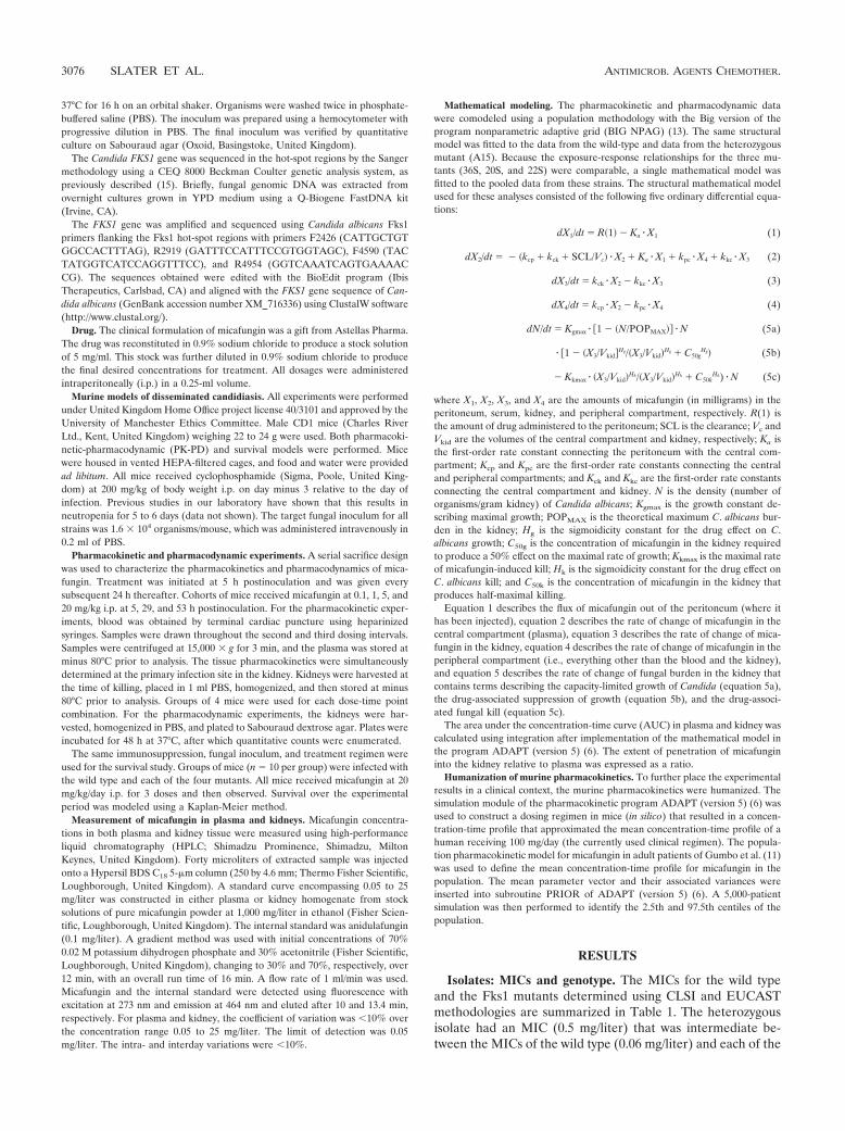
Humanization of murine pharmacokinetics. A regimen ad-ministered to mice that simulated the mean concentration-time profile for humans receiving 100 mg (Fig. 6A) who werealso infected with the wild type resulted in suppression offungal growth (Fig. 6B). In contrast, however, the same hu-manized regimen, when administered to the heterozygote andthe homozygous mutants, did not result in any predicted clin-ical antifungal activity (Fig. 6C and D, respectively). A regimenadministered to mice that simulated the mean concentration-time profile for humans receiving 400 mg (Fig. 7A) and in-fected with the wild type resulted in rapid fungicidal activity.The same humanized regimen had no effect on humans in-fected with the heterozygote or homozygous mutants (Fig. 7Cand D, respectively).
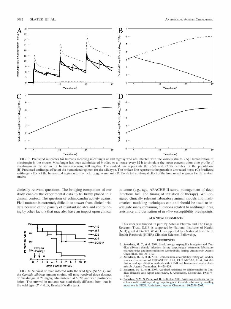
Survival. The survival of mice infected with the various C.albicans strains and treated for 3 dosages of micafungin at 20mg/kg is shown in Fig. 8. There was 100% survival of miceinfected with the wild type for at least 14 days postinoculation.In contrast, there was a statistically significant decline in thefraction of surviving mice infected with the mutant strains. Therates of survival of mice infected with isolates 36S, 22S, and20S (homozygous mutants) and treated with three dosages of20 mg/kg i.p. were 10, 50, and 60%, respectively. The survival
FIG. 2. Effect of micafungin on the density of Candida albicans SC5314 wild type in the kidneys of mice as a function of time. (A) Controls;(B) micafungin at 1 mg/kg administered at 5, 29, and 53 h; (C) micafungin at 5 mg/kg administered at 5, 29, and 53 h; (D) micafungin at 20 mg/kgadministered at 5, 29, and 53 h. Data are means � SDs. The solid line is the fit of the population mathematical model.
3078 SLATER ET AL. ANTIMICROB. AGENTS CHEMOTHER.

of mice infected with isolate A15 (heterozygous mutant)treated with three dosages of 20 mg/kg i.p. was 50%.
DISCUSSION
The echinocandins are first-line agents for the treatment ofdisseminated candidiasis (15). Their fungicidal activity, paucityof drug interactions, and wide therapeutic index facilitate theiruse in critically ill patients with this disease. Increasingly, how-ever, there are reports of isolates with reduced echinocandinsusceptibility, which usually occurs in the context of prolongedtherapy and at sites where drug penetration may be compro-mised (e.g., esophagus, respiratory tract, and abdomen) (20).Under these circumstances, cross-resistance between otherechinocandin agents is frequently observed (2).
The molecular mechanism of reduced susceptibility for themajority of isolates has been defined and is related to nonsyn-onymous nucleotide substitutions in the gene FKS1, whichencodes the putative echinocandin target, Fks1 (17). Aminoacid substitutions occur in two well-defined areas within thetarget protein: (i) hot spot 1, which is an 8-amino-acid regionbetween Phe641 and Asp648, and (ii) hot spot 2, at amino acidpositions 1345 to 1365 (17). The in vitro echinocandin expo-sure-response relationships can be defined using partially pu-rified glucan synthase with tritiated substrate (17). The con-
centration of drug that produces half-maximal inhibition ofenzyme activity (50% effective concentration [EC50]) in glucansynthase assays from Fks1 mutants is several orders of magni-tude higher than that for the wild type (17). Importantly, how-ever, the enzyme complex can still be ultimately inhibited byhigh drug concentrations (17). Because the echinocandins havesuch wide therapeutic ratios, a clinically relevant question iswhether any Fks1 mutant can be treated with either a conven-tional or an elevated dosage of drug.
The pharmacokinetic-pharmacodynamic analyses in thisstudy suggest that infections with the wild type can be treatedwith micafungin at 100 mg/day, which is consistent with clinicalexperience. In contrast, a variety of amino acid substitutions(i.e., proline [P], phenylalanine [F], and tyrosine [Y]) for theserine residue at position 645 within Fks1 render these strainsuntreatable in the kidneys of mice even with the use of dosagesof micafungin as high as 20 mg/kg. This murine dosage corre-sponds to a human dosage of �1,165 mg/day, which is signif-icantly more than the currently employed 100 to 150 mg/dayand the highest reported human dosage of 900 mg/day (21).Our bridging study suggests that the concentrations of mica-fungin that are required to have any antifungal effect are sig-nificantly in excess of those used in currently approved regi-mens. Our results suggest that Candida albicans isolates with
FIG. 3. Effect of micafungin on the density of Candida albicans SC5313 A15 heterozygous mutant in the kidneys of mice as a function of time.(A) Controls; (B) micafungin at 1 mg/kg administered at 5, 29, and 53 h; (C) micafungin at 5 mg/kg administered at 5, 29, and 53 h; (D) micafunginat 20 mg/kg administered at 5, 29, and 53 h. Data are means � SDs. The solid line is the fit of the population mathematical model.
VOL. 55, 2011 MICAFUNGIN TREATMENT OF Fks1 CANDIDA MUTANTS 3079

amino acid substitutions in Fks1 at Ser645 should be deemedresistant and that this applies to both homozygous andheterozygous strains.
Our results confirm a view that echinocandin breakpoints
should be set to exclude the vast majority of Fks1 mutants. TheCLSI originally defined isolates with MICs of �2 mg/liter to besusceptible at a time when experience with resistant isolateswas relatively limited and there was a desire not to categorizeC. parapsilosis as nonsusceptible (19). More recently, concernhas been expressed that these breakpoints may be too highbecause of reports that some Fks1 mutants have MICs that arecurrently classified as susceptible. For example, a recent studyof both wild-type and Fks1 mutants revealed that some of theseFks1 mutants have MICs that overlap the tail of the wild-typedistribution (i.e., have MICs that are within a mode and 2doubling dilutions) (2). These findings have prompted CLSI topropose a downward revision of breakpoints for all three echi-nocandin agents, and this process is under way (see www.clsi.org). The EUCAST committee is currently formulating echi-nocandin breakpoints, and a final decision will be available inthe near future.
Our study also has implications for critically ill patients re-ceiving echinocandins who have breakthrough Candida infec-tion. An increasing clinical conundrum is the interpretation ofa Candida isolate that has grown in the context of prolongedechinocandin therapy. While there may be a number of expla-nations for this phenomenon (e.g., a retained infected catheteror an unresolved deep focus of infection), the emergence ofdrug resistance is an increasing concern. Our results indicate
FIG. 4. Effect of micafungin on the density of Candida albicans SC5314 36S, 20S, and 22S homozygous mutants in the kidneys of mice as a functionof time. (A) Controls; (B) micafungin at 1 mg/kg administered at 5, 29, and 53 h; (C) micafungin at 5 mg/kg administered at 5, 29, and 53 h;(D) micafungin at 20 mg/kg administered at 5, 29, and 53 h. Data are means � SDs. The solid line is the fit of the population mathematical model.
FIG. 5. Box-and-whisker plots for the Bayesian estimates of C50k forthe wild type, heterozygote, and homozygous mutants. The median(range) values for the wild type, heterozygous mutant, and homozygousmutants are 2.14 mg/liter (0.93 to 8.63 mg/liter), 26.24 mg/liter (5.86 to49.99 mg/liter), and 39.46 mg/liter (13.57 to 125.87 mg/liter), respectively.
3080 SLATER ET AL. ANTIMICROB. AGENTS CHEMOTHER.

that echinocandin therapy is contraindicated for Ser645 Fks1mutants. Because the prior probability of an Fks1 mutation ismuch higher in an echinocandin-exposed patient, clinical mi-crobiology laboratories require a rapid and accurate way ofdetermining the genotype to inform the decision on furtherechinocandin therapy. This is probably best achieved by se-quencing both hot-spot regions in Fks1. While this is techni-cally straightforward, it may not be always possible or practical,especially for smaller regional laboratories. In these cases,alternative phenotypic approaches may suffice, and potentialapproaches might include (i) measuring an MIC with 50%serum (9) and (ii) testing isolates against anidulafungin usingepidemiological cutoff values until breakpoints that excluderesistant mutants have been established (2, 18). Regardless,there are increasing concerns that the MIC may not be auniversally reliable tool to screen for echinocandin resistanceand may not always provide correct information to guide ap-propriate antifungal therapy for critically ill patients.
A number of additional factors deserve further discussionand future study: (i) we studied relatively few strains, andfurther preclinical and clinical studies are warranted; (ii) theexposure-response relationships of Candida albicans isolates
with amino acid substitutions at positions other than Ser645are not known but could be readily determined using the invivo-to-clinical bridging methods used in this study; in theabsence of specific experimental pharmacokinetic-pharmaco-dynamic and/or clinical data, the most conservative approach isto classify these isolates as resistant, but this may ultimatelyprove to be too conservative; (iii) the PK-PD relationships ofthe other echinocandins against Candida albicans with Fks1substitutions need to be defined; (iv) the consequences ofanalogous substitutions in Fks1 in other species such as Can-dida glabrata need to be determined; (v) the response of iso-lates with elevated echinocandin MICs without substitutions inFks1 is of interest and could also be determined using the samemethods used in this study; and (vi) we used a well-validatedmodel of disseminated candidiasis using the fungal burden inthe kidney as the pharmacodynamic readout. We acknowledgethat the pharmacodynamic relationships may be different forCandida infections at other sites (e.g., bloodstream and brain)and that this could be addressed in future studies.
Finally, this study demonstrates the advantages of usingmodern pharmacokinetic-pharmacodynamic modeling tech-niques in combination with molecular microbiology to address
FIG. 6. Predicted outcomes for humans receiving micafungin at 100 mg/day who are infected with the various strains used in the murineexperiments. (A) Humanization of micafungin in the mouse. Micafungin has been administered in silico to a mouse every 12 h to simulate the meanconcentration-time profile of micafungin in the serum for humans receiving 100 mg/day. The dashed line represents the 2.5th and 97.5th centilesfor the population. (B) Predicted antifungal effect of the humanized regimen for the wild type. The broken line represents the growth in untreatedhosts. (C) Predicted antifungal effect of the humanized regimen for the heterozygous mutant. (D) Predicted antifungal effect of the humanizedregimen for the mutant strains.
VOL. 55, 2011 MICAFUNGIN TREATMENT OF Fks1 CANDIDA MUTANTS 3081
clinically relevant questions. The bridging component of ourstudy enables the experimental data to be firmly placed in aclinical context. The question of echinocandin activity againstFks1 mutants is extremely difficult to answer from clinical trialdata because of the paucity of resistant isolates and confound-ing by other factors that may also have an impact upon clinical
outcome (e.g., age, APACHE II score, management of deepinfectious foci, and timing of initiation of therapy). Well-de-signed clinically relevant laboratory animal models and math-ematical modeling techniques can and should be used to in-vestigate many remaining questions related to antifungal drugresistance and derivation of in vitro susceptibility breakpoints.
ACKNOWLEDGMENTS
This work was funded, in part, by Astellas Pharma and The FungalResearch Trust. D.S.P. is supported by National Institutes of Health(NIH) grant AI069397. W.W.H. is supported by a National Institute ofHealth Research (NIHR) Clinician Scientist Fellowship.
REFERENCES
1. Arendrup, M. C., et al. 2009. Breakthrough Aspergillus fumigatus and Can-dida albicans double infection during caspofungin treatment: laboratorycharacteristics and implication for susceptibility testing. Antimicrob. AgentsChemother. 53:1185–1193.
2. Arendrup, M. C., et al. 2010. Echinocandin susceptibility testing of Candidaspecies: comparison of EUCAST EDef 7.1, CLSI M27-A3, Etest, disk dif-fusion, and agar dilution methods with RPMI and Isosensitest media. Anti-microb. Agents Chemother. 54:426–439.
3. Baixench, M. T., et al. 2007. Acquired resistance to echinocandins in Can-dida albicans: case report and review. J. Antimicrob. Chemother. 59:1076–1083.
4. Balashov, S. V., S. Park, and D. S. Perlin. 2006. Assessing resistance to theechinocandin antifungal drug caspofungin in Candida albicans by profilingmutations in FKS1. Antimicrob. Agents Chemother. 50:2058–2063.
FIG. 7. Predicted outcomes for humans receiving micafungin at 400 mg/day who are infected with the various strains. (A) Humanization ofmicafungin in the mouse. Micafungin has been administered in silico to a mouse every 12 h to simulate the mean concentration-time profile ofmicafungin in the serum for humans receiving 400 mg/day. The dashed line represents the 2.5th and 97.5th centiles for the population.(B) Predicted antifungal effect of the humanized regimen for the wild type. The broken line represents the growth in untreated hosts. (C) Predictedantifungal effect of the humanized regimen for the heterozygous mutant. (D) Predicted antifungal effect of the humanized regimen for the mutantstrains.
FIG. 8. Survival of mice infected with the wild type (SC5314) andthe Candida albicans mutant strains. All mice received three dosagesof micafungin at 20 mg/kg administered at 5, 29, and 53 h postinocu-lation. The survival in mutants was statistically different from that inthe wild type (P � 0.05; Kruskall-Wallis test).
3082 SLATER ET AL. ANTIMICROB. AGENTS CHEMOTHER.

5. Clinical and Laboratory Standards Institute. 2008. Reference methodfor broth dilution antifungal susceptibility testing of yeasts, 3rd ed. Ap-proved standard M27-A3(28). Clinical and Laboratory Standards Insti-tute, Wayne, PA.
6. D’Argenio, D. Z., A. Schumitzky, and X. Wang. 2009. ADAPT 5 user’s guide:pharmacokinetic/pharmacodynamic systems analysis software. BiomedicalSimulations Resource, Los Angeles, CA.
7. Denning, D. W. 2003. Echinocandin antifungal drugs. Lancet 362:1142–1151.8. European Committee for Antimicrobial Susceptibility Testing. 2008. EUCAST
definitive document EDef 7.1: method for the determination of broth dilutionMICs of antifungal agents for fermentative yeasts. Clin. Microbiol. Infect. 14:398–405.
9. Garcia-Effron, G., S. Park, and D. S. Perlin. 2009. Correlating echinocandinMIC and kinetic inhibition of fks1 mutant glucan synthases for Candidaalbicans: implications for interpretive breakpoints. Antimicrob. Agents Che-mother. 53:112–122.
10. Gudlaugsson, O., et al. 2003. Attributable mortality of nosocomial candi-demia, revisited. Clin. Infect. Dis. 37:1172–1177.
11. Gumbo, T., et al. 2008. Population pharmacokinetics of micafungin in adultpatients. Diagn. Microbiol. Infect. Dis. 60:329–331.
12. Kuse, E. R., et al. 2007. Micafungin versus liposomal amphotericin B forcandidaemia and invasive candidosis: a phase III randomised double-blindtrial. Lancet 369:1519–1527.
13. Leary, R., R. Jelliffe, A. Schumitzky, and M. van Guilder. 2001. An adaptivegrid, non-parametric approach to pharmacokinetic and dynamic (PK/PD)models, p. 389–394. Proc. 14th IEEE Computer Soc.
14. Louie, A., et al. 2005. Pharmacodynamics of caspofungin in a murine model
of systemic candidiasis: importance of persistence of caspofungin in tissuesto understanding drug activity. Antimicrob. Agents Chemother. 49:5058–5068.
15. Pappas, P. G., et al. 2009. Clinical practice guidelines for the management ofcandidiasis: 2009 update by the Infectious Diseases Society of America. Clin.Infect. Dis. 48:503–535.
16. Pappas, P. G., et al. 2007. Micafungin versus caspofungin for treatment ofcandidemia and other forms of invasive candidiasis. Clin. Infect. Dis. 45:883–893.
17. Park, S., et al. 2005. Specific substitutions in the echinocandin target Fks1paccount for reduced susceptibility of rare laboratory and clinical Candida sp.isolates. Antimicrob. Agents Chemother. 49:3264–3273.
18. Pfaller, M. A., et al. 2010. Wild-type MIC distributions and epidemiologicalcutoff values for the echinocandins and Candida spp. J. Clin. Microbiol.48:52–56.
19. Pfaller, M. A., et al. 2008. Correlation of MIC with outcome for Candidaspecies tested against caspofungin, anidulafungin, and micafungin: analysisand proposal for interpretive MIC breakpoints. J. Clin. Microbiol. 46:2620–2629.
20. Pfeiffer, C. D., et al. 2010. Breakthrough invasive candidiasis in patients onmicafungin. J. Clin. Microbiol. 48:2373–2380.
21. Sirohi, B., et al. 2006. A study to determine the safety profile and maximumtolerated dose of micafungin (FK463) in patients undergoing haematopoi-etic stem cell transplantation. Bone Marrow Transplant. 38:47–51.
22. Smith, P. B., et al. 2009. Pharmacokinetics of an elevated dosage of mica-fungin in premature neonates. Pediatr. Infect. Dis. J. 28:412–415.
VOL. 55, 2011 MICAFUNGIN TREATMENT OF Fks1 CANDIDA MUTANTS 3083


