Embed Size (px)
Citation preview

Disulphide production by Ero1a–PDI relay is rapidand effectively regulated
Christian Appenzeller-Herzog1,2,3,*,Jan Riemer1,6, Ester Zito4, King-TungChin4, David Ron4,5, Martin Spiess2
and Lars Ellgaard1,*1Department of Biology, University of Copenhagen, Copenhagen,Denmark, 2Biozentrum, University of Basel, Basel, Switzerland,3Department of Pharmaceutical Sciences, University of Basel, Basel,Switzerland, 4Skirball Institute of Biomolecular Medicine, New YorkUniversity School of Medicine, New York, NY, USA and 5Institute ofMetabolic Science, University of Cambridge, Cambridge, UK
The molecular networks that control endoplasmic reticu-
lum (ER) redox conditions in mammalian cells are incom-
pletely understood. Here, we show that after reductive
challenge the ER steady-state disulphide content is
restored on a time scale of seconds. Both the oxidase
Ero1a and the oxidoreductase protein disulphide isomer-
ase (PDI) strongly contribute to the rapid recovery
kinetics, but experiments in ERO1-deficient cells indicate
the existence of parallel pathways for disulphide genera-
tion. We find PDI to be the main substrate of Ero1a, and
mixed-disulphide complexes of Ero1 primarily form with
PDI, to a lesser extent with the PDI-family members ERp57
and ERp72, but are not detectable with another homolo-
gue TMX3. We also show for the first time that the
oxidation level of PDIs and glutathione is precisely regu-
lated. Apparently, this is achieved neither through ER
import of thiols nor by transport of disulphides to the
Golgi apparatus. Instead, our data suggest that a dynamic
equilibrium between Ero1- and glutathione disulphide-
mediated oxidation of PDIs constitutes an important
element of ER redox homeostasis.
The EMBO Journal (2010) 29, 3318–3329. doi:10.1038/
emboj.2010.203; Published online 27 August 2010
Subject Categories: proteins
Keywords: disulphide-bond formation; endoplasmic reticu-
lum; Ero1; glutathione; protein disulphide isomerase
Introduction
Regulation of the redox environment in the endoplasmic
reticulum (ER) is emerging as an important aspect of cellular
homeostasis (Malhotra and Kaufman, 2007; Merksamer et al,
2008), and the thiol-disulphide oxidoreductases of the protein
disulphide isomerase (PDI) family are central to ER redox
control (Appenzeller-Herzog and Ellgaard, 2008b). PDIs con-
tain one or more thioredoxin-like domains. These typically
harbour a CXXC active-site sequence motif required for the
catalysis of thiol-disulphide exchange reactions, such as the
introduction of disulphides into substrate proteins.
The identification in yeast of the essential ER-resident
sulfhydryl oxidase Ero1p (Frand and Kaiser, 1998; Pollard
et al, 1998), which oxidizes PDI (Frand and Kaiser, 1999; Tu
et al, 2000) and reduces molecular oxygen to hydrogen
peroxide (Gross et al, 2006), has led to an improved under-
standing of oxidative folding and ER redox regulation (Sevier
et al, 2007; Sevier and Kaiser, 2008). Consistent with an
important function in ER protein oxidation, both human
isoforms Ero1a and Ero1b are transcriptionally upregulated
by the ER stress response (Pagani et al, 2000; Marciniak et al,
2004), which can be associated with a reductive shift in the
ER redox conditions (Nadanaka et al, 2007; Merksamer et al,
2008). Moreover, the activity of Ero1a is subject to negative
feedback regulation by intramolecular disulphide bonds
(Appenzeller-Herzog et al, 2008; Baker et al, 2008). The
enzyme appears in at least three redox forms: reduced, OX1
and OX2 (Benham et al, 2000). The latter is the most oxidized
form with all regulatory disulphide bonds in place
(Appenzeller-Herzog et al, 2008; our unpublished observa-
tions). The cellular activation state of Ero1a is controlled by
the availability of reduced PDI (Appenzeller-Herzog et al,
2008), which can reduce the regulatory disulphide bonds (see
also Discussion) (Sevier et al, 2007; Appenzeller-Herzog et al,
2008; Baker et al, 2008). Ero1a also controls calcium fluxes
from ER to mitochondria (Li et al, 2009), which could
correlate with its partial localization at mitochondria-asso-
ciated ER membrane domains (Gilady et al, 2010).
The dominance of Ero1 enzymes in providing the oxidizing
equivalents for the synthesis of disulphides is, however, still a
matter of debate (Thorpe and Kodali, 2010). For instance, the
slow in vitro rate of PDI oxidation by Ero1a (Baker et al, 2008;
Wang et al, 2009) appears at odds with a principal function in
disulphide-bond generation. Knockout of the single Ero1 gene
in fruit fly causes a specific defect in Notch signalling while
apparently leaving the bulk disulphide-bond repertoire
unperturbed (Tien et al, 2008). Most importantly, however,
Ero1a and Ero1b appear non-essential in the mouse, as
evidenced by the viability of an Ero1a/Ero1b double mutant
(Zito et al, 2010). Indeed, several possible Ero1-independent
pathways for disulphide generation and/or the oxidation of
PDI in the ER of mammalian cells exist (Margittai and
Banhegyi, 2010). These include the activity of quiescin-sulf-
hydryl oxidases (Thorpe and Kodali, 2010), import of dehy-
droascorbate from the cytosol and its reduction by dithiol
groups (Saaranen et al, 2010), ER-luminal detoxification of
NADPH oxidase 4-generated hydrogen peroxide (Santos et al,
2009) and a pathway that uses the oxidizing equivalents of
radicals derived from mitochondrial respiration to generateReceived: 18 June 2010; accepted: 26 July 2010; published online:27 August 2010
*Corresponding authors. C Appenzeller-Herzog, Department ofPharmaceutical Sciences, University of Basel, Klingelbergstr. 50,CH-4056 Basel, Switzerland. Tel.: þ 41 61 267 14 86; Fax: þ 41 61 2671515;E-mail: [email protected] or L Ellgaard, Department ofBiology, University of Copenhagen, Ole Maaløes Vej 5, DK-2200Copenhagen N, Denmark. Tel.: þ 45 35 32 17 25; Fax: þ 45 35 32 21 28;E-mail: [email protected] address: Department of Cell Biology, University ofKaiserslautern, 67663 Kaiserslautern, Germany
The EMBO Journal (2010) 29, 3318–3329 | & 2010 European Molecular Biology Organization | All Rights Reserved 0261-4189/10
www.embojournal.org
The EMBO Journal VOL 29 | NO 19 | 2010 &2010 European Molecular Biology Organization
EMBO
THE
EMBOJOURNAL
THE
EMBOJOURNAL
3318

disulphides in secretory compartments (Yang et al, 2007).
In analogy to a mechanism that operates in both archaea and
bacteria (Dutton et al, 2008; Singh et al, 2008), PDI could also
be oxidized through the vitamin K cycle (Wajih et al, 2007).
Currently, we lack a thorough cell biological understanding of
these pathways in relation to oxidative folding in the ER.
In addition to the PDIs and Ero1, glutathione also has a
fundamental function in ER redox homeostasis. This low-mo-
lecular weight thiol compound exists as a mixture of reduced
glutathione (GSH) and glutathione disulphide (GSSG). Cytosol-
derived GSH can enter the ER where its reducing power is
required for the rearrangement of aberrant disulphide bonds in
folding substrates (Chakravarthi et al, 2006).
On these premises, we decided to further explore the links
between Ero1, PDIs and glutathione in cultured human cells.
Our work shows a very rapid production of disulphides in the
ER whose velocity depends on both Ero1a and PDI, but
apparently less so on other PDI-family members. In cells
devoid of both Ero1a and b, however, we present evidence for
Ero1-independent pathway(s) for thiol oxidation. Finally, we
show that ER oxidation is tightly regulated and propose a
mechanistic model of ER redox homeostasis that integrates
previous and current findings.
Results
Thiol import and disulphide export have a minor
function in acute ER redox control
Although it has been shown that Ero1 activity ultimately
leads to the oxidation of GSH in the ER (Cuozzo and Kaiser,
1999; Appenzeller-Herzog et al, 2008), the mechanisms that
counteract the accumulation of ER-luminal GSSG are still
unclear (Chakravarthi et al, 2006; Thorpe and Kodali,
2010). As GSSG displays only low permeability through
microsomal membranes (Banhegyi et al, 1999), we tested
whether export of GSSG through the secretory pathway might
contribute to ER redox homeostasis. We, therefore, combined
the pharmacological inhibition of ER-to-Golgi transport with
ER redox state analysis. For this purpose, we used a combi-
nation of brefeldin A and monensin (BFA/mon), which
blocks vesicular anterograde transport from the ER, while
preserving the integrity of the Golgi apparatus (Barzilay et al,
2005; Supplementary Figure S1), and an assay in which
oxidized active-site cysteines in PDIs are modified with
4-acetamido-40-maleimidylstilbene-2,20-disulphonic acid (AMS),
resulting in slower mobility upon SDS–PAGE (Jessop and
Bulleid, 2004). Using this AMS shift assay, we have consis-
tently found the redox distribution of various PDIs to exhibit
molecules in both reduced and oxidized states (Haugstetter
et al, 2005; Appenzeller-Herzog et al, 2008; Appenzeller-
Herzog and Ellgaard, 2008a; Roth et al, 2010), the ratio of
which can be used as a readout to monitor redox variations
in the ER.
We studied the effect of BFA/mon treatment on the redox
recovery of the PDIs TMX3 (a transmembrane PDI-family
member) and ERp57 (a close homologue of PDI) upon
application and washout of the oxidant diamide. No signifi-
cant delay in the recovery was observed under conditions
of blocked ER-to-Golgi transport (Figure 1A and B). Hence,
BFA/mon-sensitive vesicular export appears to be of
minor importance as a redox-balancing mechanism against
hyper-oxidizing conditions.
Reestablishment of the ER redox state after washout of the
oxidant dipyridyl sulphide is mediated by the import of GSH
through the ER membrane (Jessop and Bulleid, 2004).
Further reductive input is brought to the ER through the
co-translational translocation of protein thiols (Cuozzo and
Kaiser, 1999). For these reasons, we examined whether
lowering cellular GSH levels by L-buthionine-sulfoximine
(BSO) or the shutdown of translation by cycloheximide
(CHX) altered the ER redox state. As shown in Figure 1C,
neither of these drugs showed a consistent effect on the redox
ratios of TMX3 and ERp57 at steady state. It should be noted
that for reasons of cytotoxicity, we did not apply combina-
tions of the above treatments so that additive effects between
the different reductive pathways cannot be excluded. Still,
these results suggested that mechanism(s) other than ER
export of disulphides or import of thiols secure ER redox
balance.
PDI withstands in situ reduction better than other ER
oxidoreductases
PDI is a known substrate of Ero1 (Sevier and Kaiser, 2008).
It is, however, less clear what regulates the redox state of
other ER oxidoreductases and whether they are substrates of
Ero1 (Mezghrani et al, 2001; Kulp et al, 2006; Jessop et al,
–
–
+
+ BSOCHX–– +
ox
BFA/mon
wo (min)0 5 10 15 5 10 15
– +
– –
ox
A
B C
αTMX3
αERp57
WB
ox
dia+ + + + + + +– –
Reductive recovery of TMX3
020406080
100
0 5 10 15Recovery (min)
ControlBFA/mon%
Oxi
dize
d
dia
ox
–
–
–––
αTMX3
αERp57
WB
41% 41% 40%
30% 32% 36%
red
red
red
red
Figure 1 Vesicular transport, glutathione concentration and proteintranslocation only moderately influence ER redox homeostasis.(A) HEK293 cells pre-treated with BFA/mon for 0.5 h or leftuntreated were incubated with 5 mM diamide (dia) for 5 min,washed twice with PBS and incubated in the same buffer for 0, 5,10 or 15 min (wo, washout). BFA/mon was present throughout thetime course. For comparison, steady-state samples±treatment withBFA/mon for 0.5 h were included. The reactions were stopped byrinsing the cells in ice-cold PBS containing 20 mM NEM, and theredox distributions of TMX3 and ERp57 visualized by westernblotting (WB) after differential alkylation with NEM and AMS.The mobility of the oxidized (ox) and reduced (red) forms ofTMX3 and ERp57 are indicated. (B) Recovery of the TMX3 redoxratio assessed by densitometry in BFA/mon-treated and control cellsas shown in panel A (n¼ 3, mean±s.d.). (C) The redox states ofTMX3 and ERp57 in HEK293 cells depleted of glutathione (usingBSO) or nascent proteins (using CHX). Treatment with BSO reducedcellular glutathione levels to 20% of control (data not shown).Oxidized fractions (%) as determined by densitometry are indi-cated. Lanes labelled with dia represent oxidized control lanes usingdiamide-treated cell lysates. The hairlines indicate where interven-ing lanes have been removed. The results are representative of threeindependent experiments.
Ero1a- and PDI-mediated ER redox regulationC Appenzeller-Herzog et al
&2010 European Molecular Biology Organization The EMBO Journal VOL 29 | NO 19 | 2010 3319

2009). We, therefore, next investigated factors that could
control the redox state of various PDI-family members.
First, we determined their in vivo dithiothreitol (DTT) resis-
tance by means of an assay where cells are challenged with
increasing concentrations of this membrane-permeant reduc-
tant. Although the two separate active-site domains in PDI
exhibit very similar reduction potentials (Darby and
Creighton, 1995), the susceptibility towards DTT-mediated
in situ reduction of the a domain was greater than of the a0
domain (Figure 2A). The a0 domain is preferentially oxidized
in vitro by Ero1a (Baker et al, 2008; Wang et al, 2009), which
becomes activated by DTT through reduction of its regulatory
disulphides (see below). Thus—although the DTT resistance
readout could also be influenced by protein quaternary
structure and differential accessibility of active-site cysteines
to DTT or N-ethylmaleimide (NEM)—this result suggested
that the DTT resistance of a PDI-family member’s active site
in the ER reflects its propensity to become reoxidized by
ER-resident oxidases.
When comparing the in vivo redox states of TMX3 and
ERp57 after treatment of cells with different concentrations of
DTT, we observed a slightly higher DTT resistance of the
AMS-shifted form of ERp57 (Figure 2B and C). This form
represents ERp57 with its a0 domain oxidized, whereas ERp57
with exclusively the a domain oxidized virtually co-migrates
with the reduced form (Appenzeller-Herzog et al, 2008).
Given that the active-site domains of ERp57 and TMX3
have approximately the same reduction potential (Frickel
et al, 2004; Haugstetter et al, 2005), these results indicated
that in cells the relative rates of oxidation for the single active
site in TMX3 and the a0 active site in ERp57 are distinct. Still,
the Ero1a-controlled active site in PDIa0 by far displayed the
highest cellular DTT resistance, likely underlining its impor-
tance as an electron donor for Ero1a in vivo.
Mixed-disulphide interactions of PDIs with Ero1aand Ero1b reflect their in vivo DTT resistance
To investigate a possible function of Ero1 in maintaining the
levels of the oxidized fractions of ERp57 and TMX3, we
searched for intracellular mixed-disulphide interactions. For
this purpose, we performed co-immunoprecipitation experi-
ments of myc-tagged Ero1a (Ero1amyc6his) or Ero1b(Ero1bmyc6his) stably expressed from a doxycyclin-inducible
promoter (Appenzeller-Herzog et al, 2008) using an in situ
acidification/in vitro NEM-alkylation protocol. Compared
with in situ NEM trapping, the acidification method was
much more effective in trapping mixed-disulphide interactors
of Ero1 (data not shown).
As expected, PDI was readily precipitated with amyc in an
Ero1amyc6his-dependent manner (Figure 3A, lanes 3 and 4),
and non-reducing gel electrophoresis revealed a prominent
Ero1amyc6his–PDI mixed-disulphide complex (Figure 3A,
lane 7). Unlike in a previous study that used NEM trapping
of mixed disulphides (Mezghrani et al, 2001), an interaction
with Ero1amyc6his was also detected for ERp57 and ERp72
(Figure 3B and C). While the pool of PDI acid trapped in a
αTMX3
αERp57
+ – – – – –– – –
DTT (μM)– 50 150 300200100 2500 104
dia
dia
DTT (μM)
DTT/TCEP
+
+
– – – – – – –
– – – – – – –
– –0 100 250 500 750 1000
a-ox
ox
a′-ox
A B
ox
ox
IP: αPDI
WB
75
50
100
**
a-ox
ox
a′-ox
Contrast-enhanced
0
20
40
60
80
100
0 50 100 150 200 250 300DTT (μM)
C
Oxi
dize
d fr
actio
n(%
of s
tead
y st
ate)red
red
red
TMX3ERp57
Figure 2 In vivo DTT resistance of PDI, TMX3 and ERp57. (A) HEK293 cells were treated with the indicated concentrations of DTT, and thein vivo redox state of the two active sites in PDI (a and a0) determined by immunoprecipitation (IP) of 35S-labelled and mPEG-mal-modified PDI.Samples completely reduced with DTTand TCEP, or oxidized with diamide (dia) served as mobility markers. The contrast enhancement of theregion marked by the rectangle more clearly shows the different behaviour of the two semi-oxidized forms of PDI (a domain oxidized/a0
domain reduced (a-ox); a domain reduced/a0 domain oxidized (a0-ox)), for which we have previously determined the relative mobility(Appenzeller-Herzog and Ellgaard, 2008a). One of two independent experiments with equal outcome is shown. Red, both active sites reduced;ox, both active sites oxidized; asterisks, reduced PDI modified with mPEG-mal on its non-catalytic cysteines (Appenzeller-Herzog and Ellgaard,2008a). (B) After treatment of HEK293 cells with the indicated concentrations of DTT, the in vivo redox states of TMX3 and ERp57 weredetermined as in Figure 1A. The mobilities of the oxidized (ox) and reduced (red) species, as verified by control samples using lysates fromdiamide- or DTT (104 mM)-treated cells, are marked. (C) Densitometric analysis of (B) (n¼ 3, mean±s.d.).
Ero1a- and PDI-mediated ER redox regulationC Appenzeller-Herzog et al
The EMBO Journal VOL 29 | NO 19 | 2010 &2010 European Molecular Biology Organization3320
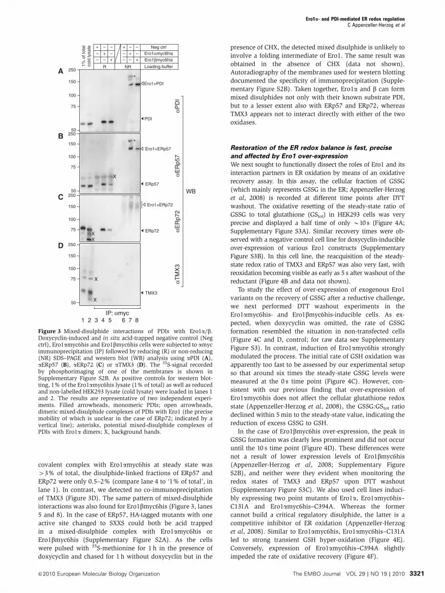
covalent complex with Ero1amyc6his at steady state was
43% of total, the disulphide-linked fractions of ERp57 and
ERp72 were only 0.5–2% (compare lane 4 to ‘1% of total’, in
lane 1). In contrast, we detected no co-immunoprecipitation
of TMX3 (Figure 3D). The same pattern of mixed-disulphide
interactions was also found for Ero1bmyc6his (Figure 3, lanes
5 and 8). In the case of ERp57, HA-tagged mutants with one
active site changed to SXXS could both be acid trapped
in a mixed-disulphide complex with Ero1amyc6his or
Ero1bmyc6his (Supplementary Figure S2A). As the cells
were pulsed with 35S-methionine for 1 h in the presence of
doxycyclin and chased for 1 h without doxycyclin but in the
presence of CHX, the detected mixed disulphide is unlikely to
involve a folding intermediate of Ero1. The same result was
obtained in the absence of CHX (data not shown).
Autoradiography of the membranes used for western blotting
documented the specificity of immunoprecipitation (Supple-
mentary Figure S2B). Taken together, Ero1a and b can form
mixed disulphides not only with their known substrate PDI,
but to a lesser extent also with ERp57 and ERp72, whereas
TMX3 appears not to interact directly with either of the two
oxidases.
Restoration of the ER redox balance is fast, precise
and affected by Ero1 over-expression
We next sought to functionally dissect the roles of Ero1 and its
interaction partners in ER oxidation by means of an oxidative
recovery assay. In this assay, the cellular fraction of GSSG
(which mainly represents GSSG in the ER; Appenzeller-Herzog
et al, 2008) is recorded at different time points after DTT
washout. The oxidative resetting of the steady-state ratio of
GSSG to total glutathione (GStot) in HEK293 cells was very
precise and displayed a half time of only B10 s (Figure 4A;
Supplementary Figure S3A). Similar recovery times were ob-
served with a negative control cell line for doxycyclin-inducible
over-expression of various Ero1 constructs (Supplementary
Figure S3B). In this cell line, the reacquisition of the steady-
state redox ratio of TMX3 and ERp57 was also very fast, with
reoxidation becoming visible as early as 5 s after washout of the
reductant (Figure 4B and data not shown).
To study the effect of over-expression of exogenous Ero1
variants on the recovery of GSSG after a reductive challenge,
we next performed DTT washout experiments in the
Ero1amyc6his- and Ero1bmyc6his-inducible cells. As ex-
pected, when doxycyclin was omitted, the rate of GSSG
formation resembled the situation in non-transfected cells
(Figure 4C and D, control; for raw data see Supplementary
Figure S3). In contrast, induction of Ero1amyc6his strongly
modulated the process. The initial rate of GSH oxidation was
apparently too fast to be assessed by our experimental setup
so that around six times the steady-state GSSG levels were
measured at the 0 s time point (Figure 4C). However, con-
sistent with our previous finding that over-expression of
Ero1amyc6his does not affect the cellular glutathione redox
state (Appenzeller-Herzog et al, 2008), the GSSG:GStot ratio
declined within 5 min to the steady-state value, indicating the
reduction of excess GSSG to GSH.
In the case of Ero1bmyc6his over-expression, the peak in
GSSG formation was clearly less prominent and did not occur
until the 10 s time point (Figure 4D). These differences were
not a result of lower expression levels of Ero1bmyc6his
(Appenzeller-Herzog et al, 2008; Supplementary Figure
S2B), and neither were they evident when monitoring the
redox states of TMX3 and ERp57 upon DTT washout
(Supplementary Figure S3C). We also used cell lines induci-
bly expressing two point mutants of Ero1a, Ero1amyc6his–
C131A and Ero1amyc6his–C394A. Whereas the former
cannot build a critical regulatory disulphide, the latter is a
competitive inhibitor of ER oxidation (Appenzeller-Herzog
et al, 2008). Similar to Ero1amyc6his, Ero1amyc6his–C131A
led to strong transient GSH hyper-oxidation (Figure 4E).
Conversely, expression of Ero1amyc6his–C394A slightly
impeded the rate of oxidative recovery (Figure 4F).
75
50
100
150
250
75
50
100
150
250
75
100
150
75
50
100
150
αPD
IαT
MX
3αE
Rp5
7αE
Rp7
2
Ero1αmyc6hisEro1βmyc6his
Loading buffer
Neg ctrl
–
+
++
+
++
– – – –
– – – –– – –
NRR
IP: αmyc
WB250
250
1 2 3 4 6 7 8
*Ero1+PDI
PDI
Ero1+ERp57
ERp57
*
Ero1+ERp72
TMX3
X
X
X
X
ERp72
5
1% o
f tot
alco
ld ly
sate
A
B
C
D
X
Figure 3 Mixed-disulphide interactions of PDIs with Ero1a/b.Doxycyclin-induced and in situ acid-trapped negative control (Negctrl), Ero1amyc6his and Ero1bmyc6his cells were subjected to amycimmunoprecipitation (IP) followed by reducing (R) or non-reducing(NR) SDS–PAGE and western blot (WB) analysis using aPDI (A),aERp57 (B), aERp72 (C) or aTMX3 (D). The 35S-signal recordedby phosphorimaging of one of the membranes is shown inSupplementary Figure S2B. As positive controls for western blot-ting, 1% of the Ero1amyc6his lysate (1% of total) as well as reducedand non-labelled HEK293 lysate (cold lysate) were loaded in lanes 1and 2. The results are representative of two independent experi-ments. Filled arrowheads, monomeric PDIs; open arrowheads,dimeric mixed-disulphide complexes of PDIs with Ero1 (the precisemobility of which is unclear in the case of ERp72; indicated by avertical line); asterisks, potential mixed-disulphide complexes ofPDIs with Ero1a dimers; X, background bands.
Ero1a- and PDI-mediated ER redox regulationC Appenzeller-Herzog et al
&2010 European Molecular Biology Organization The EMBO Journal VOL 29 | NO 19 | 2010 3321

Rapid oxidative recovery depends on Ero1 and PDI
The stimulation of ER reoxidation by exogenous Ero1 implied
that endogenous Ero1 may catalyse the fast rate of disul-
phide-bond reformation upon DTT washout in non-trans-
fected cells. To examine this, we first downregulated the
expression of Ero1a in HEK293 cells by siRNA transfection.
Partial knockdown of Ero1a slightly delayed ER reoxidation
as assessed by timed redox analysis of TMX3 and ERp57
(Supplementary Figures S4A–C). Next, we performed DTT
washout experiments in embryonic fibroblasts derived from
homozygous double mutant mice that harbour disruptive
viral insertions in the genes encoding Ero1a and Ero1b(Zito et al, 2010). In keeping with the lack of Ero1a detection
in double mutant cells (Figure 5A), we observed a strong
delay in ER reoxidation (Figure 5B and C). The steady-state
redox distribution of ERp57, however, was not affected by
Ero1 deficiency (Figure 5B). Unfortunately, these cells were
not amenable to redox analysis of TMX3, because AMS
modification only minimally shifts the electrophoretic mobi-
lity of mouse TMX3 (data not shown). We also assessed the
reformation of GSSG during oxidative recovery from DTT in
wild-type versus double mutant fibroblasts. Unexpectedly,
the GSSG:GStot ratio promptly increased upon DTT washout
not only in wild type but also in the mutant cells, whereas
complete reoxidation of GSH after a recovery period of
300 s was only achieved in wild-type cells (Figure 5D).
Furthermore, the resting value for GSSG:GStot was higher—
that is more oxidizing—in double mutant cells (Figure 5D,
inset). These findings argue that at least one Ero1-indepen-
dent pathway for GSH oxidation is operative in these cells.
We next investigated whether the efficient delivery of
disulphide bonds in the ER depended on PDI. Indeed, for
TMX3, ERp57 and glutathione, the oxidative recovery was
clearly impaired in cells stably depleted of PDI (knockdown
efficiency B90%; see Ou and Silver, 2006; Appenzeller-
Herzog et al, 2008) as compared with control cells (Figure
5E–G). Thus, PDI has a prominent function in oxidative
recovery and the direct interactions of ERp57 or ERp72 with
Ero1a (Figure 3) cannot efficiently substitute the supply of
disulphide bonds through the Ero1a–PDI relay. Nonetheless,
the diminished rate of GSSG:GStot recovery in murine
ERp57�/� cells (Garbi et al, 2006) suggested that early
after DTT washout ERp57 does contribute to the shuttling of
disulphide bonds to GSH (Figure 5H).
So far, the data indicated that upon reductive challenge the
propagation of Ero1-generated oxidative equivalents through
PDI to the ER thiol pool was a rapid process. We, therefore,
expected the complex between the two enzymes to form
quickly after DTT treatment. To investigate this, we per-
formed co-immunoprecipitation experiments using cells that
had been challenged with DTT. Hence, doxycyclin-induced
Ero1amyc6his or Ero1bmyc6his cells were treated with DTT
or left untreated, washed with ice-cold phosphate-buffered
saline (PBS) and covered with trichloroacetic acid (TCA)
followed by amyc immunoprecipitation. Co-immunoprecipi-
tated PDI was readily detectable even after DTT treatment
(Figure 6A, lanes 12, 14, 16 and 18), indicating the formation
of the Ero1–PDI mixed-disulphide complexes to be extremely
rapid. We suggest that this rapid process reflects the sulfhy-
dryl oxidase activity of Ero1, and that this in turn accounts for
the high apparent DTT resistance of the PDIa0 active site
(Figure 2A). Notably, when analysed under non-reducing
conditions, the Ero1–PDI complexes isolated from DTT-trea-
ted cells migrated more slowly in the gel than when isolated
from non-treated cells (Figure 6A, Ero1þPDI RED.), a find-
ing that was recapitulated for the endogenous proteins
(Figure 6B). This suggested that in untreated cells, the bulk
of Ero1 that is covalently attached to PDI is in an oxidized or
0
20
40
60
80
100
0 5 10 15
DTT + wo (s)dox
–+– – – – –+ + +
0 10 205
dia–
– +
ox
WB: αTMX3
A
B
GS
SG
/GS
tot
(% o
f ste
ady
stat
e)
0100200300400500600700
0 60 120 180 240 300Recovery (s)
0100200300400500600700
0 60 120 180 240 300Recovery (s)
Control
Ero1α-C131A
Control
Ero1α
Recovery (s)
0
20
40
60
80
100
0 60 120 180 240 300
0
20
40
60
80
100
0 60 120 180 240 300Recovery (s)
GS
SG
/GS
tot
(% o
f ste
ady
stat
e)
GS
SG
/GS
tot
(% o
f ste
ady
stat
e)G
SS
G/G
Sto
t(%
of s
tead
y st
ate)
GS
SG
/GS
tot
(% o
f ste
ady
stat
e)
*
**
***
**
**
******
***
***
****
C D
E F
Recovery of cellular GSSG/GStot ratioafter DTT removal
red
Control
Ero1β
Control
Ero1α-C394A
Recovery (min)20
Figure 4 ER reoxidation after DTT treatment is fast and affected by exogenous Ero1. (A) Intracellular levels of GSSG and GStot were recordedfrom DTT-treated HEK293 cells after washout of the reductant for 0, 10, 30 s, 1, 3, 5 or 20 min. The GSSG:GStot ratio is expressed as percentageof the steady-state value that was independently measured (mean±s.d., n¼ 8, for individual experiments, see Supplementary Figure S3A).(B) Negative control cells were grown on plastic coverslips, treated with or without doxycyclin (dox) for 24 h, and left untreated (�) orincubated with DTT. After 0, 5, 10 or 20 s of DTT washout (wo), the cells were processed for AMS alkylation and western blotting (WB) usingaTMX3 or aERp57. Ox, oxidized species; red, reduced species; dia, oxidized control lane using diamide-treated cells. (C–F) DTTwashout assaysfollowed by the determination of cellular levels of GSSG and GStot after 0, 10, 60 or 300 s using Ero1amyc6his (C), Ero1bmyc6his (D),Ero1amyc6his–C131A (E) and Ero1amyc6his–C394A (F) cells cultured for 24 h with or without (control) the addition of doxycyclin(mean±s.d., two independent experiments each performed in triplet; Supplementary Figures S3D–G). *Po0.05; **Po0.01; ***Po0.001(Student’s t-test). Notice the different scaling on the y axis in the individual panels.
Ero1a- and PDI-mediated ER redox regulationC Appenzeller-Herzog et al
The EMBO Journal VOL 29 | NO 19 | 2010 &2010 European Molecular Biology Organization3322

partially oxidized state. We also found the mixed-disulphide
complex between PDI and Ero1a from DTT-treated cells
to require the active-site Cys94 in Ero1a (Figure 6C).
Finally, covalent interactions of ERp57 with Ero1amyc6his,
Ero1bmyc6his and endogenous Ero1a were not unequivo-
cally detectable after DTT treatment (Supplementary
oxred
redox
DTT + wo (s)–
+– – – – –+ + +
0 10 205– – – –+ + + + + control cells
PDI kd cells
αTMX3
αERp57
WB
G
dia–+
+–
E
F
H
0
20
40
60
80
100
0 60 120 180 240 300Recovery (s)
ControlPDI kdG
SS
G/G
Sto
t(%
of s
tead
y st
ate) *
***
0
20
40
60
80
100
120
140
0 60 120 180 240 300Recovery (s)
ControlERp57 koG
SS
G/G
Sto
t(%
of s
tead
y st
ate)
***
TMX3
0102030405060708090
100
5 10 20Recovery (s)
ControlPDI kd
Oxi
dize
d fr
actio
n(%
of s
tead
y st
ate)
ERp57
0102030405060708090
100
5 10 20Recovery (s)
ControlPDI kd
Oxi
dize
d fr
actio
n(%
of s
tead
y st
ate)
ox
DTT + wo (s)–
+– – – – –+ + +
0 60 30020– – – –+ + + + + Ero1 +/+;+/+
WB: αERp57
dia––
+–Ero1 i/i ; i/i
ERp57
0102030405060708090
100110120130
20 60 300Recovery (s)
+/+;+/+i/i ; i/i
Oxi
dize
d fr
actio
n(%
of s
tead
y st
ate)
A B
C DWB: αEro1α
50–
75–
+/++/+
i/ii/i Ero1 MEFs
Loading bufferNRR
OX2
ROX1
+/++/+
i/ii/i
GS
SG
/GS
tot
(% o
f ste
ady
stat
e)
***
0
20
40
60
80
100
120
0 60 120 180 240 300Recovery (s)
+/+;+/+i/i ; i/i
00.20.40.60.8 **
Steady state
red
Figure 5 Rapid oxidative recovery of the ER depends on Ero1 and PDI. (A) Lysates of Ero1 wild type (þ /þ ;þ /þ ) or double mutant (i/i;i/i)mouse embryonic fibroblasts (MEFs) were analysed by reducing (R) or non-reducing (NR) SDS–PAGE and aEro1a western blotting after ConAprecipitation. The gel mobilities of the three known redox forms of Ero1a (R, OX1, OX2) are indicated. (B) Redox state analysis of ERp57 afterDTT washout using Ero1 wild-type (þ /þ ;þ /þ ) or double mutant (i/i;i/i) MEFs. The experiment was performed as in Figure 4B andSupplementary Figure S4B except that oxidative recovery was allowed for longer periods. Open arrowheads indicate the delayed formation ofoxidized ERp57 in double mutant cells. (C) Densitometric analysis of (B) (n¼ 3, mean±s.d.). (D) GSSG:GStot was determined in Ero1 wild-type(þ /þ ;þ /þ ) or double mutant (i/i;i/i) MEFs at the indicated intervals after DTT washout (mean±s.d., three independent experimentseach performed in triplet; Supplementary Figure S4D). For unknown reasons, GSSG:GStot rises above the steady-state value after 300 s ofoxidative recovery in wild-type cells. The GSSG:GStot ratios in Ero1 wild-type and double mutant MEFs at steady state are shown in the inset(n¼ 12). (E) Redox state analysis of TMX3 and ERp57 after DTT washout performed as in Figure 4B, but using PDI shRNA clones 5-1 (controlcells) and 4-1 (PDI knockdown (kd) cells). Open arrowheads indicate the delayed formation of oxidized TMX3/ERp57 upon knockdown of PDI.(F) Densitometric analysis of (D) (n¼ 3, mean±s.d.). (G, H) The oxidative recovery of GSSG:GStot after DTT washout was determined as inpanel D using clone 5-1 (control) and clone 4-1 (PDI kd) cells (G) or 2175þ (control) and 2175� (ERp57 ko) cells (H) (mean±s.d., twoindependent experiments each performed in triplet; Supplementary Figure S4E and F). For unknown reasons, GSSG:GStot rises above thesteady-state value after 300 s of oxidative recovery in 2175þ and 2175� cells. *Po0.05; **Po0.01; ***Po0.001 (Student’s t-test).
Ero1a- and PDI-mediated ER redox regulationC Appenzeller-Herzog et al
&2010 European Molecular Biology Organization The EMBO Journal VOL 29 | NO 19 | 2010 3323

Figure S5). Overall, the findings showed that Ero1, when
activated by DTT in situ, efficiently established a catalytic
interaction with PDI.
Discussion
A molecular model for ER redox balance: interplay
between Ero1 and glutathione
Although Ero1p is essential in yeast, the principal pathway
for disulphide-bond generation in the ER of metazoans is still
unclear (Tien et al, 2008; Thorpe and Kodali, 2010; Zito et al,
2010). Furthermore, the concept of disulphide delivery to
reduced substrate proteins and GSH through Ero1–PDI relay
has been questioned because it potentially leads to the
futile depletion of cellular reductants accompanied by the
accumulation of ER-luminal GSSG and hydrogen peroxide
(Thorpe and Kodali, 2010).
The data presented herein provide new insight on the
significance and the precise regulation of disulphide genera-
tion by Ero1. We show that reoxidation of PDI-family
members and GSH after reductive challenge is very fast.
The recovery process is hampered by genetic ablation of
Ero1 and by knockdown of PDI, indicating that the
Ero1–PDI disulphide relay represents an important pathway
for the production of disulphide bonds in the ER of mamma-
lian cells. In most cell types including mouse embryonic
* ****
* *
X
75
50
100
150
Ero1αmyc6hisEro1βmyc6hisLoading buffer
DTT–+ +++ + +– – – –––
––– –
–
NRRCol
d ly
sate
WB: αPDIIP: αmyc (Ero1)
R NR
+ ++ + + + + + + +
+ + + + + + + +– – – –– –
– –– –
– –
75
50
100
150 Ero1+PDI RED.Ero1+PDI OX.
1 2 3 4 5 6 7 8 9 10 11 12 13 14 15 16 17 18
HEK+Ero1αmyc6his-C94S (cDNA)
Loading buffer
DTT
NR
HEK+Ero1αmyc6his (cDNA)
Ero1αmyc6his (dox)
+
–– –
– – –– – –
+ + ++ +
+
Ero1+PDI RED.Ero1+PDI OX.
75
50
100
150
A C
IP: αmyc (Ero1α)
*
OX2
PDI
DTTLoading buffer
– –+ +R NR
Stripping
OX2
PDI
Ero1α+PDI RED.Ero1α+PDI OX.
B
1st: ConA 2nd: WB: αPDI 3rd: WB: αEro1α
Col
d ly
sate
Ero1α+PDI RED.Ero1α+PDI OX.
OX1
R
Contrast enhancement
Figure 6 Upon reductive challenge, activated Ero1a rapidly reacts with PDI. (A) Co-immunoprecipitation performed in analogy to theexperiment presented in Figure 3A except that, where indicated, cells were treated with DTT ahead of TCA lysis. A phosphoimager scan (IP:amyc (Ero1)) and a western blot (WB) using aPDI are shown. The mobility differences between the Ero1–PDI mixed-disulphide complexes(Ero1þPDI) under steady-state conditions (OX.) and upon DTT-mediated reduction (RED.) are marked. For unknown reasons, the intensity ofWB detection of Ero1þPDI did not reflect the relative intensities observed by phosphorimaging. Note that an NEM- and redox state-dependentmobility shift of Ero1a in reducing SDS–PAGE (compare lanes 2 and 3; see also panel B, WB: aEro1a) has been reported previously (Benhamet al, 2000). The result is representative of two independent experiments. Filled arrowhead, monomeric PDI; asterisks, Ero1a/bmyc6his–ERp57mixed-disulphide complex (inferred from Supplementary Figure S5); X, background band. (B) TCA pellets from HEK293 cells incubated withDTT or left untreated were solubilized/neutralized in the presence of NEM, and Ero1a was precipitated from the lysate using ConA-sepharose(Benham et al, 2000). The precipitate was boiled under reducing (R) or non-reducing (NR) conditions and analysed by aPDI western blotting(WB, left panel). After stripping, the membrane was probed with aEro1a (right panel). The mobilities of PDI, the known monomeric redoxforms of Ero1a (R, OX1, OX2; visible upon contrast enhancement) and of the Ero1aþPDI complex (both RED. and OX.) are indicated. Theresults are representative of three independent experiments. Asterisk, potential mixed-disulphide complex of PDI with an Ero1a dimer; doubleasterisk, unidentified, DTT-resistant mixed-disulphide complex. (C) Phosphorimager scan of a co-immunoprecipitation experiment performedas in panel A. In addition to doxycyclin-induced Ero1amyc6his cells (dox), HEK293 transiently transfected (cDNA) with pcDNA3/Ero1amyc6his or pcDNA3/Ero1amyc6his-C94S were used. For unknown reasons, the monomeric form of transiently transfectedEro1amyc6his is more exposed than stably transfected Ero1amyc6his to DTT-mediated reduction (as indicated by enhanced conversion ofOX2 into more reduced, slower migrating forms). The result is representative of two independent experiments. Asterisk, Ero1amyc6his–ERp57mixed-disulphide complex (compare Supplementary Figure S5).
Ero1a- and PDI-mediated ER redox regulationC Appenzeller-Herzog et al
The EMBO Journal VOL 29 | NO 19 | 2010 &2010 European Molecular Biology Organization3324

fibroblasts (Dias-Gunasekara et al, 2005; Zito et al, 2010), this
pathway is exclusively supported by the Ero1a isoform. In
addition, the results show that—in spite of the oxidative burst
in the ER after DTT treatment—accumulation of GSSG is very
tightly regulated. Thus, the cellular GSSG:GStot ratio levels off
to the steady-state value within a few minutes. This rapid
process could neither be explained through import of GSH or
nascent proteins from the cytosol, nor by the escape of
disulphide-bonded molecules from the ER through the secre-
tory pathway (Figure 1). Likewise, the diffusion of excess
GSSG through the ER membrane is far too slow to efficiently
counteract the luminal oxidation of GSH (Banhegyi et al,
1999). Instead, we propose that the prompt regulation of
GSSG levels involves ER-luminal reduction of GSSG to GSH
(see below).
The remarkable precision of GSSG:GStot regulation exem-
plifies the stringent redox control system in the ER. Here, PDI
fulfils a central function in regulating ER redox conditions by
its ability to adjust the activation state of Ero1a (Appenzeller-
Herzog et al, 2008). Moreover, glutathione is known to be
important for ER redox homeostasis, as its depletion com-
promises oxidative protein folding (Chakravarthi and Bulleid,
2004; Molteni et al, 2004) and sensitizes the ER to over-
expression of Ero1 (Appenzeller-Herzog et al, 2008). A model
depicting central elements of ER redox regulation that inte-
grates the PDI–Ero1a feedback loop with the redox buffering
capacity of glutathione is presented in Figure 7. We propose
that a dynamic equilibrium exists between Ero1a-driven
(Figure 7A) and GSSG-driven (Figure 7B) oxidation of
substrate proteins through PDI-family members. In the con-
text of de novo disulphide formation driven by Ero1a, GSH is
oxidized to GSSG. Rising levels of GSSG will promote GSSG-
driven oxidation of PDIs and also shutdown of Ero1a. The
interplay between the two oxidative pathways that either
produce (Figure 7A) or consume (Figure 7B) ER-luminal
GSSG maintains ER redox homeostasis by establishing a
system that can adapt to physiological changes in the
throughput of substrate proteins. It should be noted that
this model does not exclude the contribution from Ero1-
independent oxidative pathways (see also below). However,
the exact influence on ER redox control of such pathways
awaits further investigation.
The model is supported by our experiments using over-
expression of Ero1 variants. The transient overshoot in GSH
oxidation upon DTT washout resulting from the induction of
Ero1amyc6his (Figure 4C) serves to illustrate both of the
pathways depicted in Figure 7. Hence, upon DTT-mediated
breaking of the regulatory disulphides in Ero1a, the enzyme
is present in its activated form, which—when over-
expressed—will catalyse excess production of GSSG through
PDI. This process is, however, rapidly reverted. As indicated
by the slightly delayed drop of GSSG:GStot in Ero1amyc6his–
C131A-expressing cells compared with wild type (Figure 4C
and E), oxidative shutdown of Ero1a by the Cys94–Cys131
regulatory disulphide (Appenzeller-Herzog et al, 2008; Baker
et al, 2008) apparently modulates the process. A comparable
delay in GSSG peak formation after DTT washout was
observed upon over-expression of Ero1bmyc6his (Figure 4D),
which—like Ero1amyc6his–C131A—is partially deregulated
(Appenzeller-Herzog et al, 2008). As GSSG levels still de-
clined in both Ero1amyc6his–C131A and Ero1bmyc6his
cells, these experiments also point to the existence of
additional regulatory disulphide bonds in Ero1a and Ero1b,
for example the equivalent of Cys90–Cys349 in Ero1p (Sevier
et al, 2007). These in turn appear to be more stable in Ero1b,
as evidenced by the less prominent formation of GSSG in
Ero1bmyc6his cells upon DTT washout.
The decline of GSSG:GStot in Ero1-over-expressing cells,
however, cannot solely be explained by the shutdown of Ero1
activity, but must also involve reduction of GSSG. On the
basis of in vitro kinetics, it has been proposed that GSSG in
the ER preferentially reacts with reduced PDIs as compared
with folding substrate proteins (Hatahet and Ruddock, 2009).
Still, TMX3 and ERp57—as putative electron sources for
GSSG—were never completely oxidized during oxidative
recovery in both Ero1amyc6his- and Ero1bmyc6his-expressing
Ero1α(ox)
Ero1α(red)
ox
red
PDIOther PDIs?
GSSG
2 GSH
H2O2
O2
Substrate(red)
Substrate(ox)
2 H2O
?
Ero1α(ox)
Ero1α(red)
ox
red
PDIs
GSSG
2 GSH
H2O2
O2
Substrate(red)
Substrate(ox)
2 H2O
?
Ero1�-driven oxidation
GSSG-driven oxidation
A
B
Figure 7 Model for glutathione-buffered ER redox homeostasis.Graphical depiction of two disulphide relay pathways that bothlead to the oxidation of nascent proteins (substrate) in the ER.(A) In the Ero1a-driven oxidation pathway for de novo disulphideformation, oxidizing equivalents are transferred from O2 to Ero1athat in turn oxidizes PDI. The byproduct H2O2 (Wang et al, 2009;Enyedi et al, 2010) can also oxidize PDI yielding two molecules ofH2O (Karala et al, 2009). A potential in vivo catalyst of this reactionremains to be identified (question mark). Abundant levels ofreduced PDI keep Ero1a in an active state (green arrow)(Appenzeller-Herzog et al, 2008). Being the main substrate ofEro1a, disulphides are passed on primarily to PDI, but other PDI-family members (PDIs) may also participate to some extent in thispathway. GSH competes with substrate for reaction with oxidizedPDI, resulting in the formation of GSSG. (B) GSSG-driven oxidationof reduced PDIs (yellow arrows) will be prominent when ER GSSGis abundant, which will also promote shutdown of Ero1a because oflow availability of reduced PDI. Similar to the Ero1a-driven oxida-tion pathway, the PDIs will then oxidize substrate proteins (bluearrows). The interplay between the two pathways depends on theredox state of the glutathione redox couple in the ER. For instance,during oxidative recovery after DTT treatment de novo disulphidegeneration is dominant immediately after DTT washout. However,as GSSG levels rise, the GSSG-driven oxidation pathway willbecome increasingly more prominent until homeostasis is rein-stalled. For simplicity, the scheme only illustrates the net flow ofoxidizing equivalents onto substrate and excludes the reduction of,for example, aberrantly disulphide-bonded substrates by PDIs.Likewise, the direct reaction of GSSG with reduced substrates thatresults in glutathionylated substrates (Bass et al, 2004; Hansen et al,2009) has been omitted. The model does not account for thecontribution to ER thiol-disulphide homeostasis by Ero1-indepen-dent pathway(s) as the exact nature of these is not yet known.Red, reduced; ox, oxidized.
Ero1a- and PDI-mediated ER redox regulationC Appenzeller-Herzog et al
&2010 European Molecular Biology Organization The EMBO Journal VOL 29 | NO 19 | 2010 3325

cells (Supplementary Figure S3C). A potential explanation for
this observation could be that the PDIs rapidly pass on GSSG-
derived disulphides to substrate proteins.
We suggest that GSSG-driven oxidation of PDIs also takes
place in non-transfected cells to control redox homeostasis
(Figure 7B). For instance, TMX3, which is readily oxidized by
GSSG in vitro (Haugstetter et al, 2007), but not found in
mixed-disulphide complexes with Ero1 (Figure 3D), showed
rapid, Ero1a-dependent reoxidation upon DTT washout
(Supplementary Figure S4B), presumably as a result of
oxidation by GSSG. Altogether, we propose that the reaction
of GSSG with reduced PDIs followed by substrate oxidation
(Lyles and Gilbert, 1991; Zapun et al, 1998), a pathway that
has received little attention since the discovery of Ero1, is of
physiological relevance for oxidative protein folding and ER
redox homeostasis.
The in vivo rate of Ero1-mediated disulphide generation
is unexpectedly fast
The rapid kinetics of redox recovery after DTT washout
observed here was unexpected as previously published data
from mammalian (Mezghrani et al, 2001; Enyedi et al, 2010)
and yeast cells (Cuozzo and Kaiser, 1999) reported the rate of
Ero1-dependent ER reoxidation to be much slower. How can
this be explained? In our experiments, we observed that the
millimolar concentrations of DTT necessary to fully reduce
GSSG and ER oxidoreductases in situ are difficult to wash
away. Therefore, the slow recovery kinetics previously ob-
served could in part have been due to residual DTT in the
sample. In addition, the oxidation state of cellular glutathione
in yeast at ‘time point zero’ after DTT washout was not—as
should be expected after DTT treatment—fully reduced
(Cuozzo and Kaiser, 1999; Sevier et al, 2007; P N^rgaard
and JR Winther, personal communication). This suggests that
considerable amounts of GSSG had already formed in the ER
before quenching. The subsequent slow increase of GSSG
could potentially reflect the vacuolar accumulation of ER-
derived GSSG.
When assessed in vitro, the reaction kinetics of thiol-
disulphide exchange between Ero1a and PDI are surprisingly
slow compared with the rapid reaction in the ER during DTT
washout (Baker et al, 2008; Wang et al, 2009). A partial
explanation is offered by the shutdown of Ero1a activity
through formation of intramolecular disulphide bonds
(Appenzeller-Herzog et al, 2008; Baker et al, 2008).
Accordingly, the bulk of purified Ero1a used for in vitro
assays is in the inactive state (Baker et al, 2008), whereas
cellular Ero1a is fully activated by DTT at the start of the
recovery period. Although PDI is involved in their regulation
in vivo (Appenzeller-Herzog et al, 2008), the protein is not
sufficiently reducing to effectively open the stable regulatory
disulphide bonds in Ero1a (Baker et al, 2008). Addition of
GSH to maintain PDI in the reduced form or replacement of
PDI with the more reducing thioredoxin more efficiently
activated Ero1a and increased the reaction kinetics (Baker
et al, 2008), but still failed to reproduce the rapid pace of
oxidation observed in cells. It, therefore, appears that the in
vitro experiments do not faithfully reproduce the situation in
the ER where additional factors such as the ionic composition
of the solvent (e.g. the levels of calcium), conformational
changes in Ero1a induced by an as yet unknown protein, the
two N-glycans in Ero1a or the catalysed metabolic discharge
of reaction products such as hydrogen peroxide could have
important functions.
PDI is the major, but probably not the only substrate
of Ero1
Using acid quenching to trap mixed-disulphide complexes,
we identify ERp57 and ERp72 as novel interactors of Ero1aand b. Multiple lines of evidence, however, point to PDI as
being the principal interaction partner of Ero1: (1) as opposed
to ERp57, PDI is unambiguously required for efficient ER
reoxidation after DTT treatment (Figure 5E–H); (2) over-
expression of Ero1a does not affect the cellular redox state
of ERp57 while easily oxidizing PDI (Mezghrani et al, 2001;
Appenzeller-Herzog et al, 2008); (3) PDIa0 is significantly
more resistant than ERp57a0 towards in situ reduction by DTT
(Figure 2); (4) the amount of acid-trapped PDI co-immuno-
precipitating with Ero1 is relatively higher than that of ERp57
and ERp72 (Figure 3); (5) on non-reducing aEro1a western
blots after acid trapping, the mixed disulphide with PDI is the
predominant high-molecular weight species (Figure 6B); (6)
depletion and over-expression of PDI, but not of other PDI-
family members, modulate the formation of the regulatory
disulphide bonds in Ero1a (Appenzeller-Herzog et al, 2008;
K Araki and K Nagata, personal communication) and (7)
among several PDIs that interact with Ero1a in cells, PDI itself
is the best substrate in an in vitro activity assay and shows
the highest affinity for Ero1a (K Araki and K Nagata, personal
communication).
Owing to the lower prevalence of the ERp57 and ERp72
complexes with Ero1, the functional implications of these
interactions are currently unclear. Still, the increased resis-
tance of ERp57 towards in situ reduction by DTTas compared
with TMX3 (Figure 2C) and the significant delay in GSSG
reformation upon DTT washout in ERp57�/� cells
(Figure 5H) argue that at least under certain conditions,
ERp57 can accept disulphide bonds from Ero1, as has also
been observed in vitro (Kulp et al, 2006; K Araki and K
Nagata, personal communication).
We have shown that the PDI–Ero1a/b mixed disulphide in
cells at steady state predominantly involves an oxidized form
of Ero1 (Figure 6). It is noteworthy that both PDI and ERp57
also interacted with the active-site mutant Ero1a-C94S at
steady state (data not shown). It was only in DTT-treated
cells that formation of the PDI–Ero1a mixed disulphide
became strictly dependent on Cys94 (Figure 6C). Owing to
the typically short-lived nature of a mixed disulphide during
thiol-disulphide exchange (e.g. see Darby and Creighton,
1995), and since it is the reduced form of Ero1a that is
engaged in disulphide transfer to substrate (Baker et al,
2008), we reason that the surprisingly abundant Ero1 com-
plexes at steady state do not represent catalytic reaction
intermediates. It thus seems that PDI-related oxidoreductases
as well as PDI itself are engaged in as yet uncharacterized
mixed-disulphide interactions with Ero1.
Ero1-independent disulphide-bond formation
Murine B-cells depleted of both Ero1 isoforms unexpectedly
secrete nearly normal levels of disulphide-bonded immuno-
globulins (Zito et al, 2010). The results obtained here with
ERO1 double mutant cells also provide strong evidence for
Ero1-independent generation of disulphides, which may ex-
plain the viability of these cells (for a recent review, see
Ero1a- and PDI-mediated ER redox regulationC Appenzeller-Herzog et al
The EMBO Journal VOL 29 | NO 19 | 2010 &2010 European Molecular Biology Organization3326

Margittai and Banhegyi, 2010). Although we are currently
lacking an explanation for these observations, it is worth
noting the different reoxidation kinetics of cellular GSH and
of the ER enzyme ERp57 after reduction in Ero1-deficient
cells (Figure 5B and D). In addition, these cells display a
disturbed glutathione homeostasis as indicated by a higher
GSSG:GStot (Figure 5D) and a lower concentration of GStot
(our unpublished observations). The exact nature of Ero1-
independent pathways and their importance in cells harbour-
ing an intact Ero1 system will be important topics for future
investigation. It is conceivable that such studies will reveal
additional important elements of ER redox homeostasis that
must be integrated into our current thinking about this
process (Figure 7). Despite all of this, the powerful capability
of over-expressed Ero1a to boost GSH reoxidation (Figure 4C)
along with the delayed ER reoxidation in ERO1 double
mutant cells (Figure 5C) clearly indicates a prominent func-
tion of Ero1 oxidases in the net generation of disulphides in
the mammalian ER.
In conclusion, the present results emphasize the signifi-
cance of electron flow from PDI to Ero1 for effective ER
oxidation in mammalian cells. Moreover, the data indicate
that a dynamic equilibrium between Ero1- and GSSG-driven
substrate protein oxidation through PDIs constitutes a central
element of ER redox control.
Materials and methods
Recombinant DNAFor generation of the C94S mutant of Ero1a, we used pcDNA3.1/Ero1a-myc6his (Cabibbo et al, 2000; a gift from R Sitia, Milan) as atemplate for QuikChange mutagenesis (Stratagene) using theprimer pair 50-GAATGACATCAGCCAGTCTGGAAGAAGGGACTG-30/50-CAGTCCCTTCTTCCAGACTGGCTGATGTCATTC-30. pcDNA3/HA-ERp57SS1 (encoding ERp57 SXXS-CXXC) and pcDNA3/HA-ERp57SS2(encoding ERp57 CXXC-SXXS) were produced by two consecutiverounds of QuikChange using pcDNA3/HA-ERp57 (Otsu et al, 2006;a gift from R Sitia, Milan) as template. The primer pairs were: firstround SS1: 50-GCCCCCTGGTGTGGACACAGCAAGAGACTTGC-30/50-GCAAGTCTCTTGCTGTGTCCACACCAGGGGGC-30; second roundSS1: 50-GCCCCCTGGTCTGGACACAGCAAGAGACTTGC-30/50-GCAAGTCTCTTGCTGTGTCCAGACCAGGGGGC-30; first round SS2: 50-GCCCCTTGGTGTGGTCATAGCAAGAACCTGGAG-30/50-CTCCAGGTTCTTGCTATGACCACACCAAGGGGC-30; second round SS2: 50-GCCCCTTGGTCTGGTCATAGCAAGAACCTGGAG-30/50-CTCCAGGTTCTTGCTATGACCAGACCAAGGGGC-30.
Cell culture, transfection, drug treatment and antibodiesThe culturing of HEK293, Flp-In TRex-293 cells for doxycyclin-inducible expression of Ero1 variants (or transfected with emptypcDNA5/FRT/TO vector; negative control cells) and HeLa-derivedPDI shRNA cells (Ou and Silver, 2006) has been described(Appenzeller-Herzog et al, 2008). Immortalized embryonic fibro-blasts were prepared from wild-type and Ero1a/Ero1b doublemutant mice (Zito et al, 2010) and cultivated in Dulbecco’s-modifiedeagle medium (4.5 g/l glucose) supplemented with 1% non-essential amino acids and 10% foetal calf serum. 2175þ
(ERp57þ /þ ) and 2175� (ERp57�/�) mouse fibroblast cells(Garbi et al, 2006) were grown in a-minimal essential medium(Invitrogen) containing 10% foetal calf serum. Transient transfec-tion of cDNA was performed using Lipofectamine 2000 and ofsiRNA using Lipofectamine RNAiMAX (both Invitrogen). Thefollowing siRNAs were used (Qiagen, final concentrations inbrackets): negative control siRNA 1022076 (20 nM) and Hs_ER-O1L_5 HP against Ero1a (20 nM). BFA and monensin (both Sigma)were used at a concentration of 5 mg/ml and 100 nM, respectively.For the depletion of glutathione or nascent proteins, the cells weretreated with 1 mM BSO for 20 h or 100mg/ml CHX for 3 h (bothSigma). DTT resistance experiments were carried out using a fresh,aqueous DTT stock solution that was calibrated in 50 mM NaPO4,
pH 7.3, 0.1 mM EDTA using 5,50-dithiobis(2-nitrobenzoic acid)(DTNB; 1 mM; e412 14150 M�1cm�1). Cells were then incubated infull growth medium containing defined DTT concentrations for10 min at 371C.
The following mouse monoclonal antibodies were used: 9E10(amyc, Covance), AC-15 (aactin, Sigma), HA.11 (aHA, Covance)and RL90 (aPDI, abcam). The rabbit polyclonal antisera used wereas follows: aTMX3 (Haugstetter et al, 2005), aERp57 (a gift from AHelenius, Zurich), SPA-890 (aPDI, Stressgen), SPS-720 (aERp72,Stressgen) and aEro1a (D5, a gift from I Braakman, Utrecht).
Assays for the in vivo redox states of PDIs and glutathioneProtocols for alkylation of originally oxidized cysteines withmethoxy polyethylene glycol 5000 maleimide (mPEG-mal) or AMShave been published (Appenzeller-Herzog and Ellgaard, 2008a).The cellular GSSG:GSH ratio was measured using a DTNB/glutathione reductase recycling assay as previously described(Appenzeller-Herzog et al, 2008).
In situ acid trapping, immunoprecipitation and concanavalinA precipitationCells induced with 1mg/ml doxycyclin for 24 h, pulsed with 35S-methionine (Perkin Elmer) for 1 h in the presence of doxycyclin andchased for 1 h with 10 mM cold methionine in the presence orabsence of 100 mg/ml CHX were washed with cold PBS, coveredwith 10% TCA and incubated on ice for 15 min. The precipitated cellmaterial was then scraped from the culture dish with a rubberpoliceman, pelleted at 20 000 g at 41C for 15 min and the pelletcovered with a solution containing 58 mM Tris/HCl pH 7, 27%dimethyl sulphoxide, 7.3% glycerol, 1.5% SDS, 15 mM NEM,0.2 mM phenylmethylsulphonylfluoride and 0.1% bromcresolpurple. After neutralization of the supernatant by dropwise additionof 1 M Tris/Cl, pH 8 until the solution turned purple, the pellet wassolubilized using a microsonicator equipped with a 0.5 mmsonotrode (Hielscher Ultrasound Technology, Teltow, Germany)followed by incubation at room temperature for 1 h. Ten samplevolumes of cold 30 mM Tris/HCl, pH 8.1, 100 mM NaCl, 5 mM EDTAand 2% Triton X-100 were then added, and the lysate processed foramyc immunoprecipitation as described previously (Appenzeller-Herzog and Ellgaard, 2008a). Immunoprecipitates were analysed byreducing or non-reducing SDS–PAGE and western blotting, followedby exposure of the western blot membrane to a phosphor screen(GE Healthcare) for autoradiography. Ahead of precipitation usingconcanavalin A (ConA)-sepharose (Sigma), SDS–lysates of TCApellets were mixed with 10 volumes of 100 mM NaPO4, pH 6.8,1.5% TX-100.
DTT washout assaysFor measuring the recovery of cellular GSSG levels after DTTtreatment, the cells were grown in 10 cm dishes and incubated for5 min in medium containing 10 mM DTT. The cell monolayers werethen quickly washed twice with 5 ml of PBS at room temperature(a step taking B30 s) and, for oxidative recovery, covered again withPBS (defined as the 0 s time point). The reaction was stopped by theremoval of PBS and the addition of ice-cold 1% sulphosalicylic acid.
For visualization of the TMX3 and ERp57 redox states upon DTTwashout, the cells were grown on plastic coverslips (diameterB30 mm, placed in a six-well dish) that had been excised from35 mm cell culture dishes and sterilized by UV light. DTT treatment(1 mM) was for 5 min at 371C in growth medium. Subsequently, thecoverslips were picked with forceps, drained on a paper towel,consecutively dipped into three beakers containing 371C PBS (forB1 s each) and into another warm PBS bathing solution for theindicated periods. For the 0 s time point, this last incubation stepwas omitted. Oxidative recovery was terminated by dipping thecoverslips into ice-cold PBS containing 20 mM NEM. After a 20 minincubation in NEM buffer on ice, the cells were further processedfor AMS alkylation.
Densitometric analysesTo evaluate the results obtained by the AMS assay, the ratios ofoxidized to reduced species on aTMX3 and aERp57 western blotswere analysed by densitometry using the ImageJ software (availableat http://rsbweb.nih.gov/ij). Of note, the steady-state redox statesof both TMX3 and ERp57 varied between individual experiments,which likely reflected physiological fluctuations rather than lowreliability of the AMS assay (see the diamide control lanes). To
Ero1a- and PDI-mediated ER redox regulationC Appenzeller-Herzog et al
&2010 European Molecular Biology Organization The EMBO Journal VOL 29 | NO 19 | 2010 3327

normalize for these variations we, therefore, expressed the oxidizedfractions as percentage of the oxidized fraction in the steady-statelane of the same experiment.
Supplementary dataSupplementary data are available at The EMBO Journal Online(http://www.embojournal.org).
Acknowledgements
We thank Sandra Abel Nielsen and Nicole Beuret for excellenttechnical assistance, Mirko Lukic for the production of plastic cover-slips, Roberto Sitia, Ineke Braakman, Jonathan Silver, Gunter J
Hammerling and Ari Helenius for providing reagents, KazutakaAraki and Kazuhiro Nagata for sharing data before publication andthe members of the Ellgaard laboratory and Jakob R Winther forhelpful discussions and critical reading of the paper. Funding obtainedfrom the Swiss National Science Foundation (SNSF), the BohringerIngelheim Foundation, the Novartis Stiftung, Carlsbergfondet, NovoNordisk Fonden, the European Molecular Biology Organization andthe National Institutes of Health is gratefully acknowledged. CA-H isan independent ambizione fellow of the SNSF.
Conflict of interest
The authors declare that they have no conflict of interest.
References
Appenzeller-Herzog C, Ellgaard L (2008a) In vivo reduction-oxida-tion state of protein disulfide isomerase: the two active sitesindependently occur in the reduced and oxidized forms. AntioxidRedox Signal 10: 55–64
Appenzeller-Herzog C, Ellgaard L (2008b) The human PDI family:versatility packed into a single fold. Biochim Biophys Acta 1783:535–548
Appenzeller-Herzog C, Riemer J, Christensen B, Sorensen ES,Ellgaard L (2008) A novel disulphide switch mechanism inEro1alpha balances ER oxidation in human cells. EMBO J 27:2977–2987
Baker KM, Chakravarthi S, Langton KP, Sheppard AM, Lu H, BulleidNJ (2008) Low reduction potential of Ero1alpha regulatory dis-ulphides ensures tight control of substrate oxidation. EMBO J 27:2988–2997
Banhegyi G, Lusini L, Puskas F, Rossi R, Fulceri R, Braun L, Mile V,di Simplicio P, Mandl J, Benedetti A (1999) Preferential transportof glutathione versus glutathione disulfide in rat liver microsomalvesicles. J Biol Chem 274: 12213–12216
Barzilay E, Ben-Califa N, Hirschberg K, Neumann D (2005)Uncoupling of brefeldin a-mediated coatomer protein complex-Idissociation from Golgi redistribution. Traffic 6: 794–802
Bass R, Ruddock LW, Klappa P, Freedman RB (2004) A majorfraction of endoplasmic reticulum-located glutathione is presentas mixed disulfides with protein. J Biol Chem 279: 5257–5262
Benham AM, Cabibbo A, Fassio A, Bulleid N, Sitia R, Braakman I(2000) The CXXCXXC motif determines the folding, structure andstability of human Ero1-Lalpha. EMBO J 19: 4493–4502
Cabibbo A, Pagani M, Fabbri M, Rocchi M, Farmery MR, Bulleid NJ,Sitia R (2000) ERO1-L, a human protein that favors disulfidebond formation in the endoplasmic reticulum. J Biol Chem 275:4827–4833
Chakravarthi S, Bulleid NJ (2004) Glutathione is required to reg-ulate the formation of native disulfide bonds within proteinsentering the secretory pathway. J Biol Chem 279: 39872–39879
Chakravarthi S, Jessop CE, Bulleid NJ (2006) The role of glutathionein disulphide bond formation and endoplasmic-reticulum-gener-ated oxidative stress. EMBO Rep 7: 271–275
Cuozzo JW, Kaiser CA (1999) Competition between glutathione andprotein thiols for disulphide-bond formation. Nat Cell Biol 1: 130–135
Darby NJ, Creighton TE (1995) Characterization of the active sitecysteine residues of the thioredoxin-like domains of proteindisulfide isomerase. Biochemistry 34: 16770–16780
Dias-Gunasekara S, Gubbens J, van Lith M, Dunne C, Williams JA,Kataky R, Scoones D, Lapthorn A, Bulleid NJ, Benham AM(2005) Tissue-specific expression and dimerization of the endo-plasmic reticulum oxidoreductase Ero1beta. J Biol Chem 280:33066–33075
Dutton RJ, Boyd D, Berkmen M, Beckwith J (2008) Bacterial speciesexhibit diversity in their mechanisms and capacity forprotein disulfide bond formation. Proc Natl Acad Sci USA 105:11933–11938
Enyedi B, Varnai P, Geiszt M (2010) Redox state of the endoplasmicreticulum is controlled by Ero1L-alpha and intraluminal calcium.Antioxid Redox Signal 13: 721–729
Frand AR, Kaiser CA (1998) The ERO1 gene of yeast is required foroxidation of protein dithiols in the endoplasmic reticulum. MolCell 1: 161–170
Frand AR, Kaiser CA (1999) Ero1p oxidizes protein disulfideisomerase in a pathway for disulfide bond formation in theendoplasmic reticulum. Mol Cell 4: 469–477
Frickel EM, Frei P, Bouvier M, Stafford WF, Helenius A,Glockshuber R, Ellgaard L (2004) ERp57 is a multifunctionalthiol-disulfide oxidoreductase. J Biol Chem 279: 18277–18287
Garbi N, Tanaka S, Momburg F, Hammerling GJ (2006) Impairedassembly of the major histocompatibility complex class I peptide-loading complex in mice deficient in the oxidoreductase ERp57.Nat Immunol 7: 93–102
Gilady SY, Bui M, Lynes EM, Benson MD, Watts R, Vance JE,Simmen T (2010) Ero1alpha requires oxidizing and normoxicconditions to localize to the mitochondria-associated membrane(MAM). Cell Stress Chaperones 15: 619–629
Gross E, Sevier CS, Heldman N, Vitu E, Bentzur M, Kaiser CA,Thorpe C, Fass D (2006) Generating disulfides enzymatically:reaction products and electron acceptors of the endoplasmicreticulum thiol oxidase Ero1p. Proc Natl Acad Sci USA 103:299–304
Hansen RE, Roth D, Winther JR (2009) Quantifying the globalcellular thiol-disulfide status. Proc Natl Acad Sci USA 106: 422–427
Hatahet F, Ruddock LW (2009) Protein disulfide isomerase: a criticalevaluation of its function in disulfide bond formation. AntioxidRedox Signal 11: 2807–2850
Haugstetter J, Blicher T, Ellgaard L (2005) Identification andcharacterization of a novel thioredoxin-related transmembraneprotein of the endoplasmic reticulum. J Biol Chem 280:8371–8380
Haugstetter J, Maurer MA, Blicher T, Pagac M, Wider G, Ellgaard L(2007) Structure-function analysis of the endoplasmic reticulumoxidoreductase TMX3 reveals interdomain stabilization of the N-terminal redox-active domain. J Biol Chem 282: 33859–33867
Jessop CE, Bulleid NJ (2004) Glutathione directly reduces anoxidoreductase in the endoplasmic reticulum of mammaliancells. J Biol Chem 279: 55341–55347
Jessop CE, Watkins RH, Simmons JJ, Tasab M, Bulleid NJ (2009)Protein disulphide isomerase family members show distinctsubstrate specificity: P5 is targeted to BiP client proteins. J CellSci 122: 4287–4295
Karala AR, Lappi AK, Saaranen M, Ruddock LW (2009) Efficientperoxide mediated oxidative refolding of a protein at physiologi-cal Ph and implications for oxidative folding in the endoplasmicreticulum. Antioxid Redox Signal 11: 963–970
Kulp MS, Frickel EM, Ellgaard L, Weissman JS (2006) Domainarchitecture of protein-disulfide isomerase facilitates its dualrole as an oxidase and an isomerase in Ero1p-mediated disulfideformation. J Biol Chem 281: 876–884
Li G, Mongillo M, Chin KT, Harding H, Ron D, Marks AR, Tabas I(2009) Role of ERO1-alpha-mediated stimulation of inositol 1,4,5-triphosphate receptor activity in endoplasmic reticulumstress-induced apoptosis. J Cell Biol 186: 783–792
Lyles MM, Gilbert HF (1991) Catalysis of the oxidative foldingof ribonuclease A by protein disulfide isomerase: dependence ofthe rate on the composition of the redox buffer. Biochemistry 30:613–619
Malhotra JD, Kaufman RJ (2007) Endoplasmic reticulum stress andoxidative stress: a vicious cycle or a double-edged sword?Antioxid Redox Signal 9: 2277–2293
Ero1a- and PDI-mediated ER redox regulationC Appenzeller-Herzog et al
The EMBO Journal VOL 29 | NO 19 | 2010 &2010 European Molecular Biology Organization3328

Marciniak SJ, Yun CY, Oyadomari S, Novoa I, Zhang Y, Jungreis R,Nagata K, Harding HP, Ron D (2004) CHOP induces death bypromoting protein synthesis and oxidation in the stressedendoplasmic reticulum. Genes Dev 18: 3066–3077
Margittai E, Banhegyi G (2010) Oxidative folding in the endoplasmicreticulum: towards a multiple oxidant hypothesis? FEBS Lett 584:2995–2998
Merksamer PI, Trusina A, Papa FR (2008) Real-time redox measure-ments during endoplasmic reticulum stress reveal interlinkedprotein folding functions. Cell 135: 933–947
Mezghrani A, Fassio A, Benham A, Simmen T, Braakman I, Sitia R(2001) Manipulation of oxidative protein folding and PDI redoxstate in mammalian cells. EMBO J 20: 6288–6296
Molteni SN, Fassio A, Ciriolo MR, Filomeni G, Pasqualetto E, FagioliC, Sitia R (2004) Glutathione limits Ero1-dependent oxidation inthe endoplasmic reticulum. J Biol Chem 279: 32667–32673
Nadanaka S, Okada T, Yoshida H, Mori K (2007) Role of disulfidebridges formed in the luminal domain of ATF6 in sensingendoplasmic reticulum stress. Mol Cell Biol 27: 1027–1043
Otsu M, Bertoli G, Fagioli C, Guerini-Rocco E, Nerini-Molteni S,Ruffato E, Sitia R (2006) Dynamic retention of Ero1alpha andEro1beta in the endoplasmic reticulum by interactions with PDIand ERp44. Antioxid Redox Signal 8: 274–282
Ou W, Silver J (2006) Role of protein disulfide isomerase and otherthiol-reactive proteins in HIV-1 envelope protein-mediated fusion.Virology 350: 406–417
Pagani M, Fabbri M, Benedetti C, Fassio A, Pilati S, Bulleid NJ,Cabibbo A, Sitia R (2000) Endoplasmic reticulum oxidoreductin1-lbeta (ERO1-Lbeta), a human gene induced in the course of theunfolded protein response. J Biol Chem 275: 23685–23692
Pollard MG, Travers KJ, Weissman JS (1998) Ero1p: a novel andubiquitous protein with an essential role in oxidative proteinfolding in the endoplasmic reticulum. Mol Cell 1: 171–182
Roth D, Lynes E, Riemer J, Hansen HG, Althaus N, Simmen T,Ellgaard L (2010) A di-arginine motif contributes to the ERlocalization of the type I transmembrane ER oxidoreductaseTMX4. Biochem J 425: 195–205
Saaranen M, Karala AR, Lappi AK, Ruddock LW (2010) The role ofdehydroascorbate in disulfide bond formation. Antioxid RedoxSignal 12: 15–25
Santos CX, Tanaka LY, Wosniak J, Laurindo FR (2009) Mechanismsand implications of reactive oxygen species generation during the
unfolded protein response: roles of endoplasmic reticulum oxi-doreductases, mitochondrial electron transport, and NADPHoxidase. Antioxid Redox Signal 11: 2409–2427
Sevier CS, Kaiser CA (2008) Ero1 and redox homeostasis in theendoplasmic reticulum. Biochim Biophys Acta 1783: 549–556
Sevier CS, Qu H, Heldman N, Gross E, Fass D, Kaiser CA (2007)Modulation of cellular disulfide-bond formation and the ER redoxenvironment by feedback regulation of Ero1. Cell 129: 333–344
Singh AK, Bhattacharyya-Pakrasi M, Pakrasi HB (2008)Identification of an atypical membrane protein involved in theformation of protein disulfide bonds in oxygenic photosyntheticorganisms. J Biol Chem 283: 15762–15770
Thorpe C, Kodali VK (2010) Oxidative protein folding and thequiescin-sulfhydryl oxidase family of flavoproteins. AntioxidRedox Signal (doi:10.1089/ars.2010.3098)
Tien AC, Rajan A, Schulze KL, Ryoo HD, Acar M, Steller H, BellenHJ (2008) Ero1L, a thiol oxidase, is required for Notch signalingthrough cysteine bridge formation of the Lin12-Notch repeats inDrosophila melanogaster. J Cell Biol 182: 1113–1125
Tu BP, Ho-Schleyer SC, Travers KJ, Weissman JS (2000) Biochemicalbasis of oxidative protein folding in the endoplasmic reticulum.Science 290: 1571–1574
Wajih N, Hutson SM, Wallin R (2007) Disulfide-dependent proteinfolding is linked to operation of the vitamin K cycle in theendoplasmic reticulum. A protein disulfide isomerase-VKORC1redox enzyme complex appears to be responsible for vitamin K12,3-epoxide reduction. J Biol Chem 282: 2626–2635
Wang L, Li SJ, Sidhu A, Zhu L, Liang Y, Freedman RB, Wang CC(2009) Reconstitution of human Ero1-La/protein disulfide iso-merase oxidative folding pathway in vitro: position-dependentdifferences in role between the A and A0 domains of proteindisulfide isomerase. J Biol Chem 284: 199–206
Yang Y, Song Y, Loscalzo J (2007) Regulation of the protein disulfideproteome by mitochondria in mammalian cells. Proc Natl AcadSci USA 104: 10813–10817
Zapun A, Darby NJ, Tessier DC, Michalak M, Bergeron JJ, ThomasDY (1998) Enhanced catalysis of ribonuclease B folding by theinteraction of calnexin or calreticulin with ERp57. J Biol Chem273: 6009–6012
Zito E, Chin KT, Blais J, Harding HP, Ron D (2010) ERO1-beta, apancreas-specific disulfide oxidase, promotes insulin biogenesisand glucose homeostasis. J Cell Biol 188: 821–832
Ero1a- and PDI-mediated ER redox regulationC Appenzeller-Herzog et al
&2010 European Molecular Biology Organization The EMBO Journal VOL 29 | NO 19 | 2010 3329