Embed Size (px)
Citation preview

Divergent Aging Characteristics in CBA/J and CBA/CaJ MouseCochleae
KEVIN K. OHLEMILLER1,2, ASHLEY R. DAHL
1, AND PATRICIA M. GAGNON2
1Program in Audiology and Communication Sciences, Washington University School of Medicine, St. Louis, MO 63110, USA2Fay and Carl Simons Center for Biology of Hearing and Deafness, Department of Otolaryngology, Washington University School ofMedicine, 660 S. Euclid, St. Louis, MO 63110, USA
Received: 2 June 2010; Accepted: 21 July 2010; Online publication: 13 August 2010
ABSTRACT
Two inbred mouse strains, CBA/J and CBA/CaJ, havebeen used nearly interchangeably as ‘good hearing’standards for research in hearing and deafness. Werecently reported, however, that these two strainsdiverge after 1 year of age, such that CBA/CaJ miceshow more rapid elevation of compound action poten-tial (CAP) thresholds at high frequencies (Ohlemiller,Brain Res. 1277: 70–83, 2009). One contributor isprogressive decline in endocochlear potential (EP) thatappears only in CBA/CaJ. Here, we explore the cellularbases of threshold and EP disparities in old CBA/J andCBA/CaJ mice. Among the major findings, both strainsexhibit a characteristic age (∼18 months in CBA/J and24months in CBA/CaJ) when females overtakemales insensitivity decline. Strain differences in progression ofhearing loss are not due to greater hair cell loss in CBA/CaJ, but instead appear to reflect greater neuronal loss,plus more pronounced changes in the lateral wall,leading to EP decline. While both male and femaleCBA/CaJ show these pathologies, they are morepronounced in females. A novel feature that differedsharply by strain was moderate loss of outer sulcuscells (or ‘root’ cells) in spiral ligament of the upperbasal turn in old CBA/CaJ mice, giving rise to deepindentations and void spaces in the ligament. Weconclude that CBA/CaJ mice differ both quantitativelyand qualitatively from CBA/J in age-related cochlearpathology, and model different types of presbycusis.
Keywords: presbycusis, stria vascularis, spiralligament, endocochlear potential, hair cells, outersulcus cells, marginal cells, gender effects, spiral ganglion
INTRODUCTION
Age-related hearing loss is the major form of hearingloss and the single predominant neurodegenerativedisease of aging (Schacht andHawkins 2005; Ohlemillerand Frisina 2008; Frisina 2009; Schmiedt 2010). Assess-ments of human temporal bones have led to a generalframework whereby three key inner ear components(hair cells, neurons, and strial cells) are taken todegenerate independently in a manner that reflectsenvironmental and genetic risk factors that may beunique to each (Schuknecht 1974; Schuknecht andGacek 1993). An individual may show hearing loss thatprincipally reflects accelerated loss of hair cells (sensorypresbycusis), loss of neurons (neural presbycusis), lossof strial cells with attendant endocochlear potential(EP) decline (strial presbycusis)—or some combinationof these. Understanding of the interdependency ofcochlear cell types for survival, along with shared riskfactors by cell type, has benefited from the applicationof inbred mouse strains exhibiting very different agingcharacteristics. Many inbred and genetically engineeredmutant lines usefully model specific aspects of humanpresbycusis (Willott 1991; Henry and McGinn 1992;Willott 2001; Ohlemiller 2006), and have helped clarifyprinciples learned from other animals (Covell andRogers 1957; Bohne et al. 1990; Tarnowski et al. 1991;Spicer and Schulte 2005a). As part of this process,inbred strains showing rapid progressive hearing loss
Correspondence to: Kevin K. Ohlemiller & Fay and Carl Simons Centerfor Biology of Hearing and Deafness, Department of Otolaryngology &
Washington University School of Medicine & 660 S. Euclid, St. Louis,MO 63110, USA. Telephone: +1-314-7477179; fax: +1-314-7477230;email: [email protected]
JARO 11: 605–623 (2010)DOI: 10.1007/s10162-010-0228-1D 2010 Association for Research in Otolaryngology
605
JAROJournal of the Association for Research in Otolaryngology

such as C57BL/6 (B6) or BALB/c have typically beencompared with a few ‘good hearing’ standard strains,most often CBA/J or CBA/CaJ (collectively ‘CBA’; e.g.,Henry 1983; e.g., Erway et al. 1993; Hequembourg andLiberman 2001; Henry 2004). Both these strains hearwell to at least 1 year of age, and have been used nearlyinterchangeably, or even mixed in the same studies. Infact, they diverged genetically over 80 years ago, andtheir genomes differ by more than 2,200 single-nucleo-tide polymorphisms (Bult et al. 2008). We recentlyreported that CBA/J appear much more vulnerable tonoise in the first months of life (Fernandez et al. 2010).Moreover, after 1 year of age CBA/CaJ showmore rapidhearing loss, in part due to EP decline that does notoccur in CBA/J (Ohlemiller 2009). Here, we explorethe cellular bases of threshold differences in old CBA/Jand CBA/CaJ. As in a previous comparison of aging B6and BALB/c mice (Ohlemiller et al. 2006), our strategywas to use CBA/J mice to indicate which age-relatedpathology has no impact on EP maintenance, and thepathology that is magnified in CBA/CaJ to indicatewhich changes are important for EP maintenance. Weshow here that these two strains model different formsof presbycusis. While CBA/J most closely model‘uncomplicated’ sensory presbycusis (Sha et al. 2008),CBA/CaJ adds aspects of strial and neural presbycusis.These are more pronounced in females, suggestingpotentially hormonally related mechanisms of loss intwo distinct cell populations. We also report novel loss ofouter sulcus cells (or ‘root’ cells) in spiral ligament ofCBA/CaJ that presently appears unique to this strain.
MATERIALS AND METHODS
Animals
All procedures were approved by the WashingtonUniversity Institutional Animal Care and Use Com-mittee. Mice examined were offspring no more thanfive generations removed from CBA/J and CBA/CaJbreeders purchased from The Jackson Laboratory(JAX). The total sample included 105 CBA/J miceranging in age from 2–27 months, and 124 CBA/CaJ mice ranging in age from 2–34 months. Differ-ences in maximum age examined reflect life spandifferences in the two strains, reportedly ranging21–25 months in CBA/J and 24–29 months inCBA/CaJ (Fox et al. 1997). Samples were mixedby gender. Sample gender composition by data typeis given in relevant sections.
CAP recording
Compound action potential (CAP) recordings wereobtained from the left ear of 105 CBA/J and 124CBA/CaJ mice. Animals were anesthetized (60 mg/kg
sodium pentobarbital, IP) and positioned ventrally ina custom headholder. Core temperature was main-tained at 37.5±1.0°C using a thermostatically con-trolled heating pad in conjunction with a rectal probe(Yellow Springs Instruments Model 73A). An incisionwas made along the midline of the neck and softtissues were blunt dissected and displaced laterally toexpose the trachea and animal's left bulla. A trache-ostomy was then made and the musculature over thebulla was cut posteriorly to expose the bone overlyingthe round window. Using a hand drill, a small holewas made over the round window. The recordingelectrode consisted of a fine silver wire coated inplastic except at the tip, which had been melted into aball that could be inserted into round windowantrum. Additional silver electrodes inserted into theneck musculature and hind leg served as referenceand ground, respectively. Electrodes were led to aGrass P15 differential amplifier (100–3,000 Hz, ×100),then to a custom amplifier providing another ×1,000gain, then digitized at 30 kHz using a CambridgeElectronic Design Micro1401 in conjunction withSIGNAL™ and custom signal averaging softwareoperating on a 120 MHz Pentium PC. Sinewavestimuli generated by a Hewlett Packard 3325A oscil-lator were shaped by a custom electronic switch to5 ms total duration, including 1 ms rise/fall times.The stimulus was amplified by a Crown D150A poweramplifier and output to an Alpine SPS-OEOA coaxialspeaker located 10 cm directly lateral to the leftear. Stimuli were presented freefield and calibratedusing a B&K 4135 1/4 in. microphone placedwhere the external auditory meatus would normallybe. Toneburst stimuli at each frequency and levelwere presented 100 times at 3/s. The minimumsound pressure level required for visual detectionof a response (N1) was determined at 2.5, 5, 10,20, 28.3, 40, and 56.6 kHz, using a 5 dB minimumstep size.
Endocochlear potential recording
The EP was measured immediately after CAP record-ing in 89 CBA/J and 104 CBA/CaJ mice. Using afine drill, a hole was made in the left cochlearcapsule directly over scala media of the lower basalturn. Apical turn EPs were also measured in 68CBA/J and 89 CBA/CaJ mice. Glass capillary pip-ettes (40–80 MΩ) filled with 0.15 M KCl weremounted on a hydraulic microdrive (FrederickHaer) and advanced until a stable positive potentialwas observed that did not change with increasedelectrode depth. The signal from the recordingelectrode was led to an AM Systems Model 1600intracellular amplifier.
606 OHLEMILLER ET AL.: Divergent Aging in CBA/J and CBA/CaJ

Tissue processing and sectioning
At the end of recording, animals were overdosed andperfused transcardially with cold 2.0% paraformalde-hyde/2.0% glutaraldehyde in 0.1 M phosphate buffer(pH 7.4). Each cochlea was rapidly isolated, immersedin the same fixative, and the stapes was immediatelyremoved. Complete infiltration of the cochlea byfixative was ensured by making a small hole at theapex of the cochlear capsule, and gently circulatingthe fixative over the cochlea using a transfer pipet.After decalcification in sodium EDTA for 72 hours,cochleas were post-fixed in buffered 1% osmiumtetroxide, dehydrated in an ascending acetone series,and embedded in Epon. Left cochleas were sectionedin the mid-modiolar plane at 4.0 μm, then stainedwith toluidine blue for bright field viewing with aNikon Optiphot™ light microscope using a 100× oilobjective and a calibrated grid ocular. Typically, 50sections were obtained from each cochlea, spanning200 μm centered on the modiolar ‘core’.
Morphometric analysis of mid-modiolar sections
For quantitative histologic evaluation by age and strain,cochleas were taken from ‘young’ (2–4 months) and‘old’ (19–27 months) mixed-gender samples in eachstrain. ‘Young’ samples included eight CBA/J and 6CBA/CaJ mice; ‘Old’ samples included 12 CBA/J and13 CBA/CaJ. An additional ‘very old’ CBA/CaJ sample(28–34 months, n=9) was evaluated for comparisonwith other CBA/CaJ, but could not be compareddirectly with CBA/J due to differences in lifespan. Foreach animal, five mid-modiolar sections distributedevenly over the 200-μm sectioned distance were ana-lyzed by an observer blinded to strain and age. Strial andligament measures were chosen from previous studies(Ohlemiller and Gagnon 2004a; Ohlemiller et al. 2006,2008, 2009; Ohlemiller and Gagnon 2007), andincluded strial thickness, marginal cell density,intermediate cell density, basal cell density, strialcapillary density, ligament thickness, density inligament of types I, II, and IV fibrocytes, and theappearance of outer sulcus cells. Based on prelimi-nary observations of anomalies in the outer sulcuscell region of spiral ligament in old CBA/CaJ, twoadditional metrics were devised as described below.DIC images used for illustration were obtained on aZeiss LSM 700 laser scanning confocal microscopeusing ZEN™ software, then further processed usingCANVAS™.
Neurons
Estimates of spiral ganglion cell density were obtainedin the cochlear lower basal turn, upper basal turn,and apical turn. Neuronal density was estimated by
counting nucleated neuronal profiles within a 3,600-μm2 grid roughly centered on Rosenthal’s canal ateach location.
Stria vascularis
Strial thickness was measured orthogonal to themidpoint in the lower base, upper base, and lowerapex. Only nucleated profiles were included in strialcell counts. Because marginal cell density was ofspecial interest among strial cell types, marginal cellswere counted in the lower base, upper base, and lowerapex, while other cell types were quantified only inthe upper base. Marginal cells, intermediate cells, andbasal cells were counted in an 80-μm linear segmentof stria centered at the midpoint. No attempt wasmade to distinguish between lower and upper levelintermediate cells, a distinction made by Spicer andSchulte (2005b). Unconnected capillary profiles werecounted over the entire strial profile in the lowerbase, upper base, and lower apex (Fig. 13A). In aseparate analysis (see Figs. 13B, C and 15C, F),capillaries were counted coincident with other strialcell counts in an 80 μm linear segment of stria in theupper base, and the luminal diameters of thosecapillaries were measured.
Spiral ligament
Ligament thickness was measured in the lower base,upper base, and lower apex. At each location, thick-ness was measured on an axis co-linear with the strialmidline (coaxial with strial thickness measures).Types I, II, and IV fibrocytes were assessed in theupper base using a 1,600 μm2 area. These wereidentified based on location, an approach taken inprevious studies (Hequembourg and Liberman 2001;Hirose and Liberman 2003; Lang et al. 2003). Onlynucleated profiles were included.
Outer sulcus cells/root cells
OSCs were easily recognizable by light staining oftheir cytoplasm, and by their large, lightly stained,either circular or ovoid nuclei (Fig. 17; Duvall 1969;Spicer et al. 1996). These cells line the ligament wallbetween Claudius cells and spiral prominence epithe-lial cells, and are thought to be involved in spatialbuffering and recirculation of K+ and Na+ (Marcusand Chiba 1999; Jagger et al. 2010). Depending onlocation, they may either directly border scala media,or be covered by Claudius and spiral prominencecells. Similar-appearing cells compose distinct ‘roots’ or‘pegs’ (Duvall 1969) that do not border scalamedia, andproject deep into the ligament. Each root is usuallycomposed of multiple cells. It is presently not clearwhether OSCs and root cells are biochemically or
OHLEMILLER ET AL.: Divergent Aging in CBA/J and CBA/CaJ 607

functionally distinct, and it is also possible that differentbasal/apical locations and even different superior/inferior positions in the ligament contain distinct typesof these (Jagger et al. 2010). For present purposes,references to OSCs should be understood to includeroot cells. We noted that OSCs bordering scala mediaand those composing roots were often missing in oldCBA/CaJ and devised two metrics to facilitate quantita-tive analysis. For the first metric, OSC nuclei werecounted over the entire ligament profile in upper andlower basal turn. For the secondmetric, each section wasblindly scored ‘present’ or ‘absent’ for obvious inden-tations or voids in the region where OSCs are normallyobserved. To ensure that empty capillary profiles werenot scored as voids, apparent voids were always exam-ined using a ×100 oil objective for an endothelial celllining.
Hair cell counts
Right cochleas of eight old CBA/J and eight old CBA/CaJ mice were dissected using fine blades into half-turn segments, immersed in mineral oil in a depres-sion slide, and examined as surface preparations byNomarski optics using a ×20 oil objective and acalibrated grid ocular. The percent outer hair cells(OHCs) and inner hair cells (IHCs) missing (asjudged by the absence of nuclei) was estimated incontiguous 200 μm segments, and data were recordedseparately by cell type as a function of distance fromthe basal tip. For each hair cell type, cochlear distanceversus percent present was plotted as a function offrequency based on Muller et al. (2005).
Statistical analysis
CAP thresholds were analyzed for each age group bytwo-way ANOVA (threshold × strain, frequency; SIG-MASTAT™). OHC and IHC counts were separatelyanalyzed by two-way ANOVA (percent present × strain,location). For other histologic metrics, estimates ineach animal were averaged to yield an overall meanfor that animal. Strial and ligament metrics obtainedfor more than one cochlear location (ganglion celldensity, strial thickness, marginal cell density, strialcapillary density, ligament thickness, OSC density)were analyzed by two-way ANOVA (metric × group,location). Metrics obtained only for the upper basewere analyzed by one-way ANOVA. All ANOVAs werefollowed by Bonferroni multiple comparisons tests.Non-parametric data (presence versus absence ofindentations or voids in ligament) were analyzed byZ test. For some analyses, linear or nonlinear regres-sion was applied (SIGMASTAT™). For all tests,pG0.05 was taken to denote a significant difference
between groups, or regression slope significantlydifferent from zero.
RESULTS
CAP thresholds
At 1 year of age CAP thresholds were little changedfrom those in young mice (not shown), and showedno effect of strain (Fig. 1A). By 17–19 months,however, thresholds at 20 kHz and higher significantlydiffered by strain, with CBA/CaJ exhibiting poorerthresholds (Fig. 1B). Strain differences at highfrequencies were further magnified at 22–26 months(Fig. 1C). Both strains showed similar hearing declinebelow 10 kHz.
Based on differences by strain in CAP audiograms,two frequencies were chosen for more detailedanalysis by strain, age, and gender. Five kilohertz wasselected as a low frequency where both strainsindicated similar age trajectories, and 28.3 kHz wasselected as high frequency where strain differencesappeared maximal. Separate scatter plots by genderand frequency in Figure 2 compare CBA/J and CBA/CaJ explore trends across all animals. Second-orderpolynomials have been fit to each group to aid inextracting trends. Although male CBA/J are reportedto live longer than females (Fox et al. 1997), oursample favored females after 18 months, renderinggender comparisons at advanced ages tentative. Bothmale and female CBA/J showed some thresholdelevation at 5 kHz after about 10 months of age,accelerating by 17 months (Fig. 2A, B). Sensitivity at28.3 kHz in CBA/J appeared better preserved withage than at 5 kHz, although females showed accel-erated loss by 17 months (Fig. 2C, D). Thresholdtrends in CBA/CaJ were similar to those in CBA/J at5 kHz (Fig. 2A, B), with both males and femalesindicating accelerated CAP threshold shifts by 16–18 months of age. The rate of threshold elevation at28.3 kHz, however, featured a much stronger genderdependence than in CBA/J (Fig. 2C, D), with malesshowing a nearly linear progression, compared to aclear acceleration in females by 24 months. Figure 3compares just the fitted polynomials. Again, differentage endpoints reflect the fact that both male andfemale CBA/CaJ mice in our sample lived up to9 months longer than CBA/Js. While little differenceis apparent by strain or gender at 5 kHz, at 28.3 kHz,both strains show similar gender-based trends inoverall threshold trajectory. In each case, thereappears to be an age where a tendency for betterhearing in females gives way to better hearing inmales. This age, roughly 18 months in CBA/J, and24 months in CBA/CaJ is marked by acceleratedthreshold elevation in females. The acceleration in
608 OHLEMILLER ET AL.: Divergent Aging in CBA/J and CBA/CaJ

female CBA/CaJ, however, appears much sharperthan in CBA/J.
Hair cell loss
To determine the bases of threshold differences bystrain and gender, we first performed hair cell countsin old mice of both strains. As shown in Figure 4,
inner hair cell losses in both strains were similarlymodest and limited to the extreme base and apex.Since young CBA/J and CBA/CaJ mice show littlehair cell loss (Spongr et al. 1997), the losses indicatedby our counts presumably are strictly age-associated.For both strains, outer hair cells were completelypreserved only in the upper base (∼15–25 kHzregion), so that progressive threshold elevation atboth low and high frequencies appeared explainablein part by outer hair cell loss or dysfunction. Outerhair cell loss differed significantly by strain. Notably,however, the trend actually favored CBA/CaJ mice(for example see OHC profiles in Fig. 6 inset), so thatCAP threshold differences by strain could not beaccounted for in terms of hair cell survival. While nogender effects were detected, samples by gender weretoo small to rule out an effect.
Spiral ganglion cell loss
Young mice of each strain showed similar, relativelyflat, neuronal density functions by location (Fig. 5A).With age, both strains showed clear loss of neurons.However, the extent of loss was significantly greater inCBA/CaJ than in CBA/J, particularly in the apex andlower base (see example in Fig. 6). Losses in the lowerbase for CBA/CaJ mice exceeded 50% on average,compared to about 20% in CBA/J. Neuronal losses inCBA/CaJ appeared worse overall in females (Fig. 5B),although a significant difference was detected only inthe upper cochlear base.
EP reduction in old CBA/CaJ
We previously showed that CBA/CaJ mice tend towardage-related EP decline, while CBA/J do not (Ohlemiller2009). Figure 7, excerpted from the previous report,summarizes this result. Again, data in CBA/J arerestricted to approximately the first 2 years of life,reflecting the shorter lifespan of these mice. Never-theless, the relation between EPs and age is virtually flatin CBA/J, compared to a significant negative regressionslope in CBA/CaJ. Three features completely absent inCBA/J demarcate the age versus EP trajectory in CBA/CaJ. First, the incidence of EPs below∼95mV versus ageis highly variable. The earliest clearly low values appeararound 1 year of age, but then occur probabilistically atall later ages. There is no progressive downward shift ofall EPs. Only after 30 months of age did we find no EPsabove 100 mV in these mice. Second, also beginningaround 1 year of age, the upper limit of the EPdistribution in CBA/CaJ slightly decreases, leaving nocases where the EP is greater than 110mV. Although thislatter feature is subtle (see below), it can be described asa deterministic loss of the highest EPs after 1 year.Finally, it may be noted that most instances of clearly
FIG. 1. A–C Mean (−SD) CAP thresholds for CBA/J and CBA/CaJmice in three different age ranges. Sample size and gendercomposition of each sample is given in each graph. CBA/CaJ miceshow significantly higher thresholds at 20 kHz and above by 17–19 months (B). *Bonferroni multiple comparisons.
OHLEMILLER ET AL.: Divergent Aging in CBA/J and CBA/CaJ 609

reduced EP in old CBA/CaJ mice appear in females, apoint to which we return below.
We considered whether comparing EPs measuredat only a single cochlear location was adequate tosupport broad claims of strain and age dependence.Since we had obtained EP measures from both baseand apex in a number of mice, it was possible tocompare basal and apical values, both when the EPremained normal and when it was depressed. Thetight clustering of data in Figure 8A emphasizes thehomogeneity of EPs in CBA/J mice, irrespective ofage. Consistent with previous estimates (Lang et al.2002; Wu and Marcus 2003; Ohlemiller et al. 2006),the normal basal turn EP in mice averages about10 mV higher than in the apex. When the EP isdepressed, as in some older CBA/CaJ mice (Fig. 8B),basal and apical turn EPs remain highly correlated.However, when the EP is depressed, the normalspatial gradient favoring the basal turn is eliminatedand even reversed, leaving the EP slightly greater inthe apex. In this regard, the effects of aging in CBA/CaJ resemble those of noise in CBA-related strains(Ohlemiller and Gagnon 2007). From this analysis, weconclude that when the basal turn EP is depressed inthe mouse, it is depressed to a similar degree all alongthe cochlear spiral.
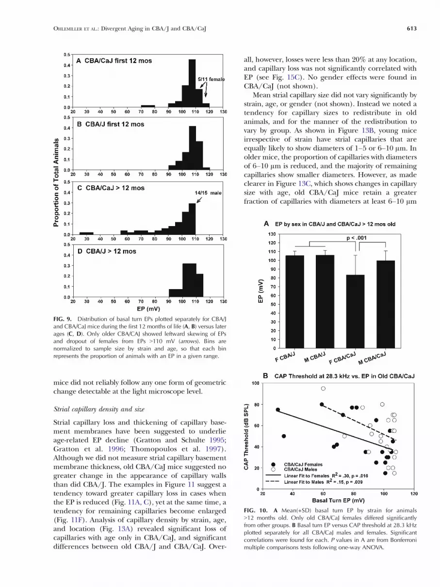
We argued from Figure 7 that one feature distin-guishing CBA/CaJ from CBA/J is that EPs greaterthan ∼110 mV disappear in CBA/CaJ after about12 months of age. This trend is confirmed in Figure 9,which shows the distribution of EP values (grouped by10 mV bins) by strain and age group. While thedistribution of EPs by strain is similar in younger mice(Fig. 9A, B), changes in the EP distribution after12 months in CBA/CaJ feature both skewingtoward lower values and complete disappearanceof EPs 9110 mV (compare Fig. 9C, D). By contrastwith CBA/CaJ, the distribution of EPs in old CBA/Jappeared little affected by age. Notably, of 15 oldCBA/CaJ mice showing EPs between 105 and110 mV, 14 were male. Among young CBA/CaJs(Fig. 9A) males and females were equally likely toshow an EP above 110 mV. Therefore, althoughboth males and females exhibit EP decline at agesgreater than 12 months, the decline is worse infemales.
Figure 10A confirms that old CBA/CaJ miceshowed lower EPs than CBA/J mice, although theoverall difference was significant only in females. Wenext considered to what extent CAP threshold differ-ences by strain could be accounted for by variation inEP. Figure 10B shows significant correlations between
FIG. 2. Scatter plots of CAP threshold versus age in months at 5 kHz (A,B) and 28.3 kHz (C,D) for both CBA/J and CBA/CaJ mice. Left and rightcolumns separate animals by gender. Lines are fitted second-order polynomials. At 28.3 kHz, CBA/CaJ exhibit more hearing loss with age thanCBA/J, irrespective of gender.
610 OHLEMILLER ET AL.: Divergent Aging in CBA/J and CBA/CaJ

CAP threshold at 28.3 kHz and basal turn EP for bothmale and female CBA/CaJ. From R2 estimates,roughly 30% of threshold variation in females and15% of threshold variation in males could beaccounted for by EP variation.
Strial anatomic correlates of EP reduction
Since variation in the EP could account in part forCAP threshold variation by strain, we considered whatanatomic changes in the cochlear lateral wall are mostclosely associated with EP decline, and whether thesechanges also show gender dependence. We begin withthe stria vascularis.
Strial thickness
Six examples in Figure 11 illustrate the variableappearance of the stria in upper basal turn of oldCBA/CaJ mice. The three examples associated with alow EP feature strial thinning (Fig. 11A), strial edema
FIG. 3. Overlay of fitted polynomial functions from Figure 2. A and B,respectively, superimpose fitted curves for both strains and genders forthresholds at 5 and 28.3 kHz.
FIG. 4. Mean (+SD) inner and outer hair cell survival for old CBA/J(22–26 months; n=8) and CBA/CaJ (23–27 months; n=8) mice. Eachgroup was roughly evenly split by gender. Frequency map is basedon Muller et al. (2005). Outer hair cell survival differed significantlyby strain and location, with CBA/CaJ mice showing better OHCsurvival. Horizontal bars above the X axis denote locations wheredifferences were significant by Bonferroni multiple comparisons test.
FIG. 5. Mean (−SD) spiral ganglion cell density at three cochlearlocations by strain and age (A), and for old male and female CBA/CaJ(B). Old CBA/CaJ mice showed significantly greater neuronal loss,particularly in the apex and lower base. Old female CAB/CaJ tendedtoward greater neuronal loss than males. In B, data from 13 ‘old’ andnine ‘very old’ CBA/CaJ mixed-gender data sets were combined.P values are from Bonferroni multiple comparisons tests followingtwo-way ANOVA.
OHLEMILLER ET AL.: Divergent Aging in CBA/J and CBA/CaJ 611

(Fig. 11C), and a typical normal profile (Fig. 11F).Strial thinning with age is commonly described inhumans and animals (Kusunoki et al. 2004; Suzukiet al. 2006; Ishiyama et al. 2007), and may reflect cellchanges that lead to EP decline (Ohlemiller et al.2008, 2009). Both CBA/J and CBA/CaJ showedsignificant strial thinning throughout most of thecochlea (Fig. 12). Counter to any simple prediction,
however, it was CBA/J that sustained the greatest degreeof thinning on average. It is possible that cases of strialthinning were offset by instances of overt or subtle strialedema. Thus impaired strial function in old CBA/CaJ
FIG. 6. Lower basal turn (mid-hookregion) Rosenthal’s canal and organ ofCorti in an old female CBA/CaJ mouseshowing loss of spiral ganglion cells(SpG). Inset shows expanded organ ofCorti, calling attention to the presence ofouter hair cells (arrowheads in bothimages), but the absence of inner haircells (asterisk) in this animal. SpLim spirallimbus, IP inner pillar, OP outer pillar, DDeiters’ cells.
FIG. 7. Scatter plots of basal turn EP versus age for CBA/J (top) andCBA/CaJ (bottom). Significant reduction (indicated by significantnon-zero regression coefficient) is seen only in CBA/CaJ. Most of themice with EPs below 95 mV are female.
FIG. 8. Scatter plots of basal turn EP versus apical turn EP for CBA/J(A) and CBA/CaJ (B) across all ages. Normal EP is ∼10 mV higher inthe base than in the apex. Significant EP reduction in CBA/CaJ isassociated with reversal of the EP spatial gradient.
612 OHLEMILLER ET AL.: Divergent Aging in CBA/J and CBA/CaJ
mice did not reliably follow any one form of geometricchange detectable at the light microscope level.
Strial capillary density and size
Strial capillary loss and thickening of capillary base-ment membranes have been suggested to underlieage-related EP decline (Gratton and Schulte 1995;Gratton et al. 1996; Thomopoulos et al. 1997).Although we did not measure strial capillary basementmembrane thickness, old CBA/CaJ mice suggested nogreater change in the appearance of capillary wallsthan did CBA/J. The examples in Figure 11 suggest atendency toward greater capillary loss in cases whenthe EP is reduced (Fig. 11A, C), yet at the same time, atendency for remaining capillaries become enlarged(Fig. 11F). Analysis of capillary density by strain, age,and location (Fig. 13A) revealed significant loss ofcapillaries with age only in CBA/CaJ, and significantdifferences between old CBA/J and CBA/CaJ. Over-
all, however, losses were less than 20% at any location,and capillary loss was not significantly correlated withEP (see Fig. 15C). No gender effects were found inCBA/CaJ (not shown).
Mean strial capillary size did not vary significantly bystrain, age, or gender (not shown). Instead we noted atendency for capillary sizes to redistribute in oldanimals, and for the manner of the redistribution tovary by group. As shown in Figure 13B, young miceirrespective of strain have strial capillaries that areequally likely to show diameters of 1–5 or 6–10 μm. Inolder mice, the proportion of capillaries with diametersof 6–10 μm is reduced, and the majority of remainingcapillaries show smaller diameters. However, as madeclearer in Figure 13C, which shows changes in capillarysize with age, old CBA/CaJ mice retain a greaterfraction of capillaries with diameters at least 6–10 μm
FIG. 9. Distribution of basal turn EPs plotted separately for CBA/Jand CBA/CaJ mice during the first 12 months of life (A, B) versus laterages (C, D). Only older CBA/CAJ showed leftward skewing of EPsand dropout of females from EPs 9110 mV (arrows). Bins arenormalized to sample size by strain and age, so that each binrepresents the proportion of animals with an EP in a given range.
FIG. 10. A Mean(+SD) basal turn EP by strain for animals912 months old. Only old CBA/CaJ females differed significantlyfrom other groups. B Basal turn EP versus CAP threshold at 28.3 kHzplotted separately for all CBA/CaJ males and females. Significantcorrelations were found for each. P values in A are from Bonferronimultiple comparisons tests following one-way ANOVA.
OHLEMILLER ET AL.: Divergent Aging in CBA/J and CBA/CaJ 613

in diameter. This trend is especially pronounced in oldCBA/CaJ females, in which∼11%of strial capillaries arelarger than 10 μm(arrow in Fig. 13B). The old CBA/CaJfemales show only a slight gain in the fraction of smallcapillaries, and uniquely, show a gain in the fraction ofcapillaries larger than 10 μm (arrows in Fig. 13C). Insummary, capillary data suggest that neither reductionin strial capillary density nor reduced capillary sizeaccount for EP decline in old CBA/CaJ. Instead,retention of larger capillaries—including an increasein the fraction of large capillaries in females—maysignal other changes that are tied to EP reduction.
Strial cell density
Both human temporal bone studies (Schuknecht etal. 1974; Pauler et al. 1988; Schuknecht and Gacek
1993) and animal studies (Spicer and Schulte 2005a;Ohlemiller et al. 2006; Ohlemiller et al. 2009) haveemphasized the frequent limiting nature of strialmarginal cell survival and function in aging. Marginalcell counts in CBA/J and CBA/CaJ likewise suggestedthat marginal cell density distinguishes these strainsboth young and old. As shown in Figure 14, CBA/Jmice may begin life with more marginal cells thanCBA/CaJ, and then retain these over their normal lifespan, showing no significant reduction in numberwith age. By contrast, old CBA/CaJ mice show anoverall significant loss of marginal cells with age at alllocations. Although no interactions with location weredetected, marginal cell losses with age in CBA/CaJappeared greatest in the basal half of the cochlea. Nosignificant effect of gender was detected (data notshown). Thus, differences in EP by sex in old CBA/
FIG. 11. A–F Example lateral wall of the cochlear upper basal turnin six old CBA/CaJ mice with very different EPs. Age, gender, and EPare given in each panel. General features include a mainly normalappearance of the spiral ligament and a large degree of heterogeneityin the stria vascularis. The most glaring pathology of the lateral wall
is the apparent loss of outer sulcus cells/root cells in the region of spiralligament just below spiral prominence (arrows). Asterisk in B highlightslarge capillaries that appeared most prominent in old female mice (seetext). Labels in A denote major landmarks: StV stria vascularis, SM scalamedia, SpP spiral prominence, I and II types I and II fibrocytes.
614 OHLEMILLER ET AL.: Divergent Aging in CBA/J and CBA/CaJ

CaJ (Figs. 9 and 10) could not clearly be explained bydifferential marginal cell survival.
We tested the ability of strial marginal cell densityin the cochlear upper basal turn to predict the EP. Asshown in Figure 15A and D, marginal cell densitycorrelates with the EP in CBA/CaJ mice and appearsto account for about 16% of EP variance. Neither of thetwo other major strial cell types (basal and intermediatecells) showed significant variation by strain, or anysignificant correlation with EP (not shown).
Spiral ligament histologic correlates of EPreduction
We previously showed that thinning of the ligamentcorrelates with EP decline in old mice of some strains
(Ohlemiller et al. 2006, 2009). We therefore com-pared ligament thickness by strain and age in CBA/Jand CBA/CaJ. Both young and old CBA/J miceshowed a significantly thicker ligament than theirCBA/CaJ counterparts (Fig. 16). Moreover, ligamentthickness in CBA/J was surprisingly stable with age.Potential thickening of the ligament with age in CBA/CaJ suggested by Figure 16 was not significant. Nogender effects were found (not shown).
FIG. 13. A Mean (−SD) number of capillaries per strial profileversus cochlear location for young and old CBA/J and CBA/CaJ. OldCBA/CaJ showed significantly greater capillary loss, yet the magni-tude of loss was G20%. B Distribution of strial capillary sizes acrossall animals, grouped by strain and age. Each symbol represent theproportion of all capillaries falling within a 5 μm diameter range (0–5, 6–10, etc.). Numbers in parentheses are the number of capillariesmeasured. For young mice of both strains, essentially all capillarieshad diameters of 10 μm or less, and appeared uniformly distributedacross this range. With age, most animals showed a redistribution ofcapillary sizes to favor diameters 5 μm or smaller. Old CBA/CaJfemales alone (arrow) also featured a notable fraction of capillaries(∼11%) with diameters 910 μm. C Re-plot of data from B normalizedusing ‘young’ data to show the change in distribution of capillarysize with age. Note decrease in proportion of capillaries withdiameters of 6–10 μm for all groups, but less pronounced in CBA/CaJ. Arrows highlight qualitatively different redistribution of capillarysize in old CBA/CaJ females, including a gain in the fraction of largecapillaries. P values in A are from Bonferroni multiple comparisonstests following two-way ANOVA. b
FIG. 12. Mean (−SD) strial thickness versus cochlear location foryoung and old CBA/J and CBA/CaJ mice. Significant differences werefound by strain and age. Both strains showed strial thinning with age,but old CBA/CaJ mice tended toward a thicker stria than old CBA/J.P values are from Bonferroni multiple comparisons tests followingtwo-way ANOVA.
OHLEMILLER ET AL.: Divergent Aging in CBA/J and CBA/CaJ 615

Loss of outer sulcus cells/root cells in CBA/CaJ
No prominent strain effects were found in the densityof any type of ligament fibrocyte (data not shown).Close examination of the ligament, however, revealeda remarkable difference between CBA/J and CBA/CaJ. In the cochlear upper base, the region ofligament between organ of Corti and spiral prom-inence that is normally bordered by outer sulcus cellsoften contained few of these. Instead, there oftenappeared deep indentations or voids, suggestive of theloss of outer sulcus cells and root processes (seearrows in Figs. 11 and 17A). Cell counts (Fig. 18A)confirmed that CBA/J mice retain more OSCsthroughout life in both lower and upper cochlearbase, and that only CBA/CaJ mice show loss of thesecells in the upper base. The proportion of sectionsshowing ligament indentations or voids also wassignificantly increased only in the cochlear upperbase of old CBA/CaJ (Fig. 18B), where it appeared inthe majority of sections. The severity of outer sulcuscell anomalies was not correlated with either EP(Fig. 15B) or thresholds (not shown).
The OSCs most affected appeared to be those lyingmore superior in the ligament, immediately below thespiral prominence (Figs. 11 and 17A), and may repre-sent a specific population. We further noted that thesewere most likely to lie in the zone where Claudius cellsand spiral prominence epithelial cells typically meet inCBA/J cochleas, leaving no OSCs directly exposed toendolymph. In Figure 17, the point of termination ofClaudius cells abutting the spiral ligament is comparedin example old CBA/CaJ and CBA/J mice (comparepoints marked by arrowheads in Figure 17A, B). In the
CBA/CaJ, a thin Claudius cell process ends well inferiorto a spiral prominence epithelium that follows theoutline of the void left by missing OSCs and their rootprocesses. In the CBA/J, a more expansive network ofClaudius cells clearly ends at the thin line of cellsmarking spiral prominence. Basal–apical variation inthe extent of ‘exposure’ to endolymph of OSCs hasbeen reported as a normal feature of gerbils and guineapigs (Duvall 1969; Spicer et al. 1996). It was ourimpression, however, that these cells are less oftenexposed in CBA/J than in CBA/CaJ, even when younganimals are compared.
DISCUSSION
We show that two intensively used ‘good hearing’inbred mouse strains, CBA/J and CBA/CaJ, differ inhearing sensitivity and cochlear pathology beginningaround one year of age. Despite similar names andgenerally similar hearing characteristics for the firstyear, these two strains appear to model differentforms of presbycusis. The 980 year span over whichthese strains have diverged (Fox et al. 1997) has beenample time for the accumulation of over 2,200 knowngenetic differences, including at least 41 non-synon-ymous exonic base changes (Bult et al. 2008). Theseor other polymorphisms must alter qualitatively thetrajectory of cochlear aging. Key qualitative differ-ences that set CBA/CaJ apart from CBA/J with age(summarized in Table 1) include EP decline, loss ofstrial marginal cells, and loss of outer sulcus cells inCBA/CaJ. These do not occur to a significant degreein CBA/J mice, irrespective of gender, at any time in atypical lifespan.
CBA/CaJ versus CBA/J
We agree with a previous assessment (Sha et al. 2008)that CBA/J mice most closely approximate sensorypresbycusis. The loss of predominantly outer hair cellsin both base and apex (Fig. 4), also described by Shaet al. (2008), reasonably corresponds with the patternof threshold shifts with age in CBA/J. According toour cell counts, old CBA/CaJ mice retain more OHCsthan CBA/J, so that they cannot simply represent amore extreme version of the same aging model. Weknow of no other study that has specifically examinedhair cell density in old CBA/CaJ mice. Previousstudies have mixed CBA/J and CBA/CaJ (Spongr etal. 1997) or used other CBA/Ca substrains (Li 1992;Li and Hultcrantz 1994).
Comparative patterns of spiral ganglion cell loss inCBA/J and CBA/CaJ mice are nearly the inverse oftheir patterns of hair cell loss. That is, CBA/CaJs losefewer OHCs than CBA/J, but more neurons, at both
FIG. 14. Mean (+SD) strial marginal cell density at three cochlearlocations by strain and age. Young CBA/J mice tended toward moremarginal cells than young CBA/CaJ, and sustained no significant losswith age. Old CBA/CaJ showed significant loss of marginal cells,particularly in the lower cochlear base, with no clear effect of gender(not shown). P values are from Bonferroni multiple comparisons testsfollowing two-way ANOVA.
616 OHLEMILLER ET AL.: Divergent Aging in CBA/J and CBA/CaJ

apical and basal ends of the cochlea (Figs. 4 and 5).Complementary loss of different cell types may helpexplain why the two strains show similar patterns oflow frequency hearing loss with age (Figs. 1 and 3).Alternatively, of course, the rate of low frequencyhearing loss may reflect a common degeneration ofsome other cell or structure we did not quantify. Sinceneuronal loss in CBA/CaJ appeared to outpace innerhair cell loss (compare Figs. 4 and 5), this loss was
likely a combination of primary and secondary loss.The pattern of accelerated neuronal loss in both baseand apex matches a trend noted for humans (Felderand Schrott-Fischer 1995). To our knowledge, thepresent study is the first suggesting gender bias inneuronal loss.
While CBA/CaJ mice lost fewer OHCs in the lowerbase than CBA/J, there was some loss. We thereforecannot say that hair cell loss played no role in
FIG. 15. Scatter plots of three anatomic metrics in the upper basal turn (marginal cell density, OSC abnormality, strial capillary density) versusbasal turn EP for CBA/CaJ (A–C) and CBA/J (d–f). Only marginal cell density in CBA/CaJ was significantly correlated with EP.
OHLEMILLER ET AL.: Divergent Aging in CBA/J and CBA/CaJ 617

progressive high frequency threshold shifts. It wouldappear more correct to say that CBA/CaJ mice addother forms of presbycusis to the sensory formdisplayed by CBA/J. Presently, no aging mouse modelthat has been well described remains free of signifi-cant hair cell loss over the normal life span (Spongret al. 1997; Willott et al. 1998; Ohlemiller and Gagnon2004a; Ohlemiller et al. 2008; Sha et al. 2008). Unlikespiral ganglion and stria vascularis, which possessredundant capacity, and for which moderate degen-eration can apparently be tolerated without hearingloss (Schulte and Schmiedt 1992; El-Badry andMcFadden 2009; Kujawa and Liberman 2009), OHCs
are not present in excess. The predominant cochlearaging mode for mice may be sensory presbycusis, withstrial and neural presbycusis added in some strains.
Threshold changes at high frequencies in bothCBA/J and CBA/CaJ appear sexually dimorphic, withfemales showing more rapid hearing loss than malesby 15 or 24 months, respectively (Fig. 3). In thisregard, our data seem to depart from a previous studyindicating that CBA/CaJ males sustain more rapidhigh frequency hearing loss (Henry 2004). At agesranging 9–24 months, however, we likewise findpoorer hearing in males, so that there appear to betwo gender patterns over the CBA/CaJ lifespan. Theoldest CBA/CaJ mice examined in the Henry (2004)study, 350 days of age, fell within a window that favorsfemales in our data as well, and Henry would havemissed a later transition. Notably, our data bringCBA/J and CBA/CaJ into register with another trendnoted by Henry, namely that C57BL/6 J mice alsofeature more rapid hearing loss in females. CBA andB6 mice may not mimic a broadly supported trend inhumans, whereby males fare more poorly with age(Jerger et al. 1993). However, they may usefully modelthe human phenomenon of accelerated hearing lossin females after menopause (Hederstierna et al. 2007)suggested to reflect loss of the protective effects ofestrogen. Unfortunately, we know of only one esti-mate of age at menopause in mice, universally placingthis event at 12–14 months (Silver 1995). It would beof interest to know whether the ∼9-month straindifference in the age at which male and femalethresholds diverge (Fig. 3) reflects a later age atmenopause in CBA/CaJ. Gender effects on thresholds
FIG. 16. Mean (−SD) spiral ligament thickness versus cochlearlocation for young and old CBA/J and CBA/CaJ mice. Significantdifferences were found by strain, so that CBA/CaJ mice showed athinner ligament, particularly in the basal half of the cochlea. P valuesare from Bonferroni multiple comparisons tests following two-wayANOVA.
FIG. 17. Example lateral wall of the cochlear upper basal turn in anold CBA/CaJ female (A same animal as in Fig. 11A) and an old CBA/Jfemale (B). Age and EP in each mouse are shown. The two imageshave been aligned to emphasize difference in survival of outer sulcuscells/root cells in the region just below spiral prominence (comparelocations at large arrows). Potentially related was a difference in the
extent of coverage of OSCs at this location by spiral prominenceepithelial cells. In CBA/J mice, the cells of spiral prominence (SpP)more often contact Claudius cells (Cc) of the organ of Corti. Smallarrowheads in each panel denote the apparent end of Claudius cellprocesses, leaving more OSCs exposed to endolymph in the CBA/CaJ.
618 OHLEMILLER ET AL.: Divergent Aging in CBA/J and CBA/CaJ

in old CBA/CaJ parallel two anatomic trends. First,loss of cochlear neurons appears more pronounced inold females than in males (Fig. 5). Second, EP declineis significantly greater in females (Figs. 9 and 10).Therefore, neuronal loss and EP decline in CBA/CaJmay both be modulated by sex hormones or othergender-related characteristics (see below).
We cannot, of course, be sure that we have identifiedthe essential cellular differences between aging CBA/Jand CBA/CaJ that account for their threshold differ-ences. We only sampled cochleas at fixed locations,although this strategy has proven successful in the past.The usual caveat—that cell counts need not correspondto cell functionality—must be injected. We found noclear anatomic correlate for more rapid hearing loss infemale CBA/J mice. In CBA/CaJ, EP variationaccounted for only 30% of threshold variation at most,and no clear pattern by gender was identified formarginal cell loss. Moderate loss of outer sulcus cells inthe upper base of CBA/CaJ could certainly affect bothEP and threshold (see below), but we detected nocontribution to either EP decline or threshold elevation.Finally, we could not show correlation between neuro-nal density and thresholds in CBA/CaJ, and thus couldnot demonstrate that greater neuronal loss in old CBA/CaJ females helps explain their more severe hearingloss. Most evidence indicates that only substantialneuronal loss is manifested in behaviorally or physio-logically determined threshold shifts (El-Badry andMcFadden 2009; Kujawa and Liberman 2009). Never-theless, the 950% loss of neurons exhibited in the lowerbase of old CBA/CaJ mice may be sufficient.
Correlates of EP decline in CBA/CaJ
Comparison with CBA/J revealed several properties ofcochlear lateral wall in old CBA/CaJ mice that havebeen linked to explicit, or suggested, EP decline.
FIG. 18. A Mean(-SD) outer sulcus cell density in the upper andlower basal turn of young and old CBA/J and CBA/CaJ mice. CBA/CaJmice show fewer of these cells, irrespective of age. B Incidence ofabnormal OSC/root cell profiles by strain and age. Uniquely in oldCBA/CaJ mice, over 60% of sections exhibited indentations or voidsin spiral ligament suggestive of OSC/root cell loss. P values are in Afrom Bonferroni multiple comparisons tests following two-wayANOVA.
TABLE 1
Comparison of features in aging CBA/J and CBA/CaJ cochleae
Feature Similarities Differences
Thresholds Similar loss at low frequencies CBA/CaJ show greater loss at highfrequencies
Thresholds by gender Females more susceptible than malesHair cell loss Similar IHC loss CBA/J show more OHC lossNeuronal loss CBA/CaJ show more lossEP by age Only CBA/CaJ show EP declineStrial capillariesa Increased fraction of capillaries G5 μm with age CBA/CaJ females show increase in
fraction of capillaries 910 μm with ageStrial marginal cells Only CBA/CaJ show significant lossStrial intermediate, basal cellsa Similar density, little change with ageSpiral ligament Thinner in CBA/CaJLigament types I, II, IV fibrocytesa Similar density, little change with ageOuter sulcus cells Only CBA/CaJ show significant loss
aBased on analysis of upper base
OHLEMILLER ET AL.: Divergent Aging in CBA/J and CBA/CaJ 619

Strial thickness
Strial thinning is a commonly reported aspect ofcochlear aging (Kusunoki et al. 2004; Suzuki et al.2006; Ishiyama et al. 2007), although it is often not clearwhen thinning is functionally meaningful. However,differences in strial thickness in our material (Fig. 12)actually favor CBA/CaJ, a trend that may reflectpathology not evident at the light microscope level.
Microvascular changes
Reduced capillary density, reduced capillary size, andchanges in capillary structure have all been implicatedin real or potential EP decline (Johnsson and Hawkins1972; Gratton and Schulte 1995; Gratton et al. 1996;Thomopoulos et al. 1997). As we have noted however(Ohlemiller et al. 2006), neither 950% reductions instrial capillary density (Di Girolamo et al. 2001), normore than doubling of capillary basement membranethickness in C57BL/6 mice promote EP decline(Lang et al. 2002). By contrast, capillary loss inCBA/CaJ mice is generally less than 20% (Fig. 13A).Probably more important was strial capillary sizeredistribution with age (Fig. 13B, C). The fact thatCBA/J mice showed the most pronounced shifttoward smaller capillary sizes renders this an unlikelykey factor in EP decline. Greater retention of largercapillaries in old CBA/CaJ—and particularly theincrease in the fraction of the largest capillaries inold female CBA/CaJ—may be more significant. Wepreviously noted a similar trend in old female BALB/cmice, in which average capillary size was greater in miceshowing EP decline (Ohlemiller et al. 2006). A shifttoward larger strial capillaries may point to anotherprocess that contributes to EP reduction, or perhaps to acompensatory response. Local stress responses maysense EP reduction, ion imbalance, or hypoxia, andincrease capillary size in response. Abnormally largestrial capillaries may emerge as an anatomic marker forthe presence of EP decline.
Spiral ligament thickness and fibrocyte density
The finding that a relatively thin spiral ligament inCBA/CaJ (Fig. 16) coincides with a tendency towardEP decline mirrors a trend in two other mouse modelswe have described (Ohlemiller et al. 2006, 2009).BALB/c mice in particular combine somewhat lowmarginal cell density and a thin spiral ligament withslightly reduced EP (∼10 mV) from an early age, andthese become more pronounced with time. There isdisagreement about the existence of any causal link—and direction thereof—between pathology of the striaand spiral ligament (e.g., Hequembourg and Liberman2001; Spicer and Schulte 2002; Wu and Marcus 2003;Ishiyama et al. 2007). Aging appears reliably associated
with progressive reduction in ligament volume and lossof fibrocytes in both humans and animals (Wright andSchuknecht 1972; Spicer and Schulte 2002; Kusunoki etal. 2004; Ishiyama et al. 2007). However, comparison ofthe extent of ligament degeneration and strial pathologyacross multiple inbred mouse models suggests that theseneed not be closely related (Ohlemiller 2009). Disrup-tion of K+ flux through the ligament can reduce the EP,but it is not clear this promotes strial degeneration (e.g.,Minowa et al. 1999). A thin spiral ligament early in lifemay signal a developmental process that also limitsmarginal cell density, and promotes eventual EP decline.
The role of gender in hearing and EP decline
Our data suggest that CBA/CaJ mice begin life withfewer marginal cells than CBA/J (Fig. 14), thenundergo further marginal cell loss with age. CBA/Jmice, by contrast, do not show significant loss ofmarginal cells over their typical life span. Primary lossor dysfunction of marginal cells is emerging as themost frequently cited basis of EP decline in humans(Pauler et al. 1988; Schuknecht and Gacek 1993) andanimal models (Spicer and Schulte 2005a; Ohlemilleret al. 2006; Ohlemiller 2009). That said, marginal cellloss did not seem to explain prominent genderdifferences in CBA/CaJ, and gender effects onmarginal cell function must be considered. The ideathat estrogen impacts strial marginal cell operation issupported by the literature (Konig et al. 2008;Motohashi et al. 2010). Interestingly, the effectappears to be one of inhibition (Lee and Marcus2001). Inhibition of strial ion transport coincides witha suggested overall protective effect of estrogen onsensory cells (Vina et al. 2005; Charitidi et al. 2009). Itis conceivable that effects of estrogen in females partlyexplain both reduction of the EP after 1 year and theacceleration of hearing loss after 2 years (that is, aftermenopause). Alternatively, sex-related differencesother than strictly hormonal processes may underliethe gender trends we observe (Willott 2009). Theapparent gender-skewing of EP decline in aging CBA/CaJ matches a trend suggested for humans (Gates et al.1999; Gates and Mills 2005), so that the underlyingmechanisms merit further study.
Loss of outer sulcus cells in CBA/CaJ
Loss of outer sulcus cells in CBA/CaJ adds a new twistto how threshold sensitivity and EP generation may bealtered in these mice. In this and other mouse modelswe have examined, we have interpreted reducedmarginal cell density as an indication that criticalmarginal cell functions in generating the EP areimpaired. We suppose—but have yet to show—thatfewer, larger, marginal cells maintain fewer critical
620 OHLEMILLER ET AL.: Divergent Aging in CBA/J and CBA/CaJ

pumps, transporters, and channels (Gratton et al.1997), or alter the critical ‘stoichiometry’ of theseamong strial cell types (Diaz et al. 2007). We have notproposed or observed that marginal cell degenerationleaves exposed intermediate cells or alters the ionbarrier of scala media. Published descriptions of aginggerbils, which also appear to model EP declineoriginating with marginal cell pathology, are likewiseconsistent with the maintenance of a continuousmarginal cell covering. By contrast with marginalcells, outer sulcus cells and the root processes theycompose project deep into the ligament (Duvall 1969;Spicer et al. 1996). OSC loss seems likely to uncovercells not normally exposed to endolymph, and notexpressing tight junctions. OSC loss therefore prob-ably violates the integrity of scala media. Sufficient lossof outer sulcus cells would act as an uncontrolled ionshunt, lowering the EP and reducing K+ currentsthrough hair cells. While this certainly could explainsome characteristics of CBA/CaJmice, we could not linkthe severity of OSC degeneration to hearing loss. Thespatially delimited pattern ofOSC loss in thesemicemayreduce its impact on hearing. As we noted, the OSCsmost affected appeared to be those lying where Claudiuscells and spiral prominence epithelial cells typicallymeet, and appeared less often exposed in CBA/J than inCBA/CaJ. However, it has not been suggested thatexposure of some OSCs represents a form of pathology.For now, loss of outer sulcus cells joins a long list ofnoted age-related cochlear changes—including capil-lary loss, ligament fibrocyte loss, limbus fibrocyte loss,organ of Corti supporting cell loss, clumping of neuro-nal cell bodies, pillar cell anomalies, and changes inReissner’s membrane (Cohen et al. 1990; Adams andSchulte 1997; Hequembourg and Liberman 2001;Ohlemiller and Gagnon 2004a, 2004b)—whose signifi-cance for the incidence and severity of presbycusis isnot clear.
The value of the nth aging model
Investigators in human presbycusis and its animalmodels must continually apply optimism that if weexamine enough cases and models, a finite number ofpatterns will emerge. Even if a manageable number ofpatterns do emerge, they will certainly reflect a muchlarger set of genetic and environmental causes. Never-theless, focusing on patterns may point the way to amanageable number of therapeutic targets. Unfortu-nately, the number of patterns divined from humantemporal bones is discouragingly large (for an excel-lent review, see Nelson and Hinojosa 2006). Mostdiscrepancies derive from the degree of presbycusisattributed to pathology of neurons versus hair cellsversus strial cells. An implicit theme that runs through
Schuknecht’s work (Schuknecht 1993; Schuknechtand Gacek 1993) is that these cells and structurescan degenerate independently. In fact, these arerarely encountered in isolation, and temporal boneanalyses rarely resolve how much hearing loss isattributable to each. The significance of their positedindependent degeneration lies in its implications forunderlying mechanisms. Invariant co-degeneration ofneurons, hair cells, and strial cells (to an extent thatall contribute to hearing loss) would suggest differentdisease mechanisms than would co-degeneration ofonly subsets of these. Just how much and what type oflateral wall degeneration is incompatible with anormal EP is currently best determined from compar-isons of mouse models. Since the EP must bemeasured to ‘diagnose’ strial presbycusis, few modelscan be considered adequately characterized.
ACKNOWLEDGMENTS
This work was supported by NIH R01 DC03454, DC08321(KKO), P30 DC04665 (R. Chole) and Washington UniversityMedical School Department of Otolaryngology.
REFERENCES
ADAMS JC, SCHULTE BA (1997) Histopathologic observations of theaging gerbil cochlea. Hear Res 104:101–111
BOHNE BA, GRUNER MM, HARDING GW (1990) Morphologicalcorrelates of aging in the chinchilla cochlea. Hear Res 48:79–91
BULT CJ, EPPIG JT, KADIN JA, RICHARDSON JE, BLAKE JA (2008) Themouse genome database (MGD): mouse biology and modelsystems. Nucleic Acids Res 36:D724–D728
CHARITIDI K, MELTSER I, TAHERA Y, CANLON B (2009) Functionalresponses of estrogen receptors in the male and female auditorysystem. Hear Res 252:71–78
COHEN GM, PARK JC, GRASSO JS (1990) Comparison of demyelinationand neural degeneration in spiral and Scarpa's ganglion ofC57BL/6 mice. J Electron Microsc Tech 15:165–172
COVELL WP, ROGERS JB (1957) Pathologic changes in the inner ear ofsenile guinea pigs. Laryngoscope 67:118–129
DI GIROLAMO S, QUARANTA N, PICCIOTTI P, TORSELLO A, WOLF F (2001)Age-related histopathological changes of the stria vascularis: anexperimental model. Audiology 40:322–326
DIAZ RC, VAZQUEZ AE, DOU H, WEI D, CARDELL EL, LINGREL J, SHULL
GE, DOYLE KJ, YAMOAH EN (2007) Conservation of hearing bysimultaneous mutation of Na, K-ATPase and NKCCl. J Assoc ResOtolaryngol 8:422–434
DUVALL AJ (1969) The ultrastructure of the external sulcus in theguinea pig cochlear duct. Laryngoscope 79:1–29
EL-BADRY MM, MCFADDEN SL (2009) Evaluation of inner hair cell andnerve fiber loss as sufficient pathologies underlying auditoryneuropathy. Hear Res 255:84–90
ERWAY LC, WILLOTT JF, ARCHER JR, HARRISON DE (1993) Genetics ofage-related hearing loss in mice: I. Inbred and F1 hybrid strains.Hear Res 65:125–132
FELDER E, SCHROTT-FISCHER A (1995) Quantitative evaluation ofmyelinated nerve fibers in cochlea of humans with age-relatedhigh-tone hearing loss. Hear Res 91:19–32
OHLEMILLER ET AL.: Divergent Aging in CBA/J and CBA/CaJ 621

FERNANDEZ EA, OHLEMILLER KK, GAGNON PM, CLARK WW (2010)Protection against noise-induced hearing loss in young CBA/Jmice by low-dose kanamycin. J Assoc Res Otolaryngol11:235–244
FOX RR, WITHAM BA, NELESKI LA, (EDS.) 1997. Handbook ofgenetically standardized JAX mice. The Jackson Laboratory,Bar Harbor ME.
FRISINA RD (2009) Age-related hearing loss: ear and brain mecha-nisms. Ann NY Acad Sci 1170:708–717
GATES GA, MILLS JH (2005) Presbycusis. Lancet 366:1111–1120GATES GA, COUROPMITREE NN, MYERS RH (1999) Genetic associations
in age-related hearing thresholds. Arch Otolaryngol Head NeckSurg 125:654–659
GRATTON MA, SCHULTE BA (1995) Alterations in microvasculature areassociated with atrophy of the stria vascularis in quiet-agedgerbils. Hear Res 82:44–52
GRATTON MA, SCHMIEDT RA, SCHULTE BA (1996) Age-relateddecreases in endocochlear potential are associated with vascularabnormalities in the stria vascularis. Hear Res 102:181–190
GRATTON MA, SMYTH BJ, LAM CF, BOETTCHER FA, SCHMIEDT RA (1997)Decline in the endocochlear potential corresponds to decreasedNa, K-ATPase activity in the lateral wall of quiet-aged gerbils.Hear Res 108:9–16
HEDERSTIERNA C, HULTCRANTZ M, COLLINS A, ROSENHALL U (2007)Hearing in women at menopause: prevalence of hearing loss,audiometric configuration and relation to hormone replace-ment therapy. Acta Otolaryngol 27:149–155
HENRY KR (1983) Ageing and audition. In: Willott JF (ed) Theauditory psychobiology of the mouse. Thomas, Springfield,pp 470–494
HENRY KR (2004) Males lose hearing earlier in mouse models of late-onset age-related hearing loss; females lose hearing earlier inmouse models of early-onset hearing loss. Hear Res 190:141–148
HENRY KR, MCGINN MD (1992) The mouse as a model for humanaudition. A review of literature. Audiology 31:181–189
HEQUEMBOURG S, LIBERMAN MC (2001) Spiral ligament pathology: amajor aspect of age-related cochlear degeneration in C57BL/6mice. J Assoc Res Otolaryngol 2:118–129
HIROSE K, LIBERMAN MC (2003) Lateral wall histopathology andendocochlear potential in the noise-damaged mouse cochlea. JAssoc Res Otolaryngol 4:339–352
ISHIYAMA G, TOKITA J, LOPEZ I, TANG Y, ISHIYAMA A (2007) Unbiasedstereological estimation of the spiral ligament and stria vascu-laris volumes in aging and Meniere's disease using archivalhuman temporal bones. J Assoc Res Otolaryngol 8:8–17
JAGGER DJ, NEVILL G, FORGE A (2010) The membrane properties ofcochlear root cells are consistent with roles in potassiumrecirculation and spatial buffering. JARO 11:435–448
JERGER J, CHMIEL R, STACH B, SPRETNJAK M (1993) Gender affectsaudiometric shape in presbycusis. J Am Acad Audiol 4:42–49
JOHNSSON L-G, HAWKINS JE (1972) Vascular changes in the human earassociated with aging. Ann Otol 81:364–376
KONIG O, RUTTIGER L, MULLER M, ZIMMERMANN U, ERDMANN B,KALBACHER H, GROSS M, KNIPPER M (2008) Estrogen and theinner ear: megalin knockout mice suffer progressive hearingloss. FASEB J 22:410–417
KUJAWA SG, LIBERMAN MC (2009) Adding insult to injury: cochlearnerve degeneration after 'temporary' noise-induced hearing loss.J Neurosci 29:14077–14085
KUSUNOKI T, CUREOGLU S, SCHACHERN PA, BABA K, KARYIA S, PAPARELLAMM (2004) Age-related histopathologic changes in the humancochlea: a temporal bone study. Arch Otolaryngol Head NeckSurg 131:897–903
LANG H, SCHULTE BA, SCHMIEDT RA (2002) Endocochlear potentialsand compound action potential recovery: functions in theC57BL/6J mouse. Hear Res 172:118–126
LANG H, SCHULTE BA, SCHMIEDT RA (2003) Effects of chronicfurosemide treatment and age on cell division in the adultgerbil inner ear. J Assoc Res Otolaryngol 4:164–175
LEE JH, MARCUS DC (2001) Estrogen acutely inhibits ion transport bystria vascularis. Hear Res 158:123–130
LIH-S (1992)Genetic influences on susceptibility of the auditory systemto aging and environmental factors. Scand Audiol Suppl 36:1–39
LI H-S, HULTCRANTZ M (1994) Age-related degeneration of the organof corti in two genotypes of mice. Oto Rhino Laryngol 56:61–67
MARCUS DC, CHIBA T (1999) K+ and Na+ absorption by outer sulcusepithelial cells. Hear Res 134:48–56
MINOWA O, IKEDA K, SUGITANI Y, OSHIMA T, NAKAI S, KATORI Y, SUZUKI M,FURUKAWA M, KAWASE T, ZHENG Y, OGURA M, ASADA Y, WATANABE K,YAMANAKA H, GOTOH S, NISHI-TAKESHIMA M, SUGIMOTO T, KIKUCHI T,TAKASAKA T, NODA T (1999) Altered cochlear fibrocytes in amouse model of DFN3 nonsyndromic deafnesss. Science285:1408–1411
MOTOHASHI R, TAKUMIDA M, SHIMIZU A, KONOMI U, FUJITA K, HIRAKAWA K,SUZUKI M, ANNIKO A (2010) Effects of age and sex on the expressionof estrogen receptor α and β in the mouise inner ear. ActaOtolaryngol 130:204–214
MULLER M, VON HUNERBEIN K, HOIDIS S, SMOLDERS JWT (2005) Aphysiological place-frequency map of the cochlea in the CBA/Jmouse. Hear Res 202:63–73
NELSON EG, HINOJOSA R (2006) Presbycusis: a human temporal bonestudy of individuals with downward sloping audiometric patternsof hearing loss and review of the literature. Laryngoscope 116(suppl 112):1–12
OHLEMILLER KK (2006) Contributions of mouse models to under-standing of age- and noise-related hearing loss. Brain Res1091:89–102
OHLEMILLER KK (2009) Mechanisms and genes in human strialpresbycusis from animal models. Brain Res 1277:70–83
OHLEMILLER KK, GAGNON PM (2004A) Cellular correlates of progressivehearing loss in 129 S6/SvEv mice. J Comp Neurol 469:377–390
OHLEMILLER KK, GAGNON PM (2004B) Apical-to-basal gradients inage-related cochlear degeneration and their relationship to'primary' loss of cochlear neurons. J Comp Neurol 479:103–116
OHLEMILLER KK, GAGNON PM (2007) Genetic dependence of cochlearcells and structures injured by noise. Hear Res 224:34–50
OHLEMILLER KK, FRISINA RD (2008) Age-related hearing loss and itscellular and molecular bases. In: Schacht J, Popper AN, Fay RR(eds) Auditory trauma, protection, and repair. Springer, NewYork, pp 145–194
OHLEMILLER KK, LETT JM, GAGNON PM (2006) Cellular correlates ofage-related endocochlear potential reduction in a mouse model.Hear Res 220:10–26
OHLEMILLER KK, RYBAK RICE ME, GAGNON PM (2008) Strial micro-vascular pathology and age-associated endocochlear potentialdecline in NOD congenic mice. Hear Res 244:85–97
OHLEMILLER KK, RICE MR, LETT JM, GAGNON PM (2009) Absence ofstrial melanin coincides with age associated marginal cell lossand endocochlear potential decline. Hear Res 249:1–14
PAULER M, SCHUKNECHT HF, WHITE JA (1988) Atrophy of the striavascularis as a cause of sensorineural hearing loss. Laryngoscope98:754–759
SCHACHT J, HAWKINS JE (2005) Sketches of otohistory. Part 9: presby[a]cusis. Audiol Neuro-Otol 10:243–247
SCHMIEDT RA (2010) Chapter 2: The physiology of cochlearpresbycusis. In: Gordon-Salant, S., Frisina, R.D., Popper, A.N.,Fay, R.R., (Eds.), Springer handbook of auditory research. TheAging Auditory System, Vol. 34. Springer, New York. pp. 9–38.
SCHUKNECHT HF (1974) Pathology of the ear, 1st edn. HarvardUniversity Press, Cambridge
SCHUKNECHT HF (1993) Pathology of the ear, 2nd edn. Lea andFebiger, Philadelphia
622 OHLEMILLER ET AL.: Divergent Aging in CBA/J and CBA/CaJ

SCHUKNECHT HF, GACEK MR (1993) Cochlear pathology in presby-cusis. Ann Otol Rhinol Laryngol 102:1–16
SCHUKNECHT HF, WATANUKI K, TAKAHASHI T, BELAL AA, KIMURA RS,JONES DD (1974) Atrophy of the stria vascularis, a common causefor hearing loss. Laryngoscope 84:1777–1821
SCHULTE BA, SCHMIEDT RA (1992) Lateral wall Na, K-ATPase andendodochlear potentials decline with age in quiet-reared gerbils.Hear Res 61:35–46
SHA S-H, KANICKI A, DOOTZ GA, TALASKA AE, HALSEY K, DOLAN DF,ALTSCHULER RA (2008) Age-related auditory pathology in theCBA/J mouse. Hear Res 243:87–94
SILVER LM (1995) Mouse genetics. Oxford, Oxford, UKSPICER SS, SCHULTE BA (2002) Spiral ligament pathology in quiet-
aged gerbils. Hear Res 172:172–185SPICER SS, SCHULTE BA (2005A) Pathologic changes of presbycusis
begin in secondary processes and spread to primary processes ofstrial marginal cells. Hear Res 205:225–240
SPICER SS, SCHULTE BA (2005B) Novel structures in marginal andintermediate cells presumably relate to functions of basal versusapical strata. Hear Res 200:87–101
SPICER SS, SAMUEL S, SCHULTE BA (1996) The fine structure of spiralligament cells relates to ion return to the stria and varies withplace-frequency. Hear Res 100:80–100
SPONGR VP, FLOOD DG, FRISINA RD, SALVI RJ (1997) Quantitativemeasures of hair cell loss in CBA and C57BL/6 mice throughouttheir life span. J Acoust Soc Am 101:3546–3553
SUZUKI T, NOMOTO Y, NAKAGAWA T, KUWAHATA N, OGAWA H, SUZUKI
Y, ITO J, OMORI K (2006) Age-dependent degeneration of the
stria vascularis in human cochleae. Laryngoscope 116:1846–1850
TARNOWSKI BI, SCHMIEDT RA, HELLSTROM LI, LEE FS, ADAMS JC (1991)Age-related changes in cochleas of Mongolian gerbils. Hear Res54:123–134
THOMOPOULOS GN, SPICER SS, GRATTON MA, SCHULTE BA (1997) Age-related thickening of basement membrane in stria vasculariscapillaries. Hear Res 111:31–41
VINA J, BORRAS C, GAMBINI J, SASTRE J, PALLARDO FV (2005) Whyfemales live longer than males? Importance of the upregulationof longevity-associated genes by oestrogenic compounds. FEBSLett 579:2541–2545
WILLOTT JF (1991) Aging and the auditory system: anatomy,physiology, and psychophysics. Singular, San Diego
WILLOTT JF (2001) Modulation of presbcusis: current status andfuture directions. Audiol Neuro-Otol 6:231–249
WILLOTT JF (2009) Effects of sex, gonadal hormones, and aug-mented acoustic environments on sensorineural hearing lossand the central auditory system: insights from research onC57BL/6J mice. Hear Res 252:89–99
WILLOTT JF, TURNER JG, CARLSON S, DING D, BROSS LS, FALLS WA(1998) The BALB/c mouse as an animal model for progressivesensorineural hearing loss. Hear Res 115:162–174
WRIGHT CG, SCHUKNECHT HF (1972) Atrophy of the spiral ligament.Arch Otolaryngol 96:16–21
WU T, MARCUS DC (2003) Age-related changes in cochlearendolymphatic potassium and potential in CD-1 and CBA/CaJmice. J Assoc Res Otolaryngol 4:353–362
OHLEMILLER ET AL.: Divergent Aging in CBA/J and CBA/CaJ 623


