Embed Size (px)
Citation preview

ClickHere
for
FullArticle
Diversity of rock varnish bacterial communities from BlackCanyon, New Mexico
Diana E. Northup,1 Jessica R. Snider,1 Michael N. Spilde,2 Megan L. Porter,3
Jodie L. van de Kamp,4 Penelope J. Boston,5 April M. Nyberg,6 and John R. Bargar7
Received 6 August 2009; revised 19 October 2009; accepted 9 November 2009; published 27 April 2010.
[1] Scientists vigorously debate the degree to which rock varnish is formed through theactions of microorganisms. To investigate this enigma, we utilized a three‐prongedapproach that combined (1) culture‐independent molecular methods to characterizebacterial communities associated with varnish that coats the rhyolitic volcanic rocks ofBlack Canyon, New Mexico, and rocks with no visible varnish; (2) culturing of varnish inmedia supplemented with reduced forms of manganese and/or iron and no or low amountsof carbon to isolate bacteria capable of precipitating iron and/or manganese oxides;and (3) scanning electron microscopy (SEM) of varnish and nearby rock that lacksmacroscopically visible varnish. Our culture‐independent studies revealed significantdifferences between varnish and nonvarnish communities. Chloroflexi and Ktedobacteriadominated one varnish site, while the other varnish site was dominated by Cyanobacteria.The nonvarnish sites were dominated by Actinobacteria and, to a lesser extent,Cyanobacteria and were the only samples to contain Deinococcus‐Thermus sequences.Approximately 65% of varnish cultures produced visible manganese precipitates. Mostculture isolates were not closely related to known manganese oxidizers, with the exceptionof Bacillus spp. SEM revealed microbial morphologies and two types of varnishmorphologies: (1) relatively smooth layers and (2) patches of botryoidal pinnacles, whichwere often associated with increased manganese concentrations. “Bare” rock showedevidence of incipient varnish. These results have important implications for the detectionof life on extraterrestrial planets such as Mars, where putative varnish coatings havebeen observed, and represent some of the first culture‐independent characterizations ofvarnish communities.
Citation: Northup, D. E., J. R. Snider, M. N. Spilde, M. L. Porter, J. L. van de Kamp, P. J. Boston, A. M. Nyberg, and J. R.Bargar (2010), Diversity of rock varnish bacterial communities from Black Canyon, New Mexico, J. Geophys. Res., 115, G02007,doi:10.1029/2009JG001107.
1. Introduction
[2] Rock varnish is a natural, black to brown coloredcoating enriched with manganese (Mn) and iron (Fe) oxideswith a typical thickness rarely exceeding 200 mm. Varnish
is ubiquitous, forming on exposed rock surfaces of diverselithology in almost every type of terrestrial weatheringenvironment, (e.g., Antarctica [Dorn et al., 1992a], Norway[Whalley et al., 1990], and Hawaii [Dorn et al., 1992b]),but is particularly abundant in arid and semiarid regions.Rock varnishes have attracted considerable research interestas a potential Quaternary dating tool for rock surfaces[Liu, 2003]; as archeological features and artifacts [e.g.,VandenDolder, 1992]; as indicators of paleoclimatic change[e.g., Dorn, 1994; Liu et al., 2000; Liu and Broecker, 2000,2007]; as environmental monitors because of the greatscavenging abilities of Mn oxides for certain heavy metals[Dorn, 1991; Fleisher et al., 1999; Wayne et al., 2006];because of the likely role of microbes in their formation[e.g., Perry and Adams, 1978; Krumbein and Jens, 1981;Nagy et al., 1991; Gorbushina, 2007]; as analogous envi-rons for the search for life on other planets [DiGregorio,2002; Gorbushina et al., 2002; Allen et al., 2004; Edwards,2004]; and to assist in interpretations of remote sensingstudies of varnished rock surfaces on Earth and Mars [e.g.,Israel et al., 1997; Kraft and Greeley, 2000].
1Department of Biology, University of New Mexico, Albuquerque,New Mexico, USA.
2Institute of Meteoritics, University of New Mexico, Albuquerque,New Mexico, USA.
3Department of Biological Sciences, University of Maryland BaltimoreCounty, Baltimore, Maryland, USA.
4CSIRO Marine and Atmospheric Research, Hobart, Tasmania,Australia.
5Earth and Environmental Science Department, New Mexico Instituteof Mining and Technology, Socorro, New Mexico, USA.
6National Clonal Germplasm Repository, USDA‐ARS, Corvallis,Oregon, USA.
7Stanford Synchrotron Radiation Laboratory, Menlo Park, California,USA.
Copyright 2010 by the American Geophysical Union.0148‐0227/10/2009JG001107
JOURNAL OF GEOPHYSICAL RESEARCH, VOL. 115, G02007, doi:10.1029/2009JG001107, 2010
G02007 1 of 19

[3] The first scientific descriptions of rock varnish and itspossible biogenic origin date as far back in the literature as1852, demonstrating that the deposition of Mn and Fe coat-ings on rocks has occupied the minds of many generations ofscientists [cf. Krumbein and Jens, 1981]. Rock varnish ismostly composed of clay minerals that are cemented to rockby Mn and Fe oxides in a laminated structure resembling thebotryoidal morphology that is occasionally seen in stroma-tolites [Perry and Adams, 1978]. Mn and Fe concentrationscan vary widely, but in most cases, Mn is highly enrichedover Fe as compared to their natural distribution [Engel andSharp, 1958; Krumbein, 1968, 1971; Potter and Rossman,1979; Knauss and Ku, 1980], Mn oxides usually account-ing for 20% or more of the total oxides. It is widely acceptedthat the Mn and Fe of rock varnish come from a variety ofsources including the atmosphere, meteoric water, dust, andfrom the surrounding soils [Allen, 1978; Engel and Sharp,1958; Scheffer et al., 1963; Krumbein and Jens, 1981;Potter and Rossman, 1979; Hooke et al., 1969; Linck, 1901;Bao et al., 2001; Thiagarajan and Lee, 2004]. Due to itsdistinct chemical composition from that of the substrate rock,varnish is usually considered to be of sedimentary origin[Hooke et al., 1969; Perry and Adams, 1978]. The questionremains as to whether processes concentrating Mn and Fe invarnish deposits are microbially or inorganically mediated.Evidence of biogenicity from the literature includes thedetection of amino acids in varnish including b‐alanine andg‐amino butyric acid, which are formed by enzymaticcarboxylation, thereby indicating possible organismal activ-ity [Perry et al., 2003].[4] There is considerable evidence suggesting that mi-
crobes can directly or indirectly control Mn precipitation[Nealson, 1983; Tebo et al., 2004] and biomineralization ofmanganese has been suggested in a variety of environmentsincluding, marine nodules [cf. Ehrlich, 2000], hot springs[Chafetz et al., 1998; Ferris et al., 1987; Mita et al., 1994],freshwater sediments [Maki et al., 1987], soils [Ghiorse,1988; Stiles et al., 2001], and caves [Spilde et al., 2005].Evidence for a biological origin for rock varnishes has beenmounting based on microscopic and culture‐based resultssuggesting that bacteria are intimately associated with varnishcoatings [Dorn and Oberlander, 1981; Jones, 1991;Hungateet al., 1987; Krumbein and Jens, 1981; Palmer et al., 1986;Perry et al., 2003; Raymond et al., 1992; Taylor‐Georgeet al., 1983]. However, it can be argued that we still do nothave sufficient evidence to state a biogenic origin of varnishemphatically.[5] It is widely accepted that only ∼1% of microbes
are cultured in the laboratory using standard techniques[Hugenholtz et al., 1998]. With the advent of culture‐independent detection techniques, particularly rRNA‐basedtechniques, we are able to characterize microbial commu-nities more fully, revealing significantly broader diversitythan previously recognized. Nevertheless, to date our knowl-edge of bacterial communities associated with rock varnishis due mostly to culture‐based studies, with the exception ofKuhlman et al. [2006, 2008] and Schelble et al. [2005].While molecular methods are valuable tools in character-izing microbial communities, simply demonstrating thepresence of an organism is not enough to prove involvementin the process of interest; we must correlate presence withfunction.
[6] The purpose of this research was to characterize thebacterial communities associated with rock varnish from asemiarid environment in New Mexico using 16S rDNAclone library analysis, to compare these communities tothose found on rock bare of macroscopically visible varnish,and to identify members of these communities able to pre-cipitate Mn oxides based on enrichment culture studies.These results represent some of the first published molecularstudies of rock varnish microbial communities. In addition,we utilize scanning electron microscopy to relate the organ-isms present to their chemical and mineral products.
2. Site Description
[7] Samples were obtained from a rock varnish sitelocated several kilometers southwest of Socorro in centralNew Mexico, USA, which is characterized as semiaridChihuahuan desert, which receives approximately 245 mmof precipitation annually. The study site is on the south slopeof Black Canyon, an east–west canyon that crosscuts part ofthe Socorro Cauldron, a dissected volcanic caldera, wherevolcanic activity has been dated from 33 MA to around7 MA [Eggleston et al., 1983]. The rocks in the area consistof hydrothermally altered rhyolite, and at the Luis Lopezmining district, just to the east of the sample site, a man-ganese mining operation exploited crytomelane and hol-landite from hydrothermal veins in the volcanic rockthrough World War II. Black rock varnish is common on thereddish brown rocks throughout the area. This site waschosen for two key characteristics. First, unlike the thick,layered varnish of places such as the Mojave Desert inCalifornia [cf. Broecker and Liu, 2001], the varnish here isdiscontinuous, often thin, and concentrated into micropitsand pockets in the rock surface. This may help preserveprimary structures and microbial remains that are destroyedin harsher, more arid environments. Second, this site isunique because the region contains elevated manganeselevels that may provide a more substantial source ofmanganese than most other rock varnish sites, although rockvarnish composition seems to be independent of theunderlying rock composition [Potter and Rossman, 1977,1979; Dorn and Oberlander, 1981]. Manganese leachedfrom the surrounding rock by weakly acidic rainwater mayprovide a source of Mn(II). With elevated manganese levelsavailable in the local country rock, the rock varnish maygrow more actively than other areas.
3. Methods
3.1. Sampling
[8] A total of five sites were sampled at Black Canyon,three containing rock varnish and two with no macro-scopically visible varnish. Varnish sample sites 1 and 2 werecollected from a dry, south facing ledge with scatteredpatches of black rock varnish, and were considered togetheras one sample (site 1‐2) for molecular analyses. Site 3 was abare rock surface several meters from site 1‐2, but on thesame rock outcrop. Site 4, approximately 10 m uphill fromthe first three sites, was also a bare rock surface, a road cutblasted approximately 60 years ago. Sites 3 and 4 were“bare” rock surfaces with no macroscopically visible varnishthat were to serve as control samples for the varnish sam-
NORTHUP ET AL.: VARNISH BACTERIAL COMMUNITY DIVERSITY G02007G02007
2 of 19

ples. Site 5 possesses a large amount of black varnish due toits location in an ephemeral watercourse on a vertical rockface that drains several hundred square meters of rock andscattered soil above it during rain and snowmelt events.Several small rock chips were aseptically cleaved from thesurface of each sample site for culturing and DNA analysis.After the aseptic sampling, additional fragments were cleaved,nonaseptically, for scanning electron microscope (SEM) studyand for processing into polished mounts for electron micro-probe (EMP) analysis. Larger hand samples were collectedfrom the immediate outcrop area around site 1‐2 for bulkchemical and X‐ray diffraction analysis.[9] Samples of rainwater and standing water were taken
from the Black Canyon site during and after a rainstorm totest for dissolved metals (e.g., Mn, Fe and Ni) in the waterthat may be the source for metals in the rock varnish, par-ticularly reduced metals in solution that could be taken upby metal oxidizing bacteria. A sample of dripping water wascollected from the ephemeral watercourse at site 5. Thestanding water sample was collected from a small pothole inthe rock surface above the watercourse. Additional rainwa-ter samples and dry deposition samples were analyzed fromthe nearest Sevilleta LTER site, Deep Well Met Station 40,approximately 51.5 km to the northeast. Samples of rain-water and dust from this site are part of an archive main-tained by the Sevilleta LTER.
3.2. Manganese Enrichment Cultures
[10] To ascertain which groups of organisms are capableof oxidizing reduced manganese, enrichment cultures wereinitiated using aseptically collected chips of the desert var-nish from the Black Canyon site. Samples were inoculatedin the field and transported to the laboratory for incubation.Approximately 45 different cultures that showed initial growthwere inoculated into replicate series of manganese complexchallenge media developed previously [Spilde et al., 2005].Based on our extensive experience with these types of rock‐inhabiting communities of organisms, we have observed thatthe ability to oxidize metals is frequently a consortiumproperty rather than the capability of an individual strain[Boston et al., 2009]. Because of this, we work to develop aminimum microbial consortium (MMC), which representswhat appear to be the organisms that are functioning togetherto produce the oxides and subsequent crystallization that weobserve in the cultures. Attempts to isolate individual strainsin pure culture often do not result in mineral precipitation thatmatches the minerals we observe in nature [Spilde et al.,2005]. In addition, these consortia tend to be very slowgrowing, particularly when we challenge them with mediacontaining no or low organic carbon. Even when we providemoderate levels of organic carbon, they can be exceedinglyslow to produce visible growth and oxides and subsequentcrystallization can take months or years to appear. Because ofthis, we have developed simple methods of keeping thegrowth media hydrated using parafilm sealing of Petri platesor tubes and we incubate in closed growth chambers whereadequate humidity levels can be maintained.[11] The media used in this study were enriched with
0.1 mmol concentrations of one of four forms of reducedmanganese (MnCO3, Mn2O3, Mn(NO3)2 · 4H2O, andMnCl2 · 4H2O). A fifth medium contained 0.1 mmol con-centration of both MnCO3 and FeCO3. Each of these media
was prepared in two forms: (1) without any organic carbonor (2) with low concentrations (0.1% w/v) of both acetateand glucose (Figure 1). Identical replicates of each culturevariant are inoculated to provide the ability to conduct killedcontrols at periodic intervals, using 2.5% glutaraldehydesolution to flood the cultures, in order to follow the processof mineralization. Uninoculated media, serving as negativecontrols, were incubated under the same conditions for thesame period of time. All cultures were incubated at 25°C forup to 2.5 years and were subcultured further onto mediadescribed above as colonies and mineral precipitates ofinterest occurred (Figure 1).
3.3. Molecular Phylogeny
3.3.1. Extraction of Nucleic Acids[12] Genomic DNA was extracted from enrichment cul-
tures using the method described by Marmur [1961]. DNAwas purified using the UltraClean™ Microbial DNA Isola-tion Kit (MoBio Laboratories, Inc.) reagents and protocol.Genomic DNA was extracted from rock chips with varnishstored in sucrose lysis buffer using the Power Soil DNAExtraction kit (MoBio Laboratories, Inc.).3.3.2. The 16S rDNA PCR Amplification and CloneLibrary Construction[13] The 16S rRNA gene was amplified from both enrich-
ment culture and environmental DNA by PCR with universalprimers, p46 forward (5′‐GCYTAAYACATGCAAGTCG‐3′)and p1409 reverse (5′‐GTGACGGGCRGTGTGTRCAA‐3′,provided by C. Takacs‐Vesbach, unpublished data, 2004)and AmpliTaq LD (Applied Biosystems) with an MJ ther-mal cycler as follows: 4 min denaturation at 94°C, followedby 35 cycles of 45 s annealing at 55°C, 2 min at 72°C(extension), and 30 s at 94°C (denaturation), with a final45 s 55°C annealing and 20 min 72°C extension step aftercycling was complete. Amplification products were clonedusing the TOPO TA Cloning kit (Invitrogen), and plated onLB/ampicillin agar plates [Sambrook et al., 1989].3.3.3. RFLP[14] To assist in determining which clones to sequence,
the 16S ribosomal DNA of 173 (site 1‐2) and 234 (site 5)varnish clones and 138 (site 3) and 140 (site 4) nonvarnishclones was digested with enzymes to produce restrictionfragment length polymorphisms (RFLPs). The restrictionenzymes HhaI and RsaI were used in double digestions ofthe whole cell PCR amplicons of clone DNA as follows:8 ml of PCR amplicon DNA, 1 ml of NE React Buffer 4,0.4 ml of double distilled water, and 0.2 ml of HhaI and0.4 ml of RsaI enzymes. RFLP patterns were visualizedusing a 4% Metaphor (FMC Rockland, Maine) electropho-resis gel in TAE, stained with 2 ml of ethidium bromide,visualized on a UV transilluminator, and compared visuallyby the authors.3.3.4. The 16S rDNA Sequencing[15] Representative clones of each RFLP pattern were
grown overnight in LB broth containing 100 mg/ml ampi-cillin and were purified with a QIAprep plasmid miniprepkit (Qiagen Inc., Chatsworth, Calif.). 125–300 ng of purifiedDNA was used as a template in cycle sequencing reac-tions with the ABI PRISM dye terminator cycle sequencingkit (Perkin‐Elmer‐Applied Biosystems). Primers used forsequencing were T7, T3 and the internal primers (533F,907R, and 765F) of the 16S rRNA gene. Some sequencing
NORTHUP ET AL.: VARNISH BACTERIAL COMMUNITY DIVERSITY G02007G02007
3 of 19

was done through the Washington University SequencingFacility in St. Louis, Missouri, while the bulk of thesequencing was done at the University of New MexicoMolecular Biology Facility.3.3.5. Phylogenetic Analysis[16] Each sequence was submitted to the CHIMERA_
CHECK program of the Ribosomal Database Project(RDP) [Maidak et al., 2001] (http://rdp.cme.msu.edu/) or tothe Mallard program (http://www.bioinformatics‐toolkit.org/Mallard/index.html [Ashelford et al., 2006]) to detectthe presence of possible chimeric artifacts. All sequenceswere analyzed using BLAST (NCBI [Altschul et al., 1997])and SIMILARITY_RANK (RDP [Maidak et al., 2001]) todetermine the taxonomic groupings of clone sequences.Sequences were submitted to GenBank and assigned the
accession numbers FJ595524–FJ595655. Each major taxo-nomic sequence group (e.g., Proteobacteria, Acidobacteria,etc.) was submitted to the Greengenes alignment tool tocreate an initial alignment, including the nearest‐neighbordatabase sequences for each clone (http://greengenes.lbl.gov/cgi‐bin/nph‐index.cgi [DeSantis et al., 2006a, 2006b]).Out‐group sequences (Aquifex aeolicus AE000657,Thermotoga maritime AE000512, and Thermus aquaticusL09663) were added to each set of sequences, and the datasets were realigned using MAFFT version 6.611 [Katoh andToh, 2008; Katoh et al., 2002, 2005]. Ambiguous regionsof each alignment were removed using GBlocks version0.91 [Castresana, 2000; Talavera and Castresana, 2007]. Amaximum likelihood phylogeny with branch support from100 bootstrap replicates was constructed for each data set
Figure 1. Logical schematic of the culturing experiments. An initial 45 cultures directly isolated fromthe varnish sites were subcultured in five different media types and each of those were subcultured inorganic carbon and no‐carbon versions. Six replicates of each medium were inoculated from the 45 pri-mary inocula to enable the potential to serially kill cultures at different development points to follow thesequence of mineral crystallization. The banding morphology was the most numerous macroscopicgrowth and Mn concentrating indicator. Most of the bands grew between 1 mm and 3 cm below the agarsurface, indicating varying degrees of microaerophilic preference. The halo and fan morphologies are cen-tered on the agar surface, implying a preference for higher oxygen partial pressures.
NORTHUP ET AL.: VARNISH BACTERIAL COMMUNITY DIVERSITY G02007G02007
4 of 19

(PhyML version 3.0 webserver [Guindon and Gascuel,2003; Guindon et al., 2005]) using the best fit model asdetermined by Modeltest version 3.7 [Posada and Crandall,1998; Posada and Buckley, 2004]. In order to performrarefaction analysis, a data set was constructed by takingthe representative sequence from each RFLP pattern andduplicating that sequence in the data set to represent thenumber of clones found in that clone group. Using this dataset, a distance matrix was constructed with the PHYLIPversion 3.6 DNADIST module under the F84 model ofevolution [Felsenstein, 1989], which was used to generaterarefaction curves at the 95% similarity level using DOTUR[Schloss and Handelsman, 2005]. The similarity betweeneach clone library was calculated using S‐Libshuff (http://www.plantpath.wisc.edu/joh/s‐libshuff.html [Schloss et al.,2004; Singleton et al., 2001]) and SONS [Schloss andHandelsman, 2006]. Richness and diversity indices werecomputed using EstimateS (version 8.0, Colwell, http://viceroy.eeb.uconn.edu/estimates).
3.4. Microscopy
[17] In addition to bulk analytical techniques, sampleswere examined on a JEOL 5800 SEM equipped with anenergy dispersive X‐ray analyzer (EDX). Rock chips fromthe varnish and bare rock sites were mounted directly onSEM sample stubs and coated with Au‐Pd metal for imag-ing. Small sections of cultures were examined with SEM/EDX to determine the presence of precipitated manganese.Several additional chips with surface varnish were embed-ded in epoxy, sectioned, and polished for cross‐sectionalimaging on a JEOL 8200 electron microprobe equippedwith a backscattered electron (BSE) detector.
3.5. Chemistry and X‐ray Diffraction
[18] Varnish was removed from the surface of the largerpieces using a reciprocating tungsten carbide‐coated wiresaw to remove as much varnish as possible in order toanalyze the varnish separately from the underlying hostrock. Major and trace elements were analyzed by means of
atomic absorption spectroscopy on samples digested in HFand HNO3. Mineralogical composition was determined onpowdered samples prior to digestion by X‐ray diffraction(XRD) using a Scintag Pad V diffractometer.[19] A varnish sample from site 1 was analyzed using
synchrotron‐based X‐ray diffraction (SR‐XRD) at beamline 10.3.2 at the Advanced Light Source synchrotron atthe Lawrence Berkeley National Laboratory (Berkeley, CA).X‐ray energy was set to 17 KeV using a crystal mono-chromator. The crushed sample, mounted on Kapton tape,was exposed to a 5 by 5 mm spot for 300 s acquisition time.[20] Black Canyon rainwater and pothole water samples
were analyzed by atomic absorption (AA) spectroscopyand by inductively coupled plasma atomic emission spec-trometry (ICP/AES). Sevilleta (see location informationabove) rainwater and dry deposition samples were analyzedfor metals and trace elements by ion chromatography usinga Dionex Ion Chromatograph DX‐100, and by ICP/AES.
4. Results
4.1. Culturing
[21] Many of the minimum microbial consortia (MMC)that we isolated from environmental samples producedamorphous Mn oxides and eventually Mn crystalline mi-nerals (Figure 2) when that element was provided in media(Figure 1). Of the 165 total MMC obtained on replicatemedia series, about 20% (33) produced black or dark brownbands, halos or fans in solid media within the first severalmonths (Figure 1). An additional 45% (76) of the MMCproduced similarly black or dark brown deposits within thefirst 18 months of incubation, albeit some at very low pro-ductivities. A remaining 56 cultures produced at least lim-ited Mn morphologies over the course of an additional year(2.5 years total incubation time). Uninoculated media, serv-ing as negative controls, produced no mineral precipitates,although 2 of a total of 20 negative controls (2 per 10 mediumvariants) did darken over time. Both of these were organiccarbon containing media variants, MnCO3 and MnCO3/
Figure 2. Electron micrographs of cultures. (a) Backscattered electron (BSE) image of surface ofbacterial culture plate showing deposition of Mn oxides by bacteria (lighter areas). Scale bar is 500 mm.(b) Mn‐coated tube‐like structure on the surface of smooth varnish from site 2. Scale bar is 100 mm. Insetcompares a similar structure from a bacterial culture taken from the same sample site. Scale bar is 1 mm.
NORTHUP ET AL.: VARNISH BACTERIAL COMMUNITY DIVERSITY G02007G02007
5 of 19

FeCO3. Electron microscopy of cultures revealed thatmineralogies ranged from amorphous oxides through variouscrystalline phases (described below). Concentrations of Mnwere confirmed with EDS of harvested material from onereplicate from each original culture type. Some of the crys-tallinemorphologies resemble crumpled tissue paper‐like andstar‐like shapes that we have seen in other environments inprior work [Spilde et al., 2005, 2009; Boston et al., 2001].
4.2. Molecular Phylogeny
[22] Rarefaction curves show that the nonvarnish clonelibraries approached saturation, particularly for site 4, whilethe varnish sites are still accumulating diversity (Figure 3).Although the clone libraries at each site represent signifi-cantly different communities (p < 0.0001), nine of the OTUsat the 95% similarity level were found at more than one ofthe different sampling sites. Seven OTUs were found in bothvarnish and nonvarnish sites, while two other OTUs werefound at both of the nonvarnish sites. Interestingly, no similarOTUs were found in both of the varnish sites.[23] Overall, our clone library from Black Canyon varnish
site 1‐2 was substantially different from varnish site 5’s clonelibrary. Chloroflexi and Ktedobacteria (unclassified) werenumerous in the site 1‐2 clone library, while the clone library
from site 5 had many Cyanobacteria clones. All three sitescontained Actinobacteria andAlphaproteobacteria, while thesite 5 clone library lacked Betaproteobacteria, but containeda small number of Gemmatimonadetes and Bacteroidetesclones. Nonvarnish site clone libraries contained numerousActinobacteria clones, and to a lesser extent, Cyanobacteria.Nonvarnish sites also were the only samples to containDeinococcus‐Thermus sequences (Table 1).[24] Clones whose closest relatives were from desert or
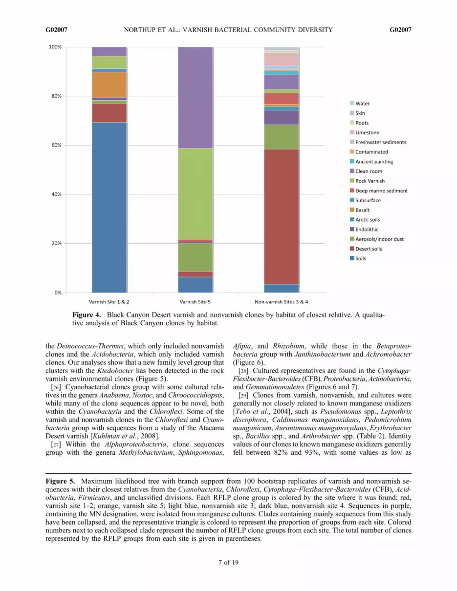
other soil environments were numerous in site 1‐2 (varnish),while the site 5 (varnish) clone library contained manyclones whose closest relatives were from other rock varnishenvironments and clean room studies (Figure 4). Interest-ingly, the clone library for site 1‐2 (varnish) also contained agreater number of closest relatives from basalt environmentsthan did site 5’s (varnish) clone library. Some closest relativeswere from studies of aerosols, especially in the site 5 (varnish)clone library. Sites 3 and 4 (nonvarnish) clones exhibited amuch greater portion of desert soil closest relatives than didthe varnish sites and showed a greater diversity in the habitatsof closest relatives.[25] The phylogenetic analyses show an intermixing of
nonvarnish and varnish clones in most phyla (Figures 5, 6,and 7 and Table 1). The exceptions to this pattern include
Figure 3. Varnish and nonvarnish sample rarefaction curves.
Table 1. Distribution of Clones Across Sample Sites by Phylum
Phylum Nonvarnish Site 3 Nonvarnish Site 4 Varnish Site 1‐2 Varnish Site 5 Cultured Phylotypes
Actinobacteria 11 22 3 3 3Gemmatimonadetes 2 2Deinococcus‐Thermus 4Cyanobacteria 8 2 4 27CFB 2 1 4 4Ktedobacter (unclassified) 1 14Chloroflexi 2 2 2Firmicutes 1 1Alphaproteobacteria 6 7 3 7 6Betaproteobacteria, Gammaproteobacteria 2 11Deltaproteobacteria 2
NORTHUP ET AL.: VARNISH BACTERIAL COMMUNITY DIVERSITY G02007G02007
6 of 19
the Deinococcus‐Thermus, which only included nonvarnishclones and the Acidobacteria, which only included varnishclones. Our analyses show that a new family level group thatclusters with the Ktedobacter has been detected in the rockvarnish environmental clones (Figure 5).[26] Cyanobacterial clones group with some cultured rela-
tives in the genera Anabaena, Nostoc, and Chroococcidiopsis,while many of the clone sequences appear to be novel, bothwithin the Cyanobacteria and the Chloroflexi. Some of thevarnish and nonvarnish clones in the Chloroflexi and Cyano-bacteria group with sequences from a study of the AtacamaDesert varnish [Kuhlman et al., 2008].[27] Within the Alphaproteobacteria, clone sequences
group with the genera Methylobacterium, Sphingomonas,
Afipia, and Rhizobium, while those in the Betaproteo-bacteria group with Janthinobacterium and Achromobacter(Figure 6).[28] Cultured representatives are found in the Cytophaga‐
Flexibacter‐Bacteroides (CFB),Proteobacteria,Actinobacteria,and Gemmatimonadetes (Figures 6 and 7).[29] Clones from varnish, nonvarnish, and cultures were
generally not closely related to known manganese oxidizers[Tebo et al., 2004], such as Pseudomonas spp., Leptothrixdiscophora, Caldimonas manganoxidans, Pedomicrobiummanganicum, Aurantimonas manganoxydans, Erythrobactersp., Bacillus spp., and Arthrobacter spp. (Table 2). Identityvalues of our clones to known manganese oxidizers generallyfell between 82% and 93%, with some values as low as
Figure 4. Black Canyon Desert varnish and nonvarnish clones by habitat of closest relative. A qualita-tive analysis of Black Canyon clones by habitat.
Figure 5. Maximum likelihood tree with branch support from 100 bootstrap replicates of varnish and nonvarnish se-quences with their closest relatives from the Cyanobacteria, Chloroflexi, Cytophaga‐Flexibacter‐Bacteroides (CFB), Acid-obacteria, Firmicutes, and unclassified divisions. Each RFLP clone group is colored by the site where it was found: red,varnish site 1‐2; orange, varnish site 5; light blue, nonvarnish site 3; dark blue, nonvarnish site 4. Sequences in purple,containing the MN designation, were isolated from manganese cultures. Clades containing mainly sequences from this studyhave been collapsed, and the representative triangle is colored to represent the proportion of groups from each site. Colorednumbers next to each collapsed clade represent the number of RFLP clone groups from each site. The total number of clonesrepresented by the RFLP groups from each site is given in parentheses.
NORTHUP ET AL.: VARNISH BACTERIAL COMMUNITY DIVERSITY G02007G02007
7 of 19

-
-
Figure 5
NORTHUP ET AL.: VARNISH BACTERIAL COMMUNITY DIVERSITY G02007G02007
8 of 19

Figure 6
NORTHUP ET AL.: VARNISH BACTERIAL COMMUNITY DIVERSITY G02007G02007
9 of 19

76%. One nonvarnish clone was more closely related at 99%identity to Pseudomonas putida.
4.3. SEM
[30] Rock varnish from Black Canyon is discontinuous inhand samples and is often isolated into surface depressions.The varnish is dark brown to black, although with a morematte texture than that found in other deserts, such as theMojave, where varnish often displays a vivid sheen. InSEM images, the varnish tends to display two distinct forms:relatively smooth surfaces and patches of botryoidal pro-trusions or pinnacles. The smooth varnish surfaces exhibita somewhat lumpy texture at high magnification. X‐rayspectra from the surface usually contain peaks for Si, Al, Mn,Fe, O, Ca, K and C, in order of peak intensity. These surfacesare commonly crosscut by tube‐like features (Figure 2b),which are significantly enriched in Mn compared to the sur-rounding varnish.[31] The second common form of varnish observed on the
Black Canyon samples is a mass of botryoidal shapes. Theseare patches or clumps of protrusions or short pinnacles a fewmicrometers tall (Figure 8). The patches are distributedmainly into low spots on the surface and are on the order ofhundreds of micrometers across but discontinuously scatteredacross the rock surface. This type of structure is somewhatless common than the smooth surface varnish. The patcheshave higher concentrations of Mn than the smooth surfacevarnish, and in X‐ray maps, Mn is usually limited to thecoverage of the botryoidal patch (Figure 8c) with little Mnoutside of that area. Close examination of these botryoidalclusters revealed a large amount of organic andmineral debristhat had been collected on the pinnacles (Figure 8b). In ad-dition to random organic debris such as pollen grains, anabundance of fine filaments, possibly dehydrated microbialexopolysaccharides, were seen looping around between thepinnacles. This network of filaments appears to be respon-sible for trapping the debris associated with the pinnaclestructures.[32] Another common surface form observed in SEM
were small, grape‐like clusters, usually in pits or holes in thesurface (Figure 8a), identified as putative microorganisms.The grape‐like clusters are sometimes spatially associatedwith the varnish but contain no Mn in X‐ray spectra.[33] In cross section, varnish from the Black Canyon sites
displays micrometer‐scale layering that can be broken intothree different types: (1) uniformly layered varnish, re-presenting the smooth surface varnish observed in Figures 9aand 9b; (2) botryoidal varnish, representing the patches ofpinnacle varnish seen in Figure 9c; and (3) chaotic layering,which had no counterpart observed on the surface. Thesmooth varnish, shown in Figure 9a, exhibits laterally con-tinuous, regular layers. The layers are generally uniform in
thickness and in backscattered electron (BSE) intensity, in-dicating constant composition. A few layers appear brighterin the BSE image, indicating a higher atomic number due toincreased Mn content. The botryoidal form, shown in crosssection in Figure 9c contains micrometer‐scale lamellae thatare organized into a pinnacle but laterally discontinuous. Inmany cases, the pinnacles exhibit arcs of lamellae that climbconcentrically upward throughout the botryoidal pinnacle.Despite the lateral discontinuities, some bright layers (in BSEimages) may be present from pinnacle to pinnacle, and somelayers, such as the bright, high manganese layer immediatelybelow the pinnacles in Figure 9b, can be traced across theentire sample. The third type, chaotic layering, was associatedwith both the layered and botryoidal forms particularly inthe lower portions. The chaotic layering is distinguished bydisorganized laminae that may form concentric circles ofvarious sizes or nonoriented layering (a wavy texture). In theuniformly layered varnish, there may be a transition to thechaotic form, as in the lower portion of Figure 9a or remainseparate and blanket the chaotic region below.[34] As a form of control sample, we compared rocks with
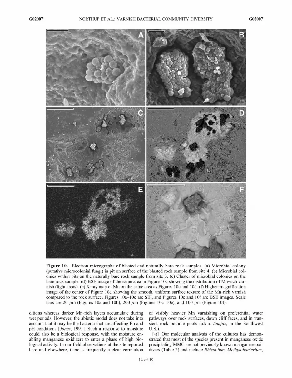
no macroscopically visible varnish (sites 3 and 4) to thosewith varnish (sites 1–2 and 5). Despite the fact that bothsamples appeared devoid of varnish to the unaided eye, eachhad microbial colonies visible in the SEM (Figure 10). The60 year old blasted surface (site 4) displayed colonies ofmicroorganisms that strongly resemble known microcolonialfungi examples hidden inmicropits in the surface.Most of thecolonies were less than 20 mm across and contained less than15 spheres attached to one another to form the colony(Figure 10a). The naturally weathered sample surface (site 3)contained colonies that were larger than those on the youngerblasted surface (site 4), but again hidden in pits in the sur-face. These colonies measured as much as 70 by 40 mmand contained many more attached spheres, indicating thatthe colonies were probably older and more developed(Figures 10b and 10c). Though not quantitatively measured,the colonies on the natural rock sample (site 3) were morecommon than those on the blasted rock surface (site 4).[35] Surrounding some of the colonies on the natural rock
sample (site 3), small Mn‐ and Fe‐rich areas were observed inthe SEM (Figures 10d, 10e, and 10f). These features, whichwere not noticeable when the sample was collected, were onlya few hundred micrometers in diameter and consist of smoothpatches containing as much as 34 wt % MnO2 and 15 wt %Fe2O3.Most of these patches partially or completely surroundcolonies in pits; the Mn‐rich area themselves are not neces-sarily within pits or depressions, although they may extendinto the pits. The exception is a small 100–150 mm patchat the bottom left side of Figure 10d that sits by itself and isnot associated with a colony.
Figure 6. Maximum likelihood tree with branch support from 100 bootstrap replicates of varnish and nonvarnish se-quences with their closest relatives from the Proteobacteria. Each RFLP clone group is colored by the site where it wasfound: red, varnish site 1‐2; orange, varnish site 5; light blue, nonvarnish site 3; dark blue, nonvarnish site 4. Sequencesin purple, containing the MN designation, were isolated from manganese cultures. Clades containing mainly sequences fromthis study have been collapsed, and the representative triangle is colored to represent the proportion of groups from eachsite. Colored numbers next to each collapsed clade represent the number of RFLP clone groups from each site. The totalnumber of clones represented by the RFLP groups from each site is given in parentheses.
NORTHUP ET AL.: VARNISH BACTERIAL COMMUNITY DIVERSITY G02007G02007
10 of 19

Figure 7. Maximum likelihood tree with branch support from 100 bootstrap replicates of varnish andnonvarnish sequences with their closest relatives from the Actinobacteria, Gemmatimonadetes, andDeinococcus‐Thermus. Each RFLP clone group is colored by the site where it was found: red, varnishsite 1‐2; orange, varnish site 5; light blue, nonvarnish site 3; dark blue, nonvarnish site 4. Sequencesin purple, containing the MN designation, were isolated from manganese cultures. Clades containingmainly sequences from this study have been collapsed, and the representative triangle is colored to rep-resent the proportion of groups from each site. Colored numbers next to each collapsed clade represent thenumber of RFLP clone groups from each site. The total number of clones represented by the RFLP groupsfrom each site is given in parentheses.
NORTHUP ET AL.: VARNISH BACTERIAL COMMUNITY DIVERSITY G02007G02007
11 of 19

Table 2. Pairwise Sequence Similarity Values for Clones Within a Bacterial Group Relative to Known Mn Oxidizing Bacteriaa
Known Mn Oxidizer Identity to Varnish Clone (%) Identity to Nonvarnish Clone (%) Identity to Cultured Clone (%)
FirmicutesBacillus sp. MB‐11 (AF326360) 94 91Bacillus sp. PL12 (AF326366) 94 90Bacillus sp. PL26 (AF326369) 95 91Bacillus sp. SG‐1 (AF326373) 95 91
AlphaproteobacteriaAurantimonas manganoxydans (AJ786360) 81–89 82–89 77–91Erythrobacter sp. SD‐21 (AF325445) 79–91 79–92 77–91Pedomicrobium manganicum (X97691) 80–87 81–87 79–87
Betaproteobacteria, GammaproteobacteriaCaldimonas manganoxidans (AB008801) 82–93 76–92Leptothrix discophora SP‐6 (L33974) 81–94 76–91Leptothrix discophora SS‐1 (NR_025916) 81–94 76–91Pseudomonas sp. GP11 (AF326375) 82–96 79–86Pseudomonas putida (AM411059) 82–99 78–86Pseudomonas chlororaphis (D84011) 81–96 78–85
ActinobacteriaArthrobacter crystallopoietes (X80738) 84–93 87–96 86–88Arthrobacter globiformis (AB089841) 83–92 87–95 86–89
aMn oxidizing bacteria from Tebo et al. [2004]. GenBank accession numbers are given in parentheses following the name.
Figure 8. Secondary electron image (SEI) of varnish from Black Canyon site 1. (a) Botryoidal coloniesare present in the center portion of the image, and small clusters of putative microorganisms can be seenalong the left side of the image (arrows). Scale bar is 500 mm. (b) Close‐up of a portion of the botryoidalvarnish in Figure 8a (outline area). Note the presence of mineral debris (such as the particle at the blackarrow), pollen grains (white arrows), and abundant microbial filaments attached to the surface of the var-nish colony. Scale bar is 100 mm. (c) Mn X‐ray map of the area in Figure 8a. Note the association ofmanganese with the botryoidal features but not the small putative microbial clusters.
NORTHUP ET AL.: VARNISH BACTERIAL COMMUNITY DIVERSITY G02007G02007
12 of 19

4.4. Chemistry and XRD
4.4.1. Rainwater/Standing Water Chemistry[36] The results indicate that the standing and flowing
water contains dissolved Si, K, and other cations andmoderate sulfate, nitrate, and chloride (4, 7, and 2 ppm,respectively). The rainwater had low dissolved cations,mainly Ca and detectable levels of Ba (0.3 ppm), but notenough sample from Black Canyon was available for furtheranalysis. Only the drip water contained detectable dissolvedmetals, specifically Fe (13.5 ppm) and Mn (0.8 ppm). Therainwater and dry deposition samples from the Sevilleta siteshowed no detectable Fe or Mn.
4.4.2. XRD[37] XRD results yielded amorphous Mn oxides with
a weak peak identified as 7‐Angstrom phyllomanganate(possibly birnessite). We were unable to identify any furtherspecific manganese phases.4.4.3. Synchrotron Analysis[38] SR‐XRD analysis of varnish from site 1 shows
the manganese oxide to have a phyllomanganate (layeredmanganese oxide) basal plane spacing of 7.17 Å and anXRD pattern consistent with hexagonal birnessite. Birnes-site consists of MnO6 sheets or layers with interlayer cationsfilling the space between the sheets; interlayer cations in-clude Na, K, Ca, Mn(II), and H2O [Post, 1999]. Hexagonalbirnessite contains vacancies in the MnO6 sheet that arecharge balanced by substitution of higher‐valence cationsinto the interlayer sites. Monoclinic birnessite, on the otherhand, contains MnO6 layers that are relatively vacancy free.
5. Discussion
5.1. Biogenicity of Varnish
[39] The strongest arguments for varnish biogenicity arethe presence of organisms able to precipitate Mn [Dorn andOberlander, 1981; Krumbein and Jens, 1981; Taylor‐George et al., 1983; Palmer et al., 1986; Jones, 1991;Hungate et al., 1987; Raymond et al., 1992]. Our cultivationresults show that there are numerous organisms present inthe varnish layers of the Black Canyon sites that have theability to produce manganese oxides in culture. This pro-vides a strong piece of circumstantial evidence that supportsa biogenic component to desert varnish production at thislocation. An active role of the microorganisms in minerali-zation processes is suggested by the time series killed con-trols, which show that the minerals being produced can behalted if the organisms are killed (details not reported here).A simple, passive precipitation on cell surfaces is much lesslikely in light of these observations.[40] Another interesting potential indicator of a biological
origin that has been cited is the stromatolitic texture ofvarnishes [Perry and Adams, 1978]. The Black Canyonvarnish sites that we examined are characterized in manycases by stromatolitic textures. Such layering may also in-dicate fluctuations in environmental conditions, which canhave both chemical and biological consequences.[41] Mn solubility and precipitation is largely controlled
by Eh and pH conditions [Taylor et al., 1964; Nealson,1983; Jones, 1991]. Namely, low Eh and pH usually leadsto dissolution of Mn whereas high Eh and pH will promoteprecipitation. Two models have been proposed to explainhow Mn is concentrated so effectively in varnish. The bioticmodel assumes that bacteria capable of bioconcentrating Mnfavor a relatively wet environment with low pH and alka-linity, whereas the abiotic model proposes that in a relativelydry environment that has high pH and alkalinity, there is noeffective way to release Mn from airborne dust [cf. Smithand Whalley, 1989]. Both models, though different inmechanism, suggest a climatic influence over the enhance-ment and depletion of Mn in varnish, supported by the workof Liu et al. [2000], who concluded that varnish micro-laminations are correlated with environmental fluctuations.They posit that Fe‐rich layers accumulate during dry con-
Figure 9. BSE images from polished sections of rock var-nish from the Black Canyon sites. (a) Layered varnish fromsite 2. Chaotic layering is present in the lower portion of thevarnish, below the uniformly layered region (dashed line).(b) Layered varnish from site 5. A discontinuous layer of sil-ica is present on the surface of the varnish. (c) Botryoidalvarnish from site 2. Inset shows a close‐up of several pinna-cles in the white outlined box. In all images, the varnish islighter gray and the substrate rock is darker gray.
NORTHUP ET AL.: VARNISH BACTERIAL COMMUNITY DIVERSITY G02007G02007
13 of 19
ditions whereas darker Mn‐rich layers accumulate duringwet periods. However, the abiotic model does not take intoaccount that it may be the bacteria that are affecting Eh andpH conditions [Jones, 1991]. Such a response to moisturecould also be a biological response, with the moisture en-abling manganese oxidizers to enter a phase of high bio-logical activity. In our field observations at the site reportedhere and elsewhere, there is frequently a clear correlation
of visibly heavier Mn varnishing on preferential waterpathways over rock surfaces, down cliff faces, and in tran-sient rock pothole pools (a.k.a. tinajas, in the SouthwestU.S.).[42] Our molecular analysis of the cultures has demon-
strated that most of the species present in manganese oxideprecipitating MMC are not previously known manganese oxi-dizers (Table 2) and include Rhizobium, Methylobacterium,
Figure 10. Electron micrographs of blasted and naturally bare rock samples. (a) Microbial colony(putative microcolonial fungi) in pit on surface of the blasted rock sample from site 4. (b) Microbial col-onies within pits on the naturally bare rock sample from site 3. (c) Cluster of microbial colonies on thebare rock sample. (d) BSE image of the same area in Figure 10c showing the distribution of Mn‐rich var-nish (light areas). (e) X‐ray map of Mn on the same area as Figures 10c and 10d. (f) Higher‐magnificationimage of the center of Figure 10d showing the smooth, uniform surface texture of the Mn‐rich varnishcompared to the rock surface. Figures 10a–10c are SEI, and Figures 10e and 10f are BSE images. Scalebars are 20 mm (Figures 10a and 10b), 200 mm (Figures 10c–10e), and 100 mm (Figure 10f).
NORTHUP ET AL.: VARNISH BACTERIAL COMMUNITY DIVERSITY G02007G02007
14 of 19

Afipia, Gemmatimonas, Sphingobacterium, Shinella,Carynebacterium, Sundsvallense, and Gemmatimonas(Figures 5, 6, and 7, clones whose names include MN).Several of these genera, Methylobacterium, Afipia, Gemma-timonas, overlap with those found in the Atacama Desertvarnish study by Kuhlman et al. [2008]. One genus, whichwas found in the manganese precipitating varnish cultures,Bacillus, is a documented manganese oxidizer in varioushabitats, including desert varnish [Perry and Adams, 1978;Hungate et al., 1987; Tebo et al., 2004]. This evidence issuggestive, rather than conclusive, and it is possible that someof these organisms provide nutrients to other organisms thatdo the actual precipitation of manganese in these mixedcultures. Further detailed analyses of individual MMC strainswill be essential to unraveling this complex ecological andgeomicrobial relationship. However, we can conclude thatthe community‐wide capability to conduct Mn oxidation andcrystallization processes is abundant and widespread, whichis consistent with the ubiquity of this material in aridenvironments worldwide.
5.2. Comparison With Other Varnish andFerromanganese Communities
[43] Our phylogenetic results for the varnish sites arestrongly congruent with those of Kuhlman et al. [2008] intheir study of the rock varnish community from the Yungayregion of the Atacama Desert, despite substantial differencesin the degree of aridity of the two sites. Both culture‐independent studies document the presence of Alphaproteo-bacteria, Betaproteobacteria, and Gammaproteobacteria,Cyanobacteria, Chloroflexi, Actinobacteria, Gemmatimo-nadetes, and CFB. Our results also document the presence ofFirmicutes, which includes the manganese‐oxidizing genus,Bacillus. Both studies included one unclassified group, whichdiffered between the two sites. Even at the level of genus, agreat deal of overlap between the two sites was observed.Both the Black Canyon and the Yungay varnish containedMethylobacterium, Afipia, Bradyrhizobiacea, Janthino-bacterium, Chrococcidiopsis, Anabaena, Nostoc, Hymeno-bacter, Actinomycetales, Geodermatophilus, Chloroflexus,andGemmatimonadetes as closest relatives of varnish clones.[44] The degree to which rock varnish communities
overlap with other ferromanganese deposits and with soilcommunities is also of interest for interpreting our results.Manganese and iron oxides occur widely in sediments andsoils worldwide. One type of such deposits in soils, ferro-manganese nodules, overlaps to some degree with rockvarnish communities in bacterial community composition.In a study of both nodules and the soil surrounding nodules,He et al. [2008] found that both the soil and the noduleshave communities dominated by Acidobacteria and Pro-teobacteria. Surrounding soil had more Acidobacteria andVerrucomicrobia than found in nodules, while soils do notcontain the Firmicutes observed in nodules. A major dif-ference between the soil ferromanganese nodules and ourvarnish communities is the lack of Acidobacteria in the BlackCanyon varnish communities. Groups found in commonbetween the soil nodules and varnish communities includethe Proteobacteria (Beta, Alpha, Gamma, and Deltasubdivisions), theGemmatimonadetes, Actinobacteria, CFB,and Firmicutes. The only knownmanganese‐oxidizing genusin common between the studies was Bacillus. Not unex-
pectedly, neither Cyanobacteria nor Chloroflexi found in theBlack Canyon varnish, were found in the soil ferromanganesenodules. The close congruence at the genus level observedbetween the Atacama Desert varnish [Kuhlman et al., 2008]and the Black Canyon varnish, was not seen with the soilferromanganese nodules, despite some overlap at the phylumlevel.[45] Soils, in general, are dominated by Proteobacteria,
Acidobacteria, Actinobacteria, Verrucomicrobia, Bacter-oidetes, Chloroflexi, Planctomycetes, Gemmatimonadetes,and Firmicutes, with Proteobacteria making up the largestpercentage (39% on average) of soil bacterial communities[Janssen, 2006]. Varnish communities, both in the Atacamain Chile and Black Canyon in New Mexico, USA, overlapto some degree with soil communities at the phylum level,but also exhibit major differences. Black Canyon varnishcommunities contain no Acidobacteria, Verrucomicrobia,or Planctomycetes. Thus, marked differences occur betweenBlack Canyon varnish communities and both soil and fer-romanganese deposits that occur in soils [He et al., 2008].These observations, plus the strong congruence with theAtacama varnish community and the presence of organismscapable of precipitating manganese in culture, suggest thatthese varnish communities are not simply derived from soilbacterial communities, but are a complex community thatcontains novel manganese oxidizers.
5.3. Overlap Among Varnish, Nonvarnish, andCultured Sequences
[46] Nonvarnish and varnish sequences intermixed withinseveral of the phyla found in our sites, with some notableexceptions (Table 1). Deinococcus‐Thermus phylotypeswere only found in nonvarnish site 4, the recently blastedrock. Because species in this group are known for theirresistance to ultraviolet radiation, we believe that these maybe early colonizers on the “cleaned” rock, which lacks anyvisible varnish and may not tolerate being covered by var-nish layers. The Ktedobacter (unclassified group), on theother hand, were predominantly found in varnish site 1‐2.The actinobacterial phylotypes are predominantly found inthe nonvarnish sites, but a few actinobacterial phylotypeswere also found from varnish sites. The greatest numberof actinobacterial phylotypes was found in the recentlyblasted rock (site 4) at the Black Canyon site. Many of theActinobacteria are heterotrophic and may provide needednutrients to other community members and may be pioneer-ing species.[47] Major differences were found between the two non-
varnish sites. Overall, many more phylotypes (eight of thenine phyla documented) were found in nonvarnish site 3, thenatural rock with no macroscopically visible varnish, than innonvarnish 4, which contained only five of the nine phyladocumented across all sites. Because site 4 represents a“cleaned” surface, the phylogenetic differences betweenthese two sites provide insights into colonization. Moreclones from the Cyanobacteria and Chloroflexi phyla werefound in the older, noncleaned nonvarnish site 3, possiblyindicating these organisms flourish in a more developedcommunity.[48] Our culturing efforts were particularly effective with
the Alphaproteobacteria, Betaproteobacteria, and Gamma-proteobacteria, and to a lesser extent with the Actinobacteria,
NORTHUP ET AL.: VARNISH BACTERIAL COMMUNITY DIVERSITY G02007G02007
15 of 19
Gemmatimonadetes, CFB, and Firmicutes. We have nowdocumented several new phylotypes that are capable ofmanganese oxide production, which were not previouslyreported to possess this capability and future studies willinvestigate the interactions of these different species and theirmineral precipitates.[49] We were not successful in growing Cyanobacteria,
Deinococcus‐Thermus, Chloroflexi, or Deltaproteobacteriathat were capable of producing manganese oxides. Wehypothesize that both the Cyanobacteria and the Chlorofleximay be organisms that provide nutrients and carbon com-pounds to other community members. The large numberof cyanobacterial phylotypes in varnish site 5 may be dueto the increased level of moisture in this site, which is anephemeral watercourse.[50] Our ability to culture a putative manganese‐oxidizing
Bacillus, while not isolating Bacillus phylotypes from thevarnish clone libraries may reflect the difficulty of lysingFirmicutes cells. Several additional cultured phylotypesrepresent unique diversity not observed in the clone librar-ies, which potentially would not have been detected withoutculturing. These results parallel those seen by Donachieet al. [2007], who suggest that a polyphasic approach tocharacterizing diversity is necessary in order to avoid thebiases inherent in different approaches.[51] The presence of the microscopic incipient varnish
on our “nonvarnish” samples would explain the extensiveoverlap between phyla found in the varnish and nonvarnishsites. Although not visible to the unaided eye, there is suf-ficient biomass to be picked up during DNA extraction andamplification.
5.4. Microstructure of Varnish
[52] Our microscopy studies of both nonvarnish sites(Figure 10) revealed the presence of incipient varnish thatwe believe represents early stages of varnish formation.Larger colonies were observed in nonvarnish site 3 than inthe blasted nonvarnish site 4. Figure 10 also shows thetongues of manganese that are coming out from beneath thecolonies from site 3, while no apparent manganese wasobserved in site 4. Most of the incipient varnish was foundsurrounding colony‐occupied pits in the site 3 sample,suggesting that rock varnish starts near the colonies or isassociated with them. Because of the resemblance of thecolonies to microcolonial fungi (MCF) [Gorbushina, 2003,2007], additional studies are being undertaken to study thefungi associated with the varnish deposits. It is also possiblethat these microcolonial growth shapes may represent col-onies of Cyanobacteria or Actinobacteria, which are knownto sometimes mimic microcolonial fungi in external ap-pearance [Gorbushina, 2003]. Both Cyanobacteria andActinobacteria are found in the varnish clone libraries. Thecolonies themselves are slightly enriched in iron and notin manganese, but may provide necessary conditions forMn‐oxidizing microorganisms to take hold. These samplesalso imply that microorganisms can quickly colonize rocksurfaces, since the blasted rock has been exposed for onlyabout 60 years. This observation certainly agrees withobservations of Krumbein and Jens [1981], who scrapedvarnish from rocks in the Negev Desert, Israel and notedregrowth in 14 years. The varnish on these samples probablyrepresents an early stage of development, where the varnish
has not yet covered the entire rock surface or becomemacroscopically visible to the unaided eye. This observationthat varnish starts growing at nucleation centers and spreadslaterally agrees with the observations of Perry and Adams[1978], although they make no mention of any micro-organisms at the varnish nucleation centers.[53] The microstructure of the rock varnish yields some
significant clues to its origin. The botryoidal varnish andchaotic layering represent deposition by upright or pinna-cled colonies such as those in Figure 8. These botryoidalstructures have been described in varnish from several lo-calities [Krinsley et al., 1995; Nagy et al., 1991; Perry et al.,2003], and Perry and Adams [1978] interpreted thesestructures as proof that varnish grows vertically and later-ally. In many ways, the botryoidal structures in cross sectionin Figure 9 resemble miniature stromatolites, a texture oftenargued as indirect evidence of deposition by living organ-isms [Allwood et al., 2006]. Certainly the botryoidal varnishpinnacles in Figure 8 represent microbial colonies that aredistinctly different, larger and upright, from the nearby MCFcolonies that are sunken into the rock. While it has beenspeculated that MCF may aid in varnish formation bytrapping dust [Dorn and Oberlander, 1981; Taylor‐Georgeet al., 1983], it is clear from Figure 8 that the pinnacles ofthe botryoidal varnish are trapping significant amounts ofmineral and organic debris. Numerous mineral particles canbe seen between the botryoidal pinnacles in addition topollen and organic debris. Mineral grains are also observedthat are trapped within the chaotic and botryoidal layers incross section in Figure 9. The trapped dust is the likelysource of the manganese, iron, and other materials that makeup the varnish [Allen, 1978; Dorn and Oberlander, 1981;Perry and Adams, 1978; Thiagarajan and Lee, 2004]. Wesuggest that metals such as Fe, Mn, Co, Ni, Pb and othertrace elements are dissolved from the dust minerals by rain-water (pH around 5.7 [Thiagarajan and Lee, 2004]) andmanganese‐oxidizing microbes oxidize the dissolved man-ganese, leaving behind altered silicates (mainly clay and sil-ica) and the oxidized manganese. Furthermore, the trappedorganic material may provide nutrients and carbon for amicrobial community building rock varnish.[54] The transition from chaotic layering to uniform
laminations (Figure 9a) suggests that conditions of depo-sition changed or diagenesis of the varnish has takenplace after deposition. Krinsley et al. [1995] studied themicrometer‐ to nanometer‐scale layering in desert varnish indetail and determined that the oxides are remobilized andreprecipitated as nanometer‐scale layers, eliminating cellwall and other evidence of microbes. Furthermore, the harshenvironmental conditions to which varnish is exposed arenot conducive to preservation of cellular material and onlythe manganese oxide respiratory waste would remain.Garvie et al. [2008] concluded that the chaotic nature ofvarnish layering and the different manganese phases presentin the varnish was an indication that varnish does not reachmineralogical equilibrium and is subject to continuouschange. The smooth varnish may then represent reworkingor weathering of the botryoidal varnish after deposition.This can be seen in the chaotic layers in Figure 9a that likelyrepresent remnant botryoidal layers that have been reformedinto smooth layers by weathering, planing off of the bot-ryoidal pinnacles and redeposition of the manganese oxide,
NORTHUP ET AL.: VARNISH BACTERIAL COMMUNITY DIVERSITY G02007G02007
16 of 19

along with clays and silica, into thin laminar layers char-acteristic of the smooth varnish.
5.5. Significance of Varnish Studies
[55] An understanding of the origin of Mn‐rich rockvarnish is important to understanding the biogeochemicalcycling of manganese in the terrestrial environment andperhaps planetary environments such as Mars. The recentrobotic exploration of the surface of Mars has renewedinterest in terrestrial rock varnish. Some of the imagesreturned by the recent MER missions and the previousPathfinder have led to speculation about coatings on rocksurfaces on the red planet. S. Murchie et al. (unpublisheddata, 2004) pointed out that certain features at the Pathfindersite resembled rock varnish. Considerable work has beendone to compare terrestrial desert varnish with potentialvarnish that may be present on Mars, in the belief thatvarnish may preserve biofabrics [Allen et al., 2004; Baoet al., 2001; Gorbushina et al., 2002; Goudie, 1980] ormay be a location that harbors extant life [DiGregorio,2002]. The surface environment of Mars is believed to behighly inhospitable for life. The Viking landers foundstrongly oxidizing conditions [Zent and McKay, 1994] andthe thin atmosphere provides little protection against solarultraviolet radiation. However, several recent discoveriesmake rock varnish an important target in the search for lifeon the surface of Mars. First, there is evidence that man-ganese‐depositing organisms may be less susceptible toradiation. Among the most radiation‐resistant bacterialgroups, Deinococcus, Enterococcus, Lactobacillus, andCyanobacteria accumulate Mn(II), while bacteria that ac-cumulate iron, such as Shewanella oneidensis and Pseudo-monas putida, are much more sensitive to radiation [Dalyet al., 2004]. Thus rock varnish may provide a niche forradiation‐resistant life forms [Kuhlman et al., 2005],although we only found Deinococcus spp. in nonvarnishsamples. Second, manganese oxides may provide protectionfrom the oxidative surface conditions. Manganese has beenshown to act catalytically as an antioxidant by associatingwith anions including phosphate, and metabolic inter-mediates such as lactate or malate [Archibald and Duong,1984; Archibald and Fridovich, 1981, 1982; Inaoka et al.,1999]. Furthermore, Horsburgh et al. [2002] describe evi-dence that suggests the accumulation of manganese mayform the basis for an alternative, catalytic detoxification ofharmful reactive oxygen species via several, recently iden-tified transporters. Understanding the origin of rock varnishmay therefore be an important step toward looking for lifeon Mars.
6. Conclusions
[56] Our cultivation experimental results suggest thatorganisms are present in the varnish layers of the BlackCanyon sites that have the ability to produce manganeseoxides in culture, including one known manganese oxi-dizer, Bacillus. Additionally, we have identified several newgroups of bacteria that were present in the varnish thatdemonstrated the ability to precipitate manganese oxides.Our culture‐independent characterization shows significantdifferences between varnish and nonvarnish communities,but also shows some overlap between the two communities,
which may be explained by the presence of incipient varnishrevealed by scanning electron microscopy. Our results alsoshow extensive congruence at both the phylum and genuslevel with those of the Atacama Desert study conducted byKuhlman et al. [2008], which is startling given the differ-ences in aridity and physical distance between the two sites.Taken together, these results suggest that microorganismsplay a role in the formation of rock varnish. The recentdiscovery of possible varnish coatings on rocks on theMartian surface and the potential for varnish to provide arefuge for life from ultraviolet radiation and other hostilesurface conditions, provide a stimulus to understand the roleof microorganisms in the formation of varnish.
[57] Acknowledgments. Special thanks go to John Craig for analysesand countless acts of assistance in the laboratory. The authors thank DougMoore, Sevilleta LTER, for the rain and dry deposition samples. DonnaPham and Casey Gilman were very helpful with initial sequencing efforts.Our gratitude goes to Rasima Bakhtiyarova for great assistance with culturewrangling. Leslie Melim and two anonymous reviewers provided valuablecomments on the manuscript, which led to substantial improvement.This work could not have been done without the generous support of theNational Science Foundation’s Geosciences Directorate (EAR0311932 toD. Northup and EAR0311930 to P. Boston) and Kenneth Ingham Consult-ing. We acknowledge technical support from the University of New MexicoDepartment of Biology’s Molecular Biology Facility, supported by NIHgrant P20RR18754 from the Institute Development Award Program of theNational Center for Research Resources.
ReferencesAllen, C. C. (1978), Desert varnish of Sonoran Desert: Optical andelectron‐probe microanalysis, J. Geol., 86, 743–752.
Allen, C. C., L. W. Probst, B. E. Flood, T. G. Longazo, R. T. Schelble, andF. Westall (2004), Meridiani Planum hematite deposit and the search forevidence of life on Mars: Iron mineralization of microorganisms in rockvarnish, Icarus, 171, 20–30, doi:10.1016/j.icarus.2004.04.015.
Allwood, A. C., M. R. Walter, B. S. Kamber, C. P. Marshall, and I. W.Burch (2006), Stromatolite reef from the early Archaean era of Australia,Nature, 441, 714–718, doi:10.1038/nature04764.
Altschul, S. F., T. L. Madden, A. A. Schäffer, J. Zhang, Z. Zhang, W.Miller,and D. J. Lipman (1997), Gapped BLAST and PSI‐BLAST: A new gener-ation of protein database search programs, Nucleic Acids Res., 25, 3389–3402, doi:10.1093/nar/25.17.3389.
Archibald, F. S., and M. N. Duong (1984), Manganese acquisition byLactobacillus plantarum, J. Bacteriol., 158, 1–8.
Archibald, F. S., and I. Fridovich (1981), Manganese and defenses againstoxygen toxicity in Lactobacillus plantarum, J. Bacteriol., 145, 442–451.
Archibald, F. S., and I. Fridovich (1982), The scavenging of superoxideradical by manganous complexes in vitro, Arch. Biochem. Biophys.,214, 452–463, doi:10.1016/0003-9861(82)90049-2.
Ashelford, K. E., N. A. Chuzhanova, J. C. Fry, A. J. Jones, and A. J.Weightman (2006), New screening software shows that most recent large16S rRNA gene clone libraries contain chimeras, Appl. Environ.Microbiol., 72, 5734–5741, doi:10.1128/AEM.00556-06.
Bao, H., G. M. Michalski, and M. H. Thiemens (2001), Sulfate oxygen‐17anomalies in desert varnishes, Geochim. Cosmochim. Acta, 65, 2029–2036, doi:10.1016/S0016-7037(00)00490-7.
Boston, P. J., et al. (2001), Cave biosignature suites: Microbes, mineralsand Mars, Astrobiology, 1, 25–55, doi:10.1089/153110701750137413.
Boston, P. J., M. N. Spilde, D. E. Northup, M. C. Curry, L. A. Melim, andL. Rosales‐Lagarde (2009), Microorganisms as speleogenetic agents:Geochemical diversity but geomicrobial unity, in Hypogene Speleogen-esis and Karst Hydrogeology of Artesian Basins, spec. pap. 1, editedby A. B. Klimchouk and D. C. Ford, pp. 51–57, Ukrainian Inst. of Spe-leol. and Karstol., Simferopol, Ukraine.
Broecker, W. S., and T. Liu (2001), Rock varnish: Recorder of desertwetness?, GSA Today, 11, 4–10, doi:10.1130/1052-5173(2001)011<0004:RVRODW>2.0.CO;2.
Castresana, J. (2000), Selection of conserved blocks from multiplealignments for their use in phylogenetic analysis, Mol. Biol. Evol., 17,540–552.
NORTHUP ET AL.: VARNISH BACTERIAL COMMUNITY DIVERSITY G02007G02007
17 of 19

Chafetz, H. S., B. Akdim, R. Julia, and A. Reid (1998), Mn‐ and Fe‐richblack travertine shrubs: Bacterially (and nanobacterially) induced precipi-tates, J. Sediment. Res., 68, 404–412.
Daly, M. J., et al. (2004), Accumulation of Mn(II) in Deinococcus radio-durans facilitates gamma‐radiation resistance, Science, 306, 1025–1028, doi:10.1126/science.1103185.
DeSantis, T. Z., P. Hugenholtz, N. Larsen, M. Rojas, E. L. Brodie, K. Keller,T. Huber, D. Dalevi, P. Hu, and G. L. Andersen (2006a), Greengenes, achimera‐checked 16S rRNA gene database and workbench compatiblewith ARB, Appl. Environ. Microbiol., 72, 5069–5072, doi:10.1128/AEM.03006-05.
DeSantis, T. Z., P. Hugenholtz, K. Keller, E. L. Brodie, N. Larsen, Y. M.Piceno, R. Phan, and G. L. Andersen (2006b), NAST: A multiplesequence alignment server for comparative analysis of 16S rRNA genes,Nucleic Acids Res., 34, W394–W399, doi:10.1093/nar/gkl244.
DiGregorio, B. E. (2002), Rock varnish as a habitat for extant life on Mars,in Instruments, Methods, and Missions for Astrobiology IV, edited byR. B. Hoover et al., Proc. SPIE Int. Soc. Opt. Eng., 4495, 120–130.
Donachie, S. P., J. S. Foster, and M. V. Brown (2007), Culture clash:Challenging the dogmas of microbial diversity, ISME J., 1, 97–99,doi:10.1038/ismej.2007.22.
Dorn, R. I. (1991), Rock varnish, Am. Sci., 79, 542–553.Dorn, R. I. (1994), Rock varnish as evidence of climatic change, inGeomorphology of Desert Environments, edited by A. D. Abrahamsand A. J. Parsons, pp. 539–552, Chapman and Hall, London.
Dorn, R. I., and T. M. Oberlander (1981), Microbial origin of desert var-nish, Science, 213, 1245–1247, doi:10.1126/science.213.4513.1245.
Dorn, R. I., D. H. Krinsley, T. Z. Liu, S. Anderson, J. Clark, T. A. Cahill,and T. E. Gill (1992a), Manganese‐rich rock varnish does occur in Ant-arctica, Chem. Geol., 99, 289–298, doi:10.1016/0009-2541(92)90182-5.
Dorn, R. I., A. J. T. Jull, D. J. Donahue, T. W. Linick, L. J. Toolin, R. B.Moore, M. Rubin, T. E. Gill, and T. A. Cahill (1992b), Rock varnish onHualalai and Mauna Kea volcanoes Hawaii, Pac. Sci., 46, 11–34.
Edwards, H. G. M. (2004), Raman spectroscopic protocol for the molecularrecognition of key biomarkers in astrobiological exploration, Origins LifeEvol. Biosphere, 34, 3–11, doi:10.1023/B:ORIG.0000009824.38510.9a.
Eggleston, T. L., D. I. Norman, C. E. Chapin, and S. Savin (1983), Geol-ogy, alteration, and genesis of the Luis Lopez manganese district, NewMexico, in New Mexico Geological Society Fall Field ConferenceGuidebook: 34—Socorro Region II, edited by C. E. Chapin and J. F.Callender, pp. 241–246, N. M. Geol. Soc., Albuquerque.
Ehrlich, H. L. (2000), Ocean manganese nodules: Biogenesis and bioleach-ing possibilities, Miner. Metall. Process., 17, 121–128.
Engel, C. G., and R. P. Sharp (1958), Chemical data on desert varnish,Geol. Soc. Am. Bull., 69, 487–518, doi:10.1130/0016-7606(1958)69[487:CDODV]2.0.CO;2.
Felsenstein, J. (1989), PHYLIP–phylogeny inference package (v3.2),Cladistics, 5, 164–166.
Ferris, F. G., W. S. Fyfe, and T. J. Beveridge (1987), Manganese oxidedeposition in a hot spring microbial mat, Geomicrobiol. J., 5, 33–42,doi:10.1080/01490458709385955.
Fleisher, M., T. Liu, W. S. Broecker, and W. Moore (1999), A clue regard-ing the origin of rock varnish, Geophys. Res. Lett., 26, 103–106, doi:10.1029/1998GL900229.
Garvie, L. A., D. M. Burt, and P. R. Busek (2008), Nanometer‐scale com-plexity, growth, and diagenesis in desert varnish, Geology, 36, 215–218,doi:10.1130/G24409A.1.
Ghiorse, W. C. (1988), The biology of manganese‐transforming micro-organisms in soil, in Manganese in Soils and Plants, edited by R. D.Graham, R. J. Hannam, and N. C. Uran, pp. 75–85, Kluwer Acad.,Dordrecht, Netherlands.
Gorbushina, A. A. (2003), Microcolonial fungi: Survival potential ofvegetative structures, Astrobiology , 3 , 543–554, doi:10.1089/153110703322610636.
Gorbushina, A. A. (2007), Life on the rocks, Environ. Microbiol., 9, 1613–1631, doi:10.1111/j.1462-2920.2007.01301.x.
Gorbushina, A. A., W. E. Krumbein, and M. Volkmann (2002), Rocksurfaces as life indicators: New ways to demonstrate life and traces offormer life, Astrobiology, 2, 203–213, doi:10.1089/15311070260192273.
Goudie, A. (1980), Arid geomorphology, Prog. Phys. Geogr., 4, 276–281,doi:10.1177/030913338000400208.
Guindon, S., and O. Gascuel (2003), A simple, fast, and accurate algorithmto estimate large phylogenies by maximum likelihood, Syst. Biol., 52,696–704, doi:10.1080/10635150390235520.
Guindon, S., F. Lethiec, P. Duroux, and O. Gascuel (2005), PHYMLOnline—A web server for fast maximum likelihood‐based phylogeneticinference, Nucleic Acids Res., 33, W557–W559, doi:10.1093/nar/gki352.
He, J., L. Zhang, S. Jin, Y. Zhu, and F. Liu (2008), Bacterial communitiesinside and surrounding soil iron‐manganese nodules, Geomicrobiol. J.,25, 14–24, doi:10.1080/01490450701829014.
Hooke, R. L., H. Y. Yang, and P. W. Weblen (1969), Desert varnish: Anelectron probe study, J. Geol., 77, 275–288, doi:10.1086/627435.
Horsburgh, M. J., S. J. Wharton, M. Karavolos, and S. J. Foster (2002),Manganese: Elemental defence for a life with oxygen, Trends Micro-biol., 10, 496–501, doi:10.1016/S0966-842X(02)02462-9.
Hugenholtz, P., B. M. Goebel, and N. R. Pace (1998), Impact of culture‐independent studies on the emerging phylogenetic view of bacterialdiversity, J. Bacteriol., 180, 4765–4774. (Correction, J. Bacteriol.,180, 6793, 1998.)
Hungate, B., A. Danin, N. B. Pellerin, J. Stemmler, P. Kjellander, J. B.Adams, and J. T. Staley (1987), Characterization of manganese‐oxidizing(MnII→MnIV) bacteria from Negev Desert rock varnish: Implications indesert varnish formation, Can. J. Microbiol., 33, 939–943.
Inaoka, T., Y. Matsumura, and T. Tsuchido (1999), SODA and manganeseare essential for resistance to oxidative stress in growing and sporulatingcells of Bacillus subtilis, J. Bacteriol., 181, 1939–1943.
Israel, E. J., R. E. Arvidson, A. Wang, J. D. Pasteris, and B. L. Jolliff(1997), Laser Raman spectroscopy of varnished basalt and implicationsfor in situ analysis of Martian rocks, J. Geophys. Res., 102, 28,705–28,716, doi:10.1029/97JE02399.
Janssen, P. H. (2006), Identifying the dominant soil bacterial taxa in librar-ies of 16S rRNA and 16S rRNA genes, Appl. Environ. Microbiol., 72,1719–1728, doi:10.1128/AEM.72.3.1719-1728.2006.
Jones, C. E. (1991), Characteristics and origin of rock varnish from thehyperarid coastal deserts of northern Peru, Quat. Res., 35, 116–129,doi:10.1016/0033-5894(91)90099-Q.
Katoh, K., and H. Toh (2008), Recent developments in the MAFFTmultiple sequence alignment program, Briefings Bioinf., 9, 286–298,doi:10.1093/bib/bbn013.
Katoh, K., K. Misawa, K. Kuma, and T. Miyata (2002), MAFFT: A novelmethod for rapid multiple sequence alignment based on fast Fouriertransform, Nucleic Acids Res., 30, 3059–3066, doi:10.1093/nar/gkf436.
Katoh, K., K. Kuma, H. Toh, and T. Miyata (2005), MAFFT version 5: Im-provement in accuracy of multiple sequence alignment, Nucleic AcidsRes., 33, 511–518, doi:10.1093/nar/gki198.
Knauss, K. G., and T.‐L. Ku (1980), Desert varnish: Potential for agedating via uranium‐series isotopes, J. Geol., 88, 95–100, doi:10.1086/628476.
Kraft, M. D., and R. Greeley (2000), Rock coatings and aeolian abrasion onMars: Application to the Pathfinder landing site, J. Geophys. Res., 105,15,107–15,116, doi:10.1029/1999JE001229.
Krinsley, D., R. I. Dorn, and N. K. Tovey (1995), Nanometer‐scale layer-ing in rock varnish: Implications for genesis and paleoenvironmentalinterpretation, J. Geol., 103, 106–113, doi:10.1086/629726.
Krumbein, W. E. (1968), Über den iinfluss der mikroflora auf die exogenedynamik (verwitterung und krustenbildung) (The influence of microfloraon the dynamics of exogene processes (weathering and crust formation)),Geol. Rundsch., 58, 333–363, doi:10.1007/BF01820710.
Krumbein, W. E. (1971), Manganese‐oxidizing fungi and bacteria,Naturwissenschaften, 58, 56–57, doi:10.1007/BF00620814.
Krumbein, W. E., and K. Jens (1981), Biogenic rock varnishes of theNegev Desert (Israel): An ecological study of iron and manganesetransformation by cyanobacteria and fungi, Oecologia, 50, 25–38,doi:10.1007/BF00378791.
Kuhlman, K. R., L. B. Allenbach, C. L. Ball, W. G. Fusco, M. T. La Duc,G. M. Kuhlman, R. C. Anderson, T. Stuecker, I. K. Erickson, andJ. Benardini (2005), Enumeration, isolation, and characterization of ultra-violet (UV‐C) resistant bacteria from rock varnish in the WhippleMountains, California, Icarus, 174, 585–595, doi:10.1016/j.icarus.2004.11.022.
Kuhlman, K. R., W. G. Fusco, M. T. La Duc, L. B. Allenbach, C. L. Ball,G. M. Kuhlman, R. C. Anderson, I. K. Erickson, T. Stuecker, andJ. Benardini (2006), Diversity of microorganisms within rock varnish inthe Whipple Mountains, California, Appl. Environ. Microbiol., 72, 1708–1715, doi:10.1128/AEM.72.2.1708-1715.2006.
Kuhlman, K. R., P. Venkat, M. T. La Duc, G. M. Kuhlman, and C. P.McKay (2008), Evidence of a microbial community associated with rockvarnish at Yungay, Atacama Desert, Chile, J. Geophys. Res., 113,G04022, doi:10.1029/2007JG000677.
Linck, G. (1901), Über die dunklen rinden der gesteine der wüste, Jena. Z.Naturwiss., 35, 329–336.
Liu, T. (2003), Blind testing of rock varnish microstratigraphy as a chrono-metric indicator: Results on Late Quaternary lava flows in the MojaveDesert, California, Geomorphology, 53, 209–234, doi:10.1016/S0169-555X(02)00331-8.
NORTHUP ET AL.: VARNISH BACTERIAL COMMUNITY DIVERSITY G02007G02007
18 of 19

Liu, T., and W. S. Broecker (2000), How fast does rock varnish grow?,Geology, 28 , 183–186, doi:10.1130/0091-7613(2000)28<183:HFDRVG>2.0.CO;2.
Liu, T., and W. S. Broecker (2007), Holocene rock varnish microstratigra-phy and its chronometric application in the drylands of western USA,Geomorphology, 84, 1–21, doi:10.1016/j.geomorph.2006.06.008.
Liu, T., W. S. Broecker, J. W. Bell, and C. W. Mandeville (2000), TerminalPleistocene wet event recorded in rock varnish from Las Vegas Valley,southern Nevada, Palaeogeogr. Palaeoclimatol. Palaeoecol., 161, 423–433, doi:10.1016/S0031-0182(00)00097-3.
Maidak, B. L., J. R. Cole, T. G. Lilburn, C. T. Parker Jr., P. R. Saxman, R. J.Farris, G. M. Garrity, G. J. Olsen, T. M. Schmidt, and J. M. Tiedje (2001),The RDP‐II (Ribosomal Database Project), Nucleic Acids Res., 29, 173–174, doi:10.1093/nar/29.1.173.
Maki, J. S., B. M. Tebo, F. E. Palmer, K. H. Nealson, and J. T. Staley(1987), The abundance and biological activity of manganese‐oxidizingbacteria and Metallogenium‐like morphotypes in Lake Washington,USA, FEMS Microbiol. Lett., 45, 21–29, doi:10.1111/j.1574-6968.1987.tb02334.x.
Marmur, L. J. (1961), A procedure for the isolation of deoxyribonucleicacid from microorganisms, J. Mol. Biol., 3, 208–218, doi:10.1016/S0022-2836(61)80047-8.
Mita, N., A. Maruyama, A. Usui, T. Higashihara, and Y. Hariya (1994), Agrowing deposit of hydrous Mn‐oxide produced by microbial mediationat a hot spring, Japan, Geochem. J., 28, 71–80.
Nagy, B., L. A. Nagy, M. J. Rigali, W. D. Jones, D. H. Krinsley, and N. A.Sinclair (1991), Rock varnish in the Sonoran Desert: Microbiologicallymediated accumulation of manganiferous sediments, Sedimentology,38, 1153–1171, doi:10.1111/j.1365-3091.1991.tb00376.x.
Nealson, K. H. (1983), The microbial manganese cycle, in MicrobialGeochemistry, edited by W. E. Krumbeln, pp. 191–221, Blackwell,Oxford, U. K.
Palmer, F. E., J. T. Staley, R. G. E. Murray, T. Counsell, and J. B. Adams(1986), Identification of manganese‐oxidizing bacteria from desert var-nish, Geomicrobiol. J., 4, 343–360, doi:10.1080/01490458609385943.
Perry, R. S., and J. B. Adams (1978), Desert varnish: Evidence for cyclicdeposition of manganese, Nature, 276, 489–491, doi:10.1038/276489a0.
Perry, R. S., M. H. Engel, O. Botta, and J. T. Staley (2003), Amino acidanalyses of desert varnish from the Sonoran and Mojave deserts, Geomi-crobiol. J., 20, 427–438, doi:10.1080/713851132.
Posada, D., and T. R. Buckley (2004), Model selection and model averag-ing in phylogenetics: Advantages of the AIC and Bayesian approachesover likelihood ratio tests, Syst. Biol., 53, 793–808, doi:10.1080/10635150490522304.
Posada, D., and K. A. Crandall (1998), MODELTEST: Testing themodel of DNA substitution, Bioinformatics, 14, 817–818, doi:10.1093/bioinformatics/14.9.817.
Post, J. E. (1999), Manganese oxide minerals: Crystal structures and eco-nomic and environmental significance, Proc. Natl. Acad. Sci. U. S. A.,96, 3447–3454, doi:10.1073/pnas.96.7.3447.
Potter, R. M., and G. R. Rossman (1977), Desert varnish: Importance ofclay‐minerals, Science, 196, 1446–1448, doi:10.1126/science.196.4297.1446.
Potter, R. M., and G. R. Rossman (1979), Manganese‐oxide and iron‐oxidemineralogy of desert varnish, Chem. Geol., 25, 79–94, doi:10.1016/0009-2541(79)90085-8.
Raymond, R. J., G. D. J. Guthrie, D. L. Bish, S. L. Reneau, and S. J. Chipera(1992), Biomineralization of manganese within rock varnish, CatenaSuppl., 21, 321–335.
Sambrook, J., E. F. Fritsch, and T. Maniatis (1989), Molecular Cloning: ALaboratory Manual, 2nd ed., Cold Spring Harbor Lab. Press, New York.
Scheffer, F., B. Meyer, and E. Kalk (1963), Biologische ursachen derwüstenlackbildung, Z. Geomorphol., 7, 112–119.
Schelble, R. T., G. D. McDonald, J. J. Hall, and K. H. Nealson (2005),Community structure comparison using FAME analysis of desert varnishand soil, Mojave Desert, California, Geomicrobiol. J., 22, 353–360,doi:10.1080/01490450500248754.
Schloss, P. D., and J. Handelsman (2005), Introducing DOTUR, acomputer program for defining operational taxonomic units and estimat-ing species richness, Appl. Environ. Microbiol., 71, 1501–1506, doi:10.1128/AEM.71.3.1501-1506.2005.
Schloss, P. D., and J. Handelsman (2006), Introducing SONS, a tool foroperational taxonomic unit‐based comparisons of microbial community
memberships and structures, Appl. Environ. Microbiol., 72, 6773–6779, doi:10.1128/AEM.00474-06.
Schloss, P. D., B. R. Larget, and J. Handelsman (2004), Integration ofmicrobial ecology and statistics: A test to compare gene libraries, Appl.Environ. Microbiol., 70, 5485–5492, doi:10.1128/AEM.70.9.5485-5492.2004.
Singleton, D. R., M. A. Furlong, S. L. Rathbun, and W. B. Whitman(2001), Quantitative comparisons of 16S rRNA gene sequence librariesfrom environmental samples, Appl. Environ. Microbiol., 67, 4374–4376, doi:10.1128/AEM.67.9.4374-4376.2001.
Smith, B. J., and W. B. Whalley (1989), A note on the characteristics andpossible origins of desert varnishes from southeast Morocco: A reply tocomments by R. I. Dorn, Earth Surf. Processes Landforms, 14, 171–172,doi:10.1002/esp.3290140208.
Spilde, M. N., D. E. Northup, P. J. Boston, R. T. Schelble, K. E. Dano, L. J.Crossey, and C. N. Dahm (2005), Geomicrobiology of cave ferromanga-nese deposits: A field and laboratory investigation, Geomicrobiol. J., 22,99–116, doi:10.1080/01490450590945889.
Spilde, M. N., A. Kooser, P. J. Boston, and D. E. Northup (2009), Speleosol:A subterranean soil, in Proceedings of the 15th International Congress ofSpeleology, edited by W. B. White, pp. 338–344, Natl. Speleol. Soc.,Huntsville, Ala.
Stiles, C. A., C. I. Mora, and S. G. Driese (2001), Pedogenic iron‐manganesenodules in vertisols: A new proxy for paleoprecipitation?, Geology, 29,943–946, doi:10.1130/0091-7613(2001)029<0943:PIMNIV>2.0.CO;2.
Talavera, G., and J. Castresana (2007), Improvement of phylogenies afterremoving divergent and ambiguously aligned blocks from proteinsequence alignments, Syst. Biol. , 56 , 564–577, doi:10.1080/10635150701472164.
Taylor, R. M., R. M. McKenzie, and K. Norrish (1964), The mineralogyand chemistry of manganese in some Australian soils, Aust. J. SoilRes., 2, 235–248, doi:10.1071/SR9640235.
Taylor‐George, S., F. Palmer, J. T. Staley, D. J. Borns, B. Curtiss, and J. B.Adams (1983), Fungi and bacteria involved in desert varnish formation,Microb. Ecol., 9, 227–245, doi:10.1007/BF02097739.
Tebo, B. M., J. R. Bargar, B. G. Clement, G. J. Dick, K. J. Murray,D. Parker, R. Verity, and S. M. Webb (2004), Biogenic manganeseoxides: Properties and mechanisms of formation, Annu. Rev. EarthPlanet. Sci., 32, 287–328, doi:10.1146/annurev.earth.32.101802.120213.
Thiagarajan, N., and C. A. Lee (2004), Trace‐element evidence for theorigin of desert varnish by direct aqueous atmospheric deposition, EarthPlanet. Sci. Lett., 224, 131–141, doi:10.1016/j.epsl.2004.04.038.
VandenDolder, E. M. (1992), Rock varnish and desert pavement providegeological and archaeological records, Ariz. Geol., 22, 4–7.
Wayne, D. M., T. A. Diaz, R. Fairhurst, R. L. Orndorff, and D. V. Pete(2006), Direct major‐ and minor‐element analysis of rock varnish byhigh resolution laser ablation inductively coupled plasma mass spec-trometry (LA‐ICPMA), Appl. Geochem., 21, 1410–1431, doi:10.1016/j.apgeochem.2006.04.005.
Whalley, W. B., A. F. Gellatly, J. E. Gordon, and J. D. Hansom (1990),Ferromanganese rock varnish in Norway—A subglacial origin, EarthSurf. Processes Landforms, 15, 265–275, doi:10.1002/esp.3290150308.
Zent, A. P., and C. P. McKay (1994), Chemical reactivity of Martian soiland implications for future missions, Icarus, 108, 146–157, doi:10.1006/icar.1994.1047.
J. R. Bargar, Stanford Synchrotron Radiation Laboratory, Menlo Park,CA 94025, USA.P. J. Boston, Earth and Environmental Science Department, New Mexico
Institute of Mining and Technology, Socorro, NM 87801, USA.D. E. Northup and J. R. Snider, Department of Biology, University of
New Mexico, Albuquerque, NM 87131, USA. ([email protected])A. M. Nyberg, National Clonal Germplasm Repository, USDA‐ARS,
Corvallis, OR 97333, USA.M. L. Porter, Department of Biological Sciences, University of Maryland
Baltimore County, Baltimore, MD 21250, USA.M. N. Spilde, Institute of Meteoritics, University of New Mexico,
Albuquerque, NM 87131, USA.J. L. van de Kamp, CSIRO Marine and Atmospheric Research, Hobart,
Tas 7000, Australia.
NORTHUP ET AL.: VARNISH BACTERIAL COMMUNITY DIVERSITY G02007G02007
19 of 19


