Embed Size (px)
Citation preview

The Journal of Molecular Diagnostics, Vol. 16, No. 1, January 20142014
JMD
CME Program
jmd.amjpathol.org
DNA Sequence Capture and Next-Generation Sequencingfor the Molecular Diagnosis of Genetic CardiomyopathiesValeria D’Argenio,*y Giulia Frisso,*y Vincenza Precone,*y Angelo Boccia,* Antonella Fienga,y Giuseppe Pacileo,z
Giuseppe Limongelli,z Giovanni Paolella,*y Raffaele Calabrò,z and Francesco Salvatore*x
From CEINGE-Biotecnologie Avanzate,* Naples; the Department of Molecular Medicine and Medical Biotechnologies,y University of Naples Federico II,Naples; the Cardiomyopathy and Inherited Heart Disease Clinic,z UOC Cardiology, Second University of Naples, Naples; and IRCCS-Fondazione SDN,x
Naples, Italy
CME Accreditation Statement: This activity (“JMD 2014 CME Program in Molecular Diagnostics”) has been planned and implemented in accordance with the Essential Areas andpolicies of the Accreditation Council for Continuing Medical Education (ACCME) through the joint sponsorship of the American Society for Clinical Pathology (ASCP) and theAmerican Society for Investigative Pathology (ASIP). ASCP is accredited by the ACCME to provide continuing medical education for physicians.
The ASCP designates this journal-based CME activity (“JMD 2014 CME Program in Molecular Diagnostics”) for a maximum of 48 AMA PRA Category 1 Credit(s)�. Physiciansshould only claim credit commensurate with the extent of their participation in the activity.
CME Disclosures: The authors of this article and the planning committee members and staff have no relevant financial relationships with commercial interests to disclose.
Accepted for publication
C
a
P
h
July 30, 2013.
Address correspondence toFrancesco Salvatore, M.D.,Ph.D., CEINGE-BiotecnologieAvanzate, Dipartimento diMedicina Molecolare eBiotecnologie Mediche, Ed. 19Via Sergio Pansini 5, 80131Naples, Italy. E-mail:[email protected].
opyright ª 2014 American Society for Inve
nd the Association for Molecular Pathology.
ublished by Elsevier Inc. All rights reserved
ttp://dx.doi.org/10.1016/j.jmoldx.2013.07.008
Hypertrophic cardiomyopathy is a relatively frequent disease with a prevalence of 0.2% worldwide and aremarkable genetic heterogeneity, with more than 30 causative genes reported to date. Current PCR-based strategies are inadequate for genomic investigations involving many candidate genes. Here, wereport a next-generation sequencing procedure associated with DNA sequence capture that is able tosequence 202 cardiomyopathy-related genes simultaneously. We developed a complementary dataanalysis pipeline to select and prioritize genetic variants. The overall procedure can screen a largenumber of target genes simultaneously, thereby potentially revealing new disease-causing and modifiergenes. By using this procedure, we analyzed hypertrophic cardiomyopathy patients in a shorter time andat a lower cost than with current procedures. The specificity of the next-generation sequencingebasedprocedure is at least as good as other techniques routinely used for mutation searching, and thesensitivity is much better. Analysis of the results showed some novel variants potentially involved in thepathogenesis of hypertrophic cardiomyopathy: a missense mutation in MYH7 and a nonsense variant inINS-IGF2 (patient 1), a splicing variant in MYBPC3 and an indel/frameshift variant in KCNQ1 (patient 2),and two concomitant variations in CACNA1C (patient 3). Sequencing of DNA from the three patientswithin a pool allowed detection of most variants identified in each individual patient, indicatingthat this approach is a feasible and cost-effective procedure. (J Mol Diagn 2014, 16: 32e44; http://dx.doi.org/10.1016/j.jmoldx.2013.07.008)
Supported by grant L.5/95 from Regione Campania, grants PS 35-126/Indand PON01_02589 (MICROMAP) 2012 from theMinistry of University andResearch, and grant RF-2010-2318372 from the Ministry of Health (F.S.).
Hypertrophic cardiomyopathy (HCM) is one of the mostcommon inherited cardiac disorders, its prevalence in adultsis 1 in 500.1 HCM is a primary myocardial disorder inheritedin most instances as an autosomal-dominant trait, and char-acterized by hypertrophy of the left ventricle with histologicfeatures of cellular hypertrophy, myofibrillar disarray, andinterstitial fibrosis.2 It is the most common cause of suddencardiac death in young people and can contribute to disabilityat any age.3 The clinical presentation and the natural history
stigative Pathology
.
of HCM vary significantly: some individuals remainasymptomatic throughout life, others may develop a widerange of symptoms, and sudden cardiac death is the firstdisease manifestation in others.2 HCM is a disease ofremarkable genetic heterogeneity. Hundreds of mutations

NGS-DNA Analysis in Cardiomyopathies
have been identified, scattered over more than 30 genes,which encode essentially sarcomeric, ionic-handling chan-nels, and metabolic regulatory proteins.4e7 Disease-causingmutations can be found in about 40% to 60% of HCM pa-tients, suggesting that other disease-causing genes remain tobe identified.8 In addition, the phenotypic expression ofHCM is believed to result not only from a single disease-causing mutation, but also from mutations in other genes(many of which have not been identified) that may act asmodifier genes.9
The genetic heterogeneity, variable penetrance, and thedaunting task of analyzing all known candidate genestemper the enthusiasm for genotyping patients with HCM.Different approaches have been used to address this prob-lem, namely procedures to test multiple known mutations viaarray hybridization or the systematic search for variants in asmall number of genes by denaturing high performance liquidchromatography (DHPLC) or direct Sanger sequencing.8
Next-generation sequencing (NGS) generates a largeamount of high-quality data,10e12 and is a new approach to thestudy of diseases with a heterogeneous genetic basis.13,14
In this study we report the application of high-throughputtechnology to the molecular diagnosis of HCM by simul-taneously sequencing a large panel of genes potentiallyrelated to cardiomyopathy onset and/or to its clinical fea-tures and evolution. Specifically, we set-up a procedure ableto detect gene variants with high sensitivity and specificity,and show that it can identify candidate causative mutationsin patients, also in cases in which traditional molecularscreening had not been informative, thereby increasing thediagnostic sensitivity of the genetic test of HCM. Finally,we evaluated the feasibility of pooling together DNA sam-ples from various patients to reduce the cost and proceduretime.
Materials and Methods
Selection of Patients and DNA Sample Preparation
Three unrelated individuals with a clinical diagnosis ofHCM were selected for this study. HCM was defined asunexplained left, or left and right, ventricular hypertrophy inthe absence of any potential cause of cardiac hypertrophy. Itwas diagnosed by echocardiographic evidence of increasedwall thickness two standard deviations or more above theupper reference limit of healthy individuals matched for age,sex, and body surface area. Maximal ventricular wallthickness (MWT) was defined as the greatest thickness inthe various segments and was measured as an absolute valueand as a z-score for age and body surface area (z-scorereference value < þ2).8 Normal reference ranges of specificECG features were as follows: PR, 0.12 to 0.20 seconds;QRS, 0.06 to 0.10 seconds; and QTc of 440 ms or less inmen, and 460 ms or less in women.
Patient 1 was diagnosed with unexplained left ventricularhypertrophy (LVH) and cleft mitral valve during intrauterine
The Journal of Molecular Diagnostics - jmd.amjpathol.org
life. HCM with asymmetric LVH was confirmed at birth[MWT anterior interventricular (IV) septum was 8 mm;z-score of þ7]. She was followed-up at the CardiomyopathyClinic of the Monaldi Hospital (Naples, Italy) with regularECG and echocardiographic evaluations. Verapamil therapywas started and the left ventricular wall thickness progres-sively normalized during childhood. No significant progres-sion of LVHwas seen at follow-up evaluation. This is similarto a previous report.15 At the last clinical evaluation, at 15years of age, she showed amitral cleft with concomitant mild-to-moderate regurgitation; her septal thickness was nearlynormal (MWT at anterior IV septum was 12 mm; z-score,þ3.1). Patient 2 (age 8 at the time of manuscript completion)was diagnosed at birth with nonobstructive HCM and multipleseptal ventricular defects (MWT at anterior IV septum at 4months of age was 8.3 mm; z-score, þ6.1). During follow-upevaluation, in the absence of cardiologic therapy, LVH didnot progress, and all ventricular septal defects, except one,closed.15 Her ECG showed signs of LVH and a long QTc (490ms), which was considered a secondary effect of the cardiachypertrophy.At last follow-up evaluation,MWTat the anteriorIV septumwas 8 mm (z-score,þ1.4). Patient 3 was diagnosedat birth with a valvular pulmonary stenosis and underwent asurgical valvulotomy at 14years of age.Hewas diagnosedwithdiabetesmellitus and polycythemia at age 44.One year later, hehad his first episode of atrial fibrillation/flutter. He also showedafirst-degree atrioventricular block, an incomplete right bundlebranch block (QRS, 110 ms), and a long QTc (QT, 495 ms).ECG showed nonobstructive HCM (MWT at anterior IV was15 mm; z-score, þ4.4). The patient and his younger daughteralso suffered from minor depressive disorders. Patient 3 un-derwent biochemical investigation and a muscle biopsy toexclude that LVH was of a secondary nature (biochemicalevaluation: normal lactate/pyruvate ratio; normal creatinekinase, aspartate aminotransferase, alanine aminotransferaselevels; muscle biopsy: negative cyclooxygenase and succi-nate dehydrogenase fibers, normal mitochondria, no inclu-sion bodies). During follow-up evaluation, we observedrecurrent episodes of atrial fibrillation/flutter and a progres-sive end-stage evolution (burn-out phase); thus, the patientunderwent an orthotopic heart transplant in 2011 at age 51(MWT at anterior IVwas 17mmbefore heart transplantation;z-score, þ5.8).
These three patients were selected for the present study toobtain genetic information from individuals showing dif-ferent phenotypes. Two of these patients (patients 1 and 2)were evaluated previously by using a DHPLC/Sanger test tosearch for causative mutations within the exons of 8 sar-comeric genes: MYH7, MYBPBC3, TNNI3, TNNT2, TPM1,ACTC, MYL2, and MYL3.8 We extended the analysis to thepatient’s relatives to study segregation of the mutations ineach family. We obtained informed consent from each pa-tient and family member, according to the procedures of theinstitutional review boards of the participating institutions.Blood samples were obtained from each subject and DNAwas isolated from lymphocytes with standard protocols.
33

D’Argenio et al
Gene Selection and Microarray Design
A list of target genes was produced by selecting all of thegenes related to cardiomyopathy onset and the genes codingfor ionic channels, membrane receptors, growth factors, andinflammatory and transcriptional factors, bearing in mindalso that cardiomyocytes respond to mechanical and neu-rohormonal stimuli.16e21 Thus, 202 genes of interest wereidentified (Supplemental Table S1), and a list with theirreference sequence identifiers (refseq IDs) was generated.Starting from this list, 2250 genomic targets were selected,each corresponding to one or more exons, plus a flankingregion of 500 bp at the 50 and 30 ends of each exon. Thesegenomic coordinates, including chromosome start and stoppositions, were used to define unique probes for the final setof 4790 probed regions (one or more for each target) for atotal of approximately 3.9 � 106 bp. The microarray wasprovided byRocheNimblegen, Inc. (454Optimized SequenceCapture 385K Arrays, Madison, WI). Probe uniqueness wasassessed by comparing the probe sequence with the genomeby Sequence Search and Alignment by Hashing Algorithm(SSAHA).22
Enrichment of Target Sequences
The selected regions were captured according to the man-ufacturer’s protocols by using the NimbleGen HybridizationSystem (NimbleGen Arrays User’s Guide: Sequence Cap-ture Array Delivery version 3.2; Roche Nimblegen).13
Sample enrichment was evaluated according to the manu-facturer’s instructions (ie, by measuring, through quantita-tive PCR analysis, the relative fold-enrichment of fourcontrol loci present in each capture array). The analysis wasperformed on a LightCycler 480 real-time PCR system(Roche); capture efficiency in the three patients typicallyranged between approximately 280- and 330-fold, which is
Table 1 Overview of the Entire Sequencing and Annotation ProcedureThree Patients Plus a Control
Patient 1
qPCR enrichment (fold) 319Total sequenced bases (bp) 197,796,849Total sequenced reads (N ) 629,332Average reads length (bp) 328.41Reads mapped on genome (n/%) 584,128/92.8Reads mapped on targets (n/%) 458,343/72.8Target DNA enrichment (fold) 578Average depth (fold � SD) 32.8 � 16.3Target coverage with depth >0 (%) 99Target coverage with depth >8 (%) 93Target coverage with depth >15 (%) 84AllDiffs 20,500HCDiffs 6894Variants in final set (all/novel) 3350/1456
The target overall gene regions (4 Mb) include the 202 genes selected in thisAllDiffs, all nucleotide differences; HCDiffs, high-confidence nucleotide differe
34
well above the suggested 200-fold threshold indicative of anacceptable capture before sequencing. A single custom arraywas used to enrich the pooled DNA sample and yielded390-fold enrichment (Table 1, first line). The amount ofcaptured DNA was measured by spectrophotometry, with anaverage yield of 10 mg per sample.
High-Throughput Sequencing
The captured and enriched DNA from each of the foursamples (ie, samples from patients 1, 2, and 3, and thepooled DNA) was processed using the GS FLX System (454Life Sciences, Roche, Branford, CT) and a shotgun approach,according to the manufacturer’s specifications.23 The nebu-lization step was not performed during library preparationbecause of the size of the captured fragments (average, 500bp). Two sequencing runs were performed with each sampleloaded in one large PicoTiterPlate region. This procedureyielded, on average, 203 Mb/sample, and about 650,000reads/sample with a read length of approximately 330 bp(Table 1). The same analytic strategy was used for the DNApool. The pooled sample was sequenced in duplicate on aPicoTiterPlate in a single run. The procedure yielded morethan 307 Mb, which is equivalent to 1067,389 sequencingreads with an average size of 298 bp (Table 1).
Read Mapping and Variant Detection
Reads weremapped using theGSReferenceMapper softwarepackage version 2.6 (Roche), using the GS FLX instrumentwith the default parameters. Variants were identified bycomparing assembled versus reference genomic sequences(hg19, http://genome.ucsc.edu/cgi-bin/hgGateway, last ac-cessedMarch1, 2013). The procedure produces a list of high-confidence nucleotide differences (HCDiffs), as well as alarger list of all nucleotide differences (AllDiffs), identified
for Patients P1, P2, and P3, and the Pool Containing DNA from the
Patient 2 Patient 3 Pool
333 288 390215,720,477 197,375,419 307,280,122687,163 627,170 1,067,389325.54 329.43 298.02646,491/94.1 582,455/92.9 1002,244/93.9525,851/76.5 474,050/75.6 793,459/74.3607 600 57934.2 � 17.3 36.1 � 18.9 45.4 � 21.499 99 9994 93 9686 85 9119,654 20,904 30,5806157 6371 91152721/985 2902/1204
study.nces; qPCR, quantitative PCR analysis; N, absolute number.
jmd.amjpathol.org - The Journal of Molecular Diagnostics

Figure 1 Coverage of targeted bases and regions. A: Relative coverageof bases contained within targeted regions is shown for each of the threeindividuals. B: Number of target regions versus average coverage depth.The color code is referred to each of the three patients who show a verysimilar behavior.
Table 2 Genotype Assignment for the Various HCDiffs, Based ona Binomial Exact Test and Reported Separately for Each Patient
Subject HCDiffs HCDiffs r/r* HCDiffs r/ay HCDiffs a/az
Patient 1 6894 92 3714 3088Patient 2 6157 69 3500 2588Patient 3 6371 77 3626 2668
*Homozygous for reference allele.yHeterozygous for reference allele.zHomozygous for nonreference (alternative) allele.HCDiffs, high-confidence nucleotide differences.
NGS-DNA Analysis in Cardiomyopathies
by a less stringent approach. The GS Reference Mapper usesa combination of flow signal and quality score informationtogether with the type of nucleotide change to determinewhether a variant should be treated as a high-confidencevariant. The strategy used to select HCDiffs is based on thefollowing criteria: i) there must be at least three nonduplicatereads with the difference, with at least five bases on both sidesof the difference, and a few other isolated sequence differ-ences in the read; ii) there must be both forward and reversereads showing the difference, unless there are at least sevenreads with quality scores higher than 20 (or 30 if the differ-ence involves a homopolymer of 5 nt or more); and iii) not allovercalls/undercalls are reported as insertions/deletions, butonly those in which the difference is the consensus of thesequenced reads. The AllDiff strategy is less stringent andrequires at least two nonduplicate reads, without restrictionsin terms of forward/reverse strands. To facilitate downstreamanalyses, the complete list of variants (AllDiffs/HCDiffs),together with relevant information concerning target cov-erage, was imported in a relational database based on Post-greSQL (http://www.postgresql.org, last accessed March 1,2013).
Genotype Calling and Statistical Analysis
For each candidate polymorphic site, the genotype wasassigned according to a procedure for evaluation of statisticalsignificance of deviations from the predicted distribution ofreference-supporting/alternative-supporting read frequencyin the case of alternative-homozygous, heterozygous, andreference-homozygous subjects. The procedure was based ona binomial exact test where the probability of producing the
The Journal of Molecular Diagnostics - jmd.amjpathol.org
observed frequencies was calculated starting from threealternative hypotheses,24 corresponding to a ratio of 0.99, 0.5,and 0.01 for alternative-homozygous, heterozygous, andreference-homozygous, respectively, as one would expect fora sequencing error rate of 0.01.25 The hypothesis with thehighest P value was taken as the result; a quality score wascalculated as the logarithm with base 10 of the ratio betweenthe highest and second-best P value. P values were calculatedby using the binom.test function from the R statistical soft-ware environment version 2.15.3 (WU, Vienna, Austria;http://www.r-project.org, last accessed March 1, 2013).
Assessment of the NGS Methodology
The sensitivity and specificity of the described NGS-basedprocedure was assessed by evaluating its ability to identifyall base changes present in a subset of eight genes, previ-ously associated with HCM.8 This test was performed ontwo patients in whom the same eight genes previously hadbeen analyzed8 by using a combination of DHPLC andSanger sequencing. For both procedures, the analysis wastargeted to 128 exons extended by 60 flanking bases on eachside, for a total of 34,067 bp (Supplemental Table S2). Weevaluated both the NGS-based and DHPLC/Sanger pro-cedures by testing their ability to correctly detect all basechanges present in the patients’ sequence. No techniqueclearly better than the two procedures being tested is yetavailable to be used as a gold standard. Therefore, for eachpatient a best estimate sequence was obtained by combiningand reviewing for each position all of the available experi-mental data. Specifically, when automated sequencing dataunivocally identified a nucleotide by all methods, it wasaccepted as such; in all other cases comparative analysis ofthe results was performed manually. When data from Sangerand NGS sequencing were both available, but divergent, theresults often could be reconciled by visual inspection ofelectropherograms (Sanger) and multiple alignment of NGSreads, and by taking into account the most common inter-pretation mistakes, namely small peaks, too few or con-flicting reads, and so forth. In these cases the resultingnucleotide was accepted and included in the best estimatesequence; when base and genotype assignment could not beunambiguously obtained, the position was not accepted andtherefore was removed from the best estimate sequence.This effort produced a sequence that best fits the deepest
35

Table 3 Number of Variants in the Final Set Annotated According to Their Predicted Features
Patient 1 Patient 2 Patient 3
Number of variants in the final set (see Table 1, last line) 3350 2721 2902Protein coding Synonymous 123 128 131
Nonsynonymous (possibly deleterious)* 60 (11) 61 (8) 73 (7)Frameshift, loss/gain of stop codon 6 3 6
Transcript Intron/UTR 1638 1518 1550Splice site 15 15 18Complex in/del 0 1 0
Genome Intergenic 1560 1014 1140Upstream/downstream 479 422 429Regulatory regiony 516 313 386Within nc-transcriptz 674 633 668
*Nonsynonymous refers to a variant resulting in an amino acid change in the encoded peptide sequence; it is classified as being possibly deleterious whenSIFT (http://sift.jcvi.org) and Polyphen (http://genetics.bwh.harvard.edu/pph2) predictions on the protein function are concordant.
yRegulatory region corresponds to those annotated as such in Ensembl, being inferred from experimental data through the Regulatory Build process (http://www.ensembl.org/info/docs/funcgen/regulatory_build.html).
zThe nc-transcript is a noncoding transcript annotated in Ensembl (http://www.ensembl.org), which does not contain an open reading frame.SIFT, sorts intolerant from tolerant amino acid substitutions.
D’Argenio et al
level of analysis performed, and that was used as a referenceto assess the sensitivity and specificity of the NGS and theDHPLC/Sanger procedures, by counting as true positive,true negative, false positive, or false negative the resultsavailable from each of the two procedures, taken indepen-dently. Sensitivity and specificity then were calculated ac-cording to the conventional formulas: sensitivity Z truepositive/(true positive þ false negative) and specificity Ztrue negative/(true negative þ false positive). Confidenceintervals for sensitivity and specificity were estimated byusing the Pearson-Klopper method, and the R statisticalsoftware environment.
The aim of the DHPLC approach we used was to identifyrare exon sequence changes; therefore, some variants iden-tified by NGS were difficult to detect or undetectablebecause they were located close to the ends of the ampliconsanalyzed by DHPLC, or they were homozygous, or locatedwithin untranslated sequences. To better assess DHPLC
Table 4 Performance Indexes of DHPLC/Sanger and NGS Methods in N
Patient 1
True nucleotidechange: n Z 26*
Wild-type sequence:n Z 34,041*
Method
DHPLC/SangerPositive TP: 14 FP: 3 PPNegative FN: 12 TN: 34,038 NSensitivity, % (95% CI) 53.8 (33.4e73.4)Specificity, % (95% CI) 99.9 (99.974e99.998)
NGSPositive TP: 25 FP: 1 PPNegative FN: 1 TN: 34,040 NSensitivity, % (95% CI) 96.2 (80.4e99.9)Specificity, % (95% CI) 99.9 (99.997e99.999)
*Numbers refer to bp evaluated with best estimate method, as described in MCI, confidence interval; FN, false negative; FP, false positive; NPV, negative p
positive.
36
sensitivity, making the results of calculations comparablewith those of other reports, the calculations were repeatedby excluding these variants, as reported in the Resultssection.
Variant Annotation
The potential consequence of nucleotide substitutions, in-sertions, and deletions on transcripts and other functional el-ementswas examined using theVariant Effect Predictor tool26
(version 2.5), and the most recent release 67 (May 2012) oftranscripts and regulatory regions annotated in Ensembl(http://www.ensembl.org, last accessed June 1, 2012).The same tool was used to map variant loci to previously
described SNPs. The allele frequency in each of the 1000genomes superpopulations (European, which is the popu-lation closest to our three subjects, East Asian, African, andMixed American) was annotated, when available, by using a
ucleotide Sequence-Variant Detection
Patient 2
True nucleotidechange: n Z 23*
Wild-type sequence:n Z 34,044*
V: 82.4% TP: 14 FP: 2 PPV: 87.5%PV: 99.9% FN: 9 TN: 34,042 NPV: 99.9%
60.9 (38.5e80.3)99.9 (99.979e99.999)
V: 96.2% TP: 22 FP: 1 PPV: 95.7%PV: 99.9% FN: 1 TN: 34,043 NPV: 99.9%
95.7 (78.05e99.89)99.9 (99.984e99.999)
aterials and Methods.redictive value; PPV, positive predictive value; TN, true negative; TP, true
jmd.amjpathol.org - The Journal of Molecular Diagnostics

Figure 2 Detection pattern of the KCNQ1 se-quence duplication. KCNQ1 c.524_534dup wasidentified in patient 2.A: Sequence alignment of thereads in the allele carrying the variant against thereference sequence (first row). The duplicate nu-cleotides are marked in yellow. B: List of all variantfeatures, including genomic coordinates, and thenumber of total and variant reads (coverage). C:Duplication was confirmed by Sanger sequencing.The arrow indicates the duplication start points. HC,high confidence; nt, nucleotide. UCSC, University ofCalifornia Santa Cruz.
NGS-DNA Analysis in Cardiomyopathies
custom-developed script based on Ensembl’s perl Applica-tion Program Interfaces (http://www.ensembl.org).27 Foreach subject, the number of variants with a nonreferenceallele frequency greater than a given threshold value is re-ported in Supplemental Table S3. Four threshold values(0.5, 0.1, 0.05, and 0.01) were considered.
Results
High-Throughput Sequencing Analysis
Each patient and genomic pool DNA samples were analyzedseparately as described in the Materials and Methods sec-tion. The sequencing results are reported in Table 1. For allsamples, >90% of the reads were mapped unambiguouslyon the human genome reference sequence, whereas a frac-tion ranging between 72.8% and 76.5% of all reads werewithin the targeted regions. Overall, this corresponds to anaverage enrichment of the targeted DNA of 595-fold,calculated as the ratio between the fraction of reads mappedon the targets and the fraction of the human genome rep-resented by them. With this approach, a variable length ofDNA sequence, adjacent to both ends of the targets, alsousually determined, in addition to targeted sequences.
Average sequencing depth within the target area rangedfrom 32.8� to 36.1� for the three patients (Table 1),however, the number of covered targets varies depending on
The Journal of Molecular Diagnostics - jmd.amjpathol.org
the depth that is defined as acceptable, and used as athreshold (Figure 1). If a minimum accepted depth of 10� ischosen, at least 91.8% of the targeted bases were covered forall patients (Figure 1A). These values are similar to, or evenhigher than, the coverage reported in other studies aimed atdetecting sequence variations in inherited diseases.28e33
Most low-depth or uncovered bases were located within asmall number of targets, ranging between 32 and 63 for thethree patients. These targets were completely unsequenced,probably because of low efficiency capture in the enrich-ment procedure (Figure 1B). The average sequencing depthof the pooled sample was 45.4�, as expected from duplicateanalysis (Table 1).
Variant Detection, Genotype Calling, and VariantAnnotation: Prioritization Pipeline
Candidate variants were detected by using the GS ReferenceMapper software. As reported in Table 1, of the many hypo-thetical variants reported in the AllDiff list, 6894, 6157, and6371 were selected by the HCDiff selection procedure in pa-tients 1, 2, and 3, respectively. For each HCDiff variant, thegenotypewasdefined according to a significance evaluation test(see Genotype Calling and Statistical Analysis). This test wasused to identify additional potential detection errors (ie,reference-homozygous loci that erroneously passed the HCDifffilter). In fact, a small number of variant loci (92, 69, and 77) are
37

Figure 3 Patient 2 family pedigree. Familial segregation shows theMYBPC3 and KCNQ1 genotypes. The two variants were inherited indepen-dently by the patient and his HCM-affected brother.
D’Argenio et al
better designated as reference homozygous (Table 2). In eachindividual, between 1036 and 1530 variants correspond to pu-tatively novel alleles not reported in the Ensembl variationdatabase (release #67) (see Variant Annotation).
A frequency filter was used to exclude variants that areeither the most frequent allele in populations close to thepopulation of our three subjects (southern Italian), or presentwith a frequency too high to be consistent with pathogenicitywithin these or other populations (Table 3). Variants inwhich the alternative allele frequency was greater than 0.05in the European superpopulation were filtered out to producethe final set (3350, 2721, and 2902 variants for patients 1,2, and 3, respectively). This multistep selection/annotationprocedure (Supplemental Figure S1) produces a prioritizedshort list of well-annotated variants that may be used tosearch for pathogenetic changes related to HCM onset and/orclinical features (Table 3 and Supplemental Table S4).
Reliability of the NGS-Based Procedure
The reliability of the NGS-based procedure was assessed byvalidating the variant bases identified within eight sarcomericgenes against a best estimate sequence obtained for each pa-tient by comparative assessment of all of the experimentaldata as described inMaterials and Methods. The results of allparameters measuredwith bothmethodologies are reported inTable 4. The sensitivity of the NGS-based procedure was96.2% in patient 1 and 95.7% in patient 2; specificitywas veryclose to 100% in both patients (Table 4). Predictive values arevery satisfactory, the positive predictive value is 96.2% and95.7% in the two patients, and the negative predictive value is99.9% in both patients. The single false-negative result ob-tained in both patients corresponded to an undetected deletionof oneG in a four-G strand. Similarly, the single false-positiveresult obtained in the two patients is the same and is localizedwithin a 20-fold CA repeat. No mistakes occurred in single-base variants. The two mistakes reflect well-known short-comings of the NGS sequencing technique.33 The previouslyused combined DHPLC/Sanger method showed a very highspecificity (practically 100%) comparable with that of theNGS method, but its sensitivity was significantly lower(53.8% and 60.9%); in fact, 12 and 9 variants were notindentified in patients 1 and 2, respectively (Table 4). Furtheranalysis of the DHPLC/Sanger false-negative results showedthat eight and six of them, respectively, were located in a smallsubset of noncoding genomic segments (half of them in asingle very long 30-UTR of the ACTC1 gene), which althoughincluded in the initial target list, were outside the ampliconsobtained for the DHPLC/Sanger analysis, although they wereimmediately upstream of their 50-UTR or downstream of their30-UTR. Therefore, these nucleotide positions failed to pro-duce an unequivocal positive or negative result but were notretested owing to their noncoding nature. Two additionalfalse-negative results per patient are homozygous variations,not detectable by the DHPLC procedure used, which origi-nally was designed to identify rare heterozygous changes. If
38
these variants are not taken into account, the sensitivity of theDHPLC/Sanger method increases to 87.5% and 93% in pa-tients 1 and 2, respectively; these revised values are close tothose obtained by others using the same technique,34 but arestill lower than those obtained with the NGS-based proceduredescribed herein (Table 4).
Mutation Identification and Genotype/PhenotypeCorrelations
The downstream analysis of the final set of variants showeda small number of variants that produced potentially dele-terious effects on protein function (number of variantsequal to 11, 8, and 7 in patients 1, 2, and 3, respectively).In patient 1 we detected a variant in the MYH7 gene(c.976G>C; p.A326P), which produces an alanine-prolinechange. Interestingly, we failed to identify this mutation inour previous DHPLC analysis of this region.8 This mutation,inherited from the patient’s healthy father, has been describedpreviously in HCM patients.35 Consequently, we suggestp.A326P is the disease-causing mutation in patient 1. Thispatient also carried a rare variant very close to the 30 end of thecoding region, at the 20th nucleotide in the 30-UTR (c.5808þ20G>A) of the MYH7 gene. This variant is reported in the1000-Genome Phase 1 data set with a frequency of 0.8% inthe European superpopulation. We found c.5808 þ 20G>A
jmd.amjpathol.org - The Journal of Molecular Diagnostics

Figure 4 Detection of a missense mutation inthe CACNA1C gene. The CACNA1C p.G490R variantwas identified in patient 3. A: Alignment of all ofthe reads in the allele carrying the variant againstthe CACNA1C reference sequence (first row). Theyellow lettering indicates the variant nucleotide.B: List of all variant features, including genomiccoordinates, and the number of total and variantreads (coverage). C: Sanger electropherogram fromthe same patient confirming the variant. The arrowindicates the variant nucleotide. HC, high confi-dence; nt, nucleotide. UCSC, University of Califor-nia Santa Cruz.
NGS-DNA Analysis in Cardiomyopathies
in only two alleles from among 520 chromosomes of healthyindividuals (a frequency of only 0.38%), and hence it does notfit the definition of a single-nucleotide polymorphism. Thisvariant was absent in the proband’s father, the only parentavailable for genetic testing. Patient 1 carried a third variant, aheterozygous nonsense mutation, in the conjoined INS-IGF2gene (namely, c.575C>T; p.Q172X) that occurs 28 aminoacids before the 30 end of the coding region. In addition, thisvariation was not found in the patient’s father. The functions ofINS-IGF2 are not completely known, although it seems to beinvolved in the regulation of insulin expression,36,37 and thusin diabetes.37,38 Glucose homeostasis was not altered in patient1, but periodic screening of the blood glucose level wouldseem warranted in this case.
In patient 2, the expected MYBPC3 c.3627 þ 2 T>Avariant previously identified with the DHPLC/Sangerapproach8 (Supplemental Table S5) was readily identified asa splicing-related variant. As we previously reported,8 thismutation causes exon 32 skipping and produces a shortermRNA than the wild-type mRNA. We identified andconfirmed a second mutation in patient 2, namely an indel/frameshift, in the KCNQ1 gene (g 2548481_2548491dup)(Figure 2). This mutation consists of a duplication of nu-cleotides 50-GTGGTCCGCCT-30 at position c.524 to 534,previously reported in a patient with long QT syndrome,39
The Journal of Molecular Diagnostics - jmd.amjpathol.org
which results in a premature stop codon (p.W176Lfs*65).The retrospective examination of the patient’s ECGs showeda long QT trait that was interpreted simply as a secondaryeffect of hypertrophy. Examination of patient 2’s familyrevealed the independent inheritance of these two variants(Figure 3). The patient’s mother carries the KCNQ1 variant.Her ECG showed no sign of LVH or of a long QT trait, but amarked sinus bradycardia (50 bpm) without a history ofathleticism. Holter ECG showed marked sinusal bradycardia(average, 53 bpm) without signs of a long QT syndrome,conduction disorders, and/or arrhythmias; therefore, she wasenrolled in a cardiomyopathy monitoring program.
We did not find variations in the conventional sarcomericgenes in patient 3, but we did find and confirm CACNA1Cp.G490R (g.2529447G>A) by Sanger sequencing (Figure 4).This mutation was previously associated with the short QTand Brugada syndromes,40 and identified in 2 of 6752 Cau-casians (rs121912775) in the ESP exome sequencing project(http://evs.gs.washington.edu/EVS, last accessed June 2012).Patient 3 carried a second missense variant in the CACNA1Cgene: the novel p.E771G variant, located in the secondmembrane-spanning domain that contributes to pore forma-tion, which is essential for the channeling function of theprotein. CACNA1C variants have been linked to a number ofcardiac phenotypes, including the Brugada and Timothy
39
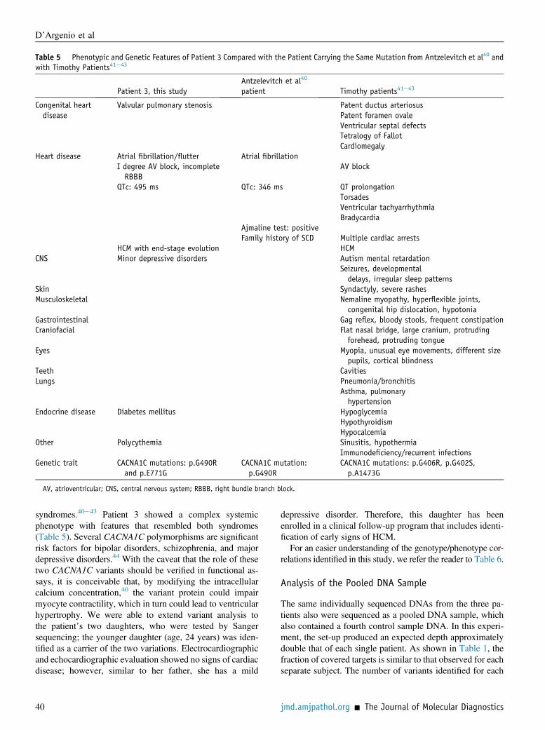
Table 5 Phenotypic and Genetic Features of Patient 3 Compared with the Patient Carrying the Same Mutation from Antzelevitch et al40 andwith Timothy Patients41e43
Patient 3, this studyAntzelevitch et al40
patient Timothy patients41e43
Congenital heartdisease
Valvular pulmonary stenosis Patent ductus arteriosusPatent foramen ovaleVentricular septal defectsTetralogy of FallotCardiomegaly
Heart disease Atrial fibrillation/flutter Atrial fibrillationI degree AV block, incompleteRBBB
AV block
QTc: 495 ms QTc: 346 ms QT prolongationTorsadesVentricular tachyarrhythmiaBradycardia
Ajmaline test: positiveFamily history of SCD Multiple cardiac arrests
HCM with end-stage evolution HCMCNS Minor depressive disorders Autism mental retardation
Seizures, developmentaldelays, irregular sleep patterns
Skin Syndactyly, severe rashesMusculoskeletal Nemaline myopathy, hyperflexible joints,
congenital hip dislocation, hypotoniaGastrointestinal Gag reflex, bloody stools, frequent constipationCraniofacial Flat nasal bridge, large cranium, protruding
forehead, protruding tongueEyes Myopia, unusual eye movements, different size
pupils, cortical blindnessTeeth CavitiesLungs Pneumonia/bronchitis
Asthma, pulmonaryhypertension
Endocrine disease Diabetes mellitus HypoglycemiaHypothyroidismHypocalcemia
Other Polycythemia Sinusitis, hypothermiaImmunodeficiency/recurrent infections
Genetic trait CACNA1C mutations: p.G490Rand p.E771G
CACNA1C mutation:p.G490R
CACNA1C mutations: p.G406R, p.G402S,p.A1473G
AV, atrioventricular; CNS, central nervous system; RBBB, right bundle branch block.
D’Argenio et al
syndromes.40e43 Patient 3 showed a complex systemicphenotype with features that resembled both syndromes(Table 5). Several CACNA1C polymorphisms are significantrisk factors for bipolar disorders, schizophrenia, and majordepressive disorders.44 With the caveat that the role of thesetwo CACNA1C variants should be verified in functional as-says, it is conceivable that, by modifying the intracellularcalcium concentration,40 the variant protein could impairmyocyte contractility, which in turn could lead to ventricularhypertrophy. We were able to extend variant analysis tothe patient’s two daughters, who were tested by Sangersequencing; the younger daughter (age, 24 years) was iden-tified as a carrier of the two variations. Electrocardiographicand echocardiographic evaluation showed no signs of cardiacdisease; however, similar to her father, she has a mild
40
depressive disorder. Therefore, this daughter has beenenrolled in a clinical follow-up program that includes identi-fication of early signs of HCM.For an easier understanding of the genotype/phenotype cor-
relations identified in this study, we refer the reader to Table 6.
Analysis of the Pooled DNA Sample
The same individually sequenced DNAs from the three pa-tients also were sequenced as a pooled DNA sample, whichalso contained a fourth control sample DNA. In this experi-ment, the set-up produced an expected depth approximatelydouble that of each single patient. As shown in Table 1, thefraction of covered targets is similar to that observed for eachseparate subject. The number of variants identified for each
jmd.amjpathol.org - The Journal of Molecular Diagnostics

Table 6 Mutations Most Likely to Exert a Pathogenic Role in the Patients Analyzed
Patients Chr Gene Mutation type Variation* (inheritance) Amino acid change
1y 14 MYH7 Missense c.976G>C (paternal) p.A326P11 INS-IGF2z Nonsense c.573C>T p.Q172X
2 11 MYBPC3 Splicing site c.3627 þ 2T>A (paternal)11 KCNQ1 Indel-frameshift c.524-534dup (maternal) p.W176Lfs*65
3y 12 CACNA1C Missense c.1781G>A p.G490R12 CACNA1C Missense c.2625A>G p.E771G
*All mutations were at heterozygous state.yBiological mother of patient 1 and parents of patient 3 were not available for molecular testing.zMutation included because it produces a truncated protein.Chr, chromosome.
NGS-DNA Analysis in Cardiomyopathies
subject is shown in Supplemental Table S6. Comparativeanalysis of the identified variants (Table 7) shows that mostallelic variants common to more than one patient also wereidentified in the pool. However, more variants were missedbecause their presence in the eight analyzed alleles decreased.
Discussion
We have successfully applied DNA sequence capture tech-nology followed by NGS to the simultaneous analysis of acombination of genes possibly related to cardiomyopathies.The analysis was focused on a restricted panel of genes thatencode proteins involved in heart functions or that might berelated to the development of cardiomyopathies, by using acustom array to simultaneously enrich 202 genes beforeNGS, thereby increasing the effectiveness of HCM molec-ular diagnosis over currently available procedures.
Limiting the analysis to eight sarcomeric genes previouslyanalyzed by a combined DHPLC/Sanger approach,8 all of theSanger-confirmed variants previously identified in patients 1and 2 (Supplemental Table S5) were confirmed by the NGSprocedure described herein except one. With the describedprocedure, we also detected 21 variants that were missed bythe previous DHPLC screening (Supplemental Table S7). Toevaluate the reliability of the new NGS-based method in
Table 7 Analysis of Nonreference (Variant) Alleles Found inSingle-Subject Sequencing Experiments and Also Identified inthe Pool
Alleles
Pool variants
Expected* Found Missed
8/8 683 677 67/8 353 353 06/8 669 659 105/8 717 695 224/8 1182 1077 1053/8 1090 966 1242/8 2976 1756 12201/8 4414 1705 2709
Variants missed in the pool (last column) increased when they were readon fewer alleles.
*Expected in the pool from the analysis performed in the individualpatient samples.
The Journal of Molecular Diagnostics - jmd.amjpathol.org
detecting variants, we compared its sensitivity and specificitywith those of the previously used DHPLC/Sanger method (seeMaterials and Methods and Results). As shown in Table 4,NGS was very sensitive and specific in detecting mutations,whereas the sensitivity of the DHPLC/Sanger procedure wasmuch lower (about 50% to 60%). These low sensitivity valuesdepend on the experimental design used for the DHPLC/Sanger approach,whichwas designed to detect variantswithinthe exon sequences, and is expected to be less sensitive, forexample, in highlighting changes in the untranslated regions orclose to the ends of the analyzed segments e a limitation thatdoes not apply to the NGS approach. In fact, when variantsexpected to be undetectable by DHPLC/Sanger sequencing(ie, variants outside amplicons and homozygous variants), arenot taken into account in calculating sensitivity, the resultsbecome comparable with those of other reports,34 althoughstill remaining worse than the new NGS-based procedure.
The considerations described earlier only refer to a smallproportion (1%) of the entire target region analyzed by theNGS-based method. Unlike the DHPLC method, the NGSprocedure produces results on a set of 202 genes, corre-sponding to about 5 million bases of the coding genomicsequence. It is noteworthy that the performance of the NGS-based method is better on the limited 8-gene set. As veryrecently stated elsewhere,45 it remains to be establishedwhether the diagnostic output of a test that detects a largenumber of variants in a relatively large number of genesmight be compared with a test that detects almost all vari-ants, but in a low number of genes.
In diagnostic terms, the high-throughput sequencingprocedure enabled us to accomplish the following: i) iden-tify a known HCM-causative mutation in patient 1, whowent previously undiagnosed; ii) better delineate the mo-lecular alteration underlying the phenotype of patient 2 bydetecting the presence in this HCM subject of a mutationcausative of long QT syndrome39,46; and iii) postulate thatthe HCM observed in patient 3 was due to CACNA1C-related mutations (Timothy syndrome41e43).
Although the bioinformatic tools we used eliminated var-iants that are most likely bereft of clinical significance, thereal pathogenic role of a given variant can be determined onlywith functional studies and/or family segregation analysisto evaluate genotype-phenotype correlations within each
41

D’Argenio et al
family. The variants we identified in the present work(Table 6) should be classified as possibly pathogenic becausethey fulfill at least one of the following conditions: i) previousfindings showing that they are disease-causing (four of them);ii) their presence on genes known to cause HCM (two); andiii) the presence of stop codons indicating a truncated protein(one) (Table 6). Further procedures are of course necessary toconfirm the causative origin of mutations to better rationalizethe genotype-phenotype relationship within each family andto better understand the molecular basis of the alterationinduced by each mutational event. The annotations and theprioritization strategy described herein could help to simplifythese steps.
Sequencing of several patients in a single run is an addi-tional improvement over the conventional Sanger procedurein terms of time and sequencing throughput. We applied ourprocedure to a pool containing DNA from the three patientsto evaluate the feasibility of sequencing several patientssimultaneously. Target enrichment and sequencing coveragewere highly consistent with the values found in the threeseparate samples. However, not all variants detected in theindividual DNA samples were identified in the pool (seeResults and Table 7), as expected, considering the reducedoverall sequence depth. The sensitivity of the pool-basedapproach easily may be enhanced by increasing thesequencing depth while still saving on the costs and workloadassociated with library preparation and titration. The use oftags to selectively label the DNA of single patients in mul-tiplexing experiments would have the advantage of univo-cally assigning the sequencing reads to each patient at thecost of a slightly more complex experiment. We have per-formed this successfully in similar experiments.
Taken together, these considerations indicate that theNGS-based procedure described here easily could be applied forroutine diagnostic purposes in this group of rather frequentimportant human diseases. Further studies will be useful tooptimize costs and sensitivity of the various approaches.
When, after more than 2 years of study, our experimentalwork was completed,47 and this manuscript was undergoinginternal editing, Meder et al48 reported a study conductedusing an approach similar to ours, but with a different NGStechnology. Their article was accompanied by a thought-provoking editorial regarding the significance and diag-nostic relevance of this technology.49 Our study reinforcesthe concept that NGS techniques open a new era in thesearch for new disease-causing genes and/or novel modifiergenes as appears to be the case in our patients 2 and 3. Italso highlights the profound complexity of inherited heartdisorders and their different phenotype expressions, whichfurther supports the concept that the worlds of cardiomy-opathies and ion channel diseases could be linked.50
In conclusion, by examining a large set of genes, and notonly genes strictly related to HCM, we identified moleculardefects that would have eluded detection by previous tech-niques and, thus, we were able to initiate personalized moni-toring and therapy in the patients and in their relatives. Our
42
findings, together with several other reports,13,14,29e31,51e56
indicate that NGS will become increasingly important forthe molecular diagnostics of complex inherited diseases suchas cardiomyopathies.
Acknowledgments
We thank Jean Ann Gilder (Scientific Communication Srl)for editing the text and Vittorio Lucignano (CEINGEeBiotecnologie Avanzate) for technical assistance related tographics.
Supplemental Data
Supplemental material for this article can be found athttp://dx.doi.org/10.1016/j.jmoldx.2013.07.008.
References
1. Maron BJ, Gardin JM, Flack JM, Gidding SS, Kurosaki TT, Bild DE:Prevalence of hypertrophic cardiomyopathy in a general population ofyoung adults. Echocardiographic analysis of 4111 subjects in theCARDIA Study. Coronary Artery Risk Development in (Young)Adults. Circulation 1995, 92:785e789
2. Fatkin D, Graham RM: Molecular mechanisms of inherited cardio-myopathies. Physiol Rev 2002, 82:945e980
3. Maron BJ: Hypertrophic cardiomyopathy: a systematic review.JAMA 2002, 287:1308e1320
4. Marian AJ, Roberts R: The molecular genetic basis for hypertrophiccardiomyopathy. J Mol Cell Cardiol 2001, 33:655e670
5. Seidman JG, Seidman C: The genetic basis for cardiomyopathy: frommutation identification to mechanistic paradigms. Cell 2001, 104:557e567
6. Tester DJ, Ackerman MJ: Cardiomyopathic and channelopathiccauses of sudden, unexplained death in infants and children. AnnuRev Med 2009, 60:69e84
7. Elliott P, Andersson B, Arbustini E, Bilinska Z, Cecchi F, Charron P,Dubourg O, Kühl U, Maisch B, McKenna WJ, Monserrat L,Pankuweit S, Rapezzi C, Seferovic P, Tavazzi L, Keren A: Classifi-cation of the cardiomyopathies: a position statement from the Euro-pean Society of Cardiology Working Group on Myocardial andPericardial Diseases. Eur Heart J 2008, 29:270e276
8. Frisso G, Limongelli G, Pacileo G, Del Giudice A, Forgione L,Calabrò P, Iacomino M, Detta N, Di Fonzo LM, Maddaloni V,Calabrò R, Salvatore F: A child cohort study from southern Italyenlarges the genetic spectrum of hypertrophic cardiomyopathy. ClinGenet 2009, 76:91e101
9. Poliac LC, Barron ME, Maron BJ: Hypertrophic cardiomyopathy.Anesthesiology 2006, 104:183e192
10. Smith MG, Gianoulis TA, Pukatzki S, Mekalanos JJ, Ornston LN,Gerstein M, Snyder M: New insights into Acinetobacter baumanniipathogenesis revealed by high-density pyrosequencing and trans-poson mutagenesis. Genes Dev 2007, 21:601e614
11. Turnbaugh PJ, Ley RE, Mahowald MA, Magrini V, Mardis ER,Gordon JI: An obesity-associated gut microbiome with increasedcapacity for energy harvest. Nature 2006, 444:1027e1031
12. Thomas RK, Nickerson E, Simons JF, Jänne PA, Tengs T, Yuza Y,Garraway LA, LaFramboise T, Lee JC, Shah K, O’Neill K, Sasaki H,Lindeman N, Wong KK, Borras AM, Gutmann EJ, Dragnev KH,DeBiasi R, Chen TH, Glatt KA, Greulich H, Desany B, Lubeski CK,Brockman W, Alvarez P, Hutchison SK, Leamon JH, Ronan MT,Turenchalk GS, Egholm M, Sellers WR, Rothberg JM, Meyerson M:
jmd.amjpathol.org - The Journal of Molecular Diagnostics

NGS-DNA Analysis in Cardiomyopathies
Sensitive mutation detection in heterogeneous cancer specimens bymassively parallel picoliter reactor sequencing. Nat Med 2006, 12:852e855
13. Albert TJ, Molla MN, Muzny DM, Nazareth L, Wheeler D, Song X,Richmon TA, Middle CM, Rodesch MJ, Packard CJ, Weinstock GM,Gibbs RA: Direct selection of human genomic loci by microarrayhybridization. Nat Methods 2007, 11:903e905
14. Chou LS, Liu CS, Boese B, Zhang X, Mao R: DNA sequence captureand enrichment by microarray followed by next-generationsequencing for targeted resequencing: neurofibromatosis type 1gene as a model. Clin Chem 2010, 56:62e72
15. Lin AE, Alexander ME, Colan SD, Kerr B, Rauen KA, Noonan J,Baffa J, Hopkins E, Sol-Church K, Limongelli G, Digilio MC,Marino B, Innes AM, Aoki Y, Silberbach M, Delrue MA, White SM,Hamilton RM, O’Connor W, Grossfeld PD, Smoot LB, Padera RF,Gripp KW: Clinical, pathological, and molecular analyses of car-diovascular abnormalities in Costello syndrome: a Ras/MAPKpathway syndrome. Am J Med Genet A 2011, 155A:486e507
16. Christensen G, Chen J, Ross J, Chien KR: Mouse models of humancardiovascular disease. Edited by Chien KR. Molecular basis ofcardiovascular disease. Philadelphia, Elsevier, 2004, pp 72e106
17. Barrans JD, Allen PD, Stamatiou D, Dzau VJ, Liew CC: Global geneexpression profiling of end-stage dilated cardiomyopathy using ahuman cardiovascular-based cDNA microarray. Am J Pathol 2002,160:2035e2043
18. Frey N, Olson EN: Cardiac hypertrophy: the good, the bad, and theugly. Annu Rev Physiol 2003, 65:45e79
19. Carreno JE, Apablaza F, Ocaranza MP, Jalil JE: Cardiac hypertrophy:molecular and cellular events. Rev Esp Cardiol 2006, 59:473e486
20. Detta N, Frisso G, Zullo A, Sarubbi B, Cozzolino C, Romeo E,Wang DW, Calabrò R, Salvatore F, George AL Jr.: Novel deletionmutation in the cardiac sodium channel inactivation gate causes longQT syndrome. Int J Cardiol 2013, 165:362e365
21. Sarubbi B, Frisso G, Romeo E, Evangelista E, Cordella A, D’Alto M,Santarpia G, Russo MG, Salvatore F, Calabrò R: Efficacy of phar-macological treatment and genetic characterization in early diagnosedpatients affected by long QT syndrome with impaired AV conduction.Int J Cardiol 2011, 149:109e113
22. Ning Z, Cox AJ, Mullikin JC: SSAHA: a fast search method for largeDNA databases. Genome Res 2001, 11:1725e1729
23. Margulies M, Egholm M, Altman WE, Attiya S, Bader JS,Bemben LA, et al: Genome sequencing in microfabricated high-density picolitre reactors. Nature 2005, 437:376e380
24. Martin ER, Kinnamon DD, Schmidt MA, Powell EH, Zuchner S,Morris RW: SeqEM: an adaptive genotype-calling approach for next-generation sequencing studies. Bioinformatics 2010, 26:2803e2810
25. Gilles A, Meglécz E, Pech N, Ferreira S, Malausa T, Martin JF:Accuracy and quality assessment of 454 GS-FLX Titanium pyrose-quencing. BMC Genomics 2011, 12:245
26. McLaren W, Pritchard B, Rios D, Chen Y, Flicek P, Cunningham F:Deriving the consequences of genomic variants with the EnsemblAPI and SNP effect predictor. BMC Bioinformatics 2010, 26:2069e2070
27. The 1000 Genomes Project Consortium: A map of human genomevariation from population-scale sequencing. Nature 2010, 467:1061e1073
28. Hedges DJ, Burges D, Powell E, Almonte C, Huang J, Young S,Boese B, Schmidt M, Pericak-Vance MA, Martin E, Zhang X,Harkins TT, Züchner S: Exome sequencing of a multigenerationalhuman pedigree. PLoS One 2009, 4:e8232
29. Wei Z, Wang W, Hu P, Lyon GJ, Hakonarson H: SNVer: a statisticaltool for variant calling in analysis of pooled or individual next-generation sequencing data. Nucleic Acids Res 2011, 39:e132
30. Amstutz U, Andrey-Zürcher G, Suciu D, Jaggi R, Häberle J,Largiadèr CR: Sequence capture and next-generation resequencing ofmultiple tagged nucleic acid samples for mutation screening of ureacycle disorders. Clin Chem 2011, 57:102e111
The Journal of Molecular Diagnostics - jmd.amjpathol.org
31. Ghosh S, Krux F, Binder V, Gombert M, Niehues T, Feyen O,Laws HJ, Borkhardt A; PID-NET: German Network on PrimaryImmunodeficiency Diseases: Array-based sequence capture and next-generation sequencing for the identification of primary immunodefi-ciencies. Scand J Immunol 2012, 75:350e354
32. Neveling K, Collin RW, Gilissen C, van Huet RA, Visser L,Kwint MP, Gijsen SJ, Zonneveld MN, Wieskamp N, de Ligt J,Siemiatkowska AM, Hoefsloot LH, Buckley MF, Kellner U,Branham KE, den Hollander AI, Hoischen A, Hoyng C,Klevering BJ, van den Born LI, Veltman JA, Cremers FP, Scheffer H:Next-generation genetic testing for retinitis pigmentosa. Hum Mutat2012, 33:963e972
33. Raca G, Jackson C, Warman B, Bair T, Schimmenti LA: Next gen-eration sequencing in research and diagnostics of ocular birth defects.Mol Genet Metab 2010, 100:184e192
34. Chou LS, Lyon E, Wittwer CT: A comparison of high-resolutionmelting analysis with denaturing high-performance liquid chroma-tography for mutation scanning: cystic fibrosis transmembraneconductance regulator gene as a model. Am J Clin Pathol 2005, 124:330e338
35. Michels M, Soliman OI, Phefferkorn J, Hoedemaekers YM,Kofflard MJ, Dooijes D, Majoor-Krakauer D, Ten Cate FJ: Diseasepenetrance and risk stratification for sudden cardiac death inasymptomatic hypertrophic cardiomyopathy mutation carriers. EurHeart J 2009, 30:2593e2598
36. Tait KF, Collins JE, Heward JM, Eaves I, Snook H, Franklyn JA,Barnett AH, Todd JA, Maranian M, Compston A, Sawcer S,Gough SC: Evidence for a type 1 diabetes-specific mechanism for theinsulin gene-associated IDDM2 locus rather than a general influenceon autoimmunity. Diabet Med 2004, 21:267e270
37. Prakash T, Sharma VK, Adati N, Ozawa R, Kumar N, Nishida Y,Fujikake T, Takeda T, Taylor TD: Expression of conjoined genes:another mechanism for gene regulation in eukaryotes. PLoS One2010, 5:e13284
38. Fendler W, Klich I, Cieœlik-Heinrich A, Wyka K, Szadkowska A,Młynarski W: Increased risk of type 1 diabetes in Polish children -association with INS-IGF2 50VNTR and lack of association withHLA haplotype. Endokrynol Pol 2011, 62:436e442
39. Lupoglazoff JM, Denjoy I, Villain E, Fressart V, Simon F, Bozio A,Berthet M, Benammar N, Hainque B, Guicheney P: Long QT syn-drome in neonates: conduction disorders associated with HERGmutations and sinus bradycardia with KCNQ1 mutations. J Am CollCardiol 2004, 43:826e830
40. Antzelevitch C, Pollevick GD, Cordeiro JM, Casis O, Sanguinetti MC,Aizawa Y, Guerchicoff A, Pfeiffer R, Oliva A, Wollnik B, Gelber P,Bonaros EP Jr., Burashnikov E, Wu Y, Sargent JD, Schickel S,Oberheiden R, Bhatia A, Hsu LF, Haïssaguerre M, Schimpf R,Borggrefe M, Wolpert C: Loss-of-function mutations in the car-diac calcium channel underlie a new clinical entity characterizedby ST-segment elevation, short QT intervals, and sudden cardiacdeath. Circulation 2007, 115:442e449
41. Bidaud I, Lory P: Hallmarks of the channelopathies associated withL-type calcium channels: a focus on the Timothy mutations in Ca(v)1.2 channels. Biochimie 2011, 93:2080e2086
42. Splawski I, Timothy KW, Decher N, Kumar P, Sachse FB,Beggs AH, Sanguinetti MC, Keating MT: Severe arrhythmia disordercaused by cardiac L-type calcium channel mutations. Proc Natl AcadSci U S A 2005, 102:8089e8096
43. Splawski I, Timothy KW, Sharpe LM, Decher N, Kumar P, Bloise R,Napolitano C, Schwartz PJ, Joseph RM, Condouris K, Tager-Flusberg H, Priori SG, Sanguinetti MC, Keating MT: Ca(V)1.2 cal-cium channel dysfunction causes a multisystem disorder includingarrhythmia and autism. Cell 2004, 119:19e31
44. Roussos P, Giakoumaki SG, Georgakopoulos A, Robakis NK,Bitsios P: The CACNA1C and ANK3 risk alleles impact on affectivepersonality traits and startle reactivity but not on cognition or gatingin healthy males. Bipolar Disord 2011, 13:250e259
43

D’Argenio et al
45. Ware JS, John S, Roberts AM, Buchan R, Gong S, Peters NS,Robinson DO, Lucassen A, Behr ER, Cook SA: Next generationdiagnostics in inherited arrhythmia syndromes: a comparison of twoapproaches. J Cardiovasc Trans Res 2013, 6:94e103
46. Chiang CE, Roden DM: The long QT syndromes: genetic basis andclinical implications. J Am Coll Cardiol 2000, 36:1e12
47. D’Argenio V, Frisso G, Limongelli G, Precone V, Pacileo G,Fienga A, Boccia A, Calabrò R, Paolella G, Salvatore F: DNAsequence capture and high throughput sequencing technology: anovel approach to identify a large number of hypertrophiccardiomyopathy-causing genes. Circulation 2010, 122:A19602
48. Meder B, Haas J, Keller A, Heid C, Just S, Borries A, Boisguerin V,Scharfenberger-Schmeer M, Stähler P, Beier M, Weichenhan D,Strom TM, Pfeufer A, Korn B, Katus HA, Rottbauer W: Targetednext-generation sequencing for the molecular genetic diagnostics ofcardiomyopathies. Circ Cardiovasc Genet 2011, 4:110e122
49. Bagnall RD, Ingles J, Semsarian C: Molecular diagnostics of cardio-myopathies the future is here. Circ Cardiovasc Genet 2011, 4:103e104
50. Monserrat L, Mazzanti A, Ortiz-Genga M, Barriales-Villa R, Garcia-Giustiniani D, Gimeno-Blanes JR: The interpretation of genetic testsin inherited cardiovascular diseases. Cardiogenetics 2011, 1:e8
51. Lalonde E, Albrecht S, Ha KC, Jacob K, Bolduc N, Polychronakos C,Dechelotte P, Majewski J, Jabado N: Unexpected allelic hetero-geneity and spectrum of mutations in Fowler syndrome revealedby next-generation exome sequencing. Hum Mutat 2010, 31:918e923
44
52. Ng SB, Bigham AW, Buckingham KJ, Hannibal MC, McMillin MJ,Gildersleeve HI, Beck AE, Tabor HK, Cooper GM, Mefford HC,Lee C, Turner EH, Smith JD, Rieder MJ, Yoshiura K, Matsumoto N,Ohta T, Niikawa N, Nickerson DA, Bamshad MJ, Shendure J: Exomesequencing identifies MLL2 mutations as a cause of Kabuki syn-drome. Nat Genet 2010, 42:790e793
53. Ng SB, Buckingham KJ, Lee C, Bigham AW, Tabor HK, Dent KM,Huff CD, Shannon PT, Jabs EW, Nickerson DA, Shendure J,Bamshad MJ: Exome sequencing identifies the cause of a mendeliandisorder. Nat Genet 2010, 42:30e35
54. Shearer AE, DeLuca AP, Hildebrand MS, Taylor KR, Gurrola J 2nd,Scherer S, Scheetz TE, Smith RJ: Comprehensive genetic testing forhereditary hearing loss using massively parallel sequencing. Proc NatlAcad Sci U S A 2010, 107:21104e21109
55. Bowne SJ, Sullivan LS, Koboldt DC, Ding L, Fulton R, Abbott RM,Sodergren EJ, Birch DG, Wheaton DH, Heckenlively JR, Liu Q,Pierce EA, Weinstock GM, Daiger SP: Identification of disease-causing mutations in autosomal dominant retinitis pigmentosa(adRP) using next-generation DNA sequencing. Invest OphthalmolVis Sci 2011, 52:494e503
56. Guelly C, Zhu PP, Leonardis L, Papi�c L, Zidar J, Schabhüttl M,Strohmaier H, Weis J, Strom TM, Baets J, Willems J, De Jonghe P,Reilly MM, Fröhlich E, Hatz M, Trajanoski S, Pieber TR, Janecke AR,Blackstone C, Auer-Grumbach M: Targeted high-throughputsequencing identifies mutations in atlastin-1 as a cause of hereditarysensory neuropathy type I. Am J Hum Genet 2011, 88:99e105
jmd.amjpathol.org - The Journal of Molecular Diagnostics


