Embed Size (px)
Citation preview
Short communication
Does Vitamin E or Vitamin E plus Selenium
improve reproductive performance of rams during hot weather?
Ammar Bin Talib Ali, Giovanni Bomboi, Basilio Floris
Dipartimento di Biologia Animale. Università di Sassari, Italy
Corresponding author: Dr. Ali Ammar Bin Talib. Dipartimento di Biologia Animale. Facoltà di Medicina Veterinaria, Università degli Studi di Sassari. Via Vienna 2, 07100 Sassari, Italy - Tel. + 39 079 229 428 - Fax: + 39 079 229 429 - Email: [email protected]
Received September 23, 2008; accepted March 19, 2009
ABSTRACT
The objective of this study was to determine the effect of Vitamin E (Vit E) and Selenium (Se) on semen quality, sexual activity, packed cell volume (PCV), and white blood cell counts (WBC) of Awassi rams du-ring the hot season.Twelve Awassi rams were subdivided into three groups and treated for 90 days. Rams in the 1st group (T1) were treated twice weekly with 175 mg/ram vitamin E at intervals of 12h; rams in the 2nd group (T2) were treated on the same schedule with 70 mg/ram vitamin E plus 2800 mg selenium; and rams in 3rd group (T3) served as controls. Sperm quality (percentage of motile cells and percentage of morphologically normal cells) and quantity (sperm volume, and concentration) were recorded weekly. Sexual activity was tested by using the pen libido test at monthly intervals. Blood samples were taken before treatment and after the 1st, 2nd, and 3rd months. Semen quality was significantly affected by treatments: the ejaculate volume increased in T2 vs T1, and T3; mass activity and individual motility were increased in T1 and T2 vs T3. Sperm concentration was increased in T2 and T1 vs T3, and the percentage of dead and abnormal spermatozoa was reduced in T1 and T2 vs T3, though the differences were not statistically significant. Pen libido test showed reduced reaction time for the first mount in the 1st month in T2 and T1 vs T3. The number of serves was increased in groups T1 and T2 vs T3. There were no significant differences in PCV among groups. After 3 months from the beginning of treatments, the percentage of lymphocytes increased in T1 and T2 vs. T3, while the percentage of neutrophils was reduced in T2 vs T3 in the 2nd and 3rd months.The results of this experiment indicate that treatments with vitamin E alone or in combination with se-lenium improved semen characteristics and reproductive performance of Awassi rams during the hot season.
Key words: Rams, Reproductive Performance, Selenium, Semen Characteristics, Vitamin E.
Ital.J.anIm.ScI. vol. 8, 743-754, 2009 743
Ital.J.anIm.ScI. vol. 8, 743-754, 2009744
alI et al.
RIASSUNTO
PUò lA VITAmINA E, DA SOlA O CON SElENIO, mIGlIORARE lE PRESTAzIONI RIPRODUTTIVE DEGlI ARIETI DURANTE lA STAGIONE CAlDA?
Lo scopo di questo studio é stato quello di determinare l’effetto di Vitamina E (Vit E) e Selenio (Se) su qualità del seme, attività sessuale, valore ematocrito (PCV) e conteggio di leucociti (WBC) in arieti Awassi durante la stagione calda.Dodici arieti Awassi vennero suddivisi in tre gruppi e sottoposti a trattamento per 90 giorni. Gli arieti del 1° gruppo (T1) furono trattati due volte alla settimana con 175 mg/ariete di vitamina E con un intervallo di 12h, gli arieti del 2° gruppo (T2) furono trattati allo stesso modo con 70 mg/ariete di vitamina E e 2800 mg di selenio, mentre gli arieti del 3° gruppo (T3) servirono da controllo. La qualità del seme (percentuale di cellule mobili e percentuale di cellule morfologicamente normali) e la quantità (volume e concentra-zione spermatica) vennero registrate settimanalmente. L’attività sessuale venne valutata tramite “pen libido test” a cadenza mensile. Campioni di sangue vennero prelevati prima del trattamento e dopo il 1°, 2° e 3° mese. La qualità del seme venne significativamente influenzata dai trattamenti, con incremento del volume di eiaculato in T2 vs T1 e T3, di attività massale e di motilità individuale in T1 e T2 vs T3. La concentrazione spermatica incrementò in T2 e T1 vs T3, e la percentuale di spermatozoi morti e anormali si ridusse in T1 e T2 vs T3, sebbene le differenze non siano risultate statisticamente significative. Il “pen libido test” mostrò un ridotto tempo di reazione alla prima monta nel 1° mese in T2 e T1 vs T3. Il numero di accoppiamenti incrementò nei gruppi T1 e T2 vs T3. Non vi furono significative differenze tra i gruppi per quanto concerne il PCV. Dopo 3 mesi di trattamento, la percentuale di linfociti risultò aumentata in T1 e T2 vs T3, mentre la percentuale di neutrofili si ridusse in T2 vs T3 nel 2° e 3° mese.I risultati di questo esperimento indicano che i trattamenti con vitamina E, da sola o in combinazione con selenio, migliorano le caratteristiche del seme e la performance riproduttiva di arieti Awassi durante la stagione calda.
Parole chiave: Arieti, Prestazioni Riproduttive, Selenio, Caratteristiche Seminali, Vitamina E.
Introduction
Sheep are important animals in many countries because they can be successfully raised under harsh conditions and cost re-latively little to maintain. The Awassi sheep is the most numerous and widespread type of sheep in southwest Asia, and has admira-bly adapted to the rigorous conditions found there. The libido and semen quality vary with season, and usually decline from May through August when the climate is dry, very hot and skies are cloudless.
Vitamin E (Vit E) and Selenium (Se) are two of the important nutrients that can af-fect several biological processes including spermatogenesis and semen quality (Marin-Guzman et al., 1997; Yousef et al., 2003), re-production (Koyuncu and Yerlikaya, 2007), metabolism (Awadeh et al., 1998), immunity
(Hernken et al., 1998), and protecting again-st oxidative stress (Bernabucci et al., 2002).
Selenium (Se) is an essential dietary tra-ce element and is always of research interest because it is required for the maintenance of male fertility. This element is also required for testosterone biosynthesis and the forma-tion and normal development of spermatozoa (Brown and Arthur, 2001). Both testis and epididymis require exogenously supplied Se in order to synthesize a variety of known se-lenoproteins, whose precise role in spermio-genesis and post testicular sperm maturation are not clearly defined. In many areas of the world plants do not provide levels of this ele-ment adequate to meet dietary requirements (Hogan et al., 1993). Insufficient dietary Se leads to oxidation, rupture of muscle mem-branes and leakage of cellular enzymes into circulation, and the animals differ in their re-
Ital.J.anIm.ScI. vol. 8, 743-754, 2009 745
vItamIn E, SElEnIum and ram pErformancE
quirements for Se and in their susceptibility. Moreover, deficiency in Vit E and Se in sheep flocks can lead to stiff lamb disease or nutri-tional muscular dystrophy.
Vitamin E compounds in plant tissues are a mixture of four tocopherols and four toco-trienols, both forms in the “D” configuration, of which D-α-tocopherol (RRR- α-tocophe-rol) has the highest biological activity in animals. These eight isomers constitute the Vit E product commonly known as DL-α-to-copherol, also denoted as all rac-α-tocophe-rol (Mahan et al., 2000). The association of Vit E deficiency with impaired male repro-duction was established more than three decades ago, and traditionally it is called the “Anti-sterility Vitamin”. Nutritional de-ficiency of Vit E has also been linked to liver necrosis in swine and rats, foetal resorption in rats, and encephalomalacia in poultry.
Furthermore, many studies have been in-vestigated for their effects on immunological responses in various animal species. In mice and chickens, Spallholz et al., (1975) demon-strated that both dietary and injectable Se enhanced the primary immune response, and increased serum IgG antibody titers in mice supplemented with 60 to 180 mg Vit E/kg diet and antigenically challenged with sheep red blood cells (RBC) or tetanus toxoid. Similarly, Smith et al. (1979) reported the di-rect evidence that Vit E and Se are related to mammary health. Vit E has been impli-cated in stimulation of antibody synthesis, particularly IgG. Reffett et al. (1988) demon-strated that Se supplementation of lambs enhanced serum IgM (P<0.05) and mean immunoglobulin concentration by increasing the number of IgM-producing cells, and Vit E supplementation appeared to stimulate the secondary immune response, generally asso-ciated with IgG antibody production.
Several researchers supported the hypothesis that seasonal alterations of the oxidative status depend more on weather
conditions such as temperature and humidi-ty (Bernabucci et al., 2002). Oxidative stress resulting from increased production of free radicals and reactive oxygen species, and/or a decrease in antioxidant defences, leads to damage of biological macromolecules and disruption of normal metabolism and phy-siology, and when reactive forms of oxygen are produced faster than they can safely be neutralized by antioxidant mechanisms, oxi-dative stress results (Tervisan et al., 2001). In response to heat stress, rams secrete ACTH from the pituitary gland which cau-ses increased adrenal cortisol release into the circulation, and the stress augments the autonomic nervous system release of nore-pinephrine and the adrenal release of nore-pnephrine and epinephrine into circulation. Studies on the effect of heat stress on oxida-tive status in ovine are lacking.
Sconberg et al. (1993) demonstrated that stressed rams or rams given a stress treat-ment of ACTH and epinephrine injections were found to have reduced circulating neutrophils and RBC, and reduced plasma α-tocopherol levels. Similarity, Nockels et al. (1996) demonstrated that stressed cat-tle may have reductions in α-tocopherol concentrations in certain tissues, and sup-plemental Vit E may be required to restore α-tocopherol in tissues following stress, and Calamari et al. (1999) observed negative ef-fects of heat stress on some plasma markers of oxidative status in midlacting cows. Whi-le, Zhu and Setchell (2004) demonstrated that increasing the temperature of testes causes a derangement in the spermatoge-nesis, which leads to a decrease in sperm number, motility and normality, and redu-ces the ability of sperm to produce normal offspring. Both Vit E and Se-containing enzyme glutathione peroxides (GPx4), are an integral part of the antioxidant system present in most mammalian cells, and play an important role in protecting cells again-
Ital.J.anIm.ScI. vol. 8, 743-754, 2009746
alI et al.
st oxidative stress and toxic agents (Flohè, 2007). Furthermore, Burk et al. (2007) ob-served a strong association of low sperm GPx4 with infertility.
To date, there have no long-term studies evaluating the connection between Vit E, Se and hot weather stress on reproductive performance and hematologic parameters in Awassi rams. Therefore, the aim of this experiment was to evaluate the effects of Vit E alone or in combination with Se on chan-ges in semen quality, sexual activity, and some hematologic parameters caused by hot weather stress in Awassi rams.
Material and methods
Animal and semen collectionThe experiment was conducted from May
through July (in Iraq, the hot season runs from May to October) and air temperatu-res hovered between 43-54°C in the shade (Source: U.S. National Climatic Data Center, Asheville, N.C). On a farm located in the Agriculture College/Baghdad University, twelve adult Iraqi Awassi rams (63±2.2 kg BW, and 30 - 36 mo of age) were kept in semi open pens. The daily hours devoted to ou-tdoor feeding - related activity supplemented with hay and some barley were randomly as-signed to three groups. Rams in the 1st group (T1) were injected twice a week 175 mg/ram Vit E at intervals of 12h, whereas rams in the 2nd group (T2) were injected twice a week with 70 mg Vit E plus 2800 mg Se /ram at in-tervals of 12h. Rams in 3rd group (T3) served as controls. Semen was collected using an artificial vagina at weekly intervals, and the ejaculates were immediately immersed in a warm water bath at 35°C until their assess-ment. Semen assessment was performed in approximately 25 min.
Analysis of standard semen parametersSemen samples were analyzed for volu-
me, pH, colour, and concentration. Progres-sive sperm motility was subjectively evalua-ted by using the standard method. Motility estimations were carried out from three different microscope fields in each sample at 35°C. The mean of the three estimations was used as final motility score. The viabi-lity of spermatozoa in samples was asses-sed by mean of the nigrosen - eosin stain method. The final composition of the stain was eosin - Y 1.67 g, and sodium citrate 2.9 g, dissolved in 100 ml distilled water.
Sperm suspension smears were prepared by mixing a drop of sperm sample with two drops of stain on a warm slide and sprea-ding the stain with a second slide; viability was assessed by counting at least 200 cells under phase-contrast at 1000x magnification. Sperm displaying partial or complete purple staining were considered nonviable; only sperm showing strict exclusion of stain were counted as viable. For abnormal sperm asses-sment, at least three drops of each sample were added to Eppendorf tubes containing 1 ml of Hancock solution [62.5 ml formalin (37%), 150 ml sodium saline solution, 150 ml buffer solution, and 500 ml of double-distilled water]. One drop of this mixture was put on a slide and covered with a cover slip. The per-centage of total sperm abnormality (acroso-mal abnormality, detached heads, abnormal mid-pieces and tail defects) was determined by counting at least 200 spermatozoa under phase contrast microscopy (magnification 1000 x, oil immersion).
Sexual activity and blood sample testIn the pen and flock mating test Iraqi
Awassi ewes ≥2.5 years old were used. Oe-strus was induced in these ewes by two injec-tions of 25 mg progesterone on d 1 and 3, and an injection of 200 μg oestradiol benzoate on d 5. Ewes displayed behavioural oestrus 18 - 22 h after the injection of oestradiol and re-mained in oestrus for at least 27h.
Ital.J.anIm.ScI. vol. 8, 743-754, 2009 747
vItamIn E, SElEnIum and ram pErformancE
In the sexual activity test, each ram was placed in a pen (16 m2) with four oestrus ewes, and usually each mount serve timed close to 5 sec. Mounts were defined as the ram straddling and clasping the ewe, and contacting her rump with his brisket. A ser-ve was defined as a mount accompanied by intromission and ejaculation, characterized by distinct pelvic thrust with head thrown back, followed by a short period during whi-ch the ram showed no interest in the ewes. All rams were given a 20 min pen mating test; reaction time for the 1st mount, time for 1st service, number of mounts, and number of services were recorded. In all groups, the test was carried out at the end of month 1, 2, 3, and before treatments.
Blood samples were taken from the jugu-lar veins of all rams before treatment and at the end of month 1, 2, and 3. The PCV, and WBC counts were measured on all animals.
Statistical analysisThe results were expressed as Ls mean
±SE. The effects of the treatment on semen quality were analyzed by using the PROC MIXED procedure of SAS (1992) according to the following model:
Yijk=µ+Ti+Wj+(TW)ij+Rk+Eijkwhere Yijk is the observation, µ is the
overall mean, Ti is the treatment effect (i=1 and 2), Wj is the time effect (j=1, 2 and 3), (TW)ij is their interaction, Rk is the ram (k=1,2,3 and 4), and Eijk the residual error.
The effects of treatment on sexual activity and hematologic parameters were analyzed by using the general linear model procedu-re of SAS (1992) according to the following model:
Yij=µ+Ti+Rj+Eijwhere Yij is the observation, µ is the ove-
rall mean, Ti is the treatment effect (i=1 and 2), Rj is the ram effect (k=1,2,3 and 4), and Eij the residual error.
Significant differences between means
were tested using the Duncan multiple ran-ge test.
Results
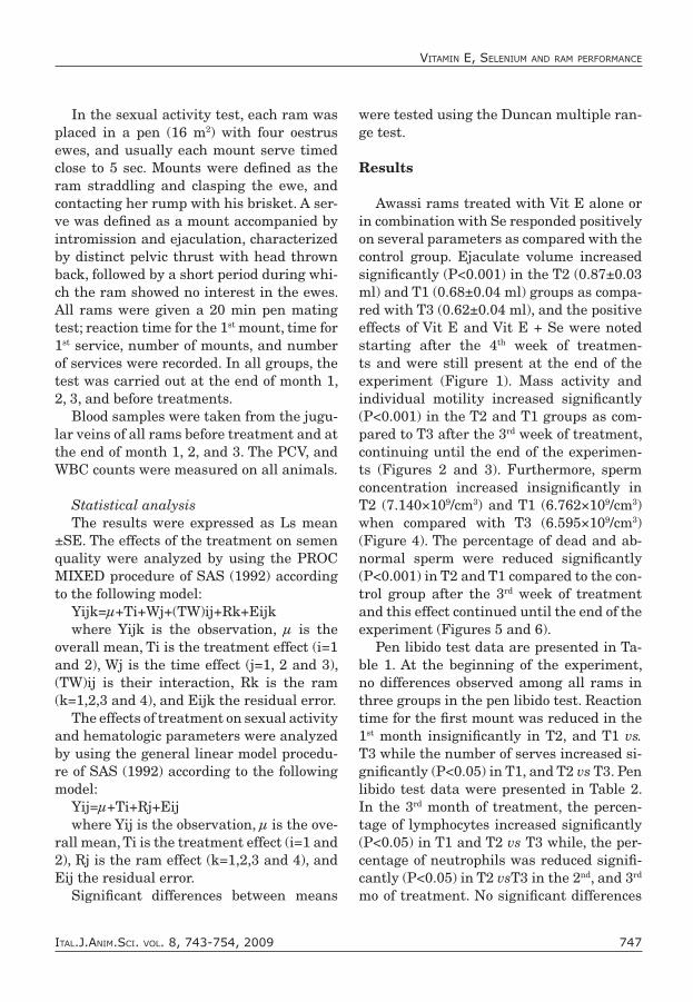
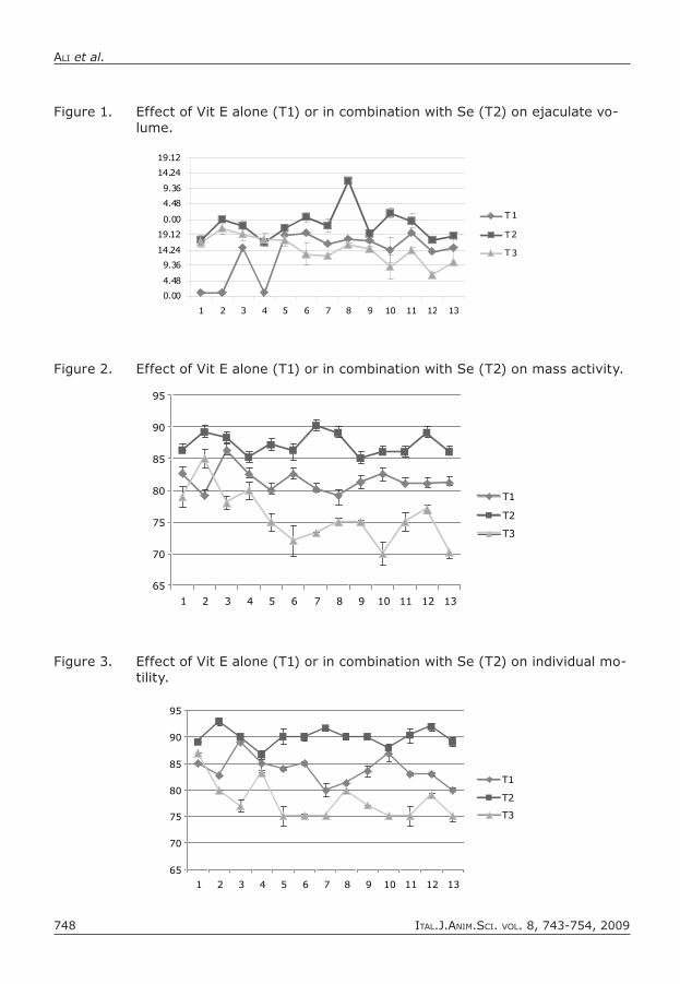
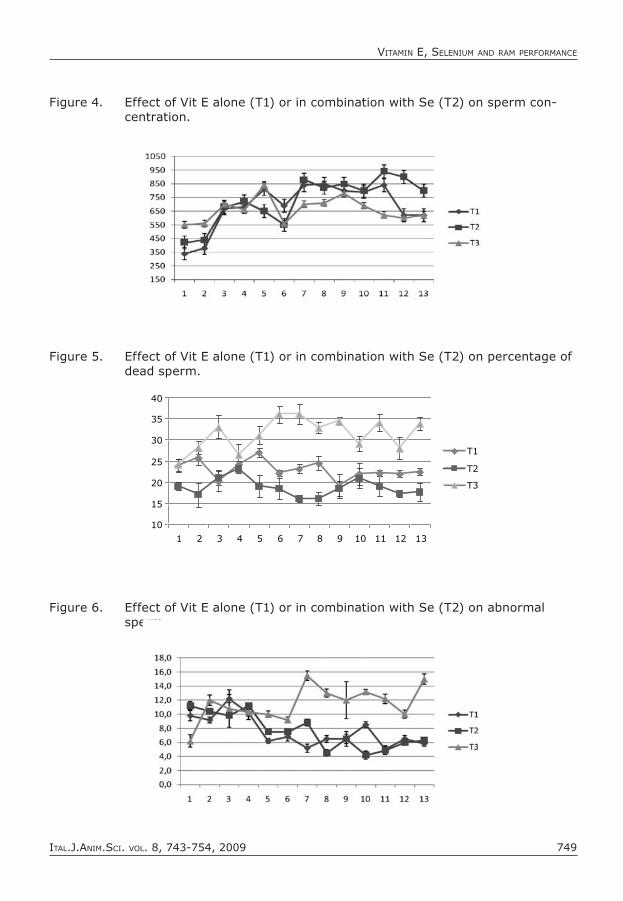
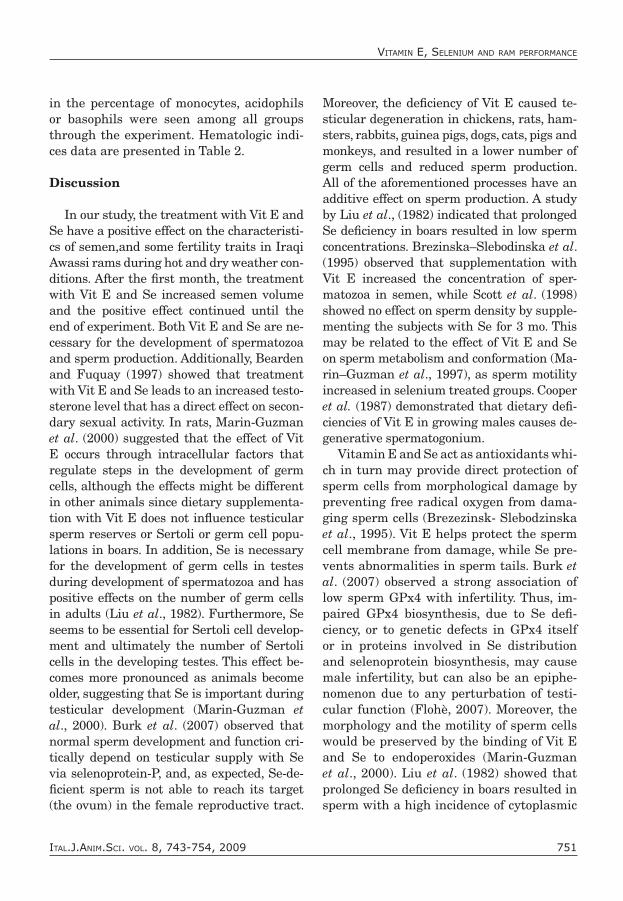
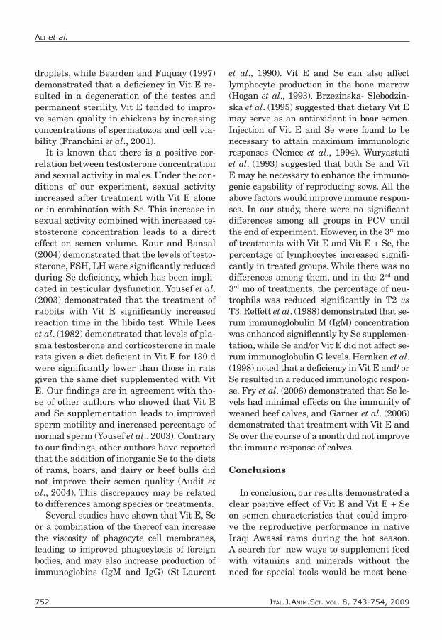
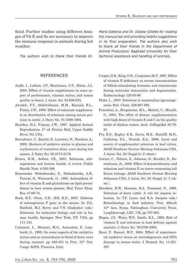
Awassi rams treated with Vit E alone or in combination with Se responded positively on several parameters as compared with the control group. Ejaculate volume increased significantly (P<0.001) in the T2 (0.87±0.03 ml) and T1 (0.68±0.04 ml) groups as compa-red with T3 (0.62±0.04 ml), and the positive effects of Vit E and Vit E + Se were noted starting after the 4th week of treatmen-ts and were still present at the end of the experiment (Figure 1). Mass activity and individual motility increased significantly (P<0.001) in the T2 and T1 groups as com-pared to T3 after the 3rd week of treatment, continuing until the end of the experimen-ts (Figures 2 and 3). Furthermore, sperm concentration increased insignificantly in T2 (7.140×10⁹/cm3) and T1 (6.762×10⁹/cm3) when compared with T3 (6.595×10⁹/cm3) (Figure 4). The percentage of dead and ab-normal sperm were reduced significantly (P<0.001) in T2 and T1 compared to the con-trol group after the 3rd week of treatment and this effect continued until the end of the experiment (Figures 5 and 6).
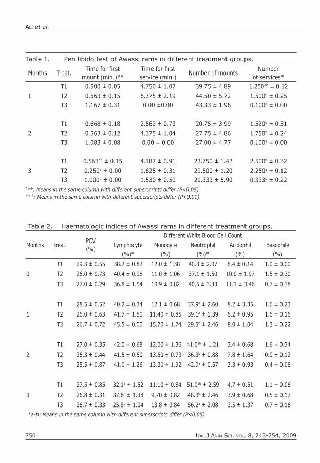
Pen libido test data are presented in Ta-ble 1. At the beginning of the experiment, no differences observed among all rams in three groups in the pen libido test. Reaction time for the first mount was reduced in the 1st month insignificantly in T2, and T1 vs. T3 while the number of serves increased si-gnificantly (P<0.05) in T1, and T2 vs T3. Pen libido test data were presented in Table 2. In the 3rd month of treatment, the percen-tage of lymphocytes increased significantly (P<0.05) in T1 and T2 vs T3 while, the per-centage of neutrophils was reduced signifi-cantly (P<0.05) in T2 vsT3 in the 2nd, and 3rd mo of treatment. No significant differences
Ital.J.anIm.ScI. vol. 8, 743-754, 2009748
alI et al.
Figure 1. Effect of Vit E alone (T1) or in combination with Se (T2) on ejaculate vo-lume.
0.00
4.48
9.36
14.24
19.12
0.00
4.48
9.36
14.24
19.12
1 2 3 4 5 6 7 8 9 10 11 12 13
T1
T2
T3
Figure 3. Effect of Vit E alone (T1) or in combination with Se (T2) on individual mo-tility.
65
70
75
80
85
90
95
1 2 3 4 5 6 7 8 9 10 11 12 13
T1
T2
T3
Figure 2. Effect of Vit E alone (T1) or in combination with Se (T2) on mass activity.
65
70
75
80
85
90
95
1 2 3 4 5 6 7 8 9 10 11 12 13
T1
T2
T3
Ital.J.anIm.ScI. vol. 8, 743-754, 2009 749
vItamIn E, SElEnIum and ram pErformancE
Figure 4. Effect of Vit E alone (T1) or in combination with Se (T2) on sperm con-centration.
Figure 5. Effect of Vit E alone (T1) or in combination with Se (T2) on percentage of dead sperm.
10
15
20
25
30
35
40
1 2 3 4 5 6 7 8 9 10 11 12 13
T1
T2
T3
Figure 6. Effect of Vit E alone (T1) or in combination with Se (T2) on abnormal sperm.
Ital.J.anIm.ScI. vol. 8, 743-754, 2009750
alI et al.
Table 1. Pen libido test of Awassi rams in different treatment groups.
months Treat.Time for first
mount (min.)**Time for first service (min.)
Number of mountsNumber
of services*
1
T1 0.500 ± 0.05 4.750 ± 1.07 39.75 ± 4.89 1.250ab ± 0.12
T2 0.563 ± 0.15 6.375 ± 2.19 44.50 ± 5.72 1.500b ± 0.25
T3 1.167 ± 0.31 0.00 ±0.00 43.33 ± 1.96 0.100a ± 0.00
2
T1 0.668 ± 0.18 2.562 ± 0.73 20.75 ± 3.99 1.520b ± 0.31
T2 0.563 ± 0.12 4.375 ± 1.04 27.75 ± 4.86 1.750b ± 0.24
T3 1.083 ± 0.08 0.00 ± 0.00 27.00 ± 4.77 0.100a ± 0.00
3
T1 0.563ab ± 0.15 4.187 ± 0.91 23.750 ± 1.42 2.500a ± 0.32
T2 0.250a ± 0.00 1.625 ± 0.31 29.500 ± 1.20 2.250a ± 0.12
T3 1.000b ± 0.00 1.530 ± 0.50 29.333 ± 5.90 0.333b ± 0.22* a-b: Means in the same column with different superscripts differ (P<0.05).**a-b: Means in the same column with different superscripts differ (P<0.01).
Table 2. Haematologic indices of Awassi rams in different treatment groups.
months Treat.PCV(%)
Different White Blood Cell Count
lymphocyte monocyte Neutrophil Acidophil Basophile
(%)* (%) (%)* (%) (%)
T1 29.3 ± 0.55 38.2 ± 0.82 12.0 ± 1.38 40.3 ± 2.07 8.4 ± 0.14 1.0 ± 0.00
0 T2 26.0 ± 0.73 40.4 ± 0.98 11.0 ± 1.06 37.1 ± 1.50 10.0 ± 1.97 1.5 ± 0.30
T3 27.0 ± 0.29 36.8 ± 1.54 10.9 ± 0.82 40.5 ± 3.33 11.1 ± 3.46 0.7 ± 0.18
T1 28.5 ± 0.52 40.2 ± 0.34 12.1 ± 0.68 37.9a ± 2.60 8.2 ± 3.35 1.6 ± 0.23
1 T2 26.0 ± 0.63 41.7 ± 1.80 11.40 ± 0.85 39.1a ± 1.39 6.2 ± 0.95 1.6 ± 0.16
T3 26.7 ± 0.72 45.5 ± 0.00 15.70 ± 1.74 29.5b ± 2.46 8.0 ± 1.04 1.3 ± 0.22
T1 27.0 ± 0.35 42.0 ± 0.68 12.00 ± 1.36 41.0ab ± 1.21 3.4 ± 0.68 1.6 ± 0.34
2 T2 25.3 ± 0.44 41.5 ± 0.50 13.50 ± 0.73 36.3b ± 0.88 7.8 ± 1.64 0.9 ± 0.12
T3 25.5 ± 0.87 41.0 ± 1.26 13.30 ± 1.92 42.0a ± 0.57 3.3 ± 0.93 0.4 ± 0.08
T1 27.5 ± 0.85 32.1a ± 1.52 11.10 ± 0.84 51.0ab ± 2.59 4.7 ± 0.51 1.1 ± 0.06
3 T2 26.8 ± 0.31 37.6a ± 1.38 9.70 ± 0.82 48.3b ± 2.46 3.9 ± 0.68 0.5 ± 0.17
T3 26.7 ± 0.33 25.8b ± 1.04 13.8 ± 0.84 56.2a ± 2.08 3.5 ± 1.37 0.7 ± 0.16*a-b: Means in the same column with different superscripts differ (P<0.05).
Ital.J.anIm.ScI. vol. 8, 743-754, 2009 751
vItamIn E, SElEnIum and ram pErformancE
in the percentage of monocytes, acidophils or basophils were seen among all groups through the experiment. Hematologic indi-ces data are presented in Table 2.
Discussion
In our study, the treatment with Vit E and Se have a positive effect on the characteristi-cs of semen,and some fertility traits in Iraqi Awassi rams during hot and dry weather con-ditions. After the first month, the treatment with Vit E and Se increased semen volume and the positive effect continued until the end of experiment. Both Vit E and Se are ne-cessary for the development of spermatozoa and sperm production. Additionally, Bearden and Fuquay (1997) showed that treatment with Vit E and Se leads to an increased testo-sterone level that has a direct effect on secon-dary sexual activity. In rats, Marin-Guzman et al. (2000) suggested that the effect of Vit E occurs through intracellular factors that regulate steps in the development of germ cells, although the effects might be different in other animals since dietary supplementa-tion with Vit E does not influence testicular sperm reserves or Sertoli or germ cell popu-lations in boars. In addition, Se is necessary for the development of germ cells in testes during development of spermatozoa and has positive effects on the number of germ cells in adults (Liu et al., 1982). Furthermore, Se seems to be essential for Sertoli cell develop-ment and ultimately the number of Sertoli cells in the developing testes. This effect be-comes more pronounced as animals become older, suggesting that Se is important during testicular development (Marin-Guzman et al., 2000). Burk et al. (2007) observed that normal sperm development and function cri-tically depend on testicular supply with Se via selenoprotein-P, and, as expected, Se-de-ficient sperm is not able to reach its target (the ovum) in the female reproductive tract.
Moreover, the deficiency of Vit E caused te-sticular degeneration in chickens, rats, ham-sters, rabbits, guinea pigs, dogs, cats, pigs and monkeys, and resulted in a lower number of germ cells and reduced sperm production. All of the aforementioned processes have an additive effect on sperm production. A study by Liu et al., (1982) indicated that prolonged Se deficiency in boars resulted in low sperm concentrations. Brezinska–Slebodinska et al. (1995) observed that supplementation with Vit E increased the concentration of sper-matozoa in semen, while Scott et al. (1998) showed no effect on sperm density by supple-menting the subjects with Se for 3 mo. This may be related to the effect of Vit E and Se on sperm metabolism and conformation (Ma-rin–Guzman et al., 1997), as sperm motility increased in selenium treated groups. Cooper et al. (1987) demonstrated that dietary defi-ciencies of Vit E in growing males causes de-generative spermatogonium.
Vitamin E and Se act as antioxidants whi-ch in turn may provide direct protection of sperm cells from morphological damage by preventing free radical oxygen from dama-ging sperm cells (Brezezinsk- Slebodzinska et al., 1995). Vit E helps protect the sperm cell membrane from damage, while Se pre-vents abnormalities in sperm tails. Burk et al. (2007) observed a strong association of low sperm GPx4 with infertility. Thus, im-paired GPx4 biosynthesis, due to Se defi-ciency, or to genetic defects in GPx4 itself or in proteins involved in Se distribution and selenoprotein biosynthesis, may cause male infertility, but can also be an epiphe-nomenon due to any perturbation of testi-cular function (Flohè, 2007). Moreover, the morphology and the motility of sperm cells would be preserved by the binding of Vit E and Se to endoperoxides (Marin-Guzman et al., 2000). Liu et al. (1982) showed that prolonged Se deficiency in boars resulted in sperm with a high incidence of cytoplasmic
Ital.J.anIm.ScI. vol. 8, 743-754, 2009752
alI et al.
droplets, while Bearden and Fuquay (1997) demonstrated that a deficiency in Vit E re-sulted in a degeneration of the testes and permanent sterility. Vit E tended to impro-ve semen quality in chickens by increasing concentrations of spermatozoa and cell via-bility (Franchini et al., 2001).
It is known that there is a positive cor-relation between testosterone concentration and sexual activity in males. Under the con-ditions of our experiment, sexual activity increased after treatment with Vit E alone or in combination with Se. This increase in sexual activity combined with increased te-stosterone concentration leads to a direct effect on semen volume. Kaur and Bansal (2004) demonstrated that the levels of testo-sterone, FSH, LH were significantly reduced during Se deficiency, which has been impli-cated in testicular dysfunction. Yousef et al. (2003) demonstrated that the treatment of rabbits with Vit E significantly increased reaction time in the libido test. While Lees et al. (1982) demonstrated that levels of pla-sma testosterone and corticosterone in male rats given a diet deficient in Vit E for 130 d were significantly lower than those in rats given the same diet supplemented with Vit E. Our findings are in agreement with tho-se of other authors who showed that Vit E and Se supplementation leads to improved sperm motility and increased percentage of normal sperm (Yousef et al., 2003). Contrary to our findings, other authors have reported that the addition of inorganic Se to the diets of rams, boars, and dairy or beef bulls did not improve their semen quality (Audit et al., 2004). This discrepancy may be related to differences among species or treatments.
Several studies have shown that Vit E, Se or a combination of the thereof can increase the viscosity of phagocyte cell membranes, leading to improved phagocytosis of foreign bodies, and may also increase production of immunoglobins (IgM and IgG) (St-Laurent
et al., 1990). Vit E and Se can also affect lymphocyte production in the bone marrow (Hogan et al., 1993). Brzezinska- Slebodzin-ska et al. (1995) suggested that dietary Vit E may serve as an antioxidant in boar semen. Injection of Vit E and Se were found to be necessary to attain maximum immunologic responses (Nemec et al., 1994). Wuryastuti et al. (1993) suggested that both Se and Vit E may be necessary to enhance the immuno-genic capability of reproducing sows. All the above factors would improve immune respon-ses. In our study, there were no significant differences among all groups in PCV until the end of experiment. However, in the 3rd mo of treatments with Vit E and Vit E + Se, the percentage of lymphocytes increased signifi-cantly in treated groups. While there was no differences among them, and in the 2nd and 3rd mo of treatments, the percentage of neu-trophils was reduced significantly in T2 vs T3. Reffett et al. (1988) demonstrated that se-rum immunoglobulin M (IgM) concentration was enhanced significantly by Se supplemen-tation, while Se and/or Vit E did not affect se-rum immunoglobulin G levels. Hernken et al. (1998) noted that a deficiency in Vit E and/ or Se resulted in a reduced immunologic respon-se. Fry et al. (2006) demonstrated that Se le-vels had minimal effects on the immunity of weaned beef calves, and Garner et al. (2006) demonstrated that treatment with Vit E and Se over the course of a month did not improve the immune response of calves.
Conclusions
In conclusion, our results demonstrated a clear positive effect of Vit E and Vit E + Se on semen characteristics that could impro-ve the reproductive performance in native Iraqi Awassi rams during the hot season. A search for new ways to supplement feed with vitamins and minerals without the need for special tools would be most bene-
Ital.J.anIm.ScI. vol. 8, 743-754, 2009 753
vItamIn E, SElEnIum and ram pErformancE
ficial. Further studies using different dosa-ges of Vit E and Se are necessary to improve the immune response in animals during hot weather.
The authors wish to thank their friends Dr.
Maria Dattena and Dr. Debbie Gillette for reading the manuscript and providing helpful suggestions in its final preparation. The authors also wish to thank all their friends in the Department of Animal Production/ Baghdad University for their technical assistance and handling of animals.
REFERENCES
Audit, I., Laforst, J.P., Martineau, G.P., Matte, J.J., 2004. Effect of vitamin supplements on some as-pect of performance, vitamin status, and semen quality in boars. J. Anim. Sci. 82:626-633.
Awadeh, F.T., Abdelrahman, M.M., Kincaid, R.L., Finley, J.W., 1998. Effect of selenium supplemen-ts on distribution of selenium among serum pro-teins in cattle. J. Dairy Sci. 81:1089-1094.
Bearden, H.J., Fuquay, J.W., 1997. Applied Animal Reproduction. 4th ed. Pentice Hall, Upper Saddle River, NJ, USA.
Bernabucci, U., Ronchi, B., Lacetera, N., Nardone, A., 2002. Markers of oxidative status in plasma and erythrocytes of transition dairy cows during hot season. J. Dairy Sci. 85:2173-2179.
Brown, K.M., Arthur, J.R., 2001. Selenium, sele-noproteins and human health: A review. Public Health Nutr. 4:593-599.
Brzezinska- Slebodzinska, E., Slebodzinska, A.B., Pietras, B., Wieczorek, G., 1995. Antioxidant ef-fect of vitamin E and glutathione on lipid peroxi-dation in boor semen plasma. Biol. Trace Elem. Res. 47:69-74.
Burk, R.F., Olson, G.E., Hill, K.E., 2007. Deletion of selenoprotein P gene in the mouse. In: D.L. Hatfield, M.J. Berry and V.N. Gladyshev (eds.) Selenium. Its molecular biology and role in hu-man health. Springer, New York, NY, USA, pp 111-122.
Calamari, L., Maianti, M.G., Amendola, F., Lom-bardi, G., 1999. On some aspects of the oxidative status and on antioxidants in blood of dairy cows during summer. pp 449-451 in Proc. 13th Nat. Congr. ASPA, Piacenza, Italy.
Cooper, D.R., King, O.R., Carpenter, M.P., 1987. Effect of vitamin E deficiency on serum concentrations of follicle-stimulating hormone and testosterone during testicular maturation and degeneration. Endocrinology 120:83-90.
Flohè, L., 2007. Selenium in mammalian spermioge-nesis. Biol. Chem. 338:987-995.
Franchini, A., Bergonzoni, M.L., Melotti, C., Minelli, G., 2001. The effect of dietary supplementation with high doses of vitamin E and C on the quality traits of chicken semen. Arch. Geflugelkd. 65:76-81.
Fry, R.S., Kegley, E.B., Davis, M.E., Ratcliff, M.D., Galloway, D.L., Dvorak, R.A., 2006. Level and source of supplemental selenium in beef calves. ASAS Southern Section Meeting Arkansas-USA. J. Anim. Sci. 83 (Suppl.2): 2 (abstr.).
Garner, C., Nihsen, Z., Johnson, D., Kreider, D., Ro-senkrans, Jr., 2006. Effect of dexamethasone and selenium and vitamin E on steers consuming tall fescue baleage. ASAS Southern Section Meeting Arkansas-USA. J. Anim. Sci. 83 (Suppl. 2): 3 (ab-str.).
Hernken, R.W., Harmon, R.J., Trammel, S., 1998. Selenium of dairy cattle: A role for organic se-lenium. In: T.P. Lyons and K.A. Jacques (eds.) Biotechnology in feed industry. Proc. Alltech 14th Ann. Symp. Nottingham University Press, Loughborough, LEC, UK, pp 797-803.
Hogan, J.S., Weiss, W.P., Smith, K.L., 1993. Role of vitamin E and selenium in host defense against mastitis. J. Dairy Sci. 76:2795-2803.
Kaur, P., Bansal, M.P., 2004. Effect of experimen-tal oxidative stress on steriodogenesis and DNA damage in mouse testis. J. Biomed. Sci. 11:391-397.
Ital.J.anIm.ScI. vol. 8, 743-754, 2009754
alI et al.
Koyuncu, M., Yerlikaya, H., 2007. Effect of sele-nium–vitamin E injections of ewes on reproduc-tion and growth of their lambs. S. Afr. J. Anim. Sci. 37:233-236.
Lees, D., Mc Brarnes, M., Cox, J.E., 1982. Testos-terone and corticosterone concentrations in the plasma of rats deficient in vitamin E. J. Reprod. Fertil. 66:543-545.
Liu, C.H., Chen, Y.M., Zhang, J.Z., Huang, M.Y., Su, Q., Lu, Z.H., Yin, R.X., Shao, G.Z., Feng, D., Zheng, P.L., 1982. Preliminary studies on influ-ence of selenium deficiency to the developments of genital organs and spermatogenesis of infancy boars. Acta Vet. Zootech. Sin. 13:73-77.
Mahan, D.C., Kim, Y.Y., Stuart, R.L., 2000. Effect of vitamin E source (RRR-or all- rac-α-tocopheryl acetate) and level on sow reproductive perfor-mance, serum tissue, and milk α-tocopherol con-tents over a five-parity period, and the effects on the progeny. J. Anim. Sci. 78:110-119.
Marin-Guzman, J., Mahan, D.C., Chung, Y.K., Pate, J.L., Pope, W.F., 1997. Effects of dietary sele-nium and vitamin E on Boar performance and tissue response, semen quality and subsequent fertilization rates in mature gilts. J. Anim. Sci. 75:2994-3003.
Marin-Guzman, J., Mahan, D.C., Pate, J.L., 2000. Ef-fect of dietary selenium and vitamin E on sper-matogenic development in boars. J. Anim. Sci. 78:1537-1543.
Nemec, M., Butler, G., Hidiroglou, M., Farnworth, E.R., Nielsen, K., 1994. Effect of supplementing gilts’ with different levels of vitamin E and diffe-rent fats on the hormonal and cellular immunity of gilts and their progeny. J. Anim. Sci. 72:665-676.
Nockels, C.F., Odde, K.G., Craig, A.M., 1996. Vita-min E supplementation and stress affect tissue α-tocopherol content of beef heifers. J. Anim. Sci. 74:672-677.
Reffet, J.K., Spears, J.W., Brown, T.T.Jr., 1988. Effect of dietary selenium and vitamin E on the pri-mary and secondary immune response in lambs challenged with parainfluenza3 virus. J. Anim. Sci. 66:1520-1528.
SAS, 1992. SAS Users Guide Statistics. SAS Insti-tute Inc., Cary, NC, USA.
Sconberg, S., Nockels, C.F., Bennett, B.W., Bruynin-ckx, W., Blancquaret, A-M.B., Craig, A.M., 1993. Effects of shipping handling, adrenocorticotropic hormone, and epinephrine on α-tocopherol con-tent of bovine blood. Am. J. Vet. Res. 54:1287 (ab-str.).
Scott, R., MacPherson, A., Yates, R.W. Hussain, B., Dixon, J., 1998. The effect of oral selenium sup-plementation on human sperm motility. Brit. J. Urol. 82:76-80.
Smith, D.G., Senger, P.L., McCutchan, J.F., Lands, C.A., 1979. Selenium and glutathione peroxidase distribution in bovine semen and selenium -75 retention by the tissues of the reproductive tract in bull. Biol. Reprod. 20:377-383.
Spallholz, J.E, Martin, J.L., Gerlach, M.L., Heinzer-ling, R.H., 1975. Injectable Se: Effect on the pri-mary immune response of mice. P. Soc. Exp. Biol. Med. 148:37-40.
St-Laurent, A., Hidiroglou, M., Snoddon, M., Nich-olson, J.W.G., 1990. Response to dietary vitamin E in the dairy cow and its effect spontaneous oxidized flavor in milk. Can. J. Anim. Sci. 70:561-570.
Trevisan, M., Browne, R., Ram, M., Muti, P., Freu-denheim, J., Carosella, A.N., Armstrong, D., 2001. Correlates of markers of oxidative status in the general population. Am. J. Epidemiol. 154:348-356.
Wuryastuti, H., Stowe, H.D., Bull, R.W., Miller, E.R., 1993. Effect of vitamin E and selenium on immune response of peripheral blood, colos-trum, and milk leukocytes of sows. J. Anim. Sci. 71:2464-2472.
Yousef, M.I., Abdallah, G.A., Kamel, K.I., 2003. Effect of ascorbic acid and vitamin E supplementation on semen quality and biochemical parameters of male rabbits. Anim. Reprod. Sci. 76:99-111.
Zhu, B., Setchell, B.P., 2004. Effect of paternal heat stress on the in vivo development of preimplan-taion embryos in mouse. Reprod. Nutr. Dev. 44:617-629.


