Embed Size (px)
Citation preview
Published online 27 November 2020 Nucleic Acids Research, 2020, Vol. 48, No. 22 12917–12928doi: 10.1093/nar/gkaa1128
Double-stranded RNA bending by AU-tract sequencesAlberto Marin-Gonzalez 1, Clara Aicart-Ramos 1, Mikel Marin-Baquero 1,Alejandro Martın-Gonzalez1, Maarit Suomalainen2, Abhilash Kannan2, J. G. Vilhena 3,4,Urs F. Greber2, Fernando Moreno-Herrero 1,* and Ruben Perez 4,5,*
1Department of Macromolecular Structures, Centro Nacional de Biotecnologıa, Consejo Superior de InvestigacionesCientıficas, 28049 Cantoblanco, Madrid, Spain, 2Department of Molecular Life Sciences, University of Zurich,Winterthurerstrasse 190, 8057 Zurich, Switzerland, 3Department of Physics, University of Basel, Klingelbergstrasse82, 4056 Basel, Switzerland, 4Departamento de Fısica Teorica de la Materia Condensada, Universidad Autonoma deMadrid, E-28049 Madrid, Spain and 5IFIMAC - Condensed Matter Physics Center, Universidad Autonoma de Madrid,E-28049 Madrid, Spain
Received June 04, 2020; Revised October 08, 2020; Editorial Decision November 03, 2020; Accepted November 10, 2020
ABSTRACT
Sequence-dependent structural deformations of theDNA double helix (dsDNA) have been extensivelystudied, where adenine tracts (A-tracts) provide astriking example for global bending in the molecule.However, in contrast to dsDNA, sequence-dependentstructural features of dsRNA have received little at-tention. In this work, we demonstrate that the nu-cleotide sequence can induce a bend in a canoni-cal Watson-Crick base-paired dsRNA helix. Using all-atom molecular dynamics simulations, we identifieda sequence motif consisting of alternating adeninesand uracils, or AU-tracts, that strongly bend the RNAdouble-helix. This finding was experimentally val-idated using atomic force microscopy imaging ofdsRNA molecules designed to display macroscopiccurvature via repetitions of phased AU-tract motifs.At the atomic level, this novel phenomenon origi-nates from a localized compression of the dsRNAmajor groove and a large propeller twist at the posi-tion of the AU-tract. Moreover, the magnitude of thebending can be modulated by changing the lengthof the AU-tract. Altogether, our results demonstratethe possibility of modifying the dsRNA curvature bymeans of its nucleotide sequence, which may be ex-ploited in the emerging field of RNA nanotechnologyand might also constitute a natural mechanism forproteins to achieve recognition of specific dsRNAsequences.
INTRODUCTION
Double-stranded RNA (dsRNA) plays a central role ina number of biological processes. For instance, dsRNAmolecules are involved in the regulation of gene expressionby RNAi (1), or in the host responses to dsRNA encoded byviruses (2). In addition, dsRNA helices perform key func-tions as an essential part of tertiary RNA structures, in-cluding tRNA and riboswitches (3,4), and of macromolecu-lar RNA-protein complexes such as ribosomal subunits andthe spliceosome (5–7).
Many of the biological processes involving dsRNA ex-ploit the conformational flexibility of dsRNA helices in or-der e.g. to achieve folding of the RNA into complex 3Dstructures (8–10) or in dsRNA:protein interactions (11–13). Therefore, a quantitative understanding of the physicalproperties of dsRNA can provide novel insights on theseprocesses, and also, can aid the design of RNA nanostruc-tures for biotechnological applications (14). Motivated bythese considerations, immense research efforts have charac-terized the effect of helical imperfections, such as bulges orinternal loops, on dsRNA conformations (15,16). However,the question of how the nucleotide sequence impacts theoverall structure of canonical, Watson–Crick base-paireddsRNA helices remains largely unanswered (17).
In contrast to dsRNA, the sequence-dependent struc-ture of the canonical DNA double-helix (dsDNA) has beencharacterized in great detail. A prime example of suchsequence-dependent features are the so-called A-tracts, runsof adenines and thymines without a TpA step that, when inphase with the helical pitch yield a significant global curva-ture of the dsDNA (18,19). Besides their bending charac-ter, A-tracts show a peculiar conformation at the molecu-lar level, with a characteristic narrow minor groove (20,21).Remarkably, both the curvature induced by A-tracts andthe molecular conformation of these sequences are thought
*To whom correspondence should be addressed. Tel: +34 914974906; Email: [email protected] may also be addressed to Fernando Moreno-Herrero. Email: [email protected]
C© The Author(s) 2020. Published by Oxford University Press on behalf of Nucleic Acids Research.This is an Open Access article distributed under the terms of the Creative Commons Attribution-NonCommercial License(http://creativecommons.org/licenses/by-nc/4.0/), which permits non-commercial re-use, distribution, and reproduction in any medium, provided the original workis properly cited. For commercial re-use, please contact [email protected]
Dow
nloaded from https://academ
ic.oup.com/nar/article/48/22/12917/6007656 by guest on 10 February 2022
12918 Nucleic Acids Research, 2020, Vol. 48, No. 22
to have biological relevance. The former seems to stabi-lize DNA tertiary structures, such as loops and supercoils(22,23), whereas the latter is used by proteins to achievebinding specificity (24). Moreover, the DNA bending in-duced by A-tracts has aided the design of DNA rings aspart of nanotechnological devices (25).
Scattered experimental evidence suggests the existenceof sequence-induced curvature in a Watson–Crick base-paired RNA duplex. Early crystallographic works reportedhelical kinks in the structure of an RNA duplex consist-ing of alternating adenines and uracils (26,27). However,this bent conformation was stabilized by the intermolecu-lar interactions among the molecules forming the crystaland, therefore, bending could not be attributed to the RNAduplex alone. In parallel, analysis of structural databasesand theoretical methods, such as molecular dynamics (MD)simulations, have provided valuable insight on sequence-dependent dsRNA conformations (17,28–31). In particu-lar, recent MD studies have predicted strong sequence ef-fects on the dsRNA shape (29,30) and flexibility (31), whichcould potentially lead to sequence-induced bending. Suchsimulation techniques hold great potential in decipheringthe sequence-dependent dsRNA conformational landscape,provided that the computational predictions are thoroughlytested against experimental measurements. However, suchcomparison remains challenging due, in part, to the limitedavailability on high-resolution dsRNA experimental struc-tures and the number of artifacts that are often found, e.g.in crystal structures (17).
Here, we present a procedure that led us to the directexperimental observation of single dsRNA molecules bentonly by their nucleotide sequence. We combined MD simu-lations and atomic force microscopy (AFM) experiments; atechnique especially suited for studying dsRNA bending, asdemonstrated by AFM measurements of the dsRNA persis-tence length (32,33). We first performed a systematic analy-sis of how the sequence affects the structure of dsRNA us-ing MD simulations. Our simulations predicted that a se-quence motif, that we named AU-tract, would cause a bendin the RNA double-helix. We then synthesized long dsRNAmolecules containing AU-tracts in phase with the helicalpitch. Analysis of AFM images of these molecules revealedthat they were indeed significantly more bent than controldsRNA molecules of arbitrary sequence. Finally, we pro-pose a molecular mechanism for AU-tract bending based ona large propeller twist at A:U base pairs. Our work unveilsthe phenomenon of sequence-induced curvature in dsRNA,challenging the traditional picture of dsRNA as an invari-ant double helix.
MATERIALS AND METHODS
Molecular dynamics simulations
Simulation details are similar to the ones from (34), onlyexcluding the external force. RNA duplexes were placed inan approximately cubic box of 110 A edge size and filledwith water and sodium counterions to balance the phos-phate charges. Only sodium ions were included in the sim-ulations. The systems were heated up to 300 K and equili-brated in the isobaric-isothermal (NPT) ensemble (P = 1atm, T = 300 K) for 20 ns. Production simulations were run
in the NVT ensemble using as input coordinates the onesfrom the last configuration of the NPT equilibration. Allsimulations were extended to ∼1 �s time.
We used the AMBER14 software suite with NVIDIAGPU acceleration (35,36). For the modeling of dsRNAmolecules we resorted to the Cornell ff99 force field (37)with the parmbsc0 (38) refinement and the � OL3 mod-ification (39). The ions were described according to theJoung/Cheatham (40) parametrization; and the TIP3Pmodel (41) was used for water molecules. Periodic bound-ary conditions and Particle Mesh Ewald (with standarddefaults and a real-space cutoff of 9 A) were used to ac-count for long-range electrostatics interactions. The samereal space cutoff was used to truncate van der Waals forces.SHAKE algorithm was used to constrain bonds contain-ing hydrogen atoms, thus allowing us to use an integrationstep of 2 fs. Coordinates were saved every 1000 steps. Av-erage structures were computed using the cpptraj softwareof the AMBER14 suite. Helical, base pair step parametersand groove dimensions were computed using Curves+ (42)and 3DNA (43) and helical bending was calculated usingCurves+ (42). The four base pairs adjacent to the terminiof the molecules were excluded from the analysis.
Production of dsRNA molecules
In order to study the mechanical properties of AU-tracts atthe single-molecule level, we produced dsRNA moleculesthat contain periodic repetitions of alternating AU nu-cleotides. We named them ExpAU-4 or ExpAU-5 depend-ing on the length of four or five nucleotides of the AU-tract.These molecules were produced by hybridizing two longcomplementary ssRNAs. To fabricate these, the sequence ofinterest was cloned after the T7 RNA polymerase promoterbetween two KpnI sites. In this way, the fragment could bedigested and ligated in the opposite orientation, allowingus to synthesize the two complementary ssRNA chains. Inaddition, a SmaI site was introduced at the end of the se-quence, enabling the linearization of the plasmid vector tolimit the length of the transcripts.
dsRNA molecules were synthesized according to a pre-viously described protocol (33,44) with slight modificationsto increase yield for single-molecule manipulation purposes.Once each pair of plasmids were obtained, plasmid vec-tors used as transcription templates were linearized withSmaI followed by purification (QIAGEN). Afterwards, in-vitro transcription using the commercial HiScribe™ T7 HighYield RNA Synthesis Kit (NEB) gave rise to two com-plementary ssRNAs without any non-complementary nu-cleotides at their ends. After 3 h at 42◦C, EDTA wasadded to a final concentration of 30 mM, and both strandswere subsequently hybridized by heating 1 h at 65◦C andslowly cooling to room temperature at a 1.2◦C/5 min rateup to 25◦C. This resulted in dsRNA molecules withoutany single-stranded overhangs. Transcription products werethen cleaned with RNeasy MinElute Cleanup Kit (QIA-GEN) followed by treatment for 1 h at 37◦C with 2.5 unitsof RNase-free DNase I (NEB). The sample was once againcleaned with RNeasy MinElute Cleanup Kit before apply-ing on a 1% agarose gel for gel extraction and purificationwith QIAGEN gel extraction kit and elution with RNase
Dow
nloaded from https://academ
ic.oup.com/nar/article/48/22/12917/6007656 by guest on 10 February 2022
Nucleic Acids Research, 2020, Vol. 48, No. 22 12919
free H2O. dsRNA constructs were stored at 4◦C in RNasefree H2O. The final sequences are shown in SupplementaryTable S1. Further details on the synthesis of the dsRNAmolecules can be found in the Supplementary Material (seesection 1, Supplementary Table S3 and Supplementary Fig-ure S12).
Atomic force microscopy measurements
Imaging conditions and data analysis were similar to thoseemployed in a previous work (33). A 10 �l solution con-taining 0.5 nM dsRNA, 2.5 mM NiCl2, 25 mM TrisAc pH7.5, 2.5 mM MgOAc and 100 mM NaCl was deposited ontofreshly cleaved mica. After ∼60 s, the sample was washedusing Milli-Q water and dried using air nitrogen. Imageswere taken in tapping mode in air, using an AFM fromNanotech Electronica S.L. with PointProbePlus tips (PPP-NCH Nanosensors). Contour lengths were obtained usingthe WSxM software (45). Persistence lengths were com-puted using the tracing routine from (46,47). Traces of 170nm were obtained with a point-to-point separation of 2.5nm.
RESULTS AND DISCUSSION
The dsRNA sequence affects the width of the major groove,the extension, and twist of dsRNA
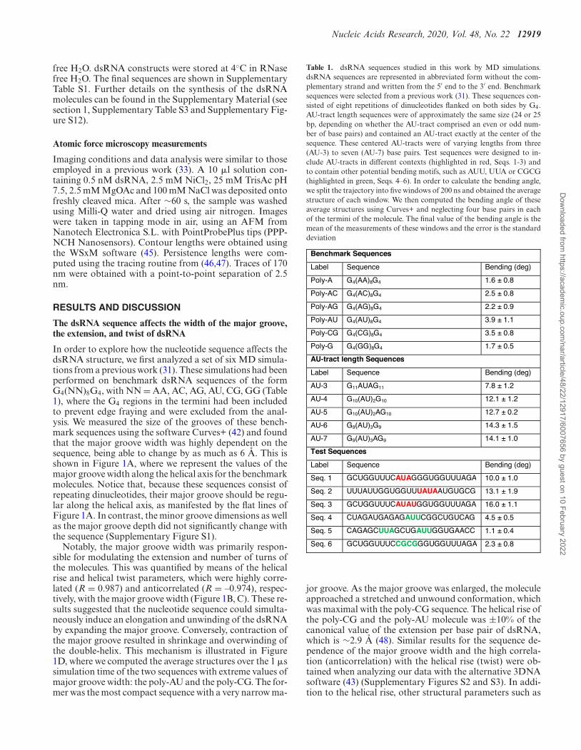
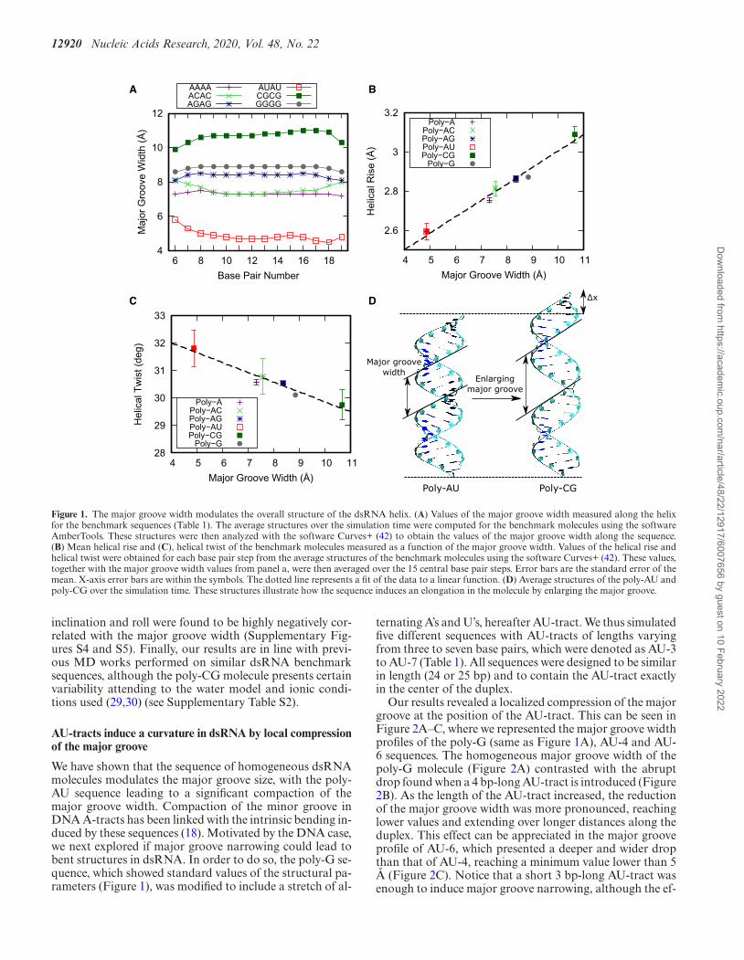
In order to explore how the nucleotide sequence affects thedsRNA structure, we first analyzed a set of six MD simula-tions from a previous work (31). These simulations had beenperformed on benchmark dsRNA sequences of the formG4(NN)8G4, with NN = AA, AC, AG, AU, CG, GG (Table1), where the G4 regions in the termini had been includedto prevent edge fraying and were excluded from the anal-ysis. We measured the size of the grooves of these bench-mark sequences using the software Curves+ (42) and foundthat the major groove width was highly dependent on thesequence, being able to change by as much as 6 A. This isshown in Figure 1A, where we represent the values of themajor groove width along the helical axis for the benchmarkmolecules. Notice that, because these sequences consist ofrepeating dinucleotides, their major groove should be regu-lar along the helical axis, as manifested by the flat lines ofFigure 1A. In contrast, the minor groove dimensions as wellas the major groove depth did not significantly change withthe sequence (Supplementary Figure S1).
Notably, the major groove width was primarily respon-sible for modulating the extension and number of turns ofthe molecules. This was quantified by means of the helicalrise and helical twist parameters, which were highly corre-lated (R = 0.987) and anticorrelated (R = –0.974), respec-tively, with the major groove width (Figure 1B, C). These re-sults suggested that the nucleotide sequence could simulta-neously induce an elongation and unwinding of the dsRNAby expanding the major groove. Conversely, contraction ofthe major groove resulted in shrinkage and overwinding ofthe double-helix. This mechanism is illustrated in Figure1D, where we computed the average structures over the 1 �ssimulation time of the two sequences with extreme values ofmajor groove width: the poly-AU and the poly-CG. The for-mer was the most compact sequence with a very narrow ma-
Table 1. dsRNA sequences studied in this work by MD simulations.dsRNA sequences are represented in abbreviated form without the com-plementary strand and written from the 5′ end to the 3′ end. Benchmarksequences were selected from a previous work (31). These sequences con-sisted of eight repetitions of dinucleotides flanked on both sides by G4.AU-tract length sequences were of approximately the same size (24 or 25bp, depending on whether the AU-tract comprised an even or odd num-ber of base pairs) and contained an AU-tract exactly at the center of thesequence. These centered AU-tracts were of varying lengths from three(AU-3) to seven (AU-7) base pairs. Test sequences were designed to in-clude AU-tracts in different contexts (highlighted in red, Seqs. 1-3) andto contain other potential bending motifs, such as AUU, UUA or CGCG(highlighted in green, Seqs. 4–6). In order to calculate the bending angle,we split the trajectory into five windows of 200 ns and obtained the averagestructure of each window. We then computed the bending angle of theseaverage structures using Curves+ and neglecting four base pairs in eachof the termini of the molecule. The final value of the bending angle is themean of the measurements of these windows and the error is the standarddeviation
Benchmark Sequences
Label Sequence Bending (deg)
Poly-A G4(AA)8G4 1.6 ± 0.8
Poly-AC G4(AC)8G4 2.5 ± 0.8
Poly-AG G4(AG)8G4 2.2 ± 0.9
Poly-AU G4(AU)8G4 3.9 ± 1.1
Poly-CG G4(CG)8G4 3.5 ± 0.8
Poly-G G4(GG)8G4 1.7 ± 0.5
AU-tract length Sequences
Label Sequence Bending (deg)
AU-3 G11AUAG11 7.8 ± 1.2
AU-4 G10(AU)2G10 12.1 ± 1.2
AU-5 G10(AU)2AG10 12.7 ± 0.2
AU-6 G9(AU)3G9 14.3 ± 1.5
AU-7 G9(AU)3AG9 14.1 ± 1.0
Test Sequences
Label Sequence Bending (deg)
Seq. 1 GCUGGUUUCAUAGGGUGGUUUAGA 10.0 ± 1.0
Seq. 2 UUUAUUGGUGGUUUAUAAUGUGCG 13.1 ± 1.9
Seq. 3 GCUGGUUUCAUAUGGUGGUUUAGA 16.0 ± 1.1
Seq. 4 CUAGAUGAGAGAUUCGGCUGUCAG 4.5 ± 0.5
Seq. 5 CAGAGCUUAGCUGAUUGGUGAACC 1.1 ± 0.4
Seq. 6 GCUGGUUUCCGCGGGUGGUUUAGA 2.3 ± 0.8
jor groove. As the major groove was enlarged, the moleculeapproached a stretched and unwound conformation, whichwas maximal with the poly-CG sequence. The helical rise ofthe poly-CG and the poly-AU molecule was ±10% of thecanonical value of the extension per base pair of dsRNA,which is ∼2.9 A (48). Similar results for the sequence de-pendence of the major groove width and the high correla-tion (anticorrelation) with the helical rise (twist) were ob-tained when analyzing our data with the alternative 3DNAsoftware (43) (Supplementary Figures S2 and S3). In addi-tion to the helical rise, other structural parameters such as
Dow
nloaded from https://academ
ic.oup.com/nar/article/48/22/12917/6007656 by guest on 10 February 2022
12920 Nucleic Acids Research, 2020, Vol. 48, No. 22
Poly-AU Poly-CG
Enlarging major groove
Major groove width
Δx
A B
C D
4
6
8
10
12
6 8 10 12 14 16 18
Maj
or G
roov
e W
idth
(Å)
Base Pair Number
AAAAACACAGAG
AUAUCGCGGGGG
2.6
2.8
3
3.2
4 5 6 7 8 9 10 11
Hel
ical
Ris
e (Å
)
Major Groove Width (Å)
Poly−APoly−ACPoly−AGPoly−AUPoly−CG
Poly−G
28
29
30
31
32
33
4 5 6 7 8 9 10 11
Hel
ical
Tw
ist (
deg)
Major Groove Width (Å)
Poly−APoly−ACPoly−AGPoly−AUPoly−CG
Poly−G
Figure 1. The major groove width modulates the overall structure of the dsRNA helix. (A) Values of the major groove width measured along the helixfor the benchmark sequences (Table 1). The average structures over the simulation time were computed for the benchmark molecules using the softwareAmberTools. These structures were then analyzed with the software Curves+ (42) to obtain the values of the major groove width along the sequence.(B) Mean helical rise and (C), helical twist of the benchmark molecules measured as a function of the major groove width. Values of the helical rise andhelical twist were obtained for each base pair step from the average structures of the benchmark molecules using the software Curves+ (42). These values,together with the major groove width values from panel a, were then averaged over the 15 central base pair steps. Error bars are the standard error of themean. X-axis error bars are within the symbols. The dotted line represents a fit of the data to a linear function. (D) Average structures of the poly-AU andpoly-CG over the simulation time. These structures illustrate how the sequence induces an elongation in the molecule by enlarging the major groove.
inclination and roll were found to be highly negatively cor-related with the major groove width (Supplementary Fig-ures S4 and S5). Finally, our results are in line with previ-ous MD works performed on similar dsRNA benchmarksequences, although the poly-CG molecule presents certainvariability attending to the water model and ionic condi-tions used (29,30) (see Supplementary Table S2).
AU-tracts induce a curvature in dsRNA by local compressionof the major groove
We have shown that the sequence of homogeneous dsRNAmolecules modulates the major groove size, with the poly-AU sequence leading to a significant compaction of themajor groove width. Compaction of the minor groove inDNA A-tracts has been linked with the intrinsic bending in-duced by these sequences (18). Motivated by the DNA case,we next explored if major groove narrowing could lead tobent structures in dsRNA. In order to do so, the poly-G se-quence, which showed standard values of the structural pa-rameters (Figure 1), was modified to include a stretch of al-
ternating A’s and U’s, hereafter AU-tract. We thus simulatedfive different sequences with AU-tracts of lengths varyingfrom three to seven base pairs, which were denoted as AU-3to AU-7 (Table 1). All sequences were designed to be similarin length (24 or 25 bp) and to contain the AU-tract exactlyin the center of the duplex.
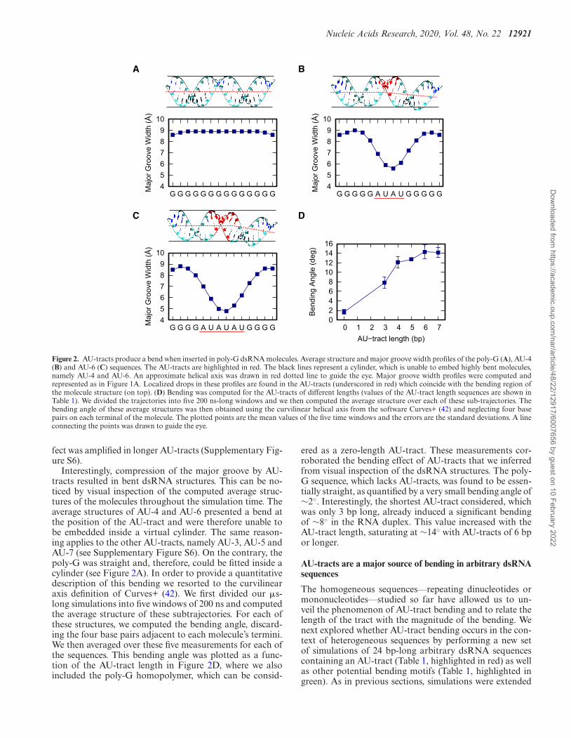
Our results revealed a localized compression of the majorgroove at the position of the AU-tract. This can be seen inFigure 2A–C, where we represented the major groove widthprofiles of the poly-G (same as Figure 1A), AU-4 and AU-6 sequences. The homogeneous major groove width of thepoly-G molecule (Figure 2A) contrasted with the abruptdrop found when a 4 bp-long AU-tract is introduced (Figure2B). As the length of the AU-tract increased, the reductionof the major groove width was more pronounced, reachinglower values and extending over longer distances along theduplex. This effect can be appreciated in the major grooveprofile of AU-6, which presented a deeper and wider dropthan that of AU-4, reaching a minimum value lower than 5A (Figure 2C). Notice that a short 3 bp-long AU-tract wasenough to induce major groove narrowing, although the ef-
Dow
nloaded from https://academ
ic.oup.com/nar/article/48/22/12917/6007656 by guest on 10 February 2022
Nucleic Acids Research, 2020, Vol. 48, No. 22 12921
A
456789
10
G G G G G G G G G G G G G GMaj
or G
roov
e W
idth
(Å)
456789
10
G G G G G A U A U G G G G GMaj
or G
roov
e W
idth
(Å)
456789
10
G G G G A U A U A U G G G GMaj
or G
roov
e W
idth
(Å)
02468
10121416
0 1 2 3 4 5 6 7
Ben
ding
Ang
le (d
eg)
AU−tract length (bp)
B
C D
Figure 2. AU-tracts produce a bend when inserted in poly-G dsRNA molecules. Average structure and major groove width profiles of the poly-G (A), AU-4(B) and AU-6 (C) sequences. The AU-tracts are highlighted in red. The black lines represent a cylinder, which is unable to embed highly bent molecules,namely AU-4 and AU-6. An approximate helical axis was drawn in red dotted line to guide the eye. Major groove width profiles were computed andrepresented as in Figure 1A. Localized drops in these profiles are found in the AU-tracts (underscored in red) which coincide with the bending region ofthe molecule structure (on top). (D) Bending was computed for the AU-tracts of different lengths (values of the AU-tract length sequences are shown inTable 1). We divided the trajectories into five 200 ns-long windows and we then computed the average structure over each of these sub-trajectories. Thebending angle of these average structures was then obtained using the curvilinear helical axis from the software Curves+ (42) and neglecting four basepairs on each terminal of the molecule. The plotted points are the mean values of the five time windows and the errors are the standard deviations. A lineconnecting the points was drawn to guide the eye.
fect was amplified in longer AU-tracts (Supplementary Fig-ure S6).
Interestingly, compression of the major groove by AU-tracts resulted in bent dsRNA structures. This can be no-ticed by visual inspection of the computed average struc-tures of the molecules throughout the simulation time. Theaverage structures of AU-4 and AU-6 presented a bend atthe position of the AU-tract and were therefore unable tobe embedded inside a virtual cylinder. The same reason-ing applies to the other AU-tracts, namely AU-3, AU-5 andAU-7 (see Supplementary Figure S6). On the contrary, thepoly-G was straight and, therefore, could be fitted inside acylinder (see Figure 2A). In order to provide a quantitativedescription of this bending we resorted to the curvilinearaxis definition of Curves+ (42). We first divided our �s-long simulations into five windows of 200 ns and computedthe average structure of these subtrajectories. For each ofthese structures, we computed the bending angle, discard-ing the four base pairs adjacent to each molecule’s termini.We then averaged over these five measurements for each ofthe sequences. This bending angle was plotted as a func-tion of the AU-tract length in Figure 2D, where we alsoincluded the poly-G homopolymer, which can be consid-
ered as a zero-length AU-tract. These measurements cor-roborated the bending effect of AU-tracts that we inferredfrom visual inspection of the dsRNA structures. The poly-G sequence, which lacks AU-tracts, was found to be essen-tially straight, as quantified by a very small bending angle of∼2◦. Interestingly, the shortest AU-tract considered, whichwas only 3 bp long, already induced a significant bendingof ∼8◦ in the RNA duplex. This value increased with theAU-tract length, saturating at ∼14◦ with AU-tracts of 6 bpor longer.
AU-tracts are a major source of bending in arbitrary dsRNAsequences
The homogeneous sequences––repeating dinucleotides ormononucleotides––studied so far have allowed us to un-veil the phenomenon of AU-tract bending and to relate thelength of the tract with the magnitude of the bending. Wenext explored whether AU-tract bending occurs in the con-text of heterogeneous sequences by performing a new setof simulations of 24 bp-long arbitrary dsRNA sequencescontaining an AU-tract (Table 1, highlighted in red) as wellas other potential bending motifs (Table 1, highlighted ingreen). As in previous sections, simulations were extended
Dow
nloaded from https://academ
ic.oup.com/nar/article/48/22/12917/6007656 by guest on 10 February 2022
12922 Nucleic Acids Research, 2020, Vol. 48, No. 22
A
B
No AU-tractsAU-tracts
4
6
8
10
UUUCAUAGGGUGGUMaj
or G
roov
e W
idth
(Å)
Seq. 1
UGAGAGAUUCGGCU
Seq. 4
4
6
8
10
UGGUGGUUUAUAAUMaj
or G
roov
e W
idth
(Å)
Seq. 2
CUUAGCUGAUUGGU
Seq. 5
4
6
8
10
UUUCAUAUGGUGGUMaj
or G
roov
e W
idth
(Å)
Seq. 3
UUUCCGCGGGUGGU
Seq. 6
0
5
10
15
20
Seq.1Seq.2
Seq.3Seq.4
Seq.5Seq.6
Ben
ding
Ang
le (d
eg)
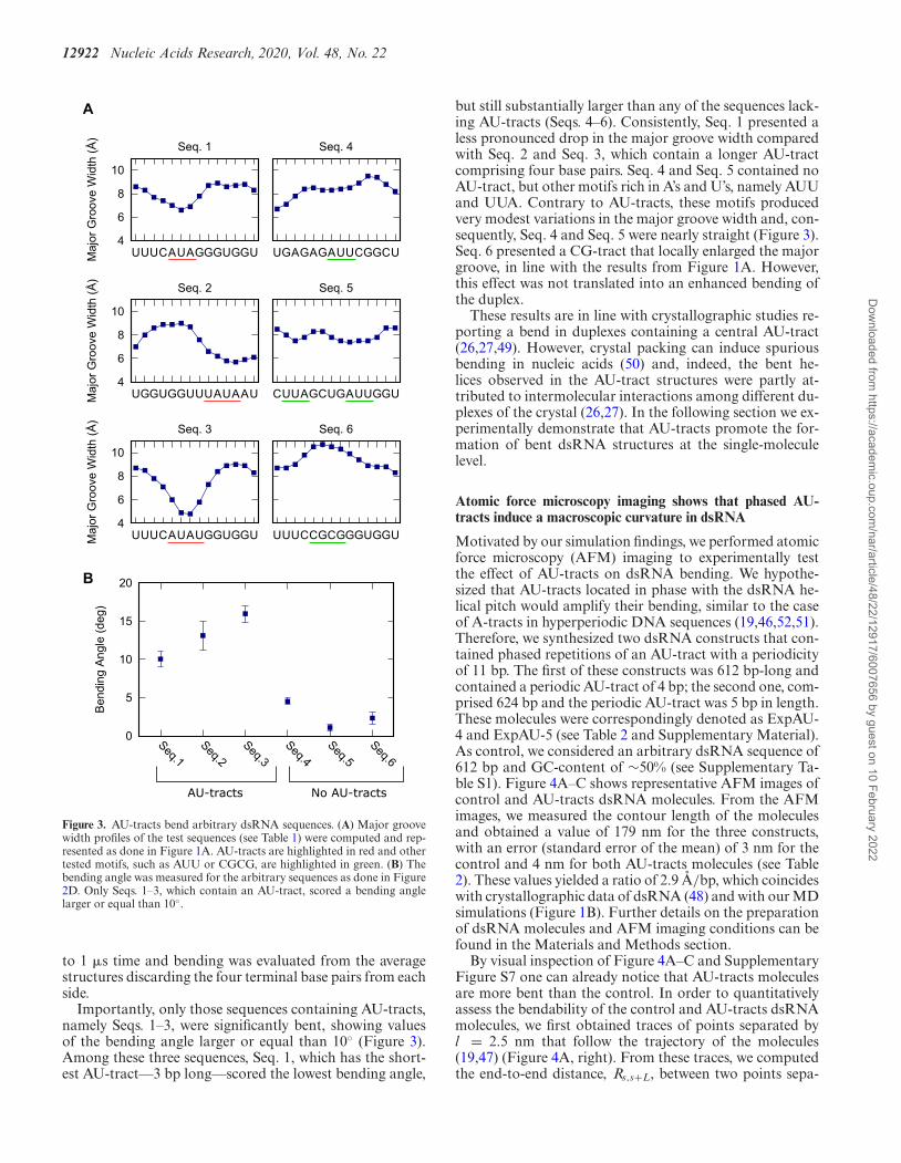
Figure 3. AU-tracts bend arbitrary dsRNA sequences. (A) Major groovewidth profiles of the test sequences (see Table 1) were computed and rep-resented as done in Figure 1A. AU-tracts are highlighted in red and othertested motifs, such as AUU or CGCG, are highlighted in green. (B) Thebending angle was measured for the arbitrary sequences as done in Figure2D. Only Seqs. 1–3, which contain an AU-tract, scored a bending anglelarger or equal than 10◦.
to 1 �s time and bending was evaluated from the averagestructures discarding the four terminal base pairs from eachside.
Importantly, only those sequences containing AU-tracts,namely Seqs. 1–3, were significantly bent, showing valuesof the bending angle larger or equal than 10◦ (Figure 3).Among these three sequences, Seq. 1, which has the short-est AU-tract––3 bp long––scored the lowest bending angle,
but still substantially larger than any of the sequences lack-ing AU-tracts (Seqs. 4–6). Consistently, Seq. 1 presented aless pronounced drop in the major groove width comparedwith Seq. 2 and Seq. 3, which contain a longer AU-tractcomprising four base pairs. Seq. 4 and Seq. 5 contained noAU-tract, but other motifs rich in A’s and U’s, namely AUUand UUA. Contrary to AU-tracts, these motifs producedvery modest variations in the major groove width and, con-sequently, Seq. 4 and Seq. 5 were nearly straight (Figure 3).Seq. 6 presented a CG-tract that locally enlarged the majorgroove, in line with the results from Figure 1A. However,this effect was not translated into an enhanced bending ofthe duplex.
These results are in line with crystallographic studies re-porting a bend in duplexes containing a central AU-tract(26,27,49). However, crystal packing can induce spuriousbending in nucleic acids (50) and, indeed, the bent he-lices observed in the AU-tract structures were partly at-tributed to intermolecular interactions among different du-plexes of the crystal (26,27). In the following section we ex-perimentally demonstrate that AU-tracts promote the for-mation of bent dsRNA structures at the single-moleculelevel.
Atomic force microscopy imaging shows that phased AU-tracts induce a macroscopic curvature in dsRNA
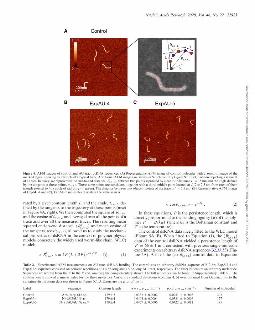
Motivated by our simulation findings, we performed atomicforce microscopy (AFM) imaging to experimentally testthe effect of AU-tracts on dsRNA bending. We hypothe-sized that AU-tracts located in phase with the dsRNA he-lical pitch would amplify their bending, similar to the caseof A-tracts in hyperperiodic DNA sequences (19,46,52,51).Therefore, we synthesized two dsRNA constructs that con-tained phased repetitions of an AU-tract with a periodicityof 11 bp. The first of these constructs was 612 bp-long andcontained a periodic AU-tract of 4 bp; the second one, com-prised 624 bp and the periodic AU-tract was 5 bp in length.These molecules were correspondingly denoted as ExpAU-4 and ExpAU-5 (see Table 2 and Supplementary Material).As control, we considered an arbitrary dsRNA sequence of612 bp and GC-content of ∼50% (see Supplementary Ta-ble S1). Figure 4A–C shows representative AFM images ofcontrol and AU-tracts dsRNA molecules. From the AFMimages, we measured the contour length of the moleculesand obtained a value of 179 nm for the three constructs,with an error (standard error of the mean) of 3 nm for thecontrol and 4 nm for both AU-tracts molecules (see Table2). These values yielded a ratio of 2.9 A/bp, which coincideswith crystallographic data of dsRNA (48) and with our MDsimulations (Figure 1B). Further details on the preparationof dsRNA molecules and AFM imaging conditions can befound in the Materials and Methods section.
By visual inspection of Figure 4A–C and SupplementaryFigure S7 one can already notice that AU-tracts moleculesare more bent than the control. In order to quantitativelyassess the bendability of the control and AU-tracts dsRNAmolecules, we first obtained traces of points separated byl = 2.5 nm that follow the trajectory of the molecules(19,47) (Figure 4A, right). From these traces, we computedthe end-to-end distance, Rs,s+L, between two points sepa-
Dow
nloaded from https://academ
ic.oup.com/nar/article/48/22/12917/6007656 by guest on 10 February 2022
Nucleic Acids Research, 2020, Vol. 48, No. 22 12923
ExpAU-4
ControlA
B C ExpAU-5
θs,s+L
L
rL
Rs,s+L
0 nm
2 nm
l
Figure 4. AFM images of control and AU-tract dsRNA sequences. (A) Representative AFM image of control molecules with a zoom-in image of themarked region showing an example of a typical trace. Additional AFM images are shown in Supplementary Figure S7. Inset, cartoon depicting a segmentof a trace. In black, we represented the end-to-end distance, Rs,s+L, between two points separated by a contour distance L = 15 nm and the angle definedby the tangents at those points, θs,s+L. Those same points are considered together with a third, middle point located at L/2 = 7.5 nm from each of them(purple points) to fit a circle of radius rL (in green). The distance between two adjacent points of the trace is l = 2.5 nm. (B) Representative AFM imagesof ExpAU-4 and (C), ExpAU-5 molecules. Z-scale is the same as in A.
rated by a given contour length L, and the angle, θs,s+L, de-fined by the tangents to the trajectory at those points (insetin Figure 4A, right). We then computed the square of Rs,s+Land the cosine of θs,s+L and averaged over all the points of atrace and over all the measured traces. The resulting meansquared end-to-end distance, 〈R2
s,s+L〉 and mean cosine ofthe tangents, 〈cos θs,s+L〉, allowed us to study the mechani-cal properties of dsRNA in the context of polymer physicsmodels, concretely the widely used worm-like chain (WLC)model:
< R2s,s+L >= 4P
(L + 2P
(e−L/2P − 1
)), (1)
< cos θs,s+L >= e− L2P . (2)
In these equations, P is the persistence length, which isdirectly proportional to the bending rigidity (B) of the poly-mer P = B/kBT (where kB is the Boltzman constant andT is the temperature).
The control dsRNA data nicely fitted to the WLC model(Figure 5A, B). When fitted to Equation (1), the 〈R2
s,s+L〉data of the control dsRNA yielded a persistence length ofP = 66 ± 1 nm, consistent with previous single-moleculeexperiments on arbitrary dsRNA sequences (32,33,53) (Fig-ure 5A). A fit of the 〈cos θs,s+L〉 control data to Equation
Table 2. Experimental AFM measurements on AU-tract dsRNA bending. The control was an arbitrary dsRNA sequence of 612 bp; ExpAU-4 andExpAU-5 sequences consisted on periodic repetitions of a 4 bp-long and a 5 bp-long AU-tract, respectively. The letter N denotes an arbitrary nucleotide.Sequences are written from the 5′ to the 3′ end, omitting the complementary strand. The full sequences can be found in Supplementary Table S1. Thecontour length showed a similar value for the three molecules. Curvature standard deviations (columns 4, 5) were obtained from Gaussian fits to thecurvature distributions data sets shown in Figure 5C, D. Errors are the error of the fit
Label Sequence Contour length σ C,L = 15 nm (nm−1) σ C,L = 25 nm (nm−1) Number of molecules
Control Arbitrary, 612 bp 179 ± 3 0.0332 ± 0.0002 0.0251 ± 0.0003 202ExpAU-4 N7 (AUAU N7)55 179 ± 4 0.0404 ± 0.0004 0.0331 ± 0.0006 127ExpAU-5 N7 (UAUAU N6)56N 179 ± 4 0.0467 ± 0.0006 0.0422 ± 0.0011 195
Dow
nloaded from https://academ
ic.oup.com/nar/article/48/22/12917/6007656 by guest on 10 February 2022
12924 Nucleic Acids Research, 2020, Vol. 48, No. 22
A B
C D
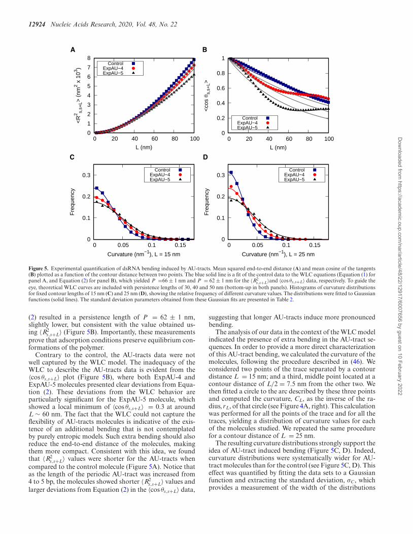
Figure 5. Experimental quantification of dsRNA bending induced by AU-tracts. Mean squared end-to-end distance (A) and mean cosine of the tangents(B) plotted as a function of the contour distance between two points. The blue solid line is a fit of the control data to the WLC equations (Equation (1) forpanel A, and Equation (2) for panel B), which yielded P =66 ± 1 nm and P = 62 ± 1 nm for the 〈R2
s,s+L〉and 〈cos θs,s+L〉 data, respectively. To guide theeye, theoretical WLC curves are included with persistence lengths of 30, 40 and 50 nm (bottom-up in both panels). Histograms of curvature distributionsfor fixed contour lengths of 15 nm (C) and 25 nm (D), showing the relative frequency of different curvature values. The distributions were fitted to Gaussianfunctions (solid lines). The standard deviation parameters obtained from these Gaussian fits are presented in Table 2.
(2) resulted in a persistence length of P = 62 ± 1 nm,slightly lower, but consistent with the value obtained us-ing 〈R2
s,s+L〉 (Figure 5B). Importantly, these measurementsprove that adsorption conditions preserve equilibrium con-formations of the polymer.
Contrary to the control, the AU-tracts data were notwell captured by the WLC model. The inadequacy of theWLC to describe the AU-tracts data is evident from the〈cos θs,s+L〉 plot (Figure 5B), where both ExpAU-4 andExpAU-5 molecules presented clear deviations from Equa-tion (2). These deviations from the WLC behavior areparticularly significant for the ExpAU-5 molecule, whichshowed a local minimum of 〈cos θs,s+L〉 = 0.3 at aroundL ∼ 60 nm. The fact that the WLC could not capture theflexibility of AU-tracts molecules is indicative of the exis-tence of an additional bending that is not contemplatedby purely entropic models. Such extra bending should alsoreduce the end-to-end distance of the molecules, makingthem more compact. Consistent with this idea, we foundthat 〈R2
s,s+L〉 values were shorter for the AU-tracts whencompared to the control molecule (Figure 5A). Notice thatas the length of the periodic AU-tract was increased from4 to 5 bp, the molecules showed shorter 〈R2
s,s+L〉 values andlarger deviations from Equation (2) in the 〈cos θs,s+L〉 data,
suggesting that longer AU-tracts induce more pronouncedbending.
The analysis of our data in the context of the WLC modelindicated the presence of extra bending in the AU-tract se-quences. In order to provide a more direct characterizationof this AU-tract bending, we calculated the curvature of themolecules, following the procedure described in (46). Weconsidered two points of the trace separated by a contourdistance L = 15 nm; and a third, middle point located at acontour distance of L/2 = 7.5 nm from the other two. Wethen fitted a circle to the arc described by these three pointsand computed the curvature, CL, as the inverse of the ra-dius, rL, of that circle (see Figure 4A, right). This calculationwas performed for all the points of the trace and for all thetraces, yielding a distribution of curvature values for eachof the molecules studied. We repeated the same procedurefor a contour distance of L = 25 nm.
The resulting curvature distributions strongly support theidea of AU-tract induced bending (Figure 5C, D). Indeed,curvature distributions were systematically wider for AU-tract molecules than for the control (see Figure 5C, D). Thiseffect was quantified by fitting the data sets to a Gaussianfunction and extracting the standard deviation, σC, whichprovides a measurement of the width of the distributions
Dow
nloaded from https://academ
ic.oup.com/nar/article/48/22/12917/6007656 by guest on 10 February 2022
Nucleic Acids Research, 2020, Vol. 48, No. 22 12925
(see Table 2). The σC values obtained for the ExpAU-5molecule were systematically larger than the σC values ofExpAU-4, which, in turn, were larger than those of the con-trol. This finding indicates that AU-tract molecules weremore prone to adopt highly curved conformations whencompared to the control. Moreover, this effect was morepronounced for the ExpAU-5 molecule, supporting that themagnitude of AU-tract bending increases with the AU-tractlength, as predicted by our MD simulations (Figure 2D).
AU-tracts: similarities and differences with DNA A-tracts
Sequence-dependent bending is known to take place indsDNA by means of A-tracts: sequences of at least fourA·T base pairs without a TpA step. When several A-tractsare located in phase with the helical pitch they producea macroscopic curvature in the DNA (18). This curvaturecan be directly observed using AFM or electron microscopy(19,46,54,55), or can be inferred from gel electrophoresisexperiments (51). In addition, A-tracts display a particu-lar conformation at the molecular level, which differs fromthat of canonical B-DNA (18,20). In the following, we com-pare these well-known features of dsDNA A-tracts – macro-scopic curvature and molecular conformation – with ourfindings on dsRNA AU-tracts.
Previous AFM works have provided a detailed picture ofbending deformations in dsDNA molecules with A-tract-induced curvature. These experiments showed that, as aconsequence of that curvature, the structural properties ofdsDNA sequences with phased A-tracts exhibit significantdeviations from the WLC model (46). This effect was alsoobserved for the dsRNA AU-tracts (see Figure 5A, B). Al-though our AU-tracts 〈R2
s,s+L〉 data showed no clear dis-crepancy with respect to the WLC prediction, such devi-ations only appeared in the A-tracts for contour lengthsgreater than ∼120 nm length (19,46), which are beyond therange studied here (<100 nm). The 〈cos θs,s+L〉 of the A-tracts, on the contrary, deviated from the WLC behavior atshorter contour lengths (∼50 nm) and is, therefore, a bet-ter indicator of the existence of intrinsic curvature. Consis-tent with the presence of intrinsic bending, our AU-tractsalso presented significant deviations from the WLC in the〈cos θs,s+L〉(L) data. Moreover, the shape of the 〈cos θ〉(L)plot for the phased AU-tract studied here is remarkably sim-ilar to an intrinsically-bent A-tract dsDNA that we recentlyreported (19).
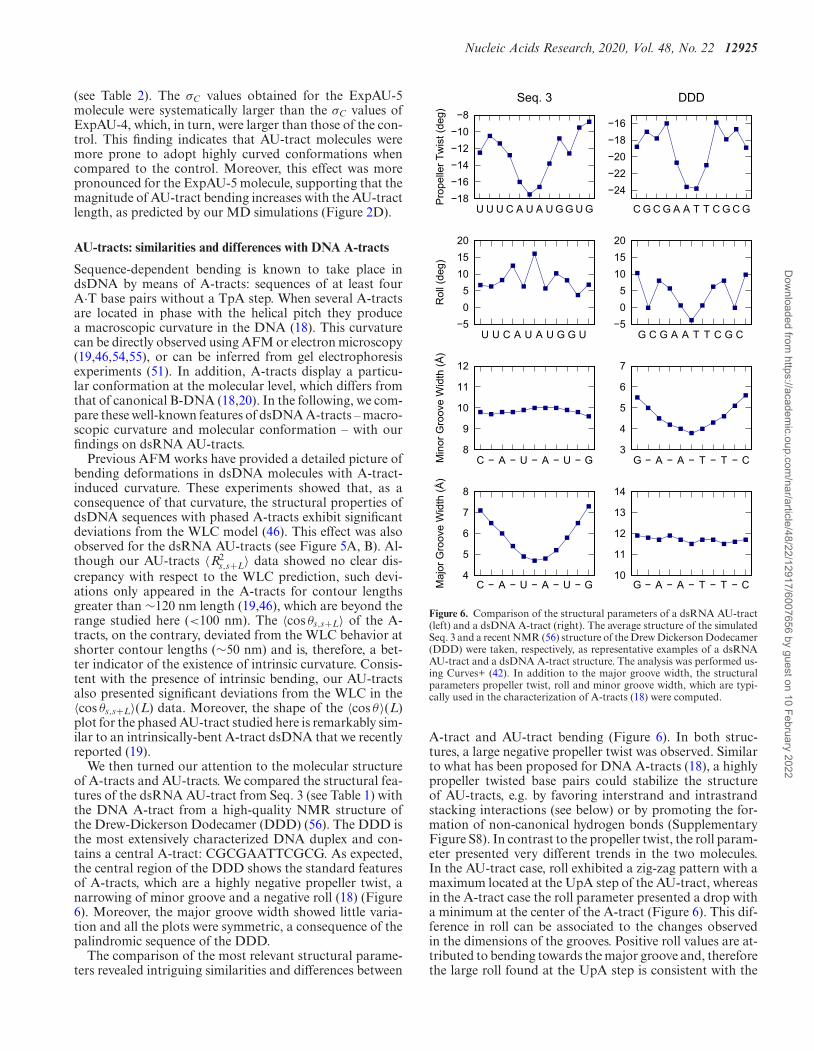
We then turned our attention to the molecular structureof A-tracts and AU-tracts. We compared the structural fea-tures of the dsRNA AU-tract from Seq. 3 (see Table 1) withthe DNA A-tract from a high-quality NMR structure ofthe Drew-Dickerson Dodecamer (DDD) (56). The DDD isthe most extensively characterized DNA duplex and con-tains a central A-tract: CGCGAATTCGCG. As expected,the central region of the DDD shows the standard featuresof A-tracts, which are a highly negative propeller twist, anarrowing of minor groove and a negative roll (18) (Figure6). Moreover, the major groove width showed little varia-tion and all the plots were symmetric, a consequence of thepalindromic sequence of the DDD.
The comparison of the most relevant structural parame-ters revealed intriguing similarities and differences between
−18−16−14−12−10−8
U U U C A U A U G G U G
Pro
pelle
r Tw
ist (
deg)
Seq. 3
−24−22−20−18−16
C G C G A A T T C G C G
DDD
−505
101520
U U C A U A U G G U
Rol
l (de
g)
−505
101520
G C G A A T T C G C
8
9
10
11
12
C − A − U − A − U − GMin
or G
roov
e W
idth
(Å)
C − A − U − A − U − G G − A − A − T − T − C
3
4
5
6
7
G − A − A − T − T − C
4
5
6
7
8
Maj
or G
roov
e W
idth
(Å)
10
11
12
13
14
Figure 6. Comparison of the structural parameters of a dsRNA AU-tract(left) and a dsDNA A-tract (right). The average structure of the simulatedSeq. 3 and a recent NMR (56) structure of the Drew Dickerson Dodecamer(DDD) were taken, respectively, as representative examples of a dsRNAAU-tract and a dsDNA A-tract structure. The analysis was performed us-ing Curves+ (42). In addition to the major groove width, the structuralparameters propeller twist, roll and minor groove width, which are typi-cally used in the characterization of A-tracts (18) were computed.
A-tract and AU-tract bending (Figure 6). In both struc-tures, a large negative propeller twist was observed. Similarto what has been proposed for DNA A-tracts (18), a highlypropeller twisted base pairs could stabilize the structureof AU-tracts, e.g. by favoring interstrand and intrastrandstacking interactions (see below) or by promoting the for-mation of non-canonical hydrogen bonds (SupplementaryFigure S8). In contrast to the propeller twist, the roll param-eter presented very different trends in the two molecules.In the AU-tract case, roll exhibited a zig-zag pattern with amaximum located at the UpA step of the AU-tract, whereasin the A-tract case the roll parameter presented a drop witha minimum at the center of the A-tract (Figure 6). This dif-ference in roll can be associated to the changes observedin the dimensions of the grooves. Positive roll values are at-tributed to bending towards the major groove and, thereforethe large roll found at the UpA step is consistent with the
Dow
nloaded from https://academ
ic.oup.com/nar/article/48/22/12917/6007656 by guest on 10 February 2022
12926 Nucleic Acids Research, 2020, Vol. 48, No. 22
1
2
2
3
3
2
2
3
3
Adenines in UpA step approach each other
A:U base pairs undergo propeller twisting
Propeller twist propagates along the AU-tract
1
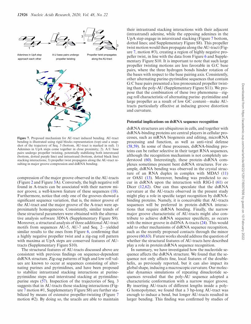
Figure 7. Proposed mechanism for AU-tract induced bending. AU-tractbending is illustrated using rigid blocks representation (top) and a snap-shot of the trajectory of Seq. 3 (bottom, AU-tract is marked in red). 1)Adenines in UpA steps come together in close proximity; 2). A:U basepairs undergo propeller twisting, potentially stabilizing both interstrand(bottom, dotted purple line) and intrastrand (bottom, dotted black line)stacking interactions; 3) propeller twist propagates along the AU-tract re-sulting in major groove compression and dsRNA bending.
compression of the major groove observed in the AU-tracts(Figure 2 and Figure 3A). Conversely, the high negative rollfound in A-tracts can be associated with their narrow mi-nor groove, a well-known feature of these sequences (18).Furthermore, notice that only one of the grooves showed asignificant sequence variation, that is, the minor groove ofthe AU-tract and the major groove of the A-tract were ap-proximately homogeneous. Consistently, similar trends ofthese structural parameters were obtained with the alterna-tive analysis software 3DNA (Supplementary Figure S9).Moreover, a structural analysis of three additional AU-tractmotifs from sequences AU-5, AU-7 and Seq. 2––yieldedsimilar results to the ones from Figure 6, confirming thata highly negative propeller twist and a zig-zag roll patternwith maxima at UpA steps are conserved features of AU-tracts (Supplementary Figure S10).
The structural features of AU-tracts discussed above areconsistent with previous findings on sequence-dependentdsRNA structure. Zig-zag patterns of high and low roll val-ues are known to occur at sequences consisting of alter-nating purines and pyrimidines, and have been proposedto stabilize intrastrand stacking interactions at purine-pyrimidine steps and interstrand stacking at pyrimidine-purine steps (57). Inspection of the trajectories of Seq. 3suggests that in AU-tracts those stacking interactions (Fig-ure 7 motion #1, Supplementary Figure S8) are further sta-bilized by means of extensive propeller-twisting (Figure 7motion #2). By doing so, the uracils are able to maintain
their intrastrand stacking interactions with their adjacent(intrastrand) adenine, while the opposing adenines in theUpA step engage in interstrand stacking (Figure 7 bottom,dotted lines; and Supplementary Figure S8). This propellertwist motion would then propagate along the AU-tract (Fig-ure 7, motion #3), creating a region of highly negative pro-peller twist, in line with the data from Figure 6 and Supple-mentary Figure S10. It is important to note that such largepropeller twisting motions are less favorable in G:C basepairs, where the three hydrogen bonds hinder rotation ofthe bases with respect to the base pairing axis. Consistently,other alternating purine-pyrimidine sequences that containG:C base pairs presented a less pronounced propeller twist-ing than the poly-AU (Supplementary Figure S11). We pro-pose that the combination of these two phenomena––zig-zag roll characteristic of alternating purine–pyrimidine andlarge propeller as a result of low GC content––make AU-tracts particularly effective at inducing groove distortionand curvature.
Potential implications on dsRNA sequence recognition
dsRNA structures are ubiquitous in cells, and together withdsRNA-binding proteins are central players in cellular pro-cesses, such as mRNA biogenesis and editing, microRNAprocessing and function, as well as anti-viral defense(58,59). In some of these processes, dsRNA-binding pro-teins can be rather selective in their target RNA sequences,although the recognition mechanism is not completely un-derstood (60). Interestingly, these protein–dsRNA com-plexes sometimes present bent dsRNA structures. For ex-ample, dsRNA bending was observed in the crystal struc-ture of an RNA duplex in complex with MDA5 (11)or OAS1 (13). Moreover, bending was predicted to oc-cur in dsRNA upon the interaction with RIG-I (61) orDicer (12,62). One can thus speculate that the dsRNAcurvature at the AU-tracts observed in the present studymight play a role in specific target recognition by dsRNA-binding proteins. Namely, it is conceivable that AU-tractssequences will be preferred in protein–dsRNA interac-tions that require dsRNA bending. Finally, the narrowmajor groove characteristic of AU-tracts might also con-tribute to achieve dsRNA sequence specificity, as occurswith the minor groove in dsDNA A-tracts (24). This wouldadd to other mechanisms of dsRNA sequence recognition,such as the recently proposed contacts through the minorgroove (60,63). Future works should address the question ofwhether the structural features of AU-tracts here describedplay a role in protein:dsRNA sequence recognition.
In summary, we have investigated how the nucleotide se-quence affects the dsRNA structure. We found that the se-quence not only affects fine, local features of the double-helix, as previously reported, but it can also impact itsglobal shape, inducing a macroscopic curvature. Our molec-ular dynamics simulations of repeating dinucleotide se-quences revealed that the poly-AU sequence adopted acharacteristic conformation with a narrow major groove.By inserting AU-tracts of different lengths inside a poly-G homopolymer, we found that a 3 bp-long AU-tract wasenough to induce a bend, but longer AU-tracts resulted inlarger bending. This finding was confirmed by studies of
Dow
nloaded from https://academ
ic.oup.com/nar/article/48/22/12917/6007656 by guest on 10 February 2022
Nucleic Acids Research, 2020, Vol. 48, No. 22 12927
AU-tracts located in arbitrary sequences. Our simulationresults guided the design of dsRNA constructs suitable formeasuring the effect of AU-tract bending in AFM experi-ments. Using AFM imaging, we found that these AU-tractmolecules exhibited higher curvature than control dsRNA’sof arbitrary sequence, confirming the prediction of our sim-ulations. A molecular mechanism for AU-tract bending isproposed, where extensive propeller twisting of the A:Ubase pairs induce major groove compression.
Intrinsic bending induced by dsDNA A-tracts has beenlinked to multiple biological functions such as nucleo-some positioning, localization of supercoils or germ-linegene silencing. It is therefore expected that the sequence-dependent bending reported here for dsRNA might alsohave important biological implications. On one hand, thebent structure of the AU-tracts could be exploited in the for-mation of tertiary contacts in the process of RNA folding.On the other hand, AU-tracts might provide a mechanismfor sequence recognition based on dsRNA shape. Finally,our finding that the global dsRNA structure is sequence de-pendent might be relevant in the field of RNA nanotech-nology. Future works might explore how this effect can beexploited in the design of complex RNA nanostructures.
SUPPLEMENTARY DATA
Supplementary Data are available at NAR Online.
ACKNOWLEDGEMENTS
The authors acknowledge the computer resources, technicalexpertise and assistance provided by the Red Espanola deSupercomputacion at the Minotauro Supercomputer (BSC,Barcelona).
FUNDING
Spanish MINECO [MAT2017-83273-R (AEI/FEDER,UE) to R.P., BFU2017-83794-P (AEI/FEDER, UEto F.M.-H.) and Comunidad de Madrid (Tec4Bio –S2018/NMT-4443 and NanoBioCancer – Y2018/BIO-4747 to F.M.-H.]; R.P. acknowledges support from theSpanish Ministry of Science and Innovation, through the‘Marıa de Maeztu’ Programme for Units of Excellencein R&D [CEX2018-000805-M]; F.M.-H. acknowledgessupport from European Research Council (ERC) under theEuropean Union Horizon 2020 research and innovation[681299]; J. G. V. acknowledges funding from a MarieSklodowska-Curie Fellowship within the Horizons 2020framework (DLV-795286) and the Swiss National ScienceFoundation (grant number CRSK-2 190731/1). AlbertoM.-G. acknowledges support from the International PhDProgram of ‘La Caixa-Severo Ochoa’ as a recipient of aPhD fellowship; U.F.G. acknowledges support from theSwiss National Science Foundation [31003A 179256/1];Alejandro M.-G. and MM-B acknowledge support fromthe Spanish Ministry of Competitiveness and Industry asa recipients of a FPI fellowship [REFs BES-2015-071244,PRE2018-083464, respectively]. Funding for open accesscharge: European Research Council (ERC) [681299].Conflict of interest statement. None declared.
REFERENCES1. Fire,A., Xu,S., Montgomery,M.K., Kostas,S.A., Driver,S.E. and
Mello,C.C. (1998) Potent and specific genetic interference bydouble-stranded RNA in Caenorhabditis elegans. Nature, 391,806–811.
2. Yoneyama,M., Kikuchi,M., Natsukawa,T., Shinobu,N., Imaizumi,T.,Miyagishi,M., Taira,K., Akira,S. and Fujita,T. (2004) The RNAhelicase RIG-I has an essential function in double-strandedRNA-induced innate antiviral responses. Nat. Immunol., 5, 730–737.
3. Schimmel,P. (2018) The emerging complexity of the tRNA world:mammalian tRNAs beyond protein synthesis. Nat. Rev. Mol. CellBiol., 19, 45–58.
4. Mandal,M. and Breaker,R.R. (2004) Gene regulation byriboswitches. Nat. Rev. Mol. Cell Biol., 5, 451–463.
5. Wimberly,B.T., Brodersen,D.E., Clemons,W.M. Jr,Morgan-Warren,R.J., Carter,A.P., Vonrhein,C., Hartsch,T. andRamakrishnan,V. (2000) Structure of the 30S ribosomal subunit.Nature, 407, 327–339.
6. Nissen,P., Ippolito,J.A., Ban,N., Moore,P.B. and Steitz,T.A. (2001)RNA tertiary interactions in the large ribosomal subunit: theA-minor motif. Proc. Natl. Acad. Sci. U.S.A., 98, 4899–4903.
7. Shi,Y. (2017) Mechanistic insights into precursor messenger RNAsplicing by the spliceosome. Nat. Rev. Mol. Cell Biol., 18, 655–670.
8. Marino,J.P., Gregorian,R.S. Jr, Csankovszki,G. and Crothers,D.M.(1995) Bent helix formation between RNA hairpins withcomplementary loops. Science, 268, 1448–1454.
9. Denny,S.K., Bisaria,N., Yesselman,J.D., Das,R., Herschlag,D. andGreenleaf,W.J. (2018) High-Throughput investigation of diversejunction elements in RNA tertiary folding. Cell, 174, 377–390.
10. Das,R., Karanicolas,J. and Baker,D. (2010) Atomic accuracy inpredicting and designing noncanonical RNA structure. Nat.Methods, 7, 291–294.
11. Uchikawa,E., Lethier,M., Malet,H., Brunel,J., Gerlier,D. andCusack,S. (2016) Structural analysis of dsRNA binding to anti-viralpattern recognition receptors LGP2 and MDA5. Mol. Cell, 62,586–602.
12. MacRae,I.J., Zhou,K. and Doudna,J.A. (2007) Structuraldeterminants of RNA recognition and cleavage by Dicer. Nat. Struct.Mol. Biol., 14, 934–940.
13. Donovan,J., Dufner,M. and Korennykh,A. (2013) Structural basis forcytosolic double-stranded RNA surveillance by humanoligoadenylate synthetase 1. Proc. Natl. Acad. Sci. U.S.A., 110,1652–1657.
14. Guo,P. (2010) The emerging field of RNA nanotechnology. Nat.Nanotechnol., 5, 833–842.
15. Salmon,L., Yang,S. and Al-Hashimi,H.M. (2014) Advances in thedetermination of nucleic acid conformational ensembles. Annu. Rev.Phys. Chem., 65, 293–316.
16. Ganser,L.R., Kelly,M.L., Herschlag,D. and Al-Hashimi,H.M. (2019)The roles of structural dynamics in the cellular functions of RNAs.Nat. Rev. Mol. Cell Biol., 20, 474–489.
17. Sponer,J., Bussi,G., Krepl,M., Banas,P., Bottaro,S., Cunha,R.A.,Gil-Ley,A., Pinamonti,G., Poblete,S., Jurecka,P. et al. (2018) RNAstructural dynamics as captured by molecular simulations: acomprehensive overview. Chem. Rev., 118, 4177–4338.
18. Haran,T.E. and Mohanty,U. (2009) The unique structure of A-tractsand intrinsic DNA bending. Q. Rev. Biophys., 42, 41–81.
19. Marin-Gonzalez,A., Pastrana,C.L., Bocanegra,R.,Martın-Gonzalez,A., Vilhena,J.G., Perez,R., Ibarra,B.,Aicart-Ramos,C. and Moreno-Herrero,F. (2020) Understanding theparadoxical mechanical response of in-phase A-tracts at differentforce regimes. Nucleic Acids Res., 48, 5024–5036.
20. Nelson,H.C., Finch,J.T., Luisi,B.F. and Klug,A. (1987) The structureof an oligo(dA).oligo(dT) tract and its biological implications.Nature, 330, 221–226.
21. Sherer,E.C., Harris,S.A., Soliva,R., Orozco,M. and Laughton,C.A.(1999) Molecular dynamics studies of DNA A-Tract structure andflexibility. J. Am. Chem. Soc., 121, 5981–5991.
22. Johnson,S., Chen,Y.J. and Phillips,R. (2013) Poly(dA:dT)-rich DNAsare highly flexible in the context of DNA looping. PLoS One, 8,e75799.
Dow
nloaded from https://academ
ic.oup.com/nar/article/48/22/12917/6007656 by guest on 10 February 2022
12928 Nucleic Acids Research, 2020, Vol. 48, No. 22
23. Kim,S.H., Ganji,M., Kim,E., van der Torre,J., Abbondanzieri,E. andDekker,C. (2018) DNA sequence encodes the position of DNAsupercoils. eLife, 7, e36557.
24. Rohs,R., Jin,X., West,S.M., Joshi,R., Honig,B. and Mann,R.S.(2010) Origins of specificity in protein-DNA recognition. Annu. Rev.Biochem., 79, 233–269.
25. Iric,K., Subramanian,M., Oertel,J., Agarwal,N.P., Matthies,M.,Periole,X., Sakmar,T.P., Huber,T., Fahmy,K. and Schmidt,T.L.(2018) DNA-encircled lipid bilayers. Nanoscale, 10, 18463–18467.
26. Dock-Bregeon,A.C., Chevrier,B., Podjarny,A., Moras,D.,deBear,J.S., Gough,G.R., Gilham,P.T. and Johnson,J.E. (1988) Highresolution structure of the RNA duplex [U(U-A)6A]2. Nature, 335,375–378.
27. Dock-Bregeon,A.C., Chevrier,B., Podjarny,A., Johnson,J., deBear,J.S., Gough,G.R., Gilham,P.T. and Moras,D. (1989)Crystallographic structure of an RNA helix: [U(UA)6A]2. J. Mol.Biol., 209, 459–474.
28. Perez,A., Noy,A., Lankas,F., Luque,F.J. and Orozco,M. (2004) Therelative flexibility of B-DNA and A-RNA duplexes: databaseanalysis. Nucleic Acids Res., 32, 6144–6151.
29. Besseova,I., Otyepka,M., Reblova,K. and Sponer,J. (2009)Dependence of A-RNA simulations on the choice of the force fieldand salt strength. Phys. Chem. Chem. Phys.: PCCP, 11, 10701–10711.
30. Besseova,I., Banas,P., Kuhrova,P., Kosinova,P., Otyepka,M. andSponer,J. (2012) Simulations of A-RNA duplexes. The effect ofsequence, solute force field, water model, and salt concentration. J.Phys. Chem. B, 116, 9899–9916.
31. Marin-Gonzalez,A., Vilhena,J.G., Moreno-Herrero,F. and Perez,R.(2019) Sequence-dependent mechanical properties of double-strandedRNA. Nanoscale, 11, 21471–21478.
32. Abels,J.A., Moreno-Herrero,F., van der Heijden,T., Dekker,C. andDekker,N.H. (2005) Single-molecule measurements of the persistencelength of double-stranded RNA. Biophys. J., 88, 2737–2744.
33. Herrero-Galan,E., Fuentes-Perez,M.E., Carrasco,C., Valpuesta,J.M.,Carrascosa,J.L., Moreno-Herrero,F. and Arias-Gonzalez,J.R. (2013)Mechanical identities of RNA and DNA double helices unveiled atthe single-molecule level. J. Am. Chem. Soc., 135, 122–131.
34. Marin-Gonzalez,A., Vilhena,J.G., Perez,R. and Moreno-Herrero,F.(2017) Understanding the mechanical response of double-strandedDNA and RNA under constant stretching forces using all-atommolecular dynamics. Proc. Natl. Acad. Sci. U.S.A., 114, 7049–7054.
35. Salomon-Ferrer,R., Gotz,A.W., Poole,D., Le Grand,S. andWalker,R.C. (2013) Routine microsecond molecular dynamicssimulations with AMBER on GPUs. 2. Explicit solvent Particle MeshEwald. J. Chem. Theory Comput., 9, 3878–3888.
36. Le Grand,S., Gotz,A.W. and Walker,R.C. (2013) SPFP: Speedwithout compromise––a mixed precision model for GPU acceleratedmolecular dynamics simulations. Comput. Phys. Commun., 184,374–380.
37. Cornell,W.D., Cieplak,P., Bayly,C.I., Gould,I.R., Merz,K.M.,Ferguson,D.M., Spellmeyer,D.C., Fox,T., Caldwell,J.W. andKollman,P.A. (1995) A second generation force field for thesimulation of proteins, nucleic acids, and organic molecules. J. Am.Chem. Soc., 117, 5179–5797.
38. Perez,A., Marchan,I., Svozil,D., Sponer,J., Cheatham,T.E. 3rd.,Laughton,C.A. and Orozco,M. (2007) Refinement of the AMBERforce field for nucleic acids: improving the description ofalpha/gamma conformers. Biophys. J., 92, 3817–3829.
39. Zgarbova,M., Otyepka,M., Sponer,J., Mladek,A., Banas,P.,Cheatham,T.E. 3rd. and Jurecka,P. (2011) Refinement of the Cornellet al. nucleic acids force field based on reference quantum chemicalcalculations of glycosidic torsion profiles. J. Chem. Theory Comput.,7, 2886–2902.
40. Joung,I.S. and Cheatham,T.E. 3rd. (2009) Molecular dynamicssimulations of the dynamic and energetic properties of alkali andhalide ions using water-model-specific ion parameters. J. Phys. Chem.B, 113, 13279–13290.
41. Jorgensen,W.L., Chandrasekhar,J., Madura,J.D., Impey,R.W. andKlein,M. (1983) Comparison of simple potential functions forsimulating liquid water. J. Chem. Phys., 79, 926.
42. Lavery,R., Moakher,M., Maddocks,J.H., Petkeviciute,D. andZakrzewska,K. (2009) Conformational analysis of nucleic acidsrevisited: Curves+. Nucleic Acids Res., 37, 5917–5929.
43. Lu,X.J. and Olson,W.K. (2003) 3DNA: a software package for theanalysis, rebuilding and visualization of three-dimensional nucleicacid structures. Nucleic Acids Res., 31, 5108–5121.
44. Dekker,N.H., Abels,J.A., Veenhuizen,P.T., Bruinink,M.M. andDekker,C. (2004) Joining of long double-stranded RNA moleculesthrough controlled overhangs. Nucleic Acids Res., 32, e140.
45. Horcas,I., Fernandez,R., Gomez-Rodriguez,J.M., Colchero,J.,Gomez-Herrero,J. and Baro,A.M. (2007) WSXM: a software forscanning probe microscopy and a tool for nanotechnology. Rev. Sci.Instrum., 78, 013705.
46. Moreno-Herrero,F., Seidel,R., Johnson,S.M., Fire,A. andDekker,N.H. (2006) Structural analysis of hyperperiodic DNA fromCaenorhabditis elegans. Nucleic Acids Res., 34, 3057–3066.
47. Wiggins,P.A., van der Heijden,T., Moreno-Herrero,F., Spakowitz,A.,Phillips,R., Widom,J., Dekker,C. and Nelson,P.C. (2006) Highflexibility of DNA on short length scales probed by atomic forcemicroscopy. Nat. Nanotechnol., 1, 137–141.
48. Bloomfield,V.A., Crothers,D.M. and Tinoco,I. (2000) Nucleic Acids:Structures, Properties and functions. University Science Books,Sausalito, California.
49. Wahl,M.C., Ban,C., Sekharudu,C., Ramakrishnan,B. andSundaralingam,M. (1996) Structure of the purine-pyrimidinealternating RNA double helix, r(GUAUAUA)d(C), with a 3’-terminaldeoxy residue. Acta Crystallogr. D. Biol. Crystallogr., 52, 655–667.
50. DiGabriele,A.D., Sanderson,M.R. and Steitz,T.A. (1989) Crystallattice packing is important in determining the bend of a DNAdodecamer containing an adenine tract. Proc. Natl. Acad. Sci. U.S.A.,86, 1816–1820.
51. Koo,H.S., Wu,H.M. and Crothers,D.M. (1986) DNA bending atadenine. thymine tracts. Nature, 320, 501–506.
52. Fire,A., Alcazar,R. and Tan,F. (2006) Unusual DNA structuresassociated with germline genetic activity in Caenorhabditis elegans.Genetics, 173, 1259–1273.
53. Lipfert,J., Skinner,G.M., Keegstra,J.M., Hensgens,T., Jager,T.,Dulin,D., Kober,M., Yu,Z., Donkers,S.P., Chou,F.C. et al. (2014)Double-stranded RNA under force and torque: similarities to andstriking differences from double-stranded DNA. Proc. Natl. Acad.Sci. U.S.A., 111, 15408–15413.
54. Griffith,J., Bleyman,M., Rauch,C.A., Kitchin,P.A. and Englund,P.T.(1986) Visualization of the bent helix in kinetoplast DNA by electronmicroscopy. Cell, 46, 717–724.
55. Rivetti,C., Walker,C. and Bustamante,C. (1998) Polymer chainstatistics and conformational analysis of DNA molecules with bendsor sections of different flexibility. J. Mol. Biol., 280, 41–59.
56. Wu,Z., Delaglio,F., Tjandra,N., Zhurkin,V.B. and Bax,A. (2003)Overall structure and sugar dynamics of a DNA dodecamer fromhomo- and heteronuclear dipolar couplings and 31P chemical shiftanisotropy. J. Biomol. NMR, 26, 297–315.
57. Sponer,J. and Kypr,J. (1991) Different intrastrand and interstrandcontributions to stacking account for roll variations at the alternatingpurine-pyrimidine sequences in A-DNA and A-RNA. J. Mol. Biol.,221, 761–764.
58. Ha,M. and Kim,V.N. (2014) Regulation of microRNA biogenesis.Nat. Rev. Mol. Cell Biol., 15, 509–524.
59. Saunders,L.R. and Barber,G.N. (2003) The dsRNA binding proteinfamily: critical roles, diverse cellular functions. FASEB J., 17,961–983.
60. Masliah,G., Barraud,P. and Allain,F.H. (2013) RNA recognition bydouble-stranded RNA binding domains: a matter of shape andsequence. Cell. Mol. Life Sci., 70, 1875–1895.
61. Beckham,S.A., Brouwer,J., Roth,A., Wang,D., Sadler,A.J., John,M.,Jahn-Hofmann,K., Williams,B.R., Wilce,J.A. and Wilce,M.C. (2013)Conformational rearrangements of RIG-I receptor on formation of amultiprotein:dsRNA assembly. Nucleic Acids Res., 41, 3436–3445.
62. Macrae,I.J., Zhou,K., Li,F., Repic,A., Brooks,A.N., Cande,W.Z.,Adams,P.D. and Doudna,J.A. (2006) Structural basis fordouble-stranded RNA processing by Dicer. Science, 311, 195–198.
63. Stefl,R., Oberstrass,F.C., Hood,J.L., Jourdan,M., Zimmermann,M.,Skrisovska,L., Maris,C., Peng,L., Hofr,C., Emeson,R.B. et al. (2010)The solution structure of the ADAR2 dsRBM-RNA complex revealsa sequence-specific readout of the minor groove. Cell, 143, 225–237.
Dow
nloaded from https://academ
ic.oup.com/nar/article/48/22/12917/6007656 by guest on 10 February 2022