Embed Size (px)
Citation preview

doi: 10.1152/ajpregu.00500.2012304:R621-R627, 2013. First published 20 February 2013;Am J Physiol Regul Integr Comp Physiol
James S. Waters, Wah-Keat Lee, Mark W. Westneat and John J. SochabeetleDynamics of tracheal compression in the horned passalus
You might find this additional info useful...
for this article can be found at: Supplementary material
0.2012.DC1.htmlhttp://ajpregu.physiology.org/http://ajpregu.physiology.org/content/suppl/2013/03/08/ajpregu.0050
36 articles, 14 of which you can access for free at: This article citeshttp://ajpregu.physiology.org/content/304/8/R621.full#ref-list-1
including high resolution figures, can be found at: Updated information and serviceshttp://ajpregu.physiology.org/content/304/8/R621.full
can be found at: and Comparative PhysiologyAmerican Journal of Physiology - Regulatory, Integrative about Additional material and information
http://www.the-aps.org/publications/ajpregu
This information is current as of April 26, 2013.
http://www.the-aps.org/. 20814-3991. Copyright © 2013 the American Physiological Society. ESSN: 1522-1490. Visit our website at24 times a year (twice monthly) by the American Physiological Society, 9650 Rockville Pike, Bethesda MD levels of biological organization, ranging from molecules to humans, including clinical investigations. It is publishedinvestigations that illuminate normal or abnormal regulation and integration of physiological mechanisms at all
publishes originalAmerican Journal of Physiology - Regulatory, Integrative and Comparative Physiology
at Princeton U
niv Library on April 26, 2013
http://ajpregu.physiology.org/D
ownloaded from

Dynamics of tracheal compression in the horned passalus beetle
James S. Waters,1 Wah-Keat Lee,2 Mark W. Westneat,3 and John J. Socha4
1Department of Ecology and Evolutionary Biology, Princeton University, Princeton, New Jersey; 2Advanced Photon Source,Argonne National Laboratory, Argonne, Illinois; 3Department of Zoology, Field Museum of Natural History, Chicago,Illinois; and 4Department of Engineering Science and Mechanics, Virginia Tech, Blacksburg, Virginia
Submitted 1 November 2012; accepted in final form 16 February 2013
Waters JS, Lee W, Westneat MW, Socha JJ. Dynamics oftracheal compression in the horned passalus beetle. Am J PhysiolRegul Integr Comp Physiol 304: R621–R627, 2013. First publishedFebruary 20, 2013; doi:10.1152/ajpregu.00500.2012.—Rhythmic pat-terns of compression and reinflation of the thin-walled hollow tubes ofthe insect tracheal system have been observed in a number of insects.These movements may be important for facilitating the transport andexchange of respiratory gases, but observing and characterizing thedynamics of internal physiological systems within live insects can bechallenging due to their size and exoskeleton. Using synchrotronX-ray phase-contrast imaging, we observed dynamical behavior in thetracheal system of the beetle, Odontotaenius disjunctus. Similar toobservations of tracheal compression in other insects, specific regionsof tracheae in the thorax of O. disjunctus exhibit rhythmic collapseand reinflation. During tracheal compression, the opposing sides of atracheal tube converge, causing the effective diameter of the tube todecrease. However, a unique characteristic of tracheal compression inthis species is that certain tracheae collapse and reinflate with awavelike motion. In the dorsal cephalic tracheae, compression beginsanteriorly and continues until the tube is uniformly flattened; reinfla-tion takes place in the reverse direction, starting with the posterior endof the tube and continuing until the tube is fully reinflated. We reportthe detailed kinematics of this pattern as well as additional observa-tions that show tracheal compression coordinated with spiracle open-ing and closing. These findings suggest that tracheal compression mayfunction to drive flow within the body, facilitating internal mixing ofrespiratory gases and ventilation of distal regions of the trachealsystem.
bessbug; biomechanics; convection; insect; tracheae
INSECT RESPIRATION IS A COMPLEX biomechanical, physiological, andbehavioral system. Physically, gas transport through insect tra-cheae involves a combination of diffusive and convective me-chanics (3, 24, 30, 40). Physiologically, the control and regulationof respiratory patterns are subject to developmental and environ-mental conditions (2, 11–13, 22, 26–28). Body posture andmovement may influence the demand for gas exchange and alsoaffect the regulation and transport of respiratory gases (1, 33).
Historically, one of the primary tools for investigating thecomplexity of insect respiration has been the study of externalgas exchange (24), with a focus on patterns of carbon dioxideemission enabled by the ease and accuracy of CO2 measure-ments using modern techniques (2, 5, 9, 23, 25). Such studieshave provided extensive knowledge and insight into the respi-ratory physiology of insects (8, 21, 29), but we know almostnothing about what happens to the air between the spiracleentrance to the tracheal system and the terminal branches of thetracheae embedded within the insect’s tissues. The specific
mechanics of insect respiratory systems largely remain a“black box” due to the difficulty of measuring and sensing themicroenvironment within live insects. Although diffusion-onlysystems can be modeled with a static geometry, patterns ofexchange in insects that create bulk flow of air by changes intracheal system volume require the understanding of patternsof compression or expansion in the tracheal system.
The applicability of synchrotron X-ray phase-contrast imag-ing for studying small animals has made it possible to observethe internal structure and dynamics of insect tracheal systemsin vivo (36, 42). Contrary to the long-standing paradigm thatinsect tracheae function largely as rigid conduits, many tra-cheae and air sacs exhibit periodic deformations. The rhythmiccollapse and reinflation of localized regions of the trachealsystem have been observed in a range of adult insects, includ-ing ants (41), beetles (35, 41), flies (34), crickets, and grass-hoppers (14, 18, 41). Recent work has shown that even soft-bodied animals can exhibit tracheae that compress; late-instarManduca sexta caterpillars switch to rhythmic compression(from none) under hypoxic conditions (15). The documentedrecords of tracheal tube deformation have suggested that com-pression is synchronous between tubes and may be spatiallysymmetrical within a tube (35, 41). However, few details onthe kinematics of tube collapse have been reported. Given thatmultiple physical mechanisms may underlie the generation oftube collapse, it is possible that insects exhibit substantialvariation in collapse kinematics. Here, we report observationsof new patterns of tracheal compression in the horned passalusbeetle, Odontotaenius disjunctus Illiger (Coleoptera: Passali-dae).
Odontotaenius disjunctus is a social, primarily flightless beetlethat excavates tunnels and galleries within rotting hardwood, witha distribution that is widespread across the United States andextends south into the tropics (10). It is relatively large for aninsect, with a body mass on the order of 1 g, and with correspond-ingly large tracheal structures. A single prior study has examinedthe respiratory anatomy of this species (32). The major structuresof the tracheal system include tubular tracheae, so-called “collaps-ible tracheae,” and air sacs (32). Robertson observed that bothtubular and collapsible tracheae contained reinforcing chitinousspiral-strand taenidiae, but the air sacs did not. Regionally, air sacsare found only in the head and thorax, and collapsible tracheae areconfined to the abdomen. Robertson hypothesized that, becauseair sac tissue lacks reinforcing taenidia, the air sacs may functionas a bellows to inflate the major tracheal trunks, and, additionally,that the collapsible tracheae might serve a similar function in theabdomen. His anecdotal support for this hypothesis was found inobservations of rhythmic bellows-like movement of the thin-walled dorsolateral metathoracic cuticle, which could produce thehypothesized tracheal movements. However, Robertson (32) wasunable to directly observe the tracheal system in the living beetle,
Address for reprint requests and other correspondence: James S. Waters,Dept. of Ecology and Evolutionary Biology, Princeton Univ., Princeton, NJ08544, USA (e-mail: [email protected]).
Am J Physiol Regul Integr Comp Physiol 304: R621–R627, 2013.First published February 20, 2013; doi:10.1152/ajpregu.00500.2012.
0363-6119/13 Copyright © 2013 the American Physiological Societyhttp://www.ajpregu.org R621
at Princeton U
niv Library on April 26, 2013
http://ajpregu.physiology.org/D
ownloaded from

and all of his functional conclusions were based on inferencesfrom anatomical dissections. Here, we use X-ray imaging tovisualize movements of the air-filled tracheae within live, intact,passalid beetles. Specifically, we identify tracheal tubes that ex-hibit substantial deformations and describe their detailed dynam-ics for the first time.
METHODS
We recorded live video of the tracheal system of adult hornedpassalus beetles (Odontotaenius disjunctus) using synchrotron X-rayphase-contrast imaging. All experiments were conducted at the Ad-vanced Photon Source (APS) at Argonne National Laboratory (Ar-gonne, IL). Beetles were purchased from Carolina Biological Supply(Burlington, NC) and shipped directly to the APS. The average wetmass of live beetles was 1.57 � 0.07 g, and the average body lengthwas 34.1 � 1.7 mm (mean � SD). Live beetles were placed within atube of X-ray transparent polyimide film (Kapton, Dupont, DE) andwere positioned horizontally above a set of mechanical translation androtation stages. Although this enclosure held the beetle in place, it wasloose enough to allow the beetle to move its head, legs, and abdomenwithin the tube, and included holes for access to ambient air. We usedmonochromatic X-rays with an energy of 25 keV and a sample-to-detector distance of 0.8 m; other beam settings and experimentalprotocols were consistent with those of previous studies (35, 36).X-ray videos from a Cohu 4920 video camera (Cohu, San Diego, CA)were recorded at standard video rates (30 Hz) onto MiniDV tapeswith an image size of 720 � 480 pixels and a field of view of 3.3 by2.5 mm.
We focused the imaging field of view on the major longitudinaltracheal tubes in the thorax. A total of 4 h of X-ray video wererecorded across five animals (average, 44 min per specimen). Duringmuch of the recorded video, the beetle’s movement made it difficultto observe the tracheae, but during stationary periods, it was possibleto repeatedly observe specific tracheal tubes. Additionally, in one
specimen, these tracheae and a mesothoracic spiracle could be iden-tified within the same field of view. We digitized video recordings oftracheal dynamics and used QuickImage software (38) to quantifypatterns of tracheal compression.
To precisely quantify the kinematics of compression in the majortracheae, we focused on the pair of superior cephalic tracheae, whichoccurred in the most prominent region of tracheal compression. Thispair of tracheal tubes originates from the tracheal atrium connected tothe mesothoracic spiracle and extends through the thorax and into thehead (Fig. 1, A and B). We quantified the tracheal diameter as afunction of both time and position along the longitudinal axis of thetube by measuring the width of the trachea at three sequentialpositions, all simultaneously visible for the duration of the compres-sion cycle (Fig. 2A). The three widths were measured �300 �m apart,covering an average trachea length of 1.0 mm. Tracheal positioncoordinate data were digitized for each frame of video for eight com-plete compression cycles, averaging �183 frames (6.1 s) per com-pression cycle.
The digitized kinematic data were used to characterize the dynam-ics of compression in the superior cephalic tracheae. We determinedthe rates of tube collapse and reinflation and the sequence of thesechanges along the length of the tracheal tube. We also calculatedthe average periods during which the tracheal tube was fully inflated(the “resting” state) and collapsed (the “compressed” state). Tocalculate the speed of propagation of compression and reinflation inthe axial direction, we calculated the distance between sequential pairsof points and determined how much time was necessary for thecompression (and reinflation) to reach its peak rate of change (i.e.,maximum derivative) at each of the three sequential positions alongthe length of the tube. For the animal in which we were able toobserve the mesothoracic spiracle concurrently with the trachealtrunks, we recorded the time codes associated with the spiracle’sopening and closing and tracheal compression cycles to determine thetemporal relationship between these behaviors. Means are reported
superior cephalic trachea (sct)
inferior cephalic trachea (ict) mesothoracic spiracle (ms)
ictict
sctict
sct
ict
ictict
sctsct
ms ms
A
B C
0.5 mm
5 mm5 mmFig. 1. The general anatomy of the trachealsystem of Odontotaenius disjunctus. The sche-matics (A and B) show the system from lateraland dorsoventral perspectives, respectively, asmodified from Robertson (32). C: superior ce-phalic tracheae (sct) and inferior cephalic tra-cheae (ict) as visualized within a live O. dis-junctus using synchrotron X-ray phase-contrastimaging. The beetle is oriented as in B, with thedorsoventral plane perpendicular to the X-raybeam; the field of view is shown by the dotted-line box in B.
R622 TRACHEAL COMPRESSION IN THE HORNED PASSALUS BEETLE
AJP-Regul Integr Comp Physiol • doi:10.1152/ajpregu.00500.2012 • www.ajpregu.org
at Princeton U
niv Library on April 26, 2013
http://ajpregu.physiology.org/D
ownloaded from
throughout with SE, and statistical analyses were carried out usingPrism 5.0 (GraphPad Software, San Diego, CA) and R 2.13 (31).
RESULTS
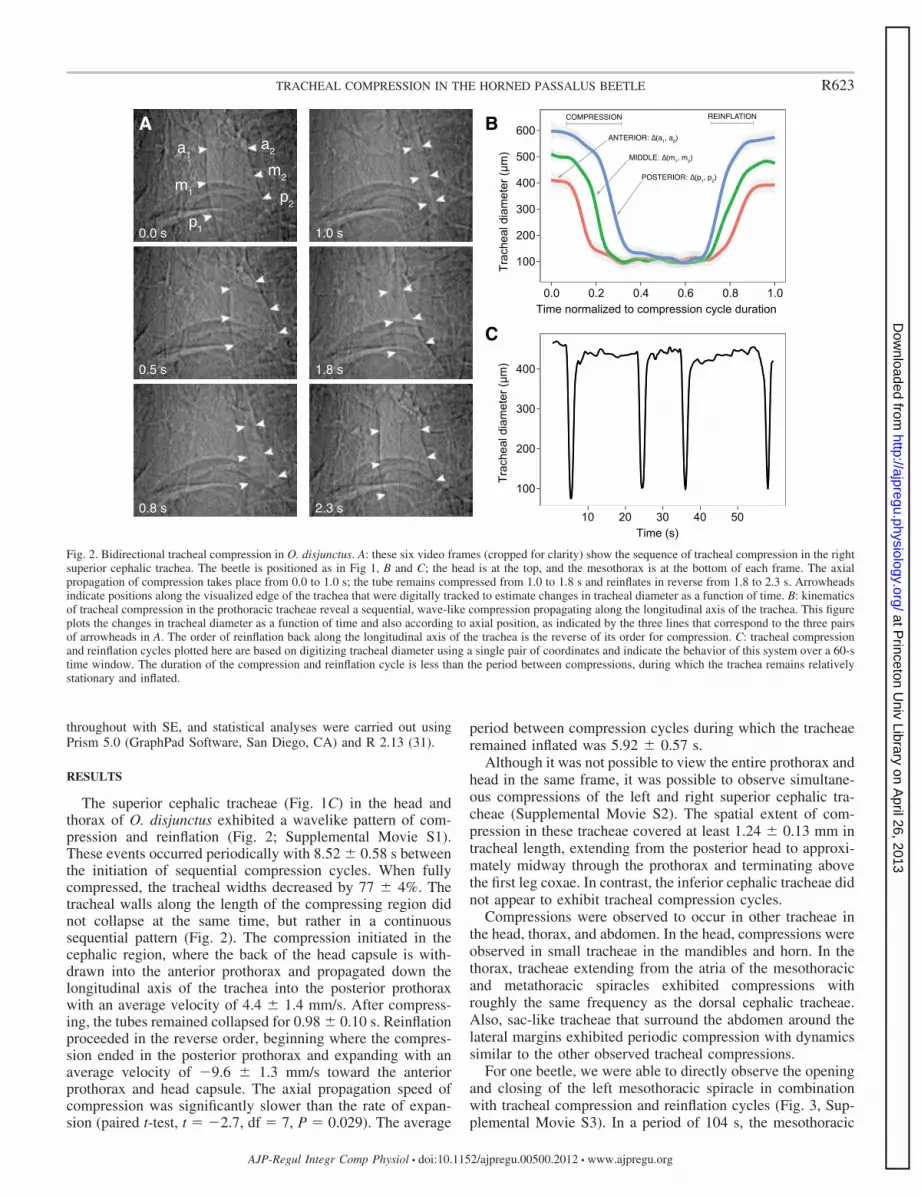
The superior cephalic tracheae (Fig. 1C) in the head andthorax of O. disjunctus exhibited a wavelike pattern of com-pression and reinflation (Fig. 2; Supplemental Movie S1).These events occurred periodically with 8.52 � 0.58 s betweenthe initiation of sequential compression cycles. When fullycompressed, the tracheal widths decreased by 77 � 4%. Thetracheal walls along the length of the compressing region didnot collapse at the same time, but rather in a continuoussequential pattern (Fig. 2). The compression initiated in thecephalic region, where the back of the head capsule is with-drawn into the anterior prothorax and propagated down thelongitudinal axis of the trachea into the posterior prothoraxwith an average velocity of 4.4 � 1.4 mm/s. After compress-ing, the tubes remained collapsed for 0.98 � 0.10 s. Reinflationproceeded in the reverse order, beginning where the compres-sion ended in the posterior prothorax and expanding with anaverage velocity of �9.6 � 1.3 mm/s toward the anteriorprothorax and head capsule. The axial propagation speed ofcompression was significantly slower than the rate of expan-sion (paired t-test, t � �2.7, df � 7, P � 0.029). The average
period between compression cycles during which the tracheaeremained inflated was 5.92 � 0.57 s.
Although it was not possible to view the entire prothorax andhead in the same frame, it was possible to observe simultane-ous compressions of the left and right superior cephalic tra-cheae (Supplemental Movie S2). The spatial extent of com-pression in these tracheae covered at least 1.24 � 0.13 mm intracheal length, extending from the posterior head to approxi-mately midway through the prothorax and terminating abovethe first leg coxae. In contrast, the inferior cephalic tracheae didnot appear to exhibit tracheal compression cycles.
Compressions were observed to occur in other tracheae inthe head, thorax, and abdomen. In the head, compressions wereobserved in small tracheae in the mandibles and horn. In thethorax, tracheae extending from the atria of the mesothoracicand metathoracic spiracles exhibited compressions withroughly the same frequency as the dorsal cephalic tracheae.Also, sac-like tracheae that surround the abdomen around thelateral margins exhibited periodic compression with dynamicssimilar to the other observed tracheal compressions.
For one beetle, we were able to directly observe the openingand closing of the left mesothoracic spiracle in combinationwith tracheal compression and reinflation cycles (Fig. 3, Sup-plemental Movie S3). In a period of 104 s, the mesothoracic
a1
m1
p1
a2
m2
p2
A B
C
0.0 s
0.5 s
0.8 s
1.0 s
1.8 s
2.3 s
Time normalized to compression cycle duration
Trac
heal
dia
met
er (µ
m)
100
200
300
400
500
600
0.0 0.2 0.4 0.6 0.8 1.0
Time (s)
Trac
heal
dia
met
er (µ
m)
100
200
300
400
10 20 30 40 50
COMPRESSION REINFLATION
1, a2)
1, m2)
1, p2)
Fig. 2. Bidirectional tracheal compression in O. disjunctus. A: these six video frames (cropped for clarity) show the sequence of tracheal compression in the rightsuperior cephalic trachea. The beetle is positioned as in Fig 1, B and C; the head is at the top, and the mesothorax is at the bottom of each frame. The axialpropagation of compression takes place from 0.0 to 1.0 s; the tube remains compressed from 1.0 to 1.8 s and reinflates in reverse from 1.8 to 2.3 s. Arrowheadsindicate positions along the visualized edge of the trachea that were digitally tracked to estimate changes in tracheal diameter as a function of time. B: kinematicsof tracheal compression in the prothoracic tracheae reveal a sequential, wave-like compression propagating along the longitudinal axis of the trachea. This figureplots the changes in tracheal diameter as a function of time and also according to axial position, as indicated by the three lines that correspond to the three pairsof arrowheads in A. The order of reinflation back along the longitudinal axis of the trachea is the reverse of its order for compression. C: tracheal compressionand reinflation cycles plotted here are based on digitizing tracheal diameter using a single pair of coordinates and indicate the behavior of this system over a 60-stime window. The duration of the compression and reinflation cycle is less than the period between compressions, during which the trachea remains relativelystationary and inflated.
R623TRACHEAL COMPRESSION IN THE HORNED PASSALUS BEETLE
AJP-Regul Integr Comp Physiol • doi:10.1152/ajpregu.00500.2012 • www.ajpregu.org
at Princeton U
niv Library on April 26, 2013
http://ajpregu.physiology.org/D
ownloaded from
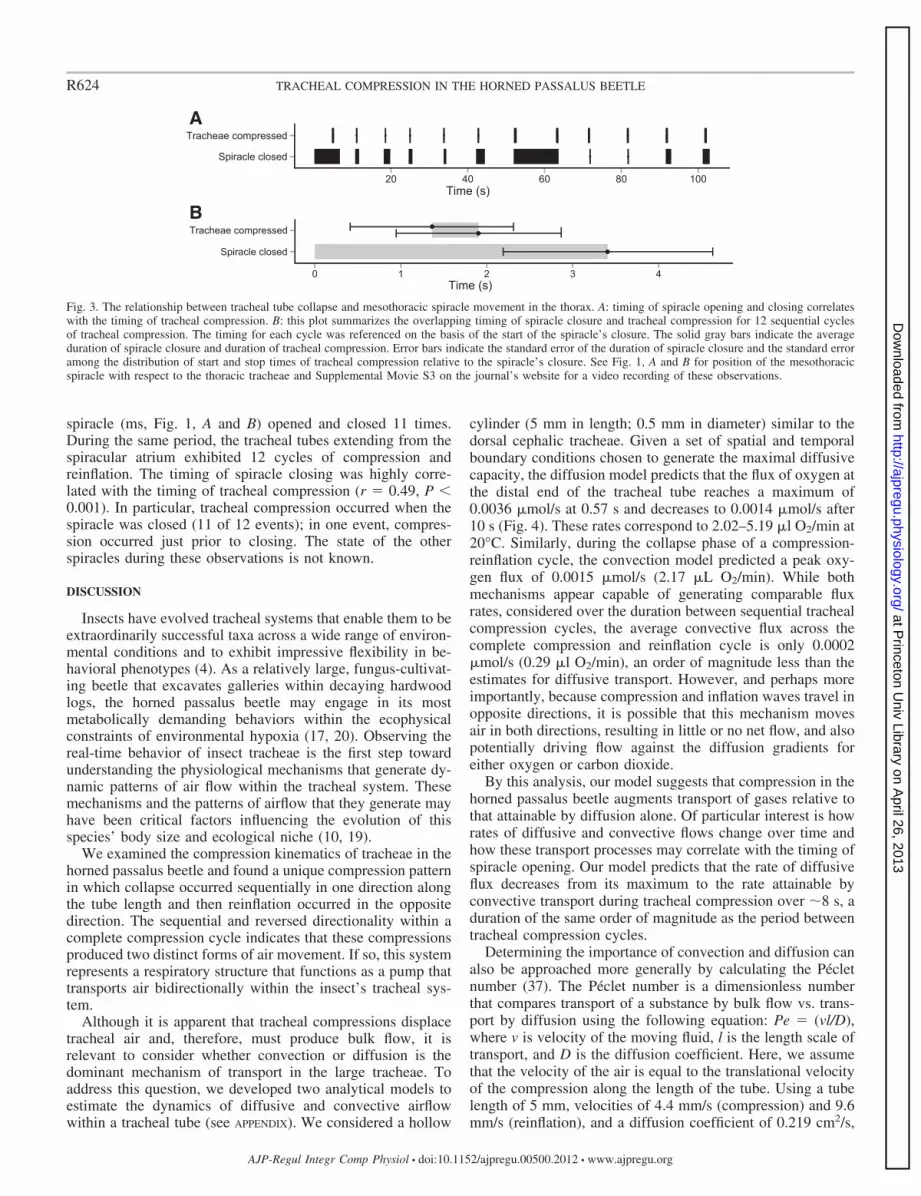
spiracle (ms, Fig. 1, A and B) opened and closed 11 times.During the same period, the tracheal tubes extending from thespiracular atrium exhibited 12 cycles of compression andreinflation. The timing of spiracle closing was highly corre-lated with the timing of tracheal compression (r � 0.49, P �0.001). In particular, tracheal compression occurred when thespiracle was closed (11 of 12 events); in one event, compres-sion occurred just prior to closing. The state of the otherspiracles during these observations is not known.
DISCUSSION
Insects have evolved tracheal systems that enable them to beextraordinarily successful taxa across a wide range of environ-mental conditions and to exhibit impressive flexibility in be-havioral phenotypes (4). As a relatively large, fungus-cultivat-ing beetle that excavates galleries within decaying hardwoodlogs, the horned passalus beetle may engage in its mostmetabolically demanding behaviors within the ecophysicalconstraints of environmental hypoxia (17, 20). Observing thereal-time behavior of insect tracheae is the first step towardunderstanding the physiological mechanisms that generate dy-namic patterns of air flow within the tracheal system. Thesemechanisms and the patterns of airflow that they generate mayhave been critical factors influencing the evolution of thisspecies’ body size and ecological niche (10, 19).
We examined the compression kinematics of tracheae in thehorned passalus beetle and found a unique compression patternin which collapse occurred sequentially in one direction alongthe tube length and then reinflation occurred in the oppositedirection. The sequential and reversed directionality within acomplete compression cycle indicates that these compressionsproduced two distinct forms of air movement. If so, this systemrepresents a respiratory structure that functions as a pump thattransports air bidirectionally within the insect’s tracheal sys-tem.
Although it is apparent that tracheal compressions displacetracheal air and, therefore, must produce bulk flow, it isrelevant to consider whether convection or diffusion is thedominant mechanism of transport in the large tracheae. Toaddress this question, we developed two analytical models toestimate the dynamics of diffusive and convective airflowwithin a tracheal tube (see APPENDIX). We considered a hollow
cylinder (5 mm in length; 0.5 mm in diameter) similar to thedorsal cephalic tracheae. Given a set of spatial and temporalboundary conditions chosen to generate the maximal diffusivecapacity, the diffusion model predicts that the flux of oxygen atthe distal end of the tracheal tube reaches a maximum of0.0036 �mol/s at 0.57 s and decreases to 0.0014 �mol/s after10 s (Fig. 4). These rates correspond to 2.02–5.19 �l O2/min at20°C. Similarly, during the collapse phase of a compression-reinflation cycle, the convection model predicted a peak oxy-gen flux of 0.0015 �mol/s (2.17 �L O2/min). While bothmechanisms appear capable of generating comparable fluxrates, considered over the duration between sequential trachealcompression cycles, the average convective flux across thecomplete compression and reinflation cycle is only 0.0002�mol/s (0.29 �l O2/min), an order of magnitude less than theestimates for diffusive transport. However, and perhaps moreimportantly, because compression and inflation waves travel inopposite directions, it is possible that this mechanism movesair in both directions, resulting in little or no net flow, and alsopotentially driving flow against the diffusion gradients foreither oxygen or carbon dioxide.
By this analysis, our model suggests that compression in thehorned passalus beetle augments transport of gases relative tothat attainable by diffusion alone. Of particular interest is howrates of diffusive and convective flows change over time andhow these transport processes may correlate with the timing ofspiracle opening. Our model predicts that the rate of diffusiveflux decreases from its maximum to the rate attainable byconvective transport during tracheal compression over �8 s, aduration of the same order of magnitude as the period betweentracheal compression cycles.
Determining the importance of convection and diffusion canalso be approached more generally by calculating the Pécletnumber (37). The Péclet number is a dimensionless numberthat compares transport of a substance by bulk flow vs. trans-port by diffusion using the following equation: Pe � (vl/D),where v is velocity of the moving fluid, l is the length scale oftransport, and D is the diffusion coefficient. Here, we assumethat the velocity of the air is equal to the translational velocityof the compression along the length of the tube. Using a tubelength of 5 mm, velocities of 4.4 mm/s (compression) and 9.6mm/s (reinflation), and a diffusion coefficient of 0.219 cm2/s,
A
BTime (s)
Spiracle closed
Tracheae compressed
20 40 60 80 100
Time (s)
Spiracle closed
Tracheae compressed
0 1 2 3 4
Fig. 3. The relationship between tracheal tube collapse and mesothoracic spiracle movement in the thorax. A: timing of spiracle opening and closing correlateswith the timing of tracheal compression. B: this plot summarizes the overlapping timing of spiracle closure and tracheal compression for 12 sequential cyclesof tracheal compression. The timing for each cycle was referenced on the basis of the start of the spiracle’s closure. The solid gray bars indicate the averageduration of spiracle closure and duration of tracheal compression. Error bars indicate the standard error of the duration of spiracle closure and the standard erroramong the distribution of start and stop times of tracheal compression relative to the spiracle’s closure. See Fig. 1, A and B for position of the mesothoracicspiracle with respect to the thoracic tracheae and Supplemental Movie S3 on the journal’s website for a video recording of these observations.
R624 TRACHEAL COMPRESSION IN THE HORNED PASSALUS BEETLE
AJP-Regul Integr Comp Physiol • doi:10.1152/ajpregu.00500.2012 • www.ajpregu.org
at Princeton U
niv Library on April 26, 2013
http://ajpregu.physiology.org/D
ownloaded from

the Péclet number ranges from 1.0 to 2.2. This suggests thatneither diffusion nor convection dominates the transport duringtracheal compression, a conclusion that is congruent with theanalytical modeling analysis.
This study also reports for the first time the exact coordina-tion of tracheal movements with spiracle opening and clos-ing. The synchronization of spiracle opening and closing withtracheal compression could have a dramatic influence on thepressure gradients and patterns of air flow during trachealcompression cycles. For instance, consider a spiracle and itslocally connected tracheal tubes. If the tracheae compresswhen the spiracle is open, a large fraction of the displaced gasis likely to be transported toward the spiracle and out of thebody, assuming that the resistance to flow is much lower in theoutward direction. Conversely, if the spiracle is closed, the airmust be transported away from the spiracle and deeper into thebody. We observed 12 cycles of tracheal compression thatcoincided with the mesothoracic spiracle closure (Fig. 3). Inevery compression event, the spiracle was closed or in theprocess of closing. The correlation between spiracle closureand tracheal compression suggests a novel hypothesis for therole of tracheal compression, which is to transport air towardmore distal regions of the body, possibly including the head,legs, and antennae. This transport of air within body regionsmay also increase the mixing of gases within the trachealsystem. Intriguingly, our results are consistent with the predic-tions of a model for the water economy of insects, in whichsimultaneous spiracle closure and tracheal compression areproposed as a mechanism to condense water vapor within thetracheal system to either redistribute water between bodyregions or minimize evaporative water loss (7). To test thismodel, future studies may manipulate relative humidity aroundan insect within a chamber and observe whether or not watervapor pressure has a causal effect on dynamics of trachealcompression or the synchrony of tracheal compression andspiracle closure.
The observation that tracheal compression is slower thanreinflation is consistent with the idea that elastic energy isstored in the tracheal wall during compression, and reinflationoccurs as a passive process of energy return, with dynamics
governed by the viscoelastic properties of the trachea (39).Variation in structural properties of the tracheae, includingthickness, second-moment of area, and taenidia geometry,might play functional roles in storing and releasing energyduring cycles of compression and reinflation. Alternatively, thecompression may be slower than expansion, if, while the spi-racle is closed, more force is required to push air into the distalregions of the tracheal system (e.g., into peripheral air sacs, thehead, or extremities) against the pressure generated by holdingthe local spiracles sealed. Lastly, it is possible that the differ-ences in kinematics can be explained by differences in the ratesof hemolymph pressure changes if this mechanism controls thetracheal deformation.
Perspectives and Significance
Investigating the behavior and dynamics of insect trachealsystem structures is necessary to develop an integrative under-standing of respiratory physiology at the organismal level.Here, we describe the kinematics of tracheal compression inthe two dorsal cephalic trunks, but further work is needed tounderstand this complex system, in particular, with respect tothe tracheal ultrastructure and with respect to the elaboratesystem of branching tracheae and air sacs. We focused on oneregion of the tracheal system of this large beetle, but sequentialand asymmetric tracheal compressions may occur in otherbody regions, influencing patterns of air sac inflation andpotentially coordinated with spiracular control. These com-pression patterns may enhance this species’ hypoxia toleranceand its performance of energetically demanding behaviors,including excavation and stridulation. Additionally, if trachealcompression cycles increase the efficiency of mixing within thetracheal system, it may allow the animal to close its spiraclesfor an extended duration, with potential beneficial conse-quences for desiccation resistance and mite protection (7, 10,16). This study suggests that air movement patterns within therespiratory systems of insects are more complex than is cur-rently appreciated and that further determination of compres-sion patterns across insects is worthy of detailed investigation.
Bee
tle m
etab
olic
rat
e
Com
pres
sion
flux
Par
tial h
ead
met
abol
ic r
ate
21 kPa
2.5 mm 5.0 mm 10 mm
To head
0 2 4 6 8 10
0.00
00.
002
0.00
40.
006
Time (s)
Oxy
gen
flux
(µm
ols
1 )
2.5 mm5.0 mm10 mm
Fig. 4. The temporal dynamics of oxygen dif-fusion at three different lengths along a tra-cheal tube, modeled using the differential formof Fick’s first law. Using the same ordinatescale, the bar graph on the right side illustratesan estimate for the peak convective flow dur-ing compression, a reported measurement forthe metabolic rate (1611.6 �W) of a 1.63 g O.disjunctus (6), and an estimate of the fractionof whole animal metabolic rate supplied by thesingle tracheal tube.
R625TRACHEAL COMPRESSION IN THE HORNED PASSALUS BEETLE
AJP-Regul Integr Comp Physiol • doi:10.1152/ajpregu.00500.2012 • www.ajpregu.org
at Princeton U
niv Library on April 26, 2013
http://ajpregu.physiology.org/D
ownloaded from

APPENDIX
Convection model. The peak bulk flow of oxygen produced bytracheal compression can be modeled on the basis of estimatingthe change in tracheal volume. We assumed that the tracheallumen contains air with an ambient oxygen concentration (u0, molof O2 cm�3) and that the tube has a cylindrical geometry charac-terized by a constant length (l, cm) and a radius (r, cm) that varieswith the extent of tracheal compression. Finally, given a compres-sion period (t, s), during which the tracheal tube collapses (i.e., notincluding the time for reinflation) and assuming that the displacedair flows in a single direction during this collapse, the flux (Q, molof O2 per second) may be calculated by
Qconvection � u0
�l
t(rmax
2 � rmin2 )
Diffusion model. Fick’s first law is an equation that predictsdiffusive flux (J, mol of O2/s per cross-sectional area) as afunction of a diffusion coefficient (D, cm2/s) and a spatialconcentration gradient (�u/�x). In one dimension, with respectto distance (x) and time (t), Fick’s first law is
J � �D�
�xu(x, t)
Modeling the temporal dynamics of the changing spatial con-centration gradient requires a solution to Fick’s second law,which describes the relationship between the concentrationgradients in time and space:
�
� tu(x, t) � D
�2u
�x2
The solution to this equation is a function, u(x,t), which givesthe concentration of the diffusing substance as a function ofdistance and time, depending on particular boundary and initialconditions. In our case, oxygen concentration at one boundarymay be fixed at a constant value (e.g., the atmospheric oxygenconcentration, u0), and it may not be predefined at the distalboundary condition. Since a second boundary condition isnecessary for a solution, we can also define the rate of changeof oxygen at the source boundary as zero. For this situation, wecan, thus, define two boundary conditions and one initialcondition for 0 � x � and 0 � t:
u(0, t) � u0
�
� tu(0, t) � 0
u(x, 0) � 0
Given these conditions, the solution to the diffusion equation inone dimension is
u(x, t) � u(0, t)2
��� x
2�Dt
�e�t2dt
Taking the derivative of this solution with respect to distance gives
�
�xu(x, t) � u(0, t)
�1
��Dte
�x2
4Dt
Substituting this expression back into Fick’s first law gives anexpression for the temporal dynamics of diffusive flux (Q,
mol/s) at a distance (L, cm) within a cylindrical tube with aconstant radius (r, cm):
Qdiffusion � u0r2��D
te
�L2
4Dt
These equations make it possible to model the spatial andtemporal dynamics of air flow within a tracheal tube, but thepredictions must be evaluated in the context of the generalassumptions and explicit boundary conditions. It is unlikely,for example, that the oxygen partial pressure gradient within asingle tracheal tube is ever as high as in the diffusion model.Future explorations of this model may incorporate the effectsof a radial concentration gradient (across the tracheal wall), aswell as experimentally parameterized boundary conditions.
ACKNOWLEDGMENTS
We thank Kamel Fezzaa for technical assistance, and Jon Harrison, JacoKlok, Hodjat Pendar, Jennifer Fewell, and Michael LaBarbera for valuablefeedback on previous drafts of the manuscript. Rebecca Zapata adaptedRobertson’s 1962 anatomical illustration in Fig. 1. We also acknowledge manyhelpful discussions with colleagues in the Harrison Laboratory at Arizona StateUniversity, the Socha Laboratory at Virginia Tech, and with students ofMichael LaBarbera’s Biomechanics of Organisms course at The University ofChicago. We are grateful for the insightful feedback offered by four reviewers.Use of the Advanced Photon Source, an Office of Science User Facilityoperated for the U.S. Department of Energy (DOE) Office of Science byArgonne National Laboratory, was provided by the U.S. DOE under contractno. DE-AC02-06CH11357.
GRANTS
The Charlotte Mangum Student Support Program made it possible for J. S.Waters to present these findings at the annual meeting of the Society forIntegrative and Comparative Biology. J. J. Socha was supported by theNational Science Foundation under Grant 0938047 and by the Virginia TechInstitute for Critical Technology and Applied Science (ICTAS).
DISCLOSURES
No conflicts of interest, financial or otherwise, are declared by the authors.
AUTHOR CONTRIBUTIONS
Author contributions: J.S.W., M.W.W., and J.J.S. conception and design ofresearch; J.S.W., W.-K.L., and J.J.S. performed experiments; J.S.W. and J.J.S.analyzed data; J.S.W., W.-K.L., M.W.W., and J.J.S. interpreted results ofexperiments; J.S.W. prepared figures; J.S.W. and J.J.S. drafted manuscript;J.S.W., W.-K.L., M.W.W., and J.J.S. edited and revised manuscript; J.S.W.,W.-K.L., M.W.W., and J.J.S. approved final version of manuscript.
REFERENCES
1. Bartholomew GA, Lighton JRB. Ventilation and oxygen consumptionduring rest and locomotion in a tropical cockroach, Blaberus giganteus. JExp Biol 118: 449, 1985.
2. Bradley TJ. Control of the respiratory pattern in insects. Hypoxia Circ618: 211–220, 2007.
3. Buck J. Some physical aspects of insect respiration. Annu Rev Entomol 7:27–56, 1962.
4. Chown S, Nicolson SW. Insect Physiological Ecology: Mechanisms andPatterns. New York: Oxford University Press, 2004.
5. Chown SL, Gibbs AG, Hetz SK, Klok CJ, Lighton JRB, Marais E.Discontinuous gas exchange in insects: a clarification of hypotheses andapproaches. Physiol Biochem Zool 79: 333–343, 2006.
6. Chown SL, Marais E, Terblanche JS, Klok CJ, Lighton JRB, Black-burn TM. Scaling of insect metabolic rate is inconsistent with the nutrientsupply network model. Funct Ecol 21: 282–290, 2007.
7. Corbet SA. Pressure cycles and the water economy of insects. PhilosTrans R Soc London B, Biol Sci 318: 377–407, 1988.
8. Duncan FD, Förster TD, Hetz SK. Pump out the volume—the effect oftracheal and subelytral pressure pulses on convective gas exchange in a
R626 TRACHEAL COMPRESSION IN THE HORNED PASSALUS BEETLE
AJP-Regul Integr Comp Physiol • doi:10.1152/ajpregu.00500.2012 • www.ajpregu.org
at Princeton U
niv Library on April 26, 2013
http://ajpregu.physiology.org/D
ownloaded from

dung beetle, Circellium bacchus (Fabricus). J Insect Physiol 56: 551–558,2010.
9. Förster TD, Hetz SK. Spiracle activity in moth pupae—the role ofoxygen and carbon dioxide revisited. J Insect Physiol 56: 492–501, 2010.
10. Gray IE. Observations on the life history of the horned passalus. Am MidlNat 35: 728–746, 1946.
11. Greenlee K, Harrison J. Development of respiratory function in theAmerican locust Schistocerca americana I. Across-instar effects. J ExpBiol 207: 497, 2004.
12. Greenlee K, Harrison J. Development of respiratory function in theAmerican locust Schistocerca americana: II. Within-instar effects. J ExpBiol 207: 509, 2004.
13. Greenlee K, Harrison J. Respiratory changes throughout ontogeny in thetobacco hornworm caterpillar, Manduca sexta. J Exp Biol 208: 1385,2005.
14. Greenlee KJ, Henry JR, Kirkton SD, Westneat MW, Fezzaa K, LeeWK, Harrison JF. Synchrotron imaging of the grasshopper trachealsystem: morphological and physiological components of tracheal hyper-metry. Am J Physiol Regul Integr Comp Physiol 297: R1343–R1350,2009.
15. Greenlee KJ, Socha JJ, Eubanks HB, Thapa G, Pedersen P, Lee W-K,Kirkton SD. Hypoxia-induced compression in the tracheal system of thetobacco hornworm caterpillar, Manduca sexta L. J Exp Biol In press.
16. Harrison JF, Camazine S, Marden JH, Kirkton SD, Rozo A, Yang X.Mite not make it home: tracheal mites reduce the safety margin for oxygendelivery of flying honeybees. J Exp Biol 204: 805–814, 2001.
17. Harrison JF, Frazier MR, Henry JR, Kaiser A, Klok CJ, Rascon B.Responses of terrestrial insects to hypoxia or hyperoxia. Respir PhysiolNeurobiol 154: 4–17, 2006.
18. Harrison JF, Waters JS, Cease AJ, Vandenbrooks JM, Callier V,Klok CJ, Shaffer K, Socha JJ. How locusts breathe. Physiology 28:18–27, 2013.
19. Kaiser A, Klok CJ, Socha JJ, Lee WK, Quinlan MC, Harrison JF.Increase in tracheal investment with beetle size supports hypothesis ofoxygen limitation on insect gigantism. Proc Natl Acad Sci USA 104:13198–13203, 2007.
20. Klok CJ, Harrison JF. Atmospheric hypoxia limits selection for largebody size in insects. PLos One 4: e3876, 2009.
21. Lehmann FO, Schützner P. The respiratory basis of locomotion inDrosophila. J Insect Physiol 56: 543–550, 2010.
22. Levy RI, Schneiderman HA. Discontinuous respiration in insects-III.The effect of temperature and ambient oxygen tension on the gaseouscomposition of the tracheal system of silkworm pupae. J Insect Physiol 12:105–121, 1966.
23. Levy RI, Schneiderman HA. Discontinuous respiration in insects. II. Thedirect measurement and significance of changes in tracheal gas composi-tion during the respiratory cycle of silkworm pupae. J Insect Physiol 12:83–104, 1966.
24. Lighton JRB. Discontinuous gas exchange in insects. Annu Rev Entomol41: 309–324, 1996.
25. Lighton JRB. Measuring Metabolic Rates: A Manual For Scientists. NewYork: Oxford University, 2008, p. 201.
26. Lighton JRB, Fukushi T, Wehner R. Ventilation in Cataglyphis bicolor:regulation of carbon dioxide release from the thoracic and abdominalspiracles. J Insect Physiol 39: 687–699, 1993.
27. Lighton JRB, Garrigan D. Ant breathing: testing regulation and mech-anism hypotheses with hypoxia. J Exp Biol 198: 1613–1620, 1995.
28. Lighton JRB, Lovegrove BG. A temperature-induced switch from dif-fusive to conective ventilation in the honeybee. J Exp Biol 154: 509–516,1990.
29. Marais E, Klok CJ, Terblanche JS, Chown SL. Insect gas exchangepatterns: a phylogenetic perspective. J Exp Biol 208: 4495–4507, 2005.
30. Miller PL. Ventilation in active and in inactive insects. In: Locomotionand Energetics in Arthropods, edited by Herreid CF and Fourtner CF. NewYork: Plenum, 1981, p. 367–390.
31. R Development Core Team. R: A Language and Environment for Sta-tistical Computing. R Foundation for Statistical Computing, Vienna,Austria. http://www.R-project.org, 2011.
32. Robertson CH Jr. The anatomy of the respiratory system of the PassalusBeetle, Popilius disjunctus (Illiger). Am Midl Nat 68: 376–393, 1962.
33. Schultz TD, Quinlan MC, Hadley NF. Preferred body temperature,metabolic physiology, and water balance of adult Cicindela longilabris: acomparison of populations from boreal habitats and climatic refugia.Physiol Zool 65: 226–242, 1992.
34. Socha JJ, Förster TD, Greenlee KJ. Issues of convection in insectrespiration: Insights from synchrotron X-ray imaging and beyond. RespirPhysiol Neurobiol 173: S65–S73, 2010.
35. Socha JJ, Lee WK, Harrison JF, Waters JS, Fezzaa K, Westneat MW.Correlated patterns of tracheal compression and convective gas exchangein a carabid beetle. J Exp Biol 211: 3409–3420, 2008.
36. Socha JJ, Westneat MW, Harrison JF, Waters JS, Lee WK. Real-timephase-contrast x-ray imaging: a new technique for the study of animalform and function. BMC Biol 5: 6, 2007.
37. Vogel S. Living in a physical world. J Biosci 29: 391–397, 2004.38. Walker JA. QuickImage: a modification of NIH Image with enhanced
digitizing tools. http://www.usm.maine.edu/�walker/software.html.39. Webster MR, Vita RD, Twigg JN, Socha JJ. Mechanical properties of
tracheal tubes in the American cockroach (Periplaneta americana). SmartMater Struct 20: 094017, 2011.
40. Weis-Fogh T. Functional design of the tracheal system of flying insects ascompared with the avian lung. J Exp Biol 41: 207–227, 1964.
41. Westneat MW, Betz O, Blob RW, Fezzaa K, Cooper WJ, Lee WK.Tracheal respiration in insects visualized with synchrotron X-ray imaging.Science 299: 558–560, 2003.
42. Westneat MW, Socha JJ, Lee WK. Advances in biological structure,function, and physiology using synchrotron X-ray imaging. Annu RevPhysiol 70: 119–142, 2008.
R627TRACHEAL COMPRESSION IN THE HORNED PASSALUS BEETLE
AJP-Regul Integr Comp Physiol • doi:10.1152/ajpregu.00500.2012 • www.ajpregu.org
at Princeton U
niv Library on April 26, 2013
http://ajpregu.physiology.org/D
ownloaded from


