Embed Size (px)
Citation preview

Dysregulation of miRNA 181b in the temporal cortexin schizophrenia
Natalie J. Beveridge1,2, Paul A. Tooney1,2, Adam P. Carroll1,2, Erin Gardiner1,2, Nikola Bowden1,2,
Rodney J. Scott1,2, Nham Tran3, Irina Dedova1,4 and Murray J. Cairns1,2,�
1Schizophrenia Research Institute, Sydney, NSW 2006, Australia, 2School of Biomedical Sciences, Faculty of Health,
and Hunter Medical Research Institute, The University of Newcastle, University Drive, Callaghan, NSW 2308,
Australia, 3Department of Infectious Diseases and Immunology and 4Department of Pathology, The University
of Sydney, Blackburn Building Level 6, Sydney, NSW 2006, Australia
Received November 1, 2007; Revised and Accepted January 4, 2008
Analysis of global microRNA (miRNA) expression in postmortem cortical grey matter from the superior tem-poral gyrus, revealed significant up-regulation of miR-181b expression in schizophrenia. This finding was sup-ported by quantitative real-time RT–PCR analysis of miRNA expression in a cohort of 21 matched pairs ofschizophrenia and non-psychiatric controls. The implications of this finding are substantial, as this miRNAis predicted to regulate many target genes with potential significance to the development of schizophrenia.They include the calcium sensor gene visinin-like 1 (VSNL1) and the ionotropic AMPA glutamate receptor sub-unit (GRIA2), which were found to be down-regulated in the same cortical tissue from the schizophrenia group.Both of these genes were also suppressed in miR-181b transfected cells and shown to contain functional miR-181b miRNA recognition elements by reporter gene assay. This study suggests altered miRNA levels could be asignificant factor in the dysregulation of cortical gene expression in schizophrenia.
INTRODUCTION
Schizophrenia is a severely debilitating psychiatric disordercharacterized by a diverse range of symptoms. While extensiveresearch has not determined the definitive cause(s), it is gener-ally accepted that a number of influences including genetic,environmental and developmental factors are involved. Gen-etics in particular has been known for many years to play akey role in the development of schizophrenia. The pattern ofinheritance, however, is far from clear with a complex arrayof associated loci and putative candidate genes each with a rela-tively small affect, disbursed across the entire genome (1–3).This picture is further complicated by environmental andepigenetic influences evident by the relatively low rate of con-cordance in monozygotic twins (�50%) (4,5).
Recent research from a variety of quarters has lent supportto the neurodevelopmental hypothesis, which suggests thatgenetic and environmental influences affect neurodevelop-mental processes that cause abnormalities in brain plasticityand connectivity leading to schizophrenia in early adulthood.In this model, regulatory factors that control the stage and
tissue expression of molecules involved in neurodevelopmen-tal processes will be particularly important. While transcrip-tion factors and protein based signal transduction pathwaysare usually considered in this context, a new class of geneexpression modulators consisting entirely of RNA is alsoemerging as a significant regulatory factor. These small non-coding RNA molecules known as microRNAs (miRNA) func-tion as guide sequences for the cellular gene silencing pathwayto bring about target gene suppression through a processknown as post-transcriptional gene silencing (PTGS) (6).These riboregulators are now believed to play a major rolein coordinating the regulation of gene expression during thedifferentiation and development of the brain (7). Indeed, alarge proportion of human miRNAs display a brain-specificor brain-enriched expression pattern (8). There is even evi-dence to suggest they are involved in brain function andneural plasticity via the regulation of molecules involved inlong-term potentiation (LTP) (9,10) and in the establishmentand maintenance of dendrites (11).
�To whom correspondence should be addressed. Tel: þ61 249218670; Fax: þ61 249217903; Email: [email protected]
# 2008 The Author(s)This is an Open Access article distributed under the terms of the Creative Commons Attribution Non-Commercial License (http://creativecommons.org/licenses/by-nc/2.0/uk/) which permits unrestricted non-commercial use, distribution, and reproduction in any medium, provided the original work isproperly cited.
Human Molecular Genetics, 2008, Vol. 17, No. 8 1156–1168doi:10.1093/hmg/ddn005Advance Access published on January 9, 2008
by guest on January 11, 2016http://hm
g.oxfordjournals.org/D
ownloaded from

Considering that each miRNA is potentially manipulating theexpression of hundreds of target genes, the clinical implicationsof an abnormality or disturbance of this system are substantial,particularly if such abnormality occurs during developmentof the central nervous system. Neurological manifestationsof abnormalities in miRNA-mediated regulation have in factalready been identified for fragile X mental retardation (9)and Tourette’s syndrome (12). The miRNA pathway is alsoimplicated in DiGeorge syndrome as one of the deletedgenes, DGCR8 is associated with the microprocessor complexwith Drosha and is essential for processing of the primarymiRNA transcript (13). Interestingly, 25% of patients withDiGeorge syndrome also develop schizophrenia (14). Inview of these findings, it is reasonable to suspect that altera-tion in miRNA gene silencing pathways may also be involvedin the development of complex psychiatric disorders suchas schizophrenia. This is supported by a recent study ofmiRNA expression in the prefrontal cortex, which identified anumber of altered miRNAs (15). In the study reported here,we examined miRNA expression in cortical grey matter fromthe superior temporal gyrus (STG). The STG contains theprimary and secondary auditory cortex thought to be involvedin the generation of auditory hallucinations and appearsto have reduced volume in schizophrenia (16,17). This analysisrevealed significant up-regulation of miR-181b, which wasfound to correlate inversely with expression of schizophrenia-associated miR-181b target genes GRIA2 and visinin-like 1(VSNL1) in STG from the same schizophrenia cohort.
RESULTS
miRNA expression in the temporal cortex
High throughput miRNA expression analysis in postmortemcortical grey matter from the STG was accomplished using acustom DNA microarray platform (18). Fluorescence signalsobserved in duplicate array features at a level 2-fold that ofbackground were considered to be indicative of miRNAexpression. This expression profile consisted of 76 miRNAsout of a total of 262 probes corresponding to miRBaseversion 7.0. Many of these miRNAs have previously beenidentified as either brain-specific, brain-enriched or constitu-tively expressed (Supplementary Material, Table S1). This isexemplified by strong expression of miR-125a/b, miR-124a,miR-9/9�, miR-128a/b as well as miR-138, miR-219 andmiR-338 which are brain-specific or brain-enriched. The con-stitutively expressed let-7 group of miRNAs were also wellrepresented.
Differential miRNA expression in schizophrenia
Differential expression of miRNA in postmortem STG fromthe schizophrenia cohort compared with their matched non-psychiatric controls was determined through two-classunpaired analysis using statistical analysis of microarrays soft-ware (Significance Analysis of Microarrays, SAM, version2.23) (19). This analysis revealed significant up-regulation oftwo miRNAs, hsa-let-7g and hsa-miR-181b in the schizo-phrenia group, to levels 1.8-fold (P ¼ 0.008) and 2.8-fold(P ¼ 0.001), respectively (Fig. 1A).
To confirm the differential expression status of maturelet-7g and miR-181b observed by the microarray analysis,RNA from the same tissue was subjected to northern blotanalysis with 32P-labelled locked nucleic acid (LNA)-modifiedprobes. As miR-181b expression in the STG was in the low tomid range compared with the entire profile and below that ofthe constitutively expressed let-7g, the naı̈ve membrane washybridized first with the miR-181b probe. After stripping,the membrane was re-probed for let-7g and then the U6snRNA loading control, respectively, with the latter being
Figure 1. Expression analysis of miR-181b and let-7g by microarray andnorthern blot. (A) Mean normalized microarray fluorescence values formiR-181b (2.8-fold, P , 0.001) and let-7g (1.8-fold, P ¼ 0.008) in the STGfrom 7 matched pairs of subjects with schizophrenia and non-psychiatric con-trols (SZ and CTR 1–7). (B) Northern blot analysis of STG RNA comparingschizophrenia samples (SZ 1–7) and non-psychiatric controls (CTR1-5). Eachpanel consists of phosphorimages of bands probed for miR-181b, let-7g andU6 snRNA, respectively. The intensity of bands corresponding to eachmiRNA species was then determined using ImageQuant v5.2 and normalizedwith respect to U6 snRNA. (C) Mean normalized intensity for miR-181b andhsa-let-7g miRNAs in the STG of schizophrenia non-psychiatric controlsdetermined by northern blot hybridization. Bars represent mean+SD; �P ,
0.05; ��P , 0.01; ���P , 0.001.
Human Molecular Genetics, 2008, Vol. 17, No. 8 1157
by guest on January 11, 2016http://hm
g.oxfordjournals.org/D
ownloaded from

used to normalize the expression value of each miRNA(Fig. 1B). Quantitative analysis with normalization to the U6band intensity confirmed a significantly higher expression ofmiR-181b in the schizophrenia samples with an averageincrease of 1.5-fold (P ¼ 0.041) compared with the controls.In contrast, the difference in let-7g expression between thetwo groups was not significant (P ¼ 0.12; Fig. 1C).
Validation of miR-181b over-expression in schizophrenia
To further validate the apparent association between alteredmiR-181b expression in the STG and schizophrenia, tissuefrom an additional 14 matched pairs was obtained and theRNA extracted to generate a substantially larger cohort(schizophrenia n ¼ 21, controls n ¼ 21). As less tissue wasavailable from these new cases yielding lower quantities oftotal RNA, a highly sensitive and specific quantitative real-time RT–PCR-based approach to determining miR-181bexpression was established. This method utilized amiR-181b reverse primer consisting of a 9-nucleotide target-specific 30 segment and an adjoining 9-nucleotide arbitrarytag sequence at the 50 terminus. While the sequence-specific9-mer component primed low-temperature cDNA extensionon the miRNA target, the adjacent 9-mer tag provided theadditional length required for primer recognition during thehigher temperature PCR analysis phase. The relativeexpression of miR-181b from this RT–PCR analysis was con-sistent with the northern blot analysis confirming a significantup-regulation of this miRNA in the schizophrenia group, withan average increase of 1.24-fold (P ¼ 0.049) that wasobserved in 14 out of 21 matched pairs. However, removalof one control sample, CTR1, that had a higher expressionof miR-181b (differed by more than 2.7 SD units from thecontrol mean), elevated the average fold change inmiR-181b expression in schizophrenia across the remaining20 pairs to 1.39-fold (P ¼ 0.029).
To determine if the differential expression observed inmiR-181b was due to alteration in 181b precursor RNA tran-scription, primers specific to the pre-181b-1 (transcribed from1q31.3) and pre-181b-2 (transcribed from 9q33.3) were usedfor quantitative real-time RT–PCR. These were both normal-ized against U6 snRNA expression as described for the maturemiRNA expression. The results of this analysis (Fig. 2C) indi-cated that the pre-181b-2 transcript is in much greater abun-dance in the STG than the pre-181b-1 transcript, with.10-fold higher relative expression. When the expressionlevels of these precursor miRNA transcripts in the schizo-phrenia samples were compared with the controls, there wasno significant difference (Fig. 2C).
MiR-181b target prediction and pathway analysis
In order to gather some clues about the functional implication ofelevated miR-181b concentrations in the cerebral cortex, anextensive list of putative target genes was assembled from avariety of publicly available miRNA data bases using theonline TargetCombo web service (http://www.diana.pcbi.upenn.edu/cgi-bin/TargetCombo.cgi) (20). This provides theoption of combining target predictions from the leading homo-logy search algorithms miRanda, PicTar, TargetScanS and
DIANA-microT (21–24). Each of these programmes screenfor motifs with various miRNA homology attributes in the phy-logenetically conserved segments of 30 UTR sequences takenfrom the genome database. Collectively, these searches ident-ified .800 putative target genes (Supplementary Material,Table S2). A number of these genes are known to play an import-ant role in human brain function and development, and could
Figure 2. Expression analysis of mature and precursor 181b molecules byquantitative RT–PCR. (A) Real-time PCR verification of altered miR-181bexpression in the STG. For each sample, the change in cycle threshold(DCt) was obtained by subtracting the Ct of U6 snRNA from the Ct ofmiR-181b. For each pair, the DDCt was obtained by subtracting the DCt ofeach schizophrenia subject from the DCt of its corresponding controlsubject. The pair-wise expression changes are represented for each of the 21matched pairs (grey bars) and the mean (+SEM) is shown to the far right(white bar). (B) Relative expression of miR-181b by RT–PCR in 21matched pairs of postmortem STG. miR-181b was up-regulated 1.24-fold inthe schizophrenia cohort (P ¼ 0.049; paired t-test). (C) Relative expressionof precursor 181b molecules. CTR, control samples; SZ, schizophreniasamples. Bars represent mean+SEM (bar graphs); �P , 0.05.
1158 Human Molecular Genetics, 2008, Vol. 17, No. 8
by guest on January 11, 2016http://hm
g.oxfordjournals.org/D
ownloaded from
conceivably be involved in schizophrenia. This was exemplifiedby genes involved in synaptic transmission, including gammaamino butyric acid A receptor (GABRA1), glutamate receptors(GRM5, GRM7, GRIK2, GRID and GRIA2), serotonin recep-tors (HTR1B and HTR2C) and the cannabinoid receptor(CNR1). Some predicted target genes are also thought to beinvolved in brain development and neurodevelopmental dis-orders, for example ataxin 1 (ATXN1), zic family member1 (ZIC1), SLIT and NTRK-like family, member 1 (SLTRK1)and fragile X mental retardation 1 (FMR1). The latter twoinvolved in Tourette’s syndrome and fragile X mental retar-dation, respectively, are already known to be associated withmiRNA-related dysfunction.
To further understand the potential of miR-181b regulation,the predicted gene lists were subjected to functional annota-tion clustering and Kyoto Encyclopedia of Genes andGenomes (KEGG) pathway analysis using the sdatabase forannotation, visualization, and integrated discovery (DAVID;http://david.abcc.ncifcrf.gov) (25). Interestingly, of the 144conserved target genes predicted by the online version of themiRanda algorithm at the Memorial Sloan-Kettering CancerCenter Computational biology web site (http://www.micro-rna.org) (21), the most highly enriched gene ontology clusterrelated to development, nervous system development, neuro-genesis and differentiation (Supplementary Material,Table S2). This was followed by a cluster of terms relatingmore broadly to signalling, signal transduction and transcrip-tion. When the combination gene list, consisting of 789miR-181b target genes was submitted, a similar pictureemerged except the ranking of the top two clusters wasreversed. The KEGG pathways were also charted and rankedaccording to their P-value. The top five most significantterms for the large list included: LTP, MAPK signallingpathway, axon guidance and neurodegenerative disorders. Inthe smaller miRanda list developmentally related GO termsagain featured highly with the top five consisting of develop-ment, neural development, cell differentiation, system devel-opment and organ development.
While miRNA target prediction algorithms provide a usefulstarting point for understanding the function of particularmiRNA, they are also prone to a degree of over predictionleading to false positives (23,26). In order to relate the increasedmiR-181b activity in the context of schizophrenia-associatedchanges in the STG, the list of putative targets for this miRNAwas cross matched with genes already shown in microarrayexperiments to be down regulated in the same tissue (Sup-plementary Material, Table S2e; Bowden et al., manuscript inpreparation). This revealed a number of interesting targetgenes potentially regulated by miR-181b in the same tissueincluding the ionotropic glutamate receptor (GRIA2), fragileX- related 2 and VSNL1. Alteration of GRIA2 and VSNL1expression in the dorsolateral prefrontal cortex and in the hippo-campus, respectively, have also been shown to be associatedwith schizophrenia (27,28).
Validation of schizophrenia associated changes in VSNL1and GRIA2 expression
To confirm the schizophrenia-associated down-regulation ofVSNL1 and GRIA2 from the microarray analysis (Bowden
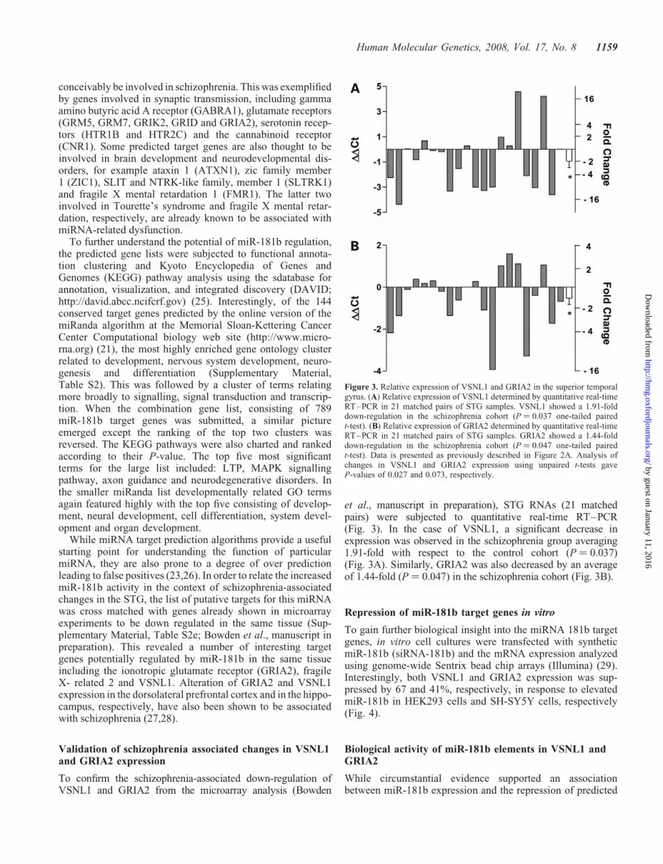
et al., manuscript in preparation), STG RNAs (21 matchedpairs) were subjected to quantitative real-time RT–PCR(Fig. 3). In the case of VSNL1, a significant decrease inexpression was observed in the schizophrenia group averaging1.91-fold with respect to the control cohort (P ¼ 0.037)(Fig. 3A). Similarly, GRIA2 was also decreased by an averageof 1.44-fold (P ¼ 0.047) in the schizophrenia cohort (Fig. 3B).
Repression of miR-181b target genes in vitro
To gain further biological insight into the miRNA 181b targetgenes, in vitro cell cultures were transfected with syntheticmiR-181b (siRNA-181b) and the mRNA expression analyzedusing genome-wide Sentrix bead chip arrays (Illumina) (29).Interestingly, both VSNL1 and GRIA2 expression was sup-pressed by 67 and 41%, respectively, in response to elevatedmiR-181b in HEK293 cells and SH-SY5Y cells, respectively(Fig. 4).
Biological activity of miR-181b elements in VSNL1 andGRIA2
While circumstantial evidence supported an associationbetween miR-181b expression and the repression of predicted
Figure 3. Relative expression of VSNL1 and GRIA2 in the superior temporalgyrus. (A) Relative expression of VSNL1 determined by quantitative real-timeRT–PCR in 21 matched pairs of STG samples. VSNL1 showed a 1.91-folddown-regulation in the schizophrenia cohort (P ¼ 0.037 one-tailed pairedt-test). (B) Relative expression of GRIA2 determined by quantitative real-timeRT–PCR in 21 matched pairs of STG samples. GRIA2 showed a 1.44-folddown-regulation in the schizophrenia cohort (P ¼ 0.047 one-tailed pairedt-test). Data is presented as previously described in Figure 2A. Analysis ofchanges in VSNL1 and GRIA2 expression using unpaired t-tests gaveP-values of 0.027 and 0.073, respectively.
Human Molecular Genetics, 2008, Vol. 17, No. 8 1159
by guest on January 11, 2016http://hm
g.oxfordjournals.org/D
ownloaded from

target genes VSNL1 and GRIA2, both in the STG inschizophrenia and in vitro, a reporter gene assay was estab-lished to further investigate the biological validity of thislink. For this purpose, miRNA recognition elements (MRE)from each of the target genes predicted to bind miR-181bwere cloned into the 30UTR of the firefly luciferase gene ofthe pMIR-REPORT vector (Fig. 5A). Mutant versions ofthese MREs containing single nucleotide polymorphisms inthe seed-pairing region were also cloned into the pMIR-REPORT vector (Fig. 5B and C). Each of these reporter con-structs and the control plasmid (pRNL-TK) encoding renillaluciferase were co-transfected (10 and 2.5 ng, respectively)with either siRNA-181b or LNA-modified antisense 181b(250 ng), into HEK-293 cells and their relative luciferaseactivity measured. In accordance with expectation, relativeluciferase expression from reporter constructs containingthe VSNL-1 MRE was found to be significantly reducedwith respect to the control (average 220%, P ¼ 0.007)when co-transfected with si181b, while being significantlyincreased in the presence of antisense 181b (average 28%,P ¼ 0.012) (Fig. 5D). The constructs containing the mutantVSNL1 MRE displayed a smaller response to transfection,with suppression of 8% after co-transfection with si181b(P ¼ 0.033), whereas it did not show a significant change inresponse to antisense 181b (Fig. 5D). The GRIA2 reportergene displayed a similar response to siRNA-181b transfection,with an average suppression of 21% (P ¼ 0.006), whereas
antisense 181b transfection increased its expression 24%(P ¼ 0.038). Surprisingly, luciferase activity from the con-struct containing the mutant GRIA2 MRE was also suppressedby si181b transfection (down 24%, P ¼ 0.003), though theantisense 181b did not induce a significant change in itsexpression (Fig. 5E).
DISCUSSION
STG, miRNA expression and schizophrenia
In recent years, a number of genome-wide expression studiesin schizophrenia including those from our own laboratory,have shown alterations in large numbers of genes (30,31). Inview of the breadth of changes in the neural genomes activityin schizophrenia, it is reasonable to speculate that these couldat least in part be due to changes in post-transcriptional genesilencing. To test this hypothesis, we initiated an investigationof miRNA expression patterns in the normal human cerebralcortex and in a matched cohort of tissue derived from subjectswith schizophrenia. Postmortem grey matter from the STG ofboth normal and schizophrenia groups were dissected and theRNA extracted and subjected to high throughput analysis oncustom miRNA-specific microarrays (18). While many post-mortem expression studies of schizophrenia have focused onthe prefrontal cortex, there is evidence to suggest the STG isalso a participant in the pathophysiology of schizophrenia.Genome-wide analysis of schizophrenia-associated geneexpression in the STG recently reported a large number ofaltered genes (Bowden et al., manuscript in preparation). Inthis study, we used the exact same tissue to enable a directcomparison between the schizophrenia-associated changes ingene and miRNA expression. The microarray analysisrevealed up-regulation of let-7g and miR-181b, and this wasalso supported for miR-181b by northern blot hybridizationand further validated by quantitative real-time RT–PCR in alarger cohort of 21 matched pairs.
miRNA 181b is encoded at two genomic loci, which giverise to primary transcripts for pre-miRNAs 181b-1(MIRN181B1; 1q31.3) and pre-miRNA 181b-2(MIRN181B2; 9q33.3). As the 181b-1 locus resides in thevicinity of a schizophrenia linkage region (1q23.3-1q31.1)(32) and the schizophrenia-associated genes for interleukin10 (IL10; 1q31-32) and plexin A2 (PLXNA2; 1q32.2) (33–35), it is conceivable that its transcription could have beenaffected by schizophrenia-associated genetic alterationspresent in this region. To determine the relative contributionsof each 181b transcript in the context of schizophrenia, weperformed precursor-specific quantitative real-time RT–PCR. This analysis showed that the MIRN181B2 locus onchromosome 9 was actually far more active (�10-fold) thanits counterpart on chromosome 1. While there was someincrease in the average pre-miR-181b-2 expression in theschizophrenia group, this was not statistically significant.One interpretation of this finding is that the up-regulation ofmature miR-181b expression was not due to changes at thelevel of transcription, but rather changes that affected its pro-cessing or maturation. This would be consistent with thehypothesis developed to explain miRNA expression changesobserved in the dorsolateral prefrontal cortex (DLPFC) in
Figure 4. Relative expression of VSNL1 and GRIA2 in response to miRNA181b transfection. (A) Relative VSNL1 expression in untransfected HEK293cells (control) and HEK293 cells transfected with synthetic miR-181b. (B)Relative expression of GRIA2 in untransfected SH-SY5Y cells and thosetransfected with synthetic miR-181b. Expression was determined in eachcase using Illumina Sentrix bead arrays. Decreased expression of bothVSNL1 and GRIA2 in both cases corresponded to elevated miR-181b levelsin vitro. Bars represent mean þ SEM.
1160 Human Molecular Genetics, 2008, Vol. 17, No. 8
by guest on January 11, 2016http://hm
g.oxfordjournals.org/D
ownloaded from

schizophrenia (15). In this study, a subset of down-regulatedmiRNAs (which incidentally did not include miR-181b)shared an upstream consensus sequence, which was suggestiveof a role in miRNA maturation. This concept is also supportedby the loss of the DiGeorge critical region 8 (DGCR8; anaccessory for the primary miRNA processing proteinDrosha) (13) in the 21q11 micro deletion responsible forDiGeorge or Velo Cardio Facial syndrome, which is knownto be the highest risk factor for schizophrenia (�25%) (14)apart from having a monozygotic twin with the disorder(�50%). However, a more specific effect could be operatingin respect to the changes in miR-181b expression, observedhere, perhaps through changes to pre-miRNA sequence
elements involved in maturation of the miRNA. Moregeneral deficits in pri-miRNA processing suggested for theDLPFC were not supported here as pre-miR-181b was notaltered in the STG and it does not contain the consensussequence reported for miRNA altered in the DLPFC (15).
MiR-181b target genes
The implications of even a small increase in miR-181bexpression in the cerebral cortex could be profound, particu-larly during development as it is capable of extending a repres-sive influence over hundreds of different target genes. WhilemiRNA 181b is known to be expressed in a variety of
Figure 5. VSNL1 and GRIA2 MRE reporter assay. (A) The pMIR-REPORTTM
miRNA expression reporter system contains a firefly luciferase gene under thecontrol of CMV promoter. The putative miR-181b MRE for VSNL1 or GRIA2 were inserted into the multiple cloning site in the 30-UTR of the luciferase gene.(B and C) Mutant versions of each MRE were also prepared such that a single base change (indicated by arrows) was introduced to the miRNA seed pairingregion. (D) Using the wild-type MRE derived from VSNL1, expression was suppressed 20% by siRNA 181b (P ¼ 0.007) and increased 28% by antisense 181b(P ¼ 0.012). Whereas the mutant version was suppressed 8% by siRNA 181b (P ¼ 0.033), and not significantly altered by antisense 181b. (E) The wild-typeMRE from GRIA2 was suppressed 21% by siRNA 181b (P ¼ 0.006) and increased 24% by antisense 181b (P ¼ 0.038). The GRIA2 mutant MRE was sup-pressed 24% by siRNA 181b (P ¼ 0.003), but was not significantly altered by antisense 181b. Bars represent mean þ SD.
Human Molecular Genetics, 2008, Vol. 17, No. 8 1161
by guest on January 11, 2016http://hm
g.oxfordjournals.org/D
ownloaded from

mammalian tissues, a number of studies have shown brainenrichment (8,36,37) with expression in both neurons andneuroglia (38,39). It is also highly expressed in the thymusand lungs, and has been shown to be directly associatedwith B-cell lineage differentiation of hematopoietic progenitorcells (40). miR-181b was also found to be up-regulated inacute promyelocytic leukaemia cells in response to retinoicacid-induced differentiation (41). It was also up in thyroidpapilliary carcinoma cells compared with those from normalthyroid tissue (42). Conversely, miR-181b was observed tobe down-regulated in glioblastoma cells (39). Most of thesefindings, however, are consistent with a role for miR-181bin differentiation and development. This is broadly supportedby miR-181b target gene predictions, which implicate anapparent bias towards genes involved in development, braindevelopment and brain function.
While it is interesting to consider the potential of genomewide interactions, we were able to examine miRNA targetsspecifically in the context of genes shown previously to bedown-regulated in schizophrenia and in the same tissue fromthe same cohort of controls and schizophrenia subjects (Sup-plementary Material, Table S2e; Bowden et al., manuscriptin preparation). Among the 23 genes in this category, twotargets including GluR2, the ionotropic glutamate/AMPAreceptor gene (GRIA2) and the calcium sensor/trkB mRNAbinding protein known as VSNL1 were particularly interestingin the context of schizophrenia. GRIA2 is a major ionotropicglutamate receptor subunit involved in fast excitatory neuro-transmission. It has been shown to have an important functionin the development of synaptic plasticity because of its invol-vement with NMDA receptors in the establishment of LTP,long-term depression (43) and by directly stimulatingincreased growth and density of dendritic spines (44).GRIA2 has also shown a consistent association with schizo-phrenia and is compatible with the glutamate hypofunctionhypothesis, with an observed decrease in both mRNA andprotein levels in postmortem samples from a number ofbrain regions including the medial temporal lobe, hippo-campus and dorsolateral prefrontal cortex (27,45,46). Thecalcium sensor protein VSNL1 has also been shown to be dif-ferentially expressed in schizophrenia (28). Specific changesin VSNL1 expression have been reported in the rat brain inphencyclidine (PCP) and ketamine models of schizophrenia(47,48). VSNL1 is thought to be a calcium sensitive signaltransduction molecule. Its location within hippocampalneurons has also been shown to be altered in response tostimulation by glutamate (49). Interestingly, VSNL1 is alsoa calcium-dependent double-stranded RNA-binding protein,that may provide activity-dependent trafficking of certainneuronal mRNAs to the dendrites and more specifically isknown to bind the 30 UTR of mRNA for the neurotrophinreceptor (trkB) (50).
To further support the plausibility of a relationship betweenGRIA2 and VSNL1 expression in the STG and miR-181b, weestablished a synthetic miRNA 181b transfection system andmonitored the expression of theses genes in response tochanges in miRNA 181b concentration in vitro. Theexpression of both of these genes responded in accordancewith expectation by showing a substantial drop in expressionafter the addition of the miRNA (Fig. 4). However, in order
to directly characterize the miR-181b target status of GRIA2and VSNL1, the putative MRE from their respective 30 UTRwere cloned downstream of the firefly luciferase gene tomeasure their response to miR-181b in vitro. The presenceof either of these MRE and si181b was observed to signifi-cantly reduce expression of luciferase expression comparedwith the control siRNA. In both cases reporter gene expressionwas also elevated in response to antisense 181b transfection,presumably as a result of depleting the bioavailability ofendogenous miR-181b. In most cases, these responses wereabsent or diminished substantially in the mutant MRE con-structs, which contained point mutations in the miRNA seedregions. The one exception to this was the reporter gene carry-ing the mutant version of the GRIA2 MRE, which was alsosilenced significantly by si181b. This was not surprising asthe mutant was predicted to be a viable miR-181b targetgene (albeit a weaker one) and thus capable of responding tosaturating levels of the cognate miRNA. In contrast, at lowerendogenous concentrations it did not display any signs ofthe silencing observed with the wild-type reporter gene,evident by the absence of response to the antisense miR-181b.From these experiments, we were able to conclude that bothVSNL1 and GRIA2 30UTR segments are sufficient to supportPTGS by miR-181b in vitro, enhancing their status as function-ing targets of this miRNA.
In summary, we have considered the possibility that altera-tion of miRNA-mediated PTGS is associated with the patho-physiology of schizophrenia. In support of this hypothesis,miR-181b was shown to be up-regulated in grey matter fromthe STG. In a similar approach, miRNA expression was alsoshown to be altered in the DLPFC in schizophrenia, althoughchanges in miRNA 181b expression was not reported in thistissue (15). Genetic analysis of polymorphisms in the vicinityof brain expressed miRNA genes hsa-miR-206 andhsa-miR-198, was recently determined to be weakly associatedwith schizophrenia (51). There has also been some interest inthe analysis of hsa-miR-103b in the context of schizophreniadue to its location in the 22q11 locus, however, no associationwas observed (52). Time will tell if schizophrenia-associatedchanges in miRNA expression are due to direct genetic influ-ence or some other upstream regulatory, epigenetic ormechanistic factors affecting miRNA maturation. In eithercase it has important implications for understanding the neuro-developmental origins of the schizophrenia, particularly aschanges in a given miRNA can affect the expression of hun-dreds of target genes. In the case of miR-181b there are upto 800 conserved targets predicted in the human genomethat have the potential to interact with the miRNA. The trueextent of its influence, however, will depend on the biologicalcontext and other so far unidentified factors. In respect toschizophrenia and changes in the STG, we have identifiedtwo important candidate target genes for miR-181b (GRIA2and VSNL1) that are suppressed in the same tissue, and maybe responding to changes in the local miRNA environment.If these specific effects are a manifestation of the observedchanges in miRNA expression, they probably represent the‘tip of the iceberg’ in terms of their global regulatory influ-ence. The full impact of this and other changes in miRNAexpression will no doubt take some time to unravel andappreciate fully.
1162 Human Molecular Genetics, 2008, Vol. 17, No. 8
by guest on January 11, 2016http://hm
g.oxfordjournals.org/D
ownloaded from

MATERIALS AND METHODS
Tissue collection
Fresh frozen postmortem STG grey matter tissue from 21 sub-jects with schizophrenia and 21 non-psychiatric controls wasobtained through the NSW Tissue Resource Centre, TheUniversity of Sydney, Australia. The grey matter tissue wastaken from the outer edge of blocks of STG tissue from themost caudal coronal brain slice containing the STG (Brodmann’sArea 22). In all cases, a diagnosis of schizophrenia in accord-ance with DSM-IV criteria was confirmed by medical filereview using the Item Group Checklist of the Schedules forClinical Assessment in Neuropsychiatry and the DiagnosticInstrument for Brain Studies. Consent was obtained from thenext of kin and subjects with a significant history of drug oralcohol abuse, or other condition or gross neuropathologythat might have influenced agonal state were excluded. Inaddition, control subjects were excluded if there was ahistory of alcoholism or suicide. All subjects were of Cauca-sian descent. Subjects with schizophrenia were matched forgender, age, brain hemisphere, postmortem interval and pH(Table 1). This cohort of tissue contained 13 matched pairsthat were previously analyzed for mRNA expression usingmicroarray analysis (Bowden el al., manuscript in prep-aration).
Tissue dissection and RNA extraction
Postmortem cortical grey matter was dissected from the outeredge of frozen coronal sections (1 cm) using a fine diameterhole punch and scalpel. In each case, �50–60 mg greymatter was removed and immediately homogenized in 1 mlof Trizol reagent and the total RNA extracted according tothe manufacturer’s instructions (Invitrogen). The RNA con-centration and integrity was determined using an Experionbioanalyser (BioRad).
miRNA expression arrays
miRNAs were labelled directly using a ligation approach con-sisting of 9 mg of total RNA, in 50 mM HEPES, pH 7.8,3.5 mM DTT, 20 mM MgCl2, 0.1 mM ATP, 10 mg/ml BSA,10% DMSO, 500 ng 50-phosphate-cytidyl-uridyl-Cy3-30
(Dharmacon) and 20 units T4 RNA ligase (Fermentas)(18,53). After incubating for 2 h on ice the labelled RNAwas precipitated with 0.3 M sodium acetate, 2 volumes100% ethanol and 20 mg glycogen at 2208C overnight. Asynthetic reference library consisting of DNA oligonucleotides(representing the entirety of miRBase version 7.0) waslabelled with Ulysis platinum conjugated AlexaFluor 647(equivalent to Cy5) for detection in the control channel,using the labelling kit, according to the manufacturer’sinstructions (Invitrogen). Unconjugated label was thenremoved by gel filtration through a Sephadex G-25 spincolumn (GE Healthcare). The labelled reference library wasused at a 1/700 dilution, along side the Cy3 labelledmiRNAs, in each array hybridization.
Microarrays were prepared using anti-sense DNA oligo-nucleotides corresponding to the miRBase Version 7.0 (SangerInstitute, UK) containing 261 human miRNAs sequences.
The oligonucleotide probes were printed in duplicate ontoGAPS-2 glass slides (Corning). The slides were then preparedand hybridized with the labelled miRNA and synthetic con-trols as described previously (18). Briefly, slides were pre-hybridized in 3� SSC, 0.1% SDS and 0.2% BSA for 1 h at658C and washed four times with RNAse-free water, oncewith 100% ethanol, and dried by centrifugation at 150g for5 min. Hybridization chambers were created around eacharray using 17 mm� 28 mm disposable frame seals andcover slides (Bio-Rad). The labelled RNA sample was addedto 100 ml hybridization buffer (400 mM Na2HPO4, pH 7.0,0.8% BSA, 5% SDS, 12% formamide) and heated for 4 minat 958C (in the dark). The mixture was injected into thechamber and hybridized for 2 h at 378C in a rotary hybridiz-ation oven. The coverslips and frames were removed and theslides washed once in 2� SSC, 0.025% SDS at room tempera-ture, three times in 0.8� SSC at room temperature and threetimes in ice-cold 0.4� SSC. Each slide was then dried by cen-trifugation for 10 min at 60g. Arrays were then scanned with aGenepix 4000B Scanner (Axon Instruments) and raw pixelintensities extracted with Genepix Pro 3.0 software (AxonInstruments).
A miRNA was considered expressed if its raw Cy3 pixelintensity was at least 200% above background. Raw Cy3median pixel intensity values were background subtractedand normalized by median centring with respect to arraysusing Cluster version 2.2 (Stanford University). DifferentialmiRNA expression was analyzed using Significance Analysisof Microarrays SAM version 2.23 (Stanford University) (19)(available from http://www-stat.stanford.edu/~tibs/SAM/).The threshold for significance was set at 5% and a two-classcomparison was performed using 5000 permutations of thedata. A list of significantly altered miRNAs was compiled(false-discovery rate ,5%).
Northern hybridization
Total RNA from each case (30 mg) was combined with equalvolumes of loading dye (0.01% bromophenol blue, 10 mM
EDTA and formamide), pre-heated at 958C for 5 min thenelectrophoresed on a 16% denaturing (8.3 M urea) polyacryl-amide sequencing gel. The RNA was then electro-transferredin a semi-dry blotter at 400 mA for 1 h to GeneScreen Plusnylon membrane (Perkin Elmer NEN) and immobilized byUV-cross linking, followed by baking at 808C for 1 h. Themembrane was then pre-hybridized and hybridized in Per-fectHyb Plus hybridization buffer (Sigma) for 3 and 16h,respectively. The later was carried out with the addition of50 pmol of 32P-labelled unmodified or LNA-modified anti-sense oligonucleotide prepared earlier using [g-32P] ATP(Perkin Elmer) and polynucleotide kinase (Fermentas) asdescribed by previously (54). After low and high stringencywashes, the radiolabelled membranes were imaged using atyphoon phosphorimager (Amersham) and analyzed usingImageQuant software (Amersham). The sequences of anti-sense probes for the detection of miR-181b, let-7g and U6snRNA are presented in Table 2. Statistical analysis, consistingof a one-tailed t-test, was performed on the normalized inten-sity values to determine the significance of observed differencesin average expression.
Human Molecular Genetics, 2008, Vol. 17, No. 8 1163
by guest on January 11, 2016http://hm
g.oxfordjournals.org/D
ownloaded from

Table 1. Demographic data for schizophrenia and non-psychiatric control subjects
Pair Diagnosis Sex Age Hemi PMI pH COD Toxicology DOI CPEi
1 CRS M 51 L 21 6.02 IHDc,f,g Thioridazine 2.2 mg/l (fatal), Mesoridazine 2.4 mg/l (toxic/fatal) 24 100–7002 CPS M 57 L 33 6.40 Coronary artery thrombosisa,e Thioridazine 0.6 mg/l, Seraline ,0.1 mg/l 26 100–4003 CPS M 52 R 8 6.10 IHDa,f Temazepam ,0.1 mg/l 31 260–6004 CUS M 44 L 35 6.55 Hanging suicidea Urine THC detected 17 500–10005 CPS M 30 L 24 6.60 CO poisoningd,h Carbon Monoxide 74% saturation, Clozapine 0.7 mg/l 3.5 130–9756 CDS M 32 L 25 6.24 Hanging suicidea N/A 13 7807 CPS M 51 R 18 6.62 IHDa,f N/A 30 300–13008 CRS F 51 L 12 5.40 Emphemaa Lithium 20 mg/l (fatal), Midazolam 0.02 mg/l 16 112–10009 CDS F 67 R 27 6.20 IHDc,f Benztropine, Mesoridazine, Thioridazine and Paracetamol detected 46 150–110010 CPS M 75 L 36 6.40 IHDa,f Olanzapine - 0.2 mg/l, Fluvoxamine - 0.7 mg/l 44 200–120011 CPS M 54 R 27 6.20 Coronary artery thrombosisa,f Chlorpromazine: 0.7 mg/l, Diazepam: ,0.1 mg/l, Nordiazepam:
0.1 mg/l, Insulin: 2 uU/ml35 50–600
12 CPS F 61 R 49 6.70 Ischaemic heart diseasec,f Clozapine 1.1 mg/l; Diazepam 0.2 mg/l; Laudanosine 0.4 mg/l;Nordiazepame 0.4 mg/l; Olanzapine 0.2 mg/l
42 800–1500
13 CUS M 67 R 5 6.40 Cardiovascular diseasec,g Negative 41 200–240014 CPS M 57 R 48 6.70 ASCVDa,e Carbamazepine 10 mg/l, Citalopram 0.2 mg/l, Quetiapine ,0.1 mg/l 17 225–97515 CPS M 40 R 21.5 6.20 Dihydrocodeine toxicity and
obstructive sleep apnoeaaValproic acid 20 mg/l, Dihydrocodeine 0.7 mg/l, Quetiapine 0.3 mg/l,
Sertraline 0.3 mg/l23 225–1800
16 CPS F 66 R 12.5 6.30 Faecoloid peritonitisa,f Negative 30 1200–250017 CPS F 61 R 39 6.60 Undetermineda,e Thioridazine and Mesoridazine detected 32 100–60018 CPS F 61 L 19 6.10 Sepsis and chronic renal failurea Morphine: 0.06 mg/l, Codeine: 0.05 mg/l, Carbamazepine: 7 mg/l,
Pethidine: 0.1 mg/l, Paracetamol: 6 mg/l, Metoclopramide 0.1 mg/l,Diazepam: ,0.1 mg/l
39 300–400
19 CPS M 33 L 48 6.70 Hanging suicidea Doxylamine: 0.9 mg/l, Olanzapine: 0.2 mg/l, Paracetamol: 3 mg/l 10 22220 CUS M 52 R 46 6.40 Cardiomegalyb,f N/A 32 17–116521 CPS F 54 R 29 6.50 Asthmaa,f Citalopram 0.6 mg/l 35 15–600Mean (SD) 52.7 (11.7) 28.9 (13.4) 6.4 (0.3)1 CON M 50 L 19 6.26 IHDe Negative2 CON M 58 L 38 6.50 IHDe N/A3 CON M 59 R 20 6.56 Coronary thrombosisf Negative4 CON M 43 L 13 6.43 Thrombotic coronary artery
occlusionfNegative
5 CON M 34 L 20.5 6.73 Asthmae N/A6 CON M 38 L 13.5 6.00 ASCVDe Negative7 CON M 46 R 25 6.70 Cardiac arrest Negative8 CON F 52 L 9.5 5.80 IHD N/A9 CON F 70 R 30 6.80 IHDg Blood EtOH: 0.251 g per 100 mL, Paracetamol ,3mgL10 CON M 73 L 10 6.20 Cardiac arrest N/A11 CON M 56 R 37 6.80 Pulmonary thromboembolus N/A12 CON F 56 R 23 6.70 Pulmonary thromboemboluse N/A13 CON M 69 R 16 6.60 Cardiac atheromaf, g Paracetamol 23 mg/l, 1% blood saturation of CO (low)14 CON M 56 R 24 6.50 Coronary artery atheromaf N/A15 CON M 37 L 21 6.60 IHD Negative16 CON F 71 L 16 6.20 Adenocarcinoma of the
pancreaseN/A
17 CON F 52 L 11 6.20 Ischaemic heart diseasef N/A18 CON M 46 L 29 6.70 Pulmonary thromboemboluse N/A19 CON M 46 L 29 6.10 MIg N/A
Continued
11
64
Hu
ma
nM
olecu
lar
Gen
etics,
20
08
,V
ol.
17
,N
o.
8
by guest on January 11, 2016 http://hmg.oxfordjournals.org/ Downloaded from
Quantitative real-time RT–PCR
Multiplex reverse transcription was performed on 500 ng ofDNaseI-treated total RNA using either random hexamers(mRNA analysis), or a combination of reverse primers(miRNA analysis) specific for mature hsa-miR-181b, the U6snRNA and b-actin, to a final concentration of 40 nM each(for sequences see Table 2). Reactions were performed usingSuperscript II reverse transcriptase in 1� first-strand bufferaccording to the manufacturer’s instructions (Invitrogen).Real-time PCR was performed essentially as previouslydescribed (31), in triplicate on diluted cDNA combined withPower SybrGreen master mix (Applied Biosystems) with1 mM of the appropriate forward and reverse primers(Table 2), in a final volume of 25 ml using an ABI prism7500 sequence detection system (PE Applied Biosystems).Relative miRNA expression was determined by the differencebetween their individual cycle threshold (Ct) value and thatproduced in the same sample for the U6 snRNA (DCt). Simi-larly, relative mRNA expression ratio was normalized withrespect to the geometric mean of b-actin and U6 snRNAexpression. Differential expression of a given miRNA ormRNA was determined by the difference between the meanDCt for the schizophrenia and control cohorts (DDCt)expressed as a ratio (22DDCt) (55). To determine the signifi-cance of any difference in average expression in a given direc-tion between the two cohorts, a paired one-tailed t-test wasapplied. No significant differences in the expression of nor-malizing genes (b-actin and U6 snRNA) were observed withrespect to each other, between the schizophrenia and controlcohorts.
Cell culture and siRNA transfection
HEK-293 and SH-SY5Y cell cultures were maintained as con-fluent monolayers at 378C with 5% CO2 and 90% humidity inDMEM with 10% (vol/vol) fetal calf serum, 20 mM HEPES,0.15% (wt/vol) sodium bicarbonate and 2 mM L-glutamine.HEK-293 cells were seeded into 10 cm petri dishes and trans-fected 24 h later using Lipofectamine 2000 (Invitrogen).SH-SY5Y cells were harvested and electroporated using theNucleofector Kit V (Amaxa), before being seeded into6-well plates at 1 � 106 cells/well. In each case transfectionswere performed according to manufacturer’s instructionswith 100 nM siRNA (si181b) oligonucleotide (Table 2).
Target gene expression profiling
RNA was extracted directly from plates 24 h post-transfectionusing 2 ml of Trizol and a disposable cell scraper (GreinerBio-One). miR-181b expression levels in transfected cellswere analyzed using real-time PCR as described above.RNA was purified further using an RNAeasy MinElute kit(Qiagen) before amplification-labelling with a TotalPrepamplification kit (Ambion) and hybridization on HumanRef-8whole-genome expression arrays (Illumina) according to themanufacturer’s instructions. Data were normalized and ana-lyzed using Illumina Beadstudio 3.0 and GeneSpringGX7.3.1 (Agilent Technologies, USA).T
ab
le1.
Conti
nued
Pai
rD
iagnosi
sS
exA
ge
Hem
iP
MI
pH
CO
DT
oxic
olo
gy
DO
IC
PE
i
20
CO
NM
53
R27
6.6
0M
IN
/A21
CO
NF
49
R15
6.9
0A
rrhyth
mogen
eic
right
ven
tric
ula
rdysp
lasi
aC
hlo
ride
ions
118
mm
ol/
l
Mea
n(S
D)
53.2
(11.4
)21.4
(8.5
)6.5
(0.3
)
CR
S,ch
ronic
resi
du
alsc
hiz
ophre
nia
;C
PS
,ch
ronic
par
anoid
schiz
ophre
nia
;C
US
,ch
ronic
undif
fere
nti
ated
schiz
ophre
nia
;C
DS
,ch
ronic
dis
org
aniz
edsc
hiz
ophre
nia
;C
ON
,co
ntr
ol
subje
ct;
Hem
i:bra
inhem
ispher
e;P
MI,
post
mort
emin
terv
al(h
);C
OD
,ca
use
of
dea
th;
DO
I,dura
tion
of
illn
ess
(yea
rs);
CP
E,ch
lorp
rom
azin
eeq
uiv
alen
t(m
g/d
ay);
IHD
,Is
chae
mic
hea
rtdis
ease
;M
I,m
yoca
rdia
lin
farc
tion;
AS
CV
D,
ather
osc
lero
tic
card
iovas
cula
rdis
ease
.aS
chiz
ophre
nia
subje
cts
med
icat
edw
ith
pre
dom
inat
ely
typic
alan
tipsy
choti
csover
thei
rli
feti
me.
bM
edic
ated
wit
hpre
dom
inat
ely
atypic
alan
tipsy
choti
csover
thei
rli
feti
me.
cM
edic
ated
wit
honly
typic
alan
tipsy
choti
csover
thei
rli
feti
me.
dM
edic
ated
equal
lyw
ith
typic
alan
dat
ypic
alan
tipsy
choti
csover
thei
rli
feti
me.
eM
oder
ate
nic
oti
ne
consu
mpti
on.
f Hea
vy
nic
oti
ne
consu
mpti
on.
gM
oder
ate
alco
hol
consu
mpti
on.
hH
eavy
alco
hol
consu
mpti
on.
i Cal
cula
ted
usi
ng
the
mea
nC
PE
dosa
ge
for
each
subje
ct.
All
toxic
olo
gy
resu
lts
are
from
blo
od
unle
ssoth
erw
ise
stat
ed(e
.g.
uri
ne)
.A
llsu
bje
cts
are
of
Cau
casi
andes
cent.
Mea
nP
MI
ishig
her
(6.5
h)
inth
esc
hiz
ophre
nia
cohort
.
Human Molecular Genetics, 2008, Vol. 17, No. 8 1165
by guest on January 11, 2016http://hm
g.oxfordjournals.org/D
ownloaded from

Target gene reporter assay
Validation of predicted miR-181b target genes VSNL1 andGRIA2 was accomplished by co-transfecting HEK293 cellswith synthetic si181b or an LNA-modified antisense inhibitorand recombinant firefly luciferase reporter gene constructscontaining 30 UTR sequences substituted from the targetgene. Oligonucleotides encoding target gene MRE (ormutant controls) were annealed to form SpeI and HindIIIrestricted overhangs of a ligatable cassette compatible withSpeI and HindIII digested pMIR-REPORT vector (Ambion).Reporter gene silencing in response to miRNA co-transfectionwas monitored with respect to a ‘spiked-in’ control plasmidexpressing renilla luciferase using the dual luciferase reporterassay (Promega). To control for non-specific effects associatedwith siRNA transfection, the controls were co-transfected withsiEGFP or miR-9� siRNA predicted to have little or no activityagainst the fusion transcripts tested here. HEK293 cells werecultured and transfected as described above (except in24-well plates) using Lipofectamine2000 (Invitrogen).
SUPPLEMENTARY MATERIAL
Supplementary Material is available at HMG Online.
ACKNOWLEDGEMENTS
Tissues were received from the Australian Brain Donor Pro-grams NSW Tissue Resource Centre, which is supported byThe University of Sydney, National Health and MedicalResearch Council of Australia, Schizophrenia Research Insti-tute, National Institute of Alcohol Abuse and Alcoholismand NSW Department of Health. The authors would like tothank Professor Vaughan Carr for critical reading of the manu-script.
Conflict of Interest statement. None declared.
FUNDING
This study was supported by the Schizophrenia Research Insti-tute, utilizing funding from NSW Health and the HendersonFoundation; a NARSAD Young Investigator Award (M.C.);a University of Newcastle pilot grant; and the M.C. AinsworthResearch Fellowship in Epigenetics (M.C.). Funding for theopen access publication charge was provided by the Schizo-phrenia Research Institute.
Table 2. Oligonucleotide sequences
Type Name Sequence Target
Antisensea 181b_antisense CþCCAþCCGþACAþGCAþATGþAATþGT miRNA 181blet7g_antisense ACTGTACAAACTACTACCTCA miRNA let-7gU6_antisense GCCATGCTAATCTTCTCTGTATC U6 snRNA
Primersb ActinB-1F TGTGGCATCCACGAAACTACC b-actinActinB-1R ACATCTGCTGGAAGGTGGACA b-actinU6_F339 CGGCAGCACATATACTAAAATTGG U6 snRNAVSNL1_F1 AAACAACCTGCCACAATGTGATATG VSNL1VSNL1_R2 ATAGTATTTTACAGGAGGGTAGTGA VSNL1GRIA2_F GTCCCTTACGTGAGTCCTG GRIA2GRIA2_R TAAACACACAAGAAAACCATT GRIA2181b_1F5 TGCAGAGATTATTTTTTAAAAGG pre-181b-1181b_1R60 TGAGCTTGTCCACACAGTTC pre-181b-1181b_2F1 CTGATGGCTGCACTCAACAT pre-181b-2181b_2R42 TGATCAGTGAGTTGATTCAGACT pre-181b-2181b_F TTTCTAACATTCATTGCT miRNA 181b181b_R CAACCTTCTCCCACCGAC miRNA 181b
Cassettesc VSNL1_181bT CTAGAAGGCTTCCAATGTGGTGGCAATAAATGTCCCAAAT VSNL1VSNL1_181bB AGCTATTTGGGACATTTATTGCCACCACATTGGAAGCCTT VSNL1VSNL1_181bmT CTAGAAGGCTTCCAATGTGGTGGCAATAAATCTCCCAAAT mutantVSNL1_181bmB AGCTATTTGGGAGATTTATTGCCACCACATTGGAAGCCTT mutantGRIA2_181bT CTAGCTTACGTGAGTCCTGGCATGGGAATGAATGTCAGTGT GRIA2GRIA2_181bB AGCTACACTGACATTCATTCCCATGCCAGGACTCACGTAAG GRIA2GRIA2_181bmT CTAGCTTACGTGAGTCCTGGCATGGGAATGAATCTCAGTGT mutantGRIA2_181bmB AGCTACACTGAGATTCATTCCCATGCCAGGACTCACGTAAG mutant
siRNAd siEGFPþ CGGCAAGCUGACCCUGAAGUU EGFPsiEGFP- GACUCCAGUGGUAAUCUACUU EGFPsi9�þ UAAAGCUAGAUAACCGAAAGU miR-9�
si9�- UUUCGGUUAUCUAGCUUUCUU miR-9�
si181bþ AACAUUCAUUGCUGUCGGUGGG miR-181bsi181b- CACCGACAGCAAUGAAUGUUUU miR-181b
aThe positions of LNA modified bases are preceded by a ‘þ’ symbol. U6 antisense was used as a probe for northern hybridization and as a reverse primerfor quantitative RTPCR.bThe direction of primers with respect to the target sequence was denoted in the name as either F or R for forward and reverse respectively. Underlined sequence isnot gene specific and was used to increase the amplicons size and primer recognition sequence.cSpeI/HindIII cassettes containing putative target MRE were used to generate recombinant luciferase reporter gene constructs.dsiRNA was used to over express miRNA.
1166 Human Molecular Genetics, 2008, Vol. 17, No. 8
by guest on January 11, 2016http://hm
g.oxfordjournals.org/D
ownloaded from

REFERENCES
1. Badner, J.A. and Gershon, E.S. (2002) Meta-analysis of whole-genomelinkage scans of bipolar disorder and schizophrenia. Mol. Psychiatry, 7,405–411.
2. Lewis, C.M., Levinson, D.F., Wise, L.H., DeLisi, L.E., Straub, R.E.,Hovatta, I., Williams, N.M., Schwab, S.G., Pulver, A.E., Faraone, S.V.et al. (2003) Genome scan meta-analysis of schizophrenia and bipolar
disorder, part II: Schizophrenia. Am. J. Hum. Genet., 73, 34–48. EpubJune 11, 2003.
3. Harrison, P.J. and Weinberger, D.R. (2005) Schizophrenia genes, geneexpression, and neuropathology: on the matter of their convergence. Mol.
Psychiatry, 10, 40–68 (image 5).
4. Tsuang, M. (2000) Schizophrenia: genes and environment. Biol.
Psychiatry, 47, 210–220.
5. Petronis, A. (2004) The origin of schizophrenia: genetic thesis, epigeneticantithesis, and resolving synthesis. Biol. Psychiatry, 55, 965–970.
6. He, L. and Hannon, G.J. (2004) MicroRNAs: small RNAs with a big rolein gene regulation. Nat Rev Genet, 5, 522–531.
7. Giraldez, A.J., Cinalli, R.M., Glasner, M.E., Enright, A.J., Thomson, J.M.,Baskerville, S., Hammond, S.M., Bartel, D.P. and Schier, A.F. (2005)MicroRNAs regulate brain morphogenesis in zebrafish. Science, 308,833–838. Epub March 17, 2005.
8. Sempere, L.F., Freemantle, S., Pitha-Rowe, I., Moss, E., Dmitrovsky, E.and Ambros, V. (2004) Expression profiling of mammalian microRNAsuncovers a subset of brain-expressed microRNAs with possible roles inmurine and human neuronal differentiation. Genome Biol, 5, R13. EpubFebruary 16, 2004.
9. Jin, P., Zarnescu, D.C., Ceman, S., Nakamoto, M., Mowrey, J., Jongens,T.A., Nelson, D.L., Moses, K. and Warren, S.T. (2004) Biochemical andgenetic interaction between the fragile X mental retardation protein andthe microRNA pathway. Nat. Neurosci., 7, 113–117. Epub January 4,2004.
10. Ashraf, S.I., McLoon, A.L., Sclarsic, S.M. and Kunes, S. (2006) Synapticprotein synthesis associated with memory is regulated by the RISCpathway in Drosophila. Cell, 124, 191–205.
11. Schratt, G.M., Tuebing, F., Nigh, E.A., Kane, C.G., Sabatini, M.E.,Kiebler, M. and Greenberg, M.E. (2006) A brain-specific microRNAregulates dendritic spine development. Nature, 439, 283–289.
12. Abelson, J.F., Kwan, K.Y., O’Roak, B.J., Baek, D.Y., Stillman, A.A.,Morgan, T.M., Mathews, C.A., Pauls, D.L., Rasin, M.R., Gunel, M. et al.
(2005) Sequence variants in SLITRK1 are associated with Tourette’ssyndrome. Science, 310, 317–320.
13. Gregory, R.I., Yan, K.P., Amuthan, G., Chendrimada, T., Doratotaj, B.,Cooch, N. and Shiekhattar, R. (2004) The Microprocessor complexmediates the genesis of microRNAs. Nature, 432, 235–240. EpubNovember 7, 2004.
14. Murphy, K.C., Jones, L.A. and Owen, M.J. (1999) High rates ofschizophrenia in adults with velo-cardio-facial syndrome. Arch. Gen.
Psychiatry, 56, 940–945.15. Perkins, D.O., Jeffries, C.D., Jarskog, L.F., Thomson, J.M., Woods, K.,
Newman, M.A., Parker, J.S., Jin, J. and Hammond, S.M. (2007)microRNA expression in the prefrontal cortex of individuals withschizophrenia and schizoaffective disorder. Genome Biol, 8, R27.
16. Rajarethinam, R.P., DeQuardo, J.R., Nalepa, R. and Tandon, R. (2000)Superior temporal gyrus in schizophrenia: a volumetric magneticresonance imaging study. Schizophr. Res., 41, 303–312.
17. Honea, R., Crow, T.J., Passingham, D. and Mackay, C.E. (2005) Regionaldeficits in brain volume in schizophrenia: a meta-analysis of voxel-basedmorphometry studies. Am. J. Psychiatry, 162, 2233–2245.
18. Thomson, J.M., Parker, J., Perou, C.M. and Hammond, S.M. (2004) Acustom microarray platform for analysis of microRNA gene expression.Nat Methods, 1, 47–53. Epub September 29, 2004.
19. Tusher, V.G., Tibshirani, R. and Chu, G. (2001) Significance analysis ofmicroarrays applied to the ionizing radiation response. Proc. Natl. Acad.
Sci. USA, 98, 5116–5121. Epub April 17, 2001.20. Sethupathy, P., Megraw, M. and Hatzigeorgiou, A.G. (2006) A guide
through present computational approaches for the identification ofmammalian microRNA targets. Nat. Methods, 3, 881–886.
21. John, B., Enright, A.J., Aravin, A., Tuschl, T., Sander, C. and Marks, D.S.(2004) Human MicroRNA targets. PLoS Biol, 2, e363. Epub October 5,2004.
22. Krek, A., Grun, D., Poy, M.N., Wolf, R., Rosenberg, L., Epstein, E.J.,MacMenamin, P., da Piedade, I., Gunsalus, K.C., Stoffel, M. et al. (2005)Combinatorial microRNA target predictions. Nat. Genet., 37, 495–500.Epub April 3, 2005.
23. Lewis, B.P., Shih, I.H., Jones-Rhoades, M.W., Bartel, D.P. and Burge,C.B. (2003) Prediction of mammalian microRNA targets. Cell, 115,787–798.
24. Kiriakidou, M., Nelson, P.T., Kouranov, A., Fitziev, P., Bouyioukos, C.,Mourelatos, Z. and Hatzigeorgiou, A. (2004) A combinedcomputational-experimental approach predicts human microRNA targets.Genes Dev., 18, 1165–1178. Epub May 6, 2004.
25. Dennis, G., Jr, Sherman, B.T., Hosack, D.A., Yang, J., Gao, W., Lane,H.C. and Lempicki, R.A. (2003) DAVID: database for annotation,visualization, and integrated discovery. Genome Biol, 4, 3. Epub April 3,2003.
26. Yousef, M., Nebozhyn, M., Shatkay, H., Kanterakis, S., Showe, L.C. andShowe, M.K. (2006) Combining multi-species genomic data formicroRNA identification using a Naive Bayes classifier. Bioinformatics,22, 1325–1334. Epub March 16, 2006.
27. Vawter, M.P., Crook, J.M., Hyde, T.M., Kleinman, J.E., Weinberger,D.R., Becker, K.G. and Freed, W.J. (2002) Microarray analysis of geneexpression in the prefrontal cortex in schizophrenia: a preliminary study.Schizophr. Res., 58, 11–20.
28. Bernstein, H.G., Braunewell, K.H., Spilker, C., Danos, P., Baumann, B.,Funke, S., Diekmann, S., Gundelfinger, E.D. and Bogerts, B. (2002)Hippocampal expression of the calcium sensor protein visinin-likeprotein-1 in schizophrenia. Neuroreport, 13, 393–396.
29. Kuhn, K., Baker, S.C., Chudin, E., Lieu, M.H., Oeser, S., Bennett, H.,Rigault, P., Barker, D., McDaniel, T.K. and Chee, M.S. (2004) A novel,high-performance random array platform for quantitative gene expressionprofiling. Genome Res., 14, 2347–2356.
30. Mirnics, K., Middleton, F.A., Marquez, A., Lewis, D.A. and Levitt, P.(2000) Molecular characterization of schizophrenia viewed by microarrayanalysis of gene expression in prefrontal cortex. Neuron, 28, 53–67.
31. Weidenhofer, J., Bowden, N.A., Scott, R.J. and Tooney, P.A. (2006)Altered gene expression in the amygdala in schizophrenia: up-regulationof genes located in the cytomatrix active zone. Mol. Cell. Neurosci., 31,243–250. Epub October 19, 2005.
32. Schwab, S.G., Hallmayer, J., Albus, M., Lerer, B., Eckstein, G.N.,Borrmann, M., Segman, R.H., Hanses, C., Freymann, J., Yakir, A. et al.(2000) A genome-wide autosomal screen for schizophrenia susceptibilityloci in 71 families with affected siblings: support for loci on chromosome10p and 6. Mol. Psychiatry, 5, 638–649.
33. Yu, L., Yang, M.S., Zhao, J., Shi, Y.Y., Zhao, X.Z., Yang, J.D., Liu, Z.J.,Gu, N.F., Feng, G.Y. and He, L. (2004) An association betweenpolymorphisms of the interleukin-10 gene promoter and schizophrenia inthe Chinese population. Schizophr. Res., 71, 179–183.
34. He, G., Zhang, J., Li, X.W., Chen, W.Y., Pan, Y.X., Yang, F.P., Gu, N.F.,Feng, G.Y., Yang, S.L., He, J.Y. et al. (2006) Interleukin-10 -1082promoter polymorphism is associated with schizophrenia in a HanChinese sib-pair study. Neurosci. Lett., 394, 1–4. Epub December 27,2005.
35. Mah, S., Nelson, M.R., Delisi, L.E., Reneland, R.H., Markward, N.,James, M.R., Nyholt, D.R., Hayward, N., Handoko, H., Mowry, B. et al.(2006) Identification of the semaphorin receptor PLXNA2 as a candidatefor susceptibility to schizophrenia. Mol. Psychiatry, 11, 471–478.
36. Krichevsky, A.M., King, K.S., Donahue, C.P., Khrapko, K. and Kosik,K.S. (2003) A microRNA array reveals extensive regulation ofmicroRNAs during brain development. RNA, 9, 1274–1281.
37. Kim, J., Krichevsky, A., Grad, Y., Hayes, G.D., Kosik, K.S., Church,G.M. and Ruvkun, G. (2004) Identification of many microRNAs thatcopurify with polyribosomes in mammalian neurons. Proc. Natl. Acad.
Sci. USA, 101, 360–365. Epub December 22, 2003.38. Dostie, J., Mourelatos, Z., Yang, M., Sharma, A. and Dreyfuss, G. (2003)
Numerous microRNPs in neuronal cells containing novel microRNAs.RNA, 9, 180–186.
39. Ciafre, S.A., Galardi, S., Mangiola, A., Ferracin, M., Liu, C.G., Sabatino,G., Negrini, M., Maira, G., Croce, C.M. and Farace, M.G. (2005)Extensive modulation of a set of microRNAs in primary glioblastoma.Biochem. Biophys. Res. Commun., 334, 1351–1358.
40. Chen, C.Z., Li, L., Lodish, H.F. and Bartel, D.P. (2004) MicroRNAsmodulate hematopoietic lineage differentiation. Science, 303, 83–86.Epub December 4, 2003.
Human Molecular Genetics, 2008, Vol. 17, No. 8 1167
by guest on January 11, 2016http://hm
g.oxfordjournals.org/D
ownloaded from

41. Garzon, R., Pichiorri, F., Palumbo, T., Visentini, M., Aqeilan, R.,Cimmino, A., Wang, H., Sun, H., Volinia, S., Alder, H. et al. (2007)MicroRNA gene expression during retinoic acid-induced differentiation ofhuman acute promyelocytic leukemia. Oncogene, 26, 4148–4157. EpubJanuary 29, 2007.
42. Pallante, P., Visone, R., Ferracin, M., Ferraro, A., Berlingieri, M.T.,Troncone, G., Chiappetta, G., Liu, C.G., Santoro, M., Negrini, M. et al.(2006) MicroRNA deregulation in human thyroid papillary carcinomas.Endocr. Relat. Cancer, 13, 497–508.
43. Carroll, R.C., Beattie, E.C., von Zastrow, M. and Malenka, R.C. (2001)Role of AMPA receptor endocytosis in synaptic plasticity. Nat. Rev.Neurosci., 2, 315–324.
44. Passafaro, M., Nakagawa, T., Sala, C. and Sheng, M. (2003) Induction ofdendritic spines by an extracellular domain of AMPA receptor subunitGluR2. Nature, 424, 677–681.
45. Eastwood, S.L., McDonald, B., Burnet, P.W., Beckwith, J.P., Kerwin,R.W. and Harrison, P.J. (1995) Decreased expression of mRNAs encodingnon-NMDA glutamate receptors GluR1 and GluR2 in medial temporallobe neurons in schizophrenia. Brain Res. Mol. Brain Res., 29, 211–223.
46. Eastwood, S.L., Kerwin, R.W. and Harrison, P.J. (1997)Immunoautoradiographic evidence for a loss ofalpha-amino-3-hydroxy-5-methyl-4-isoxazole propionate-preferringnon-N-methyl-D-aspartate glutamate receptors within the medial temporallobe in schizophrenia. Biol. Psychiatry, 41, 636–643.
47. Kajimoto, Y., Shirakawa, O., Kuno, T., Nishino, N. and Nakai, H. (1995)Delayed changes in neural visinin-like calcium-binding protein geneexpression caused by acute phencyclidine administration. J. NeuralTransm. Gen. Sect., 100, 257–262.
48. Bernstein, H.G., Becker, A., Keilhoff, G., Spilker, C., Gorczyca, W.A.,Braunewell, K.H. and Grecksch, G. (2003) Brain region-specific changesin the expression of calcium sensor proteins after repeated applications ofketamine to rats. Neurosci. Lett., 339, 95–98.
49. Spilker, C., Dresbach, T. and Braunewell, K.H. (2002) Reversibletranslocation and activity-dependent localization of the calcium-myristoylswitch protein VILIP-1 to different membrane compartments in livinghippocampal neurons. J. Neurosci., 22, 7331–7339.
50. Mathisen, P.M., Johnson, J.M., Kawczak, J.A. and Tuohy, V.K. (1999)Visinin-like protein (VILIP) is a neuron-specific calcium-dependentdouble-stranded RNA-binding protein. J. Biol. Chem., 274, 31571–31576.
51. Hansen, T., Olsen, L., Lindow, M., Jakobsen, K.D., Ullum, H., Jonsson,E., Andreassen, O.A., Djurovic, S., Melle, I., Agartz, I. et al. (2007) Brainexpressed micrornas implicated in schizophrenia etiology. PLoS ONE, 2,e873.
52. Burmistrova, O.A., Goltsov, A.Y., Abramova, L.I., Kaleda, V.G., Orlova,V.A. and Rogaev, E.I. (2007) MicroRNA in schizophrenia: genetic andexpression analysis of miR-130b (22q11). Biochemistry (Mosc)., 72,578–582.
53. Igloi, G.L. (1996) Nonradioactive labeling of RNA. Anal. Biochem., 233,124–129.
54. Valoczi, A., Hornyik, C., Varga, N., Burgyan, J., Kauppinen, S. andHavelda, Z. (2004) Sensitive and specific detection of microRNAs bynorthern blot analysis using LNA-modified oligonucleotide probes.Nucleic Acids Res, 32, e175.
55. Livak, K.J. and Schmittgen, T.D. (2001) Analysis of relative geneexpression data using real-time quantitative PCR and the 2(-Delta DeltaC(T)) Method. Methods, 25, 402–408.
1168 Human Molecular Genetics, 2008, Vol. 17, No. 8
by guest on January 11, 2016http://hm
g.oxfordjournals.org/D
ownloaded from