Embed Size (px)
Citation preview
IntroductionRickettsiae are obligate intracellular gram-negative pathogensthat are transmitted to humans via arthropod vectors(Hackstadt, 1996). Based upon the antigenicity of theirlipopolysaccharide (LPS) and the differences in the diseasesthat they cause, members are divided into two groups, thespotted fever group (SFG) and the typhus group (TG)(Vishwanath, 1991). Both groups have been classified by theNational Institute of Allergy and Infectious Diseases (NIAID)as ‘select agents’ for bioterrorism (http://www2.niaid.nih.gov/biodefense/bandc_priority.htm). Rickettsia prowazekii, thecausative agent of epidemic typhus, is transmitted via thehuman body louse whereas Rickettsia conorii, the causativeagent of Mediterranean spotted fever, is transmitted primarilyvia tick bite inoculation of bacteria into the skin of the humanhost. The genomes of both R. conorii (Ogata et al., 2001) andR. prowazekii(Andersson et al., 1998) have been sequenced;however, as there are no genetic tools currently available, theutilization of sequence data is severely limited.
Subsequent proliferation of SFG rickettsiae at the site ofinoculation, typically in endothelial cells, results in thecharacteristic dermal and epidermal necrosis known as ‘eschar’or ‘tache noire’ (Walker et al., 1988). Injury to the vascularendothelium leads to an increase in vascular permeability andleakage of fluid into the interstitial space, resulting in thecharacteristic dermal rash (Hand et al., 1970; Walker et al.,
1988). Bacteria can then spread via lymphatic vessels to thelymph nodes and via the bloodstream to various other tissuesincluding the lungs, spleen, liver, kidneys and heart (Walkerand Gear, 1985). Although advances in modern medicine havereduced the rate of fatality in infected patients, there is still anestimated 4-5% of infected patients, especially children, whodie of Mediterranean spotted fever (Yagupsky and Wolach,1993).
Adherence to the target endothelial cells is a critical step inthe establishment of a successful infection. SFG rickettsiaeputative adhesins, rOmpA (Li and Walker, 1998) and rOmpB(Uchiyama, 2003) have been described; however, currentlythere are no genetic tools available for rickettsial speciesto determine whether or not these proteins are necessaryand sufficient in mediating internalization. Subsequent toadherence, rickettsiae, like some other pathogenic bacteria,enter into non-phagocytic host cells and then quickly lyse thephagocytic vacuole (Hackstadt, 1996). Within the cytoplasm,rickettsiae begin to divide and in some cases are able topolymerize host actin filaments to propel themselves intra- andintercellularly (Gouin et al., 2004; Gouin et al., 1999; Heinzenet al., 1993; Teysseire et al., 1992).
Interactions of TG and SFG rickettsiae with variouscultured cells show that internalization is associated with aphospholipase A2 activity and host actin polymerization(Silverman et al., 1992; Walker et al., 2001; Walker, 1984).
5097
Rickettsia conorii, the causative agent of Mediterraneanspotted fever, is able to attach to and invade a variety of celltypes both in vitro and in vivo. Although previous studiesshow that entry of R. conorii into non-phagocytic cells relieson actin polymerization, little else is known about themolecular details governing Rickettsia-host cell interactionsand actin rearrangements. We determined that R. conoriirecruits the Arp2/3 complex to the site of entry foci and thatexpression of an Arp 2/3 binding derivative of the WASP-family member, Scar, inhibited bacterial entry into Verocells, establishing that Arp2/3 is an active component ofthis process. Using transient transfection with plasmidsexpressing dominant negative versions of small GTPases,we showed that Cdc42, but not Rac1 is involved in R.conorii invasion into Vero cells. Using pharmacologicalapproaches, we show that this invasion is dependent onphosphoinositide (PI) 3-kinase and on protein tyrosine
kinase (PTK) activities, in particular Src-family kinases.C-Src and its downstream target, p80/85 cortactin,colocalize at entry sites early in the infection process.R. conorii internalization correlated with the tyrosinephosphorylation of several other host proteins, includingfocal adhesion kinase (FAK), within minutes of R. conoriiinfection. Our results reveal that R. conorii entry into non-phagocytic cells is dependent on the Arp2/3 complex andthat the interplay of pathways involving Cdc42, PI 3-kinase, c-Src, cortactin and tyrosine-phosphorylatedproteins regulates Arp2/3 activation leading to the localizedactin rearrangements observed during bacterial entry. Thisis the first report that documents the mechanism of entryof a rickettsial species into mammalian cells.
Key words: Rickettsia conorii, Invasion, Arp 2/3, Cdc42, Tyrosinephosphorylation, Actin
Summary
Early signaling events involved in the entry ofRickettsia conorii into mammalian cellsJuan J. Martinez and Pascale Cossart*Institut Pasteur, Unité des Interactions Bactéries-Cellules, INSERM U604, Département de Biologie Cellulaire et Infection, 25 Rue du Dr Roux,75724 Paris CEDEX 15, France*Author for correspondence (e-mail: [email protected])
Accepted 24 June 2004Journal of Cell Science 117, 5097-5106 Published by The Company of Biologists 2004doi:10.1242/jcs.01382
Research Article
JCS ePress online publication date 21 September 2004
5098
However, little more is known about the interactions betweenSFG rickettsiae and cultured cells, in particular themechanism(s) by which R. conorii invades non-phagocyticcells. An initial investigation of proteins that could controlactin dynamics during R. conorii invasion revealed that theArp2/3 complex is recruited to the entry site. We then utilizedvarious approaches to disrupt signaling pathways that havebeen previously demonstrated to activate the Arp2/3 complexdirectly or indirectly. We found that R. conoriiuses pathwaysinvolving Cdc42, PI 3-kinase, c-Src and other PTK activitiesto enter non-phagocytic cells and that signals from thesepathways may be coordinated to ultimately activate the Arp2/3complex.
Materials and MethodsCell lines and bacterial strainsThe African green monkey kidney epithelial cell line, Vero (ATCCCRL 1587), was cultured in DMEM (Gibco-BRL) supplemented with10% fetal calf serum (Valbiotech) at 37°C, 10% CO2 and cells wereused between passages 8-18. Rickettsia conoriiwas grown in Verocells with DMEM supplemented with 10% fetal calf serum at 32°C,5% CO2 and purified and stored at –80°C as previously described(Gouin et al., 1999).
Plasmids, antibodies and other reagentsThe cDNAs encoding dominant negative Rac1 (N17Rac1-GFP) andCdc42 (N17Cdc42-GFP) in pEGFP were kind gifts from Dr PhilippeChavrier (Institut Curie, Paris, France). The cDNAs encoding c-Mycepitope-tagged full length Scar (Scar FL) and the Scar WA domain inpRK5 were kindly provided by Dr Laura Machesky and have beendescribed (Machesky et al., 1999).
Rabbit polyclonal antisera to human p85α and monoclonalantibodies against phosphotyrosine (clone 4G10), c-Src (clone GD11)and cortactin (clone 4F11) were purchased from UpstateBiotechnology (UBI). Monoclonal antisera against actin (clone AC-40), polyclonal horseradish peroxidase (HRP)-conjugated anti-mouseIgG and anti-rabbit IgG antisera used for immunoblotting wereobtained from Sigma. For immunofluorescence studies, Alexa488-phalloidin, Alexa488-conjugated goat anti-rabbit IgG and goat anti-mouse IgG, Cy3-conjugated goat anti-rabbit IgG, Cy5-conjugatedgoat anti-rabbit IgG and goat anti-mouse IgG and Alexa546-conjugated goat anti-mouse IgG antisera were purchased fromMolecular Probes. Monoclonal antibody against the c-Myc epitopetag (clone 9E10) and Cdc42 (clone B-8) and rabbit polyclonalantibody against c-Src (SRC 2) and FAK (A-17) were obtained fromSanta Cruz Biotechnology. Rabbit polyclonal anti-R. conoriiantisera(R47) (Gouin et al., 1999) and mouse polyclonal anti-R. conorii serum(S1) have been described (Gouin et al., 2004). Rabbit polyclonal Arp3antisera has also been described (David et al., 1998).
Protein phosphatase 1 (PP1) was obtained from Calbiochem andgenistein, cytochalasin D and wortmannin were obtained from Sigma.Complete protease inhibitor cocktail was purchased from BoehringerMannheim.
Bacterial internalization assayVero cells were seeded onto sterile glass coverslips in 24-well plates(Costar) 24 hours prior to use (approximately 1.2×10⁵ cells/well). Onthe day of the experiment, cells were washed three times with DMEMand serum starved in DMEM for 4 hours at 37°C, 5% CO2. 30 minutesprior to infection, cells were incubated in DMEM containingPP1, genistein, wortmannin or cytochalasin D at the indicatedconcentrations. As a control, Vero cells were incubated with DMEMcontaining 0.5% DMSO. Vero cells were infected with R. conoriiat
a multiplicity of infection (MOI) of 10-15, centrifuged for 5 minutesat 200 g at room temperature and then shifted to 37°C, 5%CO2 for 30minutes to induce bacterial internalization. Cells were washed, fixedfor 20 minutes in 3.5% paraformaldehyde at room temperature andthen processed for immunofluorescence.
For immunofluorescence staining of extracellular bacteria, infectedVero cells were incubated with rabbit anti-R. conoriiantisera (R47,1:500 in 1× PBS, 2% BSA) for 1 hour at room temperature and thenincubated with goat anti-rabbit IgG-Cy3 (1:500 in 1× PBS, 2% BSA)for 45 minutes at room temperature. In order to stain total bacteria,cells were permeabilized for 5 minutes in 0.1% Triton X-100 in 1×PBS and re-incubated with the R47 antisera and Alexa488 anti-rabbitIgG (1:500) for 45 minutes at room temperature as described above.Cells were rinsed in PBS and then glass coverslips were mounted inMowiol mounting medium. Preparations were viewed on a ZeissAxiovert 135 fluorescence microscope and images were capturedand processed using the Metamorph software package (UniversalImaging). Invasion indices are presented as intracellular bacteriaversus total bacteria per cell. Approximately 150 infected cells werecounted for each experiment and performed at least in duplicate. Pre-incubation with pharmacological inhibitors had no effect on bacterialadherence or on cellular viability. The data presented arerepresentative of at least three different experiments.
Examination of the cell cytoskeleton and colocalization of hostproteinsVero cells were seeded onto sterile 12 mm glass coverslips and theninfected with R. conoriias described above. After 15 minutes, cellswere washed with ice-cold PBS, fixed in 3.5% PFA, 1× PBS andextracellular bacteria were stained with rabbit anti-R. conorii (R47)and anti-rabbit IgG-Cy3 conjugated antibodies as described above.Actin filaments were visualized with Alexa488-conjugated phalloidin(1:500 in 1× PBS, 2% BSA). To visualize R. conorii and thedistribution of Cdc42, c-Src and cortactin in infected Vero cells (15minutes post-infection), cells were incubated with R47 antisera(1:500) and Alexa488-conjugated anti-rabbit IgG. To visualize R.conorii and the distribution of phosphotyrosine proteins, extracellularbacteria were stained as described except that the secondary antiserumused was Cy3-conjugated anti-rabbit IgG. Cells were permeabilizedwith 0.1% Triton X-100 in PBS and then incubated with the indicatedantiserum (4 µg/ml Cdc42; 5 µg/ml c-Src; 5 µg/ml 4G10 or 10 µg/mlcortactin) and Cy3-conjugated anti-mouse IgG (1:500) or Alexa488-conjugated anti-mouse IgG.
To visualize endogenous Arp3, R. conorii-infected Vero cells wereincubated with mouse anti-R. conorii polyclonal antiserum (1:100)and anti-mouse IgG Alexa488-conjugated antibodies (1:500). Cellswere then permeabilized with 0.1% Triton X-100 in 1× PBS and totalbacteria were labeled with mouse anti-R. conorii(S1) and anti-mouseIgG-Cy5 conjugated antibodies. Arp3 staining was performed usingrabbit polyclonal Arp3 antibodies (1:500) and anti-rabbit IgGAlexa546-labelled antisera. After immune staining, cells weremounted in Mowiol mounting media and images were captured asdescribed above.
Transient cell transfectionVero cells were seeded (approximately 1.2×10⁵ cells/well) onto sterileglass coverslips in 24-well plates (Costar) 24 hours prior totransfection. On the day of the experiment, cells were washed twicewith PBS and then transfected with 1 µg of the indicated DNA perwell using the Lipofectamine 2000 reagent according tomanufacturer’s instructions. 24 hours post transfection, cells werewashed with DMEM and infected with R. conorii (MOI ~20),centrifuged for 5 minutes at room temperature at 200 g and thenincubated at 37°C and 10% CO2 for 30 minutes. Infected cells werewashed with ice-cold PBS, fixed with 3.5% PFA in PBS for 5 minutes
Journal of Cell Science 117 (21)
5099Rickettsia conorii invasion mechanisms
at room temperature and then processed for immunofluorescence.Briefly, extracellular and total bacteria were stained with rabbit anti-R. conorii antisera (R47) as described above except that after cellpermeabilization, total bacteria were detected with anti-rabbit IgGCy5-conjugated secondary antisera. Transfected cells were identifiedwith anti-cMyc epitope antisera (1:500) and Alexa546 anti-mouse IgG(1:500) or through the expression of the GFP fusion protein. For eachexperiment, at least 50 transfected cells associated with bacteria werecounted in duplicate. Data are presented as internal bacteria versustotal bacteria per transfected cell and are representative of at least twoindependent experiments.
Immunoprecipitation and western immunoblottingVero cells were seeded onto six-well plates (Costar) in DMEM with10% FBS overnight at 37°C and 5% CO2 (approximately 3.0×10⁵
cells/well). On the day of the experiment, cells were washed twicewith DMEM and serum starved for 4 hours at 37°C and 5% CO2.Cells were either left uninfected or infected with R. conorii (MOI50), centrifuged at 200 g for 5 minutes at room temperature andquickly shifted to 37°C, 5% CO2 for the indicated time. After eachtime point, cells were washed three times with ice-cold PBSand then lysed in 500 µl 1% NP-40 lysis buffer (1% NP-40, 20 mMTris, pH 8.0, 150 mM NaCl, 10% glycerol, 20 mM NaF, 3 mMNa3VO4, 1× Complete Protease Inhibitor cocktail). Lysates werecentrifuged at 15,000 g for 15 minutes (4°C) to pellet insolublematter. Samples were adjusted for equal protein content andimmunoprecipitated with anti-phosphotyrosine antisera (2 µg4G10/sample) or anti-FAK antisera (2µg/sample) overnight at 4°C.Immune complexes were captured with 30 µl 50% protein A-Sepharose slurry for 1 hour at 4°C, washed three times with 1% NP-40 lysis buffer and then boiled in SDS sample buffer. Immunecomplexes were resolved by SDS-PAGE on 10% polyacrylamidegels and transferred to nitrocellulose. For western immunoblotting,membranes were incubated for 1 hour at room temperature in 1×TBST with 3% BSA containing anti-phosphotyrosine (1 µg/ml),anti-p85α (1 µg/ml) antibodies or anti-c-Src antibody (1 µg/ml, SRC2) as indicated and then incubated in anti-rabbit IgG-HRP (Sigma,1:5000) or anti-mouse IgG-HRP (Sigma, 1:5000) where appropriate.In some experiments, equal amounts of pre-immune precipitatedlysates were separated on SDS-PAGE and immunoblotted with theindicated antisera as described above to serve as a protein loadingcontrol.
Proteins were visualized with Super Signal WestPico enhancedchemiluminescence system (Pierce) and exposure to film. In somecases, films were scanned on a SNAPSCAN 1236 scanner (AGFA)and subsequent densitometric analysis of protein bands wasperformed using the ImageQuant software package (MolecularDynamics). Bands were normalized against the amount of proteinpresent in pre-immune precipitated lysates as described above. Insome experiments, blots were stripped with Restore stripping solution(Pierce) and reblotted with the indicated antisera to demonstrate equalprotein loading in each lane. Blots shown are representative of at leastthree different experiments.
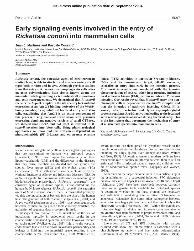
ResultsInternalization of R. conorii into Vero cells is dependenton actin polymerization.Electron micrographs of the R. conorii invasion processdemonstrated that bacterial entry requires host cytoskeletalalterations at the site of infection (Gouin et al., 1999) probablyinvolving localized actin rearrangements. The effect ofbacterial adherence on the host cell cytoskeletal network wasvisualized using Vero cells that were infected with R. conoriifor 15 minutes. As shown in Fig. 1A, F-actin colocalized with
adherent bacteria suggesting that this recruitment of actin wasinvolved in uptake (arrows). To investigate the role of theactin polymerization in the entry process, we used aspecific pharmacological inhibitor of actin polymerization(cytochalasin D) in conjunction with a fluorescence-basedinternalization assay (see Materials and Methods). Inhibitionof actin polymerization blocked R. conoriiinvasion (Fig. 1B),but had no effect on bacterial adherence (data not shown),confirming the important role of actin rearrangements in theinvasion process.
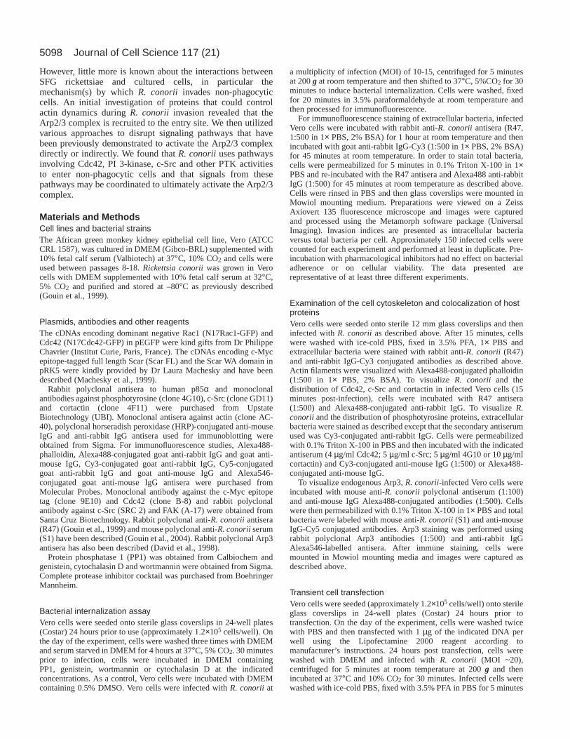
Involvement of the Arp2/3 complex in R. conoriiinvasion.To identify proteins that modulate the actin cytoskeleton andthat are potentially involved in R. conorii invasion, we firstfocused on the Arp2/3 complex. The Arp2/3 complex has adirect role in initiating actin nucleation and the branching ofactin filaments (Bear et al., 2001; Bear et al., 2002). Initially,to investigate the role of the Arp2/3 complex in R. conoriientry, Vero cells were infected with R. conoriifor 15 minutesand then processed for immunofluorescence using a polyclonalanti-Arp3 antisera (David et al., 1998). As shown in Fig. 2A,Arp3 strongly colocalizes with bacteria at the cell surface(arrow), but not with bacteria that are already internalized(arrowhead), suggesting that the Arp2/3 complex could play arole in R. conoriientry.
The Wiskott-Aldrich syndrome protein (WASp) familymember Scar1, directly binds to and activates the actinnucleating activity of the Arp2/3 complex and affects Arp2/3-dependent actin reorganization (Machesky et al., 1999).Therefore, if Arp2/3 is required for R. conorii invasion,overexpression of Scar1 and an Arp2/3 binding derivative (Scar
Fig. 1. Invasion of Rickettsia conoriiis dependent on host actinpolymerization. (A) In Vero cells infected with R. conorii; F-actin(green, left panel) colocalizes with invading bacteria (red) within 15minutes of infection, as shown in the merged image (yellow, rightpanel). (B) Preincubation of Vero cells with cytochalasin Ddiminished the ability of R. conoriito invade. Bar, 2 µm.
5100
WA) could titrate endogenous Arp2/3 complexes and inhibituptake into Vero cells. As shown in Fig. 2B, expression of full-length Scar1 (Scar FL) and the Scar1 WA domain (Scar WA)inhibited R. conoriientry into Vero cells. These results suggestthat the recruitment and activation of the Arp2/3 complex atthe site of bacterial entry may be a crucial, early event leadingto the localized actin polymerization necessary for bacterialinvasion.
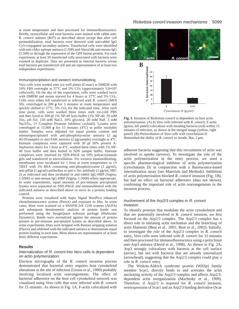
Involvement of the small GTPase Cdc42, but not Rac1in R. conorii entrySmall GTP-binding proteins, namely Rho family membersCdc42 and Rac and other members of the Ras superfamily ofGTPases, act as guanine nucleotide exchange switches toregulate diverse cellular processes within host cells (Olson etal., 1995; Ridley and Hall, 1992; Ridley et al., 1992) includingthe reorganization of the actin cytoskeleton in migrating cells(Hall, 1998) and during the phagocytosis of pathogens (Alrutzet al., 2001; Galan and Zhou, 2000; Martinez and Hultgren,2002; Tran Van Nhieu et al., 1999). It has also beendemonstrated that Cdc42 and Rac utilize proteins of the WASpfamily as downstream effector proteins to activate Arp2/3
during cortical actin polymerization (Higgs and Pollard, 2001),suggesting that activation of Cdc42 and Rac could indirectlylead to Arp2/3-mediated cytoskeletal changes. We utilizedtransfection of dominant negative versions of Cdc42 and Rac1(N17Cdc42 and N17Rac1, respectively) coupled with afluorescence-based internalization assay to assess the effects ofthese proteins on bacterial entry. As shown in Fig. 3A,expression of N17Cdc42-GFP, but not N17Rac1-GFP, intoVero cells resulted in an inhibition of R. conoriientry (~45%inhibition compared to untransfected control). To investigatethe putative role of Cdc42 in the entry process, non-transfectedVero cells were infected with R. conorii for 15 minutes andthen processed for immunofluorescence microscopy using amonoclonal anti-Cdc42 antibody. R. conoriicolocalized with
Journal of Cell Science 117 (21)
Fig. 2.The Arp2/3 complex is required for efficient bacterialinternalization. (A) Extracellular Rickettsia conorii(arrow), but notintracellular bacteria (arrowhead) strongly colocalizes with Arp3,suggesting that Arp2/3 recruitment plays an essential role in theuptake process in Vero cells. (B) Transfection of the WASp-relatedprotein, Scar (Scar FL), and the Arp2/3 binding domain (Scar WA)inhibits the ability of R. conoriito enter Vero cells, but has no effecton bacterial adherence (data not shown). Bar, 2 µm. Fig. 3.Cdc42 but not Rac1 governs actin rearrangements associated
with Rickettsia conoriiinvasion. (A) Expression of dominantnegative N17Cdc42, but not N17Rac1 in Vero cells inhibited R.conorii-mediated invasion when compared to non-transfected(Control) cells as assessed by a fluorescence-based invasion assay(see Materials and Methods). Expression of either construct had noeffect on bacterial adherence (data not shown). (B,C) Extracellular R.conorii were found to colocalize with endogenous Cdc42 (arrows) innon-transfected cells within 15 minutes of bacterial infection.(D) Colocalization of Cdc42 with bacteria is not caused by antibodycrossreactivity with bacteria (arrowhead). Bar, 4 µm.
5101Rickettsia conorii invasion mechanisms
endogenous Cdc42 at bacterial entry sites (Fig. 3B,C, arrows).This recruitment appeared to be specific to invading bacteriaas not all cell-associated R. conorii colocalized withendogenous Cdc42 (arrowhead in Fig. 3D). Taken together,
these results suggest that activation of Cdc42 contributes to thelocalized actin polymerization that occurs at bacterial entryfoci and may contribute to the activation of Arp2/3 during R.conorii invasion.
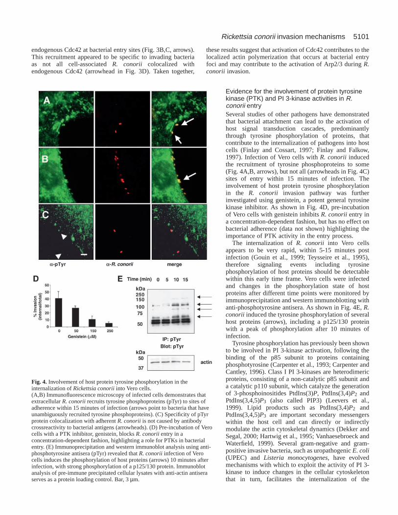
Evidence for the involvement of protein tyrosinekinase (PTK) and PI 3-kinase activities in R.conorii entrySeveral studies of other pathogens have demonstratedthat bacterial attachment can lead to the activation ofhost signal transduction cascades, predominantlythrough tyrosine phosphorylation of proteins, thatcontribute to the internalization of pathogens into hostcells (Finlay and Cossart, 1997; Finlay and Falkow,1997). Infection of Vero cells with R. conorii inducedthe recruitment of tyrosine phosphoproteins to some(Fig. 4A,B, arrows), but not all (arrowheads in Fig. 4C)sites of entry within 15 minutes of infection. Theinvolvement of host protein tyrosine phosphorylationin the R. conorii invasion pathway was furtherinvestigated using genistein, a potent general tyrosinekinase inhibitor. As shown in Fig. 4D, pre-incubationof Vero cells with genistein inhibits R. conoriientry ina concentration-dependent fashion, but has no effect onbacterial adherence (data not shown) highlighting theimportance of PTK activity in the entry process.
The internalization of R. conorii into Vero cellsappears to be very rapid, within 5-15 minutes postinfection (Gouin et al., 1999; Teysseire et al., 1995),therefore signaling events including tyrosinephosphorylation of host proteins should be detectablewithin this early time frame. Vero cells were infectedand changes in the phosphorylation state of hostproteins after different time points were monitored byimmunoprecipitation and western immunoblotting withanti-phosphotyrosine antisera. As shown in Fig. 4E, R.conorii induced the tyrosine phosphorylation of severalhost proteins (arrows), including a p125/130 proteinwith a peak of phosphorylation after 10 minutes ofinfection.
Tyrosine phosphorylation has previously been shownto be involved in PI 3-kinase activation, following thebinding of the p85 subunit to proteins containingphosphotyrosine (Carpenter et al., 1993; Carpenter andCantley, 1996). Class I PI 3-kinases are heterodimericproteins, consisting of a non-catalytic p85 subunit anda catalytic p110 subunit, which catalyze the generationof 3-phosphoinositides PtdIns(3)P, PtdIns(3,4)P2 andPtdIns(3,4,5)P3 (also called PIP3) (Leevers et al.,1999). Lipid products such as PtdIns(3,4)P2 andPtdIns(3,4,5)P3 are important secondary messengerswithin the host cell and can directly or indirectlymodulate the actin cytoskeletal dynamics (Dekker andSegal, 2000; Hartwig et al., 1995; Vanhaesebroeck andWaterfield, 1999). Several gram-negative and gram-positive invasive bacteria, such as uropathogenic E. coli(UPEC) and Listeria monocytogenes, have evolvedmechanisms with which to exploit the activity of PI 3-kinase to induce changes in the cellular cytoskeletonthat in turn, facilitates the internalization of the
Fig. 4. Involvement of host protein tyrosine phosphorylation in theinternalization of Rickettsia conoriiinto Vero cells.(A,B) Immunofluorescence microscopy of infected cells demonstrates thatextracellular R. conoriirecruits tyrosine phosphoproteins (pTyr) to sites ofadherence within 15 minutes of infection (arrows point to bacteria that haveunambiguously recruited tyrosine phosphoproteins). (C) Specificity of pTyrprotein colocalization with adherent R. conoriiis not caused by antibodycrossreactivity to bacterial antigens (arrowheads). (D) Pre-incubation of Verocells with a PTK inhibitor, genistein, blocks R. conoriientry in aconcentration-dependent fashion, highlighting a role for PTKs in bacterialentry. (E) Immunoprecipitation and western immunoblot analysis using anti-phosphotyrosine antisera (pTyr) revealed that R. conoriiinfection of Verocells induces the phosphorylation of host proteins (arrows) 10 minutes afterinfection, with strong phosphorylation of a p125/130 protein. Immunoblotanalysis of pre-immune precipitated cellular lysates with anti-actin antiseraserves as a protein loading control. Bar, 3 µm.
5102
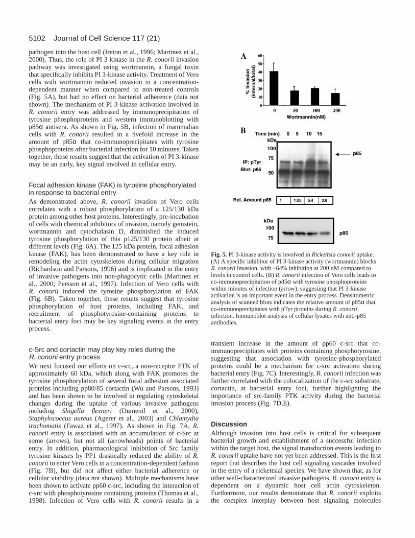
pathogen into the host cell (Ireton et al., 1996; Martinez et al.,2000). Thus, the role of PI 3-kinase in the R. conorii invasionpathway was investigated using wortmannin, a fungal toxinthat specifically inhibits PI 3-kinase activity. Treatment of Verocells with wortmannin reduced invasion in a concentration-dependent manner when compared to non-treated controls(Fig. 5A), but had no effect on bacterial adherence (data notshown). The mechanism of PI 3-kinase activation involved inR. conorii entry was addressed by immunoprecipitation oftyrosine phosphoproteins and western immunoblotting withp85α antisera. As shown in Fig. 5B, infection of mammaliancells with R. conorii resulted in a fivefold increase in theamount of p85α that co-immunoprecipitates with tyrosinephosphoproteins after bacterial infection for 10 minutes. Takentogether, these results suggest that the activation of PI 3-kinasemay be an early, key signal involved in cellular entry.
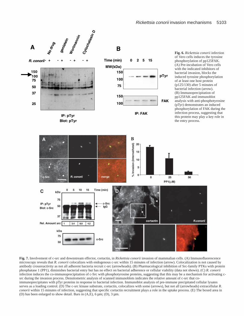
Focal adhesion kinase (FAK) is tyrosine phosphorylatedin response to bacterial entryAs demonstrated above, R. conorii invasion of Vero cellscorrelates with a robust phosphorylation of a 125/130 kDaprotein among other host proteins. Interestingly, pre-incubationof cells with chemical inhibitors of invasion, namely genistein,wortmannin and cytochalasin D, diminished the inducedtyrosine phosphorylation of this p125/130 protein albeit atdifferent levels (Fig. 6A). The 125 kDa protein, focal adhesionkinase (FAK), has been demonstrated to have a key role inremodeling the actin cytoskeleton during cellular migration(Richardson and Parsons, 1996) and is implicated in the entryof invasive pathogens into non-phagocytic cells (Martinez etal., 2000; Persson et al., 1997). Infection of Vero cells withR. conorii induced the tyrosine phosphorylation of FAK(Fig. 6B). Taken together, these results suggest that tyrosinephosphorylation of host proteins, including FAK, andrecruitment of phosphotyrosine-containing proteins tobacterial entry foci may be key signaling events in the entryprocess.
c-Src and cortactin may play key roles during theR. conorii entry processWe next focused our efforts on c-src, a non-receptor PTK ofapproximately 60 kDa, which along with FAK promotes thetyrosine phosphorylation of several focal adhesion associatedproteins including pp80/85 cortactin (Wu and Parsons, 1993)and has been shown to be involved in regulating cytoskeletalchanges during the uptake of various invasive pathogensincluding Shigella flexneri (Dumenil et al., 2000),Staphylococcus aureus(Agerer et al., 2003) and Chlamydiatrachomatis(Fawaz et al., 1997). As shown in Fig. 7A, R.conorii entry is associated with an accumulation of c-Src atsome (arrows), but not all (arrowheads) points of bacterialentry. In addition, pharmacological inhibition of Src familytyrosine kinases by PP1 drastically reduced the ability of R.conorii to enter Vero cells in a concentration-dependent fashion(Fig. 7B), but did not affect either bacterial adherence orcellular viability (data not shown). Multiple mechanisms havebeen shown to activate pp60 c-src, including the interaction ofc-src with phosphotyrosine containing proteins (Thomas et al.,1998). Infection of Vero cells with R. conorii results in a
transient increase in the amount of pp60 c-src that co-immunoprecipitates with proteins containing phosphotyrosine,suggesting that association with tyrosine-phosphorylatedproteins could be a mechanism for c-src activation duringbacterial entry (Fig. 7C). Interestingly, R. conoriiinfection wasfurther correlated with the colocalization of the c-src substrate,cortactin, at bacterial entry foci, further highlighting theimportance of src-family PTK activity during the bacterialinvasion process (Fig. 7D,E).
DiscussionAlthough invasion into host cells is critical for subsequentbacterial growth and establishment of a successful infectionwithin the target host, the signal transduction events leading toR. conoriiuptake have not yet been addressed. This is the firstreport that describes the host cell signaling cascades involvedin the entry of a rickettsial species. We have shown that, as forother well-characterized invasive pathogens, R. conoriientry isdependent on a dynamic host cell actin cytoskeleton.Furthermore, our results demonstrate that R. conorii exploitsthe complex interplay between host signaling molecules
Journal of Cell Science 117 (21)
Fig. 5.PI 3-kinase activity is involved in Rickettsia conoriiuptake.(A) A specific inhibitor of PI 3-kinase activity (wortmannin) blocksR. conoriiinvasion, with ~64% inhibition at 200 nM compared tolevels in control cells. (B) R. conoriiinfection of Vero cells leads toco-immunoprecipitation of p85α with tyrosine phosphoproteinswithin minutes of infection (arrow), suggesting that PI 3-kinaseactivation is an important event in the entry process. Densitometricanalysis of scanned blots indicates the relative amount of p85α thatco-immunoprecipitates with pTyr proteins during R. conoriiinfection. Immunoblot analysis of cellular lysates with anti-p85antibodies.
5103Rickettsia conorii invasion mechanisms
Fig. 6.Rickettsia conoriiinfectionof Vero cells induces the tyrosinephosphorylation of pp125FAK.(A) Pre-incubation of Vero cellswith the indicated inhibitors ofbacterial invasion, blocks theinduced tyrosine phosphorylationof at least one host protein(p125/130) after 5 minutes ofbacterial infection (arrow).(B) Immunoprecipitation ofpp125FAK and immunoblotanalysis with anti-phosphotyrosine(pTyr) demonstrates an inducedphosphorylation of FAK during theinfection process, suggesting thatthis protein may play a key role inthe entry process.
Fig. 7. Involvement of c-src and downstream effector, cortactin, in Rickettsia conoriiinvasion of mammalian cells. (A) Immunofluorescencemicroscopy reveals that R. conoriicolocalizes with endogenous c-src within 15 minutes of infection (arrow). Colocalization is not caused byantibody crossreactivity as not all adherent bacteria recruit c-src (arrowheads). (B) Pharmacological inhibition of Src-family PTKs with proteinphosphatase 1 (PP1), diminishes bacterial entry but has no effect on bacterial adherence or cellular viability (data not shown). (C) R. conoriiinfection induces the co-immunoprecipitation of c-Src with phosphotyrosine proteins, suggesting that this may be a mechanism for activating c-src during the invasion process. Densitometric analysis of scanned immunoblots indicates the relative amount of c-src that co-immunoprecipitates with pTyr proteins in response to bacterial infection. Immunoblot analysis of pre-immune precipitated cellular lysatesserves as a loading control. (D) The c-src kinase substrate, cortactin, colocalizes with some (arrows), but not all (arrowheads) extracellular R.conorii within 15 minutes of infection, suggesting that specific cortactin recruitment plays a role in the uptake process. (E) The boxed area in(D) has been enlarged to show detail. Bars in (A,E), 6 µm; (D), 3 µm.
5104
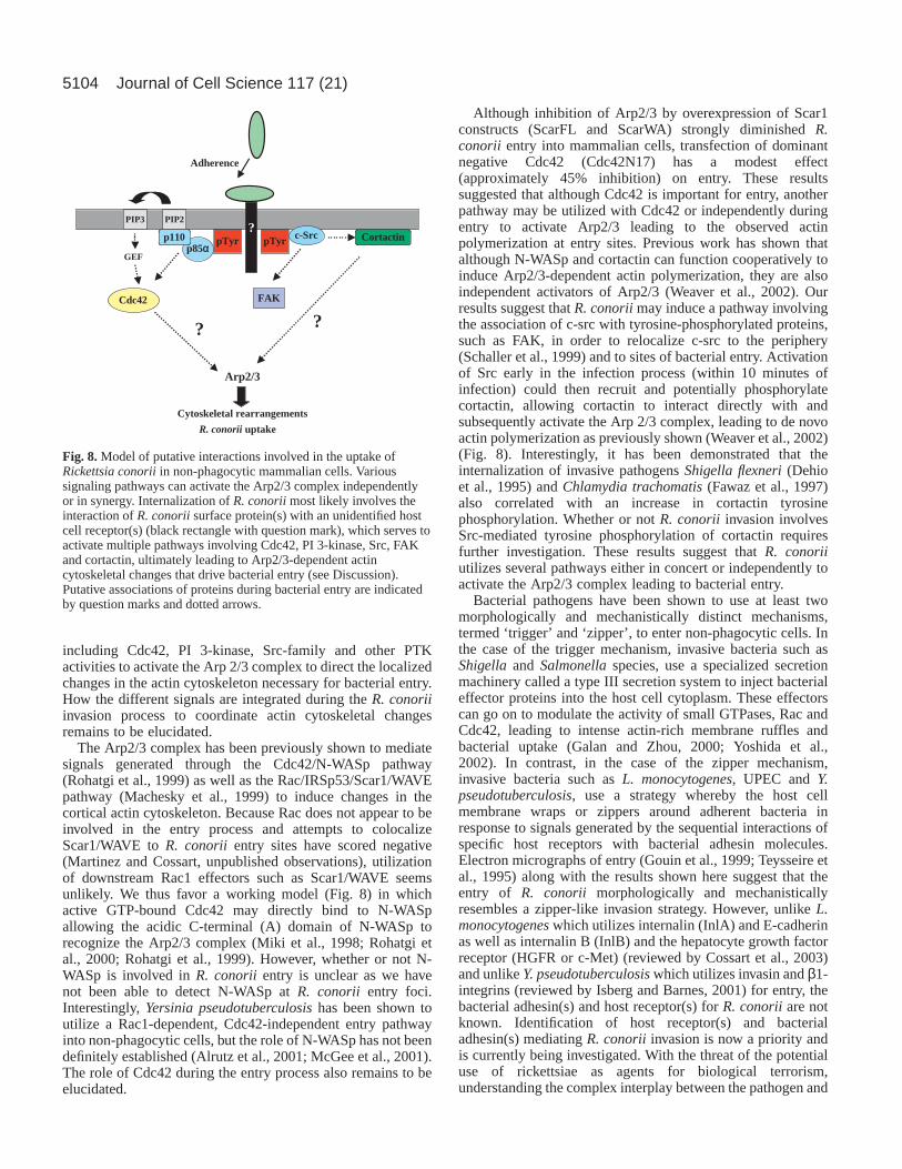
including Cdc42, PI 3-kinase, Src-family and other PTKactivities to activate the Arp 2/3 complex to direct the localizedchanges in the actin cytoskeleton necessary for bacterial entry.How the different signals are integrated during the R. conoriiinvasion process to coordinate actin cytoskeletal changesremains to be elucidated.
The Arp2/3 complex has been previously shown to mediatesignals generated through the Cdc42/N-WASp pathway(Rohatgi et al., 1999) as well as the Rac/IRSp53/Scar1/WAVEpathway (Machesky et al., 1999) to induce changes in thecortical actin cytoskeleton. Because Rac does not appear to beinvolved in the entry process and attempts to colocalizeScar1/WAVE to R. conorii entry sites have scored negative(Martinez and Cossart, unpublished observations), utilizationof downstream Rac1 effectors such as Scar1/WAVE seemsunlikely. We thus favor a working model (Fig. 8) in whichactive GTP-bound Cdc42 may directly bind to N-WASpallowing the acidic C-terminal (A) domain of N-WASp torecognize the Arp2/3 complex (Miki et al., 1998; Rohatgi etal., 2000; Rohatgi et al., 1999). However, whether or not N-WASp is involved in R. conorii entry is unclear as we havenot been able to detect N-WASp at R. conorii entry foci.Interestingly, Yersinia pseudotuberculosishas been shown toutilize a Rac1-dependent, Cdc42-independent entry pathwayinto non-phagocytic cells, but the role of N-WASp has not beendefinitely established (Alrutz et al., 2001; McGee et al., 2001).The role of Cdc42 during the entry process also remains to beelucidated.
Although inhibition of Arp2/3 by overexpression of Scar1constructs (ScarFL and ScarWA) strongly diminished R.conorii entry into mammalian cells, transfection of dominantnegative Cdc42 (Cdc42N17) has a modest effect(approximately 45% inhibition) on entry. These resultssuggested that although Cdc42 is important for entry, anotherpathway may be utilized with Cdc42 or independently duringentry to activate Arp2/3 leading to the observed actinpolymerization at entry sites. Previous work has shown thatalthough N-WASp and cortactin can function cooperatively toinduce Arp2/3-dependent actin polymerization, they are alsoindependent activators of Arp2/3 (Weaver et al., 2002). Ourresults suggest that R. conoriimay induce a pathway involvingthe association of c-src with tyrosine-phosphorylated proteins,such as FAK, in order to relocalize c-src to the periphery(Schaller et al., 1999) and to sites of bacterial entry. Activationof Src early in the infection process (within 10 minutes ofinfection) could then recruit and potentially phosphorylatecortactin, allowing cortactin to interact directly with andsubsequently activate the Arp 2/3 complex, leading to de novoactin polymerization as previously shown (Weaver et al., 2002)(Fig. 8). Interestingly, it has been demonstrated that theinternalization of invasive pathogens Shigella flexneri(Dehioet al., 1995) and Chlamydia trachomatis(Fawaz et al., 1997)also correlated with an increase in cortactin tyrosinephosphorylation. Whether or not R. conorii invasion involvesSrc-mediated tyrosine phosphorylation of cortactin requiresfurther investigation. These results suggest that R. conoriiutilizes several pathways either in concert or independently toactivate the Arp2/3 complex leading to bacterial entry.
Bacterial pathogens have been shown to use at least twomorphologically and mechanistically distinct mechanisms,termed ‘trigger’ and ‘zipper’, to enter non-phagocytic cells. Inthe case of the trigger mechanism, invasive bacteria such asShigellaand Salmonellaspecies, use a specialized secretionmachinery called a type III secretion system to inject bacterialeffector proteins into the host cell cytoplasm. These effectorscan go on to modulate the activity of small GTPases, Rac andCdc42, leading to intense actin-rich membrane ruffles andbacterial uptake (Galan and Zhou, 2000; Yoshida et al.,2002). In contrast, in the case of the zipper mechanism,invasive bacteria such as L. monocytogenes, UPEC and Y.pseudotuberculosis, use a strategy whereby the host cellmembrane wraps or zippers around adherent bacteria inresponse to signals generated by the sequential interactions ofspecific host receptors with bacterial adhesin molecules.Electron micrographs of entry (Gouin et al., 1999; Teysseire etal., 1995) along with the results shown here suggest that theentry of R. conorii morphologically and mechanisticallyresembles a zipper-like invasion strategy. However, unlike L.monocytogenes which utilizes internalin (InlA) and E-cadherinas well as internalin B (InlB) and the hepatocyte growth factorreceptor (HGFR or c-Met) (reviewed by Cossart et al., 2003)and unlike Y. pseudotuberculosiswhich utilizes invasin and β1-integrins (reviewed by Isberg and Barnes, 2001) for entry, thebacterial adhesin(s) and host receptor(s) for R. conoriiare notknown. Identification of host receptor(s) and bacterialadhesin(s) mediating R. conorii invasion is now a priority andis currently being investigated. With the threat of the potentialuse of rickettsiae as agents for biological terrorism,understanding the complex interplay between the pathogen and
Journal of Cell Science 117 (21)
pTyrp85α
p110
Cdc42
PIP2PIP3
GEF
Arp2/3
pTyr c-Src
Adherence
FAK
Cortactin
Cytoskeletal rearrangements
R. conorii uptake
?
?
?
Fig. 8.Model of putative interactions involved in the uptake ofRickettsia conoriiin non-phagocytic mammalian cells. Varioussignaling pathways can activate the Arp2/3 complex independentlyor in synergy. Internalization of R. conoriimost likely involves theinteraction of R. conoriisurface protein(s) with an unidentified hostcell receptor(s) (black rectangle with question mark), which serves toactivate multiple pathways involving Cdc42, PI 3-kinase, Src, FAKand cortactin, ultimately leading to Arp2/3-dependent actincytoskeletal changes that drive bacterial entry (see Discussion).Putative associations of proteins during bacterial entry are indicatedby question marks and dotted arrows.
5105Rickettsia conorii invasion mechanisms
host cell receptor(s) is very important and may provide insightinto the development of more efficacious therapies for thetreatment and prevention of SFG rickettsial diseases.
We would like to thank Dr Hélène Bierne and Dr Stéphanie Seveaualong with other members of the Cossart lab for helpful discussionsand critical reading of this manuscript. We would also like to thankDr Philippe Chavrier (Institut Curie, Paris, France) for the kind giftof GFP-N17Rac1 and GFP-N17Cdc42 expression constructs and DrLaura Machesky for the kind gift of c-Myc tagged Scar1 (ScarFL,Scar WA) constructs. This work was supported by the Institut Pasteurand a grant from Délégation Générale pour l’Armement (DGAcontract 01-34-068). This work was also supported in part by anEMBO Longterm Post-Doctoral Fellowship (ALTF-171-2001), apost-doctoral fellowship from the Institut Nationale de la RechercheAgronomique (INRA) and a post-doctoral fellowship from theFondation pour la Recherche Medicale (FRM) to J. J. Martinez. P.Cossart is an international research scholar of the Howard HughesMedical Institute.
ReferencesAgerer, F., Michel, A., Ohlsen, K. and Hauck, C. R. (2003). Integrin-
mediated invasion of Staphylococcus aureusinto human cells requires Srcfamily protein-tyrosine kinases. J. Biol. Chem.278, 42524-42531.
Alrutz, M. A., Srivastava, A., Wong, K. W., D’Souza-Schorey, C., Tang,M., Ch’Ng, L. E., Snapper, S. B. and Isberg, R. R. (2001). Efficient uptakeof Yersinia pseudotuberculosisvia integrin receptors involves a Rac1-Arp2/3 pathway that bypasses N-WASP function. Mol. Microbiol. 42, 689-703.
Andersson, S. G., Zomorodipour, A., Andersson, J. O., Sicheritz-Ponten,T., Alsmark, U. C., Podowski, R. M., Naslund, A. K., Eriksson, A. S.,Winkler, H. H. and Kurland, C. G. (1998). The genome sequence ofRickettsia prowazekiiand the origin of mitochondria. Nature396, 133-140.
Bear, J. E., Krause, M. and Gertler, F. B. (2001). Regulating cellular actinassembly. Curr. Opin. Cell Biol.13, 158-166.
Bear, J. E., Svitkina, T. M., Krause, M., Schafer, D. A., Loureiro, J. J.,Strasser, G. A., Maly, I. V., Chaga, O. Y., Cooper, J. A., Borisy, G. G. etal. (2002). Antagonism between Ena/VASP proteins and actin filamentcapping regulates fibroblast motility. Cell 109, 509-521.
Carpenter, C. L. and Cantley, L. C. (1996). Phosphoinositide kinases. Curr.Opin. Cell Biol.8, 153-158.
Carpenter, C. L., Auger, K. R., Chanudhuri, M., Yoakim, M.,Schaffhausen, B., Shoelson, S. and Cantley, L. C. (1993).Phosphoinositide 3-kinase is activated by phosphopeptides that bind to theSH2 domains of the 85-kDa subunit. J. Biol. Chem.268, 9478-9483.
Cossart, P., Pizarro-Cerda, J. and Lecuit, M. (2003). Invasion ofmammalian cells by Listeria monocytogenes: functional mimicry to subvertcellular functions. Trends Cell Biol.13, 23-31.
David, V., Gouin, E., Troys, M. V., Grogan, A., Segal, A. W., Ampe, C. andCossart, P. (1998). Identification of cofilin, coronin, Rac and capZ in actintails using a Listeria affinity approach. J. Cell Sci.111, 2877-2884.
Dehio, C., Prevost, M. C. and Sansonetti, P. J. (1995). Invasion of epithelialcells by Shigella flexneriinduces tyrosine phosphorylation of cortactin by app60c-src-mediated signalling pathway. EMBO J.14, 2471-2482.
Dekker, L. V. and Segal, A. W. (2000). Perspectives: signal transduction.Signals to move cells. Science287, 982-983.
Dumenil, G., Sansonetti, P. and Tran van Nhieu, G. (2000). Src tyrosinekinase activity down-regulates Rho-dependent responses during Shigellaentry into epithelial cells and stress fibre formation. J. Cell Sci.113, 71-80.
Fawaz, F. S., van Ooij, C., Homola, E., Mutka, S. C. and Engel, J. N.(1997). Infection with Chlamydia trachomatis alters the tyrosinephosphorylation and/or localization of several host cell proteins includingcortactin. Infect. Immun.65, 5301-5308.
Finlay, B. B. and Cossart, P. (1997). Exploitation of mammalian host cellfunctions by bacterial pathogens. Science276, 718-725.
Finlay, B. B. and Falkow, S. (1997). Common themes in microbialpathogenicity revisited. Microbiol. Mol. Biol. Rev.61, 136-169.
Galan, J. E. and Zhou, D. (2000). Striking a balance: modulation of the actincytoskeleton by Salmonella. Proc. Natl. Acad. Sci. USA97, 8754-8761.
Gouin, E., Gantelet, H., Egile, C., Lasa, I., Ohayon, H., Villiers, V.,Gounon, P., Sansonetti, P. J. and Cossart, P. (1999). A comparative study
of the actin-based motilities of the pathogenic bacteria Listeriamonocytogenes, Shigella flexneriand Rickettsia conorii. J. Cell Sci.112,1697-1708.
Gouin, E., Egile, C., Dehoux, P., Villiers, V., Adams, J., Gertler, F., Li, R.and Cossart, P. (2004). The RickA protein of Rickettsia conoriiactivatesthe Arp2/3 complex. Nature427, 457-461.
Hackstadt, T. (1996). The biology of rickettsiae. Infect. Agents Dis.5, 127-143.
Hall, A. (1998). G proteins and small GTPases: distant relatives keep in touch.Science280, 2074-2075.
Hand, W. L., Miller, J. B., Reinarz, J. A. and Sanford, J. P. (1970). RockyMountain spotted fever. A vascular disease. Arch. Intern. Med.125, 879-882.
Hartwig, J. H., Bokoch, G. M., Carpenter, C. L., Janmey, P. A., Taylor, L.A., Toker, A. and Stossel, T. P. (1995). Thrombin receptor ligation andactivated Rac uncap actin filament barbed ends through phosphoinositidesynthesis in permeabilized human platelets. Cell 82, 643-653.
Heinzen, R. A., Hayes, S. F., Peacock, M. G. and Hackstadt, T. (1993).Directional actin polymerization associated with spotted fever groupRickettsiainfection of Vero cells. Infect. Immun.61, 1926-1935.
Higgs, H. N. and Pollard, T. D. (2001). Regulation of actin filament networkformation through Arp2/3 complex: activation by a diverse array of proteins.Annu. Rev. Biochem.70, 649-676.
Ireton, K., Payrastre, B., Chap, H., Ogawa, W., Sakaue, H., Kasuga, M.and Cossart, P. (1996). A role for phosphoinositide 3-kinase in bacterialinvasion. Science274, 780-782.
Isberg, R. R. and Barnes, P. (2001). Subversion of integrins byenteropathogenic Yersinia. J. Cell Sci.114, 21-28.
Leevers, S. J., Vanhaesebroeck, B. and Waterfield, M. D. (1999). Signallingthrough phosphoinositide 3-kinases: the lipids take centre stage. Curr. Opin.Cell Biol. 11, 219-225.
Li, H. and Walker, D. H. (1998). rOmpA is a critical protein for the adhesionof Rickettsia rickettsiito host cells. Microb. Pathog.24, 289-298.
Machesky, L. M., Mullins, R. D., Higgs, H. N., Kaiser, D. A., Blachoin, L.,May, R. C., Hall, M. E. and Pollard, T. D. (1999). Scar, a WASp-relatedprotein, activates nucleation of actin filaments by the Arp2/3 complex. Proc.Natl. Acad. Sci. USA96, 3739-3744.
Martinez, J. J. and Hultgren, S. J. (2002). Requirement of Rho-familyGTPases in the invasion of Type 1-piliated uropathogenic Escherichia coli.Cell. Microbiol. 4, 19-28.
Martinez, J. J., Mulvey, M. A., Schilling, J. D., Pinkner, J. S. and Hultgren,S. J. (2000). Type 1 pilus-mediated bacterial invasion of bladder epithelialcells. EMBO J.19, 2803-2812.
McGee, K., Zettl, M., Way, M. and Fallman, M. (2001). A role for N-WASPin invasin-promoted internalisation. FEBS Lett.509, 59-65.
Miki, H., Sasaki, T., Takai, Y. and Takenawa, T. (1998). Induction offilopodium formation by a WASP-related actin-depolymerizing protein N-WASP. Nature391, 93-96.
Olson, M. F., Ashworth, A. and Hall, A. (1995). An essential role for Rho,Rac, and Cdc42 GTPases in cell cycle progression through G1. Science269,1270-1272.
Ogata, H., Audic, S., Renesto-Audiffren, P., Fournier, P. E., Barbe, V.,Samson, D., Roux, V., Cossart, P., Weissenbach, J., Claverie, J. M. et al.(2001). Mechanisms of evolution in Rickettsia conoriiand R. prowazekii.Science293, 2093-2098.
Persson, C., Carballeira, N., Wolf-Watz, H. and Fallman, M. (1997). ThePTPase YopH inhibits uptake of Yersinia, tyrosine phosphorylation ofp130Cas and FAK, and the associated accumulation of these proteins inperipheral focal adhesions. EMBO J.16, 2307-2318.
Richardson, A. and Parsons, T. (1996). A mechanism for regulation of theadhesion-associated protein tyrosine kinase pp125FAK. Nature 380, 538-540.
Ridley, A. J. and Hall, A. (1992). The small GTP-binding protein rhoregulates the assembly of focal adhesions and actin stress fibers in responseto growth factors. Cell 70, 389-399.
Ridley, A. J., Paterson, H. F., Johnston, C. L., Diekmann, D. and Hall, A.(1992). The small GTP-binding protein rac regulates growth factor-inducedmembrane ruffling. Cell 70, 401-410.
Rohatgi, R., Ma, L., Miki, H., Lopez, M., Kirchhausen, T., Takenawa, T.and Kirschner, M. W. (1999). The interaction between N-WASP and theArp2/3 complex links Cdc42-dependent signals to actin assembly. Cell 97,221-231.
Rohatgi, R., Ho, H. Y. and Kirschner, M. W. (2000). Mechanism of N-WASPactivation by Cdc42 and phosphatidylinositol 4,5-bisphosphate. J. Cell Biol.150, 1299-1310.
5106
Schaller, M. D., Hildebrand, J. D. and Parsons, J. T. (1999). Complexformation with focal adhesion kinase: a mechanism to regulate activity andsubcellular localization of Src kinases. Mol. Biol. Cell10, 3489-3505.
Silverman, D. J., Santucci, L. A., Meyers, N. and Sekeyova, Z. (1992).Penetration of host cells by Rickettsia rickettsiiappears to be mediated bya phospholipase of rickettsial origin. Infect. Immun.60, 2733-2740.
Teysseire, N., Chiche-Portiche, C. and Raoult, D. (1992). Intracellularmovements of Rickettsia conoriiand R. typhibased on actin polymerization.Res. Microbiol.143, 821-829.
Teysseire, N., Boudier, J. A. and Raoult, D. (1995). Rickettsia conoriientryinto Vero cells. Infect. Immun.63, 366-374.
Thomas, J. W., Ellis, B., Boerner, R. J., Knight, W. B., White, G. C., IIand Schaller, M. D. (1998). SH2- and SH3-mediated interactions betweenfocal adhesion kinase and Src. J. Biol. Chem.273, 577-583.
Tran van Nhieu, G., Caron, E., Hall, A. and Sansonetti, P. J. (1999). IpaCinduces actin polymerization and filopodia formation during Shigellaentryinto epithelial cells. EMBO J.18, 3249-3262.
Uchiyama, T. (2003). Adherence to and invasion of Vero cells by recombinantEscherichia coli expressing the outer membrane protein rOmpB ofRickettsia japonica. Ann. N.Y. Acad. Sci.990, 585-590.
Vanhaesebroeck, B. and Waterfield, M. D. (1999). Signaling by distinctclasses of phosphoinositide 3-kinases. Exp. Cell Res.253, 239-254.
Vishwanath, S. (1991). Antigenic relationships among the rickettsiae of thespotted fever and typhus groups. FEMS Microbiol. Lett.65, 341-344.
Walker, T. S. (1984). Rickettsial interactions with human endothelial cells invitro: adherence and entry. Infect. Immun.44, 205-210.
Walker, D. H. and Gear, J. H. (1985). Correlation of the distribution ofRickettsia conorii, microscopic lesions, and clinical features in SouthAfrican tick bite fever. Am. J. Trop. Med. Hyg.34, 361-371.
Walker, D. H., Occhino, C., Tringali, G. R., di Rosa, S. and Mansueto, S.(1988). Pathogenesis of rickettsial eschars: the tache noire of boutonneusefever. Hum. Pathol.19, 1449-1454.
Walker, D. H., Feng, H. M. and Popov, V. L. (2001). Rickettsialphospholipase A2 as a pathogenic mechanism in a model of cell injury bytyphus and spotted fever group rickettsiae. Am. J. Trop. Med. Hyg.65, 936-942.
Weaver, A. M., Heuser, J. E., Karginov, A. V., Lee, W. L., Parsons, J. T.and Cooper, J. A. (2002). Interaction of cortactin and N-WASp with Arp2/3complex. Curr. Biol. 12, 1270-1278.
Wu, H. and Parsons, J. T. (1993). Cortactin, an 80/85-kilodalton pp60srcsubstrate, is a filamentous actin-binding protein enriched in the cell cortex.J. Cell Biol.120, 1417-1426.
Yagupsky, P. and Wolach, B. (1993). Fatal Israeli spotted fever in children.Clin. Infect. Dis.17, 850-853.
Yoshida, S., Katayama, E., Kuwae, A., Mimuro, H., Suzuki, T. andSasakawa, C. (2002). Shigelladeliver an effector protein to trigger hostmicrotubule destabilization, which promotes Rac1 activity and efficientbacterial internalization. EMBO J.21, 2923-2935.
Journal of Cell Science 117 (21)


![Cicero [encyclopedia entry]](https://img.pdfslide.net/doc/110x75/631f3917d10f1687490fb291/cicero-encyclopedia-entry.jpg)


