Embed Size (px)
Citation preview
Complimentary Contributor Copy

Complimentary Contributor Copy
PUBLIC HEALTH IN THE 21ST CENTURY
THE VASCULITIDES
VOLUME 1
GENERAL CONSIDERATIONS
AND SYSTEMIC VASCULITIS
(SECOND EDITION)
No part of this digital document may be reproduced, stored in a retrieval system or transmitted in any form orby any means. The publisher has taken reasonable care in the preparation of this digital document, but makes noexpressed or implied warranty of any kind and assumes no responsibility for any errors or omissions. Noliability is assumed for incidental or consequential damages in connection with or arising out of informationcontained herein. This digital document is sold with the clear understanding that the publisher is not engaged inrendering legal, medical or any other professional services. Complimentary Contributor Copy

PUBLIC HEALTH IN THE 21ST CENTURY
Additional books and e-books in this series can be found
on Nova’s website under the Series tab.
Complimentary Contributor Copy

PUBLIC HEALTH IN THE 21ST CENTURY
THE VASCULITIDES
VOLUME 1
GENERAL CONSIDERATIONS
AND SYSTEMIC VASCULITIS
(SECOND EDITION)
DAVID S. YOUNGER, MD, MPH, MS
EDITOR
Complimentary Contributor Copy
Copyright © 2019 by Nova Science Publishers, Inc.
All rights reserved. No part of this book may be reproduced, stored in a retrieval system or
transmitted in any form or by any means: electronic, electrostatic, magnetic, tape, mechanical
photocopying, recording or otherwise without the written permission of the Publisher.
We have partnered with Copyright Clearance Center to make it easy for you to obtain permissions
to reuse content from this publication. Simply navigate to this publication’s page on Nova’s website
and locate the “Get Permission” button below the title description. This button is linked directly to
the title’s permission page on copyright.com. Alternatively, you can visit copyright.com and search
by title, ISBN, or ISSN.
For further questions about using the service on copyright.com, please contact:
Copyright Clearance Center
Phone: +1-(978) 750-8400 Fax: +1-(978) 750-4470 E-mail: [email protected].
NOTICE TO THE READER
The Publisher has taken reasonable care in the preparation of this book, but makes no expressed or
implied warranty of any kind and assumes no responsibility for any errors or omissions. No liability
is assumed for incidental or consequential damages in connection with or arising out of information
contained in this book. The Publisher shall not be liable for any special, consequential, or exemplary
damages resulting, in whole or in part, from the readers’ use of, or reliance upon, this material. Any
parts of this book based on government reports are so indicated and copyright is claimed for those
parts to the extent applicable to compilations of such works.
Independent verification should be sought for any data, advice or recommendations contained in
this book. In addition, no responsibility is assumed by the Publisher for any injury and/or damage
to persons or property arising from any methods, products, instructions, ideas or otherwise contained
in this publication.
This publication is designed to provide accurate and authoritative information with regard to the
subject matter covered herein. It is sold with the clear understanding that the Publisher is not
engaged in rendering legal or any other professional services. If legal or any other expert assistance
is required, the services of a competent person should be sought. FROM A DECLARATION OF
PARTICIPANTS JOINTLY ADOPTED BY A COMMITTEE OF THE AMERICAN BAR
ASSOCIATION AND A COMMITTEE OF PUBLISHERS.
Additional color graphics may be available in the e-book version of this book.
Library of Congress Cataloging-in-Publication Data
ISBN: 978-1-53615-134-3 (ebook)
Published by Nova Science Publishers, Inc. † New York
Complimentary Contributor Copy
CONTENTS
Foreword to The Vasculitides vii Charles D. Pusey, MD and Richard A. Watts, MD, MPH
Preface ix
I. General Considerations 1
Chapter 1 History and Background of Vasculitis 3 Eric L. Matteson, MD, MPH
Chapter 2 Overview of Primary and Secondary Vasculitides 17 David S. Younger, MD, MPH, MS
Chapter 3 Epidemiology of Primary Systemic Vasculitis 63 Richard A. Watts, MD, MPH and Joanna Robson, PhD
Chapter 4 Health Related Quality of Life and its Measurement
in the Vasculitides 81 Joanna Robson, PhD, and Richard A. Watts, MD, MPH
Chapter 5 Neutrophilic Cell Pathobiology in the Vasculitides 95 Akihiro Ishizu, MD, PhD
Chapter 6 Complement Factors in ANCA-Associated Vasculitis 103 Chen Wang, MD, PhD and Min Chen, MD, PhD
Chapter 7 Experimental Autoimmune Vasculitis: Insights into Human
Vasculitis Using Animal Models 125 Alan D. Salama, MA, PhD and Mark A. Little, PhD
Chapter 8 Genetic Aspects of Vasculitis 135 F. David Carmona, PhD, Ana Márquez, MD,
Javier Martín, MD, PhD and Miguel A. González-Gay
II. Systemic Vasculitis 169
Chapter 9 Nomenclature and Pathologic Features of Vasculitides 171 J. Charles Jennette, MD, Ronald J. Falk, MD
and Adil H. M. Gasim, MD
Complimentary Contributor Copy
Contents vi
Chapter 10 Classification of Pediatric Vasculitides 193 Seza Ozen, MD
Chapter 11 Detection Techniques and Clinical Relevance of ANCA Testing 203 Elena Csernok, PhD, Juliane Mahrhold and Bernhard Hellmich
Chapter 12 Classification and Pathogenicity of ANCA-Associated Vasculitis 211 Alexander Tracy MB, BCh, Constantina Yiannakis, MB, BCh,
Lorna Ward, MD and Matthew David Morgan, MB, ChB, PhD
Chapter 13 Cryoglobulinemic Vasculitis 237 Clodoveo Ferri, MD, Dilia Giuggioli, MDi,
Marco Sebastiani, MD and Michele Colaci, MD, PhDi
Chapter 14 Aortitis and Periaortitis 251 Eugenia Accorsi Buttini, MD, Chiara Marvisi, MD
and Augusto Vaglio, MD, PhD
Chapter 15 Systemic Vasculitis and the Lung 277 Christian Pagnoux, MD, MS, MPH, Noura Mustapha, MD
and Gerard P. Cox, MB
Chapter 16 Systemic Vasculitis and the Kidney: ANCA-Associated Vasculitis
and Glomerulonephritis 299 Koyal Jain, MD, Manish Saha, MD and Ronald J. Falk, MD
Chapter 17 Anti-Glomerular Basement Membrane Disease 321 Stephen P. McAdoo, PhD and Charles D. Pusey, MD
Chapter 18 Systemic Vasculitis of the Gastrointestinal Tract 347 Daniel Geh, Dimitrios Chanouzas, PhD, MB, ChB, MSc
and Matthew David Morgan, MB, ChB, PhD
Chapter 19 Rheumatoid Arthritis Vasculitis 375 Elana J. Bernstein, MD, MSc and Robert F. Spiera, MD
Chapter 20 Dermatologic Aspects of Systemic Vasculitis 391 Rami N. Al-Rohil, MBBS, Ruben Peredo-Wende, MD
and J. Andrew Carlson, MD
About the Editor 429
List of Contributors 431
Index 439
Related Nova Publications 455
Complimentary Contributor Copy

FOREWORD TO THE VASCULITIDES
Charles D. Pusey and Richard A. Watts
Clinical and basic research into the systemic vasculitides have continued to gather
momentum over the last four years since publication of the first edition of this book. We have
attended two more International Vasculitis and ANCA Workshops, in London in 2015 and
Tokyo in 2017. Both of these excellent meetings have covered the area of vasculitis more
broadly and extensively than in the past. The present edition of this book contains contributions
from many of those participating in these meetings.
Classification and nomenclature in vasculitis is increasingly harmonised and agreed across
different specialties. There have been a number of genetic studies in ANCA-associated
vasculitis (AAV) in which the genetic associations appear to be more closely related to ANCA
specificity, i.e., MPO-ANCA or PR3-ANCA, than to clinical classification. The extent of
overlap between the different vasculitic conditions is also becoming more apparent, in
particular the co-existence of AAV and anti-GBM disease.
In terms of pathogenesis, there is increasing evidence for the contribution of complement
activation in AAV. This has been nicely demonstrated in animal models, and a phase 2 trial of
a C5a receptor inhibitor has recently been reported. Another area of increasing interest is the
role of neutrophil extracellular traps (NETs) which appear to play a part both in tissue
inflammation and in the generation of autoimmunity.
The number of clinical trials in vasculitis continues to expand. The use of rituximab for
induction therapy in AAV is now well established, and different approaches to its use in
maintenance therapy have been published. There are ongoing investigations of other novel
agents, such as belimumab and abatacept. The results of the PEXIVAS study, which examines
the use of additional plasma exchange, and of standard or reduced corticosteroid dose, are
eagerly awaited.
In the area of large vessel vasculitis, there have also been rapid developments. The genetic
basis of Takayasu arteritis and giant cell arteritis is becoming clearer, with evidence of different
genetic risk factors underlying the two conditions. Imaging, especially the role of PET-CT in
disease assessment, is becoming better established. The treatment of giant cell arteritis has
taken a large step forward with the introduction of IL-6 blockade as an established therapy.
However, many questions remain, especially when to use IL-6 blockade and for how long. This
Complimentary Contributor Copy

Charles D. Pusey and Richard A. Watts viii
treatment should permit many patients to avoid the deleterious consequences of long term high
dose steroids.
We believe that the breadth and depth of the contributions in the second edition of this
book surpass the high standards set in the first edition. The publication of this new edition
precedes the 19th International Vasculitis and ANCA Workshop to be held in April 2019 in
Philadelphia.
Complimentary Contributor Copy
PREFACE
Systemic and nervous system vasculitides are a heterogeneous group of related disorders,
each characterized by vascular inflammation such that it has the potential to cause serious
morbidity and mortality if unrecognized and therefore untreated. Systemic vasculitis affect all
populations and every nationality and walk-of-life, from childhood to older age. The first
edition of The Vasculitides published in 2014 to meet the urgent need for a clear, concise and
reliable textbook regarding the epidemiology, pathogenesis, clinical presentation, laboratory
evaluation and management of these disorders, assembled participants of the 16th International
Vasculitis and ANCA Workshop in Paris, France. Five years later, two subsequent meetings
have taken place, in London and Tokyo. The 19th International Vasculitis & ANCA Workshop
in April 2019 at the University of Pennsylvania promises to be an exceptional venue to share
translational scientific discoveries, data from clinical trials, and advances in the clinical
assessment, pathophysiology, genetic biomarkers, and standard-of-care and novel therapies of
vasculitis.
The second edition, which is an update of the original two-volume book, remaining
encyclopedic in content, adds six new chapters, incorporating the participation of investigators
who did not have an opportunity to contribute the first volume, including some from the
previous meeting in Japan. The new chapters are Health Related Quality of Life and
Measurements, Neutrophilic Cell Pathology, Complement Factors in ANCA-Associated
Vasculitis, Isolated Aortitis/IgG4 Disease, Anti-GBM Disease, and the Autoimmune
Encephalitides. An additional six chapters were reassigned or incorporate new contributors.
Five chapters left out of the present edition, made room for new and updated content without
increasing the page length. Participants attending the 19th International Vasculitis & ANCA
Workshop and preparing to both share their experience and enrich their knowledge in the
clinical and scientific complexities and broad scope of organ involvement, that are the
hallmarks of vasculitis, will no doubt want to receive an advance copy of this book or obtain
one at the meeting.
I wish to express my appreciation to my coauthors, all experts in their individual field of
interest in vasculitis, for allowing me to, once again, assemble them for the task of producing
a 2nd edition of The Vasculitides. And many thanks to Ms. Lauren Bangug, Clinical
Coordinator, for assisting in the preparation of the final manuscript.
I have had the good fortune of interacting with thought-provoking medical students,
neurology trainees, public health doctoral students and professors at New York University, in
Complimentary Contributor Copy

David S. Younger x
the Department of Neurology, Division of Neuroepidemiology, and at City University of New
York, in the Department of Health Policy and Management. Like my coauthors, we strive for
the highest ethical standards in medical and public health practice and research. My wife Holly
and sons Adam and Seth encourage me to take on projects that promote core values of medicine
and humanity, as my patients educate me daily in empathy and humility.
David S. Younger, MD, MPH, MS
September 30, 2018
New York, NY
Complimentary Contributor Copy

I. GENERAL CONSIDERATIONS
Complimentary Contributor Copy

Complimentary Contributor Copy
In: The Vasculitides. Volume 1 ISBN: 978-1-53615-133-6
Editor: David S. Younger © 2019 Nova Science Publishers, Inc.
Chapter 1
HISTORY AND BACKGROUND OF VASCULITIS
Eric L. Matteson, MD, MPH Division of Rheumatology, Department of Medicine,
Division of Epidemiology, Department of Health Sciences Research,
Mayo Clinic College of Medicine, Rochester, MN, US
ABSTRACT
The descriptions of previous poorly understood and obscure diseases by early workers,
and their insights and discoveries that followed have formed many of the foundations of
modern medicine. The original descriptions of inflammatory vascular diseases in particular
shaped the cultural context of medical science and provided the catalysts and inspirations
that fueled later clinical investigation. The transition from the so-called romantic era to the
scientific era of medicine in turn mirrored advances in the pathogenesis, treatment and
classification of the vasculitides. The stories of the early contributions from Hippocrates to
Kussmaul will forever guide clinicians in the fundamental humanity of this discipline.
Keywords: vasculitis, history
INTRODUCTION
The study of the history of vasculitis provides insights into the evolution of clinical
thinking and the pathophysiologic that guided the evolution of concepts integral to the modern
appreciation of this fascinating group of diseases. Forms of idiopathic vasculitis were identified
as early as the nineteenth century, moreover accounts of idiopathic vasculitis in the form of
what is presently termed Behçet disease can be found in the writings of Hippocrates [1] as can
those related to giant cell arteritis by the oculist Ali ibn Isa in Baghdad in 1000 AD [2].
Corresponding Author’s Email: [email protected].
Complimentary Contributor Copy
Eric L. Matteson 4
VASCULITIS IN HISTORICAL CONTEXT
The introduction of scientific methodology in the nineteenth century brought to a close the
romantic era of medicine and ushered in the scientific era, promoted by the advent of university-
based systematized anatomic pathology and technical developments notably microscopic
anatomy. Such advances were critical to the development of theories of the cellular basis of
disease embraced by Rudolf Virchow and others between 1848 and 1858
[3, 4].
Yet even in 1554, Antoine Saporta [5] provided the classical description of non-traumatic
macrovascular disease related to infectious disease due in the occurrence of syphilitic
aneurysms in an affected patient. Over time, anatomic pathologists frequently noted inflamed
vessels. By the early nineteenth century, infection was accepted as a cause of vasculitis [6, 7]
and other forms of vascular disease were more clearly delineated including atherosclerotic
disease and developmental arterial disease of the arteries [8]. The first clear description of non-
infectious vascular inflammation was likely by Joseph Hodgson in 1815 [9] in his inspection
of large arteries at postmortem examination and the recognition of arterial intimal inflammation
suggesting that such changes could result from high intravascular pressure, trauma, and a
systemic inflammatory state. Moreover, vascular inflammation previously related only to
syphilitic infection, could also be associated with other diseases [9]. During the same era, John
Hunter [10] described inflammation of the veins.
The advent of microscopy enabled further investigation of the nature of vascular
inflammation and its origins. The anatomic pathologist Karl von Rokitansky, who viewed
arteritis as having its origin in the adventitia, was unlike Virchow who employed careful
microscopic studies of his anatomic specimens and postulated that inflammation instead began
in the intima and media [3, 11].
Polyarteritis Nodosa
Adolf Kussmaul and Rudolf Maier described the first clearly recognizable patient with
idiopathic vasculitis of the polyarteritis nodosa type (PAN) in 1865 [12] in a 27-year-old
journeyman, the findings of whom influenced and systematized later observations. The patient,
Carl Seufarth had improved from a prior infectious illness one month earlier when he was
arrested in destitute circumstances after wandering into the city of Freiburg, in the Black Forest
of Germany during the summer of 1865, and taken to the municipal jail where the officers
recognized his poor medical state. He was later transferred to the University of Freiburg Internal
Medicine Clinic where he was and was able to walk up the two flights of stairs to Dr.
Kussmaul’s office, where he examined him, and thereafter hospitalized. Over the ensuing
months, Seufarth developed fever, generalized muscle aching, mononeuritis multiplex of the
arms and legs, abdominal pain, and proteinuria. He died on May 30, 1865 and an English
account of his case was later published [13]. Working closely together with Kussmaul, the
anatomic pathologist Rudolf Maier, at the University of Freiburg performed detailed
postmortem microscopic examination and described the findings under the rubric of
periarteritis nodosa, and later PAN. In their words, there was a “peculiar, mostly nodular
thickening of countless arteries of and below the caliber of the liver artery and the major
Complimentary Contributor Copy
History and Background of Vasculitis 5
branches of the coronary arteries of the heart, principally in the bowel, stomach, kidneys,
spleen, heart, and voluntary muscles and, to a lesser extent, also in the liver, subcutaneous cell
tissue, and in the bronchial and phrenic arteries” [12]. The nodular thickened vessels revealed
inflammatory changes in the media and adventitia. The kidneys had changes of “acute Bright’s
disease.” Maier wrote, “The change affects the intralobular arteries, which have glomeruli at
their bifurcations, and extends into these branches and even into the glomeruli” [12].
Kussmaul and Maier ascribed the pathological alterations in the arteries to “inflammation
of the arteries affecting principally the perivascular sheath, in which the media also had a part,
at least in its outer layers,” recognizing that the inflammatory changes “often attacked
neighboring tissues in the opposite direction, for example, the renal parenchyma, connective
and muscle tissues.” The investigators initially considered the disease to be a result of infectious
causes including a worm infestation, both because of the peculiar nature of the pea-size nodules,
which appeared in the tissue below the epidermis of Seufarth’s abdomen, and the thickened
fibrotic nature of the affected vessels. Indeed, an abstract cited their patient as an example of
nematode aneurysmal disease before Maier [14] conducted a later thorough microscopic
evaluation.
There may even have been earlier cases of PAN, but none was so recognized in the medical
literature. Karl von Rokitansky provided a very brief clinical description of a patient seen in
1852 at the University of Vienna found to have fatal aneurysmal coronary and mesenteric
arterial changes [15] believed to be an early example of PAN. However, Rokitansky did not
undertake microscopic examination of the tissue, and it was not until his student Hans Eppinger
later examined the specimens microscopically that it was clear that the patient had PAN [16].
Kussmaul did not consult Rokitansky about his earlier patient because of his unfavorable
experience as a visiting clinician in Rokitansky’s Institute of Anatomic Pathology institute at
about 1840. Instead, he collaborated with Virchow who affirmed that while he might have seen
a similar patient but did understand its fundamental nature [12, 17]. Attesting to the rarity of
the disorder, only about 30 additional cases were described in Europe and the United States in
the ensuing four decades [17-19], and into the twentieth century.
Necrotizing Vasculitis
The first cases of vasculitis were macroscopically apparent and could be assessed with the
naked eye. A clear understanding of microscopic necrotizing vasculitis did not emerge until
well into the twentieth century. Davson in 1948, and Wainwright in 1950, provided the first
English language accounts of microscopic PAN or polyangiitis that affected small arterial
capillaries and venules, particularly in the kidney and lungs, often associated with necrotizing
glomerulonephritis [20, 21]. The neuropathologist Friedrich Wohlwill of Hamburg provided
the first coherent description of microscopic necrotizing vasculitis, coining the term
microscopic PAN in a description of two reported patients in 1923 [22]. Both patients described
by Wohlwill [22] had antecedent illnesses characterized by weight loss, fevers, clinical
evidence of nephritis, widespread muscle pain, and paresis consistent with mononeuritis
multiplex. At postmortem examination, there was evidence of glomerulonephritis, and
widespread inflammation of small vessels on microscopic examination. He also described
inflammatory involvement with marked polymorphonuclear cell infiltration of arterioles,
capillaries and venules [22]. Wohlwill [22] conceived of a direct relationship to the disease
Complimentary Contributor Copy
Eric L. Matteson 6
described by Kussmaul and Maier, drawing upon his findings of systemic vascular
inflammation and “a well characterized and uniform disease, which practically demand the
assumption of a unified etiology [22].
In 1931, Heinz Klinger described a patient with necrotizing granulomatous vasculitis with
glomerulonephritis that he considered a form of periarteritis nodosa [23]. He also described a
second patient with similar findings and hemoptysis on presentation postulating a possible
infectious etiopathogenesis [23]. Friedrich Wegener reported further patients which were
interpreted as forms of periarteritis nodosa, however, it was clear that he viewed the disease
course and related pathology as unique and anatomically distinctive particularly the invasive
granulomatous process, which he believed to be due to an infectious agent [24, 25], and the
granulomatous vasculitis as a form of rheumatoid or rheumatic disease [26].
In 1949, Churg and Strauss [27] described an eosinophilic form of granulomatous
polyangiitis with the constellation of “allergic granulomatosis, allergic angiitis, and periarteritis
nodosa. This combination of findings was previously reported by Ophüls [28] in 1923 in a
patient who with granulomatous nodules, particularly of the pericardium and peritoneum with
eosinophilic infiltration of the bronchi and pulmonary tissue, and concomitant arteritis,
capillaritis, venulitis, and nephritis. Otani [29] also described a variant of periarteritis nodosa
in 1924 in the description of a 35-year-old woman with asthma and eosinophilia. Churg and
Strauss [30] provided more clinical information and systemization to the disease that eventually
bore his name in a subsequent analysis of 13 patients, all of whom had asthma and
granulomatous lesions in small arteries of parenchymal organs including the epicardium. These
investigators [30] considered the disease to be essentially a “malignant expression” of allergic
granulomatosis in contrast to other more benign forms of allergic granulomatosis such as
Loeffler syndrome.
In the decades that followed, investigators provided additional clinical observations,
anatomic correlations, and pathophysiologic connections between various forms of necrotizing
vasculitis employing antineutrophil cytoplasmic antibodies (ANCA) that contrasted with other
forms of necrotizing vasculitis. First detected in 1982 in the sera of patients with Ross River
Arbovirus infection and idiopathic segmental necrotizing glomerulonephritis [31], ANCA have
advanced concepts of the immunopathogenesis of necrotizing vasculitis. Subsequently detected
in the sera of patients with GPA and MPA and to a lesser extent, in eosinophilic GPA (eGPA)
(20), further elucidation of the role of these antibodies led to considerable advances in the
understanding of the pathogenesis of systemic vasculitis [32].
Large Vessel Vasculitis
Patients with large vessel vasculitis or pulseless disease have been described for the past
3000 years [8]. Early descriptions of the condition were initially related to trauma while those
in the late eighteenth and nineteenth centuries were causally associated with arterial sclerosis
[4, 33, 34]. The formidable anatomic pathologist Morgnani [35] described an approximately
40-year-old woman with absent radial pulses for at least six years prior to her death in which
postmortem examination showed ectasia of the proximal aorta with stenosis of the lower
portion, and histologically normal radial artery vessels.
In 1839, Davy [36] described two patients with likely large vessel vasculitis including a
55-year-old male who presented with weak arm pulses and later vertigo. The physical findings
Complimentary Contributor Copy
History and Background of Vasculitis 7
were of “a throbbing pulse at the upper part of the sternum, and a slight prominence of the bone
there to some little extent”. Davy [36] suspected an aortic arch aneurysm, which was indeed
found one and one-half years later at postmortem examination. So stated, a large aneurysm of
the aortic arch was found and “all the great vessels arising from the arch were completely closed
up at their origin”. Davy [36] described absent arm pulses in a 36-year-old man who like the
first patient, was a soldier in the British Army. Postmortem examination in the second patient
showed aortic arch dilatation and occlusion of the left subclavian and carotid arteries.
A further description from this era was given by Savory [37] in 1854 of a 22-year-old
woman who was ill for five years with pulseless arteries of the arms and neck, and blindness
over the ensuing year. Postmortem examination in that patient showed marked stenosis of the
aortic arch and it branches, without aneurysms. Savory [37] was probably aware of the previous
descriptions by Rokitansky [15], and further differentiated his patient by ascribing the observed
inflammation to an origin in the intima and media, rather than the adventitia.
It has been speculated that Ali ibn Isa [2] may have described a patient with temporal
arteritis at about 1000 AD when reporting, “one treats not only migraine and headache in those
patients that are subject to chronic eye disease but also acute, sharp, catarrhal affections,
including those showing heat in and inflammation of the temporal muscles. These disease
conditions may terminate in loss of eyesight; frequently, they are attended by a considerable
degree of chemosis.” While it remains unclear whether this actually represents a case of giant
cell arteritis, there is no doubt that in 1890, Hutchinson [38] provided the first clinical
description of what is truly regarded as contemporary temporal arteritis when he reported a
“peculiar form of Thrombotic Arteritis of the aged, which is sometimes productive of
Gangrene,” in an 80-years-old man. His patient, a servant named Rumbold, presented with
headache and “red streaks on his head, which were painful and prevented him from wearing his
hat.” Hutchinson [38] recounted that that the patient lived for several years without “any other
manifestation of arterial disease.”
The clinical syndrome and histological aspects of giant cell arteritis was clearly delineated
by Horton, Magath, and Brown [39] at the Mayo Clinic in 1932. Indeed, they obtained the first
biopsies of the affected temporal arteries in living patients and described the typical findings
including “peculiar circumscribed areas of what appear to be granulation tissue…in the
adventitia of the blood vessels, which suggested granulomas,” stating further that “this
represented the most characteristic lesion present.” Like Hutchinson [38], of whose work they
apparently were not aware, these investigators [39] initially regarded the disease as benign, as
“complete recovery occurred in each case,” however on further follow-up, they observed that
two of their first patients died within two years of what they termed “unrelated conditions.” By
1938, visual loss was associated with giant cell arteritis by Jennings (40). The association of
headache with occasional jaw claudication prompted the term “cranial arteritis” in 1946 by
Kilbourne and Wolffe [41].
Much earlier in 1908, Takayasu [42] described a young woman with peculiar retinal artery
changes and “wreath-like anastomosis surrounding the optic disc at a distance of 2 or 3 mm,
and surrounding this was another circular anastomosis”. Takayasu [42] described “lumps” in
the surrounding vessels that were seen to “move from day-to-day.” These findings were
discussed at the 12th Annual Meeting of the Japanese Society of Ophthalmology whereupon
other discussants noted the relation of pulseless radial arteries to the retinal artery changes in
their own patients, an association that Takayasu had not appreciated [42]. By 1925, Beneke
[43] reported the first comprehensive histopathologic analysis of affected large vessels in
Complimentary Contributor Copy
Eric L. Matteson 8
patients with pulseless disease of Takayasu arteritis type. There was virtually complete medial
necrosis, intimal sclerosis, adventitial scarring and thickening of large arteries at postmortem
examination in affected patients. More importantly, Beneke [43] described giant cells which he
related instead atheromatous changes and white blood cell infiltrates of greater importance to
the disease pathogenesis.
Other Forms of Idiopathic Vasculitis
Henoch-Schönlein Purpura
The disease known as Henoch-Schönlein purpura (HSP) likely first appeared in a report by
William Heberden [44] in 1801 in the account of a 4-year-old with purpuric lesions of the leg,
buttock, and scrotum; as well as in a 5-year-old with similar lesions and abdominal pain.
Ollivier [4] described a youngster with purpura and abdominal pain in 1827, but the distinct
disease awaited the description of “peliosis rheumatica” by Schönlein in 1837. In 1874, Henoch
[46] reported four affected children with joint pain, purpuric rash, abdominal pain, and diarrhea
recognizing the potentially fatal aspect of the disease. The cause of the disease has been
ascribed to infection, although a specific infectious agent has yet to be definitively identified.
In 1915, Frank [47] postulated an allergic cause so termed “anaphylactoid purpura”.
Glanzmann [48] postulated that the combination of infection and hypersensitivity culminated
in the observed findings of purpura, nephritis and abdominal pain, although a potentially
causative antigen was not identified.
Behçet Disease
Oral and genital mucosal ulcers and eye inflammation of Behçet disease may have been
recognized by Hippocrates but was certainly described by Blüthe (49) in 1908, Planner and
Remenovsky [50] in 1922, Shigeta [51] in 1924, and Whitwell in 1934 [52]. One of latter
described patients [52], a 29-year-old woman, with oral and genital ulcers, likely erythema
nodosa, and venous emboli of the leg, had the cardinal features of so-called Adamantiades-
Behçet. Adamantiades [53] described a 22-year-old soldier patient with oral and genital
ulcerations who experienced recurrent iritis and hypopyon, and was thought to have syphilis
based on a positive Wassermann reaction. Treatment for syphilis was unsuccessful. In his initial
description, Behçet [54] described two patients, a 34-year-old woman and 40-year-old man
with recurrent oral and genital aphthous ulcerations, uveitis and hypopyon. Behçet [54]
speculated an infectious viral cause, undertaking a detailed examination for the presence of
viral products whereas a vasculitic etiopathogenesis was not contemplated.
Kawasaki Disease
In 1961, childhood febrile mucocutaneous lymph node syndrome was first described by
Kawasaki [55] in Tokyo. The suspected causes included allergic, infectious, and autoimmune
mechanisms. In a subsequent report in 1974 based on more than 6000 patients seen in Japan,
Kawasaki [56] indicated he noted “infantile periarteritis nodosa-like arteritis of the coronary
artery accompanied by thrombosis and aneurysm…” Kawasaki disease is increasingly regarded
as an infantile/juvenile form of PAN.
Complimentary Contributor Copy
History and Background of Vasculitis 9
Single Organ Vasculitis
A multitude of single organ vasculitides has been recognized in the past several decades.
Diaz-Perez and Winkelmann [57] at Mayo Clinic described a cutaneous form of PAN in 1974.
A nonsystemic vasculitic neuropathy (NSVN) was described by Dyck and coworkers at the
Mayo Clinic in 1986 [58]. Granulomatous angiitis (GANS), isolated angiitis of the central
nervous system (IACNS) and primary angiitis of the CNS (PACNS) are equivalent terms for a
potentially lethal adult CNS vasculitis, clinically and pathologically characterized beginning
with the description by Cravatio and Fegin in 1959 [20, 59]. An equivalent disease affects
children although curiously without granulomatous pathology. The occurrence of idiopathic
vasculitis in isolated organs which has become increasingly recognized, and on occasional may
evolve to affect other organs, was included in the 2012 Revised Chapel Hill Consensus
Conference (CHCC) Nomenclature of Vasculitides [60]. Since the initial description by
Kussmaul and Maier [14], secondary forms of vasculitis due to hypersensitivity, infection, and
vasculitis occurring in the context of other autoimmune rheumatic diseases such as rheumatoid
arthritis and systemic lupus erythematosus have also been described. Vasculitis has also been
related to the presence of systemic cancer. Indeed, in 1958, Guichard [61] introduced the term
“paraneoplastic" to describe the latter occurrence.
VASCULITIS CLASSIFICATION, NOMENCLATURE, MANAGEMENT
GUIDELINE DEVELOPMENT AND RECENT ADVANCES
Early attempts to categorize the vascular diseases were based upon the likeliest etiological
postulates to explain a given gross anatomical feature. Indeed, Rokitansky’s [11] nineteenth
century classification of aneurysms included those arising as a result of increased blood
pressure and aging, non-atheromatous muscularis inflammation, trauma, pseudomembranous
intimal proliferation of the intima or adventitial weakness of the intima at the site of atheroma.
In 1952, Zeek [62, 6]) offered the first modern classification system of vasculitis, dividing them
simplistically into hypersensitivity and angiitis, allergic granulomatous angiitis, rheumatic
arteritis, periarteritis nodosa, and temporal arteritis; granulomatosis and polyangiitis and
Takayasu arteritis were not included.
In 1990, the classification of vasculitis by the American College of Rheumatology (ACR)
distinguished one form another based on review of nearly 1200 cases of vasculitis [64]. The
1994 [60] and 2013 Revised CHCC [65] provided better systematization and nosology,
recognizing both primary idiopathic vasculitis and secondary vasculitis including those
associated with hepatitis C viral infection, while foregoing the category of hypersensitivity
vasculitis.
Classification criteria continue to be reexamined and revised. A major effort in this regard
is the prospective enrollment of now nearly 6000 patients into the Diagnostic and Classification
Criteria in Vasculitis (DCVAS) study headquartered at Oxford University and cosponsored by
the American College of Rheumatology and the European League Against Rheumatism (66).
DCVAS is a multinational, observational study that aims to develop diagnostic criteria and
Complimentary Contributor Copy
Eric L. Matteson 10
update classification criteria in vasculitis. DCVAS investigators have recruited over 6000
patients from 133 sites in 32 countries who have vasculitis, and vasculitis mimics.
A number of more recent contributions have made recent advances in the management of
vasculitis possible and certainly will be seen in their historical context. Development of
investigator cooperatives including the Vasculitis Clinical Research Consortium, the European
Vasculitis Study Group, and others has greatly enhanced the ability to pursue investigation into
the pathogenesis and genetics of vasculitis, as well pursuit of multicenter treatment studies.
These include the advent of large scale, formal randomized trials such as the Rituximab versus
Cyclophosphamide for ANCA-Associated Vasculitis (RAVE), tocilizumab in patients with
Giant Cell Arteritis (GiACTA), CYClophosphamide or AZathioprine As a REMission therapy
for vasculitis (CYCAZAREM),and many others at the national and international level has been
a major reason for improvement in vasculitis care [67, 68]. An important result of the more
systematic and collaborative efforts in developing outcome measures such as the Birmingham
Vasculitis Activity Score and its variants among many others [68, 69]. These approaches have
are improving outcomes and lowering drug toxicity rates in patients suffering from these
diseases.
Pursuit of these therapeutic studies by the various work groups has resulted in insights
about how long to use cyclophosphamide, how and when to use methotrexate, azathioprine and
more recently mycophenolate mofetil and other drugs in various forms of vasculitis. The studies
of rituximab and other biological therapies in ANCA associated vasculitis (AAV) such as
mepolizumab that targeted eosinophilic granulomatosis with polyangiitis were groundbreaking,
as were later trials of biological therapy for other forms of vasculitis. These included abatacept
and tocilizumab for giant cell arteritis (GCA). Such efforts led to the approval of rituximab by
the Food and Drug Administration (FDA) for AAV; and tocilizumab for GCA, the latter as the
first non-glucocorticoid agents ever approved by the FDA for treatment of any form of
vasculitis.
Just as investigator networks have advanced the knowledge and therapeutics of vasculitis,
support groups, partnerships and networks have emerged worldwide over the past three decades
including, the Vasculitis Foundation in the United States, Vasculitis United Kingdom (UK),
and the Arbeits-Kreis Vaskulitis in Germany. History favorably record their unique
contributions in brining patients’ voices to the forefront of vasculitis care and research, at the
same time educating patients, families, physicians and allied healthcare providers about
vasculitis.
New tests such as the ANCA (anti-neutrophil cytoplasmic autoantibodies) that came into
routine use in the late 1980’s and early 1990’s, have been supplemented by other new tests
developed in the last 30 years to more precisely image involved blood vessels and organs. These
include highly sensitive magnetic resonance imaging (hsMRI); MRI and computed
tomographic angiography (MRA and CTA), 2-deoxy-2-[fluorine-18]fluoro- D-glucose
integrated with CT (18F-FDG PET/CT), and vascular ultrasonography techniques.
While many of the systemic vasculitides described as “idiopathic,” have been discovered,
some cases of PAN are also recognized to be a result of concomitant infections with hepatitis
B and C virus (HBV and HCV). Likewise, a number of genes contribute to the risk for AAV
and GCA and determine in part, how the diseases will affect patients [70].
Complimentary Contributor Copy
History and Background of Vasculitis 11
CONCLUSION
The contributions of early workers in vasculitis had a profound impact in forming our
modern concepts of vasculitis. They reflect careful clinical observation, the application of
emerging technologies, and advances in therapy, which form the basis for our understanding of
these disease entities. They are the product of an ongoing tradition of investigation, which is
alive and well today. Our current knowledge of vasculitis is the result of a both long and recent
history of applied clinical observation and basic science investigation. Better recognition of
vasculitis in clinical practice, development of research networks and patient involvement in
vasculitis care, improved imaging modalities, outcome measure development, and application
of novel therapeutics based upon better understanding of disease pathogenesis have also led to
better effective treatment of vasculitis.
REFERENCES
[1] Matteson EL. Notes on the history of idiopathic vasculitis: The diseases of Henoch and
Schönlein, Wegener, Churg and Strauss, Horton Takayasu, Behçet, and Kawasaki.
Arthritis Care Res 2000; 13; 237-245.
[2] Wood CA. Memorandum Book of a Tenth-Century Oculist. A translation of the Tadhkirat
of Ali ibn Isa. Chicago, Northwestern University Press, 1936:225.
[3] Virchow R. Ueber die akute Entzündung der Arterien. [About the acute inflammation of
the arteries] Virchows Arch Pathol Anat 1847; 1:272-288.
[4] Virchow R. Die Cellularpathologie in ihrer Begründung auf physiologische und
pathologische Gewebelehre. [Cellular pathology in its justification on physiological and
pathological histology.] In: Vorlesungen über Pathologie. Vol. 1. Berlin, August
Hirschwald, 1858.
[5] Saporta A. Tractus de lue venerea. Ex instructissima biblioteca Ranchiniana eruti, &
publici juris facti, cura, & studio Henrici Gras. [Extension of venereal disease. The library
equipped Ranchiniana taken out and become public property, care and diligence Henry
Gras] Lyon, Sumptibus, Petri Ravaud, 1624.
[6] Broussais F-J-V. Histoire des phlegmasias ou inflammations chroniques, fondée sur de
nouvelles observations de clinique et d’anatomie pathologique. [History of phlegmasias
or chronic inflammations, based on new clinical observations and pathological
anatomy.] 2nd ed. Paris, Gabon & Crochard, 1816.
[7] Brunner JJ. Memoria Wepferiana. Miscellanea curiosa sive Ephemeridum. Academiae
imp. Leopoldinae, [Memory Wepferiana. Miscellanea curiosa: or newspapers. Academy
of IMP. Leopoldinae], Decuria 3, ann. 3, appendix pp 153-168, 1696. German translation
in Brunner C, Mural W. Aus den Briefen hervorragender Schweizer Ärzte des 17.
Jahrhunderts. Basel, Schwabe, [From the letters of outstanding Swiss doctors of the 17th
century. Basel, Schwabe,] 1919; 82-97.
[8] Matteson EL. A history of idiopathic vasculitis. Rochester, MN, Mayo Clinic Press, 1998.
[9] Hodgson J. A treatise on the diseases of arteries and veins, containing the pathology and
treatment of aneurisms and wounded arteries. London, T Underwood, 1815.
Complimentary Contributor Copy
Eric L. Matteson 12
[10] Hunter J. A treatise on the blood, inflammation, and gun-shot wounds. To which is
prefixed a short account of the author’s life by Everard Home. London, J Richardson,
1794.
[11] Rokitansky K. Handbuch der pathologischen Anatomie. [Manual of pathological
anatomy] Vienna, Braumiller & Seidel, 1842.
[12] Kussmaul A, Maier R. Ueber eine bisher nicht beschriebene eigenthümliche
Arterienerkrankung (Periarteritis nodosa), die mit Morbus Brightii und rapid
fortschreitender allgemeiner Muskellähmung einhergeht. [On a not yet described peculiar
arterial disease (Periarteritis nodosa), which is associated with Bright's disease and
rapidly progressive general muscle paralysis.] Deutsche Arch klin Med 1866; 1:484-518.
[13] Matteson EL. Polyarteritis nodosa and microscopic polyangiitis. Translation of the
original articles on classic polyarteritis nodosa by Adolf Kussmaul and Rudolf Maier and
microscopic polyarteritis nodosa by Friedrich Wohlwill, Rochester, Minnesota, Mayo
Clinic Press, 1988.
[14] Kussmaul A, Maier R. Aneurysma verminosum hominis: vorläufige Nachricht.
[Aneurysm verminosum hominis: preliminary message.] Deutsches Arch klin Med 1866;
1:125-126.
[15] Rokitansky K. Ueber einige der wichtigsten Krankheiten der Arterien. Repr. [About some
of the major diseases of the arteries.] From Denkschr Akad der Wissensch. Vienna, Hof-
Staatsdruckerei. 4:1, 1852.
[16] Eppinger H. Pathogenesis (Histogenesis and Aetiologie) der Aneurysmen einschliesslich
des Aneurysma equi verminosum. [Pathogenesis (histogenesis and aetiology) of the
aneurysms including the aneurysm equi verminosum.] Arch klin Chir 1887; 35:1-563.
[17] Kussmaul A. Jugenderinnerungen eines alten Arztes [Youth Memoirs of an Old
Physician]. Stuttgart, Adolf Bonz and Co., 1st ed., 1899.
[18] Longcope WT. Periarteritis nodosa, with a report of a case with autopsy. Bull Ayer Clin
Lab (Pennsylvania Hospital) 1908; 5:1-31.
[19] Younger DS, Kass RM. Vasculitis and the central nervous system. Neurol Clin 1997;
15(4):737-758.
[20] Davson J, Ball J, Platt R. Kidney in periarteritis nodosa. Q JM 1948; 17:175-202.
[21] Wainwright J, Davson J. Renal appearances in microscopic form of periarteritis nodosa.
J Pathol 1950; 62:189-196.
[22] Wohlwill F. Über die nur mikroskopisch erkennbare Form der Periarteritis nodosa.
[About the only microscopic form of periarteritis nodosa.] Arch Path Anat Physiol Klin
Med 1923; 246:377-411.
[23] Klinger H. Grenzformen der Periarteritis nodosa. [Boundary forms of periarteritis
nodosa.] Frankfurt Ztschr Pathol 1931; 29:202-210.
[24] Wegener F. Über generalisierte, septische Gefässerkrankungen. [About Generalized,
Septic Vascular Diseases] Verhandl Deutsch Gesellsch Pathol 1936; 29:202-210.
[25] Wegener F. Über eine eigenartige rhinogene Granulomatose mit besonderer Beteiligung
des Arteriensystems und der Nieren. [About a peculiar rhinogenic granulomatosis with
special involvement of the arterial system and the kidneys.] Beitr Pathol Anat allg Pathol
1939; 102:36-68.
[26] Wegener F. Wegener’s granulomatosis. Thoughts and observations of a pathologist. Eur
Arch Otorhinolaryngol 1990; 247:133-142.
[27] Churg J, Strauss L. Allergic granulomatosis (abstract). Am J Pathol 1949; 25:817.
Complimentary Contributor Copy
History and Background of Vasculitis 13
[28] Ophüls W. Periarteritis acuta nodosa. Arch Intern Med 1923; 32:870-898.
[29] Otani S. Zur Frage nach dem Wesen der sogenannten Periarteritis nodosa. [On the
question of the nature of the so-called periarteritis nodosa.] Frankfurt Ztschr Pathol 1924;
30:208-228.
[30] Churg J, Strauss L. Allergic granulomatosis, allergic angiitis, and periarteritis nodosa.
Am J Pathol 1951; 27:277-301.
[31] Davies DJ, Moran JE, Niall JF, et al. Segmental necrotizing glomerulonephritis with
antineutrophil antibody: Possible arbovirus aetiology? Br Med J 1948; 17:175.
[32] Cartin-Ceba R, Peikert T, Specks U. Pathogenesis of ANCA-associated vasculitis. Curr
Rheumatol Rep 2012; 14(6):481-493.
[33] Koelbing HM. Introductory lecture (Askanazy lecture). Some remarks on the history of
arterial pathology. Pathol Microbiol (Basel) 1975; 43:98-92.
[34] Lobstein JF. Traité d’Anatomie Pathologique. [Treaty of Pathological Anatomy] Vol. 2.
Paris, Levrault, 1833.
[35] Morgnani GB. De sedibus, et causis morborum per anatomen indagatis libri quinque:
dissectiones, et anima diversiones, nunc primum editas, complectuntur propemodum
innumeras, medicis, chirurgis, anatomicis profuturas. Multiplex praefixus est index
rerum, & nonimum accuratissimus. Venetiis, ex typographia Remondiniana, 1761. [The
seats and causes of diseases investigated by anatomy five dissections and is diversiones,
now published for the first time comprised almost innumerable physicians, surgeons,
anatomical beneficial. The multiple fixed for the index, and nonimum accurate. Parisiis,
apud Remondiniana, 1761.]
[36] Davy J. Researches, Physiological and Anatomical. London, Smith, Elder and Company,
1839, and Philadelphia, A Waldie, 1840.
[37] Savory WS. Case of a young woman in whom the main arteries of both upper extremities
and of the left side of the neck were throughout completely obliterated. Med Chir Tr
(London) 1856; 39:205-219.
[38] Hutchinson J. On a peculiar form of thrombotic arteritis of the aged which is sometimes
productive of gangrene. Arch Surg (London) 1890;1:323-329.
[39] Horton BT, Magath TB, Brown GE. Undescribed form of arteritis of temporal vessels.
Proc Staff Meet Mayo Clin 1932; 7:700-701.
[40] Jennings GH. Arteritis of temporal vessels. Lancet 1938; 1:424-428.
[41] Kilbourne ED, Wolff HG. Cranial arteritis: A critical evaluation of the syndrome of
temporal arteritis with report of a case. Ann Intern Med 1946;24:1-10.
[42] Takayasu M. Case with unusual changes of the central vessels in the retina [In Japanese].
Acta Soc Ophthalmol Jpn 1908;12:554-555.
[43] Beneke R. Ein eigentümlicher Fall schwieliger Aortitis. [A peculiar case of callused
aortitis] Virchows Arch Pathol Anat 1925; 254:723-733.
[44] Heberden W. Purpureae maculae. [Dark purple spots] Commentaries on the History and
Cure of Diseases. London, 1802, pp. 395-397.
[45] Ollivier C. Développement spontanea d’ecchymoses cutaneus avec oedème aigu cous-
cutané et gastro-entérite; observation recueillie par le docteur Ollivier (d’Angers).
[Spontaneous development of cutaneous ecchymoses with acute cutaneous edema and
gastroenteritis; observation collected by Dr. Ollivier (Angers).] Arch Gén Méd 1827;
15:206.
Complimentary Contributor Copy
Eric L. Matteson 14
[46] Henoch EHH. Über eine eigenthümliche Form von Purpura. [About a peculiar form of
purpura] Berl Klin Wochenschr 1874;11:641.
[47] Frank E. Die essentielle Thrombopenie. (Konstitutionelle Purpura; Pseudo-Hämophilie.)
[The essential thrombocytopenia. (Constitutional purpura, pseudo-hemophilia.)] Berl
Klin Wchnschr 1915; 52:454-458.
[48] Glanzmann E. Die Konception der anaphylaktoiden Purpura. [The concept of
anaphylactoid purpura] Jahrb Kinderh Berl 1920; 91:391-431.
[49] Blüthe L. Zur Kenntnis des recidiverenden Hypopyons. [To the knowledge of the
recidivierende Hypopyons.] Inaugural Dissertation. Heidelberg, 1908.
[50] Planner H, Remenovsky F. Beiträge zur Kenntnis der Ulcerationen am äusseren
weiblichen Genitale. [Contributions to the knowledge of the ulcerations on the external
female genitals.] Arch Dermat Syph 1922;140:162-188.
[51] Shigeta T. Recurrent iritis with hypopyon and its pathological findings. Acta Soc
Ophthalmol Jpn 1924; 28:516.
[52] Whitwell GPB. Recurrent buccal and vulval ulcers with associated embolic phenomena
in skin and eye. Br J Dermatol 1934; 46:414-419.
[53] Adamantiades B. Sur un cas d’iritis a hypopyon récidivant. [On a case of recurrent
hypopyon iritis] Ann Ocul 1931; 168:271-278.
[54] Behçet H. Über rezidivierende, aphthöse, durch ein Virus verursachte Geschwüre am
Mund, am Auge und an den Genitalien. [About recurrent, aphthous, virus-induced sores
on the mouth, on the eye and on the genitals] Dermat Wchnschr 1937; 105:1152-1157.
[55] Kawasaki T. Acute febrile mucocutaneous syndrome with lymphoid involvement with
specific desquamation of the fingers and toes in children (article in Japanese). Arerugi
1967; 16:178-222.
[56] Kawasaki T, Kosaki F, Okawa S, Shigematsu I, Yanagawa H. A new infantile acute
febrile mucocutaneous lymph node syndrome (MLNS) prevailing in Japan. Pediatrics
1974; 54:271-276.
[57] Diaz-Perez JL, Winkelmann RK. Cutaneous periarteritis nodosa. Arch Dermatol 1974;
110:407-414.
[58] Dyck PJ, Benstead TJ, Conn DL, et al. Nonsystemic vasculitic neuropathy. Brain 1987;
110:843-854.
[59] Cravioto H, Fegin I. Non-infectious granulomatous angiitis with a predeliction for the
nervous system. Neurology 1959; 9:599.
[60] Jennette JC, Falk RJ, Bacon PA, et al. 2012 Revised International Chapel Hill Consensus
Conference Nomenclature of Vasculitides. Arthritis Rheum 2013; 65(1):1-11.
[61] Guichard A, Cabanno F, Tommasi M, et al. Polynevrites chez les cancereux et
polynevrites paraneoplasiques: A propos de trois cas personnels. [Polynevritis in cancer
patients and paraneoplastic polynevritis: About three personal cases.] Lyon Medicale
1956; 41:309.
[62] Zeek PM. Periarteritis nodosa and other forms of necrotizing angiitis. N Engl J Med
1953;248:764.
[63] Zeek PM. Periarteritis nodosa: A critical review. Am J Clin Pathol 1952; 22:777-790.
[64] Hunder GG, Arend WP, Bloch DA. The American College of Rheumatology 1990
criteria for the classification of vasculitis. Introduction. Arthritis Rheum 1990; 33:1065-
1067.
Complimentary Contributor Copy
History and Background of Vasculitis 15
[65] Falk RJ, Gross WL, Guillevin L, et al. for the American College of Rheumatology, the
American Society of Nephrology, and the European League Against Rheumatism.
Granulomatosis with polyangiitis (Wegener’s): An alternative name for Wegener’s
granulomatosis. Arthritis Rheum 2011; 63:863-864.
[66] Craven A, Robson J, Ponte C, et al. ACR/EULAR-endorsed study to develop Diagnostic
and Classification Criteria for Vasculitis (DCVAS). Clin Exp Nephrol 17(5):619-621,
2013.
[67] Yates M, Watts RA, Bajema IM, et al. EULAR/ERA-EDTA recommendations for the
management of ANCA-associated vasculitis. Ann Rheum Dis doi: 10.1136/annrheumdis-
2016-209133.
[68] Ponte C, Sznajd J, O’Neill L, Luqmani RA. Optimisation of vasculitis disease
assessments in clinical trials, clinical care and long-term databases. Clin Exp Rheumatol
2014; 32(S.85):S118-S125.
[69] Tarzi RM, Mason JC, Pusey CD. Issues in trial design for ANCA-associated and large-
vessel vasculitis. Nat Rev Rheumatol 2014; 10:502–510.
[70] McInnis EA, Badhwar AK, Muthigi A, et al. Dysregulation of autoantigen genes in
ANCA-associated vasculitis involves alternative transcripts and new protein synthesis.
JASN 2015; 26:390-399.
Complimentary Contributor Copy

Complimentary Contributor Copy
In: The Vasculitides. Volume 1 ISBN: 978-1-53615-133-6
Editor: David S. Younger © 2019 Nova Science Publishers, Inc.
Chapter 2
OVERVIEW OF PRIMARY AND
SECONDARY VASCULITIDES
David S. Younger, MD, MPH, MS Department of Neurology, Division of Neuroepidemiology, New York University School
of Medicine, and the College of Global Public Health, New York University;
Department of Health Policy and Management, School of Public Health
City University of New York, New York, NY, US
ABSTRACT
The systemic vasculitides are heterogeneous clinicopathologic disorders that share the
common feature of vascular inflammation. The resulting disorder can vary depending upon
involvement of specific organs, caliber of blood vessels, the underlying inflammatory
process, and individual host factors. The cumulative result is diminished blood flow,
vascular alterations and eventual occlusion with variable ischemia, necrosis and tissue
damage. An international revised nomenclature system based on the current state of
knowledge provides clinicians and investigators alike with the necessary nosology and
findings relevant to classify each of the vasculitides. This chapter is an introduction and
overview of the clinical presentation, differential diagnosis, laboratory evaluation, and
treatment of systemic and nervous system vasculitides.
INTRODUCTION
The term vasculitides refers to heterogeneous disorders characterized by vascular
inflammation affecting vessels of different sizes from large arteries to capillaries or tiny
venules. This leads to diminished blood flow or vessel occlusion resulting in ischemia, necrosis
and subsequent tissue damage. Blood vessels themselves can also be damaged in vasculitis
resulting in permanent stenosis, aneurysmal change or rupture.
Corresponding Author’s Email: [email protected]
Complimentary Contributor Copy
David S. Younger 18
CLASSIFICATION AND NOSOLOGY
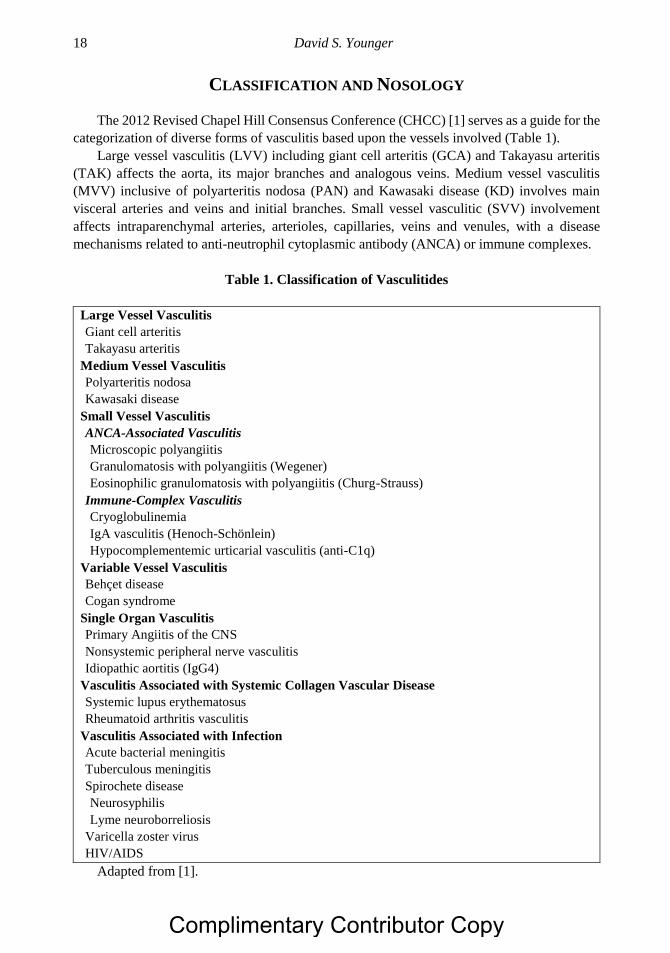
The 2012 Revised Chapel Hill Consensus Conference (CHCC) [1] serves as a guide for the
categorization of diverse forms of vasculitis based upon the vessels involved (Table 1).
Large vessel vasculitis (LVV) including giant cell arteritis (GCA) and Takayasu arteritis
(TAK) affects the aorta, its major branches and analogous veins. Medium vessel vasculitis
(MVV) inclusive of polyarteritis nodosa (PAN) and Kawasaki disease (KD) involves main
visceral arteries and veins and initial branches. Small vessel vasculitic (SVV) involvement
affects intraparenchymal arteries, arterioles, capillaries, veins and venules, with a disease
mechanisms related to anti-neutrophil cytoplasmic antibody (ANCA) or immune complexes.
Table 1. Classification of Vasculitides
Large Vessel Vasculitis
Giant cell arteritis
Takayasu arteritis
Medium Vessel Vasculitis
Polyarteritis nodosa
Kawasaki disease
Small Vessel Vasculitis
ANCA-Associated Vasculitis
Microscopic polyangiitis
Granulomatosis with polyangiitis (Wegener)
Eosinophilic granulomatosis with polyangiitis (Churg-Strauss)
Immune-Complex Vasculitis
Cryoglobulinemia
IgA vasculitis (Henoch-Schönlein)
Hypocomplementemic urticarial vasculitis (anti-C1q)
Variable Vessel Vasculitis
Behçet disease
Cogan syndrome
Single Organ Vasculitis
Primary Angiitis of the CNS
Nonsystemic peripheral nerve vasculitis
Idiopathic aortitis (IgG4)
Vasculitis Associated with Systemic Collagen Vascular Disease
Systemic lupus erythematosus
Rheumatoid arthritis vasculitis
Vasculitis Associated with Infection
Acute bacterial meningitis
Tuberculous meningitis
Spirochete disease
Neurosyphilis
Lyme neuroborreliosis
Varicella zoster virus
HIV/AIDS
Adapted from [1].
Complimentary Contributor Copy

Overview of Primary and Secondary Vasculitides 19
The category of ANCA-associated vasculitis (AAV) includes granulomatosis with
polyangiitis (GPA) (Wegener granulomatosis [WG type]), eosinophilic granulomatosis with
polyangiitis (EGPA) (Churg-Strauss syndrome [CSS]), and microscopic polyangiitis (MPA)
(microscopic polyarteritis), while vasculitic disorders associated with immune complexes (IC)
includes IgA vasculitis (IgAV) (Henoch-Schönlein purpura [HSP]), cryoglobulinemic
vasculitis (CV), and Hypocomplementemic urticarial vasculitis (HUV) associated with C1q
antibodies. Vasculitis without a predominant vessel size and caliber, respectively from small to
large, involving arteries, veins and capillaries, comprises the category of variable vessel
vasculitis (VVV), characteristic of Behçet disease (BD) and Cogan syndrome. Vascular
inflammation confirmed to a single organ system such as vasculitis restricted to the central
nervous system (CNS) and peripheral nervous system (PNS), and IgG4 related aortitis ( (IgG4-
related disease [RD]), are collectively referred to as single organ vasculitides (SOV).
There is a separate category for vasculitis associated with systemic disease notably for
connective tissue disorders such as rheumatoid arthritis vasculitis (RAV) and systemic lupus
erythematosus (SLE); and another for vasculitis associated with a probable specific etiology,
such as substance abuse and infection designated by the specific vasculitic disorder with a
prefix to denote the causative agent.
In 2008, the Pediatric Rheumatology European Society (PRES) and the European League
against Rheumatism (EULAR) and the Pediatric Rheumatology International Trials
Organization (PRINTO) reported methodology and overall clinical, laboratory and
radiographic characteristics for several childhood systemic vasculitides [2] followed by a final
validated classification [3] based upon vessel size, similar to the CHCC nomenclature [1].
Insight into effective therapies of systemic vasculitides have been guided by collaborative
evidence-based randomized clinical trials (RCT) or observational cohorts by the French
Vasculitis Study Group (FVSG) database, United States-Canadian Vasculitis Clinical Research
Consortium (VCRC), European Vasculitis Study Society (EUVAS), the European League
Against Rheumatism (EULAR), The French Vasculitis Cohort of Patients with Primary
Vasculitis of the Central Nervous System (COVAC), Diagnostic and Classification Criteria in
Vasculitis Study (DCVAS), the Pediatric Vasculitis Initiative (PedVas), the Diagnostic and
Classification Criteria in Vasculitis Study (DCVAS), and the web-based network BrainWorks.
Despite disparities in vessel involvement and end-organ damage, it is usually possible to
reach a presumptive diagnosis of primary and secondary vasculitides in the majority of patients
based upon the combination of presenting symptoms and signs, disease-specific serological
studies, and visceral and neurovascular imaging studies, while awaiting the results of tissue
histopathology.
This chapter is an overview of primary and secondary vasculitides in adults and children
for clinicians treating such patients. Five major challenges encountered in clinical practice will
be addressed and emphasized as follows.
First, clinical, pathological, and serological differentiation and diagnosis of the primary
vasculitides including, LVV (GCA, TAK), MVV (PAN, KD), SVV (AAV [MPA, GPA,
EGPA] and IC-mediated types [IgAV and anti-C1q]); and VVV (BD and Cogan syndrome),
all of which share demonstrable histopathological evidence of systemic vasculitis.
Second, recognition of secondary vasculitides associated with an underlying primary
systemic illness, in which some but not all patients will demonstrate evidence of vasculitis
including, CV, RA vasculitis (RAV), and CNS vasculitis associated with SLE, syphilis, Lyme
neuroborreliosis (LNB); bacterial meningitis, tuberculosis (TB), varicella zoster virus (VZV),
Complimentary Contributor Copy
David S. Younger 20
and human immunodeficiency virus type 1 (HIV) and acquired immune deficiency syndrome
(AIDS).
Third, the identification of the SOV, PCNSA, NSPNV, and IgG4-RD.
Fourth, a recommended laboratory approach to the diagnosis of vasculitides.
Fifth and last, evidence-based treatment options for each of the vasculitides. Interested
readers are recommended to another in-depth overview [4].
DIFFERENTIATION OF PRIMARY VASCULITIDES
Large Vessel Vasculitides
The concepts of GCA and TAK have evolved over a century, with considerable advances
in the past decade that have translated into more improved diagnosis and management.
Giant Cell Arteritis
First named temporal arteritis for the site of granulomatous giant cell inflammation and
vessel involvement [5], those with associated blindness due to vasculitic involvement of
ophthalmic and posterior ciliary vessels were subsequently classified as cranial arteritis [6], and
later generalized GCA [7] when giant cell lesions were discerned along the aorta, its branches,
and in other medium- and large-sized arteries at postmortem examination. There are five
discriminating features of GCA including, age >50 years at onset, new localized headache,
temporal artery tenderness or decreased temporal artery pulse, ESR >50 mm/hour, and biopsy
of an artery showing necrotizing arteritis and a predominance of mononuclear cells or
granulomatous process with multinucleated giant cells (Figure 1), that collectively serve as
useful guideposts in recognizing GCA.
Unrecognized and therefore untreated or inadequately treated, there is a high likelihood of
large artery complication. Nuenninghoff and coworkers [8] reported patients with large-artery
complications representing 27% of 168 patients in a GCA cohort at the Mayo Clinic between
1950 and 1999 that included aortic aneurysm or dissection in 18%, large artery stenosis in 13%,
cervical artery stenosis in 9%; and subclavian, axillary or brachial artery stenosis in 4%.
Temporal artery biopsy is the only sure way of establishing the diagnosis however false
negative findings on the contemplated affected side may be due to inadvertent sampling of a
vasculitic-free length of vessel. The pathological heterogeneity of GCA was further
exemplified by the occasional finding of intracranial lesions in several patients who also
qualified for the diagnosis of granulomatous angiitis of the nervous system (GANS) [9];
however PNS involvement in GCA remains exceedingly uncommon [10].
Takayasu Arteritis
Contemporaneously, another LVV was described in the Japanese literature as unusual
changes of the central vessels of the retina in the absence of peripheral arterial pulses in women
[11]. Patients with so called pulseless disease [12], occlusive thromboaortopathy [13] or TAK
[14], manifested constitutional complaints of malaise, fever, stiffness of the shoulders, nausea,
vomiting, night sweats, anorexia, weight loss, and irregularity of menstrual periods weeks to
Complimentary Contributor Copy
Overview of Primary and Secondary Vasculitides 21
months before the local signs of vasculitis were recognized in up to two-thirds of patients. TAK
is the commonest large vessel vasculitis among Asian women.
The non-invasive assessment of LVV includes performance of color-Doppler sonography
(CDS), contrast enhanced high-resolution magnetic resonance imaging (MRI) combined with
MRA, and contrast-enhanced computed tomography (CT) combined with CTA to visualize the
vessel wall and the lumen of large vessels. The signs of early inflammation that include vessel
wall thickening and mural inflammation, as well as the late complications of stenosis and
aneurysms, can be ascertained.
A
B
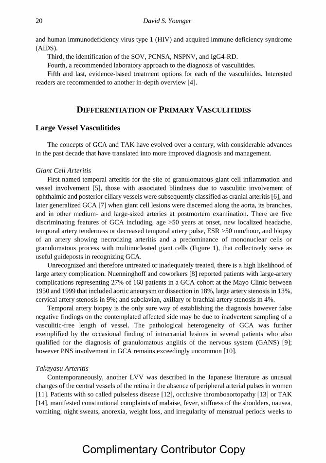
Reproduced from reference [4], with permission.
Figure 1. Giant cell arteritis. A. An early lesion of a large muscular artery, necrosis, inflammation, and
giant cell formation (single arrow) can be seen immediately adjacent to the internal elastic lamina
(arrowhead), which is undergoing degenerative changes, and there is some intimal proliferation (double
arrows) (stain, hematoxylin and eosin; original magnification, ×100). B. This more advanced lesion has
complete segmental destruction of the internal elastic lamina and virtually the entire media (arrows).
Marked intimal proliferation has nearly occluded the lumen, and few inflammatory cells remain (stain,
hematoxylin and eosin; original magnification, ×50).
18F- Fluorodeoxyglucose (FDG) positron emission tomography (PET) detects increased
FDG uptake by metabolically active cells, including inflammatory cells infiltrating the vessel
wall in vasculitis, while digital subtraction angiography (DSA) is a useful modality to
demonstrate luminal changes. Moreover, such studies can assist the surgeon in centering on an
involved segment of vessel. Performance of CDS is better suited to study superficial vessels
Complimentary Contributor Copy
David S. Younger 22
such as the internal and external carotid artery and its branches, while MR and CT are best
suited for deep vessels. When performed together, they can be used to monitor disease extent
and severity through the demonstration of early vascular changes in wall thickness and mural
inflammation, to which PET can be added to ascertain active inflammation in vessels affected
by GCA and TAK. Since the early reports of a salutary effect of corticosteroids on GCA in
1950 [15], corticosteroids have remained the standard of care because of their ability to reduce
disease-related morbidity, mortality, and symptoms that negatively impact on quality of life.
However they are not curative, do not prevent relapses, and are associated with significant
toxicity. Disease-related morbidity in GCA which largely results from cranial ischemic events
or LVV, leads to visual loss in up to 20% of patients.
The risk factors for GCA-related ischemic events include visual loss, prior ischemic events,
marked intimal hyperplasia on temporal artery biopsy, elevated inflammatory markers, older
age at diagnosis, hypertension, ischemic heart disease, and absence of systemic manifestations
[16]. While there is no treatment to date that has been found to completely reverse blindness in
GCA once it has occurred, there is strong evidence to suggest that once corticosteroids have
been started the risk of visual loss is low [17]. For this reason, corticosteroids should be started
while the diagnostic evaluation is in progress and continued for up to one year before tapering
to the lowest maintenance levels.
Ohigashi and colleagues [18] ascertained an improved prognosis among 106 consecutive
patients with TAK in those with onset before 1999 compared to those diagnosed after 2000
(4.2% versus 0%) that was attributed to reduction in the time from onset to diagnosis,
replacement of digital subtraction angiography (79% versus 9%) with ultrasound (6% versus
34%), CTA (24% versus 77%), MRA (21% versus 57%), and 18F-FDG PET (0% versus 20%);
less frequent complications of moderate or severe aortic regurgitation, and not surprisingly, an
increase in the use and maximal dose of corticosteroids (70% versus 97%); and the use of first
and second-line immunosuppressant agents (7% versus 42%). Surgical treatment of TKA was
similar between those with onset before 1999 and after 2000 (22.5% versus 22.8%).
Medium Vessel Vasculitides
Polyarteritis Nodosa and Kawasaki Disease
Kernohan and Woltman [19] summarized the clinicopathological aspects of adult PAN at
postmortem examination, while Krahulik and colleagues [20] described fulminant childhood
PAN (cPAN), three decades after the first description of the first American patient by Longcope
in 1908 [21]. The dominant clinicopathological syndrome was peripheral neuritis that occurred
in one-half of patients early in the illness with a predilection for the legs. The combination of
acute and chronic lesions correlated with known exacerbations. Arteritic lesions along nutrient
arteries of the peripheral nerves were characterized by invasion of the intima, media and
adventitia by polymorphonuclear, plasma cells, eosinophils, and lymphocytes associated with
swelling of the media, fibrinoid necrosis, and fragmentation of the internal elastic lamina
(Figure 2). So impressed was Dr. Harry Lee Parker by the frequency of arteritic lesions in the
PNS, that he conceptualized nerve and muscle biopsy as a useful mode for the diagnosis in life
during a discussion of the paper by Kernohan and Woltman [19]. Variants of cPAN were
contemporaneously recognized in infants and young children under the rubric of
mucocutaneous lymph node syndrome, infantile PAN before arrival at the preferred term KD
Complimentary Contributor Copy
Overview of Primary and Secondary Vasculitides 23
[22-25] for the childhood syndrome affecting children of all ages and races, with worldwide
occurrence.
A retrospective study of 348 adult patients registered in the French Vasculitis Study Group
(FVSG) [26] who satisfied criteria for the diagnosis of PAN between 1963 and 2005 noted
constitutional findings included fever, weight loss, myalgia, and arthralgia at presentation in
93% of patients. PNS involvement included peripheral neuropathy and mononeuritis multiplex
in nearly equal proportion in 79%, and cutaneous involvement notably, purpura, skin nodules,
and livedo reticularis were noted in 50% of patients; CNS involvement was noted in 5% of
patients.
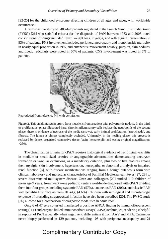
Reproduced from reference [4], with permission.
Figure 2. This small muscular artery from muscle is from a patient with polyarteritis nodosa. In the third,
or proliferative, phase illustrated here, chronic inflammatory cells replace the neutrophils of the second
phase; there is evidence of necrosis of the media (arrows), early intimal proliferation (arrowheads), and
fibrosis. The lumen is almost completely occluded. Ultimately, in the healing phase, this process is
replaced by dense, organized connective tissue (stain, hematoxylin and eosin; original magnification,
×250).
The classification criteria for cPAN requires histological evidence of necrotizing vasculitis
in medium-or small-sized arteries or angiographic abnormalities demonstrating aneurysm
formation or vascular occlusions, as a mandatory criterion, plus two of five features among
them myalgia, skin involvement, hypertension, neuropathy, or abnormal urinalysis or impaired
renal function [6], with disease manifestations ranging from a benign cutaneous form with
clinical, laboratory and molecular characteristics of Familial Mediterranean Fever [27, 28] to
severe disseminated multisystem disease. Ozen and colleagues [29] studied 110 children of
mean age 9 years, from twenty-one pediatric centers worldwide diagnosed with cPAN dividing
them into four groups including systemic PAN (57%), cutaneous PAN (30%), and classic PAN
with hepatitis B surface antigen (HBsAg) (4.6%). Children with serological and microbiologic
evidence of preceding streptococcal infection have also been described [30]. The FVSG study
[26] allowed for a comparison of diagnostic modalities in adult PAN.
Only 6 of 47 sera so tested manifested a positive ANCA finding by immunofluourescent
testing (IFT) and enzyme linked immunosorbant assay (ELISA) techniques, rendering it helpful
in support of PAN especially when negative to differentiate it from AAV and MPA. Cutaneous
nerve biopsy performed in 129 patients, including 108 with peripheral neuropathy and 21
Complimentary Contributor Copy
David S. Younger 24
without peripheral neuropathy, showed typical vasculitic lesions respectively in 83% and 81%,
compared to muscle biopsy that revealed vasculitis respectively in 68% and 60% of patients.
Angiography showed renal and gastrointestinal microaneurysms or stenosis respectively in
66% and 57% of patients. Patients with HB virus (HBV)-related PAN had more frequent
peripheral neuropathy, abdominal pain, cardiomyopathy, orchitis, and hypertension than those
with non-HBV-related PAN, with respective five-year relapse-free survival rates of 59% and
67% in scheduled therapeutic regimens depending upon involvement in clinical trials, or
according to the standard of care at the time of diagnosis, among them glucocorticoids and
cyclophosphamide [31, 32]. The predictors of a poor prognosis were age >65 years,
hypertension, and gastrointestinal involvement, and cutaneous manifestations or non-HBV-
related PAN had higher rates of relapse.
Henegar and colleagues [33] identified three positive predictive parameters including HBV
antigen and DNA in serum, arteriographic anomalies, and mononeuropathy or polyneuropathy;
and five negative predictive parameters including detection of ANCA; asthma, ear, nose and
throat signs; glomerulopathy; and cryoglobulinemia, that yielded a70.6% sensitivity for control
vasculitides and 92.3% specificity for all controls.
Small Vessel Vasculitides
ANCA-Associated Vasculitis
Microscopic Polyangiitis
Wohlwill [34], Davson and colleagues [35] and Wainwright and Davson [36] recognized
that fever, arthralgia, purpura, hemoptysis, pulmonary hemorrhage, abdominal pain, and
gastrointestinal bleeding preceded the explosive phase of systemic necrotizing vasculitis in
some patients with a disorder other than PAN that affected the kidney and lungs, with rapidly
progressive glomerulonephritis and pulmonary capillaritis. Such patients with selective
involvement of small microscopic arteries, arterioles, capillaries and venules including
glomerular and pulmonary alveolar capillaries were deemed to have microscopic PAN. Among
thirty-four such patients described by Savage and colleagues [37] the clinical symptoms and
signs at presentation were constitutional (67%), arthralgia (65%), purpura (44%), hemoptysis
in (32%), abdominal pain (32%), mouth ulcers (21%), sensory peripheral neuropathy (18%),
and CNS (headache, seizures) (18%). Eighty-five additional patients studied by Guillevin and
colleagues [38] with MPA so termed, had renal involvement (79%), weight loss (73%), skin
involvement (purpura, livedo, nodules, urticarial) (62%), mononeuritis multiplex neuropathy
(57%), fever (55%), arthralgia (50%), myalgia (48%), vascular manifestations [hypertension,
cardiac failure, pericarditis] 50%, lung involvement (alveolar hemorrhage, pneumonitis,
pleurisies) (24%), and CNS involvement (12%). Ahn and colleagues [39] noted pANCA or
anti-MPO antibody positivity in 69% of Korean MPA patients, compared to 74.5% of positive
ANCA in European patients, of whom 87% had a pANCA staining pattern. Antibodies to PR3
were present in 8% of patients compared to MPO in 61% of those as determined by ELISA.
Childhood MPA (cMPA) appears to be very uncommon and the criteria for diagnosis require
three of the following features to be present: abnormal urinalysis, granulomatous inflammation
on tissue biopsy, nasal sinus inflammation; subglottic, tracheal, or endobronchial stenosis;
Complimentary Contributor Copy
Overview of Primary and Secondary Vasculitides 25
abnormal chest radiograph or chest CT scan, and PR3 ANCA or cANCA staining [3]. Those
with cMPA accounted for four of the first 32 children in the United States/Canadian ARChiVe
registry [40].
Eosinophilic Granulomatosis with Polyangiitis
Contemporaneous with the description of MPA, the first patient with EGPA was probably
Case 1 of Lamb [41] reported in 1914 also under the heading of PAN. That patient, a 26-year-
old man with two years of worsening asthma, developed fever, palpable purpura, nodular skin
lesions, hemoptysis, vomiting, urinary difficulty and granular urinary casts. He died one month
later and postmortem examination showed necrotizing arteritis of small arteries, with dense
collections of extravascular eosinophils and tissue eosinophilia in the heart, stomach, and
kidney. Decades later, Churg and Strauss [42] described the clinical and postmortem findings
of thirteen patients with asthma, fever, and hypereosinophilia, accompanied by eosinophilic
exudation, fibrinoid change, and granulomatous proliferation or the so called allergic
granuloma. The latter was found within vessels walls and in extravascular connective tissue of
major organ systems, leading to cardiac, pulmonary, gastrointestinal, skin, PNS and CNS
manifestations. In 1990, the American College of Rheumatology (ACR) [43] developed criteria
for the classification of EGPA that included ascertainment of four or more of the following:
asthma, eosinophilia of >10%, mononeuropathy or polyneuropathy, non-fixed pulmonary
infiltrates on chest radiograph, paranasal sinus abnormality, and extravascular eosinophils on
tissue biopsy that included an artery, arteriole or venule. These criteria were inadequate since
a patient with asthma and paranasal sinusitis could also fit the designation of EGPA. The 1994
CHCC [1] characterized EGPA as an eosinophil-rich and granulomatous inflammatory process
that involved the respiratory tract, with necrotizing vasculitis that affected small to medium-
sized vessels such as capillaries, venules, arterioles and arteries, with associated asthma and
eosinophilia. Among 383 patients enrolled in the FVSG Cohort [44] who satisfied the ACR
criteria [43] or in 1994 CHCC definition for EGPA [45], the mean age at presentation was 50
years without sex predominance. Clinical manifestations at presentation included asthma
(91%), peripheral neuropathy (51%), weight loss (49%), ear, nose and throat signs (48%), non-
erosive sinusitis and polyposis (41%), skin lesions (39%), purpuric rash (22%), lung infiltrates
(38%), gastrointestinal involvement (23%), renal manifestations (22%), cardiomyopathy
(16%), CNS (5%), and cranial nerve involvement (3%). A total of 108 (31%) patients tested
positive for ANCA with significantly more frequent ear, nose and throat, peripheral nerve and
renal involvement, but less frequent cardiac manifestations. Small numbers of children have
been included in large studies of EGPA sufficient to allow comparisons to adults. Among 133
vasculitic patients in the ARChiVe registry, only two were reported to be of the EGPA type
[46].
Granulomatosis with Polyangiitis
Godman and Churg [47] described the syndrome of GPA that included granuloma in the
nasopharynx, sinuses and lower respiratory tract with focal segmental glomerulonephritis and
disseminated small vessel vasculitis. In a landmark article, Godman and Churg [47] concluded
that MPA, EGRA and GPA were related to one another yet distinct from PAN. This astute
conclusion was based mainly on pathological features was later substantiated by their common
association with ANCA, but not so for PAN [48]. Fauci and colleagues [49] and Hoffman and
colleagues [50] at the National Institutes of Health (NIH), respectively reported a prospective
Complimentary Contributor Copy
David S. Younger 26
series of eighty-five patients with GPA, and a retrospective assessment of 180 patients followed
for 6 months to 24 years. The presenting signs included pulmonary infiltrates (71%), sinusitis
(67%), arthritis and arthralgia (44%), fever (34%), cough (34%), otitis (25%), and hemoptysis
(22%), with an overall predominance of organ system involvement in the lung (94%), paranasal
sinuses (91%), kidney (85%), joints (67%), and nasopharynx and nose (64%). Fauci and
colleagues [49] established the efficacy of cyclophosphamide and prednisone in achieving
complete remissions in 93% of patients, as well as, the tendency of patients to relapse and
accrue additive mortality from both disease and treatment. The characteristic histopathology is
a necrotizing granulomatous vasculitis which may be found in lung and renal biopsy tissue
although the latter is less common. Instead, a focal, segmental glomerulonephritis is often seen.
Other inflammatory or vasculitic phenomenon can be encountered such as leukocytoclastic
vasculitis in skin lesions, and acute and chronic inflammation in sinus, retro-orbital, and
tracheal tissues. A limited form of GPA without glomerulonephritis was described [51] that
was a long-term disease stage or phenotype accounting for about 5% of all patients
characterized by destructive and space-consuming lesions associated with relapse rates of 46%
and local damage [52].
The 1990 ACR criteria for the classification of GPA [53] which preceded routine ANCA
testing, included the presence of two or more from criteria from among four including nasal or
oral inflammation (painful or painless oral ulcers or purulent or bloody nasal discharge);
abnormal chest radiograph (showing nodules, fixed infiltrates or cavities); urinary sediment
(showing microhematuria >5 red blood cells per high power field, or red cell casts in urinary
sediment); and granulomatous inflammation on biopsy tissue (within the wall of an artery or in
the perivascular or extravascular area of an artery or arteriole).
A proposed classification tree substituted hemoptysis for granulomatous inflammation on
a tissue biopsy if the latter was not available. Baseline serum samples for 180 participants in
the WG Etanercept Trial Research Group (WGET) found that when IF, direct and capture
ELISA ANCA testing were performed at baseline, 166 (92%) were seropositive, including 96%
with severe disease and 83% with limited disease.
Holle and colleagues [54] who prospectively compared a highly sensitivity (hs)PR3-
ANCA ELISA to the IFT noting an excellent performance of the hsPR3-ANCA ELISA in
identifying GPA and other AAV disorders associated with PR3-ANCA suggesting that the
former by used as screening test. Since the early observations of ANCA provided by van der
Woude and colleagues in 1985 [55], and Falk and Jennette [56] and Goldschmeding and
colleagues [57], and ensuing progress in the differentiation and understanding of ANCA
subtypes [58], the past quarter century has witnessed a renaissance in the understanding of
primary systemic vasculitis with convincing clinical evidence to support an important role for
ANCA in the development of AAV. An AAV classification appears to better recognize ANCA
disease and predict prognosis than other any existing clinical classification systems [59].
However as with other autoimmune disorders, the etiology and pathogenesis appears to be
multifactorial, involving the interplay of initiating and predisposing environment and genetic
factors [60-62]. Induction with corticosteroids and either cyclophosphamide followed by
maintenance with rituximab [63-68] or azathioprine [69-71] is recommended treatment.
Among 197 ANCA-positive patients with GPA or MPA in nine centers participating in the
Rituximab in ANCA-Associated Vasculitis-Immune Tolerance Network (RAVE-ITN), a
randomized, double-blind, double-dummy, non-inferiority trial [72] comparing rituximab 375
mg per square meter (m2) body-surface area (BSA) per week for 4 weeks to cyclophosphamide
Complimentary Contributor Copy
Overview of Primary and Secondary Vasculitides 27
2 mg per kilogram of body weight per day controls for severe AAV, 67% of study patients
compared to 42% of controls achieved the primary end point of remission of disease without
use of prednisone at six months.
Treatment and efficacy and safety data in children with AAV continue to be largely derived
from adult GPA studies, however as described in ARChiVe, pediatric patients in the United
States and Canada are being offered pulse methylprednisolone for 3 to 5 days, followed by oral
prednisone, and cyclophosphamide orally or with one of two intravenous regimens, followed
by maintenance therapy, most frequently with methotrexate [40].
Early outcome results last year for the treatment of childhood AAV, in particular GPA,
reported by Morishita and colleagues on behalf of ARCHiVe Investigators Network and the
Pediatric Vasculitis (PedVas) Initiative [73] was somewhat unencouraging. Among 105
children with AAV, mainly GPA, who received corticostereoids, cyclophosphamide,
methotrexate, or rituximab for remission-induction, and plasma exchange in conjunction with
cyclophosphamide and/or rituximab, 42% achieved remission at 12 months (Pediatric
Vasculitis Activity Score [PVAS] of 0, CS dose < 0.2 mg/kg/day), 21 (48%) of whom
discontinued CS by 12 months; all but 3 remaining on maintenance treatment at 12 months
receiving azathioprine, methotrexate, rituximab, mycophenolate mofetil, and
cyclophosphamide. However, up to 63% had a (Pediatric Vasculitis Damage Index [PVDI)
score of 1 or more by 12 months, with the presence of renal, ear, nose and throat, or pulmonary
damage; moreover, 41% of children reported hospitalizations. Thus, a significant proportion of
patients were not in remission at 12 months, and more than one‐half of the patient cohort
experienced damage early in the disease course. The 12-month remission rate of 42% in the
cohort was significantly lower than Sacri and colleagues [74] who reported 73% remission at
post-induction and 90% overall remission rate (including secondary remissions after a median
time of 6.7 months).
Immune Complex Vasculitis
The foundations for IC-mediated or hypersensitivity vasculitis were conceptualized in the
mid-twentieth century, as an immunologic response to antigenic material associated with
clinically evident purpura, and small vessel inflammation affecting arterioles, capillaries, and
post-capillary venules. It was likened by Zeek [75] to the anaphylactoid Arthus reaction
produced by the experimental injection of horse serum into rabbits [76].
IgA Vasculitis (Henoch-Schönlein Purpura)
Children with HSP were described by Gairdner [77] with anaphylactoid purpura including
one who developed rash, colic, melanotic stools, intussusception, and hematuria following by
a typical exanthema and fatal convulsion. Postmortem examination showed scattered cortical
hemorrhages associated with cerebral necrotizing arteriolitis. Levitt and Burbank [78]
described the clinicopathological findings in two previously non-allergic patients with recurrent
fatal attacks of HSP after injection of penicillin and ingestion of strawberries respectively that
included glomerulonephritis alone or with systemic arteriolitis. The findings of IgA deposits in
cutaneous blood vessel walls and in glomerular mesangial biopsies of patients with HSP and
IgA nephropathy (IgAN) [79, 80] were circumstantially convincing enough to substitute the
term IgAV for HSP. IgAV/HSP is the commonest vasculitis in children. The 1990 ACR criteria
[81] for the identification of HSP included age less than or equal to 20 years at disease onset,
Complimentary Contributor Copy
David S. Younger 28
palpable purpura, acute abdominal pain, and tissue biopsy showing granulocytes in the walls
of small arteries or venules. The presence of any two or more of these criteria distinguished 85
patients who were diagnosed as having HSP by physicians who submitted cases for the
vasculitis criteria compared to 722 patients diagnosed with other forms of vasculitis, arriving
at sensitivity of 87.1% and specificity of 87.7%. The addition of gastrointestinal bleeding in a
classification tree format respectively increased sensitivity to 89.4% and specificity to 88.1%.
The EULAR/PRINTO/PReS classification criteria [3] which recognizes the contribution of IgA
deposits, differs in the mandatory finding of purpura with predominance in the legs, and the
presence of one of the four following features: diffuse abdominal pain, arthralgia or arthritis; a
biopsy showing predominant IgA deposits, and renal involvement including proteinuria and
hematuria Derived from the analysis of 827 patients in the database, the calculated sensitivity,
specificity for the clinical and laboratory findings in between the consensus panel and specific
definition were respectively 100% and 87%. Peru and colleagues [82] studied 254 children with
IgAV/HSP between 2003 and 2006 with a distribution of skin, joint, GI and renal
manifestations respectively of 100%, 66%, 56% and 30%. The disorder commences with fever
and palpable purpura, although early lesions can be urticarial. Arthralgia and abdominal pain
precede, accompany or follow the rash. Melena is common as are signs of peritonitis.
Proteinuria and hematuria are of variably severity and renal pathology may be of a mild
glomerulitis to necrotizing or proliferative glomerulonephritis. Ozen and coworkers [3] noted
palpable purpura, commonly in crops with lower limb predominance in 89% of patients,
arthritis or arthralgia in 78%, diffuse abdominal pain in 60%, proteinuria and hematuria
combined in 33%; and IgA deposition in 10% of children. The treatment of IgAV/HSP remains
empiric and largely supportive, with conflicting conclusions in retrospective and uncontrolled
case series of immune suppression in severe HSP nephritis [46]. Extrarenal manifestations can
be managed by symptomatic treatment.
A meta-analysis of 15 studies based on a comprehensive review of the literature in the
Medline database from 1956 to 2007, and Cochrane Controlled Trials Registry among 15
studies and over 1300 patients [46] found that early treatment conferred a protective effect on
developing persistent renal disease (odds ratio [OR] .43) and the likelihood of surgical
intervention for abdominal pain (OR .75), as well as a statistically significant positive effect on
shortening the duration of abdominal symptoms (OR 5.42).
C1q (Hypocomplementemic Urticarial) Vasculitis
McDuffe and colleagues [83] later described several patients with recurrent attacks of
erythematous, urticarial, and hemorrhagic skin lesions that lasted 24 hours at a time, associated
with recurrent attacks of fever, joint swelling, abdominal distress, and depressed serum
complement. When tested by immunodiffusion against purified preparations RF and human
C1q, several patients reacted strongly with purified C1q. Skin biopsies showed leukocytoclasia,
characteristic of necrotizing vasculitis, anaphylactoid purpura, or mild nonspecific perivascular
infiltration. Immunofluorescence of skin specimens showed fixation of Ig in the patient with
necrotizing vasculitis. Renal biopsy showed mild to moderate glomerulonephritis
indistinguishable for those seen in other forms of chronic membranoproliferative
glomerulonephritis. The differences from SLE included more urticarial and purpuric skin
lesions, with relatively mild renal or absent and other visceral involvement in the patients with
HUV. An etiopathogenesis related to chronic vascular inflammation resulting from deposits of
immune complexes in small vessel walls seemed likely. Zeiss and colleagues [84] characterized
Complimentary Contributor Copy
Overview of Primary and Secondary Vasculitides 29
C1q IgG precipitins from HUV sera that precipitated C1q in agarose gel among four additional
patients. Wisnieski and Naff [85] later showed C1q binding activity in IgG from HUV sera.
Buck and colleagues [86] defined the HUV syndrome as the presence of UV with multi-organ
involvement notably arthralgia or arthritis, angioedema, pulmonary, ocular, renal, and
pericardial. However, anti-C1q antibodies are elevated and the serum low C1q levels are
reduced in virtually all children so studied. Antihistamines are the drug of choice with
cutaneous lesions to control itching, but they may be insufficient in controlling the formation
of immune complexes when given late in the inflammatory cascade. There is yet a consensus
on the most effective therapeutic regimen; however plasmapheresis and intravenous immune
globulin (IVIg) are alternative immunosuppressant modalities.
Variable Vessel Vasculitis
Behçet Disease
Behçet [87] described the clinicopathological findings of a 28-year-old Turkish patient
with relapsing oral, genital and oral eruptions over 4 years, accompanied by severe headache,
memory loss, dizziness, lethargy, fatal seizures and coma. Postmortem examination showed
perivascular inflammatory cell infiltration of the meninges, brain, and central retinal artery and
optic nerve with necrotic cerebral lesions. The first well-documented American patient with
nervous system involvement of BD was described by Wolf and coworkers [88], namely a 22-
year-old woman with a five year history of recurrent oral and genital ulceration, and a two year
course of progressive visual loss, headache, hemiparesis, ataxia, tremor, dysarthria, cranial
nerve palsy, cerebellar and corticospinal tract disease, and mental deterioration, which
responded to prednisone therapy. The most widely used diagnostic criteria of BD were
formulated by the International Study Group (ISG) [89] that included recurrent oral ulcerations
plus any two of genital ulceration, typical defined eye lesions, typical skin lesions, or a positive
pathergy. Recurrent oral ulcerations were categorized as minor aphthous, major aphthous, and
herpetiform ulcerations that recurred at least three times in a 12-month period. Recurrent genital
ulcerations were defined as aphthous ulceration and scarring. Eye lesions were defined as
anterior uveitis, posterior uveitis or cells in the vitreous on slit lamp examination; and retinal
vasculitis. Compatible skin lesions included erythema nodosum, pseudofolliculitis,
papulopustular lesions, and aceneiform nodules in post-adolescent patients not receiving
corticosteroids. A positive pathergy test of cutaneous hypersensitivity was defined as positive
when a sterile pustule developed after twenty four to forty-eight hours at the site of a needle
prick to the skin [90]. Although the usual onset of BD is in the third or fourth decade of life,
pediatric-onset patients have been described [91]. The neuropathological findings in BD in
brain biopsies and postmortem examination have been remarkably consistent among patients
over the past several decades evidencing perivascular cuffing of small meningovascular and
parenchymal arteries and veins, rarely medium-sized arteries displaying fibrinoid degeneration
and recanalization, and examples of venous thrombosis. Cortical venous sinus thrombosis
(CVST) [92] in BD presents with subacute or chronic onset of symptoms of isolated intracranial
hypertension accompanied by headache, blurred vision, and diplopia [93], and underlying
necrotizing vasculitis. Venous infarcts occur in up to 63% of those with CVST of other causes,
but in only 6% of patients with BD. The inflammatory cell infiltrates are generally comprised
of lymphocytes, both T-, and B-cells, macrophages, rarely plasma cells and eosinophils, with
Complimentary Contributor Copy
David S. Younger 30
reactive astrocytosis and microscopic gliosis in neighboring cerebral, cerebellar and brainstem
white matter. Matsumoto and colleagues [94] noted large vessel lesions in seven of eight
patients age 31 to 56 years with BD, including saccular aneurysms of the sinus of Valsalva or
aortic arch, thoracic and abdominal aorta, pulmonary, femoral, and iliac arteries, and
thrombotic occlusions in the pulmonary vein and superior and inferior vena. Aortitis was noted
histologically in six of the eight patients that was active in one, scarred in six, and intermixed
in another. Active aortitis was characterized by intense infiltration of inflammatory cells in the
media and adventitia more frequently than in the interim with occasional giant cell formation.
Although anticoagulation would not be recommended for BD-related CVST, it might be
considered in association with arterial occlusions, with both venous and arterial occlusive
episodes warranting prompt consideration of corticosteroids alone or in association with
another immunosuppressant agent.
Cogan Syndrome
The first patient with Cogan syndrome of non-syphilitic interstitial keratitis (IK) was
reported by Mogan and Baumgartner [95], that of a 26-year-old man with recurrent pain, spasm
and redness of the left eye with photophobia, excessive tearing, and marked conjunctival
injection, followed by severe attack of dizziness, tinnitus, vertigo, nausea, vomiting, ringing in
the ears, profuse perspiration, and deafness. A diagnosis of recurrent interstitial keratitis and
explosive Menière disease was made. Vestibuloauditory symptoms were later described by
Cogan [96]. Haynes and colleagues [97] set forth diagnostic criteria for typical Cogan
syndrome according to the definitions established in a review of 30 patients seen at the National
Eye Institute of the NIH by Cogan [96, 98, 99]. Symptoms of IK developed abruptly and
gradually resolved, associated with photophobia, lacrimation, and eye pain which was
unilateral or bilateral. Such symptoms tended to recur periodically for years before becoming
quiescent. Vestibuloauditory dysfunction was manifested by sudden onset of Menière-like
attacks of nausea, vomiting, tinnitus, vertigo, and frequently progressive hearing loss that
characteristically occurred before or after the onset of IK. However within one to six months
of the onset of eye symptoms, auditory symptoms progressed to deafness over a period of one
to three months, certainly no longer than two years. With less than 100 reported patients with
this rare childhood and young adult disorder, the majority of reported patients with typical
Cogan syndrome have appeared as single case reports or patient series, often without
pathological confirmation or evidence of systemic vasculitis in a biopsy or at postmortem
examination. Early recognition of the diagnosis of childhood Cogan syndrome is important in
instituting corticosteroid therapy to preserve hearing especially when hearing loss is a later
occurrence. A combination of oral and intravenous corticosteroids may be considered in
children who partly but not fully improve. Most patients with Cogan syndrome (58%) treated
with corticosteroids derived a favorable response in both vestibuloauditory and ophthalmologic
manifestations, with the remainder demonstrating only ophthalmologic (23%) or
vestibuloauditory improvement (19%) alone [100]. Other therapies include methotrexate
(23%), cyclophosphamide (10%), azathioprine (5%), entanercept (3%), hydroxchloroquine
(2%), and IVIg (2%). Surgical cochlear implantation can led to objective and subjective
benefits with improved hearing.
Complimentary Contributor Copy

Overview of Primary and Secondary Vasculitides 31
RECOGNITION OF SECONDARY VASCULITIDES
Cryoglobulinemic Vasculitis
The presence in the serum of one or more immunoglobulins (Ig) that precipitate below core
body temperatures and redissolve on rewarming is termed cryoglobulinemia [100, 101].
Wintrobue and Buell [102] described the first patient with cryoglobulinemia, that of a 56-year-
old woman who presented with progressive frontal headache, left face and eye pain; and right
shoulder, neck and lumbar discomfort after a bout of shingles. This was followed by Raynaud
symptoms, recurrent nosebleeds, exertional dyspnea and palpitation, and changes in the eye
ground attributed to central vein thrombosis. Postmortem examination showed infiltrating
myeloma of the humerus and lumbar vertebra, and splenic enlargement. A unique plasma
protein was detected that spontaneously precipitated with cold temperature and solubilized at
high temperature that differed from Bence-Jones proteinuria of other myeloma patients. Brouet
and coworkers [103] provided modern classifications of cryoglobulinemia among 86 patients
that included type 1, composed of a single monoclonal Ig; and types II and III as mixed
cryoglobulinemia (MC), composed of different Ig, with a monoclonal component in type II,
and polyclonal Ig in type III. In the absence of well-defined disease the presence of MC was
termed “essential”. Since recognition of hepatitis C virus (HCV) infection in patients with MC
[104, 105], and the recognition of the high rate of false-negative serological tests in type II MC
[106], it became evident that HCV was associated in most patients with MC. Cryoglobulinemic
vasculitis is characterized by the classical triad of purpura, weakness and arthralgia, frequent
multiple organ involvement, and infrequent late lymphatic and hepatic malignancies. The
commonest clinical manifestations of HCV- negative MC vasculitis in the CryoVas survey
[107] included purpura (75%), peripheral neuropathy (52%), arthralgia or arthritis (44%),
glomerulonephritis (36%), cutaneous ulcers (16%), and cutaneous necrosis (14%). A
connective tissue disease was diagnosed in 30% of patients, and B-cell non-Hodgkin lymphoma
(B-NHL) was noted in 22% of patients, whereas MCV was considered essential in 48% of
patients. There was a greater frequency of joint involvement (53% versus 40%), peripheral
neuropathy (74% versus 52%), CNS involvement (9% versus 2%) in those with HCV-MC
compared to those with HCV-negative MC [108], with an equal frequency of purpura (71%
versus 75%) and renal involvement (34% versus 35%). Cacoub and colleagues [109] noted five
high prevalent extrahepatic manifestations in chronic HCV infection including, arthralgia
(23%), paresthesia (17%), myalgia (15%), pruritus (15%), and sicca syndrome (11%), and a
40% prevalence of cryoglobulins. These findings suggest the possibility of an independent role
of HCV infection in the clinicopathological manifestations of MC and MCV. Among 114
patients including 18 children and 96 adults with cryoglobulinemia between 2000 and 2012,
children had more frequent prolonged fever (17% versus 3%), petechiae and purpura (27%
versus 15%), arthralgia and arthritis (66% versus 16%), and cutaneous involvement (77%
versus 50%, than adults [110]. Aggressive optimal therapy of HCV-related CV with PEGylated
(PEG)-INF alpha to improve the pharmacologic properties, and ribavirin with a protease
inhibitor in the instance of HCV genotype 1 infection, should be considered as induction
therapy for CV and administered for forty-eight weeks for all HCV genotypes [108]. An
induction phase of immunosuppression such as rituximab plus antivirals is recommended in
patients with more severe HCV-related CV exemplified by worsening renal function,
mononeuritis multiplex, extensive skin disease including ulcers and distal necrosis [111].
Complimentary Contributor Copy
David S. Younger 32
Terrier and colleagues [107] showed a greater therapeutic efficacy of rituximab and
corticosteroids compared with corticosteroids alone and alkylating agents with corticosteroids
in achieving complete clinical, renal, and immunologic responses and a prednisone dosage of
< 10 mg per day at six months. However, this regimen was associated with severe infections,
particularly when high doses of corticosteroids were employed.
Plasmapheresis combined with immunosuppression can be useful in fulminant HCV-
related CV to engender an immediate effect but should be continued to avoid post-pheresis
rebound worsening. Rituximab, fludarabine, and cyclophosphamide treatment is effective
treatment for refractory CryoVas associated with lymphoma. One-year, 2-year, 5-year, 10-year
survival rates of 91%, 89%, 79% and 65% respectively, have been reported in patients with CV
[108] with fatalities related to serious infection and disease flares of CV.
Systemic Lupus Erythematosus
Although distinctly uncommon, two collagen-vascular disorders, SLE and RA can be
associated with vasculitis of the nervous system. The early concepts of the collagen-vascular
disorders introduced by Klemperer [112, 113] stemmed from the appreciation of fibrinoid
necrosis using collagen staining in patients with SLE. As collagen swells and fragments, it
dissolves to form a homogeneous hyaline and granular periodic acid-Schiff (PAS)-positive
material. The latter fibrinoid material contains immunoglobulins, antigen–antibody complexes,
complement, and fibrinogen. The organ-specific responses to this fibrinoid material, especially
of the CNS, leads to recognizable clinical sequela due to vascular and parenchymal damage.
The ACR delineated criteria for the diagnosis of SLE [114]. The recognition of
neuropsychiatric lupus (NPSLE) was noted in 6.4% of a cohort of 1,253 SLE patients defined
by the ACR [115]. According to Tomic-Lucic and colleagues [116] those with late-onset SLE
due to development of disease after age 50 year, had a frequency of NPSLE of 6.6% compared
to 36.6% in early-onset disease despite less major organ involvement and more benign course.
Once thought to be an important cause of CNS or cerebral lupus, true vasculitis was present in
only 12% of postmortem examinations in the series of Johnson and colleagues [117], and in
26.7% of late-onset SLE patients compared to 16.6% of those with early-onset SLE. A
comparison of the cumulative incidence of clinical manifestations in the two latter groups
showed that seizures were more common in early-onset patients compared to later-onset
patients (6.6% versus 0%), similarly for multiple cerebrovascular attacks (23.3% versus 3.3%),
cranial and peripheral neuropathy (6.6% versus 3.3%). Nonetheless, there was no mention of
CNS vasculitis among 150 patients with SLE described by Estes and Christian [118], despite
the finding of neurological complications in 30% of patients that included focal deficits,
seizures, dementia, stupor, and coma. Devinsky and colleagues [119] noted a prevalence of
3.5% of psychiatric involvement that included organic affective, delusional and hallucinatory
syndromes in a cohort of 50 patients with SLE, one-half of whom had CNS lesions. Feinglass
and colleagues [120] noted neuropsychiatric manifestations at onset of SLE among 3% of 140
patients compared to 37% in the course of the illness; however headache was not specifically
tabulated. Cerebral dysfunction in SLE can be caused by large vessel or small vessel
involvement or both. In the series by Fienglass and colleagues [120] vasculitis was noted
overall in 28% of patients, as well as in 46% of those with neuropsychiatric involvement
compared to 17% of patient lacking neuropsychiatric involvement. Postmortem examination of
Complimentary Contributor Copy
Overview of Primary and Secondary Vasculitides 33
the CNS in ten of nineteen fatalities showed two cases of multiple large and small infarcts,
which in one of them, demonstrated inflammatory cells infiltrates in the walls of medium-sized
vessels, and perivascular infiltrates around small arterioles. Although active CNS vasculitis was
absent in the brain and spinal tissue of all fifty patients reported by Devinsky and colleagues
[119], two had evidence of inactive healed CNS vasculitis so suggested by focal disruption of
the elastic lamina and mild intimal proliferation of a single medium-sized artery, one of which
had active systemic vasculitis of the PAN type, both of whom evidenced Libman-Sacks
endocarditis and embolic brain infarcts. Focal angiitis of the CNS with cyst-like formation
around affected blood vessels was noted at postmortem in the patient described by Mintz and
Fraga [121] with typical SLE rash, cutaneous vasculitis, and active neuropsychiatric
involvement. Trevor and colleagues [122] summarized the literature of large named cerebral
vessel occlusions from 1958 to 1965 noting one patient with a middle cerebral artery (MCA)
stenosis progressing to occlusion and three others with angiographic internal carotid artery
(ICA) occlusions, adding three new patients and suggesting a relation of the occurrence to
cerebral arteritis. Two women, one age 21 and the other age 42 years, presented with headache
followed by focal neurological symptoms attributed respectively to lesions along the left MCA
followed by right ICA occlusions, and a right MCA stenosis progressing to occlusion in four
months. A third patient had a left ICA occlusion without mention of headache. Among the four
literature patients, one had angiographic occlusion of the MCA, and three others had occlusion
of the ICA.
Johnson and colleagues [117] attributed the vasculitic nature of this process
histopathologically to cerebral vasculitis mediated by acute inflammation and necrosis.
Younger and colleagues [123] reported large named cerebral vessel occlusion attributed to
circulating anticardiolipin antibodies in a young man in whom a vasculitic mechanism was not
evoked. A number of fluorescent antibody tests provide serological support of SLE. The
antinuclear antibody (ANA) screen produces a homogenous pattern in the majority of patients,
with antibodies to native double-stranded DNA (anti-dsDNA) and reactivity to the smith (Sm)
and ribonucleoprotein (RNP) antigens, the combination of which constitutes the extractable
nuclear antigen (ENA). Circulating IgG and IgM antibodies with an affinity for charged
phospholipids, antiphospholipid antibodies (APA), some of which have procoagulant activity
such as the lupus anticoagulant (LAC) and the generic anticardiolipin (aCL) antibody assay
using cardiolipin as the antigen probe for APA are all important determinants of prothrombotic
events, especially in the CNS wherein there is a propensity for occlusive microangiopathy.
Circulating SLE-related antibodies can impact the CNS by affecting both vascular mechanisms
and brain tissue directly. Immune complex mediated vasculitis probably affecting small vessels
is thought to account for much of the damage in CNS lupus in light of spite of the paucity of
cerebral vasculitis evident in the form of inflammatory infiltrates in vessel walls at postmortem
examination. In those with discrete vascular infarcts, there is a known association with the
presence of circulating pathogenic antibodies which predisposes some individuals to a high risk
of stroke due to both small and large vessel occlusion [123, 124]. Lupus cerebritis and
meningoencephalitis are two neurological disturbances that can be associated with preceding
headache. So noted in up to 75% of patients with SLE depending upon criteria [125], an
etiopathogenesis related to antibody-mediated neuronal dysfunction is likely given the lack of
correlation of symptoms of NPSLE and CNS lesions at postmortem examination, together with
the transient nature of the disturbance. Patients with SLE are also predisposed to infectious
episodes including those yet treated due to impaired B-cell function and humoral immunity, in
Complimentary Contributor Copy
David S. Younger 34
addition to others receiving immunosuppressant medication rendered impaired in T-cell
function and cell mediated immunity [125]. The treatment of CNS Lupus should be guided by
the tempo of the presenting neurological features, duration of disease activity, results of
serological studies, comorbid disorders, and the presence of true vasculitis, with most patients
receiving corticosteroids and those with true vasculitis of the brain considered for concomitant
immunosuppressant therapy. Despite less major organ involvement and decreased incidence of
neurological complications overall, late-onset SLE has a poor prognosis because of the higher
frequency of comorbid conditions, longer exposure to classical vascular risk factors, both the
less likelihood of treatment with higher doses of corticosteroids and the concomitantly higher
rate of complications to cyclophosphamide when administered [116].
Rheumatoid Arthritis Vasculitis
A joint working group from the ACR and the European League Against Rheumatism
(EULAR) published the 2010 classification criteria for RA [126]. Extra-articular RA (ExRA)
occurrence is associated with increased comorbidity and mortality [127]. Criteria for severe
ExRA were proposed in 2004 [128]. Active RA with high disease activity is associated with
increased risk of severe ExRA manifestations. Major cutaneous vasculitis and vasculitis
involving other organs are two such ExRA occurrences.
The diagnosis of RAV has generally been ascertained according to the criteria of Scott and
Bacon [129] according to the presence of one or more of the following: 1) mononeuritis
multiplex; 2) peripheral gangrene; 3) biopsy evidence of acute necrotizing arteritis plus fever
and weight loss; and 4) deep cutaneous ulcers or active ExRA disease if associated with typical
digital infarcts or biopsy evidence of vasculitis. Watts and colleagues [130] reported an annual
incidence of RAV of 11.6 per million inhabitants between 1988 and 1992 in the Norfolk area
of the United Kingdom, and a rate of 3.6 per million between 1998 and 2002. Myasoedova and
coworkers [131] reported a ten-year cumulative incidence of 50% of any ExRA, including
cutaneous and other organ-specific RAV in a retrospective population-based cohort study of
463 RA patients in Olmsted County, Minnesota from 1995 to 2007. The ten-year cumulative
incidence of RAV, but not other ExRA, was significantly lower in the 1995-2007 cohort (0.6%)
compared to the 1985 to 1994 cohort (3.6%). The markers of RA severity including RA
positivity, erosion and joint destructive changes (21% among those in the 1985 to 1994 cohort,
compared to 29% in 1995 to 2007), use of methotrexate, other disease-modifying anti-
rheumatic drugs, and systemic corticosteroids were significantly associated with ExRA
development between 1995 and 2007. Vollerstein and colleagues [132] studied 52 patients with
RAV at the Mayo Clinic from 1974 to 1981, who developed clinical vasculitis evidenced by
classic ischemic skin lesions, mononeuritis multiplex, or a positive tissue biopsy in comparison
to population controls. The initial manifestation of vasculitis was seen in skin (26 patients),
nerve (20 patients) or both (3 patients), and mononeuritis multiplex presented in one (2
patients), two (9 patients), three (5 patients) or four nerves (4 patients). More than 90% of tissue
biopsy specimens revealed vascular necrosis and inflammation. At diagnosis, 80% of patients
began therapy with aspirin and other non-steroidal anti-inflammatory drugs however three-
fourths continued or began corticosteroid therapy. Sixteen of the original 52 patients eventually
received cytotoxic immunosuppressive therapy. Compared to the general population, those
with RAV had decreased survival that was immediately evident and continued for six years.
Complimentary Contributor Copy
Overview of Primary and Secondary Vasculitides 35
Compared with a previously reported incidence cohort [133] and adjusted for referral bias,
survival of RAV was not different from classic RA. The factors that predicted decreased
survival in RAV in a univariate proportional-HR model included older age, failure to receive
previous non-steroidal anti-inflammatory drugs, previous administration of cytotoxic
immunosuppressive agents, a higher dose of corticosteroids at diagnosis, decision to continue
or initiate corticosteroids, and abnormal urinary sediment. Ouyang and coworkers [134]
estimated CNS involvement in RA to be extremely uncommon, and other authors [135, 136]
found CNS vasculitis to be decidedly rare. Puéchal and colleagues [137] found vasculitic
involvement of vasa nervorum of both small- and medium-sized arteries indistinguishable from
PAN in 64% of patients, with a mortality ranging from 28 to 44% according to the length of
follow-up. Epineurial and perineurial vasculitis was observed with the same frequency among
those with primary sensory neuropathy as others with predominant motor involvement,
respectively 67% versus 64%. A greater extent of the neuropathy and motor involvement
tended to predict decreased survival, however mononeuritis multiplex was not associated with
a poor five-year survival rate (57%) than was distal symmetrical sensory or sensorimotor
neuropathy (55%). Scott and Bacon [129] reported that five patients (24%) died in the group
receiving methylprednisolone and cyclophosphamide, postmortem examination in four of
whom failed to demonstrate active vasculitis.
By comparison, seven patients (29%) died receiving other treatments, of which one so
studied at postmortem examination showed active vasculitis. Three forms of vasculitis
classically occur in RA affecting all calibers of blood vessels from dermal postcapillary venules
to the aorta, usually in association with circulating IgM and IgG rheumatoid factor as measured
by the latex fixation test, decreased complement levels, and a positive ANA. The first is a
proliferative endarteritis of a few organs, notably the heart, skeletal muscle, and nerves
characterized by inflammatory infiltration of all layers of small arteries and arterioles, with
intimal proliferation, necrosis, and thrombosis. The second is fulminant vasculitis
indistinguishable from PAN with less severe leukocytosis, myalgia, renal and gastrointestinal
involvement, and bowel perforation. The third type takes the form of palpable purpura, arthritis,
cryoglobulinemia, and low complement levels. The literature contains references to RAV with
involvement of the CNS at postmortem examination in only nine patients. Detailed postmortem
findings evidencing CNS vasculitis has been reported in only nine patients [134-136, 138-142]
with accompanying clinical neurological findings including, delirium, confusion, seizures,
hemiparesis, Gerstman-like syndrome, blindness, and peripheral neuropathy. Postmortem
examination has shown widespread systemic vasculitis, single major cerebral artery
involvement, generalized PAN-like changes in the CNS, isolated CNS vasculitis affecting the
temporal lobes and brainstem with diffuse infiltrative thickening of the pia arachnoid,
rheumatoid nodular formation, and inflammatory cell infiltration of leptomeningeal vessels in
subjacent brain tissue including the midbrain, medulla, and upper cervical cord; and chronic
perivasculitis and transmural chronic inflammatory cell infiltration with severe fibrinoid
necrosis of the media of small leptomeningeal vessels and cortical arterioles.
Despite development of new and potent drugs for RA, there are no available evidence-
based recommendations for treatment of systemic rheumatoid vasculitis [143]. Complete
remission of systemic rheumatoid vasculitis occurred in nearly three-fourths treated with
rituximab, with a significant decrease in daily prednisone dosage and an acceptable toxicity
profile, making it a suitable therapeutic option to induce remission but maintenance therapy
was necessary. Bartels and Bridges [144] recommended prednisone therapy to initially decrease
Complimentary Contributor Copy
David S. Younger 36
systemic inflammation with a dose dependent upon the degree of systemic inflammation and
level of organ system involvement. The presence of CNS involvement mandated intravenous
corticosteroid therapy and consideration of cytotoxic or biologic agents from among them
methotrexate, azathioprine, cyclophosphamide, anti-TNF agents and rituximab. Bartolucci and
colleagues [145] reported the successful induction of a prompt symptomatic response in ten
patients with systemic vasculitis not responsive to conventional treatment, including two with
RA and associated vasculitis. Puéchal and colleagues [146] demonstrated evidence of efficacy
of adjunctive anti-TNF therapy and corticosteroids for treatment of active refractory systemic
RAV with remission achieved in two-thirds of patients, and a significant decrease in prednisone
dose, with a higher risk of infection in the most severely ill patients.
Bacterial Meningitis
The category of secondary vasculitides related to infection includes acute bacterial and
mycobacterial meningitis, spirochete organisms notably neurosyphilis, LNB, and viral agents
notably VZV, and HIV/AIDS.
The relationship between vascular and parenchymatous cerebral changes including
vasculitis, and acute and chronic neurological symptoms and signs was studied extensively in
9 infants and 5 children by Adam and colleagues [147] with Hemophilus (H.) influenzae
meningitis. Fever, stiff neck, and alteration of consciousness followed a respiratory infection,
with subsequent enlargement of the head and separation of the fontanels, followed later by
lethargy, coma and death despite sulfadiazine antibiotics and antiserum in the earliest stages of
meningitis lasting one to two weeks (7 patients), moderate stages of two to four weeks (4
patients) or late in the stage of meningitis of more than four weeks (three patients). Vascular
inflammation by neutrophilic cells so noted in five patients (36%) was typified by inflammation
of meningeal veins (2 patients) or meningeal arteries (1 patient) among those with early stage
meningitis involvement; with involvement of meningeal arteries in the moderate stage patient,
and in subarachnoid arteries of the patient in late stage meningitis. Vascular inflammation was
absent in nine patients (63%), four of whom manifested vascular endothelial hyperplasia of
meningeal vessels, while the remaining five were reported to have no vascular alterations.
Based upon the clinicopathological findings, the authors [147] proposed that from the earliest
stages of meningitis, pathological changes could be found in small and medium sized
subarachnoid arteries and veins comprised of neutrophilic cellular inflammation accompanied
by endothelial cell swelling, multiplication and crowding of lumina, a reaction that ensued over
48 to 72 hours. This was followed by infiltration of adventitial connection by neutrophilic cells,
and in the intima of arteries accompanied by lymphocytes forming a conspicuous layer. Foci
of vascular necrosis occurred in some cases. Vascular inflammation of the adventitia of
subarachnoid vessels was believed to be due to involvement of the arachnoid membrane which
formed the adventitia of the subarachnoid vessels, thus in a sense, the vessel wall was affected
from the very beginning by the inflammatory process arising within itself. Subintimal
inflammation was believed to have an origin in the foci of necrosis with spread beneath the
intima along the line of least resistance. Spinal and cranial nerves surrounded by purulent
exudates from the beginning of the infectious process at the base of the brain, were infiltrated
by inflammatory cells only after several days, and not very pronounced in comparison to
meningeal and cortical vessels, with comparatively more lymphocytes. Among 34 other infants
Complimentary Contributor Copy
Overview of Primary and Secondary Vasculitides 37
and children reported by Smith and Landing [148] with H. influenzae meningitis of 2 to 42
days, six postmortem studied patients showed involvement of arterial vessels and venous
lesions in 10 cases, three of whom had cortical necrosis and concomitant arteritis, and five had
phlebitis. However in only one patient, a 10-week-old infant who died the evening of admission
was arteritis believed to be responsible for brain damage. In this subject, postmortem
examination disclosed acute inflammation of the walls of several veins and small arteries in the
polymorphonuclear meningeal exudate overlying the cortical surface and extending into the
superficial cortex, with narrowing of the lumina by endothelial proliferation and fibrin thrombi,
associated with extensive early cortical necrosis.
Tuberculosis
Although the different forms of bacterial purulent meningitis was possible only after
development of modern bacteriologic methods and the introduction of the lumbar puncture as
a diagnostic measure by Quinke in 1891, TB meningitis was the first type of meningitis to be
described clinically as dropsy of the brain in 1768, and subsequently shown to be inflammatory
when meningeal tubercles and visceral tubercles were found to be identical in 1830. The
tuberculoma, once the commonest intracranial tumor, is now exceptionally rare. The chief
neurological signs and symptoms of tuberculous meningitis reflecting meningeal irritation are
neck stiffness and positive Kernig sign; and raised intracranial pressure notably headache and
vomiting with mental changes, seizures, and focal neurological signs. According to Smith and
Daniel [149], arteritis is the rule in the vicinity of tuberculous lesions, wherein vessel walls are
invaded by mononuclear cells, with the adventitia more heavily involved than the media. The
subintimal and intimal regions form a layer of homogenous fibrinoid material that later involves
the media, and the vessel lumen is reduced by inflammatory cell exudation beneath the fibrinoid
material, the end results of which are reduction or complete obliteration of the lumen,
proliferative endarteritis, and cerebral infarction. The vessels most heavily involved are those
at the base of the brain and others in the Sylvian fissure.
Pathologically-proven TB-associated CNS vasculitis was described in five heterogeneous
patients [149-151]. The first patient described by Smith and Daniel [149] was an 18-year-old
girl with fever, confusion, right hemiplegia, back and neck pain for several days followed by
incontinence, complete flaccid paraplegia, delirium dementia, generalized spasticity and death.
Postmortem examination showed advanced tuberculous meningitis with dense basal adhesions,
hydrocephalus, and obliteration of the spinal subarachnoid space by adhesions, hemorrhagic
infarction, and widespread arteritis with acute fibrinoid necrosis, without tuberculomas. The
second case was Patient 4 of Greitz [150], a 4-year-old girl with fever, left arm weakness and
increasing disorientation that rapidly progressed to coma, nuchal rigidity, and spasticity of the
legs. Vertebral angiography showed local widening of a branch of the left posterior cerebral
artery and a posterior fossa mass. The patient died soon afterward and at postmortem
examination there was tuberculous meningitis with typical tuberculous vasculitis consisting of
inflammatory changes in arteries at the base of the brain, notably in small vessels with intimal
swelling leading to concentric narrowing of vessel lumina. Headache was a presenting
symptom in one of three patients described by Leher [151], none of whom had a prior history
of tuberculosis, and all of whom had diagnostic angiographic abnormalities and histopathologic
evidence of tuberculous arteritis. A third pathologically-proven case of TB-associated CNS
Complimentary Contributor Copy
David S. Younger 38
vasculitis was Patient 1 of Leher [151], a 33-year-old man with anorexia and insomnia. Carotid
angiography showed narrowing of the supraclinoid ICA as well as narrowing of two convexity
vessels in the Sylvian fissure. At postmortem examination there was marked eccentric left
fronto-pareital region arterial narrowing due to fibroblastic proliferation of the intima with large
numbers of inflammatory cells below the elastica. Headache was not mentioned in two other
histopathologically confirmed patients with tuberculous vasculitis. The fourth case was Patient
2 of Leher [151], a 33-year-old man presented with fever and stiff neck. Cerebral angiography
demonstrated irregularity of the supraclinoid ICA and reduction in the caliber of the convexity
MCA vessels. He died shortly thereafter and postmortem examination disclosed tuberculous
leptomeningitis and arteritis of the ICA and MCA with thickened vessel walls and narrowed
lumina. The fifth case was Patient 4 of Leher [151], a 39-year-old man who presented with
confusion and stupor, was found to have indentation of the lateral aspect of the ICA as it entered
the subarachnoid space above the anterior clinoid on cerebral angiography. Ventriculography
showed a block at the aqueduct of Sylvius. He died shortly thereafter and postmortem
examination showed widely distributed systemic tuberculosis with binding of vessels at the
base of the brain that showed arteritis, accompanied by infarction of the basal ganglia and
brainstem.
Kopsachilis and colleagues [152] described a 39-year-old man without known tuberculosis,
who developed sudden visual loss in one eye. Fluorescein angiography showed an infero-
temporal branch retinal vein occlusion consisting of blockage with areas of hemorrhage,
exudation, and late leakage. This was followed by optic disc swelling and headache. Biopsy of
an enlarged cervical and submandibular lymph node revealed caseating epithelioid ant cells
confirming tuberculosis. Treatment with antituberculous treatment leads to improved visual
acuity.
Syphilis
Meningovascular syphilis comprises 39 to 61% of all symptomatic cases of neurosyphilis
and tends to occur more frequently in patients with concurrently infected HIV/AIDS. It is
characterized by obliterative endarteritis that affects blood vessels of the brain, spinal cord and
leptomeninges precipitating substantial ischemic injury.
Often referred to as Heubner arteritis, it involves medium-sized to large arteries with
lymphoplasmacytic intimal inflammation and fibrosis however there is a variant form termed
Nissl-Alzheimer arteritis that characteristically affects small vessels and produces both
adventitial and intimal thickening. Both types can lead to vascular thrombotic occlusions and
cerebral infarction, with preferential involvement of the MCA.
The search for the cause of stroke in young adults should include meningovascular syphilis
as a potential etiology. Sudden acute severe headache heralded onset of occlusion of bilateral
vertebral and proximal basilar artery documented by MRA [153] in a 35-year-old African man.
He responded to thrombectomy with restoration of blood flow but succumbed to fatal pontine
and subarachnoid hemorrhages. Postmortem examination revealed reactive plasma reagin
(RPR) and a positive Venereal Disease Research Laboratory (VDRL) test in CSF with CNS
vasculitis characterized by mural thrombi along the vertebrobasilar arteries with well-defined
lines of Zahn of alternating layers of fibrin, platelet and red blood cell aggregates, and
inflammatory cell infiltration of the arterial walls particularly in the adventitia. Headache of
Complimentary Contributor Copy
Overview of Primary and Secondary Vasculitides 39
two to three weeks in duration were the presenting features of two other patients with stroke
syndromes [27] one of whom had narrowing of bilateral M1 segments of the MCA, reactive
CSF VDRL positive Treponema pallidum hemagglutin-assay and fluorescent treponemal
antibody-absorption (FTA-Abs) staining in the CSF, similar to second patient who instead
presented with a stroke in the territory of the posterior cerebral artery (PCA) without focal
changes on cerebral angiography, neither of whom were studied pathologically for true
vasculitis.
One patient with abrupt onset of confusion, aphasia, and hemiparesis had carotid
angiography that documented normal named cerebral vessels except for smaller than average
caliber, with an abnormal complement fixation test of the blood and CSF, positive colloidal
gold curve test, and leptomeningeal biopsy that showed lymphocytic infiltration, focal fibrosis,
and chronic perivasculitis consistent with meningovascular syphilis [154].
Neither headache nor confirmatory pathological evidence of CNS vasculitis was mentioned
in a patient with basilar artery stenosis and serologically confirmed syphilis in the CSF
presumed to be due to meningovascular syphilis [155] so suggested by a positive string sign of
the mid-basilar artery at cerebral angiography.
Lyme Neuroborreliosis
The term Lyme neuroborreliosis was introduced by Veenendaal-Hilbers and colleagues
[156] in 1988 to emphasize that CNS involvement due to B. burgdorferi infection, the causative
agent of Lyme disease. Among 20 patients described in the literature with neurovascular
clinical syndromes ascribed to CNS vasculitis in which detailed information was available
including documentation of positive CSF Lyme serology, two patients [157, 158] who
presented with headache were ultimately noted to have histopathologically confirmed vasculitis
on brain biopsy. Patient 3 of Oksi and colleagues [157] was an 11-year-old boy with headache
and hyperactivity syndrome who developed gait difficulty concomitantly with a stroke
visualized on brain MRI. Subsequent craniotomy and biopsy of the area of enhancement
disclosed lymphocytic vasculitis of small vessels without fibrinoid necrosis, and CSF B.
burgdorferi serology was positive. Headache and the MRI improved with intravenous
antimicrobial therapy. Patient 2 of Topakian and coworkers [158] presented with headache,
fatigue, malaise, nausea and vomiting first considered migrainous then psychosomatic until
subsequent MRI disclosed ischemic brain infarctions, MRA was compatible with diffuse
vasculitis, and CSF showed lymphocytic pleocytosis with positive oligoclonal bands, and
diagnostic CSF and serum B. burgdorferi serology. Brain biopsy showed vasculitis involving
leptomeningeal arteries comprised of lymphoplasmacytic vessel wall infiltration with focal
necrosis. Epithelioid cells were beaded in multiple granuloma-like formations in the
leptomeninges. There was symptomatic improved after a course of intravenous antimicrobial
therapy. A third patient reported by Miklossy and colleagues [159], a 50-year-old man with leg
spasticity and CSF pleocytosis for 15 months who progressed to hemiparesis and ventilatory
support, was later found to have diagnostic B. burgdorferi serology in serum and CSF.
Postmortem examination showed perivascular lymphocytic inflammation of leptomeningeal
vessels, some of which displayed infiltration of the vessel walls, duplication of the elastic
lamina, narrowing of lumina, and complete obstruction of some leptomeningeal vessels by
organized thrombi. Seventeen other patients with presumed CNS vasculitis due to B.
Complimentary Contributor Copy
David S. Younger 40
burgdorferi infection of the CNS were reported, so suggested by complaints of headache [27,
28, 156, 162-164], while unmentioned in the case report of eight others [165-168], none of
whom had histologically-proven CNS vasculitis.
Varicella Zoster Virus Infection
VZV is the cause of childhood chickenpox and most children manifest only mild
neurologic sequela. However, after it resolves, the virus becomes latent in neurons of cranial
and spinal ganglia of nearly all individuals, and has the propensity to reactivate in elderly adults
and immunocompromised individuals to produce shingles. An uncommon but serious
complication of virus reactivation is ischemic and hemorrhagic stroke. VZV vasculopathy
affects both immunocompetent and immunocompromised individuals typically presenting with
headache and mental status changes with or without focal neurological deficits and a spectrum
of vascular damage from vasculopathy to vasculitis with stroke [169, 170]. Both large and small
vessels can be involved and MRI shows multifocal ischemic lesions, commonly at gray-white
matter junctions. The diagnosis of VZV can be missed when symptoms and signs occur months
after zoster, or in the absence of a typical zoster rash.
Fourteen patients with VZV-related vasculopathy with detailed clinicopathologic data have
been described in the literature. One patient presented with headache, fever, mental status
change, focal neurological deficits, and focal narrowing of the ICA, anterior cerebral artery
(ACA) and MCA, with CSF antibodies to VZV, but without a rash [171]. VZV DNA and VZV-
specific antigen were found in three of five cerebral arteries examined with histologically-
confirmed CNS vasculitis involving the circle of Willis. Patient 1 of Eidelberg and colleagues
[172] who presented with headache and herpes zoster ophthalmicus (HZO) rash was deemed
to have CNS vasculitis based upon complete occlusion of the MCA and so treated, however
postmortem examination showed no evidence of vasculitis. Headache was not mentioned in
five other patients despite histologically-proven widespread small vessel granulomatous
angiitis associated with lymphoma in two patients [173] and basilar branch vessel involvement
of granulomatous angiitis in another [174]. One patient with contralateral hemiplegia one
month after HZO was found at postmortem examination to have endarteritis of unilateral ACA,
MCA and PCA [175] with VZV DNA from the involved vessels. One patient with AIDS,
unilateral weakness and garbled speech due to ischemic infarction in association with
lumbosacral zoster rash, was found to have CNS vasculitis of vessels of the circle of Willis
with VZV DNA without herpetic inclusions at postmortem examination. Finally, neither
headache nor supporting histopathology was present in seven other patients with VZV-
vasculopathy including five patients with HZO and contralateral hemiparesis [176], and two
patients with HZO and contralateral delayed hemiparesis [177].
Nagel and colleagues [178] analyzed virus-infected cerebral and temporal arteries from
three patients with VZV vasculopathy. Several characteristic were noted in all VZV-infected
arteries so studied including, disrupted internal elastic lamina, hyperplastic intima composed
of cells expressing a-smooth muscle actin and smooth muscle myosin heavy chain but not
endothelial cells expressing CD31, and decreased medial smooth muscle cells. The location of
VZV antigen, degree of neointimal thickening and disruption of the media were related to the
duration of disease. The presence of VZV primarily in adventitia early in infection and later in
the media and intima supported the hypothesis that after reactivation from ganglia, VZV spread
Complimentary Contributor Copy
Overview of Primary and Secondary Vasculitides 41
transaxonally to the arterial adventitia followed by transmural spread of virus. Stroke in VZV
vasculopathy appears to result from changes in arterial caliber and contractility produced in
part by abnormal accumulation of smooth muscle cells and myofibroblasts in thickened
neointimal and disruption of the media [178]. Nagel and colleagues [178] also studied the
immune characteristics of virus-infected temporal artery three days after onset of ischemic optic
neuropathy, and after ten months of protracted CNS disease in the MCA. In both early and later
VZV vasculopathy, T-cells, activated macrophages, and rare B-cells were found in adventitia
and intima, whereas neutrophils and VZV antigen were abundant along with a thickened intima
associated with inflammatory cells in vasa vasorum. Viral antigen but not leukocytes were
found in the media in late VZV vasculopathy [179].
HIV/AIDS
Early in the HIV/AIDS epidemic, it was clear that a significant proportion of infected
persons were intravenous drug users. Their associated risk behavior exposed them to infection
through sharing of contaminated needles thereby increasing the risk of spread of HIV and other
blood borne infections. The two postulated periods in the neurobiology of HIV when
autoimmune disease manifestations and cerebral vasculitis can occur are shortly after
seroconversion and before the spread of productive infection, and after initiation of highly
active antiretroviral therapy (HAART) in association with the immune reconstitution
inflammatory syndrome (IRIS) [180]. The timing of early HIV invasion has been difficult to
ascertain based on the presence of one or well-recognized clinicopathological HIV/AIDS
syndromes including HIV encephalitis, HIV-associated dementia and AIDS-dementia complex
[181-183], all of which are indicative of symptomatic infection. Headache associated with
irritation and confusion was the presenting feature of a 42-year-old homosexual man without
evidence of immunodeficiency who developed cerebral granulomatous angiitis in association
with human-T-lymphotropic virus type II (HTLV-III). At postmortem examination there was
evidence of fibrous intimal scarring and marked luminal narrowing of the ACA, MCA and PCA
and their proximal branches, with mononuclear cell infiltration of the vessel walls and
numerous multinucleated giant cells near the internal elastic lamina. Six presymptomatic HIV-
seropositive drug abusers by Gray and colleagues [160] had non-necrotizing cerebral vasculitis
at postmortem examination.
IDENTIFICATION OF SINGLE ORGAN VASCULITIDES
Primary Angiitis of the Central Nervous System
Adult and childhood isolated angiitis or vasculitis are prototypical primary vasculitic
disorders restricted to the CNS. Childhood and adult isolated CNS angiitis (IACNS), primary
angiitis of the CNS (PACNS), granulomatous angiitis of the brain (GAB) and GANS, and adult
and childhood PACNS (cPACNS) are all equivalent terms for a prototypical primary vasculitic
disorder restricted to the CNS [184-188]. The diagnosis of PACNS [184], like IACNS [185]
originally relied upon classic angiographic (Figure 3) or histopathologic features of angiitis
Complimentary Contributor Copy
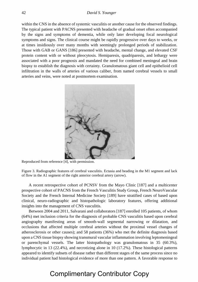
David S. Younger 42
within the CNS in the absence of systemic vasculitis or another cause for the observed findings.
The typical patient with PACNS presented with headache of gradual onset often accompanied
by the signs and symptoms of dementia, while only later developing focal neurological
symptoms and signs. The clinical course might be rapidly progressive over days to weeks, or
at times insidiously over many months with seemingly prolonged periods of stabilization.
Those with GAB or GANS [186] presented with headache, mental change, and elevated CSF
protein content with or without pleocytosis. Hemiparesis, quadriparesis, and lethargy were
associated with a poor prognosis and mandated the need for combined meningeal and brain
biopsy to establish the diagnosis with certainty. Granulomatous giant cell and epithelioid cell
infiltration in the walls of arteries of various caliber, from named cerebral vessels to small
arteries and veins, were noted at postmortem examination.
Reproduced from reference [4], with permission.
Figure 3. Radiographic features of cerebral vasculitis. Ectasia and beading in the M1 segment and lack
of flow in the A1 segment of the right anterior cerebral artery (arrow).
A recent retrospective cohort of PCNSV from the Mayo Clinic [187] and a multicenter
prospective cohort of PACNS from the French Vasculitis Study Group, French NeuroVascular
Society and the French Internal Medicine Society [189] have stratified cases of based upon
clinical, neuro-radiographic and histopathologic laboratory features, offering additional
insights into the management of CNS vasculitis.
Between 2004 and 2011, Salvarani and collaborators [187] enrolled 105 patients, of whom
(64%) met inclusion criteria for the diagnosis of probable CNS vasculitis based upon cerebral
angiography manifesting areas of smooth-wall segmental narrowing or dilatation, and
occlusions that affected multiple cerebral arteries without the proximal vessel changes of
atherosclerosis or other causes); and 58 patients (36%) who met the definite diagnosis based
upon a CNS tissue biopsy showing transmural vascular inflammation involving leptomeningeal
or parenchymal vessels. The latter histopathology was granulomatous in 35 (60.3%),
lymphocytic in 13 (22.4%), and necrotizing alone in 10 (17.2%). These histological patterns
appeared to identify subsets of disease rather than different stages of the same process since no
individual patient had histological evidence of more than one pattern. A favorable response to
Complimentary Contributor Copy
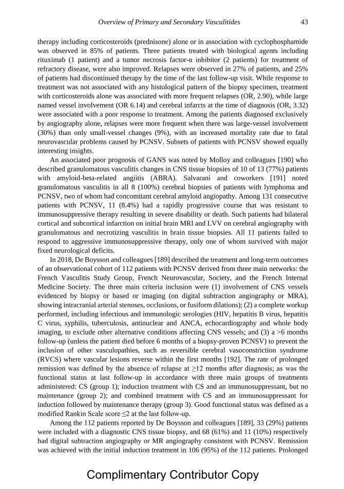
Overview of Primary and Secondary Vasculitides 43
therapy including corticosteroids (prednisone) alone or in association with cyclophosphamide
was observed in 85% of patients. Three patients treated with biological agents including
rituximab (1 patient) and a tumor necrosis factor-α inhibitor (2 patients) for treatment of
refractory disease, were also improved. Relapses were observed in 27% of patients, and 25%
of patients had discontinued therapy by the time of the last follow-up visit. While response to
treatment was not associated with any histological pattern of the biopsy specimen, treatment
with corticosteroids alone was associated with more frequent relapses (OR, 2.90), while large
named vessel involvement (OR 6.14) and cerebral infarcts at the time of diagnosis (OR, 3.32)
were associated with a poor response to treatment. Among the patients diagnosed exclusively
by angiography alone, relapses were more frequent when there was large‐vessel involvement
(30%) than only small‐vessel changes (9%), with an increased mortality rate due to fatal
neurovascular problems caused by PCNSV. Subsets of patients with PCNSV showed equally
interesting insights.
An associated poor prognosis of GANS was noted by Molloy and colleagues [190] who
described granulomatous vasculitis changes in CNS tissue biopsies of 10 of 13 (77%) patients
with amyloid-beta-related angiitis (ABRA). Salvarani and coworkers [191] noted
granulomatous vasculitis in all 8 (100%) cerebral biopsies of patients with lymphoma and
PCNSV, two of whom had concomitant cerebral amyloid angiopathy. Among 131 consecutive
patients with PCNSV, 11 (8.4%) had a rapidly progressive course that was resistant to
immunosuppressive therapy resulting in severe disability or death. Such patients had bilateral
cortical and subcortical infarction on initial brain MRI and LVV on cerebral angiography with
granulomatous and necrotizing vasculitis in brain tissue biopsies. All 11 patients failed to
respond to aggressive immunosuppressive therapy, only one of whom survived with major
fixed neurological deficits.
In 2018, De Boysson and colleagues [189] described the treatment and long-term outcomes
of an observational cohort of 112 patients with PCNSV derived from three main networks: the
French Vasculitis Study Group, French Neurovascular, Society, and the French Internal
Medicine Society. The three main criteria inclusion were (1) involvement of CNS vessels
evidenced by biopsy or based or imaging (on digital subtraction angiography or MRA),
showing intracranial arterial stenoses, occlusions, or fusiform dilations); (2) a complete workup
performed, including infectious and immunologic serologies (HIV, hepatitis B virus, hepatitis
C virus, syphilis, tuberculosis, antinuclear and ANCA, echocardiography and whole body
imaging, to exclude other alternative conditions affecting CNS vessels; and (3) a >6 months
follow-up (unless the patient died before 6 months of a biopsy-proven PCNSV) to prevent the
inclusion of other vasculopathies, such as reversible cerebral vasoconstriction syndrome
(RVCS) where vascular lesions reverse within the first months [192]. The rate of prolonged
remission was defined by the absence of relapse at ≥12 months after diagnosis; as was the
functional status at last follow-up in accordance with three main groups of treatments
administered: CS (group 1); induction treatment with CS and an immunosuppressant, but no
maintenance (group 2); and combined treatment with CS and an immunosuppressant for
induction followed by maintenance therapy (group 3). Good functional status was defined as a
modified Rankin Scale score ≤2 at the last follow-up.
Among the 112 patients reported by De Boysson and colleagues [189], 33 (29%) patients
were included with a diagnostic CNS tissue biopsy, and 68 (61%) and 11 (10%) respectively
had digital subtraction angiography or MR angiography consistent with PCNSV. Remission
was achieved with the initial induction treatment in 106 (95%) of the 112 patients. Prolonged
Complimentary Contributor Copy
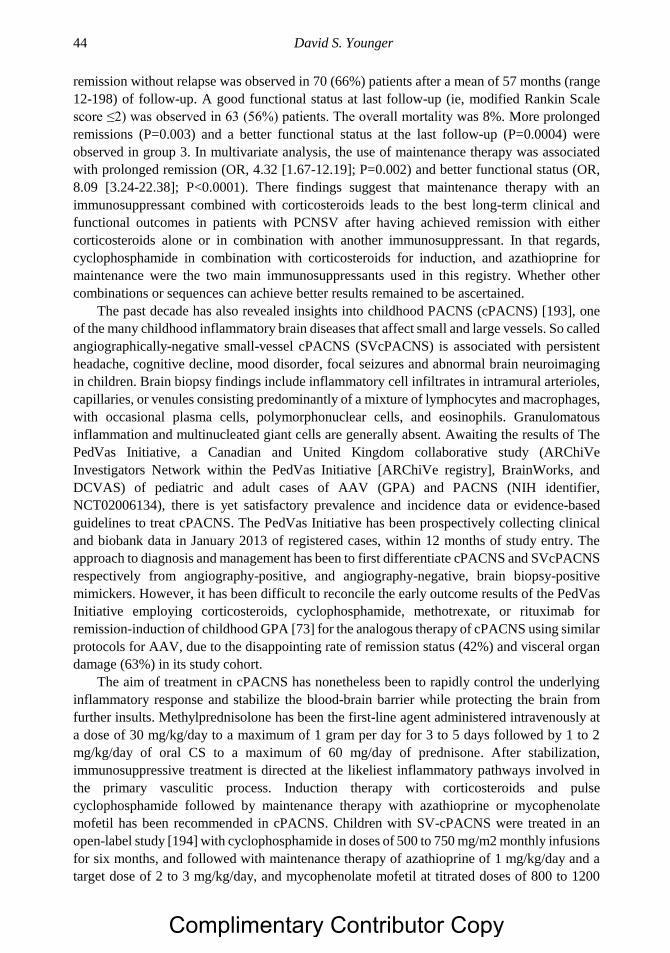
David S. Younger 44
remission without relapse was observed in 70 (66%) patients after a mean of 57 months (range
12-198) of follow-up. A good functional status at last follow-up (ie, modified Rankin Scale
score ≤2) was observed in 63 (56%) patients. The overall mortality was 8%. More prolonged
remissions (P=0.003) and a better functional status at the last follow-up (P=0.0004) were
observed in group 3. In multivariate analysis, the use of maintenance therapy was associated
with prolonged remission (OR, 4.32 [1.67-12.19]; P=0.002) and better functional status (OR,
8.09 [3.24-22.38]; P<0.0001). There findings suggest that maintenance therapy with an
immunosuppressant combined with corticosteroids leads to the best long-term clinical and
functional outcomes in patients with PCNSV after having achieved remission with either
corticosteroids alone or in combination with another immunosuppressant. In that regards,
cyclophosphamide in combination with corticosteroids for induction, and azathioprine for
maintenance were the two main immunosuppressants used in this registry. Whether other
combinations or sequences can achieve better results remained to be ascertained.
The past decade has also revealed insights into childhood PACNS (cPACNS) [193], one
of the many childhood inflammatory brain diseases that affect small and large vessels. So called
angiographically-negative small-vessel cPACNS (SVcPACNS) is associated with persistent
headache, cognitive decline, mood disorder, focal seizures and abnormal brain neuroimaging
in children. Brain biopsy findings include inflammatory cell infiltrates in intramural arterioles,
capillaries, or venules consisting predominantly of a mixture of lymphocytes and macrophages,
with occasional plasma cells, polymorphonuclear cells, and eosinophils. Granulomatous
inflammation and multinucleated giant cells are generally absent. Awaiting the results of The
PedVas Initiative, a Canadian and United Kingdom collaborative study (ARChiVe
Investigators Network within the PedVas Initiative [ARChiVe registry], BrainWorks, and
DCVAS) of pediatric and adult cases of AAV (GPA) and PACNS (NIH identifier,
NCT02006134), there is yet satisfactory prevalence and incidence data or evidence-based
guidelines to treat cPACNS. The PedVas Initiative has been prospectively collecting clinical
and biobank data in January 2013 of registered cases, within 12 months of study entry. The
approach to diagnosis and management has been to first differentiate cPACNS and SVcPACNS
respectively from angiography-positive, and angiography-negative, brain biopsy-positive
mimickers. However, it has been difficult to reconcile the early outcome results of the PedVas
Initiative employing corticosteroids, cyclophosphamide, methotrexate, or rituximab for
remission-induction of childhood GPA [73] for the analogous therapy of cPACNS using similar
protocols for AAV, due to the disappointing rate of remission status (42%) and visceral organ
damage (63%) in its study cohort.
The aim of treatment in cPACNS has nonetheless been to rapidly control the underlying
inflammatory response and stabilize the blood-brain barrier while protecting the brain from
further insults. Methylprednisolone has been the first-line agent administered intravenously at
a dose of 30 mg/kg/day to a maximum of 1 gram per day for 3 to 5 days followed by 1 to 2
mg/kg/day of oral CS to a maximum of 60 mg/day of prednisone. After stabilization,
immunosuppressive treatment is directed at the likeliest inflammatory pathways involved in
the primary vasculitic process. Induction therapy with corticosteroids and pulse
cyclophosphamide followed by maintenance therapy with azathioprine or mycophenolate
mofetil has been recommended in cPACNS. Children with SV-cPACNS were treated in an
open-label study [194] with cyclophosphamide in doses of 500 to 750 mg/m2 monthly infusions
for six months, and followed with maintenance therapy of azathioprine of 1 mg/kg/day and a
target dose of 2 to 3 mg/kg/day, and mycophenolate mofetil at titrated doses of 800 to 1200
Complimentary Contributor Copy
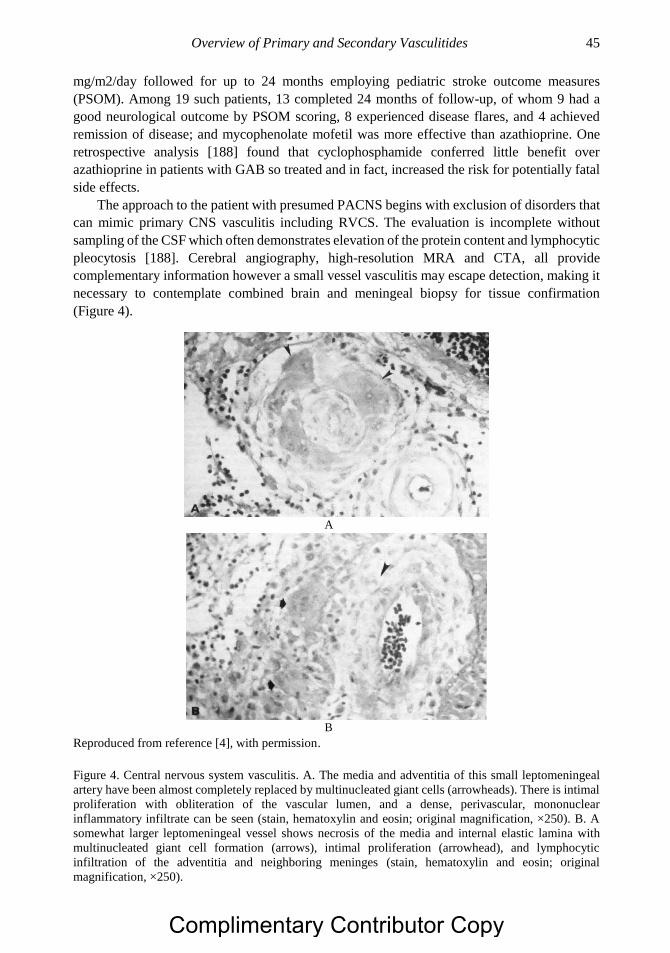
Overview of Primary and Secondary Vasculitides 45
mg/m2/day followed for up to 24 months employing pediatric stroke outcome measures
(PSOM). Among 19 such patients, 13 completed 24 months of follow-up, of whom 9 had a
good neurological outcome by PSOM scoring, 8 experienced disease flares, and 4 achieved
remission of disease; and mycophenolate mofetil was more effective than azathioprine. One
retrospective analysis [188] found that cyclophosphamide conferred little benefit over
azathioprine in patients with GAB so treated and in fact, increased the risk for potentially fatal
side effects.
The approach to the patient with presumed PACNS begins with exclusion of disorders that
can mimic primary CNS vasculitis including RVCS. The evaluation is incomplete without
sampling of the CSF which often demonstrates elevation of the protein content and lymphocytic
pleocytosis [188]. Cerebral angiography, high-resolution MRA and CTA, all provide
complementary information however a small vessel vasculitis may escape detection, making it
necessary to contemplate combined brain and meningeal biopsy for tissue confirmation
(Figure 4).
A
B
Reproduced from reference [4], with permission.
Figure 4. Central nervous system vasculitis. A. The media and adventitia of this small leptomeningeal
artery have been almost completely replaced by multinucleated giant cells (arrowheads). There is intimal
proliferation with obliteration of the vascular lumen, and a dense, perivascular, mononuclear
inflammatory infiltrate can be seen (stain, hematoxylin and eosin; original magnification, ×250). B. A
somewhat larger leptomeningeal vessel shows necrosis of the media and internal elastic lamina with
multinucleated giant cell formation (arrows), intimal proliferation (arrowhead), and lymphocytic
infiltration of the adventitia and neighboring meninges (stain, hematoxylin and eosin; original
magnification, ×250).
Complimentary Contributor Copy
David S. Younger 46
Idiopathic Aortitis-IgG4-Related Disorders
In 1972, an unusual form of inflammatory aortic disease or aortitis came to light in the
surgical literature with far-reaching implications for concepts of autoimmunity. Walker and
colleagues [193] noted that 10% of 217 patients presenting with abdominal aneurysms at
Manchester Royal Infirmary between 1958 and 1969 for resection showed excessive thickening
of aneurysm walls and perianeurysmal adhesions at operation. Subsequent histological
examination of the walls of the aneurysms showed extensive active chronic inflammatory
changes including plasma-cell infiltration.
The clinical features of patients with inflammatory aneurysms differed from those with
atherosclerotic disease due to generally younger age by a decade, lower incidence of rupture,
lack of claudication of intermittent the limbs and presence of peripheral pulses, less likelihood
of unusual presenting features, elevated ESR, and lack of calcification on preoperative
abdominal Rojo-Leyva and colleagues [196] noted idiopathic aortitis in 43% of 1,204 aortic
specimens gathered over a period of 20 years. In 96% of the patients with idiopathic aortitis
patients and aneurysm formation, aortitis was present only in the thoracic aorta. In 2001,
Hamano and colleagues [197] noted a high concentrations of IgG4 associated with sclerosing
pancreatitis characterized by obstructive jaundice, infrequent attacks of abdominal pain,
irregular narrowing of the pancreatic duct, sonolucent swelling of the parenchyma,
lymphoplasmacytic infiltration, fibrosis, and a favorable response to corticosteroid treatment.
One year later, Hamano and coworkers [198] noted the association of sclerosing pancreatitis
with raised concentrations of IgG4 among those with concomitant hydronephrosis that caused
ureteral masses later diagnosed as retroperitoneal fibrosis (RPF).
Histologic examination of ureteral and pancreatic tissues revealed abundant tissue
infiltration by IgG4-bearing plasma cells. In the same year, 2008, three important observations
were made. First, Sakata and colleagues [199] concluded that inflammatory abdominal aortic
aneurysm (IAAA) was related to IgG4 sclerosing disease. Second, Kasashima and colleagues
[200] concluded that IAAA was an IgG-RD together with RPF. Third, Ito and colleagues [201]
described a patient with IAAA, hydronephrosis caused by RPF and high levels of IgG4, in
whom treatment with corticosteroids led to clinical improvement and reduction in IgG4 levels.
Histological inspection of the aortic wall specimen showed lymphocytoplasmacytic infiltration.
Immunohistochemical analysis of the tissue showed IgG4 positive plasma cells. The findings
suggested that IAAA had an etiopathogenesis similar to autoimmune pancreatitis and that some
cases of IAAA and RPF could be aortic and periaortic lesions of an IgG4-RD. One year later
in 2009, Khosroshahi and colleagues [202] described thoracic aortitis due to IgG4-RD with
marked elevation of the serum IgG4 levels with progression to autoimmune pancreatitis, and
Stone and coworkers [203] described IgG4-related thoracic aortitis with a media-predominant
pattern of aortic wall infiltration and marked elevation of serum IgG4 levels, unequivocally
linking IgG4-RD with thoracic lymphoplasmacytic aortitis.
Color-Doppler sonography, MRI combined with MRA and CTA, which adequately
visualize the aortic wall and lumen, combined with FDG PET to detect increased uptake by
metabolically active cells including inflammatory cells infiltrating the vessel wall, are essential
in the assessment of the extent and severity of the various forms of aortitis including IgG4
types. The histopathologic analysis of biopsy specimens has been the cornerstone of the
diagnosis of IgG4-RD.
Complimentary Contributor Copy
Overview of Primary and Secondary Vasculitides 47
A 2012 consensus statement on the pathology of IgG4-RD by Deshpande and colleagues
[204] proposed a terminology scheme for the diagnosis of IgG4-RD based upon the
morphological appearance and tissue IgG4+ plasma cell counts in biopsy tissue. Three
histopathological features associated with IgG4-RD included a dense lymphoplasmacytic
infiltrate, fibrosis arranged at least focally in a storiform pattern, and obliterative phlebitis in
morphological specimens. The majority of cells were T-cell with scattered B-cells, and an
essential component of plasma cells with occasional eosinophils and macrophages. The level
IgG4 antibody, which represents less than 5% of the total IgG in healthy individuals is tightly
regulated and has a unique structure and functional property. It undergoes half antibody
exchange in vivo resulting in recombined antibodies composed of two different binding
specificities. Their production is driven in part by Th2 cytokines that mediate allergic reactions
and IgE production. It does not activate complement pathways and has reduced effector
function relative to other IgG subtypes. It remains unclear as to whether IgG4 directly mediates
the disease process or reflects a protective response induced by anti-inflammatory cytokines,
making it simply a valuable biological marker of IgG4-RD.
A Japanese consensus management guideline [205] suggested the initiation of oral
prednisolone for induction of remission at a dose of 0.6 mg per kilogram per day for 2 to 4
weeks, with tapering by 5 mg every 1 to 2 weeks based on clinical manifestations, biochemical
blood tests, and repeated imaging, to a maintenance dose of 2.5 to 5 mg per day for up to 3
months. Re-administration of corticosteroids is advised for treating relapses. Treatment with
azathioprine, mycophenolate mofetil and methotrexate can be used as corticosteroid sparing
agents or as remission-maintenance drugs after corticosteroid-induced remissions. Patients with
recurrent or refractory disease and B-cell depletion may be considered for rituximab [206].
Nonsystemic Peripheral Nerve Vasculitis
The vasculitic neuropathies are heterogeneous disorders that present in the setting of
systemic vasculitis or in the absence thereof where necrotizing arteritis may remain clinically
and pathologically restricted to the peripheral nerves as a SOV. The Peripheral Nerve Society
[207] established guidelines for the classification, diagnosis, investigation, and treatment of
NSVN. Pathologically-definite vasculitic neuropathy is defined by active or chronic peripheral
nerve and muscle tissue lesions that show cellular invasion of the walls of blood vessels with
accompanying acute vascular damage (fibrinoid necrosis, endothelial loss/disruption, internal
lamina loss/fragmentation, smooth muscle media loss/fragmentation/separation, acute
thrombosis, vascular/perivascular hemorrhage or leukocytoclasia) or chronic vascular damage
(intimal hyperplasia, fibrosis of media, adventitial/periadventitial fibrosis or chronic
thrombosis chronic thrombosis with recanalization), without evidence of another primary
disease process that could mimic vasculitis pathologically such as lymphoma, lymphomatoid
granulomatosis or amyloidosis.
Patients with NSVN lack symptoms, signs, or laboratory evidence of involvement of other
organs (demonstrable by laboratory evidence of PR3-, MPO-ANA, mixed cyroglobulins, SSA,
SSB, Sm, RNP, Scl-70, centromere, dsDNA, CCP serology; ESR >100 mm per hour, or tissue
biopsy evidence of vasculitis in another organ other than muscle; serologic, PCR or culture
evidence of a specific infection associate with vasculitis), and no predisposing factors (other
than diabetes) of a connective tissue disease, sarcoidosis, inflammatory bowel disease, active
Complimentary Contributor Copy
David S. Younger 48
malignancy, HUV, cutaneous PAN, or exposure to drugs likely to cause vasculitis.
Inflammation of microvessels less than 40 to 70 µm in diameter without vascular damage are
broadly referred to as MV.
The management of NPNV has remained uncertain because of the very concept presumes
that the vasculitic disease process is widespread within the nerves and not present elsewhere in
the body. This assumption has been called into question by four lines of evidence.
First, the definition of NPNV allows for the finding of vasculitic lesions in muscle tissue,
[207] perhaps making the syndrome more appropriately termed peripheral nervous system
vasculitis or “PNSV”. Moreover, the detection of vasculitis in cutaneous nerve and muscle
tissue specimens has been incorporated into the FVSG database to establish the diagnosis of
systemic vasculitis. Among 129 patients with PAN [208] who underwent nerve biopsy with
(108 patients) or without (21 patients) peripheral neuropathy, vasculitic lesions were noted in
cutaneous nerve tissue in 83% and 81% of patients respectively, compared to 68% and 60% of
cases where muscle biopsytissue was examined.
Second, the lack of long-term follow-up in most cases series ranging from 6 months to 22
years [209].
Third, the report of only two proposed cases, both with foci of vasculitis outside the PNS
in a visceral organ [19] or the temporal artery [210]. Patient 1 in the series of pathologically
confirmed cases of PAN by Kernohan and Woltman [19] was a 54-year-old man with five years
of progressive generalized painful peripheral neuropathy that was so severe before death that
he was partially paralyzed, and unable to speak or swallow. Postmortem examination showed
PAN limited to the nerve trunks of the arms and legs. The brain, cranial nerves, and spinal cord
were normal except for early acute changes without evidence of vasculitis. Examination of all
other organs failed to reveal a single vascular lesion, except one small artery in the capsule of
the prostate gland. Torvik and Berntzen [210] described a 76-year-old woman with diffuse
fever, pain, and central scotoma of the eye that improved with corticosteroids. A biopsy of the
temporal artery and pectoralis muscle disclosed necrotizing arteries of small arteries and
arterioles in small adventitial vessels of the temporal artery without frank temporal arteritis.
However, postmortem examination evidence of healed vasculitis in numerous small arteries
and arterioles of muscle and nerve tissue measuring 50 to 200 microns in diameter without
vasculitis in visceral organ or the CNS. .
Fourth, the inclusion of patients with diabetes according to the 2010 guidelines [210] may
be introducing selection bias. Over the years, there has been increasing support for the
contribution of autoimmune mechanism in the pathogenesis of diabetic neuropathy. Diabetes
itself appears to be caused by autoimmune mechanisms directed at insulin-producing pancreatic
beta cells, and a variety of autoantibodies have been detected in patients with type-1 diabetes
(T1D) or insulin dependent (IDDM), including anti-islet cell cytoplasmic antibodies, present in
up to 80% of newly diagnosed patients and glutamic acid decarboxylase (GAD) antibodies,
also present in patients with autoimmune stiff person syndrome. Younger and colleagues [211]
reported the clinicopathologic and immunohistochemical findings of sural nerve biopsy tissues
in a cohort of 20 patients with heterogeneous forms of diabetic neuropathy. That series was
continued to a total of 107 patients [212], of which 3 (3%) showed MV, and 3 (3%) showed
necrotizing arteritis. Although diabetes has not been considered a predisposing factor in PNV,
the presence or absence of diabetes became a defining feature of patients with LSRPN [213,
214]. In the only postmortem case of LSRPN described by Younger [215], sural nerve biopsy
showed mononuclear inflammatory cells surrounding a small epineurial artery with extension
Complimentary Contributor Copy
Overview of Primary and Secondary Vasculitides 49
into the vascular wall, with reactive luminal connective tissue suggesting recanalization of a
thrombus. An adjacent nerve fascicle showed marked loss of myelinated nerve fibers. The
patient was treated for painful diabetic lumbosacral plexopathy (DLSRPN) and peripheral
nerve vasculitis according to prevailing standards with 2 g/kg intravenous immunoglobulin for
5 days, followed by 750 mg of intravenous cyclophosphamide and 1,000 mg of
methylprednisolone intravenously for 3 additional days. Acute tubular necrosis, increasing
lethargy, unresponsiveness, and aspiration pneumonia supervened and the patient expired 4
weeks after admission. General autopsy showed no evidence of systemic or peripheral nerve
vasculitis. The brain showed diffuse loss of neurons in all sampled cortical areas, including the
cerebellum, consistent with anoxia secondary to cardiac arrest. Sections of extradural lumbar
plexus, sciatic, and femoral nerve tissue showed perivascular epineurial inflammation with
infiltration of adjacent endoneurium. This case exemplifies the restricted nature of LSRPN as
an example of a true NSVN.
Thereare far more studies of living cohorts with NSPNV. Kissel and colleagues [216]
reported that 4.5% of 350 consecutive nerve biopsies performed at a single institution
evidenced peripheral neuropathy secondary to necrotizing angiopathy. Six patients manifested
a distal symmetrical sensorimotor polyneuropathy, while ten had a mononeuritis multiplex
presentation, eight of whom had overlapping involvement of peripheral nerves that obscured
the picture of mononeuritis. In three-quarters (12 patients), a specific underlying collagen
vascular disease was not diagnosed despite extensive clinical, radiologic, and serological
evaluation. Said and colleagues [217] studied 100 patients with necrotizing arteritis in muscle
or nerve biopsy tissue that occurred in the context of a connective tissue disorder in fifty-five
patients and in association with a disorder unrelated to connective tissue pathology in thirteen
others. The commonest complaints at presentation in this cohort were specific cutaneous
manifestation of vasculitis, including livedo, cutaneous necrosis, and nodules in one-third.
Thirty-two patients had neuropathy only and necrotizing arteritis, the most common complaints
at of which were spontaneous pain of neurogenic or muscle origin (48%).
More recently, Collins and colleagues [218] described forty-eight patients with NSPNV,
85% of whom had extensive, overlapping involvement of multiple nerves. Peroneal nerve and
peroneal muscle tissue biopsy was 58% diagnostically sensitive compared to 47% for sural
nerve biopsy for the diagnosis of vasculitis. Combination therapy with corticosteroids and
cytotoxic agents was more effective than corticosteroids monotherapy for inducing remission
and improving disability, with trends toward reduced relapses and chronic pain. Overall, ten
patients died (21%) over the period of 63 months follow-up, five (10%) of whom were related
to the disease or treatment, including two patients who succumbed to pulmonary emboli as a
result of limited mobility of the legs or myocardial infarction in another; and two patients, one
of whom had fatal sepsis and another metastatic bladder cancer as a consequence of
cyclophosphamide toxicity.
There are no ongoing observational cohort studies to guide the treatment of NPNV.
Recommendations for the treatment of NSPNV [210] include prednisone monotherapy unless
there is rapidly progressive neuropathy at the dose of 1 mg per kilogram per day, with tapering
over one year to a low dose. Combination therapy employing cytotoxic drugs including
cyclophosphamide, methotrexate, and azathioprine may be employed with adjuvant plasma
exchange or IVIg. However, IVIg showed efficacy in a multicenter RCT of the treatment of
small-to-medium systemic vasculitis due to EGPA with persistent disease activity [219].
Complimentary Contributor Copy
David S. Younger 50
This suggests that IVIg may be appropriate, first-line therapeutic modality in NSPNV.
Moreover, the benefit of IVIg may be achieved without the risk of potentially fatal cytotoxic
side-effects. However there has not been a prospective RCT of IVIg in NSPNV. A variety of
mechanisms have been thought to be responsible for the beneficial effects of IVIg in NSPNV
and other systemic vasculitides including neutralization of autoantibodies, inhibition of
complement pathways, alteration of Fc receptor expression, and alteration of cytokine profiles
[220]. Careful monitoring should be performed to observe desired therapeutic responses and to
avoid potentially serious drug side effects.
CONCLUSION
In no other disorder have there been so many triumphs as in the diagnosis and treatment of
primary systemic vasculitis. Physicians in a variety of subspecialties including neurology,
rheumatology, immunology/allergy, dermatology and clinical pathologist, all working side-by-
side, aided by subspecialities of public health, epidemiology, genetics, and clinical trial
specialists, have benefited the outlook for individual patients and population cohorts around
the globe. Yet it starts with clinical acumen typically of the general practitioner. This chapter
on clinical approach was written with such diverse backgrounds in mind, from generalist to
subspecialist with the hope that it will bring the field up to date and to the present.
REFERENCES
[1] Jennette, J. C., Falk, R. J., Bacon, P. A., et al. 2012 Revised International Chapel Hill
Consensus Conference Nomenclature of Vasculitides. Arthritis Rheum. 2013; 65:1-11.
[2] Ruperto, N., Ozen, S., Pistorio, A., et al. EULAR/PINTO/PRES criteria for Henoch-
Schönlein purpura, childhood polyarteritis nodosa, childhood Wegener granulomatosis
and childhood Takayasu arteritis: Ankara 2008. Part I: Overall methodology and clinical
characterization. Ann. Rheum. Dis. 2010; 69:790-797.
[3] Ozen, S., Pistorio, A., Iusan, S. M., et al. EULAR/PRINTO/PReS criteria for Henoch-
Schönlein purpura, childhood polyarteritis nodosa, childhood Wegener granulomatosis
and childhood Takayasu arteritis. Ankara 2008. Part II: Final Classification. Ann. Rheum.
Dis. 2010; 69:798-806.
[4] Younger, D. S. Adult and childhood vasculitis of the nervous system. Chapter 14. Motor
Disorders, 3rd Ed. D. S. Younger, ed., David, S. Younger, MD PC: New York, 2013;
235-280.
[5] Jennings, G. H. Arteritis of the temporal vessels. Lancet 1938; 1:424-428.
[6] Bruce, G. M. Temporal arteritis as a cause of blindness: Review of the literature and
report of a case. Trans. Am. Ophthalmol. Soc. 1949; 47:300-316.
[7] Gilmour, J. R. Giant-cell chronic arteritis. J. Pathol. Bacteriol. 1941; 53:263-277.
[8] Nuenninghoff, D. M., Hunder, G. G., Christianson, T. J. H., et al. Incidence and
predictors of large-artery complication (aortic aneurysm, aortic dissection, and/or large
artery stenosis) in patients with giant cell arteritis. Arthritis Rheum. 2003; 48:3522-3531.
[9] Ritama, V. Temporal arteritis. Ann. Med. Exp. Fenn. 1951; 40:63-87.
Complimentary Contributor Copy
Overview of Primary and Secondary Vasculitides 51
[10] Save-Soderbergh, J., Malmvall, B., Anderson, R., et al. Giant cell arteritis as a cause of
death. JAMA 1986; 255:493-496.
[11] Takayasu, M. Case with unusual changes of the central vessels in the retina [Japanese].
Acta Soc. Ophthal. Jap. 1908; 12:554-555.
[12] Shimizu, K., Sano, K. Pulseless disease [Japanese]. Clin. Surg. 1948; 3:377-342.
[13] Ishikawa, K. Natural history and classification of occlusive thromboaortopathy
(Takayasu’s disease). Circulation 1978; 57:27-35.
[14] Nakao, K., Ikeda, M., Kimata, S.-I., et al. Takayasu’s arteritis: clinical report of eighty-
four cases and immunologic studies of seven cases. Circulation 1967; 35:1141-1155.
[15] Shick, R. M., Baggenstoss, A. H., Fuller, B. F., et al. Effects of cortisone and ACTH on
periarteritis nodosa and cranial arteritis. Mayo Clin. Proc. 1950; 25:492-494.
[16] Salvarani, C. Assessment of vasculitis extent and severity. Presse Med. 2013; 42:588-
589.
[17] Aiello, P. D., Trautmann, J. C., McPhee, T. J., et al. Visual prognosis in giant cell arteritis.
Ophthalmology 1993; 100:550-555.
[18] Ohigashi, H., Haraguchi, G., Konishi, M., et al. Improved prognosis of Takayasu arteritis
over the past decade. Circ. J. 2012; 76:1004-1011.
[19] Kernohan, J. W., Woltman, H. W. Periarteritis nodosa: a clinicopathologic study with
special reference to the nervous system. Arch. Neurol. 1938; 39:655-686.
[20] Krahulik, L., Rosenthal, M., Loughlin, E. H. Periarteritis nodosa (necrotizing
panarteritis) in childhood with meningeal involvement. Report of a case with study of
pathologic findings. Am. J. Med. Sci. 1935: 190:308-317.
[21] Longcope, W. T. Periarteritis nodosa with report of a case with autopsy. Bull. Auyer Clin.
Lab. Pennsylvania Hospital, 1908, Volume 1.
[22] Kawasaki, T. MLNS showing particular skin desquamation from the finger and toe in
infants. Allergy 1967; 16:178-189.
[23] Tanaka, N., Naoe, S., Kawasaki, T. Pathological study on autopsy cases of
mucocutaneous lymph node syndrome. J. Jap. Red Cross Cent. Hosp. 1971; 2:85-94.
[24] Chamberlain, J. L., Perry, L. W. Infantile periarteritis nodosa with coronary and brachial
aneurysms: a case diagnosed during life. J. Pediatrics 1971; 78:1039-1040.
[25] Landing, B. H., Larson, E. J. Are infantile periarteritis nodosa with coronary artery
involvement and fatal mucocutaneous lymph node syndrome the same? Comparison of
20 patients from North America with patients from Hawaii and Japan. Pediatrics 1977;
59:651-652.
[26] Pagnoux, C., Seror, R., Henegar, C., et al. Clinical features and outcomes in 348 patients
with polyarteritis nodosa. Arthritis Rheum. 2010; 62:616-626.
[27] May, E. F., Jabbari, B. Stroke in neuroborreliosis. Stroke 1990; 21: 1232-1235.
[28] Wilke, M., Eiffert, H., Christen, H. J., et al. Primary chronic and cerebrovascular course
of Lyme neuroborreliosis: case reports and literature review. Arch. Dis. Child 2000; 83:
67-71.
[29] Ozen, S., Anton, J., Arisoy, N., et al. Juvenile polyarteritis: results of a multicenter survey
of 110 children. J. Pediatr. 2004; 145:517-522.
[30] David, J., Ansell, B. M., Woo, P. Polyarteritis nodosa associated with streptococcus.
Arch. Dis. Child 1993; 69:685-688.
[31] Guillevin, L., Cohen, P., Mahr, A., et al., and the French Vasculitis Study Group.
Treatment of polyarteritis nodosa and microscopic polyangiitis with poor prognosis
Complimentary Contributor Copy
David S. Younger 52
factors: a prospective trial comparing glucocorticoids and six or twelve
cyclophosphamide pulses in sixty-five patients. Arthritis Rheum. 2003; 49:93-100.
[32] Guillevin, L., Mahr, A., Callard, P., et al., for the French Vasculitis Study Group.
Hepatitis B virus-associated polyarteritis nodosa: clinical characteristics, outcome, and
impact of treatment in 115 patients. Medicine 2005; 84:313-322.
[33] Henegar, C., Pagnoux, C., Puéchal, X., et al., for the French Vasculitis Study Group. A
paradigm of diagnostic criteria for polyarteritis nodosa. Arthritis Rheum. 2008; 58: 1528-
1538.
[34] Wohlwill, F. Uber die mur mikroskopisch erkenbarre form der periarteritis nodosa.
[About the mur microscopically recognizable form of the periarteritis nodosa.] Arch.
Pathol. Anat. 1923; 246:377-411.
[35] Davson, J., Ball, M., Platt, R. The kidney in periarteritis nodosa. QJM 1948; 17:175-202.
[36] Wainwright, J., Davson, J. The renal appearance in the microscopic form of periarteritis
nodosa. J. Pathol. Bact. 1950; 62:189-196.
[37] Savage, C. O. S., Winearls, C. G., Evans, D. J., et al. Microscopic polyarteritis:
presentation, pathology and prognosis. QJM 1985; 220:467-483.
[38] Guillevin, L., Durand-Gasselin, B., Cevallos, R., et al. Microscopic polyangiitis. Clinical
and laboratory findings in eighty-five patients. Arthritis Rheum. 1999; 42:421-430.
[39] Ahn, J. K., Hwang, J.-W., Lee, J., et al. Clinical features and outcome of microscopic
polyangiitis under a new consensus algorithm of ANCA-associated vasculitides in Korea.
Rheumatol. Int. 2012; 32:2979-2986.
[40] Cabral, D. A., Uribe, A. G., Benseler, S., et al., for the ARChiVe (A Registry for
Childhood Vasculitis: e-entry) Investigators Network. Classification, presentation, and
initial treatment of Wegener’s granulomatosis in childhood. Arthritis Rheum. 2009; 3413-
3424.
[41] Lamb, A. R. Periarteritis nodosa-a clinical and pathological review of the disease with a
report of two cases. Arch. Intern. Med. 1914; 14:481-516.
[42] Churg, J., Strauss, L. Allergic granulomatosis, allergic angiitis, and periarteritis nodosa.
Am. J. Pathol. 1951; 27:277-301.
[43] Masi, A. T., Hunder, G. G., Lie, J. T., et al. The American College of Rheumatology
1990 criteria for the classification of Churg-Strauss syndrome (Allergic granulomatosis
and angiitis). Arthritis Rheum. 1990; 33:1094-1100.
[44] Comarmond, C., Pagnoux, C., Khellaf, M., et al., for the French Vasculitis Study Group.
Eosinophilic granulomatosis with polyangiitis (Churg-Strauss). Arthritis Rheum. 2013;
65:270-281.
[45] Jennette, J. C., Falk, R. J., Andrassay, K., et al. Nomenclature of systemic vasculitides.
Proposal of an international conference. Arthritis Rheum. 1994; 37:187-192.
[46] O’Neil, K. M. Progress in pediatric vasculitis. Curr. Opin. Rheumatol. 2009; 21:538-546.
[47] Godman, G. C., Churg, J. Wegener’s granulomatosis: pathology and review of the
literature. Arch. Pathol. 1954; 58:533-553.
[48] Guillevin, L., Visser, H., Noel, L. H., et al. Antineutrophil cytoplasm antibodies in
systemic polyarteritis nodosa with and without hepatitis B virus infection and Churg-
Strauss syndrome-62 patients. J. Rheumatol. 1993; 20:1345-1349.
[49] Fauci, A. S., Haynes, B. F., Katz, P., et al. Wegener’s granulomatosis: Prospective
clinical and therapeutic experience with 85 patients over 21 years. Ann. Intern. Med.
1983; 98:76-85.
Complimentary Contributor Copy
Overview of Primary and Secondary Vasculitides 53
[50] Hoffman, G. S., Kerr, G. S., Leavitt, R. Y., et al. Wegener granulomatosis: an analysis of
158 patients. Ann. Intern. Med. 1992; 116:488-498.
[51] Carrington, C. B., Libow, A. A. Limited forms of angiitis and granulomatosis of
Wegener’s type. Am. J. Med. 1966; 41:497-527.
[52] Holle, J. U., Gross, W. L., Hull-Ulrich, K., et al. Prospective long-term followup of
patients with localized Wegener’s granulomatosis: does it occur as persistent disease
stage? Ann. Rheum. Dis. 2010; 69:1934-1939.
[53] Leavitt, R. Y., Fauci, A. S., Bloch, D. A., et al. The American College of Rheumatology
1990 criteria for the classification of Wegener’s granulomatosis. Arthritis Rheum. 1990;
33:1101-1107.
[54] Holle, J. U., Csernok, E., Fredenhagen, G., et al. Clinical evaluation of hsPR3-ANCA
ELISA for detection of antineutrophil cytoplasmic antibodies directed against PR3. Ann.
Rheum. Dis. 2010; 69:468-469.
[55] Van der Woude, F. J., Rasmussen, N., Lobatto, S., et al. Autoantibodies against
neutrophils and monocytes: tool for diagnosis and marker of disease activity in
Wegener’s granulomatosis. Lancet 1985; 1:425-429.
[56] Falk, R. J., Jennette, J. C. Anti-neutrophil cytoplasmic autoantibodies with specificity for
myeloperoxidase in patients with systemic vasculitis and idiopathic necrotizing and
crescentic glomerulonephritis. N. Engl. J. Med. 1988; 318:1651-1657.
[57] Goldschmeding, R., Cohen Tervaert, J. W., van der Schoot, C. E., et al. Autoantibodies
against myeloid lysosomal enzymes: a novel class of autoantibodies associated with
vasculitic syndrome [Abstract]. Kidney Int. 1988; 34:558-559.
[58] Gadola, S. D., Gross, W. L. The renaissance of granulomatous inflammation in AAV.
Nat. Rev. Rheumatol. 2012; 8:74-76.
[59] Lionaki, S., Blyth, E. R., Hogan, S. L., et al. Classification of anti-neutrophil cytoplasmic
autoantibody vasculitides: the role of anti-neutrophil cytoplasmic autoantibody
specificity for myeloperoxidase or proteinase 3 in disease recognition and prognosis.
Arthritis Rheum. 2012; 64:3452-3462.
[60] Savage, C. O., Gaskin, G., Pusey, C. D., et al. Myeloperoxidase binds to vascular
endothelial cells, is recognized by ANCA and can enhance complement dependent
cytotoxicity. Adv. Exp. Med. Biol. 1993; 336:121-123.
[61] Jennette, J. C., Falk, R. J. Pathogenesis of ANCA-associated vasculitis: observations,
theories, and speculations. Presse Med. 2013; 42:493-498.
[62] Free, M. E., Ciavatta, D. J., Falk, R. J. How antigens influence the disease: An
immunologist point of view. Presse Med. 2013; 42:577-579.
[63] Jones, R. B., Ferraro, A. J., Chaudhry, A. N., et al. A multicenter survey of rituximab for
refractory antineutrophil cytoplasmic antibody-associated vasculitis. Arthritis Rheum.
2009; 60:2156-2168.
[64] Cartin-Ceba, R., Golbin, J. M., Keogh, K. A., et al. Rituximab for remission induction
and maintenance in refractory granulomatosis with polyangiitis (Wegener’s): ten-year
experience at a single center. Arthritis Rheum. 2012; 64:3770-3778.
[65] Holle, J. U., Dubrau, C., Herlyn, K., et al. Rituximab for refractory granulomatosis with
polyangiitis (Wegener’s granulomatosis): comparison of efficacy in granulomatous
versus vasculitic manifestations. Ann. Rheum. Dis. 2012; 71:327-333.
Complimentary Contributor Copy
David S. Younger 54
[66] Smith, R. M., Jones, R. B., Guerry, M. J., et al. Rituximab for remission maintenance in
relapsing antineutrophil cytoplasmic antibody-associated vasculitis. Arthritis Rheum.
2012; 64:3760-3769.
[67] Roubaud-Baudron, C., Pagnoux, C., et al. Rituximab maintenance for granulomatosis
with polyangiitis and microscopic polyangiitis. J. Rheumatol. 2012; 39:125-130.
[68] Smith, R., Alberici, F., Jones, R., et al. Long term follow up of patients who received
repeat dose rituximab as maintenance therapy for ANCA associated vasculitis (AAV)
[Abstract]. Presse Med. 2013; 42:775-776.
[69] Stone, J. R., Merkel, P. A., Spiera, R., et al. Rituximab versus cyclophosphamide for
ANCA-associated vasculitis. N. Engl. J. Med. 2010; 363:221-232.
[70] Jayne, D., Smith, R., Merkel, P. An international, open label, randomised controlled trial
comparing rituximab with azathioprine as maintenance therapy in relapsing ANCA-
associated vasculitis (RITAZAREM) [Abstract]. Presse Med. 2013; 42:768.
[71] Miloslavsky, E., Specks, U., Merkel, P., et al. Retreatment with rituximab in the RAVE
trial [Abstract]. Presse Med. 2013; 42:778.
[72] Miloslavsky, E. M., Specks, U., Merkel, P., et al. Clinical outcomes of remission
induction therapy for severe antineutrophil cytoplasmic antibody-associated vasculitis.
Arthritis Rheum. 2013; 65:2441-2449.
[73] Morishita KA, Moorthy LN, Lubieniecka JM, et al. Early outcomes in children with
antineutrophil cytoplasmic antibody-associated vasculitis. Arthritis Rheum. 2017;
69:1470-1479.
[74] Sacri AS, Chambaraud T, Ranchin B, et al. Clinical characteristics and outcomes of
childhood‐onset ANCA‐associated vasculitis: a French nationwide study. Nephrol Dial
Transplant. 2015;30 Suppl 1:i104–112.
[75] Zeek, P. M. Periarteritis nodosa and other forms of necrotizing angiitis. N. Engl. J. Med.
1953; 248:764-772.
[76] Arthus, M., Breton, M. Lesions cutanées produites par les injections de sérum de cheval
chez le lapin anaphylactisé par et pour ce sérum. [M. Cutaneous lesions produced by
horse serum injections in rabbits anaphylactized with and for this serum.] Compt. Rend.
Soc. de Biol. 1903; 55: 1478-1480.
[77] Gairdner, D. The Schönlein-Henoch syndrome (anaphylactoid purpura). QJM 1948; 17:
95-122.
[78] Levitt, L. M., Burbank, B. Glomerulonephritis as a complication of the Schonlein-
Henoch syndrome. N. Engl. J. Med. 1953; 248:530-536.
[79] Faille-Kuyber, E. H., Kater, L., Kooiker, C. J., et al. IgA-deposits in cutaneous blood-
vessel walls and mesangium in Henoch-Schonlein syndrome. Lancet 1973; 1:892-893.
[80] Conley, M. E., Cooper, M. D., Michael, A. F. Selective deposition of immunoglobulin
A1 in immunoglobulin A nephropathy, anaphylactoid purpura nephritis, and systemic
lupus erythematosus. J. Clin. Invest. 1980; 66:1432-1436.
[81] Mills, J. A., Michel, B. E., Bloch, D. A., et al. The American College of Rheumatology
1990 criteria for the classification of Henoch-Schönlein purpura. Arthritis Rheum. 1990;
33:1114-1121.
[82] Peru, H., Soylemezoglu, O., Bakkaloglu, S. A., et al. Henoch Schonlein purpura in
childhood: clinical analysis of 254 cases over a 3-year period. Clin. Rheumatol. 2008;
27:1087-1092.
Complimentary Contributor Copy
Overview of Primary and Secondary Vasculitides 55
[83] McDuffie, F. C., Sams, W. M., Maldonado, J. E., et al. Hypocomplementemia with
cutaneous vasculitis and arthritis. Possible immune complex syndrome. Mayo Clin. Proc.
1973; 48:340-348.
[84] Zeiss, C. R., Burch, F. X., Marder, R. J., et al. A hypocomplementemic vasculitic
urticarial syndrome: Report of four new cases and definition of the disease. Am. J. Med.
1980; 68:867-875.
[85] Wisnieski, J. J. Naff. Serum IgG antibodies to C1q in hypocomplementemic urticarial
vasculitis syndrome. Arthritis Rheum. 1989; 32:1119-1127.
[86] Buck, A., Christensen, J., McCarty, M. Hypocomplementemic urticarial vasculitis
syndrome. A case report and literature review. J. Clin. Aesthet. Dermatol. 2012; 5:36-46.
[87] Behcet, H., Matteson, E. L. On relapsing, aphthous ulcers of the mouth, eye and genitalia
caused by a virus. 1937. Clin. Exp. Rheumatol. 2010; 28(Suppl. 60):S2-S5.
[88] Wolf, S. M., Schotland, D. L., Phillips, L. L. Involvement of nervous system in Behçet’s
syndrome. Arch. Neurol. 1965; 12:315-325.
[89] International Study Group for Behçet’s Disease. Lancet 1990; 335:1078-1080.
[90] Sobel, J. D., Haim, S., Shafrir, A., et al. Cutaneous hyper-reactivity in Behçet disease.
Dermatologica 1973; 146:350-356.
[91] Özen, S. Pediatric onset Behcet disease. Curr. Opin. Rheumatol. 2010; 22:585-589.
[92] Kawakita, H., Nishimura, M., Satoh, Y., et al. Neurological aspects of Behçet’s disease.
A case report and clinico-pathological review of the literature in Japan. J. Neurol. Sci.
1967; 5:417-439.
[93] Yesilot, N., Bahar, S., Yilmazer, S., et al. Cerebral venous thrombosis in Behcet’s disease
compared to those associated with other etiologies. J. Neurol. 2009; 256:1134-1142.
[94] Matsumoto, T., Uekusa, T., Fukuda, Y. Vasculo-Behcet’s disease: a pathologic study of
eight cases. Hum. Pathol. 1991; 22:45-51.
[95] Mogan, R. F., Baumgarten, C. J. Meniere’s disease complicated by recurrent interstitial
keratitis: Excellent results following cervical ganglionectomy. West J. Surg. 1934; 42:
628.
[96] Cogan, D. G. Syndrome of nonsyphilitic interstitial keratitis and vestibuloauditory
symptoms. Arch. Ophthal. 1945; 33:144-149.
[97] Haynes, B. F., Kaiser-Kupfer, M. I., Mason, P., et al. Cogan’s syndrome: Studies in
thirteen patients, long-term follow-up, and a review of the literature. Medicine 1980;
59:426-441.
[98] Cogan, D. G. Nonsyphilitic interstitial keratitis with vestibuloauditory symptoms. Report
of four additional cases. Arch. Ophthalmol. 1949; 42:42-49.
[99] Norton, E. W. D., Cogan, D. G. Syndrome of nonsyphylitic interstitial keratitis and
vestibuloauditory symptoms. A long-term follow-up. Arch. Ophthalmol. 1959; 61:695-
697.
[100] Ferri, C., Sebastiani, M., Giuggioli, D., et al. Mixed cryoglobulinemia: demographic,
clinical, and serologic features and survival in 231 patients. Semin. Arthritis Rheum.
2004; 33:355-374.
[101] Ferri, C. Mixed cryoglobulinemia. Orphanet J. Rare Dis. 2008; 3:25.
[102] Wintrobe, M. M., Buell, M. V. Hyperproteinemia associated with multiple myeloma.
With report of a case in which an extraordinary hyperproteinemia was associated with
thrombosis of the retinal veins and symptoms suggesting Raynauds disease. Bulletin of
the Johns Hopkins Hospital 1933; 52:156-165.
Complimentary Contributor Copy
David S. Younger 56
[103] Brouet, J. C., Clauvel, J. P., Danon, F., et al. Biologic and clinical significance of
cryoglobulins: a report of 86 cases. Am. J. Med. 1974; 57:775-788.
[104] Pascual, M., Perrin, L., Giostra, E., et al. Hepatitis C virus in patients with
Cryoglobulinemia type II. J. Inf. Dis. 1990; 162: 569-570.
[105] Ferri, C., Greco, F., Longombardo, G., et al. Association between hepatitis C virus and
mixed cryoglobulinemia. Clin. Exp. Rheum. 1991; 9: 621-624.
[106] Agnello, V., Chung, R. T., Kaplan, L. M. A role for hepatitis C virus infection in type II
cryoglobulinemia. N. Engl. J. Med. 1992; 327:1490-1495.
[107] Terrier, B., Krastinova, E., Marie, I., et al. Management of noninfectious mixed
cryoglobulinemia vasculitis: data from 242 cases included in the CryoVas survey. Blood
2012; 119:5996-6004.
[108] Terrier, B., Cacoub, P. Cryoglobulinemia vasculitis: an update. Curr. Opin. Rheumatol.
2013; 25:10-18.
[109] Cacoub, P., Poynard, T., Ghillani, P., et al., for the MULTIVIRC Group. Extrahepatic
manifestations of chronic hepatitis C. Arthritis Rheum. 1999; 42:2204-2212.
[110] Liou, Y.-T., Huang, J.-L., Ou, L.-S., et al. Comparison of cryoglobulinemia in children
and adults. J. Microbiol. Immunol. Infect. 2013; 46:59-64.
[111] Cacoub, P., Ferrier, B., Saadoun, D. Hepatitis C virus mixed cryoglobulinemia vasculitis:
Therapeutic options. Presse Med. 2013; 42:523-527.
[112] Klemperer, P. Diseases of the collagen system. Bull. NY Acad. Med. 1947; 23:581-588.
[113] Klemperer, P. The pathogenesis of lupus erythematosus and allied conditions. Ann.
Intern. Med. 1948; 28:1-11.
[114] Tan, E. M., Cohen, A. S., Fries, J. F., et al. The 1982 revised criteria for classification of
systemic lupus erythematosus. Arthritis Rheum. 1982; 25:1271-1277.
[115] The American College of Rheumatology nomenclature and case definitions for
neuropsychiatric lupus syndrome. Arthritis Rheum. 1999; 42:599-608.
[116] Tomic-Lucic, A., Petrovic, R., Radak-Perovic, M., et al. Late-onset systemic lupus
erythematosus: clinical features, course, and prognosis. Clin. Rheumatol. 2013; 32:1053-
1058.
[117] Johnson, R. T., Richardswon, E. P. The neurological manifestations of systemic lupus
erythematosus. Medicine 1968; 47:337-369.
[118] Estes, D., Christian, C. L. The natural history of systemic lupus erythematosus by
prospective analysis. Medicine 1971; 50:85-95.
[119] Devinsky, O., Petito, C. K., Alonso, D. R. Clinical and Neuropathological findings in
systemic lupus erythematosus: the role of vasculitis, heart emboli, and thrombotic
thrombocytopenic purpura. Ann. Neurol. 1988; 23:380-384.
[120] Feinglass, E. J., Arnett, F. C., Dorsch, C. A., et al. Neuropsychiatric manifestations of
systemic lupus erythematosus: Diagnosis, clinical spectrum, and relationship to other
features of the disease. Medicine 1976; 55:323-339.
[121] Mintz, G., Fraga, A. Arteritis in systemic lupus erythematosus. Arch. Intern. Med. 1965;
116:55-66.
[122] Trevor, R. P., Sondheimer, F. K., Fessel, W. J., et al. Angiographic demonstration of
major cerebral vessel occlusion in systemic lupus erythematosus. Neuroradiology 1972;
4:202-207.
[123] Younger, D. S., Sacco, R., Levine, S. R., et al. Major cerebral vessel occlusion in SLE
due to circulating anticardiolipin antibodies. Stroke 1994; 25:912-914.
Complimentary Contributor Copy
Overview of Primary and Secondary Vasculitides 57
[124] Levine, S. R., Welch, K. M. A. The spectrum of neurologic disease associated with
antiphospholipid antibodies. Arch. Neurol. 1987; 44:876-883.
[125] McCaffrey, L. M., Petelin, A., Cunha, B. A. Systemic lupus erythematosus (SLE)
cerebritis versus Listeria monocytogenes meningoencephalitis in a patient with systemic
lupus erythematosus on chronic corticosteroid therapy: The diagnostic importance of
cerebrospinal fluid (CSF) of lactic acid levels. Heart and Lung 2012; 41: 394-397.
[126] Aletaha, D., Neogi, T., Silman, A. J., et al. 2010 Rheumatoid arthritis classification
criteria. An American College of Rheumatology/European League Against Rheumatism
Collaborative Initiative. Arthritis Rheum. 2010; 62:2569-2581.
[127] Turesson, C. Extra-articular rheumatoid arthritis. Curr. Opin. Rheumatol. 2013; 25:360-
366.
[128] Turesson, C., Jacobsson, L. T. Epidemiology of extra-articular manifestations in
rheumatoid arthritis. Scand. J. Rheumatol. 2004; 33:65-72.
[129] Scott, D. G., Bacon, P. A. Intravenous cyclophosphamide plus methylprednisolone in
treatment of systemic rheumatoid vasculitis. Am. J. Med. 1984; 76:377-384.
[130] Watts, R. A., Mooney, J., Lane, S. E., et al. Rheumatoid vasculitis: becoming extinct?
Rheumatology 2004; 2012; 43:920-923.
[131] Myasoedova, E., Crowson, C. S., Turesson, C., et al. Incidence of extra-articular
rheumatoid arthritis in Olmsted County Minnesota, in 1995-2007 versus 1985-1994: a
population-based study. J. Rheumatology 2011; 38:983-989.
[132] Vollerstein, R. S., Conn, D. L., Ballard, D. J., et al. Rheumatoid vasculitis: survival and
risk factors. Medicine 1986; 65:365-375.
[133] Rasker, J. J., Cosh, J. A. Cause and age at death in a prospective study of 100 patients
with rheumatoid arthritis. Ann. Rheum. Dis. 2982; 40:115-120.
[134] Ouyang, R., Mitchell, D. M., Rozdilsky, B. Central nervous system involvement in
rheumatoid disease. Neurology 1967; 17:1099-1105.
[135] Ramos, M., Mandybur, T. I. Cerebral vasculitis in rheumatoid arthritis. Arch. Neurol.
1975; 32:271-275.
[136] Steiner, J. W., Gelbloom, A. J. Intracranial manifestations in two cases of systemic
rheumatoid disease. Arthritis Rheum. 1959; 2:537-545.
[137] Puéchal, X., Said, G., Hilliquin, P., et al. Peripheral neuropathy with necrotizing
vasculitis in rheumatoid arthritis. Arthritis Rheum. 1995; 38:1618-1629.
[138] Pirani, C. L., Bennett, G. A. Rheumatoid arthritis; a report of three cases progressing
from childhood and emphasizing certain systemic manifestations. Bull. Hosp. Joint Dis.
1951; 12:335-367.
[139] Kemper, J. W., Baggenstoss, A. H., Slocumb, C. H. The relationship of therapy with
cortisone to the incidence of vascular lesions in rheumatoid arthritis. Ann. Intern. Med.
1957; 46:831-851.
[140] Sokoloff, L., Bunim, J. J. Vascular lesions in rheumatoid arthritis. J. Chronic Dis. 1957;
5:668-687.
[141] Johnson, R. L., Smyth, C. J., Holt, G. W., et al. Steroid therapy and vascular lesions in
rheumatoid arthritis. Arthritis Rheum. 1959; 2:224-249.
[142] Watson, P., Fekete, J., Deck, J. Central nervous system vasculitis in rheumatoid arthritis.
Can. J. Neurol. Sci. 1977; 4:269-272.
Complimentary Contributor Copy
David S. Younger 58
[143] Puéchal, X., Gottenberg, J. E., Berthelot, J. M., et al. Rituximab therapy for systemic
vasculitis associated with rheumatoid arthritis: Results from the Autoimmunity and
Rituximab Registry. Arthritis Care and Research 2012; 64:331-339.
[144] Bartels, C. M., Bridges, A. J. Rheumatoid vasculitis: Vanishing menace or target for new
treatments? Curr. Rheumatol. Rep. 2010; 12:414-419.
[145] Bartolucci, P., Ramanoelina, J., Cohen, P., et al. Efficacy of the anti-TNF antibody
infliximab against refractory systemic vasculitides: an open pilot stud on 10 patients.
Rheumatology 2002; 41:1126-1132.
[146] Puéchal, X., Miceli-Richard, C., Mejjad, O., et al. Anti-tumor necrosis factor treatment
in patients with refractory systemic vasculitis associated with rheumatoid arthritis. Ann.
Rheum. Dis. 2008; 67:880-884.
[147] Adams, R., Kubik, C., Bonner, F. The clinical and pathological aspects of influenza
meningitis. Arch. Pediatr. 1948; 65: 354-376; 408-441.
[148] Smith, J. F., Landing, B. H. Mechanisms of brain damage in H. influenza meningitis. J.
Neurosurg. 1960; 19: 248-265.
[149] Smith, H. V., Daniel, P. Some clinical and pathological aspects of tuberculosis of the
central nervous system. Tuberculosis 1947; 28: 64-80.
[150] Greitz, T. Angiography in tuberculous meningitis. Acta Radiology 1964; 2:369-377.
[151] Lehrer, H. The angiographic triad in tuberculous meningitis. A radiographic and
clinicopathologic correlation. Radiology 1966; 87:829-835.
[152] Kopsachilis, N., Brar, M., Marinescu, A., et al. Central nervous system tuberculosis
presenting as branch retinal vein occlusion. Clin. Exp. Optom. 2013; 96: 121-123.
[153] Feng, W., Caplan, M., Matheus, M., et al. Meningovascular syphilis with fatal
vertebrobasilar occlusion. Am. J. Med. Sci. 2009; 338: 169-171.
[154] Rabinov, K. R. Angiographic findings in a case of brain syphilis. Radiology 1963; 80:
622-624.
[155] Flint, A. C., Liberato, B. B., Anziska, Y., et al. Meningovascular syphilis as a cause of
basilar artery stenosis. Neurology 2005; 64: 391-392.
[156] Veenendaal-Hilbers, J. A., Perquin, W. V. M., Hoogland, P. H., et al. Basal
meningovasculitis and occlusion of the basilar artery in two cases of Borrelia burgdorferi
infection. Neurology 1988; 38: 1317-1319.
[157] Oksi, J., Kalimo, H., Marttila, R. J., et al. Inflammatory brain changes in Lyme
borreliosis: a report on three patients and review of literature. Brain 1996; 119:2143-
2154.
[158] Topakian, R., Stieglbauer, K., Nussbaumer, K., et al. Cerebral vasculitis and stroke in
Lyme neuroborreliosis. Two case reports and review of current knowledge. Cerebrovasc.
Dis. 2008; 26: 455-461.
[159] Miklossy, J., Kuntzer, T., Bogousslavsky, J., et al. Meningovascular form of
neuroborreliosis: similarities between neuropathological findings in a case of Lyme
disease and those occurring in tertiary neurosyphilis. Acta Neuropathol. 1990; 80:568-
572.
[160] Gray, F., Marie-Claude, L., Keohane, C., et al. Early brain changes in HIV infection:
Neuropathological study of 11 HIV seropositive, non-AIDS cases. J. Neuropathol. Exp.
Neurol. 1992; 51:177-185.
[161] Schmiedel, J., Gahn, G., von Kummer, R., et al. Cerebral Vasculitis with multiple infarcts
caused by Lyme disease. Cerebrovasc. Dis. 2004; 17: 79-81.
Complimentary Contributor Copy
Overview of Primary and Secondary Vasculitides 59
[162] Chehrenama, M., Zagardo, M. T., Koski, C. L. Subarachnoid hemorrhage in a patient
with Lyme disease. Neurology 1997; 48:520-523.
[163] Uldry, P. A., Regli, F., Bogousslavsky, J. Cerebral angiopathy and recurrent strokes
following Borrelia burgdorferi infection. J. Neurol. Neurosurg. Psychiatry 1987; 50:
1703-1704.
[164] Klingebiel, R., Benndorf, G., Schmitt, M., et al. Large Cerebral vessel occlusive disease
in Lyme neuroborreliosis. Neuropediatrics 2002; 33:37-40.
[165] Heinrich, A., Khaw, A., Ahrens, N., et al. Cerebral Vasculitis as the only manifestation
of Borrelia burgdorferi infection in a 17-year old patient with basal ganglia infarction.
Eur. Neurol. 2003; 50: 109-112.
[166] Cox, M. G., Wolfs, T. F., Lo, T. F., et al. Neuroborreliosis causing focal cerebral
arteriopathy in a child. Neuropediatrics 2005; 36:104-107.
[167] Meurers, B., Kohlhepp, W., Gold, R., et al. Histopathological findings in the central and
peripheral nervous system in neuroborreliosis. J. Neurol. 1990; 237:113-116.
[168] Lebas, A., Toulgoat, F., Saliou, G., et al. Stroke due to Lyme neuroborreliosis: changes
in vessel wall contrast enhancement. J. Neuroimaging 2012; 22: 210-212.
[169] Kleinschmidt-DeMasters, B. K., Gilden, D. H. Varicella-zoster virus infections of the
nervous system: clinical and pathologic correlates. Arch. Pathol. Lab. Med. 2001; 125:
770-780.
[170] Kleinschmidt-DeMasters, B. K., Amlie-Lefond, C., Gilden, D. H. The patterns of
varicella zoster virus encephalitis. Hum. Pathol. 1996; 27:927-938.
[171] Gilden, D. H., Kleinschmidt-DeMasters, B. K., Wellish, M., et al. Varicella zoster virus,
a cause of waxing and waning vasculitis: the New England Journal of Medicine case 5-
1995 revisited. Neurology 1996; 47: 1441-1446.
[172] Eidelberg, D., Sotrel, A., Horoupian, S., et al. Thrombotic cerebral vasculopathy
associated with herpes zoster. Ann. Neurol. 1985; 19:7-14.
[173] Rosenblum, W. I., Hadfield, M. G. Granulomatous angiitis of the nervous system in cases
of herpes zoster and lymphosarcoma. Neurology 1972; 22: 348-354.
[174] Linnemann, C. C. Jr, Alvira, M. M. Pathogenesis of varicella-zoster angiitis in the CNS.
Arch. Neurol. 1980; 37: 329-340.
[175] Melanson, M., Chalk, C., Georgevich, L., et al. Varciella-zoster virus DNA in CSF and
arteries in delayed contralateral hemiplegia: evidence for viral invasion of cerebral
arteries. Neurology 1996; 47: 569-570.
[176] Amlie-LeFond, C., Kleinschmidt-DeMasters, B. K., Mahalingam, R., et al. The
vasculopathy of varicella-zoster virus encephalitis. Ann. Neurol. 1995; 37: 784-790.
[177] Reshef, E., Greenberg, S. B., Jankovic, J. Herpes zoster ophthalmicus followed by
contralateral hemiparesis: report of two cases and review of literature. J. Neurol.
Neurosurg. Psychiatry 1985; 48:122-127.
[178] Nagel, M. A., Traktinskiy, I., Azarkh, Y., et al. Varicella zoster virus vasculopathy.
Analysis of virus-infected arteries. Neurology 2011; 77:364-370.
[179] Nagel, M. A., Traktinskiy, I., Stenmark, K. R., et al. Varicella-zoster virus vasculopathy:
immune characteristics of virus-infected arteries. Neurology 2013; 80:62-68.
[180] Nachega, J. B., Morroni, C., Chaisson, R. E., Goliath, R., Efron, A., Ram, M., et al.
Impact of immune reconstitution inflammatory syndrome on antiretroviral therapy
adherence. Patient Preference and Adherence 2012; 6:887-891.
Complimentary Contributor Copy
David S. Younger 60
[181] Sharer, L. R., Kapila, R. Neuropathologic observations in acquired .immunodeficiency
syndrome (AIDS). Acta Neuropathol. 1985; 66:188-198.
[182] McArthur, J. C., Haughey, N., Gartner, S., et al. Human immunodeficiency virus-
associated dementia: an evolving disease. J. Neurovirol. 2003; 9:205-221.
[183] Price, R. W., Brew, B., Sidtis, J., et al. The brain in AIDS: central nervous system HIV-
1 infection and AIDS dementia complex. Science 1988; 239:586-592.
[184] Calabrese, H. L., Mallek, J. A. Primary angiitis of the central nervous system: report of
8 new cases, review of the literature, and proposal for diagnostic criteria. Medicine 1988;
67:20-39.
[185] Cupps, T. R., Moore, P. M., Fauci, A. S. Isolated angiitis of the central nervous system.
Prospsective diagnostic and therapeutic experience. Am. J. Med. 1983; 74:97-105.
[186] Younger, D. S., Hays, A. P., Brust, J. C. M., et al. Granulomatous angiitis of the brain.
An inflammatory reaction of diverse etiology. Arch. Neurol. 1988; 45:514-518.
[187] Salvarani C, Brown RD Jr, Christianson TJH, et al. Adult primary central nervous system
vasculitis: Treatment and course. Arthritis Rheum. 2015; 67:1637-1645.
[188] Younger, D. S., Calabrese, L. H., Hays, A. P. Granulomatous angiitis of the nervous
system. Neurol. Clin. 1997; 15:821-834.
[189] De Boysson H, Arquizan C, Touze E, et al. Treatment and long-term outcomes of primary
central nervous system vasculitis. Stroke 2018; 49:1946-1952.
[190] Molloy ES, Singhal AB, Calabrese LH. Tumor-like mass lesion: a under-recognized
presentation of primary angiitis of the central nervous system. Ann Rheum Dis. 2008;
67:1732-1735.
[191] Salvarani C, Brown RD Jr, Christianson TJH, et al. Primary central nervous system
vasculitis associated with lymphoma. Neurology 2018; 90:e847-e855.
[192] Ducros A, Boukobza M, Porcher R, et al. The clinical and radiological spectrum of
reversible cerebral vasoconstriction syndrome. A prospective series of 67 patients. Brain
2007; 130:3091–3101.
[193] Twilt M, Benseler SM. Central nervous system vasculitis in adults and children. Handb
Clin Neurol. 2016; 133:283-300.
[194] Hutchinson C, Elbers J, Halliday W et al. Treatment of small vessel primary CNS
vasculitis in children: an open-label cohort study. Lancet Neurol. 2010; 9:1078-1084.
[195] Walker, D. I., Bloor, K., Williams, G., et al. Inflammatory aneurysms of the abdominal
aorta. Brit. J. Surg. 1972; 59:609-614.
[196] Rojo-Leyva, F., Ratliff, N. B., Cosgrove, D. M., et al. Study of 52 patients with idiopathic
aortitis from a cohort of 1,204 surgical cases. Arthritis Rheum. 2000; 43:901-907.
[197] Hamamo, H., Kawa, S., Horiuchi, A., et al. High serum IgG4 concentrations in patients
with sclerosing pancreatitis. N. Engl. J. Med. 2001; 344:732-738.
[198] Hamano, H., Kawa, S., Ochi, Y., et al. Hydronephrosis associated with retroperitoneal
fibrosis and sclerosing pancreatitis. Lancet 2002; 359:1403-1404.
[199] Sakata, N., Tashiro, T., Uesugi, N., et al. IgG4-positive plasma cells in inflammatory
abdominal aortic aneurysm: The possibility of an aortic manifestation of IgG4-related
sclerosing disease. Am. J. Surg. Path. 2008; 32:553-559.
[200] Kasashima, S., Zen, Y., Kawashima, A., et al. Inflammatory abdominal aortic aneurysm:
Close relationship to IgG4-releated periaortitis. Am. J. Surg. Path. 2008; 32: 197-204.
Complimentary Contributor Copy
Overview of Primary and Secondary Vasculitides 61
[201] Ito, H., Kalzaki, Y., Noda, Y., et al. IgG4-related inflammatory abdominal aortic
aneurysm associated with autoimmune pancreatitis. Pathology International 2008; 58:
421-426.
[202] Khosroshahi, A., Stone, J. R., Pratt, D. S., et al. Painless jaundice with serial multiorgan
dysfunction. Lancet 2009; 373:1494.
[203] Stone, J. H., Khosroshahi, A., Hilgenberg, A., et al. IgG4-related systemic disease and
lymphoplasmacytic aortitis. Arthritis Rheum. 2009; 60:3139-3145.
[204] Deshpande, V., Zen, Y., Chan, J. K. C., et al. Consensus statement on the pathology of
IgG4-related disease. Modern Pathology 2012; 25:1181-1192.
[205] Kamisawa, T., Okazaki, K., Kawa, S., et al. Japanese consensus guidelines for
management of autoimmune pancreatitis: III. Treatment and prognosis of AIP. J.
Gastroenterol. 2010; 45:471-477.
[206] Stone, J. H., Zeri, Y., Deshpande, V. IgG-4 Related disease. N. Engl. J. Med. 2012;
366:539-551.
[207] Collins, M. P., Dyck, P. J., Gronseth, G. S., et al. Peripheral Nerve Society Guideline on
the classification, diagnosis, investigation, and immunsuppressive therapy of non-
systemic vasculitis neuropathy: executive summary. J. Peripher. Nerv. Syst. 2010; 15:
176-184.
[208] Pagnoux, C., Seror, R., Henegar, C., et al. Clinical features and outcomes in 348 patients
with polyarteritis nodosa: a systematic retrospective study of patients diagnosed between
1963 and 2005 and entered into the French Vasculitis Study Group Database. Arthritis
Rheum. 2010; 62:616-626
[209] Dyck PJ, Benstead TJ, Conn DL, et al. Nonsystemic vasculitic neuropathy. Brain 1987;
110:843-853.
[210] Torvik, A., Berntzen, A. E. Necrotizing vasculitis without visceral involvement.
Postmortem examination of three cases with affection of skeletal muscles and peripheral
nerves. Acta Med. Scand. 1968; 184:69-77.
[211] Younger DS, Rosoklija G, Hays AP, Trojaborg W, Latov N. Diabetic peripheral
neuropathy: a clinicopathologic and immunohistochemical analysis of sural nerve
biopsies. Muscle Nerve. 1996;19:722–727.
[212] Younger, D. S. Diabetic neuropathy: A clinical and neuropathological study of 107
patients. Neurol. Res. Int. 2010, Article ID: 140379.
[213] Dyck PJ, Norell JE, Dyck PJ. Non-diabetic lumbosacral radiculoplexus neuropathy:
natural history, outcome and comparison with the diabetic variety Brain 2001; 124:1197–
1207.
[214] Dyck PJB, Windebank AJ. Diabetic and nondiabetic lumbosacral radiculoplexus
neuropathies: new insights into pathophysiology and treatment. Muscle Nerve 2002;
25:477–491.
[215] Younger DS. Diabetic lumbosacral radiculoplexus neuropathy: a postmortem studied
patient and review of the literature. J Neurol. 2011; 258:1364-1367.
[216] Kissel, J. T., Slivka, A. P., Warmolts, J. R., Mendell, J. R. The clinical spectrum of
necrotizing angiopathy of the peripheral nervous system. Ann. Neurol. 1985; 18:251-257.
[217] Said, G., Lacroix-Ciaudo, C., Fujimura, H., et al. The peripheral neuropathy of
necrotizing arteritis: A clinicopathologic study. Ann. Neurol. 1988; 23:461-465.
[218] Collins, M. P., Periquet, M. I., Mendell, J. R., et al. Nonsystemic vasculitis neuropathy.
Insights from a clinical cohort. Neurology 2003; 61:623-630.
Complimentary Contributor Copy

David S. Younger 62
[219] Jayne DR, Chapel H, adu D, et al. Intravenous immunoglobulin for ANCA-associated
systemic vasculitis with persistent disease activity. QJM 2000; 93:433-439.
[220] Hartung HP. Advances in the understanding of the mechanism of IVIg. J Neurol 2008;
255 (Suppl 3):3-6.
Complimentary Contributor Copy
In: The Vasculitides. Volume 1 ISBN: 978-1-53615-133-6
Editor: David S. Younger © 2019 Nova Science Publishers, Inc.
Chapter 3
EPIDEMIOLOGY OF PRIMARY SYSTEMIC VASCULITIS
Richard A. Watts1,*, MD, MPH and Joanna Robson2, PhD 1Nuffield Department of Orthopaedics, Rheumatology and Musculoskeletal Sciences,
University of Oxford, Oxford, UK, and Norwich Medical School,
University of East Anglia, Norwich, UK 2Faculty of Health and Applied Sciences, University of the West of England, Bristol,
University Hospitals Bristol NHS Trust, Bristol, UK
ABSTRACT
The epidemiology of the vasculitides has been extensively studied over the past the
years, yet despite this, there remain a number of areas where there is a deficit of data. The
first problem is that the vasculitides are generally rare conditions and this makes
conducting accurate epidemiological studies difficult. The second is the lack for some
conditions, as for example, the anti-neutrophil cytoplasmic antibody (ANCA)-associated
vasculitides (AAV) of a reliable and validated classification system. Overall, the
vasculitides are diseases of the age spectrum, with Kawasaki disease and IgA vasculitis
being conditions of childhood, and giant cell arteritis occurring in those age 60 years or
more. The AAV also occur more commonly with increased age. The occurrence of the
vasculitides in different ancestries has been less well studied, primarily because there are
many health care systems in which it is not possible to derive epidemiological data.
Kawasaki disease is much more common in far Eastern populations than Europeans, while
IgA vasculitis is more common in Asians. Giant cell arteritis occurs in those of northern
European ancestry. The AAV have an overall uniform occurrence, but granulomatosis with
polyangiitis is more common than microscopic polyangiitis in Europeans, whereas the
reverse is true in the Far East. Improving classification systems and health care systems
will hopefully mean that some of the gaps in our knowledge will be filled over the next
few years.
Keywords: epidemiology, primary systemic vasculitis, vasculitides
* Corresponding Author’s Email: [email protected].
Complimentary Contributor Copy
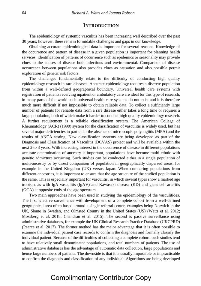
Richard A. Watts and Joanna Robson 64
INTRODUCTION
The epidemiology of systemic vasculitis has been increasing well described over the past
30 years; however, there remain formidable challenges and gaps in our knowledge.
Obtaining accurate epidemiological data is important for several reasons. Knowledge of
the occurrence and pattern of disease in a given population is important for planning health
services; identification of patterns of occurrence such as epidemics or seasonality may provide
clues to the causes of disease both infectious and environmental. Comparison of disease
occurrence between populations also provides clues as causation and also possible permit
exploration of genetic risk factors.
The challenges fundamentally relate to the difficulty of conducting high quality
epidemiology research in rare diseases. Accurate epidemiology requires a discrete population
from within a well-defined geographical boundary. Universal health care systems with
registration of patients receiving inpatient or ambulatory care are ideal for this type of research,
in many parts of the world such universal health care systems do not exist and it is therefore
much more difficult if not impossible to obtain reliable data. To collect a sufficiently large
number of patients for reliable data from a rare disease either takes a long time or requires a
large population, both of which make it harder to conduct high quality epidemiology research.
A further requirement is a reliable classification system. The American College of
Rheumatology (ACR) (1990) system for the classification of vasculitis is widely used, but has
several major deficiencies in particular the absence of microscopic polyangiitis (MPA) and the
results of ANCA testing. New classification systems are being developed as part of the
Diagnosis and Classification of Vasculitis (DCVAS) project and will be available within the
next 2 to 3 years. With increasing interest in the occurrence of disease in different populations
accurate determination of ancestry is important, populations have become multi-ethnic with
genetic admixture occurring. Such studies can be conducted either in a single population of
multi-ancestry or by direct comparison of population in geographically dispersed areas, for
example in the United Kingdom (UK) versus Japan. When comparing populations from
different ancestries, it is important to ensure that the age structure of the studied population is
the same. This is especially important for vasculitis, in which several types show a marked age
tropism, as with IgA vasculitis (IgAV) and Kawasaki disease (KD) and giant cell arteritis
(GCA) at opposite ends of the age spectrum.
Two main approaches have been used in studying the epidemiology of the vasculitides.
The first is active surveillance with development of a complete cohort from a well-defined
geographical area often based around a single referral center, examples being Norwich in the
UK, Skane in Sweden, and Olmsted County in the United States (US) (Watts et al. 2012;
Mossberg et al. 2018; Chandran et al. 2015). The second is passive surveillance using
administrative databases, for example the UK Clinical Research Practice Database (UKCPRD)
(Pearce et al. 2017). The former method has the major advantage that it is often possible to
examine the individual patient case records to confirm the diagnosis and formally classify the
individual patient. Because of the difficulties of collecting a complete cohort, such studies tend
to have relatively small denominator populations, and total numbers of patients. The use of
administrative databases has the advantage of automatic data collection, large populations and
hence large numbers of patients. The downside is that it is usually impossible or impracticable
to confirm the diagnosis and classification of any individual. Algorithms are being developed
Complimentary Contributor Copy
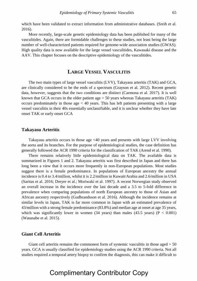
Epidemiology of Primary Systemic Vasculitis 65
which have been validated to extract information from administrative databases. (Sreih et al.
2016).
More recently, large-scale genetic epidemiology data has been published for many of the
vasculitides. Again, there are formidable challenges to these studies, not least being the large
number of well-characterized patients required for genome-wide association studies (GWAS).
High quality data is now available for the large vessel vasculitides, Kawasaki disease and the
AAV. This chapter focuses on the descriptive epidemiology of the vasculitides.
LARGE VESSEL VASCULITIS
The two main types of large vessel vasculitis (LVV), Takayasu arteritis (TAK) and GCA,
are clinically considered to be the ends of a spectrum (Grayson et al. 2012). Recent genetic
data, however, suggests that the two conditions are distinct (Carmona et al. 2017). It is well
known that GCA occurs in the older patient age > 50 years whereas Takayasu arteritis (TAK)
occurs predominately in those age < 40 years. This has left patients presenting with a large
vessel vasculitis in their 40s essentially unclassifiable, and it is unclear whether they have late
onset TAK or early onset GCA
Takayasu Arteritis
Takayasu arteritis occurs in those age <40 years and presents with large LVV involving
the aorta and its branches. For the purpose of epidemiological studies, the case definition has
generally followed the ACR 1990 criteria for the classification of TAK (Arend et al. 1990).
There remains relatively little epidemiological data on TAK. The available data is
summarized in Figures 1 and 2. Takayasu arteritis was first described in Japan and there has
long been a view that it occurs more frequently in non-European populations. Most studies
suggest there is a female predominance. In populations of European ancestry the annual
incidence is 0.4 to 3.4/million, whilst it is 2.2/million in Kuwait/Arabia and 2.6/million in USA
(Saritas et al. 2016; Dreyer et al.; Moriwaki et al. 1997). A recent Norwegian study observed
an overall increase in the incidence over the last decade and a 3.5 to 5-fold difference in
prevalence when comparing populations of north European ancestry to those of Asian and
African ancestry respectively (Gudbrandsson et al. 2016). Although the incidence remains at
similar levels in Japan, TAK is far more common in Japan with an estimated prevalence of
43/million with a strong female predominance (83.8%) and median age at onset at age 35 years,
which was significantly lower in women (34 years) than males (43.5 years) (P < 0.001)
(Watanabe et al. 2015).
Giant Cell Arteritis
Giant cell arteritis remains the commonest form of systemic vasculitis in those aged > 50
years. GCA is usually classified for epidemiology studies using the ACR 1990 criteria. Not all
studies required a temporal artery biopsy to confirm the diagnosis, this can make it difficult to
Complimentary Contributor Copy
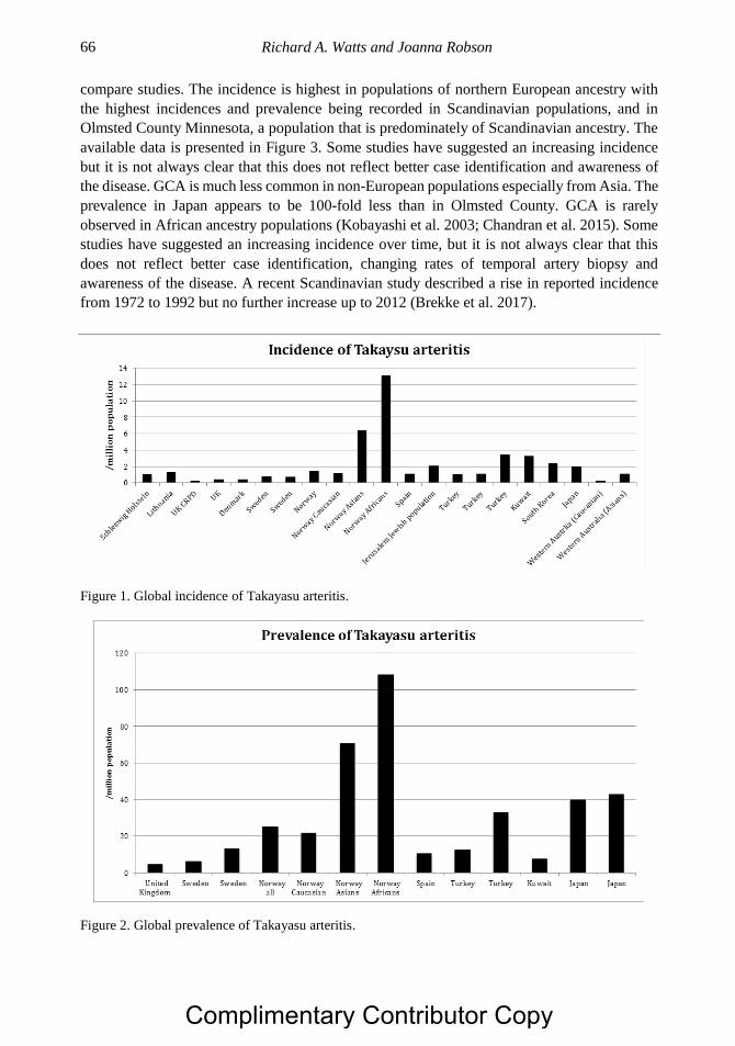
Richard A. Watts and Joanna Robson 66
compare studies. The incidence is highest in populations of northern European ancestry with
the highest incidences and prevalence being recorded in Scandinavian populations, and in
Olmsted County Minnesota, a population that is predominately of Scandinavian ancestry. The
available data is presented in Figure 3. Some studies have suggested an increasing incidence
but it is not always clear that this does not reflect better case identification and awareness of
the disease. GCA is much less common in non-European populations especially from Asia. The
prevalence in Japan appears to be 100-fold less than in Olmsted County. GCA is rarely
observed in African ancestry populations (Kobayashi et al. 2003; Chandran et al. 2015). Some
studies have suggested an increasing incidence over time, but it is not always clear that this
does not reflect better case identification, changing rates of temporal artery biopsy and
awareness of the disease. A recent Scandinavian study described a rise in reported incidence
from 1972 to 1992 but no further increase up to 2012 (Brekke et al. 2017).
Figure 1. Global incidence of Takayasu arteritis.
Figure 2. Global prevalence of Takayasu arteritis.
Complimentary Contributor Copy
Epidemiology of Primary Systemic Vasculitis 67
(Reproduced from Watts and Robson Best Practice & Research Clinical Rheumatology 2018, with
Permission).
Figure 3. Annual incidence of giant cell arteritis.
Environmental factors have long been thought to be important in the etiopathogenesis of
GCA. There is an inconsistent relation with season of onset, some studies reporting a summer
time preponderance but this has not been confirmed. Infection has also been suspected but no
clear link has emerged.
Genetic epidemiology studies have shown that HLA-DRB1*04 is the strongest risk factor
for GCA, and it has been suggested that variation in population HLA-DRB1*04 frequency may
partly explain variations in GCA incidence, with the incidence being higher in populations with
the greatest frequency of HLA-DRB1*04 (Mackie et al. 2015).
In conclusion there is still no good explanation for the striking age tropism shown by GCA,
immune senescence combined with a strong genetic background is the most likely explanation.
MEDIUM VESSEL VASCULITIS
Kawasaki Disease
Kawasaki disease is a medium sized vasculitis which predominately affects children age <
5 years, and is the predominant cause of childhood-acquired heart disease in the developed
world (Burns & Glode 2004). It was originally described by Kawasaki in Japan in the 1960 but
has been recognized as occurring in most populations, there are however, marked differences
in occurrence between populations. Populations of Asian ancestry have the highest incidence
of KD. The incidence of KD continues to increase in Japan in 2012, where the annual incidence
rates were 243.1/100,000 population aged 0 to 4 years in 2011 and 264.8 in 2012/100,000
(Makino et al. 2015a). In Japan, the cumulative incidence now means that by the age of 10
years 1.5/100 boys and 1.2/100 girls have been affected by KD (Nakamura et al. 2017). By
Complimentary Contributor Copy
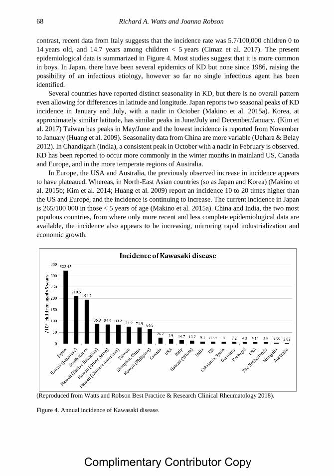
Richard A. Watts and Joanna Robson 68
contrast, recent data from Italy suggests that the incidence rate was 5.7/100,000 children 0 to
14 years old, and 14.7 years among children < 5 years (Cimaz et al. 2017). The present
epidemiological data is summarized in Figure 4. Most studies suggest that it is more common
in boys. In Japan, there have been several epidemics of KD but none since 1986, raising the
possibility of an infectious etiology, however so far no single infectious agent has been
identified.
Several countries have reported distinct seasonality in KD, but there is no overall pattern
even allowing for differences in latitude and longitude. Japan reports two seasonal peaks of KD
incidence in January and July, with a nadir in October (Makino et al. 2015a). Korea, at
approximately similar latitude, has similar peaks in June/July and December/January. (Kim et
al. 2017) Taiwan has peaks in May/June and the lowest incidence is reported from November
to January (Huang et al. 2009). Seasonality data from China are more variable (Uehara & Belay
2012). In Chandigarh (India), a consistent peak in October with a nadir in February is observed.
KD has been reported to occur more commonly in the winter months in mainland US, Canada
and Europe, and in the more temperate regions of Australia.
In Europe, the USA and Australia, the previously observed increase in incidence appears
to have plateaued. Whereas, in North-East Asian countries (so as Japan and Korea) (Makino et
al. 2015b; Kim et al. 2014; Huang et al. 2009) report an incidence 10 to 20 times higher than
the US and Europe, and the incidence is continuing to increase. The current incidence in Japan
is 265/100 000 in those < 5 years of age (Makino et al. 2015a). China and India, the two most
populous countries, from where only more recent and less complete epidemiological data are
available, the incidence also appears to be increasing, mirroring rapid industrialization and
economic growth.
(Reproduced from Watts and Robson Best Practice & Research Clinical Rheumatology 2018).
Figure 4. Annual incidence of Kawasaki disease.
Complimentary Contributor Copy
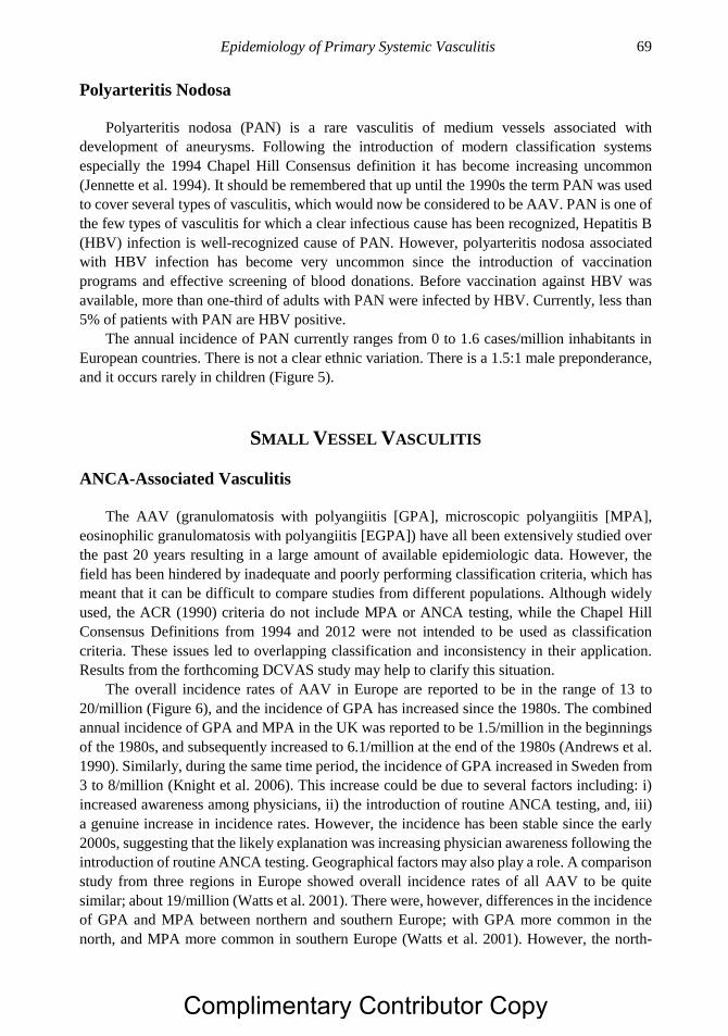
Epidemiology of Primary Systemic Vasculitis 69
Polyarteritis Nodosa
Polyarteritis nodosa (PAN) is a rare vasculitis of medium vessels associated with
development of aneurysms. Following the introduction of modern classification systems
especially the 1994 Chapel Hill Consensus definition it has become increasing uncommon
(Jennette et al. 1994). It should be remembered that up until the 1990s the term PAN was used
to cover several types of vasculitis, which would now be considered to be AAV. PAN is one of
the few types of vasculitis for which a clear infectious cause has been recognized, Hepatitis B
(HBV) infection is well-recognized cause of PAN. However, polyarteritis nodosa associated
with HBV infection has become very uncommon since the introduction of vaccination
programs and effective screening of blood donations. Before vaccination against HBV was
available, more than one-third of adults with PAN were infected by HBV. Currently, less than
5% of patients with PAN are HBV positive.
The annual incidence of PAN currently ranges from 0 to 1.6 cases/million inhabitants in
European countries. There is not a clear ethnic variation. There is a 1.5:1 male preponderance,
and it occurs rarely in children (Figure 5).
SMALL VESSEL VASCULITIS
ANCA-Associated Vasculitis
The AAV (granulomatosis with polyangiitis [GPA], microscopic polyangiitis [MPA],
eosinophilic granulomatosis with polyangiitis [EGPA]) have all been extensively studied over
the past 20 years resulting in a large amount of available epidemiologic data. However, the
field has been hindered by inadequate and poorly performing classification criteria, which has
meant that it can be difficult to compare studies from different populations. Although widely
used, the ACR (1990) criteria do not include MPA or ANCA testing, while the Chapel Hill
Consensus Definitions from 1994 and 2012 were not intended to be used as classification
criteria. These issues led to overlapping classification and inconsistency in their application.
Results from the forthcoming DCVAS study may help to clarify this situation.
The overall incidence rates of AAV in Europe are reported to be in the range of 13 to
20/million (Figure 6), and the incidence of GPA has increased since the 1980s. The combined
annual incidence of GPA and MPA in the UK was reported to be 1.5/million in the beginnings
of the 1980s, and subsequently increased to 6.1/million at the end of the 1980s (Andrews et al.
1990). Similarly, during the same time period, the incidence of GPA increased in Sweden from
3 to 8/million (Knight et al. 2006). This increase could be due to several factors including: i)
increased awareness among physicians, ii) the introduction of routine ANCA testing, and, iii)
a genuine increase in incidence rates. However, the incidence has been stable since the early
2000s, suggesting that the likely explanation was increasing physician awareness following the
introduction of routine ANCA testing. Geographical factors may also play a role. A comparison
study from three regions in Europe showed overall incidence rates of all AAV to be quite
similar; about 19/million (Watts et al. 2001). There were, however, differences in the incidence
of GPA and MPA between northern and southern Europe; with GPA more common in the
north, and MPA more common in southern Europe (Watts et al. 2001). However, the north-
Complimentary Contributor Copy
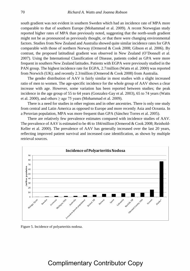
Richard A. Watts and Joanna Robson 70
south gradient was not evident in southern Sweden which had an incidence rate of MPA more
comparable to that of southern Europe (Mohammad et al. 2009). A recent Norwegian study
reported higher rates of MPA than previously noted, suggesting that the north-south gradient
might not be as pronounced as previously thought, or that there were changing environmental
factors. Studies from New Zealand and Australia showed quite similar incidence rates for GPA
comparable with those of northern Norway (Ormerod & Cook 2008; Gibson et al. 2006). By
contrast, the proposed latitudinal gradient was observed in New Zealand (O’Donnell et al.
2007). Using the International Classification of Disease, patients coded as GPA were more
frequent in southern New Zealand latitudes. Patients with EGPA were previously studied in the
PAN group. The highest incidence rate for EGPA, 2.7/million (Watts et al. 2000) was reported
from Norwich (UK); and recently 2.3/million (Ormerod & Cook 2008) from Australia.
The gender distribution of AAV is fairly similar in most studies with a slight increased
ratio of men to women. The age-specific incidence for the whole group of AAV shows a clear
increase with age. However, some variation has been reported between studies; the peak
incidence in the age group of 55 to 64 years (Gonzalez-Gay et al. 2003), 65 to 74 years (Watts
et al. 2000), and others ≥ age 75 years (Mohammad et al. 2009).
There is a need for studies in other regions and in other ancestries. There is only one study
from central and Latin America as opposed to Europe and more recently Asia and Oceania. In
a Peruvian population, MPA was more frequent than GPA (Sánchez Torres et al. 2005).
There are relatively few prevalence estimates compared with incidence studies of AAV.
The prevalence of AAV is estimated to be 46 to 184/million (Ormerod & Cook 2008; Reinhold-
Keller et al. 2000). The prevalence of AAV has generally increased over the last 20 years,
reflecting improved patient survival and increased case identification, as shown by multiple
retrieval sources.
Figure 5. Incidence of polyarteritis nodosa.
Complimentary Contributor Copy
Epidemiology of Primary Systemic Vasculitis 71
(Reproduced from Watts and Robson Best Practice & Research Clinical Rheumatology 2018, with
permission).
Figure 6. Annual incidence of ANCA-associated vasculitis.
The prevalence of GPA doubled in northern Germany from 58/million in 1994, to
98/million in 2006 (Herlyn et al. 2014). A study from an urban multi-ethnic population of Paris
showed a low prevalence of GPA of 23.7/million (Mahr et al. 2004). In Northern Europe, the
prevalence of GPA in Norway increased from 30.4/million in 1988, to 95.1/million in 1998
(Koldingsnes & Nossent 2000), and was 160/million in Sweden in 2003 (A. J. Mohammad et
al. 2007). The prevalence of GPA in the southern hemisphere was estimated at 112/million in
New Zealand and 95/million in Australia (Ormerod & Cook 2008; Gibson et al. 2006).
There is a paucity of data on the prevalence of MPA which varies from 25.1/million to
94/million. (Figure 7). As expected, the prevalence of EGPA is much lower than either GPA
or MPA, with the highest estimate being 45.7/million.
Genetic factors are clearly important as it has long been observed anecdotally that cases of
AAV are relatively rare in non-Caucasian populations. The first study to show differences in
the occurrence of AAV in different ethnic groups was reported from Paris, so suggesting that
GPA was less common in people of non-European ancestry than MPA (Mahr et al. 2004). A
study directly comparing two ethnically different populations, white UK Caucasian and the
Japanese showed that the overall similar incidence rate for AAV, but GPA and PR3-ANCA
vasculitis were much less common in Japan than in Europe (Fujimoto et al. 2011); whereas in
Japan, MPA and MPO-vasculitis were the dominant form of AAV compared to Latin America
(Peru) wherein MPA was more common than GPA (Sánchez Torres et al. 2005). In New
Zealand, GPA was twice as common in Europeans than Maoris or Asians (O’Donnell et al.
2007). Large case series from China suggest that MPA is more common than GPA (Liu et al.
2008). A multi ethnic series from Chapel Hill, US found that GPA was relatively uncommon
in African Americans (Cao et al. 2011). These differences may reflect global variation in the
frequency of the allele HLA DPB1*0401, which has been associated with GPA (Watts et al.
2014). The north-south gradient in Europe may also reflect genetic differences between
northern and southern Europeans (Tian et al. 2009). The clinical phenotype of systemic lupus
Complimentary Contributor Copy
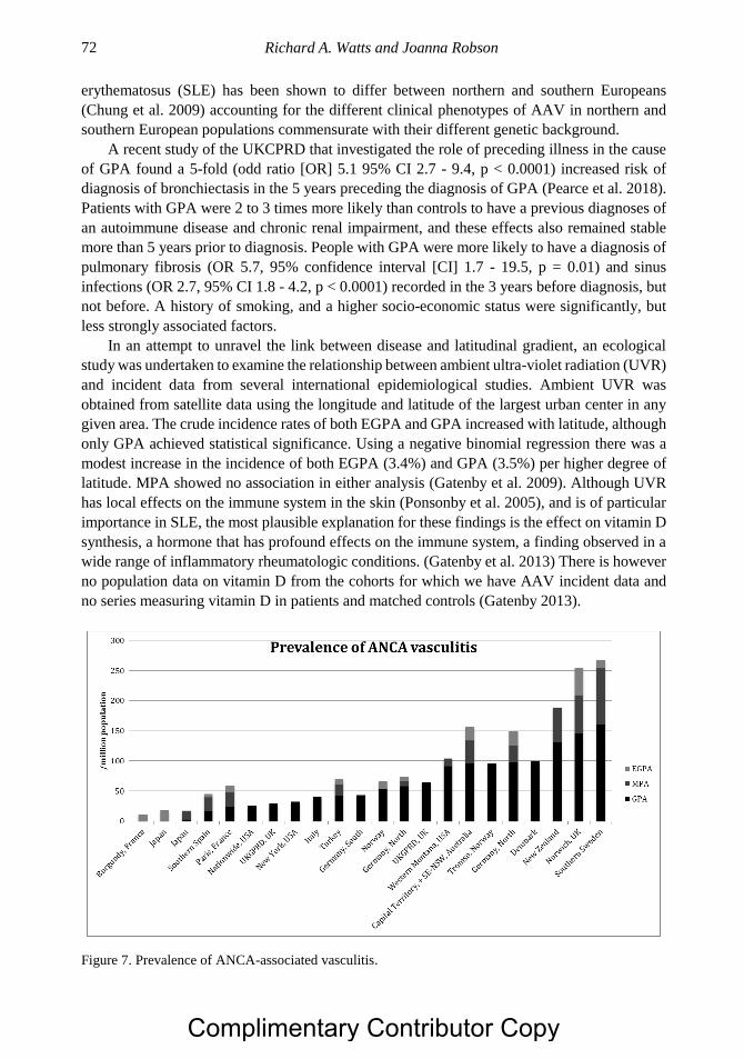
Richard A. Watts and Joanna Robson 72
erythematosus (SLE) has been shown to differ between northern and southern Europeans
(Chung et al. 2009) accounting for the different clinical phenotypes of AAV in northern and
southern European populations commensurate with their different genetic background.
A recent study of the UKCPRD that investigated the role of preceding illness in the cause
of GPA found a 5-fold (odd ratio [OR] 5.1 95% CI 2.7 - 9.4, p < 0.0001) increased risk of
diagnosis of bronchiectasis in the 5 years preceding the diagnosis of GPA (Pearce et al. 2018).
Patients with GPA were 2 to 3 times more likely than controls to have a previous diagnoses of
an autoimmune disease and chronic renal impairment, and these effects also remained stable
more than 5 years prior to diagnosis. People with GPA were more likely to have a diagnosis of
pulmonary fibrosis (OR 5.7, 95% confidence interval [CI] 1.7 - 19.5, p = 0.01) and sinus
infections (OR 2.7, 95% CI 1.8 - 4.2, p < 0.0001) recorded in the 3 years before diagnosis, but
not before. A history of smoking, and a higher socio-economic status were significantly, but
less strongly associated factors.
In an attempt to unravel the link between disease and latitudinal gradient, an ecological
study was undertaken to examine the relationship between ambient ultra-violet radiation (UVR)
and incident data from several international epidemiological studies. Ambient UVR was
obtained from satellite data using the longitude and latitude of the largest urban center in any
given area. The crude incidence rates of both EGPA and GPA increased with latitude, although
only GPA achieved statistical significance. Using a negative binomial regression there was a
modest increase in the incidence of both EGPA (3.4%) and GPA (3.5%) per higher degree of
latitude. MPA showed no association in either analysis (Gatenby et al. 2009). Although UVR
has local effects on the immune system in the skin (Ponsonby et al. 2005), and is of particular
importance in SLE, the most plausible explanation for these findings is the effect on vitamin D
synthesis, a hormone that has profound effects on the immune system, a finding observed in a
wide range of inflammatory rheumatologic conditions. (Gatenby et al. 2013) There is however
no population data on vitamin D from the cohorts for which we have AAV incident data and
no series measuring vitamin D in patients and matched controls (Gatenby 2013).
Figure 7. Prevalence of ANCA-associated vasculitis.
Complimentary Contributor Copy
Epidemiology of Primary Systemic Vasculitis 73
Silica appears to be a consistently identified risk factor among existing case-control studies
for both GPA and MPA. This is not surprising given its known association with a number of
other autoimmune syndromes (Steenland 2005). Commonly inhaled, the association with AAV
is observed even in the absence of respiratory tract disease so suggesting an impact beyond the
immediately exposed airways. Environmental dust was likely proposed as an important
determinant with an association to silica (Gómez-Puerta et al. 2013). Studies in Japan after two
large earthquakes suggested an increased incidence in the years immediately after earthquake
(Yashiro et al. 2000; Takeuchi et al. 2017). However, an increased incidence was not seen after
the Twin Towers attack in New York (Webber et al. 2015).
IMMUNE COMPLEX SMALL VESSEL VASCULITIS
IgA Vasculitis
IgA vasculitis or Henoch-Schönlein purpura, is an immune complex vasculitis that
predominantly affecting small vessels (Jennette et al. 2013). This common childhood systemic
vasculitis has the clinical characteristics of cutaneous palpable purpura, arthralgia/arthritis,
bowel angina, and hematuria/proteinuria. The annual incidence which ranges from 3.5 to
27.6/100,000 in children, is highest between the ages of 4 and 6 years (70/100,000) (Gardner-
Medwin et al. 2002). Two recent European studies reported similar incidences; a suburban area
of Paris described an annual incidence of 18.6/100 000 (13.6 -24.5) children, and 17.5/100,000
(16 - 19.1) in Sweden (Piram et al. 2017; Mossberg et al. 2018). In the French study the annual
distribution of diagnoses consistently showed a trough in summer months; 72% of children had
infectious symptoms (mainly upper respiratory tract) a few days before the onset of IgAV; and
23% had a North African background (Piram et al. 2017). The outcome of IgAV in children is
generally good with a low rate of development of renal failure.
IgA vasculitis is much less common in adults. Recent studies from Finland and Spain
suggest an incidence of about 1.4 to 1.5/million adults, whereas earlier studies from the UK
and Spain suggested an incidence up to 10-fold higher. It is not certain whether this represents
different case capture approaches or classification. Adult onset IgAV is more common in males
(63%) and has a mean age of onset of 50 years (Audemard-Verger et al. 2017).
Anti-Glomerular Basement Membrane Disease
Anti-glomerular basement membrane (GBM) disease or Goodpasture syndrome, is a
vasculitis affecting glomerular capillaries, pulmonary capillaries, or both, with basement
membrane deposition of anti-basement membrane autoantibodies. Lung involvement causes
pulmonary hemorrhage, and renal involvement causes glomerulonephritis with necrosis and
crescents. This autoimmune disorder presents mainly with rapidly progressive
glomerulonephritis and pulmonary hemorrhage in conjunction with increased serum levels of
anti-GBM antibodies. The target “Goodpasture antigen” is the non-collagenous (NC1) region
of the α3 chain of type IV collagen. Some patients also have ANCA present, leading to the
suggestion of an overlap with AAV. Authorities debate whether the disorder represents a true
Complimentary Contributor Copy
Richard A. Watts and Joanna Robson 74
vasculitis or a vasculopathy. An Irish study calculated the national incidence at 1.64/million
per year during the 11-year period of 2003 to 2014) (Canney et al. 2016).
Cryoglobulinemic Vasculitis
Cryoglobulinemic vasculitis (CV) is a rare form of immune complex vasculitis in which
circulating cryoglobulins are demonstrated in serum or tissue. There is an association with
myeloproliferative disorders, connective tissue diseases, in particular hepatitis C virus (HCV)
infection. The incidence and prevalence of CV vasculitis is unknown. The outcome is
dependent on the underlying condition. Modern antiviral therapies for HCV are highly effective
in curing the infection and consequently improving the prognosis of affected individuals.
Hypocomplementemic Urticarial Vasculitis
There is very little data on the epidemiology of hypocomplementemic urticarial vasculitis
(HUVS). A study from Sweden reported on 16 patients (14 women) all of whom were anti-
C1Q antibody positive during the years 2000 to 2015 and derived from a well-defined
population of 1,474,105. The annual incidence was 0.7/million with a point prevalence on 31
December 2015 of 9.5/million. There was no detectable age variation. The median age of onset
was 51 years. The 5-year survival was 92%, with a 10-year survival of 83% (Sjöwall et al.
2018).
OTHER VASCULITIDES
Behçet Disease
The epidemiology of Behçet disease (BD) is characterized by a striking geographic
variation with a greater incidence in an area spanning from China to the eastern Mediterranean
Sea, leading to the descriptive term, Silk Road disease. The majority of studies in BD have been
prevalence rather than incidence studies including many from countries where the health care
system is challenged to obtain accurate incidence figures. Two main methods have been used:
census surveys and sample surveys.
A recent meta-analysis of 45 population based surveys confirmed the geospatial variation
in the prevalence of BD (Maldini et al. 2018). The global overall prevalence was 10.3/100,000
inhabitants. In Turkey the pooled prevalence was 119.8/100,000 inhabitants, 31.8/100,000 in
the Middle East, 4.5/100,000 for Asia and 3.3/100,000 for Europe (Maldini et al. 2018). In
Europe, there was a north-south increase in prevalence, suggesting that BD in those of Northern
European ancestry. The case finding methodology has also strongly influenced the reported
figures, with sample surveys giving generally higher estimated of the population prevalence
compared census surveys; notwithstanding, the method employed to classify cases probably
does not influence prevalence estimates (Maldini et al. 2017).
Complimentary Contributor Copy
Epidemiology of Primary Systemic Vasculitis 75
The geospatial distribution of BD is strongly related to expression levels of HLA-B*51,
with prevalence being highest in Europe and the Middle and Far East, where HLA-B*51 is
found in > 15% of individuals, and the prevalence being lowest in Africa (Wallace 2014).
SECONDARY VASCULITIS
Rheumatoid Vasculitis
Rheumatoid vasculitis (RV) first became widely recognized and reported in the 1960s. In
the UK, the first estimate of the incidence was from Bath/Bristol in the 1970s and suggested an
incidence of 6/million (Scott et al. 1981). In Spain, the annual incidence of biopsy proven RV
during 1988–1997 was 6.4/million (González-Gay & García-Porrúa 1999). A detailed study
conducted in Norfolk, UK, between 1988 and 2010, compared the incidence of RV between
1988 and 2000 with the period 2001 to 2010 (Ntatsaki et al. 2014). The annual incidence
deceased from 9.1/million to 3.9/million. This decline occurred predominately during the late
1990s. The incidence in the Norfolk study was equal in both males and females. The decline in
RV has been attributed to better disease control especially with the much wider use of
methotrexate during the 1990s, and predates the common use of biologic drugs.
Several US studies have supported this decline in RV; a population based study of the
incidence of extra-articular RA reported a reduction in the 10 year cumulative incidence of RV
from 3.6% in 1985-1994 to 0.6% between 1995 and 2007 (Myasoedova et al. 2011). The
reduction of the prevalence of RV was also reported in a serial cross-sectional study of both
hospitalised and ambulatory patients from the US veteran population seen between 1985 and
2006, comprising a similar duration of cohort observation with our study of 22 years (Bartels
et al. 2009). There has also been a decline in hospitalisation in California of SRV patients
between 1980 and 2001 (Ward 2004).
CONCLUSION
The epidemiology of the vasculitides is much better understood now than was the case 20
- 30 years ago, however much of the data comes from populations of European descent. The
main exceptions to this are KD and BD, which have very clear variations in geographical
occurrence, for most other types of vasculitis it remains unclear how much geographical
variation occurs. These variation give clues to possible genetic differences. There is still
relatively little data from much of Asia especially the Indian subcontinent, Africa and Latin
America. Knowledge of environmental risk factors is also sparse for most of the vasculitides.
REFERENCES
Andrews, M., et al., (1990). Systemic vasculitis in the 1980s--is there an increasing incidence
of Wegener’s granulomatosis and microscopic polyarteritis? Journal of the Royal College
of Physicians of London, 24(4), pp. 284–8.
Complimentary Contributor Copy
Richard A. Watts and Joanna Robson 76
Arend, W. P., et al., (1990). The American College of Rheumatology 1990 criteria for the
classification of Takayasu arteritis. Arthritis and Rheumatism, 33(8), pp. 1129–34.
Audemard-Verger, A., et al., (2017). Characteristics and Management of IgA Vasculitis
(Henoch-Schönlein) in Adults: Data From 260 Patients Included in a French Multicenter
Retrospective Survey. Arthritis & Rheumatology, 69(9), pp. 1862–1870.
Bartels, C., et al., (2009). Decline in rheumatoid vasculitis prevalence among US veterans: a
retrospective cross-sectional study. Arthritis and Rheumatism, 60(9), pp. 2553–7.
Brekke, L. K., et al., (2017). Incidence of giant cell arteritis in Western Norway 1972–2012: a
retrospective cohort study. Arthritis Research & Therapy, 19(1), p. 278.
Burns, J. C. & Glode, M. P. (2004). Kawasaki syndrome. Lancet, 364(9433), pp. 533–544.
Canney, M., et al., (2016). Spatial and Temporal Clustering of Anti-Glomerular Basement
Membrane Disease. Clinical Journal of the American Society of Nephrology, 11(8), pp.
1392–1399.
Cao, Y., et al., (2011). DRB1*15 allele is a risk factor for PR3-ANCA disease in African
Americans. Journal of the American Society of Nephrology: JASN, 22(6), pp. 1161–7.
Carmona, F. D., et al., (2017). Analysis of the common genetic component of large-vessel
vasculitides through a meta-Immunochip strategy. Scientific Reports, 7, p. 43953.
Chandran, A., et al., (2015). The incidence of giant cell arteritis in Olmsted County, Minnesota,
over a 60-year period 1950-2009. Scandinavian Journal of Rheumatology, pp. 1–14.
Chung, S. A., et al., (2009). European population substructure is associated with
mucocutaneous manifestations and autoantibody production in systemic lupus
erythematosus. Arthritis and Rheumatism, 60(8), pp. 2448–56.
Cimaz, R., et al., (2017). Epidemiology of Kawasaki disease in Italy: surveillance from national
hospitalization records. European Journal of Pediatrics, 176(8), pp. 1061–1065.
Dreyer, L., Faurschou, M. & Baslund, B. A population-based study of Takayasu´s arteritis in
eastern Denmark. Clinical and Experimental Rheumatology, 29, (1 Suppl 64), pp. S40-2.
Fujimoto, S., et al., (2011). Comparison of the epidemiology of anti-neutrophil cytoplasmic
antibody-associated vasculitis between Japan and the U.K. Rheumatology (Oxford,
England), 50(10), pp. 1916–20.
Gardner-Medwin, J. M. M., et al., (2002). Incidence of Henoch-Schönlein purpura, Kawasaki
disease, and rare vasculitides in children of different ethnic origins. Lancet, 360(9341), pp.
1197–202.
Gatenby, P. (2013). The role of environmental factors in the pathogenesis of anti-neutrophil
antibody assocaited vasculitis.e. Immunome Research, 9(1), pp. 1–7.
Gatenby, P., Lucas, R. & Swaminathan, A. (2013). Vitamin D deficiency and risk for rheumatic
diseases: an update. Current Opinion in Rheumatology, 25(2), pp. 184–91.
Gatenby, P. A., et al., (2009). Antineutrophil cytoplasmic antibody-associated vasculitides:
could geographic patterns be explained by ambient ultraviolet radiation? Arthritis and
Rheumatism, 61(10), pp. 1417–24.
Gibson, A., et al., (2006). The epidemiology of Wegener’s granulomatosis and microscopic
polyangiitis in a Southern Hemisphere region. Rheumatology (Oxford, England), 45(5), pp.
624–8.
Gómez-Puerta, J. A., Gedmintas, L. & Costenbader, K. H. (2013). The association between
silica exposure and development of ANCA-associated vasculitis: systematic review and
meta-analysis. Autoimmunity Reviews, 12(12), pp. 1129–35.
Complimentary Contributor Copy
Epidemiology of Primary Systemic Vasculitis 77
Gonzalez-Gay, M. a., et al., (2003). The epidemiology of the primary systemic vasculitides in
northwest Spain: implications of the Chapel Hill Consensus Conference definitions.
Arthritis and Rheumatism, 49(3), pp. 388–93.
González-Gay, M. A. & García-Porrúa, C. (1999). Systemic vasculitis in adults in northwestern
Spain, 1988-1997. Clinical and epidemiologic aspects. Medicine, 78(5), pp. 292–308.
Grayson, P. C., et al., (2012). Distribution of arterial lesions in Takayasu’s arteritis and giant
cell arteritis. Annals of the Rheumatic Diseases, 71(8), pp. 1329–34.
Gudbrandsson, B., et al., (2016). Prevalence, incidence and disease characteristics of Takayasu
Arteritis differ by ethnic background; data from a large, population based cohort resident
in Southern Norway. Arthritis Care & Research.
Herlyn, K., et al., (2014). Doubled prevalence rates of ANCA-associated vasculitides and giant
cell arteritis between 1994 and 2006 in northern Germany. Rheumatology (Oxford,
England), 53(5), pp. 882–889.
Huang, W. C., et al., (2009). Epidemiologic Features of Kawasaki Disease in Taiwan, 2003-
2006. Pediatrics, 123(3), pp. e401–e405.
Jennette, J., et al., (2013). 2012 Revised International Chapel Hill Consensus Conference
Nomenclature of Vasculitides. Arthritis and Rheumatism, 65, pp. 1–11.
Jennette, J. C., et al., (1994). Nomenclature of systemic vasculitides. Proposal of an
international consensus conference. Arthritis and Rheumatism, 37(2), pp. 187–92.
Kim, G. B., et al., (2014). Epidemiologic Features of Kawasaki Disease in South Korea. The
Pediatric Infectious Disease Journal, 33(1), pp. 24–27.
Kim, G. B., et al., (2017). Epidemiology and Clinical Features of Kawasaki Disease in South
Korea, 2012–2014. The Pediatric Infectious Disease Journal, 36(5), pp. 482–485.
Knight, A., et al., (2006). Increasing incidence of Wegener’s granulomatosis in Sweden, 1975-
2001. The Journal of Rheumatology, 33(10), pp. 2060–3.
Kobayashi, S., et al., (2003). Clinical and epidemiologic analysis of giant cell (temporal)
arteritis from a nationwide survey in 1998 in Japan: the first government-supported
nationwide survey. Arthritis and Rheumatism, 49(4), pp. 594–8.
Koldingsnes, W. & Nossent, H. (2000). Epidemiology of Wegener’s granulomatosis in
northern Norway. Arthritis and Rheumatism, 43(11), pp. 2481–7.
Liu, L. J., et al., (2008). Evaluation of a new algorithm in classification of systemic vasculitis.
Rheumatology (Oxford, England), 47(5), pp. 708–12.
Mackie, S. L., et al., (2015). Association of HLA-DRB1 amino acid residues with giant cell
arteritis: genetic association study, meta-analysis and geo-epidemiological investigation.
Arthritis Research & Therapy, 17(1), p. 195.
Mahr, A., et al., (2004). Prevalences of polyarteritis nodosa, microscopic polyangiitis,
Wegener’s granulomatosis, and Churg-Strauss syndrome in a French urban multiethnic
population in 2000: a capture-recapture estimate. Arthritis and Rheumatism, 51(1), pp. 92–
9.
Makino, N., et al., (2015a). Descriptive epidemiology of Kawasaki disease in Japan, 2011-
2012: from the results of the 22nd nationwide survey. Journal of epidemiology/Japan
Epidemiological Association, 25(3), pp. 239–45.
Makino, N,. et al., (2015b). Descriptive Epidemiology of Kawasaki Disease in Japan, 2011-
2012: From the Results of the 22nd Nationwide Survey. Journal of Epidemiology, 25(3),
pp. 239–245.
Complimentary Contributor Copy
Richard A. Watts and Joanna Robson 78
Maldini, C., et al., (2018). Exploring the variability in Behçet’s disease prevalence: a meta-
analytical approach. Rheumatology, 57(1), pp. 185–195.
Mohammad, a. J., et al., (2007). Prevalence of Wegener’s granulomatosis, microscopic
polyangiitis, polyarteritis nodosa and Churg-Strauss syndrome within a defined population
in southern Sweden. Rheumatology (Oxford, England), 46(8), pp. 1329–37.
Mohammad, A. J., et al., (2009). Incidence and survival rates in Wegener’s granulomatosis,
microscopic polyangiitis, Churg-Strauss syndrome and polyarteritis nodosa. Rheumatology
(Oxford, England), 48(12), pp. 1560–5.
Moriwaki, R., et al., (1997). Clinical manifestations of Takayasu arteritis in India and Japan--
new classification of angiographic findings. Angiology, 48(5), pp. 369–79.
Mossberg, M., et al., (2018). Epidemiology of primary systemic vasculitis in children: a
population-based study from southern Sweden. Scandinavian Journal of Rheumatology,
pp. 1–8.
Myasoedova, E., et al., (2011). Incidence of extraarticular rheumatoid arthritis in Olmsted
County, Minnesota, in 1995-2007 versus 1985-1994: a population-based study. The
Journal of Rheumatology, 38(6), pp. 983–9.
Nakamura, Y., et al., (2018). Cumulative incidence of Kawasaki disease in Japan. Pediatrics
International, 60(1), pp 19-22
Ntatsaki, E., et al., (2014). Systemic rheumatoid vasculitis in the era of modern
immunosuppressive therapy. Rheumatology (Oxford, England), 53(1), pp. 145–52.
O’Donnell, J. L., et al., (2007). Wegener’s granulomatosis in New Zealand: evidence for a
latitude-dependent incidence gradient. Internal Medicine Journal, 37(4), pp. 242–6.
Ormerod, A. S. & Cook, M. C. (2008). Epidemiology of primary systemic vasculitis in the
Australian Capital Territory and south-eastern New South Wales. Internal medicine
journal, 38(11), pp. 816–23.
Pearce, F. A., et al., (2018). Novel insights into the aetiology of granulomatosis with
polyangiitis—a case–control study using the Clinical Practice Research Datalink.
Rheumatology, 57(10), pp 1002-10.
Pearce, F. A., et al., (2017). The incidence, prevalence and mortality of granulomatosis with
polyangiitis in the UK Clinical Practice Research Datalink. Rheumatology (United
Kingdom), 56(4), pp 589-96.
Piram, M., et al., (2017). Incidence of IgA vasculitis in children estimated by four-source
capture–recapture analysis: a population-based study. Rheumatology., 56(8), pp 1358-66.
Ponsonby, A. L., Lucas, R. M. & van der Mei, I. A. F. (2005). UVR, vitamin D and three
autoimmune diseases--multiple sclerosis, type 1 diabetes, rheumatoid arthritis.
Photochemistry and photobiology, 81(6), pp. 1267–75.
Reinhold-Keller, E., et al., (2000). Giant cell arteritis is more prevalent in urban than in rural
populations: results of an epidemiological study of primary systemic vasculitides in
Germany. Rheumatology (Oxford, England), 39(12), pp. 1396–402.
Sánchez Torres, A., et al., (2005). Epidemiología de las vasculitis sistémicas primarias en una
población latinoamericana. [Epidemiology of primary systemic vasculitis in a Latin
American population.] Reumatología, 21, pp. 145–150.
Saritas, F., et al., (2016). The epidemiology of Takayasu arteritis: a hospital-based study from
northwestern part of Turkey. Rheumatology international.
Scott, D. G., Bacon, P. A. & Tribe, C. R. (1981). Systemic rheumatoid vasculitis: a clinical and
laboratory study of 50 cases. Medicine, 60(4), pp. 288–97.
Complimentary Contributor Copy
Epidemiology of Primary Systemic Vasculitis 79
Sjöwall, C., et al., (2018). Epidemiology of hypocomplementaemic urticarial vasculitis (anti-
C1q vasculitis). Rheumatology, 57(8), pp 1400-7.
Sreih, A. G., et al., (2016). Development and validation of case-finding algorithms for the
identification of patients with anti-neutrophil cytoplasmic antibody-associated vasculitis in
large healthcare administrative databases. Pharmacoepidemiology and Drug Safety,
25(12), pp. 1368–1374.
Steenland, K. (2005). One agent, many diseases: exposure-response data and comparative risks
of different outcomes following silica exposure. American Journal of Industrial Medicine,
48(1), pp. 16–23.
Takeuchi, Y., et al., (2017). The influence of the Great East Japan earthquake on microscopic
polyangiitis: A retrospective observational study H. Abe, ed. PLOS ONE, 12(5), p.
e0177482.
Tian, C., et al., (2009). European population genetic substructure: further definition of ancestry
informative markers for distinguishing among diverse European ethnic groups. Molecular
Medicine (Cambridge, Mass.), 15(11–12), pp. 371–83.
Uehara, R. & Belay, E. D. (2012). Epidemiology of Kawasaki disease in Asia, Europe, and the
United States. Journal of Epidemiology, 22(2), pp. 79–85.
Wallace, G. R. (2014). HLA-B*51 the primary risk in Behçet disease. Proceedings of the
National Academy of Sciences of the United States of America, 111(24), pp. 8706–7.
Ward, M. M. (2004). Decreases in rates of hospitalizations for manifestations of severe
rheumatoid arthritis, 1983-2001. Arthritis and Rheumatism, 50(4), pp. 1122–31.
Watanabe, Y., Miyata, T. & Tanemoto, K. (2015). Current Clinical Features of New Patients
with Takayasu Arteritis Observed From a Cross-Country Research in Japan: Age and Sex
Specificity. Circulation, p. CIRCULATIONAHA.114.012547.
Watts, R. A., et al., (2000). Epidemiology of systemic vasculitis: a ten-year study in the United
Kingdom. Arthritis and Rheumatism, 43(2), pp. 414–9.
Watts, R. A., et al., (2001). Epidemiology of vasculitis in Europe. Annals of the Rheumatic
Diseases, 60(12), pp. 1156–7.
Watts, R. A., et al., (2012). The contrasting epidemiology of granulomatosis with polyangiitis
(Wegener’s) and microscopic polyangiitis. Rheumatology (Oxford, England), 51(5), pp.
926–31.
Watts, R. A., Mackie, S. L. & Macgregor, A. J. (2014). HLA-DPB alleles and granulomatosis
with polyangiitis. Rheumatology, 53, suppl 1, pp. i186-7.
Watts, R.A., and Robson, J. (2018). Introduction, Epidemiology and classification of vasculitis.
Best Prac Res Clin Rheumatol 2018; 32(1), pp 3-20.
Webber, M. P., et al., (2015). Nested Case-Control Study of Selected Systemic Autoimmune
Diseases in World Trade Center Rescue/Recovery Workers. Arthritis Rheumatology
(Hoboken, N.J.), p.n/a-n/a.
Yashiro, M., et al., (2000). Significantly high regional morbidity of MPO-ANCA-related
angitis and/or nephritis with respiratory tract involvement after the 1995 great earthquake
in Kobe (Japan). American journal of kidney diseases : the official journal of the National
Kidney Foundation, 35(5), pp. 889–95.
Complimentary Contributor Copy

Complimentary Contributor Copy
In: The Vasculitides. Volume 1 ISBN: 978-1-53615-133-6
Editor: David S. Younger © 2019 Nova Science Publishers, Inc.
Chapter 4
HEALTH RELATED QUALITY OF LIFE AND ITS
MEASUREMENT IN THE VASCULITIDES
Joanna Robson, PhD1,, and Richard A. Watts, MD, MPH2,† 1Faculty of Health and Applied Sciences, University of the West of England,
Bristol, and University Hospitals Bristol NHS Trust, Bristol, UK 2Nuffield Department of Orthopaedics, Rheumatology and Musculoskeletal Sciences,
University of Oxford, Oxford, UK, and Norwich Medical School,
University of East Anglia, Norwich, UK
ABSTRACT
Patient reported outcomes (PRO) using mainly the generic Short-Form 36 have been
studied in diverse systemic vasculitides, in particular in patients with anti neutrophil
cytoplasmic antibody–associated vasculitis (AAV), who report significant impairment in
their health-related quality of life (HRQOL). Patients report different priorities regarding
disease assessment compared with their physicians and this should be captured in clinical
studies. The Outcome Measures in Rheumatology (OMERACT) Vasculitis Working
Group, has gained endorsement by OMERACT for use of a disease-specific PRO, the
AAV-PRO and generic Patient Reported Outcome Measurement Information System
(PROMIS) instruments in clinical trials of vasculitis. Generic and disease-specific
instruments are complimentary to each other and require further study to assess their utility
in longitudinal settings, including their ability to distinguish between treatments of varying
efficacy in randomized clinical trials (RCTs). This chapter reviews HRQOL measures in
systemic vasculitis further separable by the caliber of vessels involved.
INTRODUCTION
Systemic vasculitis can be organ and life-threatening [1-3]. International randomized
controlled trials with standardized outcome measurement have revolutionized their treatment
Corresponding Author’s Email: [email protected]. † Corresponding Author’s Email: [email protected].
Complimentary Contributor Copy
Joanna Robson and Richard A. Watts 82
[4-6]. Irreversible damage, ongoing disease activity and adverse effects of medications can all
impact on Health-related quality of life (HRQoL) scores [7-9]. This chapter describes HRQoL
and its measurement in systemic vasculitis.
CLASSIFICATION OF VASCULITIDES
The Revised 2012 International Chapel Hill Consensus Conference established distinct
categories for the classification of vasculitis based upon the caliber vessels involved [10]. Large
vessel vasculitis (LVV) including giant cell arteritis (GCA) and Takayasu arteritis (TAK)
affects the aorta, its major branches and analogous veins. Medium vessel vasculitis (MVV)
inclusive of polyarteritis nodosa (PAN) and Kawasaki disease (KD), involves main visceral
arteries and veins and initial branches. Small vessel vasculitis (SVV) involvement affects
intraparenchymal arteries, arterioles, capillaries, veins and venules, with disease mechanisms
related to anti-neutrophil cytoplasmic antibody (ANCA) or immune complexes. The group of
AAV includes granulomatosis with polyangiitis (GPA), microscopic polyangiitis (MPA), and
eosinophilic granulomatosis with polyangiitis (EGPA). The subgroup of variable vessel
vasculitis (VVV) includes Behçet disease (BD).
BACKGROUND
Measurement of HRQoL scores in systemic vasculitis has historically been based on
generic patient reported outcomes (PROs), mainly the Short-Form 36 (SF-36) [11]. The
benefits of the SF-36 is that it is a well- recognized and validated outcome measure [11] and
allows comparison between patients with systemic vasculitis and other conditions. Limitations
in the use of generic PROs can include a reduction in face and content validity, as they have
not been designed to capture areas of most interest to patients with that specific disease [12].
This lack of specificity reduces the ability to detect differences in state between patients and in
the same patient over time [12]. In GCA, the SF-36 does not detect differences between patients
with and without visual loss or systemic involvement for example [13]. Trials in AAV fail to
show difference in SF-36 scores between arms, despite use of less toxic medications such as
cyclophosphamide versus rituximab [14]. Whether this is a lack of sensitivity with the SF-36
or due to glucocorticoid effects in the arms remains unanswered. The difference in HRQoL
scores as measured by the SF-36 in a randomized controlled trial (RCT) of tocilizumab in GCA,
which may be attributable to the reduction in glucocorticoid-related adverse effects in the
tocilizumab only arm [15], requires further analysis.
The Outcome Measurement in Rheumatology (OMERACT) initiative is an international
collaboration to define core sets of outcome measurements for use in RCTs [16]. Working
groups include researchers, clinicians, patient partners, and methodologists; other stakeholders
including the Food and Drug Administration (FDA) and representatives from pharmaceutical
companies participate in the workshops [16]. The Vasculitis Working Group published a core
set of domains and outcome measures for use in clinical trials in AAV in 2011 [17], and LVV
in 2017 [18] and a core-set of domains for BD at OMERACT 2018. Due to the appreciation
that patients and their physicians commonly rank aspects of their disease in different orders
Complimentary Contributor Copy
Health Related Quality of Life and its Measurement in the Vasculitides 83
[19], groups have recently been focused on the patient perspective in systemic vasculitis. This
has included the development of a new disease-specific PRO, the AAV-PRO [20],
underpinning qualitative work in TAK and BD [21, 22] and evaluation of alternative generic
PROs such as the Patient Reported Outcome Measurement Information System (PROMIS)
[23].
Disease-specific PROs developed with patient involvement at every stage are in line with
guidance from the FDA on the development of PROs [24]. They should be based on
underpinning qualitative research with patients and the specific disease to identify the full range
of impacts of the disease and its treatment, ensuring good face and content validity [25].
Themes rewritten as candidate questionnaire items, including stems and response categories,
and corresponding questionnaire items are then piloted and tested within cognitive interviews
to check readability and understanding [26]. Large scale testing within a survey is then
performed, using statistical analyses such as Exploratory Factor analysis [27] and Rasch
analysis [28] to confirm the ideal structure of the PROM and validate its measurement
properties [20, 29].
Generic and disease-specific PROs are complimentary [30] and can be used together, and
adapted to the specific context of the study. The growing recognition of the importance of PROs
in the assessment of vasculitis, and the availability of validated instruments to capture PROs in
vasculitis, will likely mean patients’ perspectives will also be incorporated into composite
outcome measures in future trials.
SMALL VESSEL VASCULITIS
ANCA-Associated Vasculitis
Vasculitic involvement in the subgroup of AAV variably effects the kidneys, lungs,
nervous system, skin, ears nose and throat, and eyes, resulting in intrusive symptoms, and
irreversible damage and impact on HRQoL [2, 31].
The generic SF-36 has been used in clinical studies of patients with AAV and is a core
outcome measure listed within the 2010 OMERACT core set [17]. Data from four European
RCTs of newly diagnosed patients with AAV demonstrated impairment in HRQoL at entry
[32]. Physical functioning scores were the most affected, particularly in those with neurological
involvement and of older age [33]. A Scottish case-control study demonstrated that patients
with AAV were twice as likely to have fatigue than matched controls, with levels of fatigue
strongly associated with impairment in physical function [34]. Survey data suggests that AAV-
related fatigue is multifactorial, with associations seen with increasing levels of pain, sleep
disturbance and inflammation [35]. Fatigue was ranked by patients as being of greatest
importance to their HRQoL, in contrast to physicians, who ranked being dialysis or oxygen
dependent as more important [19].
The OMERACT Vasculitis Group has identified the lack of a disease-specific PRO for
AAV [36]. An international collaboration of researchers and patients has overseen the
development of a new PRO to fulfill this unmet need. Qualitative interviews with 50 patients
with AAV from the United Kingdom (UK), United States (US) and Canada identified the
following domains of interest: symptom severity, and the impact of problems and limitations
Complimentary Contributor Copy
Joanna Robson and Richard A. Watts 84
imposed by patients’ ANCA-associated vasculitis and treatment on their work and domestic
roles, family and social interactions, including activities and interests outside the home; and
psychological state [31]. Themes used as underpinning data to develop candidate items for the
new PRO and tested using cognitive interviewing and psychometric evaluation to create the
final validated PRO [20].
The AAV-PRO 29-item questionnaire has six subscales/domains: “Organ-Specific
Symptoms,” “Systemic Symptoms,” “Treatment Side Effects,” “Social and Emotional Impact,”
“Concerns about the Future,” and “Physical Function.” The domains offer a comprehensive
profile of the impact of AAV and its treatment on patients’ everyday life [20].
AAV-PRO domain scores distinguish between patients who self-report active disease
versus disease in remission, and therefore can be used as an outcome measure to define
treatment success from the patient’s perspective [20]. Scores for each separate domain are
examined separately to provide a profile of the overall impact of the disease and its treatment
on HRQoL. A greater focus is placed on pre-specified domains depending on the aims of the
trial. For example, in a non-inferiority trial of a glucocorticoid-sparing agent, the treatment
domain of the AAV-PRO might be key, but other investigators may instead wish to collect and
analyze all domain scores to examine any differences across the symptoms and HRQoL
domains. Further work is necessary to determine whether summary component scores are
applicable to AAV-PRO.
Women scores are higher (i.e., worse) on all six subscales of the AAV-PRO [20] and in
other conditions [37, 38]. Such trends in women were previously been reported in AAV [32].
Younger people (<65) scored higher (worse) on the Social and Emotional Impact subscale of
the AAV-PRO, a trend also seen in other chronic diseases in this age group, potentially due to
feeling different to peers, being unable to achieve ambitions and uncertainty at key stages of
usual transition into adulthood [37, 39]. Younger age is a possible risk factor for negative illness
perceptions in AAV, and increased levels of fatigue [40].
The PROMIS, which is a collection of generic item banks of self-reported health drawn
originally from different diseases, in relation to fatigue and physical function, has been
examined in patients with GCA, and appears feasible, with scores correlating with relevant SF-
36 domain scores [23]. Further testing of different PROMIS item banks across other HRQoL
domains within the systemic vasculitides is ongoing.
LARGE VESSEL VASCULITIS
Takayasu Arteritis
The symptoms of TAK are attributable to involvement of the aorta, its major branches, and
the pulmonary arteries [41]. Constitutional symptoms related to systemic disease include
weight loss, fever and fatigue; while those associated with vascular inflammation and occlusion
lead to pain, claudication and tissue loss [41].
Complimentary Contributor Copy
Health Related Quality of Life and its Measurement in the Vasculitides 85
Health-related quality of life examined in Turkish patients via generic SF-36 showed worse
scores in patients with TAK than healthy controls, in line with scores from patients with
ankylosing spondylitis and rheumatoid arthritis [42]. A patient survey in the US identified an
association between use of immunosuppressant medications and worse HRQoL probably due
to severity of disease, while younger patients and those in remission had better HRQoL [7].
Patients with TAK have higher levels of anxiety and depression than healthy controls as well
as physical limitations [43].
A review by the OMERACT Large Vessel Vasculitis Working Group identified the lack
of a disease–specific PRO for TAK [44]. Qualitative research, including individual interviews
and focus groups in the US and Turkey have identified domains of importance to patients with
TAK [21]. Salient themes include, “Pain and Discomfort,” “Fatigue and Low Energy Levels”
and “Emotional Effects,” that could underpin the future development of a disease-specific PRO
[21].
Giant Cell Arteritis
Giant cell arteritis is associated with inflammation of cranial and neck vessels [45] and is
a medical emergency due to the risk of blindness in 20% [46, 47]. Prompt treatment with high
dose glucocorticoids (GC) is needed to protect vision [48, 49]. Presenting features include vice-
like headache, jaw claudication, flu-like symptoms and inflammatory pain and stiffness in the
shoulders and hips (polymyalgia rheumatica, present in 50%) [47]. For 60 years GC alone have
been the mainstay of treatment, but patients are concerned about the risks [50-53]. Patient report
the psychological impact of weighing up the necessity of treatment with GCs to prevent
blindness, versus fears about adverse effects [50]. The use of novel biologic medications such
as tocilizumab versus standard GC regimens, does appear to benefit patients in terms of their
HRQoL at one year [15], and this should be examined further.
Qualitative research in the UK has revealed the impact of GCA on patients' lives is due to
their symptoms (e.g., pain, visual disturbance and musculoskeletal symptoms), adverse effects
of glucocorticoids, and the disruption to normal life [54]. Patients fear blindness and have
concerns about delay in diagnosis [54]. Patients with GCA ranked “losing sight in both eyes
permanently,” “having intense or severe pain” and “feeling weak, tired or exhausted” as the
most important domains of health-related quality of life [55].
The OMERACT Vasculitis Working Group has highlighted the development of a disease-
specific PROM for GCA as one of the next steps in their research agenda [18, 56]. Generic
PROs alone may not be specific enough for use in GCA. SF-36 scores in patients with GCA do
not correlate with visual loss or systemic complications, so may be unable to differentiate
between clinically important groups [13, 57].
Underpinning qualitative work with patients in the UK and Australia was presented,
revealing themes including: “Anxieties around getting a diagnosis of GCA,” “Description of
symptoms related to GCA and its treatment,” “Lack of bodily strength, stability and stamina;
difficulties with completing daily tasks,” “Difficulties with participating in social activities,
work and caring roles,” “Not feeling normal and impact on general perception of health,”
“Anxiety and fear of the future” [58]. Themes are being developed further into candidate
questionnaire items for a disease specific PRO for GCA.
Complimentary Contributor Copy
Joanna Robson and Richard A. Watts 86
VARIABLE VESSEL VASCULITIS
Behçet Disease
Behçet disease is a variable vessel vasculitis affecting veins and arteries of different sizes
[10]. Male and female patients present in young adulthood with a spectrum of different disease
manifestations [59]. Organ involvement can include mucocutaneous (such as oral and genital
ulcers, nodular and papulopustular skin lesions), ophthalmological (panuveitus),
musculoskeletal (usually monoarthritis or arthralgia), neurological (from parenchymal stroke-
like syndromes, to cerebral venous thrombosis [60]) and/or gastro-intestinal involvement
(which can present in a similar to inflammatory bowel disease) [59].
Patients with BD have worse SF-36 scores compared with healthy controls, particularly in
women and those with high disease activity [61]. Mucucutaneous (oral and genital ulcers),
central nervous system, musculoskeletal and ocular involvement are all independent predictors
of worse HRQoL [61]. Physical functioning is most impaired in panuveitus compared with
other ocular presentations [61]. Sexual function can be impaired in men and women [62].
Patients reporting high activity in relation to their joints, oral ulcers and fatigue, scored highly
on the physical domains of the Nottingham Health Profile (NHP); joint and oral ulcer activity
was also correlated with high psychosocial subscale NHP scores [8].
A systematic review of outcome measures used in BD by the OMERACT Vasculitis
Working Group revealed a lack of standardization and large variability in terms of outcomes
used across trials [63].
There is a validated disease specific HRQoL measure, namely, the BD-QoL scale [29, 64-
66] which has undergone cross-cultural adaptation and validation in Korean and Arabic [64,
67]. Item development was based on underpinning qualitative work with patients with BD in
the UK, and included the following salient themes: “Relationships,” “Emotions,” “Limitations
in Day to Day Activities” and “Self-Image” [29].
Generic measures to evaluate HRQOL in trials in BD have largely been the SF-36 [68] and
the EQ-5D [63]. A range of different symptom-specific tools have been used, including the
Oral Health and Related Quality of Life scale, which has been validated in Turkey and the UK
in BD [69]. Symptoms specific tools to measure impact on Sexual Function (e.g., the Arizona
Sexual Experience Scale [62] and Sleep (e.g., the Pittsburgh Sleep Quality Index [70]) have
also been used, but not in a systematic way across trials [63]. Psychological impact has most
commonly been measured within trials of BD using the Beck Depression Index [71] and Beck
Anxiety Scale [66, 72].
The OMERACT Vasculitis Working Group continues to work towards standardized
assessment of BD within clinical trials [22]. A new core set of outcome domains for BD,
including HRQoL as a core domain, was ratified at OMERACT 2018. The core domain set will
be published this year; next steps within the OMERACT process will be to evaluate and
recommend appropriate tools with which to measure these domains, including HRQoL [73].
Use of generic, disease-specific and symptom specific tools may be appropriate depending on
context, i.e., whether the trial is focused on a specific organ manifestation.
Complimentary Contributor Copy
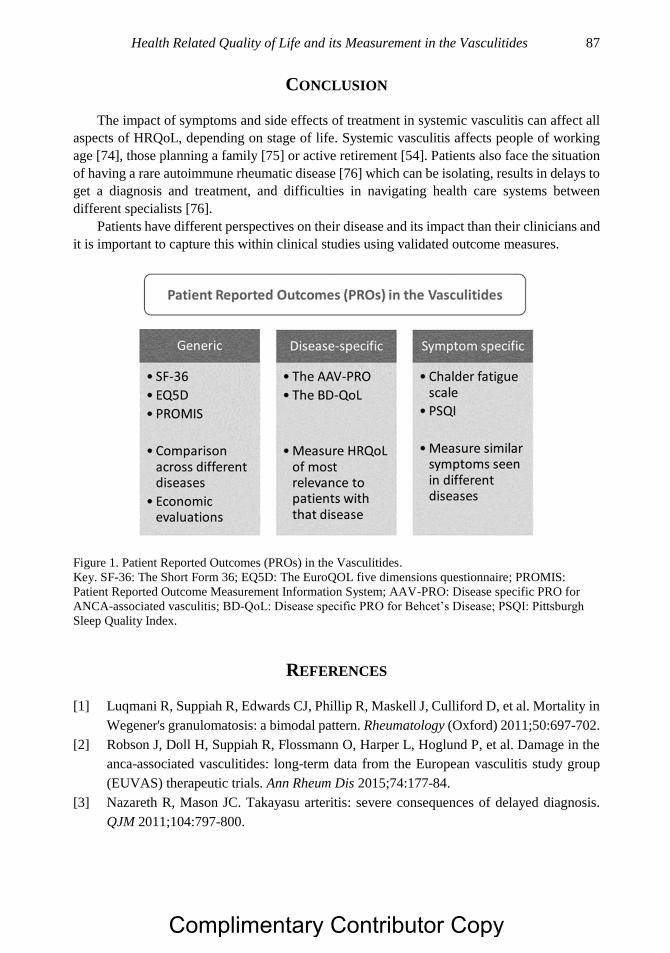
Health Related Quality of Life and its Measurement in the Vasculitides 87
CONCLUSION
The impact of symptoms and side effects of treatment in systemic vasculitis can affect all
aspects of HRQoL, depending on stage of life. Systemic vasculitis affects people of working
age [74], those planning a family [75] or active retirement [54]. Patients also face the situation
of having a rare autoimmune rheumatic disease [76] which can be isolating, results in delays to
get a diagnosis and treatment, and difficulties in navigating health care systems between
different specialists [76].
Patients have different perspectives on their disease and its impact than their clinicians and
it is important to capture this within clinical studies using validated outcome measures.
Figure 1. Patient Reported Outcomes (PROs) in the Vasculitides.
Key. SF-36: The Short Form 36; EQ5D: The EuroQOL five dimensions questionnaire; PROMIS:
Patient Reported Outcome Measurement Information System; AAV-PRO: Disease specific PRO for
ANCA-associated vasculitis; BD-QoL: Disease specific PRO for Behcet’s Disease; PSQI: Pittsburgh
Sleep Quality Index.
REFERENCES
[1] Luqmani R, Suppiah R, Edwards CJ, Phillip R, Maskell J, Culliford D, et al. Mortality in
Wegener's granulomatosis: a bimodal pattern. Rheumatology (Oxford) 2011;50:697-702.
[2] Robson J, Doll H, Suppiah R, Flossmann O, Harper L, Hoglund P, et al. Damage in the
anca-associated vasculitides: long-term data from the European vasculitis study group
(EUVAS) therapeutic trials. Ann Rheum Dis 2015;74:177-84.
[3] Nazareth R, Mason JC. Takayasu arteritis: severe consequences of delayed diagnosis.
QJM 2011;104:797-800.
Complimentary Contributor Copy
Joanna Robson and Richard A. Watts 88
[4] Jones RB, Tervaert JW, Hauser T, Luqmani R, Morgan MD, Peh CA, et al. Rituximab
versus cyclophosphamide in ANCA-associated renal vasculitis. N Engl J Med
2010;363:211-20.
[5] Stone JH, Tuckwell K, Dimonaco S, Klearman M, Aringer M, Blockmans D, et al. Trial
of Tocilizumab in Giant-Cell Arteritis. N Engl J Med 2017;377:317-28.
[6] Guillevin L, Pagnoux C, Karras A, Khouatra C, Aumaitre O, Cohen P, et al. Rituximab
versus azathioprine for maintenance in ANCA-associated vasculitis. N Engl J Med 2014;
371:1771-1780.
[7] Abularrage CJ, Slidell MB, Sidawy AN, Kreishman P, Amdur RL, Arora S. Quality of
life of patients with Takayasu's arteritis. J Vasc Surg 2008;47:131-6; discussion 6-7.
[8] Bodur H, Borman P, Ozdemir Y, Atan C, Kural G. Quality of life and life satisfaction in
patients with Behcet's disease: relationship with disease activity. Clin Rheumatol 2006;
25:329-333.
[9] Koutantji M, Harrold E, Lane SE, Pearce S, Watts RA, Scott DG. Investigation of quality
of life, mood, pain, disability, and disease status in primary systemic vasculitis. Arthritis
Rheum 2003; 49:826-837.
[10] Jennette JC, Falk RJ, Bacon PA, Basu N, Cid MC, Ferrario F, et al. 2012 revised
International Chapel Hill Consensus Conference Nomenclature of Vasculitides. Arthritis
Rheum 2013;65:1-11.
[11] Ware JE, Jr., Sherbourne CD. The MOS 36-item short-form health survey (SF-36). I.
Conceptual framework and item selection. Med Care 1992; 30:473-483.
[12] Fitzpatrick R, Davey C, Buxton MJ, Jones DR. Evaluating patient-based outcome
measures for use in clinical trials. Health Technol Assess 1998;2:i-iv, 1-74.
[13] Kupersmith MJ, Speira R, Langer R, Richmond M, Peterson M, Speira H, et al. Visual
function and quality of life among patients with giant cell (temporal) arteritis. J
Neuroophthalmol 2001; 21:266-273.
[14] Stone JH, Merkel PA, Spiera R, Seo P, Langford CA, Hoffman GS, et al. Rituximab
versus cyclophosphamide for ANCA-associated vasculitis. N Engl J Med 2010; 363:221-
232.
[15] Strand V, Dimonaco S, Tuckwell K, Klearman M, Collinson N, Stone J. Health-Related
Quality of Life in Patients with Giant Cell Arteritis Treated with Tocilizumab in a
Randomized Controlled Phase 3 Trial [abstract]. Arthritis Rheum 2017;69 (suppl 10).
[16] Tugwell P, Boers M, Brooks P, Simon L, Strand V, Idzerda L. OMERACT: an
international initiative to improve outcome measurement in rheumatology. Trials
2007;8:38.
[17] Merkel PA, Aydin SZ, Boers M, Direskeneli H, Herlyn K, Seo P, et al. The OMERACT
core set of outcome measures for use in clinical trials of ANCA-associated vasculitis. J
Rheumatol 2011; 38:1480-1486
[18] Sreih AG, Alibaz-Oner F, Kermani TA, Aydin SZ, Cronholm PF, Davis T, et al.
Development of a Core Set of Outcome Measures for Large-vessel Vasculitis: Report
from OMERACT 2016. J Rheumatol 2017; 44:1933-1937.
Complimentary Contributor Copy
Health Related Quality of Life and its Measurement in the Vasculitides 89
[19] Herlyn K, Hellmich B, Seo P, Merkel PA. Patient-reported outcome assessment in
vasculitis may provide important data and a unique perspective. Arthritis Care Res
(Hoboken) 2010; 62:1639-1645.
[20] Robson JC, Dawson J, Doll H, Cronholm PF, Milman N, Kellom K, et al. Validation of
the ANCA-associated vasculitis patient-reported outcomes (AAV-PRO) questionnaire.
Ann Rheum Dis 2018; 77:1157-1164.
[21] Sreih AG, Alibaz-Oner F, Easley E, Davis T, Mumcu G, Milman N, et al. Health-related
outcomes of importance to patients with Takayasu's arteritis. Clin Exp Rheumatol 2018;
36 Suppl 111:51-57.
[22] Hatemi G, Meara A, Ozguler Y, Direskeneli H, Mahr A, Easley E, et al. Developing a
Core Set of Outcome Measures for Behcet Disease: Report from OMERACT 2016. J
Rheumatol 2017; 44:1750-1753.
[23] Tomasson G, Farrar JT, Cuthbertson D, Ashdown S, Gebhart D, Lanier G, et al. Fatigue
and Physical Functioing in Patients with Giant Cell Arteritis [abstract]. Arthritis Rheum
2016;68 (suppl 10).
[24] Patrick DL, Burke LB, Powers JH, Scott JA, Rock EP, Dawisha S, et al. Patient-reported
outcomes to support medical product labeling claims: FDA perspective. Value in Health
2007; 10:S125-S137.
[25] Fereday J, Muir-Cochrane E. Demonstrating rigor using thematic analysis: a hybrid
approach of inductive and deductive coding and theme development. Int J Qual Meth
2006;5:1-22.
[26] Drennan J. Cognitive interviewing: verbal data in the design and pretesting of
questionnaires. J Adv Nurs 2003;42:57-63.
[27] Costello A, Osborne J. Best practices in Exploratory Factor Analysis: four
recommendations for getting the most from your analysis. Practical assessment, research
and evaluation 2005;10:1-9.
[28] Tennant A, Conaghan PG. The Rasch measurement model in rheumatology: what is it
and why use it? When should it be applied, and what should one look for in a Rasch
paper? Arthritis Rheum 2007; 57:1358-1362.
[29] Gilworth G, Chamberlain MA, Bhakta B, Haskard D, Silman A, Tennant A.
Development of the BD-QoL: a quality of life measure specific to Behcet's disease. J
Rheumatol 2004; 31:931-937.
[30] Kirwan JR, Hewlett SE, Heiberg T, Hughes RA, Carr M, Hehir M, et al. Incorporating
the patient perspective into outcome assessment in rheumatoid arthritis--progress at
OMERACT 7. J Rheumatol 2005; 32:2250-2256.
[31] Robson J, Dawson J, Cronholm PF, Milman N, Kellom K, Ashdown S, et al. Health
related quality of life in ANCA associated vasculitis and item generation for a disease
specific patient reported outcome measure. Patient Related Outcome Measures 2017; In
Press.
[32] Walsh M, Mukhtyar C, Mahr A, Herlyn K, Luqmani R, Merkel PA, et al. Health related
quality of life in patients with newly diagnosed anti-neutrophil cytoplasm antibody
associated vasculitis. Arthritis Care Res (Hoboken) 2011.
Complimentary Contributor Copy
Joanna Robson and Richard A. Watts 90
[33] Walsh M, Mukhtyar C, Mahr A, Herlyn K, Luqmani R, Merkel PA, et al. Health-related
quality of life in patients with newly diagnosed antineutrophil cytoplasmic antibody-
associated vasculitis. Arthritis Care Res (Hoboken) 2011; 63:1055-1061.
[34] Basu N, Jones GT, Fluck N, MacDonald AG, Pang D, Dospinescu P, et al. Fatigue: a
principal contributor to impaired quality of life in ANCA-associated vasculitis.
Rheumatology (Oxford) 2010; 49:1383-1390.
[35] Basu N, McClean A, Harper L, Amft EN, Dhaun N, Luqmani RA, et al. Explaining
fatigue in ANCA-associated vasculitis. Rheumatology (Oxford) 2013; 52:1680-1685.
[36] Robson JC, Milman N, Tomasson G, Dawson J, Cronholm PF, Kellom K, et al.
Exploration, Development, and Validation of Patient-reported Outcomes in
Antineutrophil Cytoplasmic Antibody-associated Vasculitis Using the OMERACT
Process. J Rheumatol 2015; 42:2204-2209.
[37] Matcham F, Scott IC, Rayner L, Hotopf M, Kingsley GH, Norton S, et al. The impact of
rheumatoid arthritis on quality-of-life assessed using the SF-36: a systematic review and
meta-analysis. Semin Arthritis Rheum 2014; 44:123-130.
[38] Singh K, Kondal D, Shivashankar R, Ali MK, Pradeepa R, Ajay VS, et al. Health-related
quality of life variations by sociodemographic factors and chronic conditions in three
metropolitan cities of South Asia: the CARRS study. BMJ Open 2017;7:e018424.
[39] Bailey PK, Hamilton AJ, Clissold RL, Inward CD, Caskey FJ, Ben-Shlomo Y, et al.
Young adults' perspectives on living with kidney failure: a systematic review and
thematic synthesis of qualitative studies. BMJ Open 2018;8:e019926.
[40] Grayson PC, Amudala NA, McAlear CA, Leduc RL, Shereff D, Richesson R, et al. Illness
perceptions and fatigue in systemic vasculitis. Arthritis Care Res (Hoboken) 2013;
65:1835-1843.
[41] Mason JC. Takayasu arteritis--advances in diagnosis and management. Nat Rev
Rheumatol 2010; 6:406-415.
[42] Akar S, Can G, Binicier O, Aksu K, Akinci B, Solmaz D, et al. Quality of life in patients
with Takayasu's arteritis is impaired and comparable with rheumatoid arthritis and
ankylosing spondylitis patients. Clin Rheumatol 2008; 27:859-865.
[43] Yilmaz N, Can M, Oner FA, Kalfa M, Emmungil H, Karadag O, et al. Impaired quality
of life, disability and mental health in Takayasu's arteritis. Rheumatology (Oxford) 2013;
52:1898-1904.
[44] Direskeneli H, Aydin SZ, Kermani TA, Matteson EL, Boers M, Herlyn K, et al.
Development of outcome measures for large-vessel vasculitis for use in clinical trials:
opportunities, challenges, and research agenda. J Rheumatol 2011; 38:1471-1479.
[45] Petri H, Nevitt A, Sarsour K, Napalkov P, Collinson N. Incidence of giant cell arteritis
and characteristics of patients: data-driven analysis of comorbidities. Arthritis Care Res
(Hoboken) 2015; 67:390-395.
[46] Salvarani C, Cimino L, Macchioni P, Consonni D, Cantini F, Bajocchi G, et al. Risk
factors for visual loss in an Italian population-based cohort of patients with giant cell
arteritis. Arthritis Rheum 2005; 53:293-297.
Complimentary Contributor Copy
Health Related Quality of Life and its Measurement in the Vasculitides 91
[47] Salvarani C, Pipitone N, Versari A, Hunder GG. Clinical features of polymyalgia
rheumatica and giant cell arteritis. Nat Rev Rheumatol 2012; 8:509-521.
[48] Mukhtyar C, Guillevin L, Cid MC, Dasgupta B, de Groot K, Gross W, et al. EULAR
recommendations for the management of large vessel vasculitis. Ann Rheum Dis 2009;
68:318-323.
[49] Dasgupta B, Borg FA, Hassan N, Alexander L, Barraclough K, Bourke B, et al. BSR and
BHPR guidelines for the management of giant cell arteritis. Rheumatology (Oxford)
2010; 49:1594-1597.
[50] Robson JC, Dawson J, Cronholm PF, Ashdown S, Easley E, Kellom KS, et al. Patient
perceptions of glucocorticoids in anti-neutrophil cytoplasmic antibody-associated
vasculitis. Rheumatol Int 2017.
[51] Black RJ, Goodman SM, Ruediger C, Lester S, Mackie SL, Hill CL. A Survey of
Glucocorticoid Adverse Effects and Benefits in Rheumatic Diseases: The Patient
Perspective. J Clin Rheumatol 2017; 23:416-420.
[52] McDonough AK, Curtis JR, Saag KG. The epidemiology of glucocorticoid-associated
adverse events. Curr Opin Rheumatol 2008; 20:131-137.
[53] Fardet L, Flahault A, Kettaneh A, Tiev KP, Genereau T, Toledano C, et al.
Corticosteroid-induced clinical adverse events: frequency, risk factors and patient's
opinion. Br J Dermatol 2007; 157:142-148.
[54] Liddle J, Bartlam R, Mallen CD, Mackie SL, Prior JA, Helliwell T, et al. What is the
impact of giant cell arteritis on patients' lives? A UK qualitative study. BMJ Open
2017;7:e017073.
[55] Hellmann DB, Uhlfelder ML, Stone JH, Jenckes MW, Cid MC, Guillevin L, et al.
Domains of health-related quality of life important to patients with giant cell arteritis.
Arthritis Rheum 2003; 49:819-825.
[56] Aydin SZ, Direskeneli H, Sreih A, Alibaz-Oner F, Gul A, Kamali S, et al. Update on
Outcome Measure Development for Large Vessel Vasculitis: Report from OMERACT
12. J Rheumatol 2015; 42:2465-2469.
[57] Jobard S, Magnant J, Blasco H, Ferreira-Maldent N, Griffoul I, Diot E, et al. Quality of
life of patients treated for giant cell arteritis: a case-control study. Clin Rheumatol
2017;36:2055-2062.
[58] Robson J, Almeida C, Dawson J, Bromhead A, Mackie SL, Hill CL, et al. A multinational
qualitative study in Giant Cell Arteritis: patient perceptions of diagnosis, treatment,
impact on health-related quality of life and contextual factors. Ann Rheum Dis [Abstract]
2018;17:777.
[59] Hatemi G, Christensen R, Bang D, Bodaghi B, Celik AF, Fortune F, et al. 2018 update
of the EULAR recommendations for the management of Behcet's syndrome. Ann Rheum
Dis 2018;77:808-818.
[60] Kalra S, Silman A, Akman-Demir G, Bohlega S, Borhani-Haghighi A, Constantinescu
CS, et al. Diagnosis and management of Neuro-Behcet's disease: international consensus
recommendations. J Neurol 2014;261:1662-76.
Complimentary Contributor Copy
Joanna Robson and Richard A. Watts 92
[61] Fabiani C, Vitale A, Orlando I, Sota J, Capozzoli M, Franceschini R, et al. Quality of life
impairment in Behcet's disease and relationship with disease activity: a prospective study.
Intern Emerg Med 2017; 12:947-955.
[62] Gul IG, Kartalci S, Cumurcu BE, Karincaoglu Y, Yologlu S, Karlidag R. Evaluation of
sexual function in patients presenting with Behcet's disease with or without depression.
J Eur Acad Dermatol Venereol 2013; 27:1244-1251.
[63] Hatemi G, Merkel PA, Hamuryudan V, Boers M, Direskeneli H, Aydin SZ, et al.
Outcome measures used in clinical trials for Behcet syndrome: a systematic review. J
Rheumatol 2014;41:599-612.
[64] Touma Z, Ghandour L, Sibai A, Puzantian H, Hamdan A, Hamdan O, et al. Cross-cultural
adaptation and validation of Behcet's disease quality of life questionnaire. BMC Med Res
Methodol 2011;11:52.
[65] Lee J, Kim SS, Jeong HJ, Son CN, Kim JM, Cho YW, et al. Association of sleep quality
in Behcet disease with disease activity, depression, and quality of life in Korean
population. Korean J Intern Med 2017; 32:352-359.
[66] Hatemi G, Melikoglu M, Tunc R, Korkmaz C, Turgut Ozturk B, Mat C, et al. Apremilast
for Behcet's syndrome--a phase 2, placebo-controlled study. N Engl J Med 2015;
372:1510-1518.
[67] Choi HJ, Seo MR, Ryu HJ, Baek HJ. Cross-cultural adaptation and validation of the
Behcet's Disease Current Activity Form in Korea. Korean J Intern Med 2015; 30:714-
718.
[68] Ertam I, Kitapcioglu G, Aksu K, Keser G, Ozaksar A, Elbi H, et al. Quality of life and
its relation with disease severity in Behcet's disease. Clin Exp Rheumatol 2009; 27:S18-
522.
[69] Mumcu G, Niazi S, Stewart J, Hagi-Pavli E, Gokani B, Seoudi N, et al. Oral health and
related quality of life status in patients from UK and Turkey: a comparative study in
Behcet's disease. J Oral Pathol Med 2009; 38:406-409.
[70] Tascilar NF, Tekin NS, Ankarali H, Sezer T, Atik L, Emre U, et al. Sleep disorders in
Behcet's disease, and their relationship with fatigue and quality of life. J Sleep Res 2012;
21:281-288.
[71] Melikoglu MA, Melikoglu M. The relationship between disease activity and depression
in patients with Behcet disease and rheumatoid arthritis. Rheumatol Int 2010;30:941-6.
[72] Calikoglu E, Onder M, Cosar B, Candansayar S. Depression, anxiety levels and general
psychological profile in Behcet's disease. Dermatology 2001; 203:238-240.
[73] Tugwell P, Boers M, D'Agostino MA, Beaton D, Boonen A, Bingham CO, 3rd, et al.
Updating the OMERACT filter: implications of filter 2.0 to select outcome instruments
through assessment of "truth": content, face, and construct validity. J Rheumatol 2014;
41:1000-1004.
[74] Benarous L, Terrier B, Laborde-Casterot H, Berezne A, Dunogue B, Cohen P, et al.
Employment, work disability and quality of life in patients with ANCA-associated
vasculitides. The EXPOVAS study. Clin Exp Rheumatol 2017; Suppl 103:4046.
Complimentary Contributor Copy

Health Related Quality of Life and its Measurement in the Vasculitides 93
[75] Fredi M, Lazzaroni MG, Tani C, Ramoni V, Gerosa M, Inverardi F, et al. Systemic
vasculitis and pregnancy: a multicenter study on maternal and neonatal outcome of 65
prospectively followed pregnancies. Autoimmun Rev 2015; 14:686-691.
[76] (RAIRDA) RARDA. Reduce, Improve, Empower. Addressing the shared needs of rare
autoimmune rheumatic diseases. 2018.
Complimentary Contributor Copy

Complimentary Contributor Copy
In: The Vasculitides. Volume 1 ISBN: 978-1-53615-133-6
Editor: David S. Younger © 2019 Nova Science Publishers, Inc.
Chapter 5
NEUTROPHILIC CELL PATHOBIOLOGY
IN THE VASCULITIDES
Akihiro Ishizu, MD, PhD Faculty of Health Sciences, Hokkaido University,
Kitaku, Sapporo, Japan
ABSTRACT
Neutrophils play a critical role in the pathogenesis of vasculitides, especially in anti-
neutrophil cytoplasmic antibody (ANCA)-associated vasculitis (AAV). ANCA cooperates
with pro-inflammatory cytokines in activating neutrophils. Activated neutrophils
contribute to the development of small vessel vasculitis. The ANCA-cytokine sequence is
pivotal to the understanding the pathogenesis of AAV. Recent studies reveal that neutrophil
extracellular traps (NETs) extruded from activated neutrophils are implicated in vascular
endothelial cell (VEC) injury. NETs are also implicated in the mechanism of ANCA
production. These findings suggest that the NETs-ANCA vicious cycle is involved in the
pathogenesis of AAV. This chapter updates the literature relating to ANCA-cytokine
sequence theory.
Keywords: ANCA-associated vasculitis, ANCA-cytokine sequence, NETs-ANCA vicious
cycle, NETs-ANCA-cytokine sequence
INTRODUCTION
Neutrophils play critical roles in the pathogenesis of vasculitides, especially in anti-
neutrophil cytoplasmic antibody (ANCA)-associated vasculitis (AAV). They are primed by
pro-inflammatory cytokines, such as TNF-α, and express myeloperoxidase (MPO) and
proteinase 3 (PR3) on the cell surface. ANCA binds to the antigen and simultaneously bridges
the antigen and bystander Fc receptor on the cell, which activates neutrophils. Consequently,
Corresponding Author’s E-mail: [email protected].
Complimentary Contributor Copy
Akihiro Ishizu 96
the activated neutrophil releases reactive oxygen species (ROS) and lytic enzymes, in turn
contributing to the development of small vessel vasculitis (SVV). This ANCA-cytokine
sequence therapy is pivotal for the understanding of AAV [1]. Recent studies have revealed
that neutrophil extracellular traps (NETs) extruded from the activated neutrophils are also
implicated in vascular endothelial cell (VEC) injury [2, 3]. Recent reports suggest involvement
of NETs in the mechanism of ANCA production [4-6], as well as in ANCA-mediated
cytotoxicity.
This chapter focuses on NETosis and the neutrophilic cell metamorphosis of NET
formation in AAV.
Discovery of NETs
In 2004, Brinkmann and coworkers [7] reported that phorbol myristate acetate (PMA)-
stimulated neutrophils extrude decondensated DNA that form extracellular web-like structure
decorated with bactericidal proteins, such as MPO and PR3. These substances, termed NETs,
bind to, and kill bacteria. NET formation is regarded as an important event of innate immunity.
Patients with chronic granulomatous diseases (CGD) who fail to generate NETs are
susceptible to diverse bacterial and fungal infections. However, restoration of NET formation
in CGD results in resistance to such infections [8]. NETs appear to be an innate response that
binds microorganisms, prevents them from spreading, and ensures a high local concentration
of antimicrobial activity derived from neutrophils [9].
Process of NET Formation
PMA-stimulated neutrophils undergo cell death with NET formation [10]. Since the
characteristics of cell death resembles neither typical necrosis nor apoptosis, Steinberg and
colleagues [11] coin NETosis for neutrophilic cell death with NET formation. NETosis
induction by PMA requires activation of the Raf-MEK-ERK pathway, with NADPH oxidase-
dependent production of ROS, and RIPK1-RIPK3-MLKL signaling [12, 13]. In this pathway,
peptidylarginine deiminase 4 (PAD4)-dependent citrullination of histones induces
decondensation of DNA resulting in a mixture of DNA and bactericidal proteins, contained
originally in intracytoplasmic granules [14]. Thereafter, these substances extrude from the
ruptured plasma membranes of neutrophils.
NETs in AAV
Kessenbrock and coworkers [4] first showed that ANCA derived from AAV induces NETs
from TNF-α-primed neutrophils obtained from healthy donors. They also demonstrated co-
localization of extracellular DNA, neutrophilic enzymes, and histones. NET formation was
evident in crescents of glomeruli affected by AAV.
Subsequent investigations noted similar findings using citrullinated histones as a specific
marker of NETs [15-17].
Complimentary Contributor Copy
Neutrophilic Cell Pathobiology in the Vasculitides 97
Vascular Endothelial Cell Toxicity of NETs
Carmona-Rivera and coinvestigators [18] demonstrated that direct incubation of VEC lines
with NETs promoted VEC death. The cytotoxicity of NETs is mediated by matrix
metalloproteinase (MMP)-9, which is externalized from neutrophils. NET formation and
activation of MMP-2 in VECs culminates in cell death. The neutralization of MMP-9 prevents
MMP-2 activation in VECs leading to a decrease in NET-medicated cytotoxicity.
Thrombosis Induced by NETs
Patients with AAV are predisposed to deep vein thrombosis (DVT), especially during the
active stage of the disease [19]. As ANCA induces NET from the primed neutrophils, NETs
are critically associated with DVT as histones within NETs bind platelets and coagulate blood
[20, 21]. Such findings are consistent with an abundance of NETs detected in DVTs of patients
with AAV [15, 17]. The collective findings suggest a link between AAV, accelerated
thrombosis and DVT via NETs.
Degradation of NETs
NETs play an essential role in the innate immune system with excessive formation and
persistence of NETs, a consequence of which is the heightened risk of adverse events.
Notwithstanding, NETs are strictly regulated in vivo. The most important NET degradation
factor is serum DNase I [22].
Propylthiouracil Induces DNase I-Resistant NETs
Propylthiouracil (PTU) is an anti-thyroid drug used for the treatment of hyperthyroidism.
Approximately 30% of patients administered with PTU produce MPO-ANCA, some of whom
develop MPO-AAV. The majority of PTU is metabolized in the liver. However, MPO in
neutrophils also modifies PTU [23]. We hypothesize that PTU influences the formation or
regulation of NETs and induces MPO-ANCA, and subsequently MPO-AAV [5]. We examined
whether the addition of PTU created an impact on the formation or regulation of NETs in vitro.
Widely extended DNA fibers studded with MPO are observed when human neutrophils are
treated with PMA alone. To the contrary, treatment with PMA with PTU leads to a rounded-
shape distribution of DNA fibers. The ensuing NETs surround dead neutrophils. Although
DNase completely digests PMA-induced normal NETs, abnormal NETs induced by PMA with
PTU is barely digested by DNase I. These findings clearly indicate that PTU influences NET
formation and degradation by DNase I.
Complimentary Contributor Copy
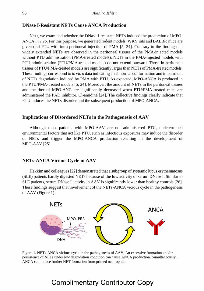
Akihiro Ishizu 98
DNase I-Resistant NETs Cause ANCA Production
Next, we examined whether the DNase I-resistant NETs induced the production of MPO-
ANCA in vivo. For this purpose, we generated rodent models. WKY rats and BALB/c mice are
given oral PTU with intra-peritoneal injection of PMA [5, 24]. Contrary to the finding that
widely extended NETs are observed in the peritoneal tissues of the PMA-injected models
without PTU administration (PMA-treated models), NETs in the PMA-injected models with
PTU administration (PTU/PMA-treated models) do not extend outward. Those in peritoneal
tissues of PTU/PMA-treated models are significantly larger than NETs of PMA-treated models.
These findings correspond to in vitro data indicating an abnormal conformation and impairment
of NETs degradation induced by PMA with PTU. As expected, MPO-ANCA is produced in
the PTU/PMA-treated models [5, 24]. Moreover, the amount of NETs in the peritoneal tissues
and the titer of MPO-ANC are significantly decreased when PTU/PMA-treated mice are
administered the PAD inhibitor, Cl-amidine [24]. The collective findings clearly indicate that
PTU induces the NETs disorder and the subsequent production of MPO-ANCA.
Implications of Disordered NETs in the Pathogenesis of AAV
Although most patients with MPO-AAV are not administered PTU, undetermined
environmental factors that act like PTU, such as infectious exposures may induce the disorder
of NETs and trigger the MPO-ANCA production resulting in the development of
MPO-AAV [25].
NETs-ANCA Vicious Cycle in AAV
Hakkim and colleagues [22] demonstrated that a subgroup of systemic lupus erythematosus
(SLE) patients hardly digested NETs because of the low activity of serum DNase I. Similar to
SLE patients, serum DNase I activity in AAV is significantly lower than healthy controls [26].
These findings suggest that involvement of the NETs-ANCA vicious cycle in the pathogenesis
of AAV (Figure 1).
Figure 1. NETs-ANCA vicious cycle in the pathogenesis of AAV. An excessive formation and/or
persistence of NETs under low degradation condition can cause ANCA production. Simultaneously,
ANCA can induce further NET formation from primed neutrophils.
Complimentary Contributor Copy
Neutrophilic Cell Pathobiology in the Vasculitides 99
The Formation of the Pauci-Immune Lesion in AAV
AAV is characterized by necrotizing crescentic glomerulonephritis and SVV. Although the
pathogenic role of ANCA in the formation of necrotizing lesions can be demonstrated, there is
little deposition of immunoglobulins (Ig) in affected tissues. This histological feature is termed,
“pauci-immune”. Futamata and colleagues [27] reported the release of elastase from NET-
forming neutrophils, digesting Ig, including ANCA, in pauci-immune lesions in AAV.
CONCLUSION
Neutrophils play a critical role in the pathogenesis of vasculitides, especially AAV. The
ANCA-cytokine sequence is pivotal to the etiopathogenesis of AAV. There is an increasing
literature supporting the role for an NETs-ANCA vicious cycle in AAV and the related
contribution of NETs-ANCA-cytokine sequence theory.
REFERENCES
[1] Csernok, E.: Anti-neutrophil cytoplasmic antibodies and pathogenesis of small vessel
vasculitides. Autoimmun. Rev., 2003, 2:158 - 64.
[2] Jennette, J. C., Falk, R. J.: Pathogenesis of antineutrophil cytoplasmic autoantibody-
mediated disease. Nat. Rev. Rheumatol., 2014, 10:463 - 73.
[3] Kallenberg, C. G.: Pathogenesis and treatment of ANCA-associated vasculitides. Clin.
Exp. Rheumatol., 2015, 33:S11-4.
[4] Kessenbrock, K., Krumbholz, M., Schonermarck, U., Back, W., Gross, W. L., Werb, Z.,
Grone, H. J., Brinkmann, V., Jenne, D. E.: Netting neutrophils in autoimmune small-
vessel vasculitis. Nat. Med., 2009, 15:623 - 5.
[5] Nakazawa, D., Tomaru, U., Suzuki, A., Masuda, S., Hasegawa, R., Kobayashi, T.,
Nishio, S., Kasahara, M., Ishizu, A.: Abnormal conformation and impaired degradation
of propylthiouracil-induced neutrophil extracellular traps: implications of disordered
neutrophil extracellular traps in a rat model of myeloperoxidase antineutrophil
cytoplasmic antibody-associated vasculitis. Arthritis Rheum., 2012, 64:3779 - 87.
[6] Sangaletti, S., Tripodo, C., Chiodoni, C., Guarnotta, C., Cappetti, B., Casalini, P.,
Piconese, S., Parenza, M., Guiducci, C., Vitali, C., Colombo, M. P.: Neutrophil
extracellular traps mediate transfer of cytoplasmic neutrophil antigens to myeloid
dendritic cells toward ANCA induction and associated autoimmunity. Blood, 2012,
120:3007 - 18.
[7] Brinkmann, V., Reichard, U., Goosmann, C., Fauler, B., Uhlemann, Y., Weiss, D. S.,
Weinrauch, Y., Zychlinsky, A.: Neutrophil extracellular traps kill bacteria. Science, 2004,
303:1532 - 5.
[8] Bianchi, M., Hakkim, A., Brinkmann, V., Siler, U., Seger, R. A., Zychlinsky, A.,
Reichenbach, J.: Restoration of NET formation by gene therapy in CGD controls
aspergillosis. Blood, 2009, 114:2619 - 22.
Complimentary Contributor Copy
Akihiro Ishizu 100
[9] Papayannopoulos, V., Zychlinsky, A.: NETs: a new strategy for using old weapons.
Trends Immunol., 2009, 30:513 - 21.
[10] Fuchs, T. A., Abed, U., Goosmann, C., Hurwitz, R., Schulze, I., Wahn, V., Weinrauch,
Y., Brinkmann, V., Zychlinsky, A.: Novel cell death program leads to neutrophil
extracellular traps. J. Cell Biol., 2007, 176:231 - 41.
[11] Steinberg, B. E., Grinstein, S.: Unconventional roles of the NADPH oxidase: signaling,
ion homeostasis, and cell death. Sci. STKE, 2007, 2007:pe11.
[12] Hakkim, A., Fuchs, T. A., Martinez, N. E., Hess, S., Prinz, H., Zychlinsky, A.,
Waldmann, H.: Activation of the Raf-MEK-ERK pathway is required for neutrophil
extracellular trap formation. Nat. Chem. Biol., 2011, 7:75 - 7.
[13] Desai, J., Kumar, S. V., Mulay, S. R., Konrad, L., Romoli, S., Schauer, C., Herrmann,
M., Bilyy, R., Muller, S., Popper, B., Nakazawa, D., Weidenbusch, M., Thomasova, D.,
Krautwald, S., Linkermann, A., Anders, H. J.: PMA and crystal-induced neutrophil
extracellular trap formation involves RIPK1-RIPK3-MLKL signaling. Eur. J. Immunol.,
2016, 46:223 - 9.
[14] Li, P., Li, M., Lindberg, M. R., Kennett, M. J., Xiong, N., Wang, Y.: PAD4 is essential
for antibacterial innate immunity mediated by neutrophil extracellular traps. J. Exp. Med.,
2010, 207:1853 - 62.
[15] Nakazawa, D., Tomaru, U., Yamamoto, C., Jodo, S., Ishizu, A.: Abundant neutrophil
extracellular traps in thrombus of patient with microscopic polyangiitis. Front Immunol.,
2012, 3:333.
[16] Yoshida, M., Sasaki, M., Sugisaki, K., Yamaguchi, Y., Yamada, M.: Neutrophil
extracellular trap components in fibrinoid necrosis of the kidney with myeloperoxidase-
ANCA-associated vasculitis. Clin. Kidney J., 2013, 6:308 - 12.
[17] Imamoto, T., Nakazawa, D., Shida, H., Suzuki, A., Otsuka, N., Tomaru, U., Ishizu, A.:
Possible linkage between microscopic polyangiitis and thrombosis via neutrophil
extracellular traps. Clin. Exp. Rheumatol., 2014, 32:149 - 50.
[18] Carmona-Rivera, C., Zhao, W., Yalavarthi, S., Kaplan, M. J.: Neutrophil extracellular
traps induce endothelial dysfunction in systemic lupus erythematosus through the
activation of matrix metalloproteinase-2. Ann. Rheum. Dis., 2015, 74:1417 - 24.
[19] Stassen, P. M., Derks, R. P., Kallenberg, C. G., Stegeman, C. A.: Venous
thromboembolism in ANCA-associated vasculitis--incidence and risk factors.
Rheumatology (Oxford), 2008, 47:530 - 4.
[20] Fuchs, T. A., Brill, A., Duerschmied, D., Schatzberg, D., Monestier, M., Myers, D. D.,
Jr., Wrobleski, S. K., Wakefield, T. W., Hartwig, J. H., Wagner, D. D.: Extracellular
DNA traps promote thrombosis. Proc. Natl. Acad. Sci. USA, 2010, 107:15880 - 5.
[21] Fuchs, T. A., Brill, A., Wagner, D. D.: Neutrophil extracellular trap (NET) impact on
deep vein thrombosis. Arterioscler. Thromb. Vasc. Biol., 2012, 32:1777 - 83.
[22] Hakkim, A., Furnrohr, B. G., Amann, K., Laube, B., Abed, U. A., Brinkmann, V.,
Herrmann, M., Voll, R. E., Zychlinsky, A.: Impairment of neutrophil extracellular trap
degradation is associated with lupus nephritis. Proc. Natl. Acad. Sci. USA, 2010,
107:9813 - 8.
[23] Waldhauser, L., Uetrecht, J.: Oxidation of propylthiouracil to reactive metabolites by
activated neutrophils. Implications for agranulocytosis. Drug Metab. Dispos., 1991, 19:
354 - 9.
Complimentary Contributor Copy
Neutrophilic Cell Pathobiology in the Vasculitides 101
[24] Kusunoki, Y., Nakazawa, D., Shida, H., Hattanda, F., Miyoshi, A., Masuda, S., Nishio,
S., Tomaru, U., Atsumi, T., Ishizu, A.: Peptidylarginine Deiminase Inhibitor Suppresses
Neutrophil Extracellular Trap Formation and MPO-ANCA Production. Front Immunol.,
2016, 7:227.
[25] Nakazawa, D., Tomaru, U., Ishizu, A.: Possible implication of disordered neutrophil
extracellular traps in the pathogenesis of MPO-ANCA-associated vasculitis. Clin. Exp.
Nephrol., 2013, 17:631 - 3.
[26] Nakazawa, D., Shida, H., Tomaru, U., Yoshida, M., Nishio, S., Atsumi, T., Ishizu, A.:
Enhanced formation and disordered regulation of NETs in myeloperoxidase-ANCA-
associated microscopic polyangiitis. J. Am. Soc. Nephrol., 2014, 25:990 - 7.
[27] Futamata, E., Masuda, S., Nishibata, Y., Tanaka, S., Tomaru, U., Ishizu, A.: Vanishing
immunoglobulins: the formation of pauci-immune lesions in myeloperoxidase-
antineutrophil cytoplasmic antibody-associated vasculitis. Nephron, 2018, 138:328 - 30.
Complimentary Contributor Copy

Complimentary Contributor Copy
In: The Vasculitides. Volume 1 ISBN: 978-1-53615-133-6
Editor: David S. Younger © 2019 Nova Science Publishers, Inc.
Chapter 6
COMPLEMENT FACTORS
IN ANCA-ASSOCIATED VASCULITIS
Chen Wang*, MD, PhD and Min Chen, MD, PhD Renal Division of the Department of Medicine, Peking University First Hospital;
Peking University Institute of Nephrology; Key Laboratory of Renal Disease,
Ministry of Health of China; Key Laboratory of Chronic Kidney Disease
Prevention and Treatment, Peking University, Ministry of Education,
Beijing, P.R. China
ABSTRACT
Anti-neutrophil cytoplasmic antibody (ANCA)-associated vasculitis (AAV) are
potentially life-threatening autoimmune diseases. The histopathological hallmark is a
“pauci-immune” necrotizing crescentic glomerulonephritis. It has been assumed that the
complement system is not involved in the development of AAV, however, new evidence
suggests that the complement system is crucial in the pathogenesis of AAV. This article
reviews updated information on clinical, in vivo and in vitro evidence for the role of
complement activation in the development of AAV. Activation of the alternative pathway
is crucial for the development of AAV, and the complement activation product C5a plays
a central role. Activation of the alternative pathway of complement, at least in part via
activated neutrophils, results in the generation of C5a, which is a strong chemoattractant
for neutrophils. C5a is also effective in neutrophil priming, a process leading to increased
membrane expression of the ANCA antigens, enabling neutrophils to be further activated
by ANCA. Blocking complement, especially C5, is a potentially promising approach in the
treatment of AAV.
Keywords: ANCA, complement, renal, vasculitides
* Corresponding Author’s Email: [email protected].
Complimentary Contributor Copy
Chen Wang and Min Chen 104
INTRODUCTION
Anti-neutrophil cytoplasmic antibody (ANCA)-associated vasculitis (AAV) are life-
threatening systemic autoimmune diseases, comprising granulomatosis with polyangiitis
[GPA, previously named Wegener’s granulomatosis (WG)], microscopic polyangiitis (MPA)
and eosinophilic granulomatosis with polyangiits (EGPA) [1]. ANCA are serological markers
for the abovementioned vasculitides, predominantly IgG class autoantibodies against primary
granule constituents of neutrophils and lysosomes of monocytes. By indirect
immunofluorescence (IIF) on ethanol-fixed neutrophils, two fluorescence patterns of ANCA
are distinguished, a cytoplasmic staining pattern (cANCA) and a perinuclear staining pattern
(pANCA). Most patients with a cANCA pattern have ANCA directed against proteinase-3
(PR3) while pANCA is associated with several antigens, in particular, myeloperoxidase (MPO)
[2].
Renal involvement occurs frequently in AAV and is histopathologically characterized by
pauci-immune necrotizing crescentic glomerulonephritis (NCGN), characterized by little or no
glomerular staining for immunoglobulins or complement. It was previously assumed that the
complement system was not involved in the development of AAV, however, emerging
evidence has suggests that activation of the complement system plays a significant role in the
pathogenesis of AAV. This article reviews updated information on clinical, in vivo and in vitro
evidence for the role of complement system in the development of AAV.
THE COMPLEMENT SYSTEM
At the turn of the nineteenth century, the complement system was already considered a
major part of the innate immune system and is one of the main effector mechanisms of
antibody-mediated immunity. A wide range of its biological activity is due to the cooperation
of more than 30 plasma proteins [3, 4]. In humans, the complement can be activated via the
three pathways: classical pathway, alternative pathway and lectin pathway.
The classical pathway is initialized when C1q binds to antibody attached to antigen,
activating C1r and C1s, which cleave C4 and C2. The lectin pathway is activated when
mannose-binding lectin (MBL) encounters conserved pathogenic carbohydrate motifs,
activating the MBL-associated serine proteases (MASPs) and cleaving C4 and C2 the cleavage
products of which form the classical and lectin pathway C3 convertase, including C4bC2a,
which cleaves C3 into C3b and C3a. The latter is a chemo-attractant factor while sufficient C3b
associates with C4bC2a to form the C5 convertase of the classical and lectin pathways,
including C4bC2aC3b.
The alternative pathway is activated when C3 undergoes spontaneous hydrolysis and forms
the initial alternative pathway C3 convertase, including C3(H2O)Bb, in the presence of factors
B and D, leading to additional C3 cleavage and eventual formation of both alternative pathway
components C3 (C3bBb) and C5 convertase (C3bBbC3b). Properdin facilitates alternative
pathway activation by stabilizing alternative pathway convertases.
All three pathways culminate in the formation of the convertases, which in turn generate
the major effectors of the complement system that includes among others, anaphylatoxins
C4a/C3a/C5a, membrane attack complex (MAC) and the opsonin C3b. Anaphylatoxins are
Complimentary Contributor Copy
Complement Factors in ANCA-Associated Vasculitis 105
potent proinflammatory molecules derived from the cleavage of C4, C3, and C5. MAC is the
terminal assembly of complement components C5b through C9, which directly lyse targeted
surfaces. C3b induces phagocytosis of opsonized targets and also serves to amplify complement
activation through the alternative pathway.
Due to the destructive potential of complement activation, especially in light of the potent
feedback amplification ability of the alternative pathway, complement activities need to be
confined to appropriate pathogenic surfaces. The generation of potent effectors needs to be
tightly regulated to prevent collateral damage to healthy host tissues. Several steps are involved
in complement activation checked by inhibitors so that the final system represents an intricate,
homeostatic balance between the efficient detection and destruction of pathogens and the
minimization of bystander tissue damage. Among them, complement factor H (CFH) is a key
regulatory component of the alternative complement pathway. CFH impedes the formation of
alternative pathway C3 convertases (C3bBb) by competing with factor B for binding to C3b,
and accelerates the decay of C3bBb. CFH acts as a co-factor for the factor I-mediated
proteolytic inactivation of C3b.
CD46, CD55 and CD59 are major membrane-associated regulators of complement in
human cells. CD46 (membrane cofactor protein), is a ubiquitously expressed type 1
transmembrane glycoprotein that acts as a cofactor for factor I mediated cleavage of C3b and
C4b, preventing formation of the classical and alternative C3 convertase. CD46 also regulates
the amplification loop of the alternative pathway of activation of complement [5]. CD55 (decay
accelerating factor) is a glycosylphosphatidylinositol (GPI) anchored protein, which serves to
inhibit assembly of new C3 convertases and shorten the half-life of preformed convertases,
thereby limiting their ability to participate in further complement activation [6]. CD59
(protectin) is a naturally occurring inhibitor of MAC found tethered to the membranes of cells
via a GPI anchor [7].
Additional complement regulators include the above mentioned factor I, which mediates
cleavage of C3b and C4b, preventing formation of active convertases; in addition to cofactor
activity and surface-expressed complement receptor 1 (CR1). The latter exhibits decay
accelerating activity and cofactor activity for factor I. C1 inhibitor, a serine protease that
irreversibly binds to and inactivates C1r, C1s, MASP-1, and MASP-2, limits classical and lectin
pathway activation, as well as C4-binding protein (C4BP) [8].
CROSS-TALK BETWEEN COMPLEMENT AND OTHER SYSTEMS
Cross-Talk between Complement and the Coagulation System
The complement and coagulation systems have a number of common characteristics and
many potential implications for health and diseases. Activation of both systems lead to
conversion of zymogens and assembly of proteolytic complexes that are mainly serine protease-
mediated activation steps. Recent evidence indicates that some serine proteases, in particular,
plasmin and thrombin from the coagulation cascade, directly activate C3 and C5, independent
of the traditional C3/C5 convertase, generating biologically active C3a and C5a [9, 10].
The interaction between platelets and complement has important implications for innate
immunity especially when the latter is in an activated form [11]. Platelets may express the
Complimentary Contributor Copy
Chen Wang and Min Chen 106
receptors of C3a and C5a (C3aR and C5aR) [12], while C5b-9 may induce platelets to release
α-granules and microparticles [13]. Del Conde and co-workers [14] found that activated
platelets activate the complement system which in turn increases C3a generation, C3b
deposition, and C5b-9 formation. For detailed information of the interaction between
complement and platelets, see the comprehensive review by Speth and colleagues [15].
Cross-Talk between Complement and Pattern Recognition Receptors
Pattern recognition receptors (PRRs) are essential components in the innate immunity,
recognizing pathogen-associated molecular patterns (PAMPs) and danger-associated molecular
patterns (DAMPs) [16]. PRRs include Toll-like receptors (TLRs), leucine-rich repeat-
containing receptors (NLRPs), nucleotide-binding oligomerization domain (NOD)-like recep-
tors, RIG-I-like receptors, and C-type lectin receptors [17]. Examples of PAMPs include
endotoxin or lipopolysaccharide (LPS) of gram-negative bacteria, lipoteichoic acid of gram-
positive bacteria, and β-glucan of fungi [18]. The crosstalk starts when MBL binds to common
carbohydrate PAMPs on gram-positive and gram-negative bacteria and yeast, as well as viruses
and parasites, activating the lectin pathway through the abovementioned chain reaction [19].
Furthermore, substantial studies have revealed more intense interactions between
complement and TLRs as well as NLRPs in a variety of inflammatory diseases. For example,
Raby and co-workers [20] demonstrated in vitro activation of TLRs that mediated the down-
regulation of C5a-like receptor 2 (C5L2), thus enhancing C5a-induced inflammatory responses.
In a mouse model of polymicrobial sepsis, factor B, which is the unique component of the
alternative pathway, acts as a downstream effector of TLR signaling [21].
Cross-Talk between Complement and Adaptive Immunity
The complement system provides a link between innate and adaptive immunity.
Complement depletion can impair antibody production, as there might well be a requirement
for functional C3 in the induction of thymus-dependent, but not thymus-independent antibody
production [22]. The deeper mechanism might involve antigen-bound C3dg, and iC3b cleavage
product binding to B-cell-expressed CR2 (CD21), which facilitates antigen presentation to B-
cells, lowering the threshold for its activation [23].
There are other unexpected roles for complement as a regulator of T-cell immunity.
Immune cells, including T-cells and antigen-presenting cells (APCs), produce alternative
pathway complement components during cognate interactions [24]. The subsequent local
complement activation yields production of the anaphylatoxins C3a and C5a, which bind to
C3aR and C5aR respectively on both partners to augment effector T-cell proliferation and
survival, while simultaneously inhibiting regulatory T-cell induction and function [25]. CFH,
as an immunological brake, may directly affect the function of adaptive immunity by
decreasing the production of key proinflammatory Th1-cytokines, including interleukin (IL)-
12, interferon (IFN)-γ, IL-6 and IL-8, from dendritic cells, while promoting the production of
immunomodulatory mediators (IL-10 and transforming growth factor [TGF]-β), impairing
effector CD4 (+) T cell alloproliferation and conversely, inducing Foxp3 (+) regulatory T-cells
Complimentary Contributor Copy
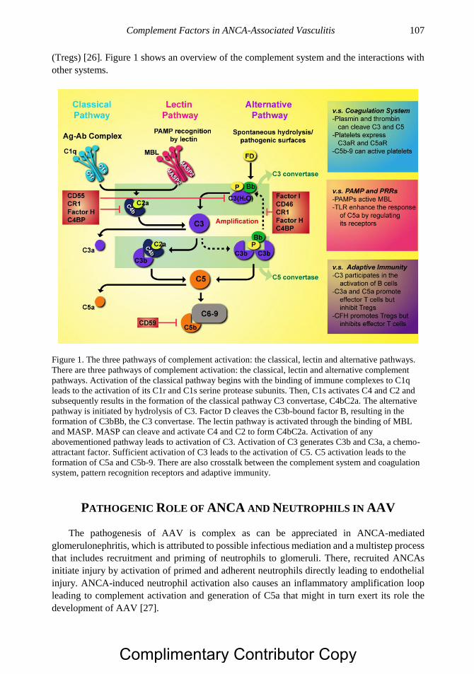
Complement Factors in ANCA-Associated Vasculitis 107
(Tregs) [26]. Figure 1 shows an overview of the complement system and the interactions with
other systems.
Figure 1. The three pathways of complement activation: the classical, lectin and alternative pathways.
There are three pathways of complement activation: the classical, lectin and alternative complement
pathways. Activation of the classical pathway begins with the binding of immune complexes to C1q
leads to the activation of its C1r and C1s serine protease subunits. Then, C1s activates C4 and C2 and
subsequently results in the formation of the classical pathway C3 convertase, C4bC2a. The alternative
pathway is initiated by hydrolysis of C3. Factor D cleaves the C3b-bound factor B, resulting in the
formation of C3bBb, the C3 convertase. The lectin pathway is activated through the binding of MBL
and MASP. MASP can cleave and activate C4 and C2 to form C4bC2a. Activation of any
abovementioned pathway leads to activation of C3. Activation of C3 generates C3b and C3a, a chemo-
attractant factor. Sufficient activation of C3 leads to the activation of C5. C5 activation leads to the
formation of C5a and C5b-9. There are also crosstalk between the complement system and coagulation
system, pattern recognition receptors and adaptive immunity.
PATHOGENIC ROLE OF ANCA AND NEUTROPHILS IN AAV
The pathogenesis of AAV is complex as can be appreciated in ANCA-mediated
glomerulonephritis, which is attributed to possible infectious mediation and a multistep process
that includes recruitment and priming of neutrophils to glomeruli. There, recruited ANCAs
initiate injury by activation of primed and adherent neutrophils directly leading to endothelial
injury. ANCA-induced neutrophil activation also causes an inflammatory amplification loop
leading to complement activation and generation of C5a that might in turn exert its role the
development of AAV [27].
Complimentary Contributor Copy
Chen Wang and Min Chen 108
ANCA IN THE PATHOGENESIS OF AAV
The most convincing evidence for the pathogenic role of ANCA comes from animal
studies. Xiao and co-workers [28] immunized MPO-/- mice with mouse MPO and obtained
MPO-ANCA IgGs or MPO-reacting splenocytes. MPO-ANCA IgGs or MPO-reacting
splenocytes were injected into recipient mice. All the mice that received MPO-ANCA IgGs
developed pauci-immune crescentic glomerulonephritis. The mice that received high doses of
MPO-reacting splenocytes developed severe NCGN with systemic necrotizing vasculitis.
Schreiber and co-workers [29] reported a modified mouse model of anti-MPO induced disease,
further documenting the pathogenic potential of ANCA. MPO knockout (MPO-/-) mice were
immunized with mouse MPO and exposed to irradiation and transplanted bone marrow (BM)
cells from either MPO-/- or MPO+/+ wild type mice. Transfer of MPO+/+ BM cells into
irradiated MPO-/- mice led to disease development, while transfer of MPO-/- BM cells into
irradiated MPO+/+ mice did not, demonstrating that MPO+/+ BM-derived cells were
pathogenic targets for anti-MPO IgG in the cause of disease. Little and co-workers [30]
immunized rats with human MPO, inducing the generation of antibodies against human MPO,
cross-reacting with rat neutrophils. Almost all the recipient rats developed pauci-immune
glomerulonephritis and pulmonary capillaritis, indicating a direct pathogenic role of ANCA in
vivo.
In vitro studies have verified the pathogenic role of ANCAs. Neutrophils stimulated by
pro-inflammatory factors, especially TNF-α or C5a, target antigens of ANCAs, MPO and PR3,
move from the cytoplasm to the cell membrane in a process termed priming. ANCAs combined
with their target antigens, and cross-linked with Fc receptors on neutrophils, are induced to
undergo a respiratory burst and degranulate, playing a direct pathogenic role in the development
of systemic vasculitis [31]. A randomized controlled trial by the European Vasculitis Study
Group (EUVAS) [32, 33] showed superiority of plasma exchange (PEX) over intravenous
methylprednisolone in restoring renal function, thus supporting a pathogenic role for circulating
ANCA in renal vasculitis.
NEUTROPHILS IN THE PATHOGENESIS OF AAV
Investigators [28] reported the development of NCGN in an experimental mouse model of
accompanied by glomerular accumulation of neutrophils and macrophages providing direct
evidence that neutrophils played a major role in the pathogenesis of anti-MPO-induced NCGN
in this animal model and implicated neutrophils in the induction of human ANCA disease. Later
studies [34] showed that neutrophil depletion prevented disease development in recipient mice.
While pro-inflammatory mediators could prime neutrophils by inducing translocation of more
ANCA antigens from the cytoplasm to the cell surface in vitro; ANCAs further activate primed
neutrophils to undergo respiratory burst and degranulation, suggesting the pathogenic role of
neutrophils in AAV.
One other important mechanism of neutrophil effector function in the development of AAV
was reported by Kessenbrock and colleagues [35] who found that ANCA-mediated neutrophil
activation induced the formation of neutrophil extracellular traps (NETs), which are fibrous
networks containing DNA, histones, and neutrophil granule proteins, triggering vasculitis and
Complimentary Contributor Copy
Complement Factors in ANCA-Associated Vasculitis 109
promoting the autoimmune response against neutrophil components. Excessive NETs
formation was further found to be related to active clinical disease in patients with AAV [36].
NETs could enhance the uptake of ANCA antigens PR3 and MPO by myeloid dendritic cells
[37], and activate autoreactive B-cells and ultimately the induction of ANCAs [38].
EMERGING ROLE OF THE COMPLEMENT SYSTEM
IN THE PATHOGENESIS OF AAV
ANCA-associated glomerulonephritis, characterized by pauci-immune NCGN
histopathologically, showed little, if any, immunoglobulin and complement deposition in the
glomeruli of AAV patients leading to the initial assumption that the complement system might
not be involved in the development of AAV. However, observations in animal models and
human disease, as well as increasingly sophisticated mechanism studies in vitro and in rodent
models in vivo, have allowed a more nuanced understanding of complement system in the
pathogenesis of AAV.
Evidence from the Animal Studies
Pre-treatment of mice with cobra venom factor to deplete complement, and C5 deficient
mice fail to develop glomerulonephritis and vasculitis whereas disease development was
comparable to wild type mice in C4-deficient mice, and C5- or factor B-deficient mice were
completely protected from the disease [39]. This suggests that complement activation via the
alternative pathway of complement system is critical to the development of AAV. The
importance of inflammatory cell activation by C5a in amplifying the anti-MPO IgG-induced
inflammatory response, was later appreciated. Huugen and colleagues [40] found that
pretreatment with anti-C5 inhibiting monoclonal antibody prevented or strongly attenuated the
development of ANCA-associated glomerulonephritis in mice. Schreiber and co-workers [41]
investigated the role of the C5aR (CD88) in a model of ANCA-induced NCGN noting that
C5aR-deficient mice were protected from developing NCGN. Xiao and investigators [42]
further elucidated the role of C5a and its receptors in a mouse model of anti-MPO-induced
NCGN, noting that CD88 deficiency ameliorated the disease, while C5L2 deficiency resulted
in more severe disease. This indicated that CD88 enhances, while C5L2 suppresses, the
inflammatory reactions. A recent study by Dick and co-workers [43] delineated the a role of
CD88 in the generation of anti-MPO autoimmunity, and effector responses that resulted in renal
injury by employing an experimental active autoimmunity to MPO by an anti-GBM globulin
trigger model, however the pathological changes were prevented by CD88 inhibition.
Evidence from Human Studies
Several studies found a certain degree of immune complex and complement C3c
deposition, which correlated with poorer renal function [44, 45]. Chen and colleagues [46]
analysed renal biopsies in 112 patients with ANCA-associated pauci-immune
Complimentary Contributor Copy
Chen Wang and Min Chen 110
glomerulonephritis noting that C3c could be detected in glomeruli of a third of specimens.
However, the intensity of C3c deposition was mild to moderate, and patients with C3c
deposition had more severe lesions in the kidneys than those without C3c deposition, along
with higher levels of proteinuria, poorer renal function at presentation, higher percentage of
crescents and more severe tubulointerstitial lesions.
Scaglioni and co-workers [47] found that 26.4% of patients with ANCA-associated
glomerulonephritis were actually not pauci-immune, and immune deposits were associated
with higher proteinuria. Researchers investigated various components of complement in kidney
specimens of patient. Xing and coinvestigators [48] found that C5b-9, C3d, and factor B, but
not C4d, could be detected in the renal specimens; while C3d and factor B co-localized with
C5b-9 in active glomerular lesions. The deposition of Bb, a marker of the alternative
complement pathway activation in glomeruli of AAV patients correlated positively with the
proportion of crescents, the extent of interstitial infiltrates, interstitial fibrosis and tubular
atrophy; and negatively with the proportion of normal glomeruli [49]. Similar results were
found in a study by Hilhorst and colleagues [50] who analyzed 187 renal biopsies of ANCA-
associated pauci-immune glomerulonephritis detecting C3c, C3d, C4d and C5b-9 in the
majority of tissue specimens. C3d and properdin staining were particularly associated with the
proportion of cellular crescents, and presence of properdin correlated with the level of
proteinuria. The staining of the two receptors of C5a, including CD88 and C5L2, were noted
mainly on infiltrating neutrophils and macrophages in kidney specimens of patients with AAV.
The staining of CD88 was down-regulated, while the staining of C5L2 was up-regulated, and
that of CD88 was associated with initial renal function and the extent of interstitial fibrosis
[51]. Given that CD88 can be rapidly internalized after the interaction with C5a [52], the down-
regulation of CD88 in renal biopsies of AAV might be a result of C5a-mediated internalization
and contribute to the self-protection mechanism to alleviate the C5a-mediated inflammation
[51].
Besides the components of complement deposited in kidneys, Gou and co-workers [49]
studied urinary levels of various components of complement in AAV patients noting that
urinary levels of Bb, C3a, C5a and C5b-9 in active stage of AAV were higher than those in
remission. In particular, correlation analysis showed that in patients with active AAV, urinary
levels of Bb correlated with disease severity, including initial serum creatinine and the
proportion of normal glomeruli in renal specimens. Circulating levels of various components
of complement in AAV were also detected and similar circumstances were found in the
circulation of AAV patients. Plasma levels of C3a, C5a, soluble C5b-9 and Bb were also
elevated, while plasma levels of properdin were lower in active AAV compared to the remission
phase. Plasma levels of C4d did not significantly differ between the active and remission stages.
Disease activity was related to plasma levels of Bb, the proportion of crescents in the renal
specimen, erythrocyte sedimentation rate (ESR), and the Birmingham Vasculitis Activity Score
(BVAS) [53].
Other studies [54-57] examined serum C3 and C4 levels in those with
hypocomplementemia at diagnosis of AAV, noting a worse prognosis when present. A study
by Manenti and colleagues [58] that included 46 patients with AAV, observing that 8 out of 30
renal biopsies had evidence of a thrombotic microangiopathy (TMA), similar to a large-Chinese
cohort [59]. Manenti and colleagues [58] also noted that 35% of patients had a decreased C3
level in the circulation, and a low C3 level was associated with decreased renal survival and
the presence of TMA.
Complimentary Contributor Copy
Complement Factors in ANCA-Associated Vasculitis 111
Using proteomic analysis by mass spectrometry, Sethi and co-workers [60] found amounts
of C3 and moderate amounts of C9, implicating activation of the alternate and terminal pathway
of complement in pauci-immune NCGN with negative ANCA serology, suggesting that its
cause may be promoted by a genetic or acquired defect in in the alternative complement
pathway.
CFH, which is a key regulator of the alternative complement pathway, inhibits
amplification of the complement cascade both in the fluid phase and on host cell surfaces.
Quantitative deficiency or impaired function of CFH, resulting in dysregulation of the
alternative complement pathway, is involved in the pathogenesis of several autoimmune
diseases, including TMA and C3 glomerulopathy [61-63]. Chen and colleagues [64] found that
plasma CFH levels in active AAV patients were lower than those in remission, and related to
parameters of disease activity, including BVAS, initial serum creatinine, and proportion of
crescents in renal specimens. In multivariate survival analysis, plasma CFH levels were
independently associated with the composite outcome of end stage renal disease (ESRD) and
death. The functional activities of CFH including its interaction with, and the regulation of C3b;
binding to mCRP and endothelial cells, and rendering of protection of host cells against
complement attack, are all impaired in AAV patients [65].
Cheng and colleagues [66] reported that the expression levels of CD46, CD55 and CD59,
the three major membrane associated regulators, were dysregulated in kidneys of patients with
AAV, and associated with the severity of renal injury of AAV patients.
Evidence from In Vitro Studies
Among the various components of the complement system, C5a plays a central role in the
pathogenesis of AAV. Schreiber and co-workers [41] found that recombinant C5a dose-
dependently primed neutrophils for an ANCA-induced respiratory burst. At the cellular level,
activation of p38 mitogen-activated protein kinase (p38MAPK), extracellular signal-regulated
kinase (ERK), phosphoinositol 3-kinase (PI3K) and protein kinase C (PKC) pathways were all
indispensable steps in the translocation of ANCA antigens and activation of neutrophils by
ANCAs [67, 68].
Among the two receptors of C5a, CD88 contributes to the initiation of acute inflammatory
responses, such as the respiratory burst and chemotaxis, while the function of C5L2 remains
more controversial. C5L2 may function as a “default” or “scavenger” receptor, by competing
with CD88 for binding of C5a [69, 70]; and a proinflammatory role for C5L2 has been
suggested [71, 72]. By blocking C5L2 with neutralized antibodies, Hao and co-workers [73]
found that C5a primed neutrophils could be mitigated, as well as the ANCA-induced C5a-
primed neutrophils respiratory burst and degranulation, suggesting C5L2 might also play a pro-
inflammatory role in C5a-primed neutrophils in ANCA-induced activation. However, that
premise was inconsistent with the findings of Xiao and colleagues [42] who demonstrated that
knockout of C5L2 was associated with more severe disease.
The reason for discrepancies in the function of C5L2 in AAV is not well understood but
may be related to different in vitro and in vivo experimental environments, and differences in
human and murine models of disease. Moreover, the function of C5L2 may be different in
various cells, species, and disease contexts [74]. Whether C5L2 plays a pro- or anti-
inflammatory role in the development of AAV will require further investigation.
Complimentary Contributor Copy
Chen Wang and Min Chen 112
Two key down-stream molecules, sphingosine-1-phosphate (S1P) and high mobility group
box 1 (HMGB1), appear to play important roles in the cellular and molecular mechanisms of
neutrophil activation by C5a. As a potent bioactive sphingolipid metabolite, S1P can regulate
a number of cellular processes in autoimmune disorder [75-77], and participates in the
regulation of the balance between expression of CD88 and C5L2 in endotoxin-induced lung
inflammation [78]. According to Hao, and Sun and colleagues [79, 80] plasma levels of S1P
are elevated in the active stage of AAV; and S1P is released from neutrophils in an autocrine
or paracrine manner upon the C5a-priming process, which further activates neutrophils and
injure glomerular endothelial. S1P may up-regulate the expression of CD88 on the surface of
neutrophils, and blocking S1P receptors could attenuate activation of C5a-primed neutrophils
by ANCAs and exert a protective effect in endothelial injury.
HMGB1 exists within the nucleus ubiquitously, but it becomes a pro-inflammatory
mediator when released extracellularly [81]. Circulating and urinary levels of HMGB1 are
associated with disease activity in AAV [82, 83], and HMGB1 could prime neutrophils by
increasing translocation of the ANCA antigens PR3 and MPO. These primed neutrophils may
be further activated by ANCAs, resulting in the respiratory burst and degranulation of
neutrophils, and the formation of NETs [84, 85]. During the process of C5a priming, HMGB1
may be released from the neutrophil cytoplasm into the extracellular space, further enhancing
the effects of C5a on neutrophil activation. Blocking HMGB1 could mitigate C5a-mediated
translocation of ANCA antigens, and ANCA-induced respiratory burst and degranulation of
C5a-primed neutrophils [86]. HMGB1 could exert its direct or indirect role on the endothelial
injury in the presence of ANCAs [87, 88]. Hence the interactions between S1P and C5a, as well
as HMGB1 and C5a, play a notable role in neutrophils for ANCA-mediated activation.
Complement activation could lead to the activation of neutrophils and further activate the
complement system especially via the alternative pathway. In the study by Xiao and co-workers
[39] supernatant from human neutrophils stimulated with ANCAs activated complement in
normal human sera, a finding indicating that ANCA-activated neutrophils may release certain
substances that subsequently activate the complement system. Camous and colleagues [89]
demonstrated that activated neutrophils could further activate the alternative complement
pathway via their cell membranes and microparticles.
Leffler and colleagues [90] discovered that NETs could activate the complement system
via the classical pathway by the interaction with C1q. Wang and co-workers [91] noted that
NETs released from ANCA-activated neutrophils could activate the complement system via
the alternative pathway. Schreiber and colleagues [92] observed that NETs provided a scaffold
for alternative pathway activation that in turn contributed to endothelial cells damage. They
further established the in vivo relevance of NETs and the requirement of RIPK1/3/MLKL-
dependent necroptosis for disease induction using both murine AAV models and in human
kidney biopsies.
An in vitro study by Chen and coinvestigators [93] revealed pathophysiological
mechanisms of CFH in the pathogenesis of AAV. On one hand, CFH inhibited ANCA-induced
neutrophil activation by interacting with neutrophils and protected against glomerular
endothelial injury, indicating a deficient function of CFH from patients with active AAV in
their ability to bind neutrophils and inhibit neutrophil activation by ANCAs. On the other hand,
MPO, which could be released from ANCA-stimulated neutrophils, influenced the complement
regulatory activity of CFH [94]. Those would contribute to better understanding of complement
system in the pathogenesis of AAV.
Complimentary Contributor Copy
Complement Factors in ANCA-Associated Vasculitis 113
EVIDENCE OF CROSS-TALK BETWEEN COMPLEMENT
AND OTHER SYSTEMS IN AAV
Cross-Talk between Complement and Coagulation System in AAV
The complement and coagulation systems have a number of common characteristics and
have many potential implications for health and diseases.
Several studies have reported that patients with AAV are in a hypercoagulable state, with
an increased risk of developing venous thromboembolic events (VTEs) [95-97], indicating the
involvement of coagulation system in AAV. C5a was found to induce expression of tissue
factor (TF) on neutrophils and endothelial cells, thus triggering the extrinsic coagulation system
[98, 99]. Huang and colleagues [100] found that stimulation of neutrophils with C5a and
ANCAs not only resulted in the respiratory burst and degranulation, but also led to release of
TF bearing microparticles and NETs; and subsequently activated the coagulation system and
thrombin generation.
Platelet counts are usually elevated in patients with active AAV. Willeke and co-workers
[101] found that platelet counts correlated with disease activity in AAV, while Miao and
colleagues [102] demonstrated that platelets were activated in AAV via thrombin-PARs
pathway, and could activate the alternative pathway of complement, further enlightening the
connection between the complement system and platelets.
Cross-Talk between Complement and TLRs in AAV
In an animal model of MPO-AAV mediated by passive transfer of anti-MPO antibodies,
LPS can aggravate the disease via a TLR4 dependent manner [103]. Wang and colleagues [104]
investigated the renal expression of TLRs in kidneys of patients with AAV by
immunohistochemistry noting that expression of TLR2 and TLR4 was up-regulated in AAV,
while that of glomerular TLR4 was inversely related to baseline serum creatinine and the
proportion of crescents. HMGB1 is a typical ligand of TLR2 and TLR4. The interaction of
HMGB1 and C5a likely plays an important role in ANCA-induced neutrophil activation [86]
like that of both complement and TLRs in AAV. The role of PRRs other than TLRs have
received little attention in AAV.
Cross-Talk between Complement and Adaptive Immunity in AAV
The study by Dick and colleagues [43] revealed the genetic absence or pharmacological
inhibition of CD88 resulting in reduced autoimmunity to MPO, with an attenuated Th1
response, increased Tregs, and reduced generation of MPO-ANCA.
One other study using murine models of MPO-AAV by the same author [105] found that
C3aR did not alter histological disease severity. However, it promoted macrophage recruitment
to the inflamed glomerulus and inhibited the generation of MPO-ANCA while not influencing
T cell autoimmunity. Thus, complement activation, especially via the alternative pathway,
plays an important role in the pathogenesis of human AAV, indicating potent and promising
Complimentary Contributor Copy
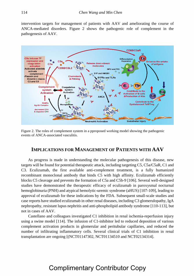
Chen Wang and Min Chen 114
intervention targets for management of patients with AAV and ameliorating the course of
ANCA-mediated disorders. Figure 2 shows the pathogenic role of complement in the
pathogenesis of AAV.
Figure 2. The roles of complement system in a pproposed working model showing the pathogenic
events of ANCA-associated vasculitis.
IMPLICATIONS FOR MANAGEMENT OF PATIENTS WITH AAV
As progress is made in understanding the molecular pathogenesis of this disease, new
targets will be found for potential therapeutic attack, including targeting C5, C5a/C5aR, C1 and
C3. Eculizumab, the first available anti-complement treatment, is a fully humanized
recombinant monoclonal antibody that binds C5 with high affinity. Eculizumab efficiently
blocks C5 cleavage and prevents the formation of C5a and C5b-9 [106]. Several well-designed
studies have demonstrated the therapeutic efficacy of eculizumab in paroxysmal nocturnal
hemoglobinuria (PNH) and atypical hemolytic-uremic syndrome (aHUS) [107-109], leading to
approval of eculizumab for these indications by the FDA. Subsequent small-scale studies and
case reports have studied eculizumab in other renal diseases, including C3 glomerulopathy, IgA
nephropathy, resistant lupus nephritis and anti-phospholipid antibody syndrome [110-113], but
not in cases of AAV.
Castellano and colleagues investigated C1 inhibition in renal ischemia-reperfusion injury
using a swine model [114]. The infusion of C1-inhibitor led to reduced deposition of various
complement activation products in glomerular and peritubular capillaries, and reduced the
number of infiltrating inflammatory cells. Several clinical trials of C1 inhibition in renal
transplantation are ongoing [(NCT01147302, NCT01134510 and NCT02134314].
Complimentary Contributor Copy
Complement Factors in ANCA-Associated Vasculitis 115
Paixão-Cavalcante and colleagues [115] reported a humanized monoclonal antibody
binding to C3 that inhibited C3 cleavage by the nephritic factor-stabilized convertase. Zhang
and co-workers [116] employed soluble CR1 therapy in C3 glomerulopathy noting that it
prevented the dysregulation of the alternative pathway C3 convertase.
Blocking of C5aR has been regarded as a novel therapeutic target for the treatment of
patients with AAV. A multicenter phase 2 randomized, double-blinded placebo-controlled trial
led by the EUVAS evaluated the efficacy and safety of CCX168 (avacopan), a small molecule
that blocks CD88 (ClinicalTrials.gov Identifier: NCT01363388) among 67 patients randomly
divided into three arms: (1) prednisone 60 mg/day; standard of care control, (2) CCX168 30
mg twice a day plus prednisone 20 mg/day; and (3) CCX168 30 mg twice a day without
prednisone. All patients received intravenous cyclophosphamide (15 mg/kg per 2 to 4 wks) or
rituximab (375 mg/m2 intravenous weekly for 4 weeks). The primary end point at 12 weeks
was reached in 70% of patients in the control group, 86% of patients in the reduced-steroid
group, and 81% of patients in the avacopan plus placebo group, demonstrating noninferiority
for both avacopan groups. There were no apparent differences in the response rates with respect
to disease category or ANCA type. However, assessment of the change in BVAS and urinary
albumin to creatinine ratio suggested that patients in the avacopan groups had a more rapid and
consistent improvement in the study parameters by 4 weeks. Quality-of-life indicators were
improved with the steroid-free protocol. The incidence of total and serious adverse events was
similar in the three arms suggesting that C5a receptor inhibition with avacopan was effective
in replacing high-dose glucocorticoids in treating vasculitis [117]. A phase 3 randomized trial
was launched (ClinicalTrials.gov Identifier: NCT02994927) to include patients with AAV and
eGFR≥15 ml/min/1.73 m2 with sustained remission as an end-point.
Other agents that target complement include TP10, which is a C3 soluble complement
receptor; and two inhibitory peptides of C3: AMY-101 and compstatin; and small molecule
factor D inhibitors to block complement activation. Although there are trials of the treatment
of C3 glomerulopathy, TMA and IgA nephropathy, there are none so far in AAV [118].
CONCLUSION
In conclusion, activation of the complement system, especially the alternative pathway,
plays a central role in the development of AAV. Among various components of the complement
system, C5a plays a central role priming neutrophils to undergo the respiratory burst with
degranulation and release of NETs in the presence of ANCAs, and contributing to the
development of systemic vasculitic lesions. The stimulation of neutrophils with C5a in
conjunction with ANCAs leads to the release of TF-bearing microparticles and NETs that
subsequently activate the coagulation system and generate thrombin. Neutrophils, ANCA and
complement system form a positive feedback loop and lead to the development of AAV. The
inhibition of complement, especially C5a, is a promising approach for remission induction in
AAV. However, whether the same mechanism can also maintain long term remission through
control of neutrophil activation remains to be explored.
Complimentary Contributor Copy
Chen Wang and Min Chen 116
REFERENCES
[1] Jennette JC, Falk RJ, Bacon PA, Basu N, Cid MC, Ferrario F, Flores-Suarez LF, Gross
WL, Guillevin L, Hagen EC et al.: 2012 revised International Chapel Hill Consensus
Conference Nomenclature of Vasculitides. Arthritis and rheumatism 2013, 65(1):1-11.
[2] Hagen EC, Daha MR, Hermans J, Andrassy K, Csernok E, Gaskin G, Lesavre P,
Ludemann J, Rasmussen N, Sinico RA et al.: Diagnostic value of standardized assays
for anti-neutrophil cytoplasmic antibodies in idiopathic systemic vasculitis. EC/BCR
Project for ANCA Assay Standardization. Kidney international 1998, 53(3):743-753.
[3] Walport MJ: Complement. First of two parts. The New England journal of medicine
2001, 344(14):1058-1066.
[4] Walport MJ: Complement. Second of two parts. The New England journal of medicine
2001, 344(15):1140-1144.
[5] Epstein J, Eichbaum Q, Sheriff S, Ezekowitz RA: The collectins in innate immunity.
Current opinion in immunology 1996, 8(1):29-35.
[6] Azumi K, De Santis R, De Tomaso A, Rigoutsos I, Yoshizaki F, Pinto MR, Marino R,
Shida K, Ikeda M, Ikeda M et al..: Genomic analysis of immunity in a Urochordate and
the emergence of the vertebrate immune system: “waiting for Godot”. Immunogenetics
2003, 55(8):570-581.
[7] Davies A, Lachmann PJ: Membrane defence against complement lysis: the structure and
biological properties of CD59. Immunologic research 1993, 12(3):258-275.
[8] Turnberg D, Botto M: The regulation of the complement system: insights from
genetically-engineered mice. Molecular immunology 2003, 40(2-4):145-153.
[9] Amara U, Flierl MA, Rittirsch D, Klos A, Chen H, Acker B, Bruckner UB, Nilsson B,
Gebhard F, Lambris JD et al..: Molecular intercommunication between the complement
and coagulation systems. Journal of immunology (Baltimore, Md: 1950) 2010,
185(9):5628-5636.
[10] Conway EM: Reincarnation of ancient links between coagulation and complement.
Journal of thrombosis and haemostasis: JTH 2015, 13 Suppl 1:S121-132.
[11] Lood C, Tyden H, Gullstrand B, Sturfelt G, Jonsen A, Truedsson L, Bengtsson AA:
Platelet activation and anti-phospholipid antibodies collaborate in the activation of the
complement system on platelets in systemic lupus erythematosus. PloS one 2014,
9(6):e99386.
[12] Martel C, Cointe S, Maurice P, Matar S, Ghitescu M, Theroux P, Bonnefoy A:
Requirements for membrane attack complex formation and anaphylatoxins binding to
collagen-activated platelets. PloS one 2011, 6(4):e18812.
[13] Sims PJ, Wiedmer T: Induction of cellular procoagulant activity by the membrane attack
complex of complement. Seminars in cell biology 1995, 6(5):275-282.
[14] Del Conde I, Cruz MA, Zhang H, Lopez JA, Afshar-Kharghan V: Platelet activation
leads to activation and propagation of the complement system. The Journal of
experimental medicine 2005, 201(6):871-879.
[15] Speth C, Rambach G, Wurzner R, Lass-Florl C, Kozarcanin H, Hamad OA, Nilsson B,
Ekdahl KN: Complement and platelets: Mutual interference in the immune network.
Molecular immunology 2015, 67(1):108-118.
Complimentary Contributor Copy
Complement Factors in ANCA-Associated Vasculitis 117
[16] Mogensen TH: Pathogen recognition and inflammatory signaling in innate immune
defenses. Clinical microbiology reviews 2009, 22(2):240-273, Table of Contents.
[17] Iwasaki A, Medzhitov R: Control of adaptive immunity by the innate immune system.
Nature immunology 2015, 16(4):343-353.
[18] Gordon S: Pattern recognition receptors: doubling up for the innate immune response.
Cell 2002, 111(7):927-930.
[19] Fujita T, Endo Y, Nonaka M: Primitive complement system--recognition and activation.
Molecular immunology 2004, 41(2-3):103-111.
[20] Raby AC, Holst B, Davies J, Colmont C, Laumonnier Y, Coles B, Shah S, Hall J, Topley
N, Kohl J et al.: TLR activation enhances C5a-induced pro-inflammatory responses by
negatively modulating the second C5a receptor, C5L2. European journal of immunology
2011, 41(9):2741-2752.
[21] Zou L, Feng Y, Li Y, Zhang M, Chen C, Cai J, Gong Y, Wang L, Thurman JM, Wu X
et al.: Complement factor B is the downstream effector of TLRs and plays an important
role in a mouse model of severe sepsis. Journal of immunology (Baltimore, Md: 1950)
2013, 191(11):5625-5635.
[22] Pepys MB: Role of complement in induction of antibody production in vivo. Effect of
cobra factor and other C3-reactive agents on thymus-dependent and thymus-independent
antibody responses. The Journal of experimental medicine 1974, 140(1):126-145.
[23] Dempsey PW, Allison ME, Akkaraju S, Goodnow CC, Fearon DT: C3d of complement
as a molecular adjuvant: bridging innate and acquired immunity. Science (New York, NY)
1996, 271(5247):348-350.
[24] Heeger PS, Lalli PN, Lin F, Valujskikh A, Liu J, Muqim N, Xu Y, Medof ME: Decay-
accelerating factor modulates induction of T cell immunity. The Journal of experimental
medicine 2005, 201(10):1523-1530.
[25] Strainic MG, Liu J, Huang D, An F, Lalli PN, Muqim N, Shapiro VS, Dubyak GR,
Heeger PS, Medof ME: Locally produced complement fragments C5a and C3a provide
both costimulatory and survival signals to naive CD4+ T cells. Immunity 2008,
28(3):425-435.
[26] Olivar R, Luque A, Cardenas-Brito S, Naranjo-Gomez M, Blom AM, Borras FE,
Rodriguez de Cordoba S, Zipfel PF, Aran JM: The Complement Inhibitor Factor H
Generates an Anti-Inflammatory and Tolerogenic State in Monocyte-Derived Dendritic
Cells. Journal of immunology (Baltimore, Md: 1950) 2016, 196(10):4274-4290.
[27] Van Timmeren MM, Chen M, Heeringa P: Review article: Pathogenic role of
complement activation in anti-neutrophil cytoplasmic auto-antibody-associated
vasculitis. Nephrology (Carlton, Vic) 2009, 14(1):16-25.
[28] Xiao H, Heeringa P, Hu P, Liu Z, Zhao M, Aratani Y, Maeda N, Falk RJ, Jennette JC:
Antineutrophil cytoplasmic autoantibodies specific for myeloperoxidase cause
glomerulonephritis and vasculitis in mice. The Journal of clinical investigation 2002,
110(7):955-963.
[29] Schreiber A, Xiao H, Falk RJ, Jennette JC: Bone marrow-derived cells are sufficient and
necessary targets to mediate glomerulonephritis and vasculitis induced by anti-
myeloperoxidase antibodies. Journal of the American Society of Nephrology: JASN
2006, 17(12):3355-3364.
Complimentary Contributor Copy
Chen Wang and Min Chen 118
[30] Little MA, Smyth CL, Yadav R, Ambrose L, Cook HT, Nourshargh S, Pusey CD:
Antineutrophil cytoplasm antibodies directed against myeloperoxidase augment
leukocyte-microvascular interactions in vivo. Blood 2005, 106(6):2050-2058.
[31] Kettritz R: How anti-neutrophil cytoplasmic autoantibodies activate neutrophils. Clinical
and experimental immunology 2012, 169(3):220-228.
[32] de Lind van Wijngaarden RA, Hauer HA, Wolterbeek R, Jayne DR, Gaskin G,
Rasmussen N, Noel LH, Ferrario F, Waldherr R, Bruijn JA et al.: Chances of renal
recovery for dialysis-dependent ANCA-associated glomerulonephritis. Journal of the
American Society of Nephrology: JASN 2007, 18(7):2189-2197.
[33] Jayne DR, Gaskin G, Rasmussen N, Abramowicz D, Ferrario F, Guillevin L, Mirapeix
E, Savage CO, Sinico RA, Stegeman CA et al.: Randomized trial of plasma exchange or
high-dosage methylprednisolone as adjunctive therapy for severe renal vasculitis.
Journal of the American Society of Nephrology: JASN 2007, 18(7):2180-2188.
[34] Xiao H, Heeringa P, Liu Z, Huugen D, Hu P, Maeda N, Falk RJ, Jennette JC: The role
of neutrophils in the induction of glomerulonephritis by anti-myeloperoxidase
antibodies. The American journal of pathology 2005, 167(1):39-45.
[35] Kessenbrock K, Krumbholz M, Schonermarck U, Back W, Gross WL, Werb Z, Grone
HJ, Brinkmann V, Jenne DE: Netting neutrophils in autoimmune small-vessel vasculitis.
Nature medicine 2009, 15(6):623-625.
[36] Kraaij T, Kamerling SWA, van Dam LS, Bakker JA, Bajema IM, Page T, Brunini F,
Pusey CD, Toes REM, Scherer HU et al.: Excessive neutrophil extracellular trap
formation in ANCA-associated vasculitis is independent of ANCA. Kidney international
2018.
[37] Lande R, Gregorio J, Facchinetti V, Chatterjee B, Wang YH, Homey B, Cao W, Wang
YH, Su B, Nestle FO et al.: Plasmacytoid dendritic cells sense self-DNA coupled with
antimicrobial peptide. Nature 2007, 449(7162):564-569.
[38] Sangaletti S, Tripodo C, Chiodoni C, Guarnotta C, Cappetti B, Casalini P, Piconese S,
Parenza M, Guiducci C, Vitali C et al.: Neutrophil extracellular traps mediate transfer of
cytoplasmic neutrophil antigens to myeloid dendritic cells toward ANCA induction and
associated autoimmunity. Blood 2012, 120(15):3007-3018.
[39] Xiao H, Schreiber A, Heeringa P, Falk RJ, Jennette JC: Alternative complement pathway
in the pathogenesis of disease mediated by anti-neutrophil cytoplasmic autoantibodies.
The American journal of pathology 2007, 170(1):52-64.
[40] Huugen D, van Esch A, Xiao H, Peutz-Kootstra CJ, Buurman WA, Tervaert JW, Jennette
JC, Heeringa P: Inhibition of complement factor C5 protects against anti-
myeloperoxidase antibody-mediated glomerulonephritis in mice. Kidney international
2007, 71(7):646-654.
[41] Schreiber A, Xiao H, Jennette JC, Schneider W, Luft FC, Kettritz R: C5a receptor
mediates neutrophil activation and ANCA-induced glomerulonephritis. Journal of the
American Society of Nephrology: JASN 2009, 20(2):289-298.
[42] Xiao H, Dairaghi DJ, Powers JP, Ertl LS, Baumgart T, Wang Y, Seitz LC, Penfold ME,
Gan L, Hu P et al.: C5a receptor (CD88) blockade protects against MPO-ANCA GN.
Journal of the American Society of Nephrology: JASN 2014, 25(2):225-231.
[43] Dick J, Gan PY, Ford SL, Odobasic D, Alikhan MA, Loosen SH, Hall P, Westhorpe CL,
Li A, Ooi JD et al.: C5a receptor 1 promotes autoimmunity, neutrophil dysfunction and
Complimentary Contributor Copy
Complement Factors in ANCA-Associated Vasculitis 119
injury in experimental anti-myeloperoxidase glomerulonephritis. Kidney international
2018, 93(3):615-625.
[44] Haas M, Eustace JA: Immune complex deposits in ANCA-associated crescentic
glomerulonephritis: a study of 126 cases. Kidney international 2004, 65(6):2145-2152.
[45] Yu F, Chen M, Gao Y, Wang SX, Zou WZ, Zhao MH, Wang HY: Clinical and
pathological features of renal involvement in propylthiouracil-associated ANCA-
positive vasculitis. American journal of kidney diseases: the official journal of the
National Kidney Foundation 2007, 49(5):607-614.
[46] Chen M, Xing GQ, Yu F, Liu G, Zhao MH: Complement deposition in renal
histopathology of patients with ANCA-associated pauci-immune glomerulonephritis.
Nephrology, dialysis, transplantation: official publication of the European Dialysis and
Transplant Association - European Renal Association 2009, 24(4):1247-1252.
[47] Scaglioni V, Scolnik M, Catoggio LJ, Christiansen SB, Varela CF, Greloni G, Rosa-Diez
G, Soriano ER: ANCA-associated pauci-immune glomerulonephritis: always pauci-
immune? Clinical and experimental rheumatology 2017, 35 Suppl 103(1):55-58.
[48] Xing GQ, Chen M, Liu G, Heeringa P, Zhang JJ, Zheng X, E J, Kallenberg CG, Zhao
MH: Complement activation is involved in renal damage in human antineutrophil
cytoplasmic autoantibody associated pauci-immune vasculitis. Journal of clinical
immunology 2009, 29(3):282-291.
[49] Gou SJ, Yuan J, Wang C, Zhao MH, Chen M: Alternative complement pathway
activation products in urine and kidneys of patients with ANCA-associated GN. Clinical
journal of the American Society of Nephrology: CJASN 2013, 8(11):1884-1891.
[50] Hilhorst M, van Paassen P, van Rie H, Bijnens N, Heerings-Rewinkel P, van Breda
Vriesman P, Cohen Tervaert JW: Complement in ANCA-associated glomerulonephritis.
Nephrology, dialysis, transplantation: official publication of the European Dialysis and
Transplant Association - European Renal Association 2017, 32(8):1302-1313.
[51] Yuan J, Gou SJ, Huang J, Hao J, Chen M, Zhao MH: C5a and its receptors in human
anti-neutrophil cytoplasmic antibody (ANCA)-associated vasculitis. Arthritis research
& therapy 2012, 14(3):R140.
[52] Huey R, Hugli TE: Characterization of a C5a receptor on human polymorphonuclear
leukocytes (PMN). Journal of immunology (Baltimore, Md: 1950) 1985, 135(3):2063-
2068.
[53] Gou SJ, Yuan J, Chen M, Yu F, Zhao MH: Circulating complement activation in patients
with anti-neutrophil cytoplasmic antibody-associated vasculitis. Kidney international
2013, 83(1):129-137.
[54] Augusto JF, Langs V, Demiselle J, Lavigne C, Brilland B, Duveau A, Poli C, Chevailler
A, Croue A, Tollis F et al.: Low Serum Complement C3 Levels at Diagnosis of Renal
ANCA-Associated Vasculitis Is Associated with Poor Prognosis. PloS one 2016,
11(7):e0158871.
[55] Fukui S, Iwamoto N, Umeda M, Nishino A, Nakashima Y, Koga T, Kawashiri SY,
Ichinose K, Hirai Y, Tamai M et al.: Antineutrophilic cytoplasmic antibody-associated
vasculitis with hypocomplementemia has a higher incidence of serious organ damage
and a poor prognosis. Medicine 2016, 95(37):e4871.
[56] Crnogorac M, Horvatic I, Kacinari P, Ljubanovic DG, Galesic K: Serum C3 complement
levels in ANCA associated vasculitis at diagnosis is a predictor of patient and renal
outcome. Journal of nephrology 2018, 31(2):257-262.
Complimentary Contributor Copy
Chen Wang and Min Chen 120
[57] Deshayes S, Aouba A, Khoy K, Mariotte D, Lobbedez T, Martin Silva N:
Hypocomplementemia is associated with worse renal survival in ANCA-positive
granulomatosis with polyangiitis and microscopic polyangiitis. PloS one 2018,
13(4):e0195680.
[58] Manenti L, Vaglio A, Gnappi E, Maggiore U, Allegri L, Allinovi M, Urban ML, Delsante
M, Galetti M, Nicastro M et al.: Association of Serum C3 Concentration and Histologic
Signs of Thrombotic Microangiopathy with Outcomes among Patients with ANCA-
Associated Renal Vasculitis. Clinical journal of the American Society of Nephrology:
CJASN 2015, 10(12):2143-2151.
[59] Chen SF, Wang H, Huang YM, Li ZY, Wang SX, Yu F, Zhao MH, Chen M:
Clinicopathologic characteristics and outcomes of renal thrombotic microangiopathy in
anti-neutrophil cytoplasmic autoantibody-associated glomerulonephritis. Clinical
journal of the American Society of Nephrology: CJASN 2015, 10(5):750-758.
[60] Sethi S, Zand L, De Vriese AS, Specks U, Vrana JA, Kanwar S, Kurtin P, Theis JD,
Angioi A, Cornell L et al.: Complement activation in pauci-immune necrotizing and
crescentic glomerulonephritis: results of a proteomic analysis. Nephrology, dialysis,
transplantation: official publication of the European Dialysis and Transplant
Association - European Renal Association 2017, 32(suppl_1):i139-i145.
[61] Ferreira VP, Herbert AP, Cortes C, McKee KA, Blaum BS, Esswein ST, Uhrin D,
Barlow PN, Pangburn MK, Kavanagh D: The binding of factor H to a complex of
physiological polyanions and C3b on cells is impaired in atypical hemolytic uremic
syndrome. Journal of immunology (Baltimore, Md: 1950) 2009, 182(11):7009-7018.
[62] Noris M, Remuzzi G: Atypical hemolytic-uremic syndrome. The New England journal
of medicine 2009, 361(17):1676-1687.
[63] Servais A, Noel LH, Roumenina LT, Le Quintrec M, Ngo S, Dragon-Durey MA, Macher
MA, Zuber J, Karras A, Provot F et al.: Acquired and genetic complement abnormalities
play a critical role in dense deposit disease and other C3 glomerulopathies. Kidney
international 2012, 82(4):454-464.
[64] Chen SF, Wang FM, Li ZY, Yu F, Zhao MH, Chen M: Plasma complement factor H is
associated with disease activity of patients with ANCA-associated vasculitis. Arthritis
research & therapy 2015, 17:129.
[65] Chen SF, Wang FM, Li ZY, Yu F, Chen M, Zhao MH: The functional activities of
complement factor H are impaired in patients with ANCA-positive vasculitis. Clinical
immunology (Orlando, Fla) 2017, 175:41-50.
[66] Cheng L, Gou SJ, Qiu HY, Ma L, Fu P: Complement regulatory proteins in kidneys of
patients with anti-neutrophil cytoplasmic antibody (ANCA)-associated vasculitis.
Clinical and experimental immunology 2018, 191(1):116-124.
[67] Hao J, Chen M, Zhao MH: Involvement of protein kinase C in C5a-primed neutrophils
for ANCA-mediated activation. Molecular immunology 2013, 54(1):68-73.
[68] Hao J, Meng LQ, Xu PC, Chen M, Zhao MH: p38MAPK, ERK and PI3K signaling
pathways are involved in C5a-primed neutrophils for ANCA-mediated activation. PloS
one 2012, 7(5):e38317.
[69] Gao H, Neff TA, Guo RF, Speyer CL, Sarma JV, Tomlins S, Man Y, Riedemann NC,
Hoesel LM, Younkin E et al.: Evidence for a functional role of the second C5a receptor
C5L2. FASEB journal: official publication of the Federation of American Societies for
Experimental Biology 2005, 19(8):1003-1005.
Complimentary Contributor Copy
Complement Factors in ANCA-Associated Vasculitis 121
[70] Kalant D, MacLaren R, Cui W, Samanta R, Monk PN, Laporte SA, Cianflone K: C5L2
is a functional receptor for acylation-stimulating protein. The Journal of biological
chemistry 2005, 280(25):23936-23944.
[71] Chen NJ, Mirtsos C, Suh D, Lu YC, Lin WJ, McKerlie C, Lee T, Baribault H, Tian H,
Yeh WC: C5L2 is critical for the biological activities of the anaphylatoxins C5a and C3a.
Nature 2007, 446(7132):203-207.
[72] Rittirsch D, Flierl MA, Nadeau BA, Day DE, Huber-Lang M, Mackay CR, Zetoune FS,
Gerard NP, Cianflone K, Kohl J et al.: Functional roles for C5a receptors in sepsis.
Nature medicine 2008, 14(5):551-557.
[73] Hao J, Wang C, Yuan J, Chen M, Zhao MH: A pro-inflammatory role of C5L2 in C5a-
primed neutrophils for ANCA-induced activation. PloS one 2013, 8(6):e66305.
[74] Li R, Coulthard LG, Wu MC, Taylor SM, Woodruff TM: C5L2: a controversial receptor
of complement anaphylatoxin, C5a. FASEB journal: official publication of the
Federation of American Societies for Experimental Biology 2013, 27(3):855-864.
[75] Choi JW, Gardell SE, Herr DR, Rivera R, Lee CW, Noguchi K, Teo ST, Yung YC, Lu
M, Kennedy G et al.: FTY720 (fingolimod) efficacy in an animal model of multiple
sclerosis requires astrocyte sphingosine 1-phosphate receptor 1 (S1P1) modulation.
Proceedings of the National Academy of Sciences of the United States of America 2011,
108(2):751-756.
[76] Niessen F, Schaffner F, Furlan-Freguia C, Pawlinski R, Bhattacharjee G, Chun J, Derian
CK, Andrade-Gordon P, Rosen H, Ruf W: Dendritic cell PAR1-S1P3 signalling couples
coagulation and inflammation. Nature 2008, 452(7187):654-658.
[77] Samy ET, Meyer CA, Caplazi P, Langrish CL, Lora JM, Bluethmann H, Peng SL:
Cutting edge: Modulation of intestinal autoimmunity and IL-2 signaling by sphingosine
kinase 2 independent of sphingosine 1-phosphate. Journal of immunology (Baltimore,
Md: 1950) 2007, 179(9):5644-5648.
[78] Bachmaier K, Guzman E, Kawamura T, Gao X, Malik AB: Sphingosine kinase 1
mediation of expression of the anaphylatoxin receptor C5L2 dampens the inflammatory
response to endotoxin. PloS one 2012, 7(2):e30742.
[79] Hao J, Huang YM, Zhao MH, Chen M: The interaction between C5a and sphingosine-
1-phosphate in neutrophils for antineutrophil cytoplasmic antibody mediated activation.
Arthritis research & therapy 2014, 16(4):R142.
[80] Sun XJ, Chen M, Zhao MH: Sphingosine-1-phosphate (S1P) enhances glomerular
endothelial cells activation mediated by anti-myeloperoxidase antibody-positive IgG.
Journal of cellular and molecular medicine 2018, 22(3):1769-1777.
[81] Seong SY, Matzinger P: Hydrophobicity: an ancient damage-associated molecular
pattern that initiates innate immune responses. Nature reviews Immunology 2004,
4(6):469-478.
[82] Ma TT, Wang H, Wang C, Chang DY, Zhao MH, Chen M: Urinary levels of high
mobility group box-1 are associated with disease activity in antineutrophil cytoplasmic
autoantibody-associated vasculitis. PloS one 2015, 10(4):e0123586.
[83] Wang C, Gou SJ, Chang DY, Yu F, Zhao MH, Chen M: Association of circulating level
of high mobility group box 1 with disease activity in antineutrophil cytoplasmic
autoantibody-associated vasculitis. Arthritis care & research 2013, 65(11):1828-1834.
Complimentary Contributor Copy
Chen Wang and Min Chen 122
[84] Ma YH, Ma TT, Wang C, Wang H, Chang DY, Chen M, Zhao MH: High-mobility group
box 1 potentiates antineutrophil cytoplasmic antibody-inducing neutrophil extracellular
traps formation. Arthritis research & therapy 2016, 18:2.
[85] Wang C, Wang H, Chang DY, Hao J, Zhao MH, Chen M: High mobility group box 1
contributes to anti-neutrophil cytoplasmic antibody-induced neutrophils activation
through receptor for advanced glycation end products (RAGE) and Toll-like receptor 4.
Arthritis research & therapy 2015, 17:64.
[86] Wang C, Wang H, Hao J, Chang DY, Zhao MH, Chen M: Involvement of high mobility
group box 1 in the activation of C5a-primed neutrophils induced by ANCA. Clinical
immunology (Orlando, Fla) 2015, 159(1):47-57.
[87] Deng H, Wang C, Chang DY, Hu N, Chen M, Zhao MH: High mobility group box-1
contributes to anti-myeloperoxidase antibody-induced glomerular endothelial cell injury
through a moesin-dependent route. Arthritis research & therapy 2017, 19(1):125.
[88] Wang C, Chang DY, Chen M, Zhao MH: HMGB1 contributes to glomerular endothelial
cell injury in ANCA-associated vasculitis through enhancing endothelium-neutrophil
interactions. Journal of cellular and molecular medicine 2017, 21(7):1351-1360.
[89] Camous L, Roumenina L, Bigot S, Brachemi S, Fremeaux-Bacchi V, Lesavre P,
Halbwachs-Mecarelli L: Complement alternative pathway acts as a positive feedback
amplification of neutrophil activation. Blood 2011, 117(4):1340-1349.
[90] Leffler J, Martin M, Gullstrand B, Tyden H, Lood C, Truedsson L, Bengtsson AA, Blom
AM: Neutrophil extracellular traps that are not degraded in systemic lupus erythematosus
activate complement exacerbating the disease. Journal of immunology (Baltimore, Md:
1950) 2012, 188(7):3522-3531.
[91] Wang H, Wang C, Zhao MH, Chen M: Neutrophil extracellular traps can activate
alternative complement pathways. Clinical and experimental immunology 2015,
181(3):518-527.
[92] Schreiber A, Rousselle A, Becker JU, von Massenhausen A, Linkermann A, Kettritz R:
Necroptosis controls NET generation and mediates complement activation, endothelial
damage, and autoimmune vasculitis. Proceedings of the National Academy of Sciences
of the United States of America 2017, 114(45):E9618-e9625.
[93] Chen SF, Wang FM, Li ZY, Yu F, Chen M, Zhao MH: Complement Factor H Inhibits
Anti-Neutrophil Cytoplasmic Autoantibody-Induced Neutrophil Activation by
Interacting With Neutrophils. Frontiers in immunology 2018, 9:559.
[94] Chen SF, Wang FM, Li ZY, Yu F, Chen M, Zhao MH: Myeloperoxidase influences the
complement regulatory activity of complement factor H. Rheumatology (Oxford,
England) 2018.
[95] Ma TT, Huang YM, Wang C, Zhao MH, Chen M: Coagulation and fibrinolysis index
profile in patients with ANCA-associated vasculitis. PloS one 2014, 9(5):e97843.
[96] Merkel PA, Lo GH, Holbrook JT, Tibbs AK, Allen NB, Davis JC, Jr., Hoffman GS,
McCune WJ, St Clair EW, Specks U et al.: Brief communication: high incidence of
venous thrombotic events among patients with Wegener granulomatosis: the Wegener’s
Clinical Occurrence of Thrombosis (WeCLOT) Study. Annals of internal medicine 2005,
142(8):620-626.
[97] Stassen PM, Derks RP, Kallenberg CG, Stegeman CA: Venous thromboembolism in
ANCA-associated vasculitis--incidence and risk factors. Rheumatology (Oxford,
England) 2008, 47(4):530-534.
Complimentary Contributor Copy
Complement Factors in ANCA-Associated Vasculitis 123
[98] Kambas K, Chrysanthopoulou A, Vassilopoulos D, Apostolidou E, Skendros P, Girod
A, Arelaki S, Froudarakis M, Nakopoulou L, Giatromanolaki A et al.: Tissue factor
expression in neutrophil extracellular traps and neutrophil derived microparticles in
antineutrophil cytoplasmic antibody associated vasculitis may promote
thromboinflammation and the thrombophilic state associated with the disease. Annals of
the rheumatic diseases 2014, 73(10):1854-1863.
[99] Ritis K, Doumas M, Mastellos D, Micheli A, Giaglis S, Magotti P, Rafail S, Kartalis G,
Sideras P, Lambris JD: A novel C5a receptor-tissue factor cross-talk in neutrophils links
innate immunity to coagulation pathways. Journal of immunology (Baltimore, Md: 1950)
2006, 177(7):4794-4802.
[100] Huang YM, Wang H, Wang C, Chen M, Zhao MH: Promotion of hypercoagulability in
antineutrophil cytoplasmic antibody-associated vasculitis by C5a-induced tissue factor-
expressing microparticles and neutrophil extracellular traps. Arthritis & rheumatology
(Hoboken, NJ) 2015, 67(10):2780-2790.
[101] Willeke P, Kumpers P, Schluter B, Limani A, Becker H, Schotte H: Platelet counts as a
biomarker in ANCA-associated vasculitis. Scandinavian journal of rheumatology 2015,
44(4):302-308.
[102] Miao D, Li DY, Chen M, Zhao MH: Platelets are activated in ANCA-associated
vasculitis via thrombin-PARs pathway and can activate the alternative complement
pathway. Arthritis research & therapy 2017, 19(1):252.
[103] Huugen D, Xiao H, van Esch A, Falk RJ, Peutz-Kootstra CJ, Buurman WA, Tervaert
JW, Jennette JC, Heeringa P: Aggravation of anti-myeloperoxidase antibody-induced
glomerulonephritis by bacterial lipopolysaccharide: role of tumor necrosis factor-alpha.
The American journal of pathology 2005, 167(1):47-58.
[104] Wang H, Gou SJ, Zhao MH, Chen M: The expression of Toll-like receptors 2, 4 and 9
in kidneys of patients with anti-neutrophil cytoplasmic antibody (ANCA)-associated
vasculitis. Clinical and experimental immunology 2014, 177(3):603-610.
[105] Dick J, Gan PY, Kitching AR, Holdsworth SR: The C3aR promotes macrophage
infiltration and regulates ANCA production but does not affect glomerular injury in
experimental anti-myeloperoxidase glomerulonephritis. PloS one 2018, 13(1):e0190655.
[106] Woodruff TM, Nandakumar KS, Tedesco F: Inhibiting the C5-C5a receptor axis.
Molecular immunology 2011, 48(14):1631-1642.
[107] Hillmen P, Hall C, Marsh JC, Elebute M, Bombara MP, Petro BE, Cullen MJ, Richards
SJ, Rollins SA, Mojcik CF et al.: Effect of eculizumab on hemolysis and transfusion
requirements in patients with paroxysmal nocturnal hemoglobinuria. The New England
journal of medicine 2004, 350(6):552-559.
[108] Hillmen P, Young NS, Schubert J, Brodsky RA, Socie G, Muus P, Roth A, Szer J,
Elebute MO, Nakamura R et al.: The complement inhibitor eculizumab in paroxysmal
nocturnal hemoglobinuria. The New England journal of medicine 2006, 355(12):1233-
1243.
[109] Legendre CM, Licht C, Muus P, Greenbaum LA, Babu S, Bedrosian C, Bingham C,
Cohen DJ, Delmas Y, Douglas K et al.: Terminal complement inhibitor eculizumab in
atypical hemolytic-uremic syndrome. The New England journal of medicine 2013,
368(23):2169-2181.
Complimentary Contributor Copy
Chen Wang and Min Chen 124
[110] Pickering MC, Ismajli M, Condon MB, McKenna N, Hall AE, Lightstone L, Terence
Cook H, Cairns TD: Eculizumab as rescue therapy in severe resistant lupus nephritis.
Rheumatology (Oxford, England) 2015, 54(12):2286-2288.
[111] Rosenblad T, Rebetz J, Johansson M, Bekassy Z, Sartz L, Karpman D: Eculizumab
treatment for rescue of renal function in IgA nephropathy. Pediatric nephrology (Berlin,
Germany) 2014, 29(11):2225-2228.
[112] Shapira I, Andrade D, Allen SL, Salmon JE: Brief report: induction of sustained
remission in recurrent catastrophic antiphospholipid syndrome via inhibition of terminal
complement with eculizumab. Arthritis and rheumatism 2012, 64(8):2719-2723.
[113] Vivarelli M, Emma F: Treatment of C3 glomerulopathy with complement blockers.
Seminars in thrombosis and hemostasis 2014, 40(4):472-477.
[114] Castellano G, Melchiorre R, Loverre A, Ditonno P, Montinaro V, Rossini M, Divella C,
Battaglia M, Lucarelli G, Annunziata G et al.: Therapeutic targeting of classical and
lectin pathways of complement protects from ischemia-reperfusion-induced renal
damage. The American journal of pathology 2010, 176(4):1648-1659.
[115] Paixao-Cavalcante D, Torreira E, Lindorfer MA, Rodriguez de Cordoba S, Morgan BP,
Taylor RP, Llorca O, Harris CL: A humanized antibody that regulates the alternative
pathway convertase: potential for therapy of renal disease associated with nephritic
factors. Journal of immunology (Baltimore, Md: 1950) 2014, 192(10):4844-4851.
[116] Zhang Y, Nester CM, Holanda DG, Marsh HC, Hammond RA, Thomas LJ, Meyer NC,
Hunsicker LG, Sethi S, Smith RJ: Soluble CR1 therapy improves complement regulation
in C3 glomerulopathy. Journal of the American Society of Nephrology: JASN 2013,
24(11):1820-1829.
[117] Jayne DRW, Bruchfeld AN, Harper L, Schaier M, Venning MC, Hamilton P, Burst V,
Grundmann F, Jadoul M, Szombati I et al.: Randomized Trial of C5a Receptor Inhibitor
Avacopan in ANCA-Associated Vasculitis. Journal of the American Society of
Nephrology: JASN 2017, 28(9):2756-2767.
[118] Reddy YN, Siedlecki AM, Francis JM: Breaking down the complement system: a review
and update on novel therapies. Current opinion in nephrology and hypertension 2017,
26(2):123-128.
Complimentary Contributor Copy
In: The Vasculitides. Volume 1 ISBN: 978-1-53615-133-6
Editor: David S. Younger © 2019 Nova Science Publishers, Inc.
Chapter 7
EXPERIMENTAL AUTOIMMUNE VASCULITIS:
INSIGHTS INTO HUMAN VASCULITIS USING
ANIMAL MODELS
Alan D. Salama, MA, PhD and Mark A. Little, PhD UCL Centre for Nephrology, London and Trinity Health,
Kidney Centre, Trinity College, Dublin, Ireland
ABSTRACT
Over the last decade several different experimental models have been described that
try to recapitulate the clinical features of ANCA associated vasculitis. Although all are
limited to some extent in their ability to mirror human disease, together they have provided
important and sometimes unexpected insights into disease pathogenesis, as well as new
avenues for therapeutic innovation.
Rodent models of both MPO-, and more recently, PR3-ANCA associated vasculitis
have replicated some of the clinical features found in affected patients with ANCA-
associated vasculitis (AAV), notably with pulmonary and renal involvement. The vast
majority of successful work in this field has concerned MPO-AAV, yet there remains the
need for a better model for aspects of PR3-AAV, including the granulomatous lesions
found in granulomatosis with polyangiitis, and the relapsing nature of the illness. By virtue
of the means by which they are induced and their time course, such experimental animal
models have not replicated the complex and varied human disease AAV phenotype.
However, the effector pathways and the final organ damage appear very similar to those
found clinically, allowing many conclusions to be drawn regarding disease etiology. This
chapter reviews the salient experimental animal models that replicate the vasculitic lesions
in human AAV. Further refinement of these models may yield the full spectrum of the
human condition, providing insight regarding organ tropism and therapeutic targets for
disease subtypes.
Keywords: vasculitis, ANCA, animal, models
Corresponding Author’s E-mail: [email protected].
Complimentary Contributor Copy
Alan D. Salama and Mark A. Little 126
INTRODUCTION
Anti-neutrophil cytoplasm antibodies (ANCA) are associated with diseases characterized
by relapsing necrotising vasculitis of small calibre blood vessels, frequently involving the
kidneys and lungs respectively leading to pauci-immune glomerulonephritis and pulmonary
capillaritis.
Many organs may be affected and, as a result, the conditions cause significant morbidity
and mortality. The precise disease triggers are not known, but clinical associations with
infection have been reported, with experimental data implicating altered responses to
microorganisms as potentially important initiators of autoimmune responses.
ANCA are directed towards two autoantigen, proteinase 3 (PR3) and myeloperoxidase
(MPO), found in neutrophil granules and monocyte lysosomes that are involved in innate
immune responses to infection, while the clinical significance of other proposed ANCA targets,
including lysosome associated membrane protein (LAMP)-2 [1] and moesin [2] remain to be
completely characterised. The ANCA-associated vasculitides (AAV) are more common in
older age and although they can be treated with immunosuppressive therapies, the disease
relapses in up to 50% of treated patients and with re-treatment, there is the increased likelihood
of treatment related adverse events.
Animal models of experimental autoimmune vasculitis (EAV) have been developed to
dissect the pathological basis of disease, to understand the role of ANCA in the
etiopathogenesis of AAV, and to replicate the clinical features. The use of murine models has
been favoured because they allow the use of genetically altered animals so that key molecules
and pathways involved in pathogenicity can be investigated.
The most novel insight gained from such an approach was the discovery the importance of
the alternate complement pathway employing passive anti-MPO antibody transfer in C5 and
factor B deficient animals [3], a finding that later prompted a therapeutic trial targeting
complement C5a receptor as adjunctive therapy in patients with AAV and renal disease.
Development of adequate models has taken considerable time, with many attempts and
refinements over the years. There remain major deficiencies in modelling granulomatous PR3-
AAV, relapsing disease overlap syndromes and non-pulmonary-renal involvement such as
cardiac, neurological and gastrointestinal disease.
Moreover, the impact of age and immune senescence, infectious triggers, and latent viral
infections on disease susceptibility and initiation need further exploration. The inability to
model PR3-AAV in mice is in part due to differences between human and rodent neutrophil
PR3 expression.
These important omissions and differences should lend some caution to translating the
findings from one animal model to all forms of human AAV. PR3-and MPO-AAV may have
certain common features, such as the occurrence of pauci-immune glomerulonephritis and
pulmonary capillaritis, but they also display many diverse characteristics that translate into
important differences in disease phenotype and outcome.
These in turn influence the long-term clinical management. Moreover, genetic studies that
demonstrate the differing molecular associations of PR3- and MPO highlight the necessity of
considering the two entities separately.
Complimentary Contributor Copy

Experimental Autoimmune Vasculitis 127
MODELS OF MPO-AAV
There have been many historical attempts at developing models of MPO-AAV, but most
have failed to mirror the clinical findings, as they were associated with significant immune
complex deposition, a major deviation from the pauci-immune characteristics of clinical
disease. Spontaneous models of crescentic nephritis and vasculitis characterised by the
presence of circulating MPO-ANCA have been reported, for example the spontaneous
crescentic glomerulonephritis/Kinjoh (SCG/Kinjoh) mice. However, they are not pauci
immune and have an array of other autoantibodies besides MPO-ANCA [4]. Significant
advances were made in this field through use of knockout and genetically manipulated mice
strains, in particular rat strains inherently susceptible to glomerulonephritis. Those strategies
included the passive transfer of pathogenic antibody, which proved useful in modelling the
acute vascular injury response, and active immunization with MPO that resulted in activation
of both humoral and cellular immune elements, consequently providing more insight into the
adaptive autoimmune response and potential responses to novel therapies.
Murine Models
Xiao and colleagues [5] developed an acute passive transfer model using purified antibody
or splenocytes taken from MPO-deficient mice immunized with purified murine MPO. This
took advantage of the allo-immune response to MPO in MPO-deficient mice, generating
antibodies and lymphocyte responses that were sufficiently potent to induce disease. Anti-MPO
antibodies injected into wild type or RAG-deficient recipient mice led to development of
necrotizing pauci-immune glomerulonephritis, and in some cases, pulmonary capillaritis over
6 to 13 days. It was further noted that splenocytes transfer recipients developed glomerular
immune deposits, whereas antibody transfer recipients remained pauci-immune. Virtually all
subsequent work was conducted with the model that used a passive transfer strategy even
though the degree of induced disease was much milder.
The development of glomerulonephritis using cell transfer was dependent on B-cells rather
than T-cells, and required the presence of neutrophils [6]. This model was replicated at other
centres and further refined to produce augmented disease through the addition of
lipopolysaccharide (LPS) [7], mimicking the effects of infection, and exacerbating
autoimmunity via toll like receptor (TLR) activation and up-regulation of tumour necrosis
factor-α. Further investigation of the model using different mouse strains, demonstrated that
the genetic background of the mice had a profound effect on the severity of the model, with
129S6 mice developing worse crescentic disease than C57BL/6 mice in which the model was
first generated [8], and with augmented pulmonary-renal disease in the absence of the inhibitory
Fcγ-II receptors [9]. That model demonstrated a critical role for alternative, but not classical,
complement components in disease induction [3], which was surprising given the lack of
clinical evidence of complement consumption in AAV. Since the available small molecule
inhibitors of C5a receptor were only active against the human form of the receptor, Xiao and
colleagues [10] adapted the murine anti-MPO transfer model by generating mice transgenic for
the human C5a receptor allowing them to test the inhibitor in the murine model and demonstrate
its efficacy in reducing renal disease.
Complimentary Contributor Copy
Alan D. Salama and Mark A. Little 128
A subsequent human trial using this C5a receptor antagonist in treatment of AAV was
initiated [11]. Further modifications of this model used a bone marrow transplant approach in
which immunization of MPO-deficient mice with murine MPO, were subjected to lethal
irradiation followed by transfer of either MPO-expressing or MPO-deficient bone marrow.
MPO expression on hematopoietic cells was necessary for disease induction [12], as only
transfer of MPO-sufficient cells resulted in disease manifested as necrotizing
glomerulonephritis. With various refinements, this model [5] revolutionised the understanding
of the mechanisms of acute anti-MPO induced vascular injury. However, all of the model
variations had limitations, as these were not autoimmune in nature, but relied instead on
alloimmune responses to MPO. Comparisons of the avidity of alloimmune anti-MPO
antibodies and other autoimmune ones formed by immunizing WT mice with MPO have not
been reported, although it is suspected that there would be significant differences reflecting the
propensity for disease induction with this model compared to previous failed attempts by direct
MPO immunization. Furthermore, as with all induced models, they begin downstream of the
break in clinical immune tolerance with a pre-formed anti-MPO antibody, restricting some of
the potential insights into clinical disease initiation. A slightly different strategy for modelling
MPO autoimmunity was adopted by Ruth and co-workers [13]. The investigators [13]
immunized C57BL/6 mice with MPO in adjuvant, both human and murine, generating cellular
and humoral anti-MPO responses, followed by low dose sub-nephritogenic nephrotoxic serum
to induce a local renal immune response within glomeruli. In contrast to the passive MPO
model, this technique depended upon T-cell and not B-cell immunity. Effector CD4+ cell
depletion attenuated crescentic glomerulonephritis and effector cell influx without altering
ANCA titers. However, B cell-deficient mice, with no ANCA, still developed severe crescentic
glomerulonephritis with accumulation of effector cells [13]. The proposed explanation of the
model was that it relied upon deposition of MPO in the glomerular circulation following
neutrophil migration and de-granulation in response to deposited nephrotoxic serum. There was
in turn a subsequent adaptive cellular immune response towards the deposited MPO already
induced by the prior immunization of MPO in adjuvant. This model was used by Gan and
colleagues [14] to illustrate the importance of the key Th17 effector cytokine IL-17A. To test
whether IL-17A also drives autoimmune delayed-type hypersensitivity in the kidney, the
investigators [14] injected MPO into the kidneys of MPO-sensitized mice noting that IL-17A
deficiency reduced accumulation of renal macrophages and renal CCL5 mRNA expression,
lending support to the clinical findings of elevated IL-17 levels in patients with acute and more
severe disease in AAV [15]. This model also demonstrated the importance of innate immune
pathway stimulation as a co-stimulus for disease induction as well as the pathogenic role of
TLR in initiating autoimmune AAV in a series of experiments in which TLR-2 induced Th17
CD4 cells while TLR-9 directed vasculitis by enhanced Th1 autoimmunity [16]. These
observations were likely due in part to the mimicking influence of infection and TLR signalling
on development of ANCA autoimmunity and organ related disease leading to
glomerulonephritis.
Rat Models
A model of MPO-AAV was developed by immunization of a susceptible Wistar-Kyoto
(WKY) rat strain with human MPO, in Complete Freund’s adjuvant (CFA).
Complimentary Contributor Copy
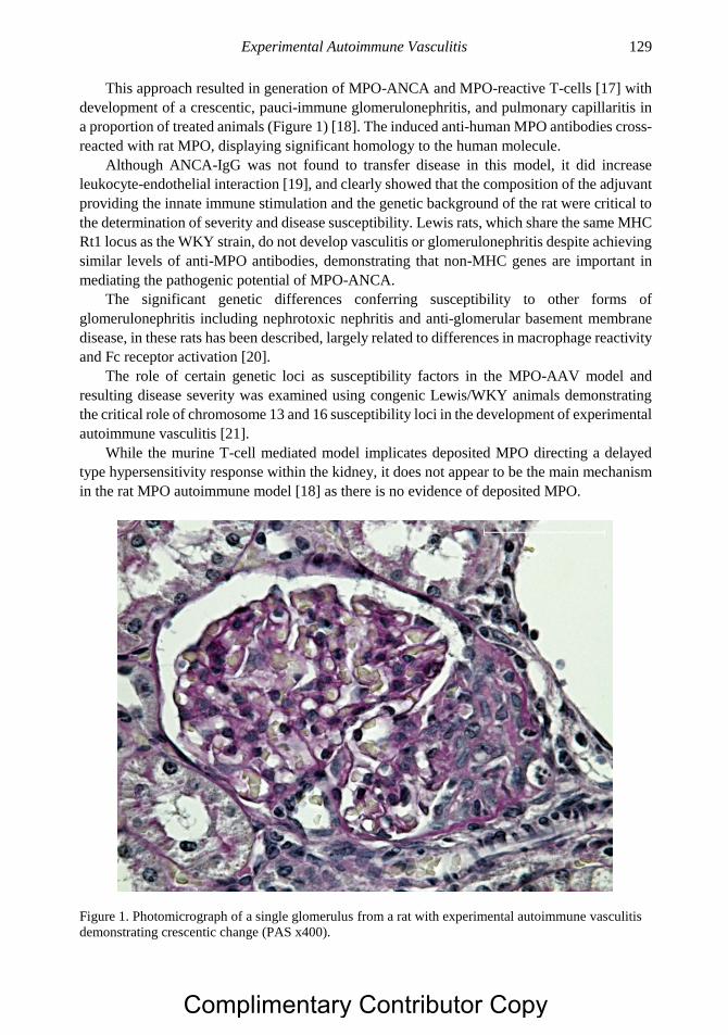
Experimental Autoimmune Vasculitis 129
This approach resulted in generation of MPO-ANCA and MPO-reactive T-cells [17] with
development of a crescentic, pauci-immune glomerulonephritis, and pulmonary capillaritis in
a proportion of treated animals (Figure 1) [18]. The induced anti-human MPO antibodies cross-
reacted with rat MPO, displaying significant homology to the human molecule.
Although ANCA-IgG was not found to transfer disease in this model, it did increase
leukocyte-endothelial interaction [19], and clearly showed that the composition of the adjuvant
providing the innate immune stimulation and the genetic background of the rat were critical to
the determination of severity and disease susceptibility. Lewis rats, which share the same MHC
Rt1 locus as the WKY strain, do not develop vasculitis or glomerulonephritis despite achieving
similar levels of anti-MPO antibodies, demonstrating that non-MHC genes are important in
mediating the pathogenic potential of MPO-ANCA.
The significant genetic differences conferring susceptibility to other forms of
glomerulonephritis including nephrotoxic nephritis and anti-glomerular basement membrane
disease, in these rats has been described, largely related to differences in macrophage reactivity
and Fc receptor activation [20].
The role of certain genetic loci as susceptibility factors in the MPO-AAV model and
resulting disease severity was examined using congenic Lewis/WKY animals demonstrating
the critical role of chromosome 13 and 16 susceptibility loci in the development of experimental
autoimmune vasculitis [21].
While the murine T-cell mediated model implicates deposited MPO directing a delayed
type hypersensitivity response within the kidney, it does not appear to be the main mechanism
in the rat MPO autoimmune model [18] as there is no evidence of deposited MPO.
Figure 1. Photomicrograph of a single glomerulus from a rat with experimental autoimmune vasculitis
demonstrating crescentic change (PAS x400).
Complimentary Contributor Copy
Alan D. Salama and Mark A. Little 130
Experimental animal models of vasculitis that may be contrived or manipulated to produce
the desired phenotype do not necessarily have to be mediated through identical pathways.
MPO-ANCA induced in MPO deficient animals by an alloimmune anti-MPO response may be
very different from the autoimmune anti-MPO response.
Indeed, it is now appreciated that there is significant heterogeneity in anti-MPO antibodies
found in patients with different epitope reactivity and with interference of immune reactive
sites under certain circumstances [22]. It would therefore be of interest to investigate the
differences between induced anti-MPO antibodies from different rodent models. Epitope
differences may contribute to why the model cannot be transferred with anti-MPO T-cells. This
may relate in part to the short duration of follow up in comparison to the immunized rat model,
which takes four or more weeks to develop similar glomerular lesions.
In the combined anti-MPO/nephrotoxic nephritis model, the anti-MPO response is a recall
response in a previously immunized animal.
When considered together, the rodent models demonstrate that vasculitis, as manifested by
pulmonary and renal disease, may be induced when a sufficient anti-MPO immunity threshold
is achieved. Patients rarely display an exclusively T- or B-cell response, or a uniquely innate
or adaptive immune response.
Rather, the observed disease represents the sum of these different pathways. Reductionist
models can provide critical insights into individual mechanisms or pathways of disease
induction, even though they may be incapable of defining them all. In particular, none of the
models currently explains why or how immune tolerance to self-antigens is broken in the first
instance, with later destructive anti MPO reactivity.
RODENT MODELS OF PR3-AAV
There have been no spontaneous experimental models in rodents of glomerulonephritis or
vasculitis in association with anti-PR3 antibodies, nor PR3-ANCA in association with other
autoantibodies. This is likely related to differences between humans and rodents in PR3 and its
cellular expression. Despite immunizing rats and mice with combinations of human or murine
PR3, chimeric molecules, and demonstrating induction of PR3-ANCA, van der Geld and
colleagues [23] were unable to experimentally generate vasculitis. Pfister and colleagues [24]
immunized PR3- and neutrophil elastase-deficient mice with murine PR3, transferred the
resultant antibodies into LPS-primed mice, and did not observe significant renal or pulmonary
pathology. There was a mild increase in panniculitis following intradermal TNF injection and
PR3-ANCA transfer compared to controls. Primo and colleagues [25] described the successful
transfer of PR3-ANCA producing splenocytes isolated from recombinant murine PR3-
immunized autoimmune-prone non-obese diabetic (NOD) mice into naïve NOD-severe
combined immunodeficient (SCID) recipients lacking endogenous T-cells and B-cells.
The immunized NOD mice developed PR3-ANCA without apparent disease despite
prolonged follow up while the NOD-SCID mice that received PR3-reactive splenocytes rapidly
developed signs of disease with vasculitis and necrotizing crescentic glomerulonephritis.
Moreover, there were no granulomata observed despite significant anti-PR3 immune reactivity;
and C57BL/6-RAG mouse recipients of the same splenocytes did not develop disease.
Complimentary Contributor Copy
Experimental Autoimmune Vasculitis 131
These findings suggested that there were protective mechanisms operating in immunized
NOD mice mediated by regulatory lymphocyte populations, while the genetic background and
degree of immunodeficiency appeared to play an important role in disease susceptibility. In the
successful anti-MPO splenocyte transfer model of Xiao et al. [5], the recipient strain was
C57BL/6-RAG2-/- further underscoring the difference between MPO and PR3 immunity. The
presence of glomerular immune deposits was not discussed in the report by Primo and
colleagues [25]. Their results were of limited applicability to human anti-PR3 associated AAV.
Relle and colleagues [26] successfully created transgenic FVB mice that expressed human PR3
into which they infused monoclonal anti-PR3 antibodies.
However, expression of PR3 was under the control of the promoter meaning that protein
was expressed only in glomeruli. Not surprisingly, they [26] did not detect any pathological
features of vasculitis as the current AAV paradigm for pathogenesis proposes interaction of the
antibody with PR3 on neutrophils and monocytes, not glomerular cells, with resultant activation
of these cells and bystander microvascular injury.
One hallmark of ANCA vasculitis is that the glomerular lesions are pauci-immune,
meaning that there is little or no local glomerular immune complex formation. One wonders
whether different results would have been obtained if human PR3 had been expressed on the
surface of myeloid cells, although it is likely that one would need to link it to other membrane
bound proteins such as CD177 to see the intracellular effect of ANCA binding.
Little and co-workers [27] recently described the use of humanized immunodeficient NOD
SCID IL2-receptor knockout mice, which received human hematopoietic stem cells and
developed a human-mouse chimeric immune system. These mice developed mild
glomerulonephritis and lung hemorrhage without evidence of granulomata following passive
transfer of PR3-ANCA containing IgG derived from patients with severe systemic vasculitis.
These exciting developments open the way for further investigation of pathogenic
mechanisms and definition of immune requirements for the induction of anti-PR3 associated
vasculitis.
CONCLUSION
By inducing an abnormal immune response to MPO, investigators have succeeded in
generating pathological changes in mice and rats that mimic human MPO-ANCA-AVV,
although most models exhibit much milder disease than that usually seen in patients. Passive
transfer of pathogenic antibody has proven a useful tool for investigating the molecular
mechanisms underlying acute vascular injury, and supports the concept that anti-MPO
antibodies, in conjunction with neutrophils, are capable of inducing such injury.
This provides a rationale in support of therapeutic strategies that aim to remove these
antibodies, such as plasma exchange and rituximab.
Active immunization of mice and rats with MPO has provided a more accurate model of
an immune response that involves both cellular and humoral immunity allowing investigation
of the role of T-cells, specifically those of the Th17 axis that have been implicated in
autoimmune disease. Such active immunization strategies are well suited to long-term
therapeutic trials of novel agents, although for this to be truly useful, the disease phenotypes of
the particular models will need to be more severe.
Complimentary Contributor Copy
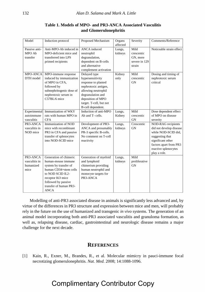
Alan D. Salama and Mark A. Little 132
Table 1. Models of MPO- and PR3-ANCA Associated Vasculitis
and Glomerulonephritis
Model Induction protocol Proposed Mechanism Organs
affected
Severity Comments/Reference
Passive anti-
MPO Ab
transfer
Anti-MPO-Ab induced in
MPO-deficient mice and
transferred into LPS
primed recipients
ANCA induced
neutrophil
degranulation,
dependent on B-cells
and alternative
complement activation
Lungs,
kidneys
Mild
crescentic
GN, more
severe in 129
strain
Noticeable strain effect
MPO-ANCA
DTH model
MPO-immune response
induced by immunization
of MPO in CFA,
followed by
subnephritogenic dose of
nephrotoxic serum in
C57BL/6 mice
Delayed type
hypersensitivity
response to planted
nephrotoxic antigen,
allowing neutrophil
degranulation and
deposition of MPO
target. T-cell, but not
B-cell dependent.
Kidney
only
Mild
crescentic
GN
Dosing and timing of
nephrotoxic serum
critical
Experimental
autoimmune
vasculitis
Immunization of WKY
rats with human MPO in
CFA
Induction of anti-MPO
Ab and T- cells.
Lungs,
Kidney
Mild
crescentic
GN
Dose dependent effect
of MPO on disease
severity
PR3-ANCA
vasculitis in
NOD mice
Immunization of NOD
mice with recombinant
PR3 in CFA and passive
transfer of splenocytes
into NOD-SCID mice
Development of PR3-
ANCA and presumably
PR-3 specific B-cells.
No comment on T-cell
reactivity
Lungs,
kidneys
Crescentic
GN
NOD-RAG recipients
did not develop disease
while NOD-SCID did,
suggesting that
significant other
factors apart from PR3
reactive splenocytes
play a role.
PR3-ANCA
vasculitis in
humanized
mice
Generation of chimeric
human-mouse immune
system by transfer of
human CD34+stem cells
to NOD SCID IL2-
receptor KO mice
followed by passive
transfer of human PR3-
ANCA
Generation of myeloid
and lymphoid
chimerism providing
human neutrophil and
monocyte targets for
PR3-ANCA
Lungs,
kidneys
Mild
proliferative
GN
Modelling of anti-PR3 associated disease in animals is significantly less advanced and, by
virtue of the differences in PR3 structure and expression between mice and men, will probably
rely in the future on the use of humanized and transgenic in vivo systems. The generation of an
animal model incorporating both anti-PR3 associated vasculitis and granuloma formation, as
well as, relapsing disease, cardiac, gastrointestinal and neurologic disease remains a major
challenge for the next decade.
REFERENCES
[1] Kain, R., Exner, M., Brandes, R., et al. Molecular mimicry in pauci-immune focal
necrotizing glomerulonephritis. Nat. Med. 2008; 14:1088-1096.
Complimentary Contributor Copy
Experimental Autoimmune Vasculitis 133
[2] Suzuki, K., Nagao, T., Nakayama, T. Proposal of anti-moesin as a novel biomarker for
ANCA-associated vasculitis. Clin. Exp. Nephrol. 2013; 17:638-641.
[3] Xiao, H., Schreiber, A., Heeringa, P., et al. Alternative complement pathway in the
pathogenesis of disease mediated by anti-neutrophil cytoplasmic autoantibodies. Am. J.
Pathol. 2007; 170:52-64.
[4] Neumann, I., Birck, R., Newman, M., et al. SCG/Kinjoh mice: a model of ANCA-
associated crescentic glomerulonephritis with immune deposits. Kidney Int. 2003; 64:
140-148.
[5] Xiao, H., Heeringa, P., Hu, P., et al. Antineutrophil cytoplasmic autoantibodies specific
for myeloperoxidase cause glomerulonephritis and vasculitis in mice. J. Clin. Invest.
2002; 110:955-963.
[6] Xiao, H., Heeringa, P., Liu, Z., et al. The role of neutrophils in the induction of
glomerulonephritis by anti-myeloperoxidase antibodies. Am. J. Pathol. 2005; 167:39-45.
[7] Huugen, D., Xiao, H., van Esch, A., et al. Aggravation of anti-myeloperoxidase antibody-
induced glomerulonephritis by bacterial lipopolysaccharide: role of tumor necrosis
factor-alpha. Am. J. Pathol. 2005; 167:47-58.
[8] Xiao, H. The role of genetic background in an animal model of ANCA associated
vasculitis. Presse Med. 2013; 42:517-520.
[9] Jennette, J. C., Xiao, H., Falk, R. J., et al. Experimental models of vasculitis and
glomerulonephritis induced by antineutrophil cytoplasmic autoantibodies. Contrib.
Nephrol. 2011; 169:211-220.
[10] Xiao, H., Dairaghi, D. J., Powers, J. P., et al. C5a Receptor (CD88) Blockade Protects
against MPO-ANCA GN. J. Am. Soc. Nephrol. 2014; 25(2):225-231.
[11] http://clinicaltrials.gov/show/NCT01363388.
[12] Schreiber, A., Xiao, H., Falk, R. J., et al. Bone marrow-derived cells are sufficient and
necessary targets to mediate glomerulonephritis and vasculitis induced by anti-
myeloperoxidase antibodies. J. Am. Soc. Nephrol. 2006; 17:3355-3364.
[13] Ruth, A. J., Kitching, A. R., Kwan, R. Y., et al. Anti-neutrophil cytoplasmic antibodies
and effector CD4+ cells play nonredundant roles in anti-myeloperoxidase crescentic
glomerulonephritis. J. Am. Soc. Nephrol. 2006; 17:1940-1949.
[14] Gan, P. Y., Steinmetz, O. M., Tan, D. S., et al. Th17 cells promote autoimmune anti-
myeloperoxidase glomerulonephritis. J. Am. Soc. Nephrol. 2010; 21:925-931.
[15] Nogueira, E., Hamour, S., Sawant, D., et al. Serum IL-17 and IL-23 levels and
autoantigen-specific Th17 cells are elevated in patients with ANCA-associated vasculitis.
Nephrol. Dial. Transplant. 2010; 25:2209-2217.
[16] Summers, S. A., Steinmetz, O. M., Gan, P. Y., et al. Toll-like receptor 2 induces Th17
myeloperoxidase autoimmunity while Toll-like receptor 9 drives Th1 autoimmunity in
murine vasculitis. Arthritis Rheum. 2011; 63:1124-1135.
[17] Chavele, K. M., Shukla, D., Keteepe-Arachi, T., et al. Regulation of myeloperoxidase-
specific T cell responses during disease remission in antineutrophil cytoplasmic
antibody-associated vasculitis: the role of Treg cells and tryptophan degradation.
Arthritis Rheum. 2010; 62:1539-1548.
[18] Little, M. A., Smyth, L., Salama, A. D., et al. Experimental autoimmune vasculitis: an
animal model of anti-neutrophil cytoplasmic autoantibody-associated systemic
vasculitis. Am. J. Pathol. 2009; 174:1212-1220.
Complimentary Contributor Copy
Alan D. Salama and Mark A. Little 134
[19] Little, M. A., Smyth, C. L., Yadav, R., et al. Antineutrophil cytoplasm antibodies directed
against myeloperoxidase augment leukocyte-microvascular interactions in vivo. Blood
2005; 106:2050-2058.
[20] Aitman, T. J., Dong, R., Vyse, T. J., et al. Copy number polymorphism in Fcgr3
predisposes to glomerulonephritis in rats and humans. Nature 2006; 439:851-855.
[21] McDaid, J. P., Tanna, A., Ghangal, G., et al. The role of quantitative trait loci (QTL) in
the pathogenesis of experimental autoimmune vasculitis. Presse Medicale 2013; 42: 688.
[22] Roth, A. J., Ooi, J. D., Hess, J. J., et al. Epitope specificity determines pathogenicity and
detectability in ANCA-associated vasculitis. J. Clin. Invest. 2013; 123:1773-1783.
[23] Van der Geld, Y. M., Hellmark, T., Selga, D., et al. Rats and mice immunised with
chimeric human/mouse proteinase 3 produce autoantibodies to mouse Pr3 and rat
granulocytes. Ann. Rheum. Dis. 2007; 66:1679-1682.
[24] Pfister, H., Ollert, M., Fröhlich, L. F., et al. Antineutrophil cytoplasmic autoantibodies
against the murine homolog of proteinase 3 (Wegener autoantigen) are pathogenic in
vivo. Blood 2004; 104:1411-1418.
[25] Primo, V. C., Marusic, S., Franklin, C. C., et al. Anti-PR3 immune responses induce
segmental and necrotizing glomerulonephritis. Clin. Exp. Immunol. 2010; 159:327-337.
[26] Relle, M., et al. PR3 antibodies do not induce renal pathology in a novel PR3-humanized
mouse model for Wegener's granulomatosis. Rheumatol. Int. 2013; 33:613-622.
[27] Little, M. A., Al-Ani, B., Ren, S., et al. Anti-proteinase 3 anti-neutrophil cytoplasm
autoantibodies recapitulate systemic vasculitis in mice with a humanized immune system.
PLoS One 2012; 7:e28626.
Complimentary Contributor Copy
In: The Vasculitides. Volume 1 ISBN: 978-1-53615-133-6
Editor: David S. Younger © 2019 Nova Science Publishers, Inc.
Chapter 8
GENETIC ASPECTS OF VASCULITIS
F. David Carmona1, PhD, Ana Márquez2,3, MD,
Javier Martín3, MD, PhD and Miguel A. González-Gay4,5,6, 1Departamento de Genética e Instituto de Biotecnología,
Universidad de Granada, Granada, Spain 2Systemic Autoimmune Disease Unit, Instituto de Investigación Biosanitaria de Granada,
Granada, Spain 3Instituto de Parasitología y Biomedicina ‘López-Neyra,’ CSIC,
Granada, Spain 4Epidemiology, Genetics and Atherosclerosis Research Group
on Systemic Inflammatory Diseases, Rheumatology Division, IDIVAL,
Santander, Spain 5School of Medicine, University of Cantabria, Santander, Spain 6Cardiovascular Pathophysiology and Genomics Research Unit,
School of Physiology, Faculty of Health Sciences, University of the Witwatersrand,
Johannesburg, South Africa
ABSTRACT
The generation of large-scale genotyping data is substantially increasing the
understanding of human diseases with a complex genetic component. Such data analysis
has been applied to the vasculitides, which are characterized by inflammatory damage of
the blood vessels. They show a very complex etiology in which both environmental and
genetic factors seem to contribute to the predisposition and clinical phenotype of the
disease. During the last decade, a large number of genetic studies, genome-wide association
studies in some cases, have been performed on vasculitides and, strikingly, most of the
identified risk loci shared susceptibility factors amongst them. This reinforces the idea that
some immunological pathways are key players in the major hallmarks of these diseases.
Corresponding Author’s Email: [email protected].
Complimentary Contributor Copy
F. David Carmona, Ana Márquez, Javier Martín et al. 136
However, since the current knowledge of the genetic basis of the vasculitides is often
based on studies performed in reduced populations, further collaborative studies on larger
cohorts are required to provide the last pieces of the genetic puzzle underlying vasculitides,
giving us a better perspective of their etiopathogenesis and allowing novel approaches for
the development of more effective therapies.
INTRODUCTION
Vasculitides present a complex etiology in which both environmental and genetic factors
appear to influence the development and progression of disease. The genetic component of
these disorders is supported by the identification of an increased risk among first-degree
relatives of affected patients and monozygotic twins. For most vasculitides familial aggregation
has been described in case reports or small series without quantification. In the absence of
family studies, differing prevalence of these diseases in different ethnic groups supports the
existence of important genetic components.
Two main approaches, candidate gene studies and high-throughput genotyping, including
genome-wide association studies (GWAS), have been used to identify disease-associated genes
in complex diseases both of which involve genotyping of genetic variations, mainly the single-
nucleotide polymorphisms (SNP) in cases and controls. Until recently, all genetic studies in
vasculitides were designed as candidate gene studies, examining a SNP or a set of
polymorphisms within certain loci with a potential role in a particular phenotype or disease. In
these studies, the selection of a candidate gene or genomic region was based on its biological
function or its location in a region implicated by previous associations or linkage studies. In
contrast to candidate gene studies, the GWAS approach, in which millions of polymorphisms
are analyzed across the whole genome, is unbiased and it does not require prior hypotheses
about the type of genes or polymorphisms most likely to be associated with the phenotypes of
interest. The recent publication of GWAS in vasculitides has increased the understanding of
potential underlying genetic mechanisms. Moreover, the two methodologies have shown that
the human leukocyte antigen (HLA) region harbors the strongest genetic contribution to the
pathophysiology of vasculitides. This genomic region occupies a large segment of DNA
extending about 3.6 megabases on the short arm of chromosome 6, and containing hundreds of
genes involved in immune function.
This chapter summarizes the genetic aspects of vasculitides giving an overview of the
recent progress in the elucidation of their genetic component. Two Chapel Hill Consensus
Conference (CHCC) nomenclatures, one in 1994 and a Revised version in 2012 [1, 2], provided
consensus on nosology and definitions for the commonest forms of vasculitis in adults based
upon the caliber of vessels involved. The Pediatric Rheumatology European Society (PRES),
European League against Rheumatism (EULAR), and the Pediatric Rheumatology
International Trials Organization (PRINTO) proposed specific classification criteria, as well
as, clinical, laboratory and radiographic characteristics for the commonest childhood vasculitis
syndromes, based upon vessel size [3, 4], similar to the 2012 Revised CHCC nomenclature [2].
These overlapping nomenclatures form the basis for the definition of most recent cohorts of
vasculitides to which reference is made in this chapter.
Complimentary Contributor Copy
Genetic Aspects of Vasculitis 137
LARGE VESSEL VASCULITIS
Giant Cell Arteritis
Increasing knowledge suggests that giant cell arteritis (GCA) has an important genetic
basis [5]. Familial aggregation with sharing of HLA alleles has been reported for GCA from
several decades ago [6-13].
However, until recently most studies were performed in unreplicated Spanish and Italian
populations [14] usually with small sample sizes. Despite these initial limitations, GCA was
always considered as one of the best examples of vasculitis in which a genetic influence has
been implicated in both disease susceptibility and severity. As discussed below, the
establishment of international collaborative groups and the recent large scale genetic studies
performed in this type of vasculitis have represented a turning point in the elucidation of its
pathogenic mechanisms [15, 16].
HLA Association
Early candidate gene studies identified HLA class II region as the most consistent
susceptibility locus for GCA. Carriage of HLA-DRB1*04 alleles (specifically DRB1*0401 and
DRB1*0404) was proposed as the main contributor of the GCA genetic component [17-29].
An increased predisposition for the development of visual manifestations as well as resistance
to corticosteroid treatment in GCA patients was related to the presence of HLA-DRB1*04 allele
[26, 30, 31], which gave an idea of the crucial role that this allele may have in GCA
pathophysiology. On the other hand, associations between GCA and alleles of the HLA class I
region, including HLA-B*15, HLA-A*31, HLA-B*8, HLA-Cw3 and HLA-Cw6, as well as the
nearby gene MHC class I polypeptide-related sequence A (MICA), encoding a stress-inducible
transmembrane molecule, were also reported [17, 32-34], although not as consistent as those
of the class II region.
The publication of the first large scale genetic study of GCA in 2015 definitively
represented a considerable leap in the understanding of the genetic contribution of the HLA
system to the susceptibility of this form of vasculitis. In collaboration with the UKGCA
Consortium, the Vasculitis Clinical Research Consortium (VCRC), and many other research
groups and hospitals from Europe, our group carried out a dense fine-mapping of immune-
related loci using the Immunochip platform, a custom high-density genotyping array designed
for immunogenetics studies [35]. One of the advantages of this chip is the presence of a high
SNP coverage in the HLA region, which allows imputation of two- and four-digit classical
HLA alleles as well as polymorphic amino acids using reference panels [36, 37]. We performed
a comprehensive interrogation of the HLA region at the amino acid level using this
bioinformatics approach in a total of 1,651 GCA patients and 15,306 unaffected controls. Our
results highlighted the class II molecules as the main contributors to GCA risk, specifically
HLA-DRβ1 and HLA-DQα1. The class I gene HLA-B was also associated, but with a
considerable lower statistical significance. We proposed a model of 3 amino acid positions that
explained most of the HLA association with GCA, which included positions 13 and 56 of HLA-
DRβ1 and HLA-DQα1, respectively, as well as 45 of HLA-B. All three positions were located
at the binding pocket of the protein, suggesting a possible involvement in antigen recognition.
The top signal corresponded to presence of histidine at position 13 of HLA-DRβ1, consistent
Complimentary Contributor Copy
F. David Carmona, Ana Márquez, Javier Martín et al. 138
with the previous associations between GCA and HLA-DRB1*04 classical alleles, as His13 is
one of the amino acids that define them [16].
Non-HLA Association
The protein tyrosine phosphatase nonreceptor type 22 (PTPN22) gene encodes a tyrosine
phosphatase known as LYP that it is expressed mainly in lymphoid tissues and is involved in
several signaling pathways associated with the immune response, including the T-cell receptor
(TCR) pathway in which LYP plays a central role in its negative control, but also the humoral
activity of B-cells. A functional PTPN22 variant (rs2476601/R620W) has been associated with
several autoimmune diseases, representing the clearest example of a common genetic risk factor
in autoimmunity [38]. PTPN22 R620W represented the strongest non-HLA association signal
in the Immunochip of GCA [16], which confirmed previously reported observations through
the candidate gene approach [39]. This association was initially identified in a large discovery
cohort from Spain and subsequently replicated in three European replication cohorts from the
United Kingdom (UK), Germany and Norway. It has been demonstrated that the R620W
change interferes with the ability of LYP to bind its partner c-src tyrosine kinase (CSK) [40]
which prevents the formation of the complex between both proteins and the suppression of the
T-cell activation.
The first GWAS performed in GCA was published on 2017. The involvement of the
European Vasculitis Genetics Consortium (EVGC) allowed the recruitment of three additional
cohorts to the first case/control set of the Immunochip, comprising a total of 2,134 GCA
patients and 9,125 controls from 11 populations of European origin (Spain, Italy, UK, USA,
Canada, France, Germany, Norway, The Netherlands, Ireland and Switzerland) [15]. The
analysis of a total of 1,844,133 genetic markers across the whole genome led to the
identification of two genes involved in angiogenesis and vascular remodeling as the most
relevant non-HLA genetic factors of GCA, i.e., plasminogen (PLG) and prolyl 4-hydroxylase
subunit alpha 2 (P4HA2). The first one encodes a precursor of both plasmin and angiostatin
proteins [41]. The GCA-associated PLG variants were predicted to affect their gene expression,
which could unbalance the plasminogen/plasmin system favoring a pro-inflammatory
environment [15]. On the other hand, P4HA2 encodes the α subunit of the collagen prolyl 4-
hydroxylase, a hypoxia responsive enzyme involved in collagen biosynthesis [42]. Some of the
P4HA2 alleles that conferred risk to GCA were predicted to alter P4HA2 expression in
lymphoblastoid cells and the aorta [15].
Different lines of evidence clearly suggest that Th17 cells are crucial players in GCA
development. They include both presence of this cell type and increased levels of IL17 in the
vascular lesions of GCA patients. Interestingly, corticosteroid therapy seems to be more
effective on Th17 cells than on Th1 cells under a GCA background [43-45]. Consistent with
these observations, associations between GCA and IL17A gene polymorphisms were described
in a candidate gene study performed on 2014 [46].
The NLR family pyrin domain containing 1 (NLRP1) gene, another autoimmune disease-
associated locus, was also confirmed as a GCA susceptibility gene in a study on Spanish and
Italian populations [47]. This gene encodes a cytoplasmic protein involved in the assembly of
the inflammasome that activates caspases 1 and 5 which results in the activation of
proinflammatory cytokines. As expected, considering its inflammatory nature, different
cytokines have been implicated in GCA pathogenesis. Amongst them, the interleukin 10 (IL10)
gene represents the most consistent association [48, 49]. This gene encodes an anti-
Complimentary Contributor Copy
Genetic Aspects of Vasculitis 139
inflammatory Th2 cytokine with pleiotropic effects in the immune system, including the
negative control of Th1 cytokines such as interferon gamma (IFN-γ), the regulation of the B
cell function, and the repression of nuclear factor kappa-light-chain-enhancer of activated B
cells (NF-κB) activity. Two independent studies in Italian [48] and Spanish [49] populations
indicated that variation of the IL10 promoter region may be involved in the pathogenic
mechanisms leading to GCA.
Other genes encoding cytokine or cytokine receptors that have been suggested as possible
genetic risk factors for GCA: IL33, encoding an IL-1 family member with strong
immunomodulatory functions; IL4, encoding a protein that acts reciprocally with IFN-γ
inducing the Th2 immune response; IL6, with pleiotropic effects that may exert both pro-
inflammatory and anti-inflammatory functions; IL18, encoding a pro-inflammatory cytokine
with cytotoxic T-cell functions; monocyte chemotactic protein-1 (MCP1), encoding a
chemoattractant protein for mononuclear cells to the sites of inflammation; regulated and
normal T-cell expressed and secreted (RANTES), encoding a potent chemotactic factor for
immune cells; the IL2/IL21 region of cytokines important in the correct function of the immune
system; IL12 receptor beta 2 (IL12RB2), a key player in the inflammatory response; tumor
necrosis factor (TNF), encoding a proinflammatory cytokine involved in the regulation of
immune cells; and IFNG, encoding a key cytokine involved in innate and adaptive immunity
against intracellular pathogens and tumor control [50-59].
The possible role of a number of important molecules for the endothelial function in GCA
predisposition and severity has been also explored. Although most of the studies were low-
powered and without replication, evidence of association was observed for intercellular
adhesion molecule 1 (ICAM1), encoding a cell surface glycoprotein involved in the interactions
between immune and endothelial cells during inflammation; vascular endothelial growth factor
(VEGF), encoding an important vasculogenic mediator; cytokine-inducible and endothelial
nitric oxide synthases (NOS2A and NOS3), involved in the regulation of the oxidative stress;
and matrix metalloproteinase 9 (MMP9), encoding an enzyme with proteolytic activity on the
extracellular matrix [52, 60-66].
Finally, genes of the innate immunity may also have a role in GCA pathogenesis.
Significant associations have been reported for toll-like receptor 4 (TLR4), implicated in signal
transduction events induced by lipopolysaccharide from Gram-negative bacteria; Fc gamma
receptor 2A and 3A (FCGR2A and FCGR3A), encoding cell-surface proteins of the
immunoglobulin superfamily; and myeloperoxidase (MPO), an important gene in the
neutrophil function [67-69]. However, all these associations require confirmation in larger and
independent cohorts.
Takayasu Arteritis
A few candidate gene studies exploring the possible role of genetic polymorphisms in
Takayasu arteritis (TAK) were performed before the publication of three recent large scale
analyses, i.e., a genome scanning of exonic regions in a Japanese cohort [70], an Immunochip
in Turkish and North Americans [71], and an unbiased GWAS also in Turkish and North
Americans [72].
Complimentary Contributor Copy
F. David Carmona, Ana Márquez, Javier Martín et al. 140
HLA Association
Different genetic studies showed that HLA-B alleles are involved in the disease
susceptibility. Specifically, HLA-B*5201 was associated with TAK in multiple cohorts of
diverse ethnicities including different populations from Asia and Mexico [73-78], which was
confirmed in the GWAS of Japanese [70]. However, this association was not observed in North
American and Arab populations [79, 80]. In the former [79], a positive correlation of HLA-
DR4/MB3 and a strongly negative association of HLA-DR1 with TAK were reported, whereas
in Arab patients [80] the association with this region was narrowed down to HLA-A2, HLA-
A9, HLA-Bw35 and HLA-DR7 alleles.
Studies on polymorphisms of the gene encoding the human complement factor 4 (C4),
located at the class III region of the HLA, showed a significantly high frequency of C4A2 and
C4BQ0 allotypes in strong association with HLA-Bw52 [81]. Furthermore, the analysis of
MICA, situated between the class I and class III regions, revealed an association between the
MICA-1.2 allele and TAK, even in the absence of HLA-B52 [82].
A subsequent study [83] in which five microsatellites, C1-2-A, MIB, C1-4-1, C1-2-5 and
C1-3-1, around the HLA-B and MICA genes were analyzed in Japanese TAK patients, suggested
two different disease-susceptibility loci for TAK, one that mapped near the C1-2-A locus, and
another closely linked to the HLA-B gene. Very recently, the MICA and the HLA-DQB1/HLA-
DRB1 regions were also described as TAK risk factors in the Immunochip study on Turkish
and American cohorts [71].
Non-HLA Association
The most consistent associations with TAK outside the HLA region rely on IL6, the
leukocyte immunoglobulin like receptor B3 (LILRB3) region, IL12B and the
FCGR2A/FCGR3A loci. The two first loci were identified in the GWAS on 2015 [72]. IL6 was
previously suggested as a TAK locus by candidate gene studies [84]. This gene encodes an
important cytokine with key roles in different aspects of the immune response, including the
differentiation of Th17 cells and regulatory T-cells (Treg). Consistent with this association, an
altered production of several cytokines, including IL-6, IL-2 and IL-12, were detected in TAK
patients. Increased levels of IL-6 and IL-12 and a lower number of CD3+ T cells producing IL-
2 in the active phase of the disease were described [85-87]. Polymorphisms of the genes
encoding these cytokines were correlated with altered protein levels [84, 88-93]. Regarding the
second non-HLA signal of the GWAS, LILRB3 is located in an immune-regulatory gene rich
region on chromosome 19q13.4, which contains genes encoding for killer immunoglobulin-
like receptors (KIR), leucocyte immunoglobulin-like receptors (LILR), and leucocyte-
associated immunoglobulin-like receptors (LAIR). Remarkably, the top marker within the
associated region was related to reduced transcription levels of multiple genes including
LILRB3 [72]. On the other hand, IL12B showed a high association peak in both the exome
screening study and the Immunochip, whereas FCGR2A/FCGR3A was identified as a
susceptibility region for TAK in the Immunochip [70, 71].
Two genes encoding members of the IL-1 cytokine family, IL1B encoding IL-1β, and IL-
1 receptor antagonist (IL1RN) encoding IL1-Ra, were involved in TAK predisposition [94]. IL-
1 may mediate the inflammatory response occurring in the vascular wall by activating
monocytes and expression of adhesion molecules on endothelial cells, inducing secretion of
other inflammatory mediators. IL1-Ra inhibits the activities of IL-1α and IL-1β, and modulates
a variety of IL-1 related immune and inflammatory responses.
Complimentary Contributor Copy
Genetic Aspects of Vasculitis 141
Functional and genetic studies [95-97] have reported an implication of both genes in
cardiovascular disease, a common feature in TAK patients. In addition, different
polymorphisms of IL1B and IL1RN were associated with TAK in the Mexican population [94],
which suggests a crucial role of IL-1 in the vasculitis lesions of TAK.
Genes encoding cardiovascular-related molecules may also play a role in TAK
development [98, 99]. For instance, paraoxonase 1 (PON1), a high density lipoprotein (HDL)-
associated enzyme involved in prevention of lipid peroxidation, was diminished in activity in
TAK patients suggesting a possible role of these lipoproteins in TAK vascular dysfunction [98].
A recent study in the Mexican population [99] suggested that variation within PON1 may be
involved in TAK susceptibility by influencing development and progression of arterial damage.
MEDIUM VESSEL VASCULITIS
Kawasaki Disease
A large number of genetic studies have been conducted in order to unravel the genetic
component of Kawasaki disease (KD) [100] focused mainly on candidate genes implicated in
the immune response. In addition, since the most serious complication of KD is the
development of coronary artery lesions (CAL), the role of cardiovascular-related loci has been
extensively studied. However, sample sizes have often been insufficient and only a few
associations have been replicated in subsequent studies [100].
In the last years, six GWAS on KD have been published [101-106]. This approach has
represented an important step forwards to the understanding of the genetic basis of this
vasculitis.
HLA Association
The involvement of the HLA region in KD is controversial. Association between KD and
HLA class I molecules was evidenced [107-111], but the results were inconsistent. Early studies
reported an association between HLA-Bw22 now called Bw54, and KD in the Japanese
population [107, 108], whereas the HLA-Bw51 and HLA-B44 alleles were predominantly
associated with the disease in Caucasians [109-111]. On the contrary, in a recent GWAS [105],
a significant association of HLA class II region with Japanese KD patients was identified.
Specifically, the strongest association signal was observed at the intergenic region between
HLA-DQB2 and HLA-DOB (rs2857151). This association was replicated in a subsequent Han
Chinese study [112]. A suggestive association signal was also observed in the HLA class III
region (rs2516390), close to the nuclear factor of kappa light polypeptide gene enhancer in the
B-cell inhibitor-like 1 (NFKBIL1) and lymphotoxin alpha (LTA) genes.
However, no significant associations of HLA alleles were documented in other published
GWAS. Thus, a consistent allele or haplotype of HLA conferring KD susceptibility has not yet
been identified.
Non-HLA Association
Several genes have been suggested to play a role in KD susceptibility through candidate
gene studies [113-125]. Susceptibility loci include 1) cytokines and their receptors, such as IL4,
Complimentary Contributor Copy
F. David Carmona, Ana Márquez, Javier Martín et al. 142
TNF, the chemokine (C-C motif) receptor 2-3-5 (CCR2-CCR3-CCR5) gene cluster which
encodes three receptors of different chemokines that play a critical role in the selective
accumulation and activation of inflammatory cells in affected tissues; 2) genes encoding
cardiovascular-related molecules, including VEGF implicated in the predisposition to this
vasculitis and CALs development in different populations, and MMP3, 9, 13 also associated
with CAL; and 3) genes involved in innate immunity, like mannose binding lectin 2 (MBL2)
involved in complement activation and associated with an increased risk of CAL, and NLRP1
involved in the inflammasome assembly.
Potential loci implicated in KD susceptibility in Japanese families include the 19q32.2 and
4q35 regions. Subsequent fine-scale studies [126] of the 19q32.2 region have led to the
identification of functional polymorphisms of the inositol 1,4,5-triphosphate 3-kinase C
(ITPKC) gene significantly associated with the susceptibility to KD, with particular risk of
developing CAL in both Japanese and Caucasian patients. This gene encodes one of the three
isoenzymes that phosphorylate inositol 1, 4,5-trisphosphate (IP3) acting as an important
molecule in the regulation of T-cell activation.
Regarding the 4q35 linkage region, several immune genes have been mapped around the
peak of linkage, including the IFN regulatory factor 2 (IRF2), TLR3 and caspase 3 (CASP3)
that play central roles in apoptosis. A subsequent positional candidate gene study [127] in
Japanese and Americans of European ancestry subjects identified a functional CASP3 variant
that altered the gene expression in immune effector cells. This SNP was shown to influence
KD susceptibility. Both the ITPKC and CASP3 associations with KD were replicated in the
Taiwanese population [128, 129].
More recently, based on previously published suggestive linkage signals at chromosome
12q24 [125], a missense SNP located within exon 2 of the calcium release-activated calcium
modulator 1 (ORAI1) gene was described as a risk factor for KD in the Japanese population.
This gene encodes a membrane calcium selective ion channel involved in T-cell activation
through the Ca2+/NFAT pathway. Strikingly, the frequency of the risk allele is more than 20
times higher in Japanese compared to that in Europeans, suggesting that ORAI1 may be related
to the differential KD incidence between populations of Asian and European descent [130].
In 2009, the first GWAS performed on KD Caucasian patients [101] led to the
identification of eight putative novel susceptibility loci. Five of these genes,
calcium/calmodulin-dependent protein kinase II delta (CAMK2D), CUB and Sushi multiple
domains 1 (CSMD1), ligand of numb-protein X1 (LNX1), N-acetylated alpha-linked acidic
dipeptidase-like 2 (NAALADL2), and t-complex 1 (TCP1), formed a functionally close network
of 35 genes defined in the Ingenuity database (Ingenuity Systems, Redwood City, CA,
www.ingenuity.com). This gene network suggests possible mechanisms by which one or more
infectious triggers may lead to deregulated inflammation and apoptosis, and cardiovascular
pathology.
In 2011, another GWAS [103] was conducted in a Korean population, and two novel
susceptibility signals were identified: a SNP close to the Dab reelin signal transducer homolog
1 (DAB1) gene associated with KD predisposition, and an intronic variant of pellino E3
ubiquitin protein ligase 1 (PELI1) gene which is involved in the TLR pathway associated with
CAL development. The first GWAS in the Han Chinese population [106] identified several
putative genetic variants associated with KD susceptibility, although none of them reached
GWAS statistical significance due to a limited sample size.
Complimentary Contributor Copy
Genetic Aspects of Vasculitis 143
The highest peaks included three SNP close to coatomer protein complex beta-2 subunit
(COPB2) gene, one located within the intronic region of endoplasmic reticulum amino
peptidase 1 (ERAP1), and six clustered in an area containing immunoglobulin heavy chain
variable region (IGHV) genes.
A well-powered GWAS [102] in KD patients from different populations of European
ancestry (Australia, UK, Netherlands and US) identified two loci that exceeded the threshold
for GWAS significance. One of them was ITPKC, confirming this gene as a risk factor for KD,
and a functional polymorphism (H131R, rs1801274) of FCGR2A. Finally, both Taiwanese
[104] and Japanese [105] groups independently reported GWAS results in 2012 [104, 105]. In
both studies, significant associations were observed in the B-lymphoid tyrosine kinase (BLK)
region. The most strongly associated polymorphism in the Taiwanese study [104] was in high
linkage disequilibrium (LD) with a genetic variant associated with systemic levels of BLK and
increased risk of systemic lupus erythematosus (SLE). SNP around cluster domain 40 (CD40,
a member of the TNFR superfamily that potentially contributes to inflammation and
autoimmune disease processes through the selection of autoreactive T cells in the thymus and
the activation of B- and T- cells) were also associated with KD at GWAS level in both studies.
Interestingly, increased cell surface expression of CD40L, the ligand of CD40, and elevated
serum levels of soluble CD40L have been detected in KD. On the other hand, the Japanese
study also replicated the association of the previously identified FCGR2A functional SNP.
On 2016, Shimizu and colleagues [131] performed a pathway-based analysis followed by
gene stability selection using the previously published GWAS data of individuals of European
ancestry [102]. They identified variants associated with both susceptibility to KD and aneurysm
formation within the solute carrier family 8 member A1 (SLC8A1) gene, which encoded protein
contributes to the regulation of calcium flux and calcium-dependent cellular processes. The
results were further replicated in an independent cohort of Japanese origin [131].
Finally, a recent whole genome screening of a 6-member African American family with
two affected KD children identified rare compound heterozygous variants in another TLR
member, TLR6, shared between both affected siblings [132].
Small Vessel ANCA-Associated Vasculitis
Early genetic studies in antineutrophil cytoplasmic antibody (ANCA)-associated vasculitis
(AAV), which comprises granulomatosis with polyangiitis (GPA) [Wegener type], microscopic
polyangiitis (MPA) and eosinophlic granulomatosis with polyangiitis (EGPA) (Churg-Strauss
type), focused on self-evident candidate genes encoding respective ANCA target antigens,
proteinase 3 (PR3) and myeloperoxidase (MPO).
Subsequent studies reported a clear familial aggregation of GPA, with a relative risk of
1.56 for first-degree relatives, which indicated the importance of the genetic component for this
disease [133]. In 2012, the ‘European Vasculitis Study Group’ (EUVAS) conducted the first
GWAS in AAV [134] (including a large cohort of GPA and MPA patients), reporting more
consistent associations with the disease, and suggesting genetic distinctions between the
clinical entities of AAV that may be associated with ANCA specificity. Subsequently, two
additional GWAS were performed by the VCRC (one only in patients diagnosed with GPA and
another in a cohort including GPA and MPA cases) [135, 136]. Those studies definitively
helped to increase the knowledge on AAV genetics.
Complimentary Contributor Copy
F. David Carmona, Ana Márquez, Javier Martín et al. 144
HLA Association
Several early low-power studies [137-139] suggested that different HLA class I and class
II loci might be involved in the predisposition to GPA including HLA-DQw7, HLA-DR3,
HLA-DR1, HLA-DR9 and HLA-B50. However, only the HLA DPB1*0401 allele was firmly
associated with GPA, although other neighboring loci such as ring finger protein 1 (RING1), a
transcriptional repressor, and retinoid X receptor beta (RXRB), involved in mediating the effects
of retinoic acid, may have independent effects [140,141].
HLA-DP also showed the highest association signals in the three AAV GWAS [134-136],
with HLA-DPB1*04 as main contributor of GPA risk [135]. Interestingly, the main
associations in the HLA region were observed in the subgroup of patients with presence of anti-
PR3 antibodies, although there were also associations in the cohort of AAV patients positive
for anti-MPO [134]. Hence, it is likely that HLA-DP alleles are a key factor for autoantibody
production in AAV, particularly for anti-PR3.
An increased prevalence of HLA-DR4 was also associated with AAV in general, and more
specifically with GPA. One study [142] performed in a Dutch population showed that the three
subtypes of AAV had a similar HLA antigen distribution, except for HLA-DR1, which was
more prevalent in GPA patients, and HLA-DR8, which showed a decreased frequency in EGPA
patients in comparison with the other two subtypes. Other evidences suggested that HLA-
DRB4 could represent a risk factor specific for EGPA [143].
Non-HLA Association
PRTN3 and MPO encode the two major ANCA autoantigens PR3 and MPO respectively.
PR3 is a neutrophil intracellular protease that can be also located on the plasma membrane.
It has been proposed that elevated membrane expression of PR3 is involved in progression
of chronic inflammatory diseases, including AAV, and that this fact is likely to be genetically
determined.
More than one decade ago, the entire coding and promoter sequences of PRTN3 were
analyzed to identify polymorphisms that could influence the pathophysiology of GPA. The
genetic variations described in that study [144] included seven SNPs, one amino acid change
(Val119Ile), one 84 base pair insertion/deletion (INDEL), and a microsatellite.
Among them, only one SNP affecting a putative transcription factor-binding site
(rs62132295, A-564G) was associated with GPA. In the AAV GWAS [134], this PRTN3
variant was not associated at GWAS level, although a suggestive P-value with GPA but not
with MPA was yielded. However, a stronger size effect was observed when the GPA patients
were stratified accordingly with the type of autoantibody production instead of the clinically
defined syndromes.
Patients positive for anti-PR3 show a stronger association with PRTN3 rs62132295 than
those positive for anti-MPO, when compared with cohort controls. Additionally, the allele
frequencies of this SNP also differed considerably between both subsets of patients. Similar
results were observed for the serpin A1 (SERPINA1) gene [134], which encodes a serine
protease inhibitor known as α1-antitrypsin whose targets include PR3 that protects tissues
against enzymes from inflammatory cells like neutrophils. The Z (null) allele seems to be
specifically associated with anti-PR3 presence rather than with the clinical subtypes, since the
most significant P-value was obtained when the AAV patients showing this autoantibody were
compared against controls. SERPINA1 was the only locus outside the HLA region that reached
GWAS significance. The association between this gene and ANCA positivity has been
Complimentary Contributor Copy
Genetic Aspects of Vasculitis 145
confirmed in many candidate gene studies [145-150]. The autoimmune disease-associated
PTPN22 functional variant R620W was also reported to confer risk to AAV through candidate
gene studies [151,152]. Specifically, it was first implicated in the susceptibility to develop GPA
[152], and this association was further confirmed in a higher cohort in which both GPA and
MPA patients were included [151].
The second GWAS was performed in North American GPA patients of European descent.
The study reported semaphorin 6A (SEMA6A), on chromosome 5, as a novel susceptibility
marker for GPA. Although the role of semaphorins remains unclear, they seem to control axon
guidance, vasculogenesis, cardiogenesis, osteoclastogenesis, tumor metastasis, and immune
regulation [135]. The same consortium conducted another GWAS in GPA and MPA patients
from European origin on 2017, and confirmed PRTN3, SERPINA1 and PTPN22 as the strongest
non-HLA susceptibility genes for this forms of vasculitis [136].
Another gene that has been associated with AAV predisposition is cytotoxic T-lymphocyte
associated antigen 4 (CTLA4), which encodes a central regulatory molecule expressed on T-
cells that inhibits T-cell function and peripheral tolerance. It has been reported that elevated
levels of CTLA4 are expressed in GPA patients [153].
In concordance, many studies have described associations of different CTLA4
polymorphisms with AAV [154-159], thus supporting the idea that CTLA4 represents another
common susceptibility factor in autoimmunity.
There are other genes of the immune system that showed less consistent evidence of
association with AAV, and confirmation in independent cohorts is still required [160]. These
include, IL10, CD226, an adhesion molecule expressed on the surface of natural killer cells,
platelets, monocytes and T cells; IL2 receptor alpha (IL2RA), an important Treg marker;
leukocyte immunoglobulin-like receptor A2 (LILRA2), an immunoreceptor expressed in
immune cells; integrin, beta 2 complement component 3 receptor 3 and 4 subunit (ITGB2),
which participates in cell adhesion as well as cell-surface mediated signaling; and FCGR2A/3B
members of IgG receptor family. Except for SERPINA1, PRTN3, CTLA4 and CD226, none of
the above described genetic associations have been confirmed at the 95% significance level in
the AAV GWAS [134].
Small Vessel Immune Complex Vasculitis
IgA Vasculitis/Henoch-Schönlein Purpura
Similar to the other vasculitides, the understanding of the genetic background of IgA
vasculitis/Henoch-Schönlein purpura (IgAV/HSP) remains elusive, and further research on
well-powered cohorts is still needed to unravel the genetic basis and pathogenic mechanisms
leading to this vasculitis.
Until the publication of the first GWAS of IgAV/HSP in 2017, the current knowledge of
the genetic component of IgAV/HSP was based exclusively on candidate gene studies including
genes mainly involved in the immune and inflammatory responses, but also in endothelial
function, mechanisms of complement system activation, coagulation and fibrinolysis, and
antioxidation [161].
Complimentary Contributor Copy
F. David Carmona, Ana Márquez, Javier Martín et al. 146
HLA Association
There is a limited number of studies on the HLA region in IgAV/HSP, all of which have
been performed in Spanish, Italian and Turkish populations [161, 162]. In relation to the HLA
class II region, an association of the HLA-DRB1*01, HLA-DRB1*07, and HLA-DRB1*11
alleles with IgAV/HSP was first reported in Italy [163] and subsequently confirmed in
independent cohorts from Spain [164, 165] and Turkish [166]. The HLA alleles DRB1*01 and
DRB1*11 increase the risk for the disease onset, whereas DRB1*07 confer a protective effect
to the disease. Also, a protective effect against IgAV/HSP development was described in
patients carrying the HLA-DRB1*03 haplotype [165].
The publication of the first GWAS of IgAV/HSP confirmed the HLA class II region as the
major susceptibility locus for IgAV/HSP. The authors also used the imputation method
described in the GCA section to narrow down the HLA association to a region between HLA-
DQA1 and HLA-DQB1. As observed in GCA, positions 13 and 11 of the HLA-DRβ1 carried
the strongest disease risk [167].
Other HLA class II alleles have been associated with specific IgAV/HSP clinical
characteristics in Turkish cohorts [166] including HLA-DRB1*14, significantly reduced in
IgAV/HSP patients with joint involvement, and HLA-DRB1*13, increased in IgAV/HSP
patients with nephrotic proteinuria. An association between HLA class I alleles and IgAV/HSP
have been also reported, although they remain controversial.
In relation to the class I region, HLA-B35 was first described as a susceptibility factor for
nephritis development in IgAV/HSP patients from Spain [168], whereas this allele was
subsequently associated with increased risk of the overall disease in a Turkish cohort [169].
The study on IgAV/HSP patients from Turkey [169] also suggested that the presence of the
antigens HLA-A2, HLA-A11, and the absence of HLA-A1, HLA-B49, and HLA-B50,
influenced IgAV/HSP predisposition. In a recent study, López-Mejías and collaborators [170]
reported an association between HLA-B*41:02 and predisposition to IgAV/HSP in Spaniards
irrespective of the HLA-DRB1 status.
In addition, HLA-A3 and HLA-B44 were associated with joint involvement, and HLA-A1,
HLA-B56, and HLA-B58 were associated with disease severity.
Regarding class III, there is evidence of association between the C4 locus (involved in the
clearance of immune complexes) and IgAV/HSP [171, 172].
Non-HLA Association
In relation to genes involved in inflammatory pathways, polymorphisms of IL1RN, IL8 and
IL1B have been shown to influence renal involvement in IgAV/HSP patients of European origin
[173-176]. Interestingly, a SNP of IL1B has been recently described as a potential marker of
severe renal manifestations and renal sequelae in patients with IgAV/HSP from Spain [177].
On the other hand, a promoter polymorphism of the TGFB1 gene and a non-synonymous
variant of the Mediterranean fever gene (MEFV) has been also associated with susceptibility
and severity of IgAV/HSP in Chinese children [178, 179]. Regarding MEFV, this gene encodes
a protein known as pyrin that is an important modulator of innate immunity. The name of the
gene is based on the fact that variations within this locus are related to the phenotype of the
Mediterranean fever (a hereditary periodic fever syndrome). Interestingly, a connection
between MEFV mutations and IgAV/HSP has been suggested [179-181].
Molecules involved in the endothelial function could be also important players in
IgAV/HSP. Several studies [182-186] have explored the role of an INDEL polymorphism in
Complimentary Contributor Copy
Genetic Aspects of Vasculitis 147
the angiotensin I converting enzyme (ACE) gene, which is involved in the control of the blood
pressure, with contradictory results. Some of them [182-184] suggested that the presence of the
deletion may increase the predisposition to IgAV/HSP, while others did not find evidence of
association with the disease [185, 186].
Another member of the rennin–angiotensin system, the angiotensinogen (AGT) gene has
been also suggested to be involved in HPS pathophysiology [184].
On the other hand, a CCTTT repeat polymorphism within NOS2A, involved in the
metabolism of nitric oxide as indicated before, was associated with IgAV/HSP predisposition
and nephritis presence in a cohort from northwest Spain [187]. Similarly, genetic
polymorphisms of the VEGF gene have been suggested to have a potential implication in the
development of nephritis in patients with IgAV/HSP [188, 189].
Other loci involved in IgAV/HSP susceptibility or nephritis development include CTLA4
and PON1 (which inhibits T cell function and regulates the formation of oxidized lipoproteins,
respectively, as indicated before in this chapter) [166, 190].
VARIABLE VESSEL VASCULITIS
Behçet Disease
Although the pathogenesis of Behçet disease (BD) is still poorly understood, major
contributors of BD genetics have been identified mainly due to the publication of three GWAS
conducted in Turkish, Japanese, Korean and Italian populations [191-193], a follow-up study
of one of them [194], and a Immunochip in Spaniards [195].
HLA Association
As observed in most autoimmune diseases, the HLA region harbors the highest
susceptibility loci for BD, specifically the HLA class I region. In this sense, HLA-B51 (in
particular HLA-B51*01 and HLA-51*08) represents the most robust genetic association with
BD. This association has been confirmed in populations of different ancestry groups since it
was first described in the early eighties [191-193, 196, 197]. Subsequently, the HLA-A26 allele
was also reported to have an independent contribution to disease predisposition [198]. In the
Immunochip study HLA-B51 also represented the primary-association marker for BD, but two
independent susceptibility signals, HLA-B*57 and HLA-A*03, were also described. Besides,
the authors proposed an amino acid model comprising the position 97 of HLA-B and the
position 66 of HLA-A which accounted for most of the genetic risk of the HLA region [195].
However, a recently published analysis of imputed GWAS data from a Turkish cohort
[194] evidenced that the HLA-B51 association with BD is explained by a SNP located between
the HLA-B and MICA loci (rs116799036). The authors also identified three additional
independent signals within the extended HLA region located at the psoriasis susceptibility 1
candidate 1 (PSORS1C1) gene, HLA-F antisense RNA 1 (HLA-F-AS1), and HLA-Cw*1602.
Complimentary Contributor Copy
F. David Carmona, Ana Márquez, Javier Martín et al. 148
Non-HLA Association
The three GWAS and the Immunochip performed in BD [191-193, 195] represented a
turning point in the understanding of the genetic contribution of the non-HLA loci to the
disease.
One of the most significant associations with BD described to date is IL10. This gene was
previously suggested as a genetic factor for BD in candidate gene studies [199, 200], and
confirmed by the GWAS data on the Turkish [191] and Japanese [192] populations. However,
the recent GWAS on Koreans [193] failed to reproduce this finding. As indicated previously in
this chapter, this genetic association is shared with GCA and AAV, but also with other
autoimmune diseases such as ulcerative colitis, type 1 diabetes or SLE [197].
Another genomic region associated with BD at GWAS level is IL23R/IL12RB2 [191, 192,
201]. As stated before, the latter gene encodes a subunit of the IL-12 receptor whose expression
is regulated by IFN-γ.
This pathway is crucial in the Th1 and Th17 cell differentiation and it is involved in the
inflammatory response. On the other hand, IL-23 is another important immunoregulatory
cytokine that also promote Th17 function associated with the active intraocular inflammation
in BD patients. Shortly before the publication of the two first BD GWASs [191, 192], an
association between IL23R and BD was reported in the Han Chinese population [201].
Moreover, this gene harbored the strongest non-HLA signal in the Immunochip [195]. Similarly
to IL10, the association was confirmed using the GWAS methodology in Turkish and Japanese
[191, 192], but not in Koreans [193].
In 2013, the GWAS on Koreans [193], which had a lower statistical power than those on
Turkish [191] and Japanese [192] cohorts, identified the GTPase IMAP family member
(GIMAP) cluster, whose members play an important role in T cell function as well as T cell
development and selection, as a novel genetic factor for BD. The peak signal outside the HLA
region was observed in GIMAP4.
Functional studies performed by the authors evidenced that the minor allele of the
associated polymorphism led to lower protein activity than the major allele, and that BD
patients showed a lower GIMAP4 expression in CD4 T cells.
Regarding the Immunochip, besides IL23R, two additional non-HLA risk loci for BD were
established at the genome-wide level of significance, IL12A (which encodes a subunit of the
heterodimeric cytokines IL-12 and IL-35) and the jerky like (JRKL)/contactin 5 (CNTN5)
region. The function of JRKL is unknown, and CTCN5 encodes a member of the
immunoglobulin superfamily involved in the control of cell surface interactions during the
development of the nervous system [195].
A large number of additional genetic associations with BD have been suggested through
candidate gene studies, although most of them do not rely on consistent results. For example,
associated variants have been identified in 1) genes encoding cytokines/chemokines, such as
transforming growth factor beta 1 (TGFB1), TNF, IFNG, IL1A, IL1B, IL2, IL4, IL6, IL8, IL17A,
IL18, and MCP1 amongst others [200, 202-217]; 2) genes involved in the endothelial function,
including VEGF, ICAM1, MMP9, NOS3, and glutathione S-transferase (GST) [218-225]; 3)
important immunomodulatory genes, like NFKB, IRF1, CTLA4, STAT4, and CD40 [226-232];
and 4) genes encoding receptors of the innate immunity, as TLR2, TLR4, TLR9, TNF receptor
superfamily 1A (TNFRSF1A), FCGR2A, FCGR3A and FCGR3B [233-238]. Interestingly, an
epistatic interaction between the KD-associated ERAP1 gene and HLA-B was proposed to
Complimentary Contributor Copy
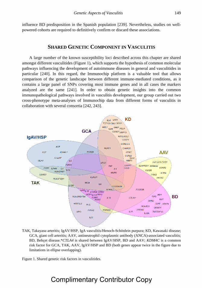
Genetic Aspects of Vasculitis 149
influence BD predisposition in the Spanish population [239]. Nevertheless, studies on well-
powered cohorts are required to definitively confirm or discard these associations.
SHARED GENETIC COMPONENT IN VASCULITIS
A large number of the known susceptibility loci described across this chapter are shared
amongst different vasculitides (Figure 1), which supports the hypothesis of common molecular
pathways influencing the development of autoimmune diseases in general and vasculitides in
particular [240]. In this regard, the Immunochip platform is a valuable tool that allows
comparison of the genetic landscape between different immune-mediated conditions, as it
contains a large panel of SNPs covering most immune genes and in all cases the markers
analyzed are the same [241]. In order to obtain genetic insights into the common
immunopathological pathways involved in vasculitis development, our group carried out two
cross-phenotype meta-analyses of Immunochip data from different forms of vasculitis in
collaboration with several consortia [242, 243].
TAK, Takayasu arteritis; IgAV/HSP, IgA vasculitis/Henoch-Schönlein purpura; KD, Kawasaki disease;
GCA, giant cell arteritis; AAV, antineutrophil cytoplasmic antibody (ANCA)-associated vasculitis;
BD, Behçet disease.*CTLA4 is shared between IgAV/HSP, BD and AAV; KDM4C is a common
risk factor for GCA, TAK, AAV, IgAV/HSP and BD (both genes appear twice in the figure due to
limitations in ellipse overlapping).
Figure 1. Shared genetic risk factors in vasculitides.
Complimentary Contributor Copy
F. David Carmona, Ana Márquez, Javier Martín et al. 150
In the first of them, the previously published Immunochip studies of GCA and TAK [16,
71] were combined to evaluate the shared genetic component of large vessel vasculitis (LVV).
As expected, no genetic pleiotropy was observed within the HLA, consistent with the fact that
GCA and TAK are archetypal HLA class II and class I diseases, respectively. However, a
significant genetic correlation was detected outside the HLA region, with IL12B representing
the strongest shared susceptibility factor for both forms of LVV. This common association is
in agreement with the key role proposed for Th1 and Th17 cells in the granuloma formation of
LVV, as its encoded protein is the P40 subunit of both IL-12 and IL-23 (involved in Th1 and
Th17 cell differentiation, respectively) [244]. Other genes that showed suggestive signals of
common association were NOS2A, ERAP1, REL and PRKQC [242].
The second inter disease analysis was aimed at deciphering the shared genetic component
of all forms of vasculitis. Hence, Immunochip data from GCA, TAK, AAV, and IgAV/HSP
were combined and meta-analyzed. The pooled cohort included a total of 2,465 patients
diagnosed with any of these four types of vasculitis and 4,632 unaffected controls. Interestingly,
a common signal was identified within the lysine demethylase 4C (KDM4C) gene, which
encodes a histone demethylase involved in epigenetic mechanisms. The shared association was
confirmed in an additional cohort that included a subset of BD patients [243].
CONCLUSION
Despite the recent advances in the identification of the risk factors underlying vasculitides
predisposition, there is still a long road ahead before completing the big picture of their genetic
landscape.
In most studies, the limited statistical power and the lack of replication in independent
cohorts have made difficult the identification of consistent genetic association signals.
However, the establishment of big consortia has represented an important step forward that has
facilitated the collection of cohorts large enough to perform adequately powered studies. These
international collaborative efforts will definitively help to identify novel risk factors and to
confirm previously reported genetic associations.
The use of high throughput genotyping has been also of great help, and data mining of the
generated large-scale genetic data is producing valuable insights into the specific and common
immunopathological pathways that lead to the development of the different forms of vasculitis.
REFERENCES
[1] Jennette JC, Falk RJ, Andrassy K, Bacon PA, Churg J, Gross WL, et al. Nomenclature
of systemic vasculitides. Proposal of an international consensus conference. Arthritis and
rheumatism. 1994; 37:187-192.
[2] Jennette JC, Falk RJ, Bacon PA, Basu N, Cid MC, Ferrario F, et al. 2012 revised
International Chapel Hill Consensus Conference Nomenclature of Vasculitides. Arthritis
and rheumatism. 2013; 65:1-11.
Complimentary Contributor Copy
Genetic Aspects of Vasculitis 151
[3] Ozen S, Ruperto N, Dillon MJ, Bagga A, Barron K, Davin JC, et al. EULAR/PReS
endorsed consensus criteria for the classification of childhood vasculitides. Annals of the
rheumatic diseases. 2006; 65:936-941.
[4] Ruperto N, Ozen S, Pistorio A, Dolezalova P, Brogan P, Cabral DA, et al.
EULAR/PRINTO/PRES criteria for Henoch-Schonlein purpura, childhood polyarteritis
nodosa, childhood Wegener granulomatosis and childhood Takayasu arteritis: Ankara
2008. Part I: Overall methodology and clinical characterisation. Annals of the rheumatic
diseases. 2010; 69:790-797.
[5] Carmona FD, Martin J, Gonzalez-Gay MA. New insights into the pathogenesis of giant
cell arteritis and hopes for the clinic. Expert review of clinical immunology. 2016; 12:57-
66.
[6] Bartolome MJ, Martinez-Taboda VM, Lopez-Hoyos M, Blanco R, Rodriguez-Valverde
V. Familial aggregation of polymyalgia rheumatica and giant cell arteritis: genetic and T
cell repertoire analysis. Clinical and experimental rheumatology. 2001; 19:259-264.
[7] Fietta P, Manganelli P, Zanetti A, Neri TM. Familial giant cell arteritis and polymyalgia
rheumatica: aggregation in 2 families. The Journal of rheumatology. 2002; 29:1551-
1555.
[8] Granato JE, Abben RP, May WS. Familial association of giant cell arteritis. A case report
and brief review. Archives of internal medicine. 1981; 141:115-117.
[9] Gros F, Maillefert JF, Behin A, Guignier F, Piroth C, Creuzot-Garcher C, et al. Giant cell
arteritis with ocular complications discovered simultaneously in two sisters. Clinical
rheumatology. 1998; 17:58-61.
[10] Liang GC, Simkin PA, Hunder GG, Wilske KR, Healey LA. Familial aggregation of
polymyalgia rheumatica and giant cell arteritis. Arthritis and rheumatism. 1974; 17:
19-24.
[11] Liozon E, Ouattara B, Rhaiem K, Ly K, Bezanahary H, Loustaud V, et al. Familial
aggregation in giant cell arteritis and polymyalgia rheumatica: a comprehensive literature
review including 4 new families. Clinical and experimental rheumatology. 2009; 27:S89-
94.
[12] Mathewson JA, Hunder GG. Giant cell arteritis in two brothers. The Journal of
rheumatology. 1986; 13:190-192.
[13] Raptis L, Pappas G, Akritidis N. Horton’s three sisters: familial clustering of temporal
arteritis. Clinical rheumatology. 2007; 26:1997-1998.
[14] Carmona FD, Gonzalez-Gay MA, Martin J. Genetic component of giant cell arteritis.
Rheumatology (Oxford). 2014; 53:6-18.
[15] Carmona FD, Vaglio A, Mackie SL, Hernandez-Rodriguez J, Monach PA, Castaneda S,
et al. A Genome-wide Association Study Identifies Risk Alleles in Plasminogen and
P4HA2 Associated with Giant Cell Arteritis. American journal of human genetics. 2017;
100:64-74.
[16] Carmona FD, Mackie SL, Martin JE, Taylor JC, Vaglio A, Eyre S, et al. A large-scale
genetic analysis reveals a strong contribution of the HLA class II region to giant cell
arteritis susceptibility. American journal of human genetics. 2015; 96:565-580.
[17] Armstrong RD, Behn A, Myles A, Panayi GS, Welsh KI. Histocompatibility antigens in
polymyalgia rheumatica and giant cell arteritis. The Journal of rheumatology. 1983;
10:659-661.
Complimentary Contributor Copy
F. David Carmona, Ana Márquez, Javier Martín et al. 152
[18] Barrier J, Bignon JD, Soulillou JP, Grolleau J. Increased prevalence of HLA-DR4 in
giant-cell arteritis. The New England journal of medicine. 1981; 305:104-105.
[19] Bignon JD, Barrier J, Soulillou JP, Martin P, Grolleau JY. HLA DR4 and giant cell
arteritis. Tissue antigens. 1984; 24:60-62.
[20] Bignon JD, Ferec C, Barrier J, Pennec Y, Verlingue C, Cheneau ML, et al. HLA class II
genes polymorphism in DR4 giant cell arteritis patients. Tissue antigens. 1988; 32:254-
258.
[21] Combe B, Sany J, Le Quellec A, Clot J, Eliaou JF. Distribution of HLA-DRB1 alleles of
patients with polymyalgia rheumatica and giant cell arteritis in a Mediterranean
population. The Journal of rheumatology. 1998; 25:94-98.
[22] Dababneh A, Gonzalez-Gay MA, Garcia-Porrua C, Hajeer A, Thomson W, Ollier W.
Giant cell arteritis and polymyalgia rheumatica can be differentiated by distinct patterns
of HLA class II association. The Journal of rheumatology. 1998; 25:2140-2145.
[23] Jacobsen S, Baslund B, Madsen HO, Tvede N, Svejgaard A, Garred P. Mannose-binding
lectin variant alleles and HLA-DR4 alleles are associated with giant cell arteritis. The
Journal of rheumatology. 2002; 29:2148-2153.
[24] Lowenstein MB, Bridgeford PH, Vasey FB, Germain BF, Espinoza LR. Increased
frequency of HLA-DR3 and DR4 in polymyalgia rheumatica-giant cell arteritis. Arthritis
and rheumatism. 1983; 26:925-927.
[25] Martinez-Taboda VM, Bartolome MJ, Lopez-Hoyos M, Blanco R, Mata C, Calvo J,
et al. HLA-DRB1 allele distribution in polymyalgia rheumatica and giant cell arteritis:
influence on clinical subgroups and prognosis. Seminars in arthritis and rheumatism.
2004; 34:454-464.
[26] Rauzy O, Fort M, Nourhashemi F, Alric L, Juchet H, Ecoiffier M, et al. Relation between
HLA DRB1 alleles and corticosteroid resistance in giant cell arteritis. Annals of the
rheumatic diseases. 1998; 57:380-382.
[27] Richardson JE, Gladman DD, Fam A, Keystone EC. HLA-DR4 in giant cell arteritis:
association with polymyalgia rheumatica syndrome. Arthritis and rheumatism. 1987;
30:1293-1297.
[28] Weyand CM, Hicok KC, Hunder GG, Goronzy JJ. The HLA-DRB1 locus as a genetic
component in giant cell arteritis. Mapping of a disease-linked sequence motif to the
antigen binding site of the HLA-DR molecule. The Journal of clinical investigation.
1992; 90:2355-2361.
[29] Weyand CM, Hunder NN, Hicok KC, Hunder GG, Goronzy JJ. HLA-DRB1 alleles in
polymyalgia rheumatica, giant cell arteritis, and rheumatoid arthritis. Arthritis and
rheumatism. 1994; 37:514-520.
[30] Gonzalez-Gay MA, Garcia-Porrua C, Hajeer AH, Dababneh A, Ollier WE. HLA-
DRB1*04 may be a marker of severity in giant cell arteritis. Annals of the rheumatic
diseases. 2000; 59:574-575.
[31] Gonzalez-Gay MA, Garcia-Porrua C, Llorca J, Hajeer AH, Branas F, Dababneh A,
et al. Visual manifestations of giant cell arteritis. Trends and clinical spectrum in 161
patients. Medicine. 2000; 79:283-292.
[32] Gonzalez-Gay MA, Rueda B, Vilchez JR, Lopez-Nevot MA, Robledo G, Ruiz MP,
et al. Contribution of MHC class I region to genetic susceptibility for giant cell arteritis.
Rheumatology (Oxford). 2007; 46:431-434.
Complimentary Contributor Copy
Genetic Aspects of Vasculitis 153
[33] Hansen JA, Healey LA, Wilske KR. Association between giant cell (temporal) arteritis
and HLA-Cw3. Human immunology. 1985; 13:193-198.
[34] Kemp A, Marner K, Nissen SH, Heyn J, Kissmeyer-Nielsen F. HLA antigens in cases of
giant cell arteritis. Acta ophthalmologica. 1980; 58:1000-1004.
[35] Cortes A, Brown MA. Promise and pitfalls of the Immunochip. Arthritis research &
therapy. 2011; 13:101.
[36] de Bakker PI, Raychaudhuri S. Interrogating the major histocompatibility complex with
high-throughput genomics. Human molecular genetics. 2012; 21:R29-36.
[37] Jia X, Han B, Onengut-Gumuscu S, Chen WM, Concannon PJ, Rich SS, et al. Imputing
amino acid polymorphisms in human leukocyte antigens. PloS one. 2013; 8:e64683.
[38] Stanford SM, Bottini N. PTPN22: the archetypal non-HLA autoimmunity gene. Nature
reviews Rheumatology. 2014; 10:602-611.
[39] Serrano A, Marquez A, Mackie SL, Carmona FD, Solans R, Miranda-Filloy JA, et al.
Identification of the PTPN22 functional variant R620W as susceptibility genetic factor
for giant cell arteritis. Annals of the rheumatic diseases. 2013; 72:1882-1886.
[40] Zhong MC, Veillette A. Immunology: Csk keeps LYP on a leash. Nature chemical
biology. 2012; 8:412-413.
[41] Law RH, Abu-Ssaydeh D, Whisstock JC. New insights into the structure and function of
the plasminogen/plasmin system. Current opinion in structural biology. 2013; 23:836-
841.
[42] Myllyharju J. Prolyl 4-hydroxylases, the key enzymes of collagen biosynthesis. Matrix
biology: journal of the International Society for Matrix Biology. 2003; 22:15-24.
[43] Espigol-Frigole G, Corbera-Bellalta M, Planas-Rigol E, Lozano E, Segarra M, Garcia-
Martinez A, et al. Increased IL-17A expression in temporal artery lesions is a predictor
of sustained response to glucocorticoid treatment in patients with giant-cell arteritis.
Annals of the rheumatic diseases. 2013; 72:1481-1487.
[44] Deng J, Younge BR, Olshen RA, Goronzy JJ, Weyand CM. Th17 and Th1 T-cell
responses in giant cell arteritis. Circulation. 2010; 121:906-915.
[45] Hernandez-Rodriguez J, Segarra M, Vilardell C, Sanchez M, Garcia-Martinez A, Esteban
MJ, et al. Tissue production of pro-inflammatory cytokines (IL-1beta, TNFalpha and IL-
6) correlates with the intensity of the systemic inflammatory response and with
corticosteroid requirements in giant-cell arteritis. Rheumatology (Oxford). 2004; 43:294-
301.
[46] Marquez A, Hernandez-Rodriguez J, Cid MC, Solans R, Castaneda S, Fernandez-
Contreras ME, et al. Influence of the IL17A locus in giant cell arteritis susceptibility.
Annals of the rheumatic diseases. 2014; 73:1742-1745.
[47] Serrano A, Carmona FD, Castaneda S, Solans R, Hernandez-Rodriguez J, Cid MC,
et al. Evidence of association of the NLRP1 gene with giant cell arteritis. Annals of the
rheumatic diseases. 2013; 72:628-630.
[48] Boiardi L, Casali B, Farnetti E, Pipitone N, Nicoli D, Macchioni P, et al. Interleukin-10
promoter polymorphisms in giant cell arteritis. Arthritis and rheumatism. 2006; 54:4011-
4017.
[49] Rueda B, Roibas B, Martin J, Gonzalez-Gay MA. Influence of interleukin 10 promoter
polymorphisms in susceptibility to giant cell arteritis in Northwestern Spain. The Journal
of rheumatology. 2007; 34:1535-1539.
Complimentary Contributor Copy
F. David Carmona, Ana Márquez, Javier Martín et al. 154
[50] Amoli MM, Gonzalez-Gay MA, Zeggini E, Salway F, Garcia-Porrua C, Ollier WE.
Epistatic interactions between HLA-DRB1 and interleukin 4, but not interferon-gamma,
increase susceptibility to giant cell arteritis. The Journal of rheumatology. 2004; 31:2413-
2417.
[51] Amoli MM, Salway F, Zeggini E, Ollier WE, Gonzalez-Gay MA. MCP-1 gene haplotype
association in biopsy proven giant cell arteritis. The Journal of rheumatology. 2005;
32:507-510.
[52] Enjuanes A, Benavente Y, Hernandez-Rodriguez J, Queralt C, Yague J, Jares P, et al.
Association of NOS2 and potential effect of VEGF, IL6, CCL2 and IL1RN
polymorphisms and haplotypes on susceptibility to GCA--a simultaneous study of 130
potentially functional SNPs in 14 candidate genes. Rheumatology (Oxford). 2012;
51:841-851.
[53] Gonzalez-Gay MA, Hajeer AH, Dababneh A, Garcia-Porrua C, Amoli MM, Llorca J,
et al. Interferon-gamma gene microsatellite polymorphisms in patients with biopsy-
proven giant cell arteritis and isolated polymyalgia rheumatica. Clinical and
experimental rheumatology. 2004; 22:S18-20.
[54] Gonzalez-Gay MA, Hajeer AH, Dababneh A, Garcia-Porrua C, Mattey DL, Amoli MM,
et al. IL-6 promoter polymorphism at position -174 modulates the phenotypic expression
of polymyalgia rheumatica in biopsy-proven giant cell arteritis. Clinical and
experimental rheumatology. 2002; 20:179-184.
[55] Makki RF, al Sharif F, Gonzalez-Gay MA, Garcia-Porrua C, Ollier WE, Hajeer AH.
RANTES gene polymorphism in polymyalgia rheumatica, giant cell arteritis and
rheumatoid arthritis. Clinical and experimental rheumatology. 2000; 18:391-393.
[56] Mattey DL, Hajeer AH, Dababneh A, Thomson W, Gonzalez-Gay MA, Garcia-Porrua
C, et al. Association of giant cell arteritis and polymyalgia rheumatica with different
tumor necrosis factor microsatellite polymorphisms. Arthritis and rheumatism. 2000;
43:1749-1755.
[57] Palomino-Morales RJ, Vazquez-Rodriguez TR, Torres O, Morado IC, Castaneda S,
Miranda-Filloy JA, et al. Association between IL-18 gene polymorphisms and biopsy-
proven giant cell arteritis. Arthritis research & therapy. 2010; 12:R51.
[58] Rodriguez-Rodriguez L, Carmona FD, Castaneda S, Miranda-Filloy JA, Morado IC,
Narvaez J, et al. Role of rs1343151 IL23R and rs3790567 IL12RB2 polymorphisms in
biopsy-proven giant cell arteritis. The Journal of rheumatology. 2011; 38:889-892.
[59] Marquez A, Solans R, Hernandez-Rodriguez J, Cid MC, Castaneda S, Ramentol M,
et al. A candidate gene approach identifies an IL33 genetic variant as a novel genetic risk
factor for GCA. PloS one. 2014; 9:e113476.
[60] Salvarani C, Casali B, Boiardi L, Ranzi A, Macchioni P, Nicoli D, et al. Intercellular
adhesion molecule 1 gene polymorphisms in polymyalgia rheumatica/giant cell arteritis:
association with disease risk and severity. The Journal of rheumatology. 2000; 27:1215-
1221.
[61] Boiardi L, Casali B, Nicoli D, Farnetti E, Chen Q, Macchioni P, et al. Vascular
endothelial growth factor gene polymorphisms in giant cell arteritis. The Journal of
rheumatology. 2003; 30:2160-2164.
[62] Rueda B, Lopez-Nevot MA, Lopez-Diaz MJ, Garcia-Porrua C, Martin J, Gonzalez-Gay
MA. A functional variant of vascular endothelial growth factor is associated with severe
Complimentary Contributor Copy
Genetic Aspects of Vasculitis 155
ischemic complications in giant cell arteritis. The Journal of rheumatology. 2005;
32:1737-1741.
[63] Amoli MM, Garcia-Porrua C, Llorca J, Ollier WE, Gonzalez-Gay MA. Endothelial nitric
oxide synthase haplotype associations in biopsy-proven giant cell arteritis. The Journal
of rheumatology. 2003; 30:2019-2022.
[64] Gonzalez-Gay MA, Oliver J, Sanchez E, Garcia-Porrua C, Paco L, Lopez-Nevot MA,
et al. Association of a functional inducible nitric oxide synthase promoter variant with
susceptibility to biopsy-proven giant cell arteritis. The Journal of rheumatology. 2005;
32:2178-2182.
[65] Salvarani C, Casali B, Nicoli D, Farnetti E, Macchioni P, Catanoso MG, et al. Endothelial
nitric oxide synthase gene polymorphisms in giant cell arteritis. Arthritis and
rheumatism. 2003; 48:3219-3223.
[66] Rodriguez-Pla A, Beaty TH, Savino PJ, Eagle RC, Jr., Seo P, Soloski MJ. Association of
a nonsynonymous single-nucleotide polymorphism of matrix metalloproteinase 9 with
giant cell arteritis. Arthritis and rheumatism. 2008; 58:1849-1853.
[67] Palomino-Morales R, Torres O, Vazquez-Rodriguez TR, Morado IC, Castaneda S,
Callejas-Rubio JL, et al. Association between toll-like receptor 4 gene polymorphism and
biopsy-proven giant cell arteritis. The Journal of rheumatology. 2009; 36:1501-1506.
[68] Morgan AW, Robinson JI, Barrett JH, Martin J, Walker A, Babbage SJ, et al. Association
of FCGR2A and FCGR2A-FCGR3A haplotypes with susceptibility to giant cell arteritis.
Arthritis research & therapy. 2006; 8:R109.
[69] Salvarani C, Casali B, Farnetti E, Pipitone N, Nicoli D, Macchioni PL, et al. -463 G/A
myeloperoxidase promoter polymorphism in giant cell arteritis. Annals of the rheumatic
diseases. 2008; 67:485-488.
[70] Terao C, Yoshifuji H, Kimura A, Matsumura T, Ohmura K, Takahashi M, et al. Two
susceptibility loci to Takayasu arteritis reveal a synergistic role of the IL12B and HLA-
B regions in a Japanese population. American journal of human genetics. 2013; 93:289-
297.
[71] Saruhan-Direskeneli G, Hughes T, Aksu K, Keser G, Coit P, Aydin SZ, et al.
Identification of multiple genetic susceptibility loci in Takayasu arteritis. American
journal of human genetics. 2013; 93:298-305.
[72] Renauer PA, Saruhan-Direskeneli G, Coit P, Adler A, Aksu K, Keser G, et al.
Identification of Susceptibility Loci in IL6, RPS9/LILRB3, and an Intergenic Locus on
Chromosome 21q22 in Takayasu Arteritis in a Genome-Wide Association Study.
Arthritis Rheumatol. 2015; 67:1361-1368.
[73] Yajima M, Numano F, Park YB, Sagar S. Comparative studies of patients with Takayasu
arteritis in Japan, Korea and India--comparison of clinical manifestations, angiography
and HLA-B antigen. Japanese circulation journal. 1994; 58:9-14.
[74] Kimura A, Kitamura H, Date Y, Numano F. Comprehensive analysis of HLA genes in
Takayasu arteritis in Japan. International journal of cardiology. 1996; 54 Suppl:S61-69.
[75] Mehra NK, Jaini R, Balamurugan A, Kanga U, Prabhakaran D, Jain S, et al.
Immunogenetic analysis of Takayasu arteritis in Indian patients. International journal of
cardiology. 1998; 66 Suppl 1:S127-132; discussion S133.
[76] Charoenwongse P, Kangwanshiratada O, Boonnam R, Hoomsindhu U. The association
between the HLA antigens and Takayasu’s arteritis in Thai patients. International journal
of cardiology. 1998; 66 Suppl 1:S117-120.
Complimentary Contributor Copy
F. David Carmona, Ana Márquez, Javier Martín et al. 156
[77] Sahin Z, Bicakcigil M, Aksu K, Kamali S, Akar S, Onen F, et al. Takayasu’s arteritis is
associated with HLA-B*52, but not with HLA-B*51, in Turkey. Arthritis research &
therapy. 2012; 14:R27.
[78] Vargas-Alarcon G, Flores-Dominguez C, Hernandez-Pacheco G, Zuniga J, Gamboa R,
Soto ME, et al. Immunogenetics and clinical aspects of Takayasu’s arteritis patients in a
Mexican Mestizo population. Clinical and experimental rheumatology. 2001; 19:439-
443.
[79] Volkman DJ, Mann DL, Fauci AS. Association between Takayasu’s arteritis and a B-cell
alloantigen in North Americans. The New England journal of medicine. 1982; 306:464-
465.
[80] Sattar MA, White AG, Eklof B, Fenech FF. Takayasu’s disease in Arabs. Postgraduate
medical journal. 1985; 61:387-390.
[81] Numano F, Namba K, Suzuki K, Matsumoto H. Hereditary factors in Takayasu disease.
III. Polymorphism of human complements. Experimental and clinical immunogenetics.
1989; 6:236-244.
[82] Kimura A, Kobayashi Y, Takahashi M, Ohbuchi N, Kitamura H, Nakamura T, et al.
MICA gene polymorphism in Takayasu’s arteritis and Buerger’s disease. International
journal of cardiology. 1998; 66 Suppl 1:S107-113; discussion S115.
[83] Kimura A, Ota M, Katsuyama Y, Ohbuchi N, Takahashi M, Kobayashi Y, et al. Mapping
of the HLA-linked genes controlling the susceptibility to Takayasu’s arteritis.
International journal of cardiology. 2000; 75 Suppl 1:S105-110; discussion S111-102.
[84] Saruhan-Direskeneli G, Bicakcigil M, Yilmaz V, Kamali S, Aksu K, Fresko I, et al.
Interleukin (IL)-12, IL-2, and IL-6 gene polymorphisms in Takayasu’s arteritis from
Turkey. Human immunology. 2006; 67:735-740.
[85] Park MC, Lee SW, Park YB, Lee SK. Serum cytokine profiles and their correlations with
disease activity in Takayasu’s arteritis. Rheumatology (Oxford). 2006; 45:545-548.
[86] Verma DK, Tripathy NK, Verma NS, Tiwari S. Interleukin 12 in Takayasu’s arteritis:
plasma concentrations and relationship with disease activity. The Journal of
rheumatology. 2005; 32:2361-2363.
[87] Tripathy NK, Gupta PC, Nityanand S. High TNF-alpha and low IL-2 producing T cells
characterize active disease in Takayasu’s arteritis. Clin Immunol. 2006; 118:154-158.
[88] Seegers D, Zwiers A, Strober W, Pena AS, Bouma G. A TaqI polymorphism in the
3’UTR of the IL-12 p40 gene correlates with increased IL-12 secretion. Genes and
immunity. 2002; 3:419-423.
[89] Yilmaz V, Yentur SP, Saruhan-Direskeneli G. IL-12 and IL-10 polymorphisms and their
effects on cytokine production. Cytokine. 2005; 30:188-194.
[90] Morahan G, Huang D, Ymer SI, Cancilla MR, Stephen K, Dabadghao P, et al. Linkage
disequilibrium of a type 1 diabetes susceptibility locus with a regulatory IL12B allele.
Nature genetics. 2001; 27:218-221.
[91] Fishman D, Faulds G, Jeffery R, Mohamed-Ali V, Yudkin JS, Humphries S, et al. The
effect of novel polymorphisms in the interleukin-6 (IL-6) gene on IL-6 transcription and
plasma IL-6 levels, and an association with systemic-onset juvenile chronic arthritis. The
Journal of clinical investigation. 1998; 102:1369-1376.
[92] Panoulas VF, Stavropoulos-Kalinoglou A, Metsios GS, Smith JP, Milionis HJ, Douglas
KM, et al. Association of interleukin-6 (IL-6)-174G/C gene polymorphism with
Complimentary Contributor Copy
Genetic Aspects of Vasculitis 157
cardiovascular disease in patients with rheumatoid arthritis: the role of obesity and
smoking. Atherosclerosis. 2009; 204:178-183.
[93] Pascual M, Nieto A, Mataran L, Balsa A, Pascual-Salcedo D, Martin J. IL-6 promoter
polymorphisms in rheumatoid arthritis. Genes and immunity. 2000(5):338-340.
[94] Soto Lopez ME, Gamboa Avila R, Hernandez E, Huesca-Gomez C, Castrejon-Tellez V,
Perez-Mendez O, et al. The interleukin-1 gene cluster polymorphisms are associated with
Takayasu’s arteritis in Mexican patients. Journal of interferon & cytokine research: the
official journal of the International Society for Interferon and Cytokine Research. 2013;
33:369-375.
[95] Fragoso JM, Delgadillo H, Llorente L, Chuquiure E, Juarez-Cedillo T, Vallejo M, et al.
Interleukin 1 receptor antagonist polymorphisms are associated with the risk of
developing acute coronary syndrome in Mexicans. Immunology letters. 2010; 133:106-
111.
[96] Olsson S, Holmegaard L, Jood K, Sjogren M, Engstrom G, Lovkvist H, et al. Genetic
variation within the interleukin-1 gene cluster and ischemic stroke. Stroke. 2012;
43:2278-2282.
[97] Bhaskar V, Yin J, Mirza AM, Phan D, Vanegas S, Issafras H, et al. Monoclonal
antibodies targeting IL-1 beta reduce biomarkers of atherosclerosis in vitro and inhibit
atherosclerotic plaque formation in Apolipoprotein E-deficient mice. Atherosclerosis.
2011; 216:313-320.
[98] Espinola-Zavaleta N, Soto-Lopez ME, Carreon-Torres E, Gamboa R, Mejia AM,
Marquez-Velasco R, et al. Altered flow-mediated vasodilatation, low paraoxonase-1
activity, and abnormal high-density lipoprotein subclass distribution in Takayasu’s
arteritis. Circulation journal: official journal of the Japanese Circulation Society. 2009;
73:760-766.
[99] Huesca-Gomez C, Soto ME, Castrejon-Tellez V, Perez-Mendez O, Gamboa R. PON1
gene polymorphisms and plasma PON1 activities in Takayasu’s arteritis disease.
Immunology letters. 2013; 152:77-82.
[100] Onouchi Y. The genetics of Kawasaki disease. International journal of rheumatic
diseases. 2018 Jan; 21(1):26-30.
[101] Burgner D, Davila S, Breunis WB, Ng SB, Li Y, Bonnard C, et al. A genome-wide
association study identifies novel and functionally related susceptibility Loci for
Kawasaki disease. PLoS genetics. 2009; 5:e1000319.
[102] Khor CC, Davila S, Breunis WB, Lee YC, Shimizu C, Wright VJ, et al. Genome-wide
association study identifies FCGR2A as a susceptibility locus for Kawasaki disease.
Nature genetics. 2011; 43:1241-1246.
[103] Kim JJ, Hong YM, Sohn S, Jang GY, Ha KS, Yun SW, et al. A genome-wide association
analysis reveals 1p31 and 2p13.3 as susceptibility loci for Kawasaki disease. Human
genetics. 2011; 129:487-495.
[104] Lee YC, Kuo HC, Chang JS, Chang LY, Huang LM, Chen MR, et al. Two new
susceptibility loci for Kawasaki disease identified through genome-wide association
analysis. Nature genetics. 2012; 44:522-525.
[105] Onouchi Y, Ozaki K, Burns JC, Shimizu C, Terai M, Hamada H, et al. A genome-wide
association study identifies three new risk loci for Kawasaki disease. Nature genetics.
2012; 44:517-521.
Complimentary Contributor Copy
F. David Carmona, Ana Márquez, Javier Martín et al. 158
[106] Tsai FJ, Lee YC, Chang JS, Huang LM, Huang FY, Chiu NC, et al. Identification of
novel susceptibility Loci for kawasaki disease in a Han chinese population by a genome-
wide association study. PloS one. 2011; 6:e16853.
[107] Matsuda I, Hattori S, Nagata N, Fruse A, Nambu H. HLA antigens in mucocutaneous
lymph node syndrome. Am J Dis Child. 1977; 131:1417-1418.
[108] Kato S, Kimura M, Tsuji K, Kusakawa S, Asai T, Juji T, et al. HLA antigens in Kawasaki
disease. Pediatrics. 1978; 61:252-255.
[109] Kaslow RA, Bailowitz A, Lin FY, Koslowe P, Simonis T, Israel E. Association of
epidemic Kawasaki syndrome with the HLA-A2, B44, Cw5 antigen combination.
Arthritis and rheumatism. 1985; 28:938-940.
[110] Krensky AM, Berenberg W, Shanley K, Yunis EJ. HLA antigens in mucocutaneous
lymph node syndrome in New England. Pediatrics. 1981; 67:741-743.
[111] Krensky AM, Grady S, Shanley KM, Berenberg W, Yunis EJ. Epidemic and endemic
HLA-B and DR associations in mucocutaneous lymph node syndrome. Human
immunology. 1983; 6:75-77.
[112] Yan Y, Ma Y, Liu Y, Hu H, Shen Y, Zhang S, et al. Combined analysis of genome-wide-
linked susceptibility loci to Kawasaki disease in Han Chinese. Human genetics. 2013;
132:669-680.
[113] Arj-Ong S, Thakkinstian A, McEvoy M, Attia J. A systematic review and meta-analysis
of tumor necrosis factor alpha-308 polymorphism and Kawasaki disease. Pediatrics
international: official journal of the Japan Pediatric Society. 2010; 52:527-532.
[114] Biezeveld MH, Geissler J, Weverling GJ, Kuipers IM, Lam J, Ottenkamp J, et al.
Polymorphisms in the mannose-binding lectin gene as determinants of age-defined risk
of coronary artery lesions in Kawasaki disease. Arthritis and rheumatism. 2006; 54:369-
376.
[115] Biezeveld MH, Kuipers IM, Geissler J, Lam J, Ottenkamp JJ, Hack CE, et al. Association
of mannose-binding lectin genotype with cardiovascular abnormalities in Kawasaki
disease. Lancet. 2003; 361:1268-1270.
[116] Breunis WB, Biezeveld MH, Geissler J, Kuipers IM, Lam J, Ottenkamp J, et al.
Polymorphisms in chemokine receptor genes and susceptibility to Kawasaki disease.
Clinical and experimental immunology. 2007; 150:83-90.
[117] Breunis WB, Biezeveld MH, Geissler J, Ottenkamp J, Kuipers IM, Lam J, et al. Vascular
endothelial growth factor gene haplotypes in Kawasaki disease. Arthritis and
rheumatism. 2006; 54:1588-1594.
[118] Burns JC, Shimizu C, Gonzalez E, Kulkarni H, Patel S, Shike H, et al. Genetic variations
in the receptor-ligand pair CCR5 and CCL3L1 are important determinants of
susceptibility to Kawasaki disease. The Journal of infectious diseases. 2005; 192:344-
349.
[119] Burns JC, Shimizu C, Shike H, Newburger JW, Sundel RP, Baker AL, et al. Family-
based association analysis implicates IL-4 in susceptibility to Kawasaki disease. Genes
and immunity. 2005; 6:438-444.
[120] Hsueh KC, Lin YJ, Chang JS, Wan L, Tsai YH, Tsai CH, et al. Association of vascular
endothelial growth factor C-634 g polymorphism in taiwanese children with Kawasaki
disease. Pediatric cardiology. 2008; 29:292-296.
Complimentary Contributor Copy
Genetic Aspects of Vasculitis 159
[121] Ikeda K, Ihara K, Yamaguchi K, Muneuchi J, Ohno T, Mizuno Y, et al. Genetic analysis
of MMP gene polymorphisms in patients with Kawasaki disease. Pediatric research.
2008; 63:182-185.
[122] Kariyazono H, Ohno T, Khajoee V, Ihara K, Kusuhara K, Kinukawa N, et al. Association
of vascular endothelial growth factor (VEGF) and VEGF receptor gene polymorphisms
with coronary artery lesions of Kawasaki disease. Pediatric research. 2004; 56:953-959.
[123] Onoyama S, Ihara K, Yamaguchi Y, Ikeda K, Yamaguchi K, Yamamura K, et al. Genetic
susceptibility to Kawasaki disease: analysis of pattern recognition receptor genes. Human
immunology. 2012; 73:654-660.
[124] Park JA, Shin KS, Kim YW. Polymorphism of matrix metalloproteinase-3 promoter gene
as a risk factor for coronary artery lesions in Kawasaki disease. Journal of Korean
medical science. 2005; 20:607-611.
[125] Onouchi Y, Tamari M, Takahashi A, Tsunoda T, Yashiro M, Nakamura Y, et al. A
genomewide linkage analysis of Kawasaki disease: evidence for linkage to chromosome
12. Journal of human genetics. 2007; 52:179-190.
[126] Onouchi Y, Gunji T, Burns JC, Shimizu C, Newburger JW, Yashiro M, et al. ITPKC
functional polymorphism associated with Kawasaki disease susceptibility and formation
of coronary artery aneurysms. Nature genetics. 2008; 40:35-42.
[127] Onouchi Y, Ozaki K, Buns JC, Shimizu C, Hamada H, Honda T, et al. Common variants
in CASP3 confer susceptibility to Kawasaki disease. Human molecular genetics. 2010;
19:2898-2906.
[128] Kuo HC, Yang KD, Juo SH, Liang CD, Chen WC, Wang YS, et al. ITPKC single
nucleotide polymorphism associated with the Kawasaki disease in a Taiwanese
population. PloS one. 2011; 6:e17370.
[129] Kuo HC, Yu HR, Juo SH, Yang KD, Wang YS, Liang CD, et al. CASP3 gene single-
nucleotide polymorphism (rs72689236) and Kawasaki disease in Taiwanese children.
Journal of human genetics. 2011; 56:161-165.
[130] Onouchi Y, Fukazawa R, Yamamura K, Suzuki H, Kakimoto N, Suenaga T, et al.
Variations in ORAI1 Gene Associated with Kawasaki Disease. PloS one. 2016;
11:e0145486.
[131] Shimizu C, Eleftherohorinou H, Wright VJ, Kim J, Alphonse MP, Perry JC, et al. Genetic
Variation in the SLC8A1 Calcium Signaling Pathway Is Associated With Susceptibility
to Kawasaki Disease and Coronary Artery Abnormalities. Circulation Cardiovascular
genetics. 2016; 9:559-568.
[132] Kim J, Shimizu C, Kingsmore SF, Veeraraghavan N, Levy E, Ribeiro Dos Santos AM,
et al. Whole genome sequencing of an African American family highlights toll like
receptor 6 variants in Kawasaki disease susceptibility. PloS one. 2017; 12:e0170977.
[133] Knight A, Sandin S, Askling J. Risks and relative risks of Wegener’s granulomatosis
among close relatives of patients with the disease. Arthritis and rheumatism. 2008;
58:302-307.
[134] Lyons PA, Rayner TF, Trivedi S, Holle JU, Watts RA, Jayne DR, et al. Genetically
distinct subsets within ANCA-associated vasculitis. The New England journal of
medicine. 2012; 367:214-223.
[135] Xie G, Roshandel D, Sherva R, Monach PA, Lu EY, Kung T, et al. Association of
granulomatosis with polyangiitis (Wegener’s) with HLA-DPB1*04 and SEMA6A gene
Complimentary Contributor Copy
F. David Carmona, Ana Márquez, Javier Martín et al. 160
variants: evidence from genome-wide analysis. Arthritis and rheumatism. 2013; 65:2457-
2468.
[136] Merkel PA, Xie G, Monach PA, Ji X, Ciavatta DJ, Byun J, et al. Identification of
functional and expression polymorphisms associated with risk for antineutrophil
cytoplasmic autoantibody-associated vasculitis. Arthritis and rheumatology. 2017;
69:1054-1066.
[137] Cotch MF, Fauci AS, Hoffman GS. HLA typing in patients with Wegener
granulomatosis. Annals of internal medicine. 1995; 122:635.
[138] Papiha SS, Murty GE, Ad’Hia A, Mains BT, Venning M. Association of Wegener’s
granulomatosis with HLA antigens and other genetic markers. Annals of the rheumatic
diseases. 1992; 51:246-248.
[139] Spencer SJ, Burns A, Gaskin G, Pusey CD, Rees AJ. HLA class II specificities in
vasculitis with antibodies to neutrophil cytoplasmic antigens. Kidney international. 1992;
41:1059-1063.
[140] Jagiello P, Gencik M, Arning L, Wieczorek S, Kunstmann E, Csernok E, et al. New
genomic region for Wegener’s granulomatosis as revealed by an extended association
screen with 202 apoptosis-related genes. Human genetics. 2004; 114:468-477.
[141] Heckmann M, Holle JU, Arning L, Knaup S, Hellmich B, Nothnagel M, et al. The
Wegener’s granulomatosis quantitative trait locus on chromosome 6p21.3 as
characterised by tagSNP genotyping. Annals of the rheumatic diseases. 2008; 67:972-
979.
[142] Stassen PM, Cohen-Tervaert JW, Lems SP, Hepkema BG, Kallenberg CG, Stegeman
CA. HLA-DR4, DR13(6) and the ancestral haplotype A1B8DR3 are associated with
ANCA-associated vasculitis and Wegener’s granulomatosis. Rheumatology (Oxford).
2009; 48:622-625.
[143] Vaglio A, Martorana D, Maggiore U, Grasselli C, Zanetti A, Pesci A, et al. HLA-DRB4
as a genetic risk factor for Churg-Strauss syndrome. Arthritis and rheumatism. 2007;
56:3159-3166.
[144] Gencik M, Meller S, Borgmann S, Fricke H. Proteinase 3 gene polymorphisms and
Wegener’s granulomatosis. Kidney international. 2000; 58:2473-2477.
[145] Griffith ME, Lovegrove JU, Gaskin G, Whitehouse DB, Pusey CD. C-antineutrophil
cytoplasmic antibody positivity in vasculitis patients is associated with the Z allele of
alpha-1-antitrypsin, and P-antineutrophil cytoplasmic antibody positivity with the S
allele. Nephrology, dialysis, transplantation: official publication of the European Dialysis
and Transplant Association - European Renal Association. 1996; 11:438-443.
[146] Savige JA, Chang L, Cook L, Burdon J, Daskalakis M, Doery J. Alpha 1-antitrypsin
deficiency and anti-proteinase 3 antibodies in anti-neutrophil cytoplasmic antibody
(ANCA)-associated systemic vasculitis. Clinical and experimental immunology. 1995;
100:194-197.
[147] Baslund B, Szpirt W, Eriksson S, Elzouki AN, Wiik A, Wieslander J, et al. Complexes
between proteinase 3, alpha 1-antitrypsin and proteinase 3 anti-neutrophil cytoplasm
autoantibodies: a comparison between alpha 1-antitrypsin PiZ allele carriers and non-
carriers with Wegener’s granulomatosis. European journal of clinical investigation.
1996; 26:786-792.
Complimentary Contributor Copy
Genetic Aspects of Vasculitis 161
[148] Borgmann S, Endisch G, Urban S, Sitter T, Fricke H. A linkage disequilibrium between
genes at the serine protease inhibitor gene cluster on chromosome 14q32.1 is associated
with Wegener’s granulomatosis. Clin Immunol. 2001; 98:244-248.
[149] Callea F, Gregorini G, Sinico A, Consalez GG, Bossolasco M, Salvidio G, et al. alpha 1-
Antitrypsin (AAT) deficiency and ANCA-positive systemic vasculitis: genetic and
clinical implications. European journal of clinical investigation. 1997; 27:696-702.
[150] Lhotta K, Vogel W, Meisl T, Buxbaum M, Neyer U, Sandholzer C, et al. Alpha 1-
antitrypsin phenotypes in patients with anti-neutrophil cytoplasmic antibody-positive
vasculitis. Clin Sci (Lond). 1994; 87:693-695.
[151] Carr EJ, Niederer HA, Williams J, Harper L, Watts RA, Lyons PA, et al. Confirmation
of the genetic association of CTLA4 and PTPN22 with ANCA-associated vasculitis.
BMC medical genetics. 2009; 10:121.
[152] Jagiello P, Aries P, Arning L, Wagenleiter SE, Csernok E, Hellmich B, et al. The PTPN22
620W allele is a risk factor for Wegener’s granulomatosis. Arthritis and rheumatism.
2005; 52:4039-4043.
[153] Steiner K, Moosig F, Csernok E, Selleng K, Gross WL, Fleischer B, et al. Increased
expression of CTLA-4 (CD152) by T and B lymphocytes in Wegener’s granulomatosis.
Clinical and experimental immunology. 2001; 126:143-150.
[154] Kamesh L, Heward JM, Williams JM, Gough SC, Chavele KM, Salama A, et al. CT60
and +49 polymorphisms of CTLA 4 are associated with ANCA-positive small vessel
vasculitis. Rheumatology (Oxford). 2009; 48:1502-1505.
[155] Slot MC, Sokolowska MG, Savelkouls KG, Janssen RG, Damoiseaux JG, Tervaert JW.
Immunoregulatory gene polymorphisms are associated with ANCA-related vasculitis.
Clin Immunol. 2008; 128:39-45.
[156] Zhou Y, Huang D, Paris PL, Sauter CS, Prock KA, Hoffman GS. An analysis of CTLA-
4 and proinflammatory cytokine genes in Wegener’s granulomatosis. Arthritis and
rheumatism. 2004; 50:2645-2650.
[157] Giscombe R, Wang X, Huang D, Lefvert AK. Coding sequence 1 and promoter single
nucleotide polymorphisms in the CTLA-4 gene in Wegener’s granulomatosis. The
Journal of rheumatology. 2002; 29:950-953.
[158] Huang D, Giscombe R, Zhou Y, Lefvert AK. Polymorphisms in CTLA-4 but not tumor
necrosis factor-alpha or interleukin 1beta genes are associated with Wegener’s
granulomatosis. The Journal of rheumatology. 2000; 27:397-401.
[159] Lee YH, Choi SJ, Ji JD, Song GG. CTLA-4 and TNF-alpha promoter-308 A/G
polymorphisms and ANCA-associated vasculitis susceptibility: a meta-analysis.
Molecular biology reports. 2012; 39:319-326.
[160] Willcocks LC, Lyons PA, Rees AJ, Smith KG. The contribution of genetic variation and
infection to the pathogenesis of ANCA-associated systemic vasculitis. Arthritis research
& therapy. 2010; 12:202.
[161] Lopez-Mejias R, Castaneda S, Genre F, Remuzgo-Martinez S, Carmona FD, Llorca J, et
al. Genetics of immunoglobulin-A vasculitis (Henoch-Schonlein purpura): An updated
review. Autoimmunity reviews. 2018; 17:301-315.
[162] Saulsbury FT. Henoch-Schonlein purpura. Current opinion in rheumatology. 2010;
22:598-602.
[163] Amoroso A, Berrino M, Canale L, Coppo R, Cornaglia M, Guarrera S, et al.
Immunogenetics of Henoch-Schoenlein disease. European journal of immunogenetics:
Complimentary Contributor Copy
F. David Carmona, Ana Márquez, Javier Martín et al. 162
official journal of the British Society for Histocompatibility and Immunogenetics. 1997;
24:323-333.
[164] Amoli MM, Thomson W, Hajeer AH, Calvino MC, Garcia-Porrua C, Ollier WE, et al.
Henoch-Schonlein purpura and cutaneous leukocytoclastic angiitis exhibit different
HLA-DRB1 associations. The Journal of rheumatology. 2002; 29:945-947.
[165] Lopez-Mejias R, Genre F, Perez BS, Castaneda S, Ortego-Centeno N, Llorca J, et al.
HLA-DRB1 association with Henoch-Schonlein purpura. Arthritis Rheumatol. 2015;
67:823–827.
[166] Soylemezoglu O, Peru H, Gonen S, Cetinyurek A, Ozkaya O, Bakkaloglu S, et al. CTLA-
4 +49 A/G genotype and HLA-DRB1 polymorphisms in Turkish patients with Henoch-
Schonlein purpura. Pediatr Nephrol. 2008; 23:1239-1244.
[167] Lopez-Mejias R, Carmona FD, Castaneda S, Genre F, Remuzgo-Martinez S, Sevilla-
Perez B, et al. A genome-wide association study suggests the HLA Class II region as the
major susceptibility locus for IgA vasculitis. Scientific reports. 2017; 7:5088.
[168] Amoli MM, Thomson W, Hajeer AH, Calvino MC, Garcia-Porrua C, Ollier WE, et al.
HLA-B35 association with nephritis in Henoch-Schonlein purpura. The Journal of
rheumatology. 2002; 29:948-949.
[169] Peru H, Soylemezoglu O, Gonen S, Cetinyurek A, Bakkaloglu SA, Buyan N, et al. HLA
class 1 associations in Henoch Schonlein purpura: increased and decreased frequencies.
Clinical rheumatology. 2008; 27:5-10.
[170] Lopez-Mejias R, Genre F, Perez BS, Castaneda S, Ortego-Centeno N, Llorca J, et al.
Association of HLA-B*41:02 with Henoch-Schonlein Purpura (IgA Vasculitis) in
Spanish individuals irrespective of the HLA-DRB1 status. Arthritis research & therapy.
2015; 17:102.
[171] Jin DK, Kohsaka T, Koo JW, Ha IS, Cheong HI, Choi Y. Complement 4 locus II gene
deletion and DQA1*0301 gene: genetic risk factors for IgA nephropathy and Henoch-
Schonlein nephritis. Nephron. 1996; 73:390-395.
[172] Stefansson Thors V, Kolka R, Sigurdardottir SL, Edvardsson VO, Arason G, Haraldsson
A. Increased frequency of C4B*Q0 alleles in patients with Henoch-Schonlein purpura.
Scandinavian journal of immunology. 2005; 61:274-278.
[173] Amoli MM, Calvino MC, Garcia-Porrua C, Llorca J, Ollier WE, Gonzalez-Gay MA.
Interleukin 1beta gene polymorphism association with severe renal manifestations and
renal sequelae in Henoch-Schonlein purpura. The Journal of rheumatology. 2004;
31:295-298.
[174] Amoli MM, Thomson W, Hajeer AH, Calvino MC, Garcia-Porrua C, Ollier WE, et al.
Interleukin 8 gene polymorphism is associated with increased risk of nephritis in
cutaneous vasculitis. The Journal of rheumatology. 2002; 29:2367-2370.
[175] Amoli MM, Thomson W, Hajeer AH, Calvino MC, Garcia-Porrua C, Ollier WE, et al.
Interleukin 1 receptor antagonist gene polymorphism is associated with severe renal
involvement and renal sequelae in Henoch-Schonlein purpura. The Journal of
rheumatology. 2002; 29:1404-1407.
[176] Tabel Y, Mir S, Berdeli A. Interleukin 8 gene 2767 A/G polymorphism is associated with
increased risk of nephritis in children with Henoch-Schonlein purpura. Rheumatology
international. 2012; 32:941-947.
[177] Lopez-Mejias R, Genre F, Remuzgo-Martinez S, Sevilla Perez B, Castaneda S, Llorca J,
et al. Interleukin 1 beta (IL1ss) rs16944 genetic variant as a genetic marker of severe
Complimentary Contributor Copy
Genetic Aspects of Vasculitis 163
renal manifestations and renal sequelae in Henoch-Schonlein purpura. Clinical and
experimental rheumatology. 2016; 34:S84-88.
[178] Yang YH, Lai HJ, Kao CK, Lin YT, Chiang BL. The association between transforming
growth factor-beta gene promoter C-509T polymorphism and Chinese children with
Henoch-Schonlein purpura. Pediatr Nephrol. 2004; 19:972-975.
[179] He X, Lu H, Kang S, Luan J, Liu Z, Yin W, et al. MEFV E148Q polymorphism is
associated with Henoch-Schonlein purpura in Chinese children. Pediatr Nephrol. 2010;
25:2077-2082.
[180] Dogan CS, Akman S, Koyun M, Bilgen T, Comak E, Gokceoglu AU. Prevalence and
significance of the MEFV gene mutations in childhood Henoch-Schonlein purpura
without FMF symptoms. Rheumatology international. 2013; 33:377-380.
[181] Gershoni-Baruch R, Broza Y, Brik R. Prevalence and significance of mutations in the
familial Mediterranean fever gene in Henoch-Schonlein purpura. The Journal of
pediatrics. 2003; 143:658-661.
[182] Zhou J, Tian X, Xu Q. Angiotensin-converting enzyme gene insertion/deletion
polymorphism in children with Henoch-Schonlein purpua nephritis. Journal of Huazhong
University of Science and Technology Medical sciences = Hua zhong ke ji da xue xue
bao Yi xue Ying De wen ban = Huazhong keji daxue xuebao Yixue Yingdewen ban.
2004; 24:158-161.
[183] Ozkaya O, Soylemezoglu O, Gonen S, Misirlioglu M, Tuncer S, Kalman S, et al. Renin-
angiotensin system gene polymorphisms: association with susceptibility to Henoch-
Schonlein purpura and renal involvement. Clinical rheumatology. 2006; 25:861-865.
[184] Desong L, Fang L, Songhui Z, Liu W, Shi M, Xiuying C, et al. Renin-angiotensin system
gene polymorphisms in children with Henoch-Schonlein purpura in West China. Journal
of the renin-angiotensin-aldosterone system: JRAAS. 2010; 11:248-255.
[185] Amoroso A, Danek G, Vatta S, Crovella S, Berrino M, Guarrera S, et al. Polymorphisms
in angiotensin-converting enzyme gene and severity of renal disease in Henoch-
Schoenlein patients. Italian Group of Renal Immunopathology. Nephrology, dialysis,
transplantation: official publication of the European Dialysis and Transplant
Association - European Renal Association. 1998; 13:3184-3188.
[186] Dudley J, Afifi E, Gardner A, Tizard EJ, McGraw ME. Polymorphism of the ACE gene
in Henoch-Schonlein purpura nephritis. Pediatr Nephrol. 2000; 14:218-220.
[187] Martin J, Paco L, Ruiz MP, Lopez-Nevot MA, Garcia-Porrua C, Amoli MM, et al.
Inducible nitric oxide synthase polymorphism is associated with susceptibility to
Henoch-Schonlein purpura in northwestern Spain. The Journal of rheumatology. 2005;
32:1081-1085.
[188] Rueda B, Perez-Armengol C, Lopez-Lopez S, Garcia-Porrua C, Martin J, Gonzalez-Gay
MA. Association between functional haplotypes of vascular endothelial growth factor
and renal complications in Henoch-Schonlein purpura. The Journal of rheumatology.
2006; 33:69-73.
[189] Zeng HS, Xiong XY, Chen YY, Luo XP. Gene polymorphism of vascular endothelial
growth factor in children with Henoch-Schonlein purpura nephritis. Zhongguo dang dai
er ke za zhi = Chinese journal of contemporary pediatrics. 2009; 11:417-421.
[190] Yilmaz A, Emre S, Agachan B, Bilge I, Yilmaz H, Ergen A, et al. Effect of paraoxonase
1 gene polymorphisms on clinical course of Henoch-Schonlein purpura. Journal of
nephrology. 2009; 22:726-732.
Complimentary Contributor Copy
F. David Carmona, Ana Márquez, Javier Martín et al. 164
[191] Remmers EF, Cosan F, Kirino Y, Ombrello MJ, Abaci N, Satorius C, et al. Genome-wide
association study identifies variants in the MHC class I, IL10, and IL23R-IL12RB2
regions associated with Behcet’s disease. Nature genetics. 2010; 42:698-702.
[192] Mizuki N, Meguro A, Ota M, Ohno S, Shiota T, Kawagoe T, et al. Genome-wide
association studies identify IL23R-IL12RB2 and IL10 as Behcet’s disease susceptibility
loci. Nature genetics. 2010; 42:703-706.
[193] Lee YJ, Horie Y, Wallace GR, Choi YS, Park JA, Choi JY, et al. Genome-wide
association study identifies GIMAP as a novel susceptibility locus for Behcet’s disease.
Annals of the rheumatic diseases. 2013; 72:1510-1516.
[194] Hughes T, Coit P, Adler A, Yilmaz V, Aksu K, Duzgun N, et al. Identification of multiple
independent susceptibility loci in the HLA region in Behcet’s disease. Nature genetics.
2013; 45:319-324.
[195] Ortiz-Fernandez L, Carmona FD, Montes-Cano MA, Garcia-Lozano JR, Conde-Jaldon
M, Ortego-Centeno N, et al. Genetic Analysis with the Immunochip Platform in Behcet
Disease. Identification of Residues Associated in the HLA Class I Region and New
Susceptibility Loci. PloS one. 2016; 11:e0161305.
[196] Ohno S, Ohguchi M, Hirose S, Matsuda H, Wakisaka A, Aizawa M. Close association of
HLA-Bw51 with Behcet’s disease. Arch Ophthalmol. 1982; 100:1455-1458.
[197] Pineton de Chambrun M, Wechsler B, Geri G, Cacoub P, Saadoun D. New insights into
the pathogenesis of Behcet’s disease. Autoimmunity reviews. 2012; 11:687-698.
[198] Meguro A, Inoko H, Ota M, Katsuyama Y, Oka A, Okada E, et al. Genetics of Behcet
disease inside and outside the MHC. Annals of the rheumatic diseases. 2010; 69:747-
754.
[199] Wallace GR, Kondeatis E, Vaughan RW, Verity DH, Chen Y, Fortune F, et al. IL-10
genotype analysis in patients with Behcet’s disease. Human immunology. 2007; 68:122-
127.
[200] Dilek K, Ozcimen AA, Saricaoglu H, Saba D, Yucel A, Yurtkuran M, et al. Cytokine
gene polymorphisms in Behcet’s disease and their association with clinical and
laboratory findings. Clinical and experimental rheumatology. 2009; 27:S73-78.
[201] Jiang Z, Yang P, Hou S, Du L, Xie L, Zhou H, et al. IL-23R gene confers susceptibility
to Behcet’s disease in a Chinese Han population. Annals of the rheumatic diseases. 2010;
69:1325-1328.
[202] Park K, Kim N, Nam J, Bang D, Lee ES. Association of TNFA promoter region haplotype
in Behcet’s Disease. Journal of Korean medical science. 2006; 21:596-601.
[203] Akman A, Sallakci N, Coskun M, Bacanli A, Yavuzer U, Alpsoy E, et al. TNF-alpha
gene 1031 T/C polymorphism in Turkish patients with Behcet’s disease. The British
journal of dermatology. 2006; 155:350-356.
[204] Bonyadi M, Jahanafrooz Z, Esmaeili M, Kolahi S, Khabazi A, Ebrahimi AA, et al. TNF-
alpha gene polymorphisms in Iranian Azeri Turkish patients with Behcet’s Disease.
Rheumatology international. 2009; 30:285-289.
[205] Touma Z, Farra C, Hamdan A, Shamseddeen W, Uthman I, Hourani H, et al. TNF
polymorphisms in patients with Behcet disease: a meta-analysis. Archives of medical
research. 2010; 41:142-146.
[206] Ates O, Dalyan L, Hatemi G, Hamuryudan V, Topal-Sarikaya A. Analyses of functional
IL10 and TNF-alpha genotypes in Behcet’s syndrome. Molecular biology reports. 2010;
37:3637-3641.
Complimentary Contributor Copy
Genetic Aspects of Vasculitis 165
[207] Chang HK, Jang WC, Park SB, Nam YH, Lee SS, Park YW, et al. The novel -G646A
polymorphism of the TNFalpha promoter is associated with the HLA-B51 allele in
Korean patients with Behcet’s disease. Scandinavian journal of rheumatology. 2007;
36:216-221.
[208] Coskun M, Bacanli A, Sallakci N, Alpsoy E, Yavuzer U, Yegin O. Specific interleukin-
1 gene polymorphisms in Turkish patients with Behcet’s disease. Experimental
dermatology. 2005; 14:124-129.
[209] Htoon J, Nadig A, Hughes T, Yavuz S, Direskeneli H, Saruhan-Direskeneli G, et al. IL18
polymorphism is associated with Behcet’s disease but not lupus in patients from Turkey.
The Journal of rheumatology. 2011; 38:962-963.
[210] Jang WC, Nam YH, Ahn YC, Lee SH, Park SH, Choe JY, et al. Interleukin-17F gene
polymorphisms in Korean patients with Behcet’s disease. Rheumatology international.
2008; 29:173-178.
[211] Jang WC, Park SB, Nam YH, Lee SS, Kim JW, Chang IS, et al. Interleukin-18 gene
polymorphisms in Korean patients with Behcet’s disease. Clinical and experimental
rheumatology. 2005; 23:S59-S63.
[212] Karasneh J, Hajeer AH, Barrett J, Ollier WE, Thornhill M, Gul A. Association of specific
interleukin 1 gene cluster polymorphisms with increased susceptibility for Behcet’s
disease. Rheumatology (Oxford). 2003; 42:860-864.
[213] Lee YJ, Kang SW, Park JJ, Bae YD, Lee EY, Lee EB, et al. Interleukin-18 promoter
polymorphisms in patients with Behcet’s disease. Human immunology. 2006; 67:812-
818.
[214] Mojtahedi Z, Ahmadi SB, Razmkhah M, Azad TK, Rajaee A, Ghaderi A. Association of
chemokine receptor 5 (CCR5) delta32 mutation with Behcet’s disease is dependent on
gender in Iranian patients. Clinical and experimental rheumatology. 2006; 24:S91-94.
[215] Alayli G, Aydin F, Coban AY, Sullu Y, Canturk F, Bek Y, et al. T helper 1 type cytokines
polymorphisms: association with susceptibility to Behcet’s disease. Clinical
rheumatology. 2007; 26:1299-1305.
[216] Shahram F, Nikoopour E, Rezaei N, Saeedfar K, Ziaei N, Davatchi F, et al. Association
of interleukin-2, interleukin-4 and transforming growth factor-beta gene polymorphisms
with Behcet’s disease. Clinical and experimental rheumatology. 2011; 29:S28-31.
[217] Kim SK, Jang WC, Ahn YC, Lee SH, Lee SS, Hur JW. Promoter -2518 single nucleotide
polymorphism of monocyte chemoattractant protein-1 is associated with clinical severity
in Behcet’s disease. Inflammation research: official journal of the European Histamine
Research Society [et al]. 2012; 61:541-545.
[218] Saadat M. Association between K469E polymorphism of intracellular adhesion
molecules-1 ICAM-1 and Behcet’s disease. Saudi medical journal. 2006; 27:1934.
[219] Boiardi L, Salvarani C, Casali B, Olivieri I, Ciancio G, Cantini F, et al. Intercellular
adhesion molecule-1 gene polymorphisms in Behcet’s Disease. The Journal of
rheumatology. 2001; 28:1283-1287.
[220] Verity DH, Vaughan RW, Kondeatis E, Madanat W, Zureikat H, Fayyad F, et al.
Intercellular adhesion molecule-1 gene polymorphisms in Behcet’s disease. European
journal of immunogenetics: official journal of the British Society for Histocompatibility
and Immunogenetics. 2000; 27:73-76.
Complimentary Contributor Copy
F. David Carmona, Ana Márquez, Javier Martín et al. 166
[221] Kim EH, Mok JW, Bang DS, Lee ES, Lee SN, Park KS. Intercellular adhesion molecule-
1 polymorphisms in Korean patients with Behcet s disease. Journal of Korean medical
science. 2003; 18:415-418.
[222] Lee YJ, Kang SW, Baek HJ, Choi HJ, Bae YD, Kang EH, et al. Association between
matrix metalloproteinase 9 promoter polymorphisms and Behcet’s disease. Human
immunology. 2010; 71:717-722.
[223] Kim JU, Chang HK, Lee SS, Kim JW, Kim KT, Lee SW, et al. Endothelial nitric oxide
synthase gene polymorphisms in Behcet’s disease and rheumatic diseases with vasculitis.
Annals of the rheumatic diseases. 2003; 62:1083-1087.
[224] Salvarani C, Boiardi L, Casali B, Olivieri I, Cantini F, Salvi F, et al. Vascular endothelial
growth factor gene polymorphisms in Behcet’s disease. The Journal of rheumatology.
2004; 31:1785-1789.
[225] Tursen U, Tamer L, Eskandari G, Kaya TI, Ates NA, Ikizoglu G, et al. Glutathione S-
transferase polymorphisms in patients with Behcet’s disease. Archives of dermatological
research. 2004; 296:185-187.
[226] Lee YJ, Kang SW, Song JK, Baek HJ, Choi HJ, Bae YD, et al. Associations between
interferon regulatory factor-1 polymorphisms and Behcet’s disease. Human immunology.
2007; 68:770-778.
[227] Hou S, Kijlstra A, Yang P. The genetics of Behcet’s disease in a Chinese population.
Frontiers of medicine. 2012; 6:354-359.
[228] Ben Dhifallah I, Chelbi H, Braham A, Hamzaoui K, Houman MH. CTLA-4 +49A/G
polymorphism is associated with Behcet’s disease in a Tunisian population. Tissue
antigens. 2009; 73:213-217.
[229] Du L, Yang P, Hou S, Zhou H, Kijlstra A. No association of CTLA-4 polymorphisms
with susceptibility to Behcet disease. The British journal of ophthalmology. 2009;
93:1378-1381.
[230] Duzgun N, Duman T, Keskin O, Morris Y, Tutkak H. Cytotoxic T lymphocyte antigen-
4 (CTLA-4) polymorphism in patients with Behcet’s disease. Clinical and experimental
rheumatology. 2010; 28:S100.
[231] Gunesacar R, Erken E, Bozkurt B, Ozer HT, Dinkci S, Erken EG, et al. Analysis of CD28
and CTLA-4 gene polymorphisms in Turkish patients with Behcet’s disease.
International journal of immunogenetics. 2007; 34:45-49.
[232] Yalcin B, Atakan N, Alli N. The functional role of nuclear factor kappa-kappaB1 -94
ins/del ATTG promotor gene polymorphism in Behcet’s disease: an exploratory study.
Clinical and experimental dermatology. 2008; 33:629-633.
[233] Amoura Z, Dode C, Hue S, Caillat-Zucman S, Bahram S, Delpech M, et al. Association
of the R92Q TNFRSF1A mutation and extracranial deep vein thrombosis in patients with
Behcet’s disease. Arthritis and rheumatism. 2005; 52:608-611.
[234] Ben Dhifallah I, Lachheb J, Houman H, Hamzaoui K. Toll-like-receptor gene
polymorphisms in a Tunisian population with Behcet’s disease. Clinical and
experimental rheumatology. 2009; 27:S58-S62.
[235] Aksu K, Kitapcioglu G, Keser G, Berdeli A, Karabulut G, Kobak S, et al. FcgammaRIIa,
IIIa and IIIb gene polymorphisms in Behcet’s disease: do they have any clinical
implications? Clinical and experimental rheumatology. 2008; 26:S77-83.
Complimentary Contributor Copy
Genetic Aspects of Vasculitis 167
[236] Horie Y, Meguro A, Ota M, Kitaichi N, Katsuyama Y, Takemoto Y, et al. Association of
TLR4 polymorphisms with Behcet’s disease in a Korean population. Rheumatology
(Oxford). 2009; 48:638-642.
[237] Meguro A, Ota M, Katsuyama Y, Oka A, Ohno S, Inoko H, et al. Association of the toll-
like receptor 4 gene polymorphisms with Behcet’s disease. Annals of the rheumatic
diseases. 2008; 67:725-727.
[238] Sakamoto N, Sekine H, Kobayashi H, Sato Y, Ohira H. Association of the toll-like
receptor 9 gene polymorphisms with Behcet’s disease in a Japanese population.
Fukushima journal of medical science. 2012; 58:127-135.
[239] Conde-Jaldon M, Montes-Cano MA, Garcia-Lozano JR, Ortiz-Fernandez L, Ortego-
Centeno N, Gonzalez-Leon R, et al. Epistatic interaction of ERAP1 and HLA-B in Behcet
disease: a replication study in the Spanish population. PloS one. 2014; 9:e102100.
[240] Carmona FD, Martin J, Gonzalez-Gay MA. Genetics of vasculitis. Current opinion in
rheumatology. 2015; 27:10-17.
[241] Parkes M, Cortes A, van Heel DA, Brown MA. Genetic insights into common pathways
and complex relationships among immune-mediated diseases. Nature reviews Genetics.
2013; 14:661-673.
[242] Carmona FD, Coit P, Saruhan-Direskeneli G, Hernandez-Rodriguez J, Cid MC, Solans
R, et al. Analysis of the common genetic component of large-vessel vasculitides through
a meta-Immunochip strategy. Scientific reports. 2017:43953.
[243] Ortiz-Fernandez L, Carmona FD, Lopez-Mejias R, Gonzalez-Escribano MF, Lyons PA,
Morgan AW, et al. Cross-phenotype analysis of Immunochip data identifies KDM4C as
a relevant locus for the development of systemic vasculitis. Annals of the rheumatic
diseases. 2018; 77:589-595.
[244] Weyand CM, Goronzy JJ. Immune mechanisms in medium and large-vessel vasculitis.
Nature reviews Rheumatology. 2013:731-740.
Complimentary Contributor Copy

Complimentary Contributor Copy

II. SYSTEMIC VASCULITIS
Complimentary Contributor Copy

Complimentary Contributor Copy
In: The Vasculitides. Volume 1 ISBN: 978-1-53615-133-6
Editor: David S. Younger © 2019 Nova Science Publishers, Inc.
Chapter 9
NOMENCLATURE AND PATHOLOGIC FEATURES
OF VASCULITIDES
J. Charles Jennette, MD, Ronald J. Falk, MD
and Adil H. M. Gasim, MD Department of Pathology and Laboratory Medicine,
University of North Carolina at Chapel Hill, Chapel Hill, NC, US
ABSTRACT
The goal of this chapter is to establish a general framework for categorizing vasculitis,
which also serves as a framework for diagnosing vasculitis. The basis for the names and
definitions used in this chapter is the 2012 Chapel Hill consensus conference nomenclature
system for vasculitides. The major subdivisions are large vessel vasculitis, medium vessel
vasculitis, small vessel vasculitis, variable vessel vasculitis, single-organ vasculitis,
vasculitis associated with systemic disease, and vasculitis associated with a probable
etiology. Although the names of the first four categories imply that vessel diameter is a
defining feature of many forms of vasculitis, the different functional and structural
characteristic of vessels are more important than the diameter of the vessel in determining
their involvement in different forms of vasculitis, as well as the nature of the pathogenic
events that are inducing the inflammation. Vasculitides that will be defined in this chapter
include Takayasu arteritis, giant cell arteritis, polyarteritis nodosa, Kawasaki disease,
microscopic polyangiitis, granulomatosis with polyangiitis (Wegener), eosinophilic
granulomatosis with polyangiitis (Churg-Strauss), anti-glomerular basement membrane
disease, cryoglobulinemic vasculitis, IgA vasculitis (Henoch-Schönlein),
hypocomplementemic urticarial vasculitis (anti-C1q vasculitis), Behçet disease and Cogan
syndrome. Many factors must be taken into consideration to make an accurate and precise
clinical diagnosis of vasculitis. These include the type of vessels affected, as for example
arteries versus venules; the histopathologic features of the inflammatory process such as
leukocytoclastic inflammation or granulomatous inflammation; the immunopathologic
features that may include IgA-dominant deposits or a paucity of immunoglobulin in vessel
walls; the serologic findings such as cryoglobulins or anti- neutrophil cytoplasmic
autoantibodies; any significantly associated systemic diseases such as rheumatoid arthritis
Corresponding Author’s E-mail: [email protected].
Complimentary Contributor Copy
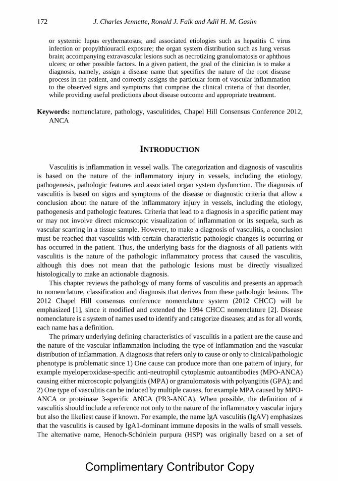
J. Charles Jennette, Ronald J. Falk and Adil H. M. Gasim 172
or systemic lupus erythematosus; and associated etiologies such as hepatitis C virus
infection or propylthiouracil exposure; the organ system distribution such as lung versus
brain; accompanying extravascular lesions such as necrotizing granulomatosis or aphthous
ulcers; or other possible factors. In a given patient, the goal of the clinician is to make a
diagnosis, namely, assign a disease name that specifies the nature of the root disease
process in the patient, and correctly assigns the particular form of vascular inflammation
to the observed signs and symptoms that comprise the clinical criteria of that disorder,
while providing useful predictions about disease outcome and appropriate treatment.
Keywords: nomenclature, pathology, vasculitides, Chapel Hill Consensus Conference 2012,
ANCA
INTRODUCTION
Vasculitis is inflammation in vessel walls. The categorization and diagnosis of vasculitis
is based on the nature of the inflammatory injury in vessels, including the etiology,
pathogenesis, pathologic features and associated organ system dysfunction. The diagnosis of
vasculitis is based on signs and symptoms of the disease or diagnostic criteria that allow a
conclusion about the nature of the inflammatory injury in vessels, including the etiology,
pathogenesis and pathologic features. Criteria that lead to a diagnosis in a specific patient may
or may not involve direct microscopic visualization of inflammation or its sequela, such as
vascular scarring in a tissue sample. However, to make a diagnosis of vasculitis, a conclusion
must be reached that vasculitis with certain characteristic pathologic changes is occurring or
has occurred in the patient. Thus, the underlying basis for the diagnosis of all patients with
vasculitis is the nature of the pathologic inflammatory process that caused the vasculitis,
although this does not mean that the pathologic lesions must be directly visualized
histologically to make an actionable diagnosis.
This chapter reviews the pathology of many forms of vasculitis and presents an approach
to nomenclature, classification and diagnosis that derives from these pathologic lesions. The
2012 Chapel Hill consensus conference nomenclature system (2012 CHCC) will be
emphasized [1], since it modified and extended the 1994 CHCC nomenclature [2]. Disease
nomenclature is a system of names used to identify and categorize diseases; and as for all words,
each name has a definition.
The primary underlying defining characteristics of vasculitis in a patient are the cause and
the nature of the vascular inflammation including the type of inflammation and the vascular
distribution of inflammation. A diagnosis that refers only to cause or only to clinical/pathologic
phenotype is problematic since 1) One cause can produce more than one pattern of injury, for
example myeloperoxidase-specific anti-neutrophil cytoplasmic autoantibodies (MPO-ANCA)
causing either microscopic polyangiitis (MPA) or granulomatosis with polyangiitis (GPA); and
2) One type of vasculitis can be induced by multiple causes, for example MPA caused by MPO-
ANCA or proteinase 3-specific ANCA (PR3-ANCA). When possible, the definition of a
vasculitis should include a reference not only to the nature of the inflammatory vascular injury
but also the likeliest cause if known. For example, the name IgA vasculitis (IgAV) emphasizes
that the vasculitis is caused by IgA1-dominant immune deposits in the walls of small vessels.
The alternative name, Henoch-Schönlein purpura (HSP) was originally based on a set of
Complimentary Contributor Copy
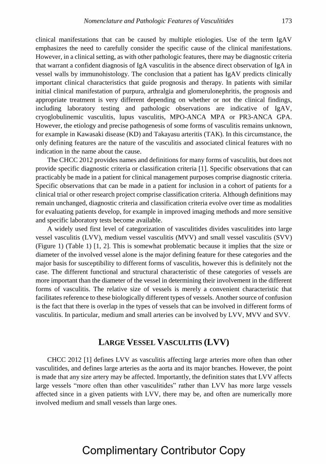
Nomenclature and Pathologic Features of Vasculitides 173
clinical manifestations that can be caused by multiple etiologies. Use of the term IgAV
emphasizes the need to carefully consider the specific cause of the clinical manifestations.
However, in a clinical setting, as with other pathologic features, there may be diagnostic criteria
that warrant a confident diagnosis of IgA vasculitis in the absence direct observation of IgA in
vessel walls by immunohistology. The conclusion that a patient has IgAV predicts clinically
important clinical characteristics that guide prognosis and therapy. In patients with similar
initial clinical manifestation of purpura, arthralgia and glomerulonephritis, the prognosis and
appropriate treatment is very different depending on whether or not the clinical findings,
including laboratory testing and pathologic observations are indicative of IgAV,
cryoglobulinemic vasculitis, lupus vasculitis, MPO-ANCA MPA or PR3-ANCA GPA.
However, the etiology and precise pathogenesis of some forms of vasculitis remains unknown,
for example in Kawasaki disease (KD) and Takayasu arteritis (TAK). In this circumstance, the
only defining features are the nature of the vasculitis and associated clinical features with no
indication in the name about the cause.
The CHCC 2012 provides names and definitions for many forms of vasculitis, but does not
provide specific diagnostic criteria or classification criteria [1]. Specific observations that can
practicably be made in a patient for clinical management purposes comprise diagnostic criteria.
Specific observations that can be made in a patient for inclusion in a cohort of patients for a
clinical trial or other research project comprise classification criteria. Although definitions may
remain unchanged, diagnostic criteria and classification criteria evolve over time as modalities
for evaluating patients develop, for example in improved imaging methods and more sensitive
and specific laboratory tests become available.
A widely used first level of categorization of vasculitides divides vasculitides into large
vessel vasculitis (LVV), medium vessel vasculitis (MVV) and small vessel vasculitis (SVV)
(Figure 1) (Table 1) [1, 2]. This is somewhat problematic because it implies that the size or
diameter of the involved vessel alone is the major defining feature for these categories and the
major basis for susceptibility to different forms of vasculitis, however this is definitely not the
case. The different functional and structural characteristic of these categories of vessels are
more important than the diameter of the vessel in determining their involvement in the different
forms of vasculitis. The relative size of vessels is merely a convenient characteristic that
facilitates reference to these biologically different types of vessels. Another source of confusion
is the fact that there is overlap in the types of vessels that can be involved in different forms of
vasculitis. In particular, medium and small arteries can be involved by LVV, MVV and SVV.
LARGE VESSEL VASCULITIS (LVV)
CHCC 2012 [1] defines LVV as vasculitis affecting large arteries more often than other
vasculitides, and defines large arteries as the aorta and its major branches. However, the point
is made that any size artery may be affected. Importantly, the definition states that LVV affects
large vessels “more often than other vasculitides” rather than LVV has more large vessels
affected since in a given patients with LVV, there may be, and often are numerically more
involved medium and small vessels than large ones.
Complimentary Contributor Copy
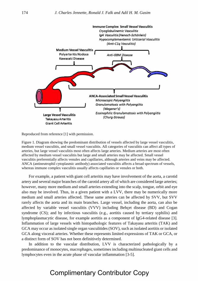
J. Charles Jennette, Ronald J. Falk and Adil H. M. Gasim 174
Reproduced from reference [1] with permission.
Figure 1. Diagram showing the predominant distribution of vessels affected by large vessel vasculitis,
medium vessel vasculitis, and small vessel vasculitis. All categories of vasculitis can affect all types of
arteries, but large vessel vasculitis most often affects large arteries. Medium arteries are most often
affected by medium vessel vasculitis but large and small arteries may be affected. Small vessel
vasculitis preferentially affects venules and capillaries, although arteries and veins may be affected.
ANCA (antineutrophil cytoplasmic antibody) associated vasculitis affects a broad spectrum of vessels,
whereas immune complex vasculitis usually affects capillaries or venules or both.
For example, a patient with giant cell arteritis may have involvement of the aorta, a carotid
artery and several major branches of the carotid artery all of which are considered large arteries;
however, many more medium and small arteries extending into the scalp, tongue, orbit and eye
also may be involved. Thus, in a given patient with a LVV, there may be numerically more
medium and small arteries affected. These same arteries can be affected by SVV, but SVV
rarely affects the aorta and its main branches. Large vessel, including the aorta, can also be
affected by variable vessel vasculitis (VVV) including Behçet disease (BD) and Cogan
syndrome (CS); and by infectious vasculitis (e.g., aortitis caused by tertiary syphilis) and
lymphoplasmacytic disease, for example aortitis as a component of IgG4-related disease [3].
Inflammation of large vessels with histopathologic features of Takayasu arteritis (TAK) and
GCA may occur as isolated single organ vasculitides (SOV), such as isolated aortitis or isolated
GCA along visceral arteries. Whether these represents limited expressions of TAK or GCA, or
a distinct form of SOV has not been definitively determined.
In addition to the vascular distribution, LVV is characterized pathologically by a
predominance of monocytes, macrophages, sometimes including multinucleated giant cells and
lymphocytes even in the acute phase of vascular inflammation [3-5].
Complimentary Contributor Copy
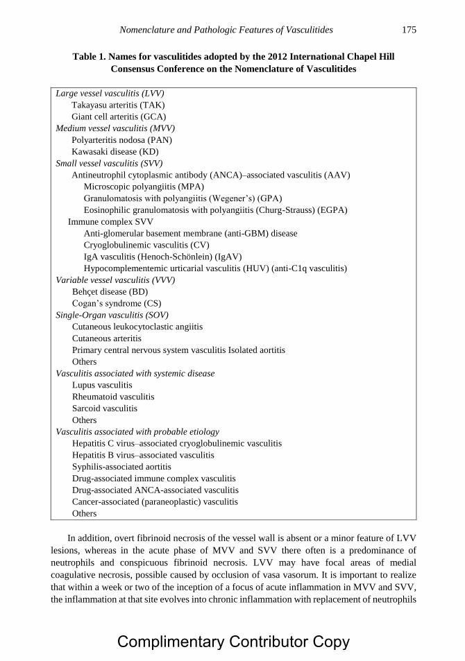
Nomenclature and Pathologic Features of Vasculitides 175
Table 1. Names for vasculitides adopted by the 2012 International Chapel Hill
Consensus Conference on the Nomenclature of Vasculitides
Large vessel vasculitis (LVV)
Takayasu arteritis (TAK)
Giant cell arteritis (GCA)
Medium vessel vasculitis (MVV)
Polyarteritis nodosa (PAN)
Kawasaki disease (KD)
Small vessel vasculitis (SVV)
Antineutrophil cytoplasmic antibody (ANCA)–associated vasculitis (AAV)
Microscopic polyangiitis (MPA)
Granulomatosis with polyangiitis (Wegener’s) (GPA)
Eosinophilic granulomatosis with polyangiitis (Churg-Strauss) (EGPA)
Immune complex SVV
Anti-glomerular basement membrane (anti-GBM) disease
Cryoglobulinemic vasculitis (CV)
IgA vasculitis (Henoch-Schönlein) (IgAV)
Hypocomplementemic urticarial vasculitis (HUV) (anti-C1q vasculitis)
Variable vessel vasculitis (VVV)
Behçet disease (BD)
Cogan’s syndrome (CS)
Single-Organ vasculitis (SOV)
Cutaneous leukocytoclastic angiitis
Cutaneous arteritis
Primary central nervous system vasculitis Isolated aortitis
Others
Vasculitis associated with systemic disease
Lupus vasculitis
Rheumatoid vasculitis
Sarcoid vasculitis
Others
Vasculitis associated with probable etiology
Hepatitis C virus–associated cryoglobulinemic vasculitis
Hepatitis B virus–associated vasculitis
Syphilis-associated aortitis
Drug-associated immune complex vasculitis
Drug-associated ANCA-associated vasculitis
Cancer-associated (paraneoplastic) vasculitis
Others
In addition, overt fibrinoid necrosis of the vessel wall is absent or a minor feature of LVV
lesions, whereas in the acute phase of MVV and SVV there often is a predominance of
neutrophils and conspicuous fibrinoid necrosis. LVV may have focal areas of medial
coagulative necrosis, possible caused by occlusion of vasa vasorum. It is important to realize
that within a week or two of the inception of a focus of acute inflammation in MVV and SVV,
the inflammation at that site evolves into chronic inflammation with replacement of neutrophils
Complimentary Contributor Copy
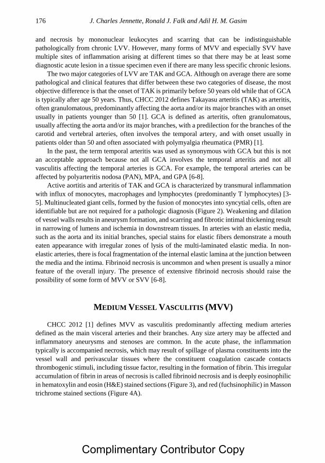
J. Charles Jennette, Ronald J. Falk and Adil H. M. Gasim 176
and necrosis by mononuclear leukocytes and scarring that can be indistinguishable
pathologically from chronic LVV. However, many forms of MVV and especially SVV have
multiple sites of inflammation arising at different times so that there may be at least some
diagnostic acute lesion in a tissue specimen even if there are many less specific chronic lesions.
The two major categories of LVV are TAK and GCA. Although on average there are some
pathological and clinical features that differ between these two categories of disease, the most
objective difference is that the onset of TAK is primarily before 50 years old while that of GCA
is typically after age 50 years. Thus, CHCC 2012 defines Takayasu arteritis (TAK) as arteritis,
often granulomatous, predominantly affecting the aorta and/or its major branches with an onset
usually in patients younger than 50 [1]. GCA is defined as arteritis, often granulomatous,
usually affecting the aorta and/or its major branches, with a predilection for the branches of the
carotid and vertebral arteries, often involves the temporal artery, and with onset usually in
patients older than 50 and often associated with polymyalgia rheumatica (PMR) [1].
In the past, the term temporal arteritis was used as synonymous with GCA but this is not
an acceptable approach because not all GCA involves the temporal arteritis and not all
vasculitis affecting the temporal arteries is GCA. For example, the temporal arteries can be
affected by polyarteritis nodosa (PAN), MPA, and GPA [6-8].
Active aortitis and arteritis of TAK and GCA is characterized by transmural inflammation
with influx of monocytes, macrophages and lymphocytes (predominantly T lymphocytes) [3-
5]. Multinucleated giant cells, formed by the fusion of monocytes into syncytial cells, often are
identifiable but are not required for a pathologic diagnosis (Figure 2). Weakening and dilation
of vessel walls results in aneurysm formation, and scarring and fibrotic intimal thickening result
in narrowing of lumens and ischemia in downstream tissues. In arteries with an elastic media,
such as the aorta and its initial branches, special stains for elastic fibers demonstrate a mouth
eaten appearance with irregular zones of lysis of the multi-laminated elastic media. In non-
elastic arteries, there is focal fragmentation of the internal elastic lamina at the junction between
the media and the intima. Fibrinoid necrosis is uncommon and when present is usually a minor
feature of the overall injury. The presence of extensive fibrinoid necrosis should raise the
possibility of some form of MVV or SVV [6-8].
MEDIUM VESSEL VASCULITIS (MVV)
CHCC 2012 [1] defines MVV as vasculitis predominantly affecting medium arteries
defined as the main visceral arteries and their branches. Any size artery may be affected and
inflammatory aneurysms and stenoses are common. In the acute phase, the inflammation
typically is accompanied necrosis, which may result of spillage of plasma constituents into the
vessel wall and perivascular tissues where the constituent coagulation cascade contacts
thrombogenic stimuli, including tissue factor, resulting in the formation of fibrin. This irregular
accumulation of fibrin in areas of necrosis is called fibrinoid necrosis and is deeply eosinophilic
in hematoxylin and eosin (H&E) stained sections (Figure 3), and red (fuchsinophilic) in Masson
trichrome stained sections (Figure 4A).
Complimentary Contributor Copy
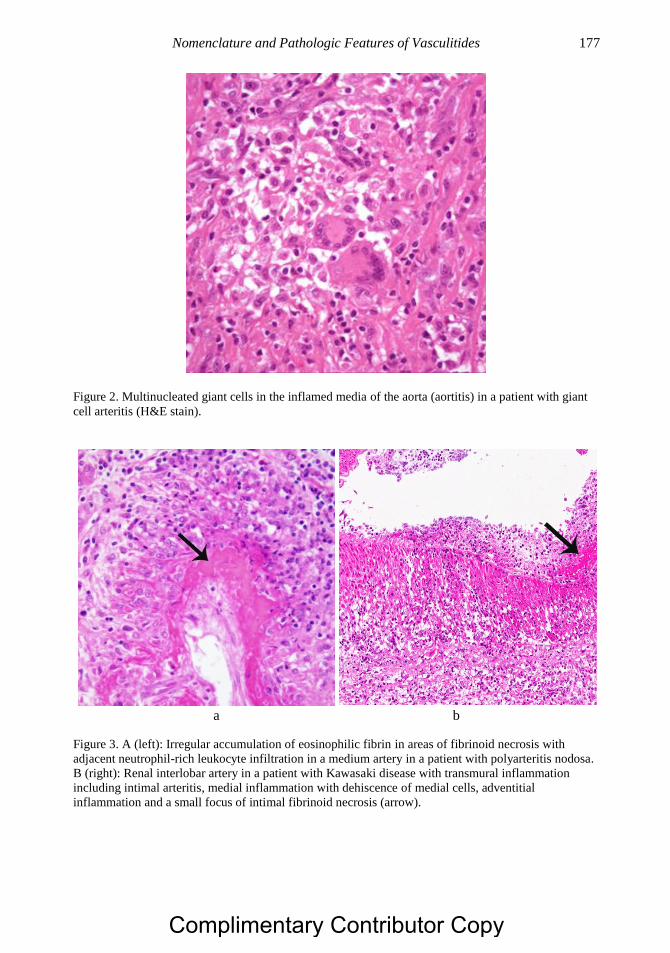
Nomenclature and Pathologic Features of Vasculitides 177
Figure 2. Multinucleated giant cells in the inflamed media of the aorta (aortitis) in a patient with giant
cell arteritis (H&E stain).
a b
Figure 3. A (left): Irregular accumulation of eosinophilic fibrin in areas of fibrinoid necrosis with
adjacent neutrophil-rich leukocyte infiltration in a medium artery in a patient with polyarteritis nodosa.
B (right): Renal interlobar artery in a patient with Kawasaki disease with transmural inflammation
including intimal arteritis, medial inflammation with dehiscence of medial cells, adventitial
inflammation and a small focus of intimal fibrinoid necrosis (arrow).
Complimentary Contributor Copy
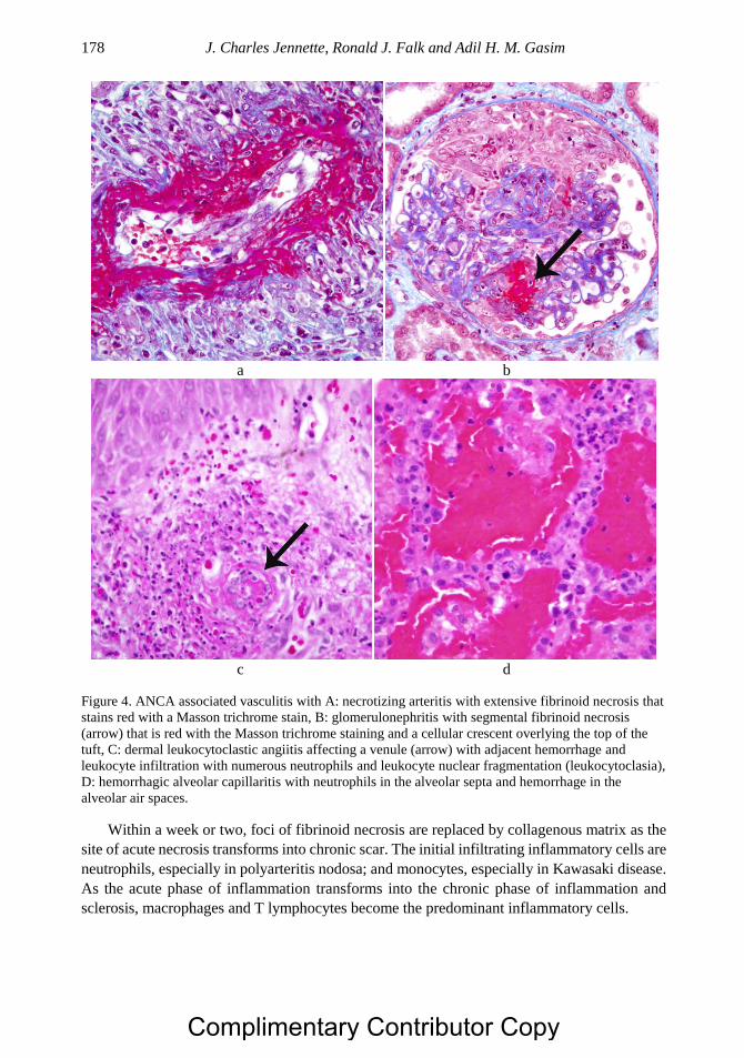
J. Charles Jennette, Ronald J. Falk and Adil H. M. Gasim 178
a b
c d
Figure 4. ANCA associated vasculitis with A: necrotizing arteritis with extensive fibrinoid necrosis that
stains red with a Masson trichrome stain, B: glomerulonephritis with segmental fibrinoid necrosis
(arrow) that is red with the Masson trichrome staining and a cellular crescent overlying the top of the
tuft, C: dermal leukocytoclastic angiitis affecting a venule (arrow) with adjacent hemorrhage and
leukocyte infiltration with numerous neutrophils and leukocyte nuclear fragmentation (leukocytoclasia),
D: hemorrhagic alveolar capillaritis with neutrophils in the alveolar septa and hemorrhage in the
alveolar air spaces.
Within a week or two, foci of fibrinoid necrosis are replaced by collagenous matrix as the
site of acute necrosis transforms into chronic scar. The initial infiltrating inflammatory cells are
neutrophils, especially in polyarteritis nodosa; and monocytes, especially in Kawasaki disease.
As the acute phase of inflammation transforms into the chronic phase of inflammation and
sclerosis, macrophages and T lymphocytes become the predominant inflammatory cells.
Complimentary Contributor Copy
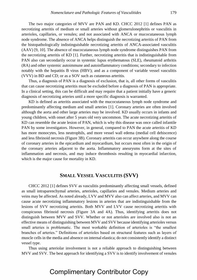
Nomenclature and Pathologic Features of Vasculitides 179
The two major categories of MVV are PAN and KD. CHCC 2012 [1] defines PAN as
necrotizing arteritis of medium or small arteries without glomerulonephritis or vasculitis in
arterioles, capillaries, or venules; and not associated with ANCA or mucocutaneous lymph
node syndrome. The absence of ANCA helps distinguish the necrotizing arteritis of PAN from
the histopathologically indistinguishable necrotizing arteritis of ANCA-associated vasculitis
(AAV) [9, 10]. The absence of mucocutaneous lymph node syndrome distinguishes PAN from
the necrotizing arteritis of KD [1]. Further, necrotizing arteritis that is indistinguishable from
PAN also can secondarily occur in systemic lupus erythematous (SLE), rheumatoid arthritis
(RA) and other systemic autoimmune and autoinflammatory conditions; secondary to infection
notably with the hepatitis B virus (HBV); and as a component of variable vessel vasculitis
(VVV) in BD and CD; or as a SOV such as cutaneous arteritis.
Thus, a diagnosis of PAN is a diagnosis of exclusion, that is, all other forms of vasculitis
that can cause necrotizing arteritis must be excluded before a diagnosis of PAN is appropriate.
In a clinical setting, this can be difficult and may require that a patient initially have a generic
diagnosis of necrotizing arteries until a more specific diagnosis is warranted.
KD is defined as arteritis associated with the mucocutaneous lymph node syndrome and
predominantly affecting medium and small arteries [1]. Coronary arteries are often involved
although the aorta and other large arteries may be involved. KD usually occurs in infants and
young children, with onset after 5 years old very uncommon. The acute necrotizing arteritis of
KD can resemble the acute lesion of PAN, which is why this disease was once called infantile
PAN by some investigators. However, in general, compared to PAN the acute arteritis of KD
has more monocytes, less neutrophils, and more vessel wall edema (medial cell dehiscence)
and less fibrinoid necrosis (Figure 3B). Coronary arteritis can occur anywhere along the course
of coronary arteries in the epicardium and myocardium, but occurs most often in the origin of
the coronary arteries adjacent to the aorta. Inflammatory aneurysms form at the sites of
inflammation and necrosis, and may induce thrombosis resulting in myocardial infarction,
which is the major cause for mortality in KD.
SMALL VESSEL VASCULITIS (SVV)
CHCC 2012 [1] defines SVV as vasculitis predominantly affecting small vessels, defined
as small intraparenchymal arteries, arterioles, capillaries and venules. Medium arteries and
veins may be affected. As noted already, LVV and MVV also can affect arteries, and MVV can
cause acute necrotizing inflammatory lesions in arteries that are indistinguishable from the
lesions of SVV necrotizing arteritis. Both MVV and LVV cause necrotizing arteritis with
conspicuous fibrinoid necrosis (Figure 3A and 4A). Thus, identifying arteritis does not
distinguish between MVV and SVV. Whether or not arterioles are involved also is not an
effective means of distinguishing between MVV and SVV because identifying arterioles versus
small arteries is problematic. The most workable definition of arterioles is “the smallest
branches of arteries.” Definitions of arterioles based on structural features such as layers of
muscle cells in the media and absence on internal elastica; do not consistently identify a distinct
vessel type.
Thus using arteriolar involvement is not a reliable approach to distinguishing between
MVV and SVV. The best approach for identifying a SVV is to identify involvement of venules
Complimentary Contributor Copy
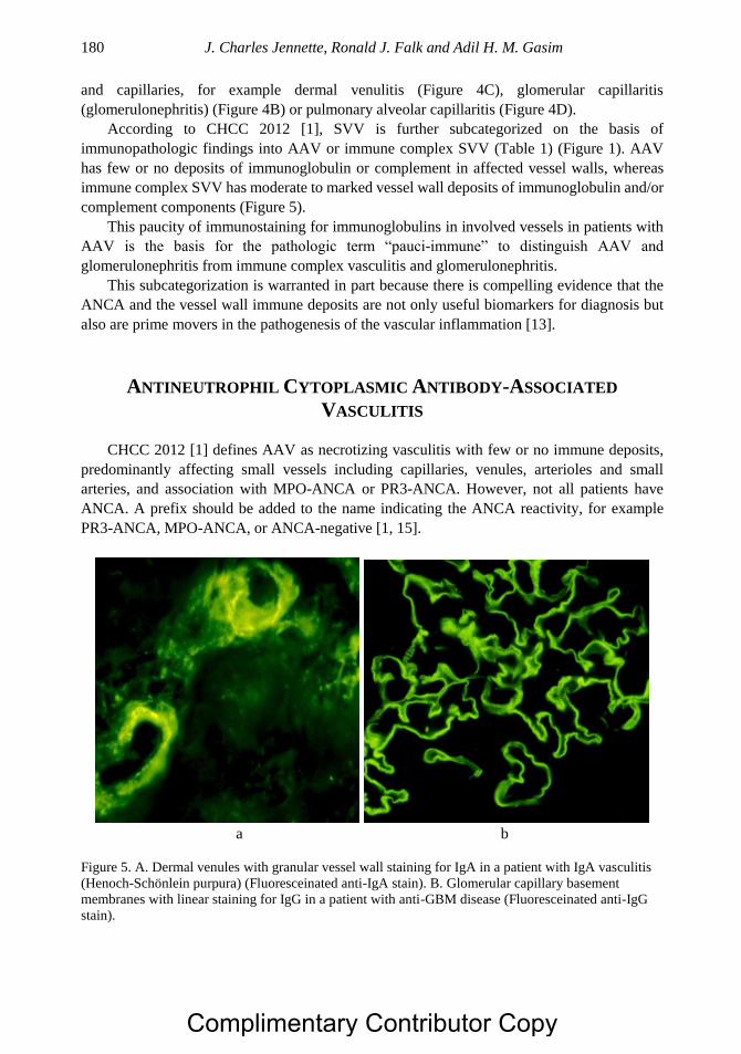
J. Charles Jennette, Ronald J. Falk and Adil H. M. Gasim 180
and capillaries, for example dermal venulitis (Figure 4C), glomerular capillaritis
(glomerulonephritis) (Figure 4B) or pulmonary alveolar capillaritis (Figure 4D).
According to CHCC 2012 [1], SVV is further subcategorized on the basis of
immunopathologic findings into AAV or immune complex SVV (Table 1) (Figure 1). AAV
has few or no deposits of immunoglobulin or complement in affected vessel walls, whereas
immune complex SVV has moderate to marked vessel wall deposits of immunoglobulin and/or
complement components (Figure 5).
This paucity of immunostaining for immunoglobulins in involved vessels in patients with
AAV is the basis for the pathologic term “pauci-immune” to distinguish AAV and
glomerulonephritis from immune complex vasculitis and glomerulonephritis.
This subcategorization is warranted in part because there is compelling evidence that the
ANCA and the vessel wall immune deposits are not only useful biomarkers for diagnosis but
also are prime movers in the pathogenesis of the vascular inflammation [13].
ANTINEUTROPHIL CYTOPLASMIC ANTIBODY-ASSOCIATED
VASCULITIS
CHCC 2012 [1] defines AAV as necrotizing vasculitis with few or no immune deposits,
predominantly affecting small vessels including capillaries, venules, arterioles and small
arteries, and association with MPO-ANCA or PR3-ANCA. However, not all patients have
ANCA. A prefix should be added to the name indicating the ANCA reactivity, for example
PR3-ANCA, MPO-ANCA, or ANCA-negative [1, 15].
a b
Figure 5. A. Dermal venules with granular vessel wall staining for IgA in a patient with IgA vasculitis
(Henoch-Schönlein purpura) (Fluoresceinated anti-IgA stain). B. Glomerular capillary basement
membranes with linear staining for IgG in a patient with anti-GBM disease (Fluoresceinated anti-IgG
stain).
Complimentary Contributor Copy
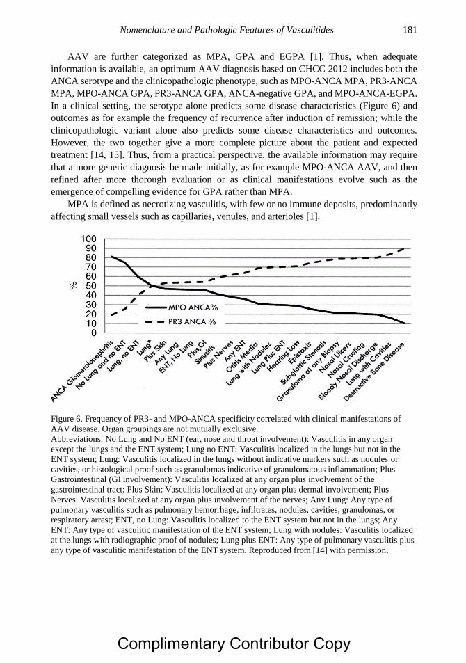
Nomenclature and Pathologic Features of Vasculitides 181
AAV are further categorized as MPA, GPA and EGPA [1]. Thus, when adequate
information is available, an optimum AAV diagnosis based on CHCC 2012 includes both the
ANCA serotype and the clinicopathologic phenotype, such as MPO-ANCA MPA, PR3-ANCA
MPA, MPO-ANCA GPA, PR3-ANCA GPA, ANCA-negative GPA, and MPO-ANCA-EGPA.
In a clinical setting, the serotype alone predicts some disease characteristics (Figure 6) and
outcomes as for example the frequency of recurrence after induction of remission; while the
clinicopathologic variant alone also predicts some disease characteristics and outcomes.
However, the two together give a more complete picture about the patient and expected
treatment [14, 15]. Thus, from a practical perspective, the available information may require
that a more generic diagnosis be made initially, as for example MPO-ANCA AAV, and then
refined after more thorough evaluation or as clinical manifestations evolve such as the
emergence of compelling evidence for GPA rather than MPA.
MPA is defined as necrotizing vasculitis, with few or no immune deposits, predominantly
affecting small vessels such as capillaries, venules, and arterioles [1].
Figure 6. Frequency of PR3- and MPO-ANCA specificity correlated with clinical manifestations of
AAV disease. Organ groupings are not mutually exclusive.
Abbreviations: No Lung and No ENT (ear, nose and throat involvement): Vasculitis in any organ
except the lungs and the ENT system; Lung no ENT: Vasculitis localized in the lungs but not in the
ENT system; Lung: Vasculitis localized in the lungs without indicative markers such as nodules or
cavities, or histological proof such as granulomas indicative of granulomatous inflammation; Plus
Gastrointestinal (GI involvement): Vasculitis localized at any organ plus involvement of the
gastrointestinal tract; Plus Skin: Vasculitis localized at any organ plus dermal involvement; Plus
Nerves: Vasculitis localized at any organ plus involvement of the nerves; Any Lung: Any type of
pulmonary vasculitis such as pulmonary hemorrhage, infiltrates, nodules, cavities, granulomas, or
respiratory arrest; ENT, no Lung: Vasculitis localized to the ENT system but not in the lungs; Any
ENT: Any type of vasculitic manifestation of the ENT system; Lung with nodules: Vasculitis localized
at the lungs with radiographic proof of nodules; Lung plus ENT: Any type of pulmonary vasculitis plus
any type of vasculitic manifestation of the ENT system. Reproduced from [14] with permission.
Complimentary Contributor Copy
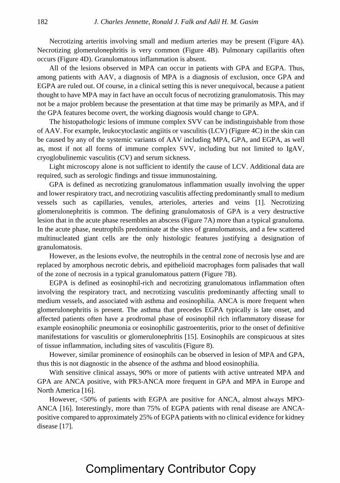
J. Charles Jennette, Ronald J. Falk and Adil H. M. Gasim 182
Necrotizing arteritis involving small and medium arteries may be present (Figure 4A).
Necrotizing glomerulonephritis is very common (Figure 4B). Pulmonary capillaritis often
occurs (Figure 4D). Granulomatous inflammation is absent.
All of the lesions observed in MPA can occur in patients with GPA and EGPA. Thus,
among patients with AAV, a diagnosis of MPA is a diagnosis of exclusion, once GPA and
EGPA are ruled out. Of course, in a clinical setting this is never unequivocal, because a patient
thought to have MPA may in fact have an occult focus of necrotizing granulomatosis. This may
not be a major problem because the presentation at that time may be primarily as MPA, and if
the GPA features become overt, the working diagnosis would change to GPA.
The histopathologic lesions of immune complex SVV can be indistinguishable from those
of AAV. For example, leukocytoclastic angiitis or vasculitis (LCV) (Figure 4C) in the skin can
be caused by any of the systemic variants of AAV including MPA, GPA, and EGPA, as well
as, most if not all forms of immune complex SVV, including but not limited to IgAV,
cryoglobulinemic vasculitis (CV) and serum sickness.
Light microscopy alone is not sufficient to identify the cause of LCV. Additional data are
required, such as serologic findings and tissue immunostaining.
GPA is defined as necrotizing granulomatous inflammation usually involving the upper
and lower respiratory tract, and necrotizing vasculitis affecting predominantly small to medium
vessels such as capillaries, venules, arterioles, arteries and veins [1]. Necrotizing
glomerulonephritis is common. The defining granulomatosis of GPA is a very destructive
lesion that in the acute phase resembles an abscess (Figure 7A) more than a typical granuloma.
In the acute phase, neutrophils predominate at the sites of granulomatosis, and a few scattered
multinucleated giant cells are the only histologic features justifying a designation of
granulomatosis.
However, as the lesions evolve, the neutrophils in the central zone of necrosis lyse and are
replaced by amorphous necrotic debris, and epithelioid macrophages form palisades that wall
of the zone of necrosis in a typical granulomatous pattern (Figure 7B).
EGPA is defined as eosinophil-rich and necrotizing granulomatous inflammation often
involving the respiratory tract, and necrotizing vasculitis predominantly affecting small to
medium vessels, and associated with asthma and eosinophilia. ANCA is more frequent when
glomerulonephritis is present. The asthma that precedes EGPA typically is late onset, and
affected patients often have a prodromal phase of eosinophil rich inflammatory disease for
example eosinophilic pneumonia or eosinophilic gastroenteritis, prior to the onset of definitive
manifestations for vasculitis or glomerulonephritis [15]. Eosinophils are conspicuous at sites
of tissue inflammation, including sites of vasculitis (Figure 8).
However, similar prominence of eosinophils can be observed in lesion of MPA and GPA,
thus this is not diagnostic in the absence of the asthma and blood eosinophilia.
With sensitive clinical assays, 90% or more of patients with active untreated MPA and
GPA are ANCA positive, with PR3-ANCA more frequent in GPA and MPA in Europe and
North America [16].
However, <50% of patients with EGPA are positive for ANCA, almost always MPO-
ANCA [16]. Interestingly, more than 75% of EGPA patients with renal disease are ANCA-
positive compared to approximately 25% of EGPA patients with no clinical evidence for kidney
disease [17].
Complimentary Contributor Copy
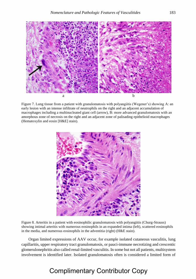
Nomenclature and Pathologic Features of Vasculitides 183
a b
Figure 7. Lung tissue from a patient with granulomatosis with polyangiitis (Wegener’s) showing A: an
early lesion with an intense infiltrate of neutrophils on the right and an adjacent accumulation of
macrophages including a multinucleated giant cell (arrow), B: more advanced granulomatosis with an
amorphous zone of necrosis on the right and an adjacent zone of palisading epithelioid macrophages
(Hematoxylin and eosin [H&E] stain).
Figure 8. Arteritis in a patient with eosinophilic granulomatosis with polyangiitis (Churg-Strauss)
showing intimal arteritis with numerous eosinophils in an expanded intima (left), scattered eosinophils
in the media, and numerous eosinophils in the adventitia (right) (H&E stain).
Organ limited expressions of AAV occur, for example isolated cutaneous vasculitis, lung
capillaritis, upper respiratory tract granulomatosis, or pauci-immune necrotizing and crescentic
glomerulonephritis also called renal-limited vasculitis. In some but not all patients, multisystem
involvement is identified later. Isolated granulomatosis often is considered a limited form of
Complimentary Contributor Copy
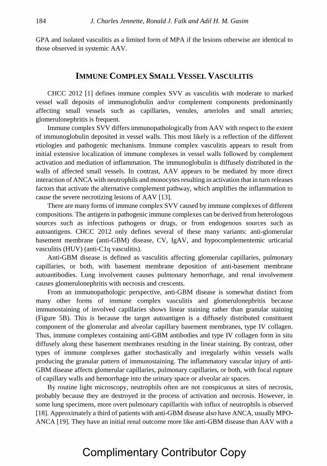
J. Charles Jennette, Ronald J. Falk and Adil H. M. Gasim 184
GPA and isolated vasculitis as a limited form of MPA if the lesions otherwise are identical to
those observed in systemic AAV.
IMMUNE COMPLEX SMALL VESSEL VASCULITIS
CHCC 2012 [1] defines immune complex SVV as vasculitis with moderate to marked
vessel wall deposits of immunoglobulin and/or complement components predominantly
affecting small vessels such as capillaries, venules, arterioles and small arteries;
glomerulonephritis is frequent.
Immune complex SVV differs immunopathologically from AAV with respect to the extent
of immunoglobulin deposited in vessel walls. This most likely is a reflection of the different
etiologies and pathogenic mechanisms. Immune complex vasculitis appears to result from
initial extensive localization of immune complexes in vessel walls followed by complement
activation and mediation of inflammation. The immunoglobulin is diffusely distributed in the
walls of affected small vessels. In contrast, AAV appears to be mediated by more direct
interaction of ANCA with neutrophils and monocytes resulting in activation that in turn releases
factors that activate the alternative complement pathway, which amplifies the inflammation to
cause the severe necrotizing lesions of AAV [13].
There are many forms of immune complex SVV caused by immune complexes of different
compositions. The antigens in pathogenic immune complexes can be derived from heterologous
sources such as infectious pathogens or drugs, or from endogenous sources such as
autoantigens. CHCC 2012 only defines several of these many variants: anti-glomerular
basement membrane (anti-GBM) disease, CV, IgAV, and hypocomplementemic urticarial
vasculitis (HUV) (anti-C1q vasculitis).
Anti-GBM disease is defined as vasculitis affecting glomerular capillaries, pulmonary
capillaries, or both, with basement membrane deposition of anti-basement membrane
autoantibodies. Lung involvement causes pulmonary hemorrhage, and renal involvement
causes glomerulonephritis with necrosis and crescents.
From an immunopathologic perspective, anti-GBM disease is somewhat distinct from
many other forms of immune complex vasculitis and glomerulonephritis because
immunostaining of involved capillaries shows linear staining rather than granular staining
(Figure 5B). This is because the target autoantigen is a diffusely distributed constituent
component of the glomerular and alveolar capillary basement membranes, type IV collagen.
Thus, immune complexes containing anti-GBM antibodies and type IV collagen form in situ
diffusely along these basement membranes resulting in the linear staining. By contrast, other
types of immune complexes gather stochastically and irregularly within vessels walls
producing the granular pattern of immunostaining. The inflammatory vascular injury of anti-
GBM disease affects glomerular capillaries, pulmonary capillaries, or both, with focal rupture
of capillary walls and hemorrhage into the urinary space or alveolar air spaces.
By routine light microscopy, neutrophils often are not conspicuous at sites of necrosis,
probably because they are destroyed in the process of activation and necrosis. However, in
some lung specimens, more overt pulmonary capillaritis with influx of neutrophils is observed
[18]. Approximately a third of patients with anti-GBM disease also have ANCA, usually MPO-
ANCA [19]. They have an initial renal outcome more like anti-GBM disease than AAV with a
Complimentary Contributor Copy
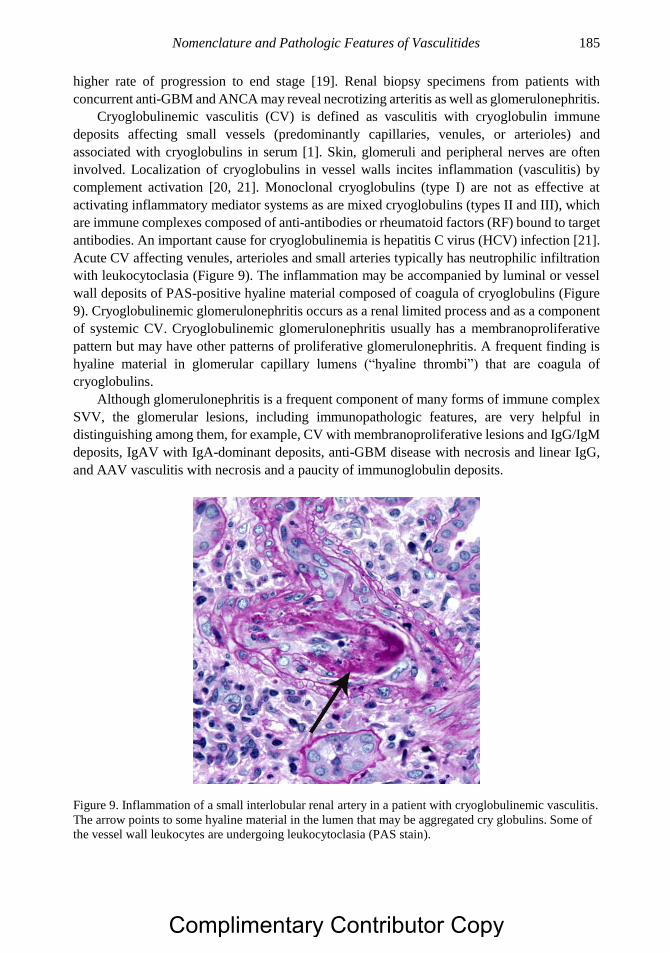
Nomenclature and Pathologic Features of Vasculitides 185
higher rate of progression to end stage [19]. Renal biopsy specimens from patients with
concurrent anti-GBM and ANCA may reveal necrotizing arteritis as well as glomerulonephritis.
Cryoglobulinemic vasculitis (CV) is defined as vasculitis with cryoglobulin immune
deposits affecting small vessels (predominantly capillaries, venules, or arterioles) and
associated with cryoglobulins in serum [1]. Skin, glomeruli and peripheral nerves are often
involved. Localization of cryoglobulins in vessel walls incites inflammation (vasculitis) by
complement activation [20, 21]. Monoclonal cryoglobulins (type I) are not as effective at
activating inflammatory mediator systems as are mixed cryoglobulins (types II and III), which
are immune complexes composed of anti-antibodies or rheumatoid factors (RF) bound to target
antibodies. An important cause for cryoglobulinemia is hepatitis C virus (HCV) infection [21].
Acute CV affecting venules, arterioles and small arteries typically has neutrophilic infiltration
with leukocytoclasia (Figure 9). The inflammation may be accompanied by luminal or vessel
wall deposits of PAS-positive hyaline material composed of coagula of cryoglobulins (Figure
9). Cryoglobulinemic glomerulonephritis occurs as a renal limited process and as a component
of systemic CV. Cryoglobulinemic glomerulonephritis usually has a membranoproliferative
pattern but may have other patterns of proliferative glomerulonephritis. A frequent finding is
hyaline material in glomerular capillary lumens (“hyaline thrombi”) that are coagula of
cryoglobulins.
Although glomerulonephritis is a frequent component of many forms of immune complex
SVV, the glomerular lesions, including immunopathologic features, are very helpful in
distinguishing among them, for example, CV with membranoproliferative lesions and IgG/IgM
deposits, IgAV with IgA-dominant deposits, anti-GBM disease with necrosis and linear IgG,
and AAV vasculitis with necrosis and a paucity of immunoglobulin deposits.
Figure 9. Inflammation of a small interlobular renal artery in a patient with cryoglobulinemic vasculitis.
The arrow points to some hyaline material in the lumen that may be aggregated cry globulins. Some of
the vessel wall leukocytes are undergoing leukocytoclasia (PAS stain).
Complimentary Contributor Copy
J. Charles Jennette, Ronald J. Falk and Adil H. M. Gasim 186
IgAV is defined as vasculitis, with IgA1-dominant immune deposits, predominantly
affecting small vessels such as capillaries, venules, and arterioles (Figure 5A) [1]. IgAV often
involves skin and gastrointestinal tract, and frequently causes arthritis. Glomerulonephritis
indistinguishable from IgA nephropathy may occur. The shift in terminology to IgA vasculitis
is prompted in part by mounting evidence that abnormally reduced glycosylation of the O-
linked glycans in the hinge region of IgA1 molecules play a pivotal pathogenic role in IgA
vasculitis and IgA nephropathy [22, 23].
In patients with these diseases, serum IgA1 has reduced terminal galactosylation and
sialylation, resulting in increased exposure of N-acetylgalactosamine at the ends of the hinge
region glycans. There also is evidence that patients have autoantibodies that recognize these
abnormally glycosylated IgA1 hinge regions, which could result in immune complex
formation. In addition to forming pathogenic immune complexes, abnormal glycosylation of
the glycans in the hinge region of IgA1 molecules could play a pathogenic role through a variety
of other mechanisms, such as reduced clearance from the circulation because of lack of receptor
engagement by the abnormal IgA, increased aggregation of IgA in the circulation resulting in
mesangial trapping, and increased affinity of the abnormal IgA for mesangial matrix.
Purpura hematuria and proteinuria are frequent clinical manifestations of IgA vasculitis
that also are shared by other forms of SVV, including AAV and cryoglobulinemic vasculitis
[24]. Demographic, clinical, laboratory and pathology observations all help resolve the
differential diagnosis. For example, IgAV is most frequent in children, especially children
under 10 years old, whereas AAV and CV are more common in older patients. However, all
can occur at any age. Useful disease markers include immunohistologic findings such as IgA1
deposits, cryoglobulin deposits, and a paucity of deposits; and serologic tests under
glycosylated IgA1, RF and ANCA.
The glomerular lesions in the kidney are indistinguishable from those of IgA nephropathy
although necrosis, crescents and capillary wall deposits are somewhat more frequent in IgA
vasculitis patients who undergo renal biopsy. In addition to the glomeruli, IgA1-dominant
immune deposits occur in renal arterioles and peritubular capillaries in <20% of patients. IgA1-
dominant immune deposits also can be identified in small vessels of the skin and gut; however,
this is not of value in differentiating IgA vasculitis from IgA nephropathy because many IgA
nephropathy patients also have IgA in dermal venules.
HUV is accompanied by urticaria and hypocomplementemia affects small vessels such as
capillaries, venules, and arterioles, and is associated with anti-C1q antibodies [1];
glomerulonephritis, arthritis, obstructive pulmonary disease, and ocular inflammation are
common.
HUV is characterized by recurrent episodes of urticaria with underlying leukocytoclastic
vasculitis that causes hemorrhage into the lesions [24]. Skin manifestations include painful,
tender, burning or pruritic lesions that persist for greater than 24 hours, often with central or
diffuse areas of hemorrhage. HUV can be isolated or associated with autoimmune diseases
(including lupus), infections, drug reactions, or neoplasms. Proteinuria and hematuria occur in
20-30% of patients and the underlying glomerular lesion usually has a membranoproliferative
pattern, but other proliferative and focal necrotizing lesions may occur. Glomeruli and dermal
vessels have granular deposits of IgG, IgA, IgM, C3 and C1q, which resembles the “full house”
immunostaining seen in SLE.
IgG autoantibodies to the collagen-like region of C1q (anti-C1q) are usually present and
may play a role in pathogenesis [25]. The hypocomplementemia appears to be secondary to
Complimentary Contributor Copy
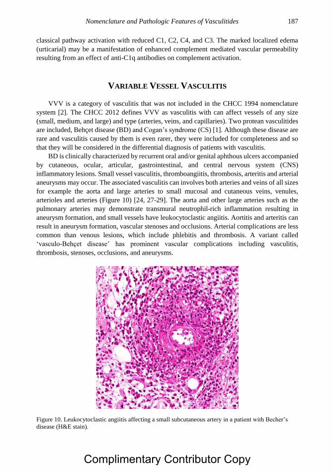
Nomenclature and Pathologic Features of Vasculitides 187
classical pathway activation with reduced C1, C2, C4, and C3. The marked localized edema
(urticarial) may be a manifestation of enhanced complement mediated vascular permeability
resulting from an effect of anti-C1q antibodies on complement activation.
VARIABLE VESSEL VASCULITIS
VVV is a category of vasculitis that was not included in the CHCC 1994 nomenclature
system [2]. The CHCC 2012 defines VVV as vasculitis with can affect vessels of any size
(small, medium, and large) and type (arteries, veins, and capillaries). Two protean vasculitides
are included, Behçet disease (BD) and Cogan’s syndrome (CS) [1]. Although these disease are
rare and vasculitis caused by them is even rarer, they were included for completeness and so
that they will be considered in the differential diagnosis of patients with vasculitis.
BD is clinically characterized by recurrent oral and/or genital aphthous ulcers accompanied
by cutaneous, ocular, articular, gastrointestinal, and central nervous system (CNS)
inflammatory lesions. Small vessel vasculitis, thromboangiitis, thrombosis, arteritis and arterial
aneurysms may occur. The associated vasculitis can involves both arteries and veins of all sizes
for example the aorta and large arteries to small mucosal and cutaneous veins, venules,
arterioles and arteries (Figure 10) [24, 27-29]. The aorta and other large arteries such as the
pulmonary arteries may demonstrate transmural neutrophil-rich inflammation resulting in
aneurysm formation, and small vessels have leukocytoclastic angiitis. Aortitis and arteritis can
result in aneurysm formation, vascular stenoses and occlusions. Arterial complications are less
common than venous lesions, which include phlebitis and thrombosis. A variant called
‘vasculo-Behçet disease’ has prominent vascular complications including vasculitis,
thrombosis, stenoses, occlusions, and aneurysms.
Figure 10. Leukocytoclastic angiitis affecting a small subcutaneous artery in a patient with Becher’s
disease (H&E stain).
Complimentary Contributor Copy
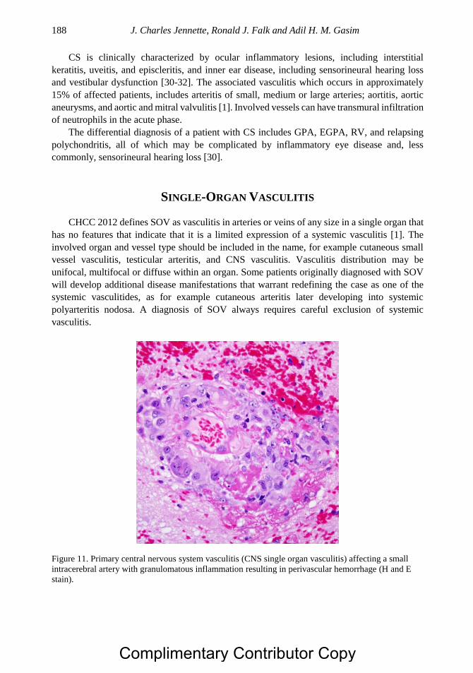
J. Charles Jennette, Ronald J. Falk and Adil H. M. Gasim 188
CS is clinically characterized by ocular inflammatory lesions, including interstitial
keratitis, uveitis, and episcleritis, and inner ear disease, including sensorineural hearing loss
and vestibular dysfunction [30-32]. The associated vasculitis which occurs in approximately
15% of affected patients, includes arteritis of small, medium or large arteries; aortitis, aortic
aneurysms, and aortic and mitral valvulitis [1]. Involved vessels can have transmural infiltration
of neutrophils in the acute phase.
The differential diagnosis of a patient with CS includes GPA, EGPA, RV, and relapsing
polychondritis, all of which may be complicated by inflammatory eye disease and, less
commonly, sensorineural hearing loss [30].
SINGLE-ORGAN VASCULITIS
CHCC 2012 defines SOV as vasculitis in arteries or veins of any size in a single organ that
has no features that indicate that it is a limited expression of a systemic vasculitis [1]. The
involved organ and vessel type should be included in the name, for example cutaneous small
vessel vasculitis, testicular arteritis, and CNS vasculitis. Vasculitis distribution may be
unifocal, multifocal or diffuse within an organ. Some patients originally diagnosed with SOV
will develop additional disease manifestations that warrant redefining the case as one of the
systemic vasculitides, as for example cutaneous arteritis later developing into systemic
polyarteritis nodosa. A diagnosis of SOV always requires careful exclusion of systemic
vasculitis.
Figure 11. Primary central nervous system vasculitis (CNS single organ vasculitis) affecting a small
intracerebral artery with granulomatous inflammation resulting in perivascular hemorrhage (H and E
stain).
Complimentary Contributor Copy
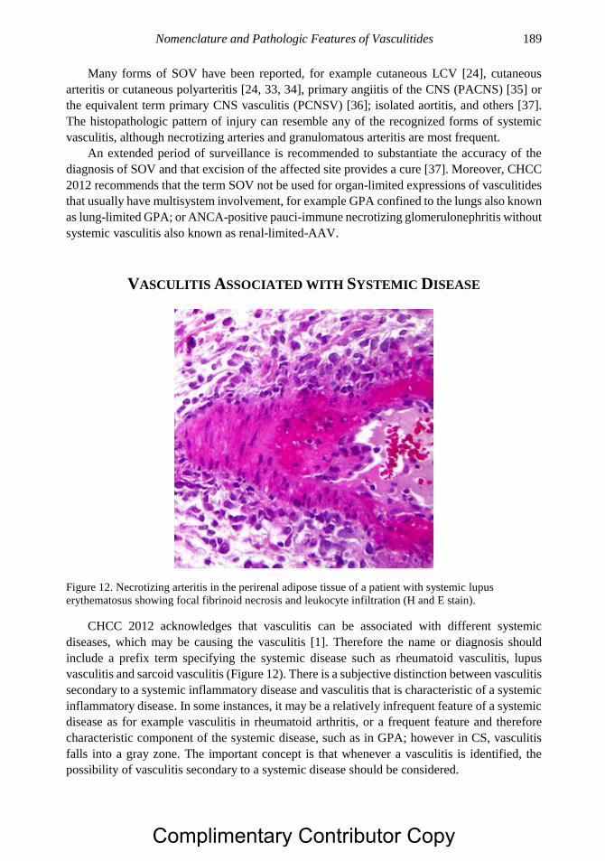
Nomenclature and Pathologic Features of Vasculitides 189
Many forms of SOV have been reported, for example cutaneous LCV [24], cutaneous
arteritis or cutaneous polyarteritis [24, 33, 34], primary angiitis of the CNS (PACNS) [35] or
the equivalent term primary CNS vasculitis (PCNSV) [36]; isolated aortitis, and others [37].
The histopathologic pattern of injury can resemble any of the recognized forms of systemic
vasculitis, although necrotizing arteries and granulomatous arteritis are most frequent.
An extended period of surveillance is recommended to substantiate the accuracy of the
diagnosis of SOV and that excision of the affected site provides a cure [37]. Moreover, CHCC
2012 recommends that the term SOV not be used for organ-limited expressions of vasculitides
that usually have multisystem involvement, for example GPA confined to the lungs also known
as lung-limited GPA; or ANCA-positive pauci-immune necrotizing glomerulonephritis without
systemic vasculitis also known as renal-limited-AAV.
VASCULITIS ASSOCIATED WITH SYSTEMIC DISEASE
Figure 12. Necrotizing arteritis in the perirenal adipose tissue of a patient with systemic lupus
erythematosus showing focal fibrinoid necrosis and leukocyte infiltration (H and E stain).
CHCC 2012 acknowledges that vasculitis can be associated with different systemic
diseases, which may be causing the vasculitis [1]. Therefore the name or diagnosis should
include a prefix term specifying the systemic disease such as rheumatoid vasculitis, lupus
vasculitis and sarcoid vasculitis (Figure 12). There is a subjective distinction between vasculitis
secondary to a systemic inflammatory disease and vasculitis that is characteristic of a systemic
inflammatory disease. In some instances, it may be a relatively infrequent feature of a systemic
disease as for example vasculitis in rheumatoid arthritis, or a frequent feature and therefore
characteristic component of the systemic disease, such as in GPA; however in CS, vasculitis
falls into a gray zone. The important concept is that whenever a vasculitis is identified, the
possibility of vasculitis secondary to a systemic disease should be considered.
Complimentary Contributor Copy
J. Charles Jennette, Ronald J. Falk and Adil H. M. Gasim 190
VASCULITIS ASSOCIATED WITH PROBABLE ETIOLOGY
CHCC 2012 acknowledges that vasculitis may be associated with a probable specific
etiology [1]. In this case, the name or diagnosis should have a prefix term specifying the
association, for example hydralazine-associated microscopic polyangiitis, HBV-associated
vasculitis and HCV-associated CV, minocycline-associated vasculitis, and paraneoplastic
vasculitis.
The likelihood that an association with a probable etiology is because of a causal
relationship depends on the credibility of the supporting data. Some etiologic relationships are
reflected in the names of major categories and others designate categorical subsets. For
example, propylthiouracil-associated MPO-ANCA and HCV-associated CV are designations
that each refer to two different etiologies that act in sequence to cause vasculitis, highlighting
the importance of using diagnostic terms that provide information about likely or definite
causes of vasculitis in a given patient.
Emerging data from genomic, proteomic and metabolomic studies are demonstrating
previously unrecognized probable etiologies and pathogenic mechanisms [38]. Monogenic
mutations have been discovered associated with various patterns of vasculitis. For example,
Deficiency of Adenosine deaminase 2 (DADA2) resulting from mutations in the CECR1 gene
can cause a necrotizing vasculopathy that resembles PAN pathologically and clinically.
However, DADA differs clinically from typical PAN because the onset usually is in childhood
[39].
CONCLUSION
Many factors must be taken into consideration in rendering an accurate and precise clinical
diagnosis of vasculitis in a given patient, such as the clinical signs and symptoms, results of
laboratory tests, and the observed histopathology of involved organ tissue. Important
parameters include the types of vessels affected, as for example arteries or venules; the
pathologic features of the inflammatory process such as leukocytoclastic inflammation versus
granulomatous inflammation; immunopathologic features such as IgA-dominant deposits or
paucity of immunoglobulin in vessel walls; serologic findings such as cryoglobulins or ANCA;
associated systemic disease such as RA or SLE; proximate etiologies such as HCV and
propylthiouracil; the distribution of lesions to affected organ systems such as the lungs versus
the gut; and any accompanying extravascular lesions such as necrotizing granulomatosis or
aphthous ulcers, and other factors.
REFERENCES
[1] Jennette, J. C., Falk, R. J., Bacon, P. A., et al. 2012 Revised International Chapel Hill
Consensus Conference Nomenclature of Vasculitides. Arthritis Rheum. 2013; 65:1-11.
[2] Jennette, J. C., Falk, R. J., Andrassy, K., et al. Nomenclature of systemic vasculitides:
The proposal of an international consensus conference. Arthritis Rheum. 1994; 37:187-
192.
Complimentary Contributor Copy
Nomenclature and Pathologic Features of Vasculitides 191
[3] Miller, D. V., Maleszewski, J. J. The pathology of large-vessel vasculitides. Clin. Exp.
Rheumatol. 2011; 29 (Suppl. 64):S92-S98.
[4] Churg, J. Large vessel vasculitis. Clin. Exp. Immunol. 1993; 93 (Suppl. 1):11.
[5] Vaideeswar, P., Deshpande, J. R. Pathology of Takayasu arteritis: A brief review. Ann.
Pediatr. Cardiol. 2013; 6:52-58.
[6] Morinaga, A., Ono, K., Komai, K., et al. Microscopic polyangiitis presenting with
temporal arteritis and multiple cranial neuropathies. J. Neurol. Sci. 2007; 2 56:81-83.
[7] Vidal, E., Liozon, F., Rogues, A. M., et al. Concurrent temporal arteritis and Churg-
Strauss syndrome. J. Rheumatol. 1992; 19:1312-1314.
[8] Hamidou, M. A., Moreau, A., Toquet, C., et al. Temporal arteritis associated with
systemic necrotizing vasculitis. J. Rheumatol. 2003; 30: 2165-2169.
[9] Guillevin, L., Lhote, F., Amouroux, J., et al. Antineutrophil cytoplasmic antibodies,
abnormal angiograms and pathological findings in polyarteritis nodosa and Churg-
Strauss syndrome: indications for the classification of vasculitides of the polyarteritis
Nodosa Group. Br. J. Rheumatol. 1996; 35:958-964.
[10] Pagnoux, C., Seror, R., Henegar, C., et al. Clinical features and outcomes in 348 patients
with polyarteritis nodosa: a systematic retrospective study of patients diagnosed between
1963 and 2005 and entered into the French Vasculitis Study Group Database. Arthritis
Rheum. 2010; 62:616-626.
[11] Amano, S., Hazama, F., Hamashima, Y. Pathology of Kawasaki disease: II. Distribution
and incidence of the vascular lesions. Jpn. Circ. J. 1979; 43:741-748.
[12] Amano, S., Hazama, F., Hamashima, Y. Pathology of Kawasaki disease: I. Pathology and
morphogenesis of the vascular changes. Jpn. Circ. J. 1979; 43:633-643.
[13] Jennette JC, Falk RJ. Pathogenesis of Antineutrophil Cytoplasmic Autoantibody-
Mediated Disease. Nature Rev Rheumatol. 2014; 10:463-473.
[14] Lionaki, S., Blyth, E. R., Hogan, S. L., et al. Classification of antineutrophil cytoplasmic
autoantibody vasculitides: The role of antineutrophil cytoplasmic autoantibody
specificity for myeloperoxidase or proteinase 3 in disease recognition and prognosis.
Arthritis Rheum. 2012; 64:3452-3462.
[15] Jennette JC, Nachman PH. ANCA glomerulonephritis and vasculitis. Clin J Am Soc
Nephrol. 2017; 12:1680-1691.
[16] Cohen Tervaert, J. W., Damoiseaux, J. Antineutrophil cytoplasmic autoantibodies: how
are they detected and what is their use for diagnosis, classification and follow-up? Clin.
Rev. Allergy Immunol. 2012; 43:211-219.
[17] Sinico, R. A., Di Toma, L., Maggiore, U., et al. Renal involvement in Churg-Strauss
syndrome. Am. J. Kidney Dis. 2006; 47:770-779.
[18] Lombard, C. M., Colby, T. V., Elliott, C. G. Surgical pathology of the lung in anti-
basement membrane antibody-associated Goodpasture’s syndrome. Hum. Pathol. 1989;
20:445-451.
[19] McAdoo SP, Tanna A, Hrušková Z, et al. Patients double-seropositive for ANCA and
anti-GBM antibodies have varied renal survival, frequency of relapse, and outcomes
compared to single-seropositive patients. Kidney Int. 2017; 92:693-702.
[20] Tedeschi, A., Baratè, C., Minola, E., et al. Cryoglobulinemia. Blood Rev. 2007; 21:183-
200.
[21] Lauletta, G., Russi, S., Conteduca, V., et al. Hepatitis C virus infection and mixed
cryoglobulinemia. Clin. Dev. Immunol. 2012; 2012:502156.
Complimentary Contributor Copy
J. Charles Jennette, Ronald J. Falk and Adil H. M. Gasim 192
[22] Novak J, Barratt J, Julian BA, Renfrow MB. Aberrant Glycosylation of the IgA1
Molecule in IgA Nephropathy. Semin Nephrol. 2018; 38:461-476.
[23] Suzuki, H., Fan, R., Zhang, Z., et al. Aberrantly glycosylated IgA1 in IgA nephropathy
patients is recognized by IgG antibodies with restricted heterogeneity. J. Clin. Invest.
2009; 119:1668-1677.
[24] Sunderkötter CH, Zelger B, Chen KR, et al. Dermatological addendum to the 2012
International Chapel Hill Consensus Conference Nomenclature of Vasculitides. Arthritis
Rheumatol. 2018; 70:171-184.
[25] Kallenberg, C. G. Anti-C1q autoantibodies. Autoimmun. Rev. 2008; 7:612-615.
[26] Jara, L. J., Navarro, C., Medina, G., et al. Hypocomplementemic urticarial vasculitis
syndrome. Curr. Rheumatol. Rep. 2009; 11:410-415.
[27] Saadoun, D., Asli, B., Wechsler, B., et al. Long-term outcome of arterial lesions in Behçet
disease: a series of 101 patients. Medicine 2012; 91:18-24.
[28] Calamia, K. T., Schirmer, M., Melikoglu, M. Major vessel involvement in Behcet
disease. Curr. Opin. Rheumatol. 2005; 17:1-8.
[29] Kalkan, G., Karadag, A. S., Astarci, H. M., et al. A histopathological approach: when
papulopustular lesions should be in the diagnostic criteria of Behçet's disease? J. Eur.
Acad. Dermatol. Venereol. 2009; 23:1056-1060.
[30] Gaubitz, M., Lübben, B., Seidel, M., et al. Cogan's syndrome: organ-specific autoimmune
disease or systemic vasculitis? A report of two cases and review of the literature. Clin.
Exp. Rheumatol. 2001; 19:463-469.
[31] Gasparovic, H., Djuric, Z., Bosnic, D., et al. Aortic root vasculitis associated with
Cogan's syndrome. Ann. Thorac. Surg. 2011; 92:340-341.
[32] Van Doornum, S., McColl, G., Walter, M., et al. Prolonged prodrome, systemic
vasculitis, and deafness in Cogan's syndrome. Ann. Rheum. Dis. 2001; 60:69-71.
[33] Morgan, A. J., Schwartz, R. A. Cutaneous polyarteritis nodosa: a comprehensive review.
Int. J. Dermatol. 2010; 49:750-756.
[34] Ishiguro, N., Kawashima, M. Cutaneous polyarteritis nodosa: a report of 16 cases with
clinical and histopathological analysis and a review of the published work. J. Dermatol.
2010; 37:85-93.
[35] Hajj-Ali, R. A., Calabrese, L. H. Central nervous system vasculitis. Curr. Opin.
Rheumatol. 2009; 21:10-18.
[36] Salvarani, C., Brown, R. D. Jr, Calamia, K. T., et al. Primary central nervous system
vasculitis: analysis of 101 patients. Ann. Neurol. 2007; 62:442-451.
[37] Hernandez-Rodriguez, J., Hoffman, G. S. Updating single-organ vasculitis. Curr. Opin.
Rheumatol. 2012; 24:38-45.
[38] Ozen S, Batu ED. Vasculitis Pathogenesis: Can We Talk About Precision Medicine?
Front Immunol. 2018; 9:1892.
[39] Ozen S. The changing face of polyarteritis nodosa and necrotizing vasculitis. Nat Rev
Rheumatol. 2017; 13:381-386.
Complimentary Contributor Copy
In: The Vasculitides. Volume 1 ISBN: 978-1-53615-133-6
Editor: David S. Younger © 2019 Nova Science Publishers, Inc.
Chapter 10
CLASSIFICATION OF PEDIATRIC VASCULITIDES
Seza Ozen*, MD From the Department of Pediatric Rheumatology,
Hacettepe University School of Medicine, Sıhhiye/Ankara, Turkey
ABSTRACT
The primary systemic vasculitides in childhood are quite uncommon with the
exception of Ig A vasculitis (Henoch-Schönlein purpura) and Kawasaki disease. Although
children and adults share many characteristics in vasculitis, they also differ in certain
aspects of different vasculitic diseases. The criteria of the 2012 Chapel Hill Consensus
nomenclature and the validated Ankara 2008 classification for childhood vasculitides can
be applied to pediatric practice. Advances in the understanding and diagnosis of childhood
primary systemic vasculitis will continue to shape classification criteria in the future.
Keywords: pediatric, vasculitis, classification
INTRODUCTION
Vasculitis is characterized by the inflammation of the blood vessels leading to vascular
stenosis, occlusion, aneurysm, or rupture [1]. The vasculitides have specific vessel
involvements, specific organ system choices and sometimes age selections. In fact, the types
of vasculitis and their course differ in childhood. With an overall estimated incidence of
childhood vasculitis of about 50 cases per 100,000 children per year [2], the most frequent
vasculitic disorders are IgA Vasculitis (Henoch Schonlein purpura) (IgAV (HSP)) and
Kawasaki disease (KD) [2]. The prevalence of childhood HSP and KD range from 13.5/100,000
for IgAV (HSP) [3, 4], and 135 to 200/100,000 IgAV/for KD in Japan to 9 to 17/100,000 in
Caucasians [5]. This compares with the lower prevalence of adult vasculitides wherein the
estimated annual incidence for IgAV (HSP) is 0.8 to 1.8/100 000 [6], and around 10/million
* Corresponding Author’s E-mail: [email protected].
Complimentary Contributor Copy
Seza Ozen 194
total population for both granulomatosis with polyangiitis (GPA) (Wegener granulomatosis)
and microscopic polyangiitis (MPA) [7, 8].
The 2012 revised definitions of the Chapel Hill consensus conference vastly improved the
understanding of the vaculitides [9]. The new nomenclature is reviewed elsewhere in this
volume, however neither are they intended to be classification or diagnostic criteria.
Many vasculitides affect both children and adults. However, some, like KD, occur
exclusively in childhood, while others such as temporal arteritis rarely if ever occur in
childhood. Children and adults also differ in the etiology, relative frequency of some clinical
manifestations, and prognostic characteristics of different vasculitides [9, 10]. This may be due
to immune system differences between adults and children. With aging, certain changes occur
in the immune system. With aging, there is progressive thymic involution that decreases the
number of naïve and regulatory T-cells. There is continuous reshaping of the immune repertoire
by persistent antigenic challenges as well as, reduced production of naïve B cells and
dysregulation of toll-like receptors that have major roles in innate immunity [11]. These
changes may have direct linkages with different autoimmune disease susceptibility.
Disease classification systems are based on arbitrary combinations of different disease
characteristics with the purpose of differentiation of closely related syndromes [12]. The
primary objective of classification criteria is to define homogenous patient populations mainly
for research purposes and epidemiological data; not to diagnose disease in individuals [13]. A
diagnosis is made by considering all clinical features of the patient, not only the classification
criteria [14]. However, classification criteria are used for diagnosis in practice, albeit with a
low sensitivity [14]. In 1990, the American College of Rheumatology (ACR) proposed
classification criteria for adult patients with vasculitides [16-20]. However, it had a number of
shortcomings. For example, ACR criteria excluded MPA as a disease category, and there was
no reference to antineutrophil cytoplasmic antibodies (ANCA)-associated vasculitides (AAV)
or KD [16-20]. In as much as the ACR criteria were not validated in children, they were not
suitable for childhood vasculitis. With this background, in 2005, the vasculitis-working group
of the Pediatric Rheumatology European Society (PRES) proposed preliminary classification
criteria for some of the commonest childhood vasculitides: HSP/IgAV, childhood polyarteritis
nodosa (PAN), GPA, childhood Takayasu arteritis (TA) and KD [21]. These criteria were
validated and took the final form at the 2008 Ankara Consensus Conference with support from
the European League Against Rheumatism (EULAR) and the Pediatric Rheumatology
International Trials Organization (PRINTO) [22, 23]. In a recent study [24], the Ankara 2008
criteria performed better in a large cohort of North American GPA patients.
IGA VASCULITIS / HENOCH-SCHÖNLEIN PURPURA
HSP/IgAV is the most common form of childhood vasculitis with an incidence of at least
135 per 1 million children [3, 4]. About 90% of IgAV/HSP patients are younger than 10 years
of age [25]. The disease is less severe in children than in adults [26]. To fulfill ACR
classification criteria for HSP/IgAV, two of the following are required: age < 20 years, palpable
purpura, abdominal pain, and vessel wall granulocytes on biopsy [17].
During the revision process for children, the major change was to make palpable purpura
a mandatory criterion [23]. The biopsy criterion was also revised as presence of IgA deposition
Complimentary Contributor Copy
Classification of Pediatric Vasculitides 195
in any biopsy to increase the specificity of the criteria in children. In addition, it is important to
define IgA deposits as “predominant” to differentiate HSP/IgAV from other diseases with IgA
deposits [22-24].
It was agreed to use the United Nations/UNICEF definition of a “child” as 18 years or less
[27]. Thus, the age criterion in IgAV/HSP was deleted [21]. As the joint involvement is more
common in children, it was added to the group of criteria [23]. Renal involvement is the main
because of late morbidity and mortality in pediatric IgAV/HSP patients and approximately 40%
of children with IgAV/HSP develop nephritis within six weeks of presentation [28]. Renal
involvement was also considered as a new criterion to underline its importance in long-term
prognosis [23].
In its final form, for IgAV/HSP classification, the Ankara 2008 criteria required the
presence of palpable purpura with lower limb predominance plus one of the following four
features: diffuse abdominal pain, any biopsy showing predominant IgA deposition, arthritis or
arthralgia, and renal involvement (any hematuria and/or proteinuria). If purpura has an atypical
distribution, the demonstration of IgA deposits in a biopsy is required [23]. The sensitivity and
specificity of these criteria were found as 100% and 87% respectively in children [23].
KAWASAKI DISEASE
KD is an acute, self-limited systemic vasculitis of medium- and small-sized vessels
predominantly occurring in children aged 6 months to 5 years [29]. It is the second most
common childhood vasculitis and the leading cause of acquired heart disease in children in
developed countries [30]. The distribution is worldwide. However, among children younger
than 5 years of age, the incidence in Japan (135 to 200 per 100,000) is 10 to 15 times greater
than the Caucasian incidence (9 to 17 per 100,000) [5].
KD classification was based on either Japanese or American classification criteria [31, 32].
The Japanese classification criteria for KD requires the presence of five of the following six
features: characteristic fever, bilateral conjunctivitis, changes in lips and oral cavity,
polymorphous exanthema, changes of peripheral extremities, and cervical lymphadenopathy
[32]. The American classification required fever plus four of the remaining five criteria [31].
The major complication of KD is damage to coronary arteries; and while 25% of untreated
KD patients suffer from coronary artery aneurysm, this decreases to 4% with proper treatment
[33]. In the aforementioned 2006 criteria, it was agreed that a child with typical
echocardiographic changes could be classified as KD without fulfilling four of the remaining
criteria [21]. The second modification was the addition of perianal desquamation to the criterion
describing the changes in extremities [21]. After these modifications, the classification criteria
for KD included fever persisting for at least five days (mandatory criterion) plus four of five
additional features: changes in the peripheral extremities or perianal area, polymorphous
exanthema, bilateral conjunctival injection, changes of lips and oral cavity and/or injection of
oral and pharyngeal mucosa, and cervical lymphadenopathy. In the case of fever and coronary
artery disease detected by echocardiography, fever than four of the remaining five criteria are
enough to classify a patient as having KD [21]. The American Heart Association recently
published suggestions for the diagnosis and management of KD [33].
Complimentary Contributor Copy
Seza Ozen 196
POLYARTERITIS NODOSA
PAN is a necrotizing vasculitis characterized by aneurysmal nodules along the walls of
predominantly medium-sized arteries and multi-organ involvement that affects individuals of
all ages [30]. The estimated annual incidence is 2 to 9/million in adults in Europe and the United
States [34]. The epidemiologic data are scarce in childhood. However, it is the third most
common vasculitis after HSP and KD in children [10, 35].
The ACR requires at least three of the following 10 criteria for the diagnosis of PAN:
granulocytic or mixed leukocytic infiltrates in a biopsy of an arterial wall, arteriographic
abnormalities, livedo reticularis, myalgia, diastolic blood pressure >90 mmHg, mono or
polyneuropathy, elevated blood urea nitrogen or creatinine, testicular pain/tenderness, hepatitis
B reactants, and weight loss >4 kg [19].
Modified forms of ACR criteria were being used for PAN classification in childhood [36,
37], however, they were found insufficient, and so, all criteria were revised again.
As hepatitis B serology positivity is unusual in childhood PAN probably due to the
vaccination protocols [10], this criterion was removed. Furthermore, according to the 2012
Chapel Hill nomenclature, if the vasculitis is related to hepatitis B infection, it should be
classified under “vasculitis associated with probable etiology” [9]. Monogenic diseases may
mimic common vasculitic disorders such that deficiency of adenosine deaminase2 mimics PAN
with similar histopathological features [38].
The childhood PAN criteria designates the typical histopathology or angiographic
abnormalities as a mandatory criterion [23]. In the final validation, the criteria for the signs and
symptoms of vasculitis in specific organ systems and testicular pain and tenderness were
removed [23]. With all these modifications, the criteria had a sensitivity of 89.6% and a
specificity of 99.6% [23].
The Ankara 2008 classification for childhood PAN requires histologic evidence of
necrotizing vasculitis in medium- or small-sized arteries or angiographic abnormalities
(conventional angiography if magnetic resonance angiography is negative) as a mandatory
criterion, plus one of the following five: skin involvement, myalgia or muscle tenderness,
hypertension, peripheral neuropathy, and renal involvement [23]. Renal involvement is
specified as involvement of medium to small size arteries excluding glomeruli.
CUTANEOUS PAN
CPAN is a form of PAN affecting small- and medium-sized vessels limited to the skin [39].
It constitutes a large group in pediatric practice. In an international survey of childhood
vasculitis, approximately one-third of the children with PAN were categorized as CPAN [10].
Although there are no formal classification criteria for CPAN, in 2005, it was defined as
cutaneous polyarteritis characterized by the presence of subcutaneous nodular, painful, non-
purpuric lesions with or without livedo reticularis, and with no systemic involvement (except
for myalgia, arthralgia, and non-erosive arthritis) [22]. The other defined characteristic features
were skin biopsy demonstrating necrotizing non-granulomatous vasculitis, negative tests for
ANCA and association with evidence of streptococcal infection [22].
Complimentary Contributor Copy
Classification of Pediatric Vasculitides 197
MICROSCOPIC POLYANGIITIS
MPA is a pauci-immune necrotizing small vessel vasculitis. Necrotizing
glomerulonephritis occurs in most of the patients with MPA [40]. There are no specific
classification criteria for MPA. However, it is described in the 2012 Chapel Hill Consensus
Conference (CHCC) [9].
GRANULOMATOSIS WITH POLYANGIITIS (GPA)
(WEGENER GRANULOMATOSIS)
GPA is a necrotizing ANCA-associated vasculitis affecting small- to medium-sized vessels
[40]. AAV are rare in childhood with an incidence of 0.24/100,000 children annually with GPA
as the most frequently diagnosed form [41].
For classification of GPA, the ACR criteria require two of the following four features:
nasal-oral inflammation, abnormal chest X ray, abnormal urinalysis, and granulomatous
inflammation on biopsy [20].
Approximately 90% of GPA patients are ANCA (mostly classical ANCA) positive [42].
So, any ANCA positivity was added as a new criterion. Most features of GPA are similar in
children and adults. However, subglottic stenosis has been reported to be more common in
pediatric patients [43, 44]. Based on these data, the presence of subglottic, tracheal, or
endobronchial stenosis was added to the group of criteria [23]. Another minor modification was
the addition of chest computed tomography (CT) scan results to the definitions based on
radiological imaging [23].
According to the final classification, childhood GPA requires at least three of the following
six criteria: granulomatous inflammation on biopsy, upper airway involvement, laryngo-
trachea-bronchial stenosis, pulmonary involvement (chest radiograph or CT), any ANCA
positivity, and renal involvement [23]. It had a sensitivity and specificity of 93.3% and 89.2%
respectively, in the validation cohort [23]. In a study on 100 children with GPA, the Ankara
2008 criteria was significantly more sensitive than the ACR 1990 criteria in classifying these
patients [24]
TAKAYASU ARTERITIS
TA is a granulomatous large vessel vasculitis involving the aorta and its main branches
leading to stenosis and aneurysms [40]. The mean prevalence of TA was 4.7/million in adults
[45]; however, pediatric incidence data for TA are not available. It is predominantly the disease
of the third decade, but it can occur in children, as well [46]. In fact, 20% of all TA patients are
younger than 19 years of age at disease onset [47] and it has been suggested that age at onset
younger than 15 years was associated with a probability of delay in diagnosis [48]. Thus, it is
important for pediatricians to suspect and classify children with TA.
The ACR criteria for TA requires the presence of three of the following six criteria: age <
40 years, claudication of the extremities, decreased brachial pulse, and blood pressure
Complimentary Contributor Copy
Seza Ozen 198
difference >10 mmHg between arms, bruit over subclavian arteries or aorta, and arteriographic
abnormalities [18].
During the process of revising these criteria for children, the age criterion was deleted and
the “angiographic abnormalities” was made a mandatory criterion and modified with imaging
modalities such as CT and magnetic resonance imaging (MRI) [23]. Hypertension may be the
only symptom in children with TA at presentation, so it was included as a new criterion [21].
The increased acute phase reactants were also included in the criteria as this was an important
finding before the onset of complications and could help with the differential diagnosis. With
all these modifications, the criteria had a sensitivity of 100% and a specificity of 99% in
children [23].
The final classification criteria for childhood TA are angiographic abnormalities of the
aorta and its main branches and pulmonary arteries showing aneurysm/dilatation (mandatory
criterion) plus one of the five as follows: pulse deficit or claudication, four-limb blood pressure
discrepancy, bruits, hypertension, and elevation of acute phase reactants [23].
Finally yet importantly, a group of pediatricians has suggested new classification criteria
for childhood Behçet diseases based on a large cohort of cases [49].
CONCLUSION
Primary systemic vasculitides are relatively uncommon in children as compared to adults.
However, certain vasculitides such as KD do occur almost exclusively in childhood. Although
children and adults may share many common characteristics in vasculitis, they differ in certain
aspects such as the etiology, the frequency of some clinical manifestations, and prognosis. As
most of the pediatric vasculitides are rare diseases, collaborative multicenter studies are needed
to improve these criteria contemporaneously with advances in the diagnostic approach and
improved etiopathogenic understanding of these disorders. The pediatric community is also
awaiting the classification task led by the adult rheumatologists, which will surely affect our
practice as well.
REFERENCES
[1] Brogan PA, Dillon MJ. Vasculitis from the pediatric perspective. Curr Rheumatol Rep
2000;2:411-416.
[2] Rowley AH, Ozen S, Sundel RP, et al. A Clinician’s Pearls and Myths in Rheumatology,
London, Springer, 2009;219-229.
[3] Allen DM, Diamond LK, Howell DA. Anaphylactoid purpura in children. American
Journal of Disease in Childhood 1960;99:833-854.
[4] Gedalia A. Henoch-Schönlein purpura. Curr Rheumatol Rep 2004;6:195-202.
[5] Burns JC, Glode MP. Kawasaki syndrome. Lancet 2004;364:533-544.
[6] Piram M, Mahr A. Epidemiology of immunoglobulin A vasculitis (Henoch-Schönlein):
current state of knowledge. Curr Opin Rheumatol. 2013;25(2):171-8.
Complimentary Contributor Copy
Classification of Pediatric Vasculitides 199
[7] Watts RA, Al-Taiar A, Scott DG, et al. Prevalence and incidence of Wegener’s
granulomatosis in the UK general practice research database. Arthritis Rheum
2009;61:1412-1416.
[8] Mohammad AJ, Jacobsson LT, Westman KW, et al. Incidence and survival rates in
Wegener’s granulomatosis, microscopic polyangiitis, Churg-Strauss syndrome and
polyarteritis nodosa. Rheumatology (Oxford) 2009;48:1560-1565.
[9] Jennette JC, Falk RJ, Bacon PA, et al. 2012 revised International Chapel Hill Consensus
Conference Nomenclature of Vasculitides. Arthritis Rheum 2013;65:1-11.
[10] Ozen S, Anton J, Arisoy N, et al. Juvenile polyarteritis: results of a multicenter survey
of 110 children. J Pediatr 2004;145:517-522.
[11] Lang PO, Govind S, Aspinall R. Reversing T cell immunosenescence: why, who, and
how? Age 2013; 35:609-620.
[12] Ozen S. Problems in classifying vasculitis in children. Pediatr Nephrol 2005; 20:1214-
1218.
[13] Sundel RP. Paediatric rheumatic disease: Classification warfare. Nat Rev Rheumatol.
2012 Oct; 8(10):570-572.
[14] Hunder GG. The use and misuse of classification and diagnostic criteria for complex
diseases. Ann Intern Med 1998; 129:417–418.
[15] Rao JK, Allen NB, Pincus T. Limitations of the 1990 American College of
Rheumatology classification criteria in the diagnosis of vasculitis. Ann Intern Med 1998;
129:345–352.
[16] Bloch DA, Michel BA, Hunder GG, et al. The American College of Rheumatology 1990
criteria for the classification of vasculitis. Patients and methods. Arthritis Rheum 1990;
33:1068–1073.
[17] Mills JA, Michel BA, Bloch DA, et al. The American College of Rheumatology 1990
criteria for the classification of Henoch-Schönlein purpura. Arthritis Rheum 1990;
33:1114–1121.
[18] Arend WP, Michel BA, Bloch DA, et al. The American College of Rheumatology 1990
criteria for the classification of Takayasu arteritis. Arthritis Rheum 1990; 33:1129–34.
[19] Lightfoot RW Jr, Michel BA, Bloch DA, et al. The American College of Rheumatology
1990 criteria for the classification of polyarteritis nodosa. Arthritis Rheum
1990;33:1088–1093.
[20] Leavitt RY, Fauci AS, Bloch DA, et al. The American College of Rheumatology 1990
criteria for the classification of Wegener’s granulomatosis. Arthritis Rheum 1990;
33:1101–7.
[21] Ozen S, Ruperto N, Dillon MJ, et al. EULAR/PRES endorsed consensus criteria for the
classification of childhood vasculitides. Ann Rheum Dis 2006; 65:936-941.
[22] Ruperto N, Ozen S, Pistorio A, et al. EULAR/PRINTO/PRES criteria for Henoch-
Schönlein purpura, childhood polyarteritis nodosa, childhood Wegener granulomatosis
and childhood Takayasu arteritis: Ankara 2008. Part I: Overall methodology and clinical
characterization. Ann Rheum Dis 2010; 69:790-797.
[23] Ozen S, Pistoiro A, Iusan SM, et al. EULAR/PRINTO/PRES criteria for Henoch-
Schönlein purpura, childhood polyarteritis nodosa, childhood Wegener granulomatosis
and childhood Takayasu arteritis: Ankara 2008. Part II: Final classification criteria. Ann
Rheum Dis 2010; 69:798-806.
Complimentary Contributor Copy
Seza Ozen 200
[24] Uribe AG, Huber AM, Kim S, et al. for ARChiVe. Increased sensitivity of the European
medicines agency algorithm for classification of childhood granulomatosis with
polyangiitis. J Rheumatol. 2012; 39(8):1687-1697.
[25] Saulsbury FT. Epidemiology of Henoch-Schönlein purpura. Cleve Clin J Med 2002;
69:SII87-SII89.
[26] Saulsbury FT. Henoch-Schönlein purpura. Current Opinion in Rheumatology 2001;
13:35-40.
[27] Pediatrics Council on Child and Adolescent Health. Age limits of pediatrics. Pediatrics
1988; 81:736.
[28] Kawasaki Y. The pathogenesis and treatment of pediatric Henoch-Schönlein purpura
nephritis. Clin Exp Nephrol 2011; 15(5):648-57.
[29] Shulman ST, Rowley AH. Advances in Kawasaki disease. Eur J Pediatr 2004, 163:285-
291.
[30] Dillon MJ, Eleftheriou D, Brogan PA. Medium-sized vessel vasculitis. Pediatr Nephrol
2010; 25:1641-1652.
[31] Newburger JW, Takahashi M, Gerber MA, et al. Diagnosis, treatment and long-term
management of Kawasaki disease: a statement for health professionals from the
Committee on Rheumatic Fever, Endocarditis, and Kawasaki Disease, Council on
Cardiovascular Disease in the Young, American Heart Association Pediatrics
2004;114:1708-1733.
[32] Japan Kawasaki Disease Research Committee. Diagnostic guidelines for Kawasaki
disease, 5th edition, Tokyo, 2002.
[33] McCrindle BW, Rowley AH, Newburger JW, Burns JC, Bolger AF, Gewitz M, Baker
AL, Jackson MA, Takahashi M, Shah PB, Kobayashi T, Wu MH, Saji TT, Pahl E;.
Diagnosis, Treatment, and Long-Term Management of Kawasaki Disease: A Scientific
Statement for Health Professionals From the American Heart Association. Circulation.
2017; 135:e927-e999.
[34] Watts RA, Scott DGI. Epidemiology of vasculitis. In: Ball GV, Bridges SL (Eds)
Vasculitis, 2nd Ed. Oxford University Press, Oxford, 2008:7–21.
[35] Ozen S, Bakkaloglu A, Dusunsel R, et al. Childhood vasculitis in Turkey: a nationwide
survey. Clin Rheumatol 2007; 26:196-200.
[36] Brogan PA, Davies R, Gordon I, et al. Renal angiography in children with PAN. Pediatr
Nephrol 2002; 17:277–83.
[37] Ozen S, Besbas N, Saatci U, Bakkaloglu A. Diagnostic criteria for polyarteritis nodosa
in childhood. J Pediatr 1992; 2:206–209.
[38] Ozen S. The changing face of polyarteritis nodosa and necrotizing vasculitis. Nat Rev
Rheumatol. 2017; 13(6):381-386.
[39] Bastian HM Cutaneous polyarteritis nodosa. In: Ball GV, Bridges SL (Eds) Vasculitis,
2nd Ed. Oxford University Press, Oxford, 2008:365–372.
[40] Jennette JC, Falk RJ, Andrassy K, et al. Nomenclature of systemic vasculitides. Proposal
of an international consensus conference. Arthritis Rheum 1994; 37:187–92.
[41] Gardner-Medwin JM, Dolezalova P, Cummins C, et al. Incidence of Henoch-Schönlein
purpura, Kawasaki disease, and rare vasculitides in children of different ethnic origins.
Lancet 2002; 360(9341):1197-1202.
[42] Vanoni F, Bettinelli A, Keller F, et al. Vasculitides associated with IgG antineutrophil
cytoplasmic autoantibodies in childhood. Pediatr Nephrol 2010; 25:205-212.
Complimentary Contributor Copy
Classification of Pediatric Vasculitides 201
[43] Rottem M, Fauci A, Hallahan C, et al. Wegener granulomatosis in children and
adolescents: clinical presentation and outcome. J Pediatr 1993; 122:26–31.
[44] Belostotsky VM, Shah V, Dillon MJ. Clinical features in 17 paediatric patients with
Wegener granulomatosis. Pediatr Nephrol 2002; 17:754–761.
[45] Watts R, Al-Taiar A, Mooney J, et al. The epidemiology of Takayasu arteritis in the UK.
Rheumatology (Oxford) 2009; 48:1008-1011.
[46] Lane SE, Watts R, Scott DG. Epidemiology of systemic vasculitis. Curr Rheumatol Rep
2005; 7:270-275.
[47] Ozen S. The spectrum of vasculitis in children. Best Practice and Research Clinical
Rheumatology 2002; 16(3):411-425.
[48] Vanoli M, Daina E, Salvarini C, et al. Takayasu arteritis: a study of 104 Italian patients.
Arthritis Rheum 2005; 53:100-107.
[49] Koné-Paut I, Shahram F, Darce-Bello M, Cantarini L, Cimaz R, Gattorno M, Anton J,
Hofer M, Chkirate B, Bouayed K, Tugal-Tutkun I, Kuemmerle-Deschner J, Agostini H,
Federici S, Arnoux A, Piedvache C, Ozen S; PEDBD group. Consensus classification
criteria for paediatric Behçet's disease from a prospective observational cohort: PEDBD.
Ann Rheum Dis. 2016 Jun; 75(6):958-64.
Complimentary Contributor Copy

Complimentary Contributor Copy
In: The Vasculitides. Volume 1 ISBN: 978-1-53615-133-6
Editor: David S. Younger © 2019 Nova Science Publishers, Inc.
Chapter 11
DETECTION TECHNIQUES AND CLINICAL
RELEVANCE OF ANCA TESTING
Elena Csernok*, PhD, Juliane Mahrhold and Bernhard Hellmich Klinik für Innere Medizin, Rheumatologie und Immunologie,
Medius Kliniken Kirchheim,
Akademisches Lehrkrankenhaus der Universität Tübingen, Deutschland
ABSTRACT
Antineutrophil cytoplasmic antibodies (ANCA) testing is an established diagnostic
tool in the necrotizing small vessel vasculitis and their presence define ANCA-associated
vasculitides (AAV) including granulomatosis with polyangiitis (GPA), microscopic
polyangiitis (MPA), and eosinophilic GPA (EGPA). These autoantibodies are also detected
in a wide range of inflammatory and infectious diseases leading to a critical reappraisal in
the diagnostic significance. The diagnostic utility of ANCA depends on the type of assay
performed and the clinical setting. Methods of ANCA detection have been standardized in
large multicenter studies. Classically, laboratories screen for ANCA by indirect
immunofluorescence (IIF) on ethanol-fixed neutrophils, and positive IIF samples are
further analyzed for antibodies to proteinase 3 (PR3) or myeloperoxidase (MPO) by
immunoassay. Such diagnostic algorithm is based on an international consensus statement
on testing and reporting of ANCA published in 1999. Novel methods for PR3- and MPO-
ANCA detection have been developed to improve the performance of traditional ANCA-
specific assays and new high quality immunoassays have been shown to have superior
diagnostic performance compared to IFT. The 2017 revised consensus recommendations
on ANCA testing state that high quality antigen-specific immunoassays are the preferred
screening methodology for the diagnosis of ANCA-associated vasculitis. ANCA subtype
(PR3-ANCA and MPO-ANCA) are associated with different epidemiological, genetic and
clinical features. The remaining challenges in routine clinical practice include the
methodological aspects of test performance by antigen-specific immunoassays, the
application of testing in clinical settings with low pretest probability of vasculitis, and the
value of serial testing in the prediction of disease relapse.
* Corresponding Author’s E-mail: [email protected].
Complimentary Contributor Copy
Elena Csernok, Juliane Mahrhold and Bernhard Hellmich 204
Keywords: ANCA, proteinase 3, myeloperoxidase, ANCA-associated vasculitides
INTRODUCTION
In 1982, Davies and colleagues [2] discovered the association of the antineutrophil
antibody in segmental necrotizing glomerulonephritis, and three years later, Van der Woude
and coworkers [3] characterized their presence as a diagnostic tool and marker of disease in
Wegener’s granulomatosis. The spectrum of diseases associated with antineutrophil
cytoplasmic antibodies (ANCA) has since increased. Two ANCA are highly associated markers
for ANCA-associated vasculitides (AAV), the latter of which includes granulomatosis with
polyangiitis (GPA, [Wegener`s]), microscopic polyangiitis (MPA), eosinophil granulomatosis
with polyangiitis (EGPA), and primary pauci-immune crescentic glomerulonephritis, namely
C-ANCA synonymous with cytoplasmic fluorescence and specificity for proteinase 3 (PR3-
ANCA) and P-ANCA with perinuclear fluorescence and specificity for myeloperoxidase
(MPO-ANCA).
However, ANCA can be demonstrated in patients with inflammatory disease due to
irritable bowel disease (IBD), autoimmune liver disease, rheumatoid arthritis (RA), drug-
induced vasculitides and infections, often with multiple antigen specificities and unclear
clinical significance. Accurate identification of all patients with AAV and the avoidance of
misdiagnosis can be achieved by use a “gating policy” based on clinical information given to
the laboratory at the time of request. This policy limits requests for ANCA testing exclusively
to clinical scenarios that may suggest a diagnosis of necrotizing vasculitis.
The clinical utility of serial ANCA measurements for predicting and assessing clinical
relapses is under discussion, but may be informative in subsets of patients, such as patients with
renal involvement or alveolar hemorrhage and in patients treated with rituximab.
The new testing strategy for ANCA in vasculitis directly identifies the ANCA target
antigen (PR3- and MPO-ANCA) and has a particular value for the AAV sub classification.
Indeed, new studies have shown that AAV can be classified based on ANCA serotype, since
PR3- and MPO-ANCA- diseases are strongly associated with distinguishable genetic alleles,
different clinical and histological features. ANCA presence and the antigen specificity also may
have important value as a prognostic factor and may serve as a guide for immunosuppressive
therapy. This chapter considers current data on ANCA testing, the application of particular
ANCA assays, and how such testing can be used in the management of patients with small-
vessel vasculitis.
ANCA TESTING IN SMALL-VESSEL VASCULITIS
According to the international consensus statement of 1999 [5], ANCA testing by IFT for
screening followed by mandatory antigen-specific ELISA for positive IFT tests [5, 6] has
become the standard in many laboratories. The three major patterns demonstrated with IFT
(Figure 1). The first is granular cytoplasmic neutrophil fluorescence with central interlobular
accentuation (“C-ANCA”) so noted overall in 90% of patients, in particular in active
generalized GPA. The second is perinuclear neutrophil staining often with nuclear extension
Complimentary Contributor Copy
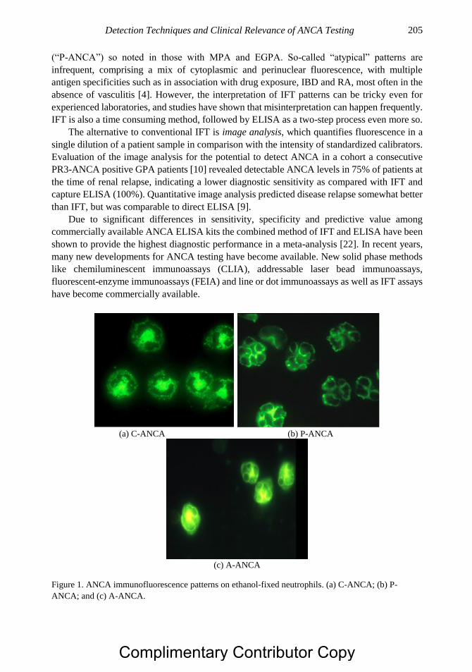
Detection Techniques and Clinical Relevance of ANCA Testing 205
(“P-ANCA”) so noted in those with MPA and EGPA. So-called “atypical” patterns are
infrequent, comprising a mix of cytoplasmic and perinuclear fluorescence, with multiple
antigen specificities such as in association with drug exposure, IBD and RA, most often in the
absence of vasculitis [4]. However, the interpretation of IFT patterns can be tricky even for
experienced laboratories, and studies have shown that misinterpretation can happen frequently.
IFT is also a time consuming method, followed by ELISA as a two-step process even more so.
The alternative to conventional IFT is image analysis, which quantifies fluorescence in a
single dilution of a patient sample in comparison with the intensity of standardized calibrators.
Evaluation of the image analysis for the potential to detect ANCA in a cohort a consecutive
PR3-ANCA positive GPA patients [10] revealed detectable ANCA levels in 75% of patients at
the time of renal relapse, indicating a lower diagnostic sensitivity as compared with IFT and
capture ELISA (100%). Quantitative image analysis predicted disease relapse somewhat better
than IFT, but was comparable to direct ELISA [9].
Due to significant differences in sensitivity, specificity and predictive value among
commercially available ANCA ELISA kits the combined method of IFT and ELISA have been
shown to provide the highest diagnostic performance in a meta-analysis [22]. In recent years,
many new developments for ANCA testing have become available. New solid phase methods
like chemiluminescent immunoassays (CLIA), addressable laser bead immunoassays,
fluorescent-enzyme immunoassays (FEIA) and line or dot immunoassays as well as IFT assays
have become commercially available.
(a) C-ANCA (b) P-ANCA
(c) A-ANCA
Figure 1. ANCA immunofluorescence patterns on ethanol-fixed neutrophils. (a) C-ANCA; (b) P-
ANCA; and (c) A-ANCA.
Complimentary Contributor Copy
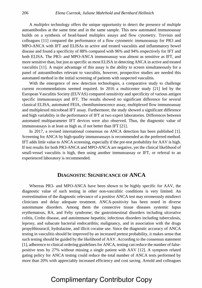
Elena Csernok, Juliane Mahrhold and Bernhard Hellmich 206
A multiplex technology offers the unique opportunity to detect the presence of multiple
autoantibodies at the same time and in the same sample. This new automated immunoassay
builds on a synthesis of bead-based multiplex assays and flow cytometry. Trevisin and
colleagues [11] compared the performance of a flow cytometric immunoassay for PR3 and
MPO-ANCA with IFT and ELISAs in active and treated vasculitis and inflammatory bowel
disease and found a specificity of 88% compared with 96% and 94% respectively for IFT and
both ELISA. The PR3- and MPO-ANCA immunoassay was almost as sensitive as IFT, and
more sensitive than, but just as specific as most ELISA in detecting ANCA in active and treated
vasculitis [11]. A major advantage of this assay is the ability to screen simultaneously for a
panel of autoantibodies relevant to vasculitis, however, prospective studies are needed this
automated method in the initial screening of patients with suspected vasculitis.
With the emergence of new detection technologies, a comparative study to challenge
current recommendations seemed required. In 2016 a multicenter study [21] led by the
European Vasculitis Society (EUVAS) compared sensitivity and specificity of various antigen
specific immunoassays and IFT. The results showed no significant difference for several
classical ELISA, automated FEIA, chemiluminescence assay, multiplexed flow immunoassay
and multiplexed microbead IFT assay. Furthermore, the study showed a significant difference
and high variability in the performance of IFT at two expert laboratories. Differences between
automated multiparameter IFT devices were also observed. Thus, the diagnostic value of
immunoassays is at least as high as, if not better than IFT [21].
In 2017, a revised international consensus on ANCA detection has been published [1].
Screening for ANCA by high-quality immunoassays is recommended as the preferred method.
IFT adds little value to ANCA screening, especially if the pre-test probability for AAV is high.
If test results for both PR3-ANCA and MPO-ANCA are negative, yet the clinical likelihood of
small-vessel vasculitis is high, then using another immunoassay or IFT, or referral to an
experienced laboratory is recommended.
DIAGNOSTIC SIGNIFICANCE OF ANCA
Whereas PR3- and MPO-ANCA have been shown to be highly specific for AAV, the
diagnostic value of such testing in other non-vasculitic conditions is very limited. An
overestimation of the diagnostic relevance of a positive ANCA test may erroneously misdirect
clinicians and delay adequate treatment. ANCA-positivity has been noted in diverse
autoimmune disorders. Among them the connective tissue diseases systemic lupus
erythematous, RA, and Felty syndrome; the gastrointestinal disorders including ulcerative
colitis, Crohn disease, and autoimmune hepatitis; infectious disorders including tuberculosis,
leprosy, and subacute bacterial endocarditis; malignancy, and in association with the drugs
propylthiouracil, hydralazine, and illicit cocaine use. Since the diagnostic accuracy of ANCA
testing in vasculitis should be improved by an increased pretest probability, it makes sense that
such testing should be guided by the likelihood of AAV. According to the consensus statement
[1], adherence to clinical ordering guidelines for ANCA, testing can reduce the number of false-
positive tests by 27% without missing a single patient with AAV [12]. A symptom related
gating policy for ANCA testing could reduce the total number of ANCA tests performed by
more than 20% with appreciably increased efficiency and cost saving. Arnold and colleagues
Complimentary Contributor Copy
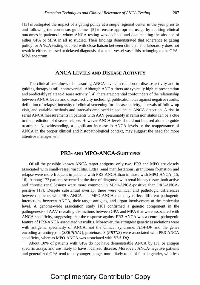
Detection Techniques and Clinical Relevance of ANCA Testing 207
[13] investigated the impact of a gating policy at a single regional center in the year prior to
and following the consensus guidelines [5] to ensure appropriate usage by auditing clinical
outcomes in patients in whom ANCA testing was declined and documenting the absence of
either GPA or MPA in all so studied. Their findings demonstrated that adherence to gating
policy for ANCA testing coupled with close liaison between clinician and laboratory does not
result in either a missed or delayed diagnosis of a small-vessel vasculitis belonging to the GPA-
MPA spectrum.
ANCA LEVELS AND DISEASE ACTIVITY
The clinical usefulness of measuring ANCA levels in relation to disease activity and in
guiding therapy is still controversial. Although ANCA titers are typically high at presentation
and predictably relate to disease activity [14], there are potential confounders of the relationship
between ANCA levels and disease activity including, publication bias against negative results,
definition of relapse, intensity of clinical screening for disease activity, intervals of follow-up
visit, and variable methods and intervals employed in sequential ANCA detection. A rise in
serial ANCA measurements in patients with AAV presumably in remission status can be a clue
to the prediction of disease relapse. However ANCA levels should not be used alone to guide
treatment. Notwithstanding, a significant increase in ANCA levels or the reappearance of
ANCA in the proper clinical and histopathological context, may suggest the need for more
attentive management.
PR3- AND MPO-ANCA-SUBTYPES
Of all the possible known ANCA target antigens, only two, PR3 and MPO are closely
associated with small-vessel vasculitis. Extra renal manifestations, granuloma formation and
relapse were more frequent in patients with PR3-ANCA than in those with MPO-ANCA [15,
16]. Among 173 patients screened at the time of diagnosis with renal biopsy tissue, both active
and chronic renal lesions were more common in MPO-ANCA-positive than PR3-ANCA-
positive [17]. Despite substantial overlap, there were clinical and pathologic differences
between patients with PR3-ANCA and MPO-ANCA that may reflect different pathogenic
interactions between ANCA, their target antigens, and organ involvement at the molecular
level. A genome-wide association study [18] confirmed a genetic component in the
pathogenesis of AAV revealing distinctions between GPA and MPA that were associated with
ANCA specificity, suggesting that the response against PR3-ANCA was a central pathogenic
feature of PR3-ANCA associated vasculitis. Moreover, the strongest genetic associations were
with antigenic specificity of ANCA, not the clinical syndrome. HLA-DP and the genes
encoding a1-antitrypsin (SERPINA1), proteinase 3 (PRTN3) were associated with PR3-ANCA
specificity, whereas MPO-ANCA was associated with HLA-DQ.
About 10% of patients with GPA do not have demonstrable ANCA by IFT or antigen
specific assays and are likely to have localized disease. Moreover, ANCA-negative patients
and generalized GPA tend to be younger in age, more likely to be of female gender, with less
Complimentary Contributor Copy
Elena Csernok, Juliane Mahrhold and Bernhard Hellmich 208
lung and kidney involvement, lower rate of relapse, and improved outcome compared to those
ANCA-positive patients.
Since ANCA specificity seems to affect the phenotype of small vessel vasculitis, the initial
response to remission-inducing drug therapy, long-term prognosis and relapse risk more than
the clinical diagnosis, it has been suggested that classification of AAV by ANCA specificity
could provide more useful information than clinical diagnosis [19].
CONCLUSION
ANCA serology and the associated clinical manifestation of AAV continue to attract the
attention of clinicians and investigators alike. In addition, to be providing a useful diagnostic
tool for small vessel vasculitis, ANCA testing may be useful in predicting relapses and in
guiding therapy. Worldwide, most laboratories currently perform IFT to screen for ANCA
positivity patients with small-vessel vasculitis, with later confirmation employing PR3- and
MPO-ANCA immunoassays. However, in recent studies new high-quality antigen-specific
immuno-assays have been shown to have equal or better diagnostic performance than IFT.
Therefore, the new international consensus of 2016 recommends screening for ANCA with
immunoassays primarily since there is no apparent benefit of combining it with IFT.
Notwithstanding, any testing strategy applicable to vasculitis should be able to identify relevant
ANCA target antigens since these correlate best with the clinicopathological aspects, disease
activity, and propensity for relapse. The solution to problems regarding ANCA-diagnosis lies
in focusing upon the implementation of high-quality immunoassay testing according to the
revised international consensus, and restriction of such tests to those with the highest pretest
probability of AAV.
REFERENCES
[1] Bossuyt, X; Cohen Tervaert, JW; Arimura, Y; Blockmans, D; Flores Suárez, LP;
Guillevin, L; Hellmich, B; Jayne, D; Jennette, JC; Kallenberg, CM; Radice, A; Savige,
JA; Sinico, A; Specks, U; van Paassen, P; Zhao, MH; Rasmussen, N; Damoiseaux, J;
Csernok, E. Revised international consensus statement on testing of anti-neutrophil
cytoplasm antibodies in small vessel vasculitis. Nat Rev Rheumatol, 2017, 13(11), 683-
692.
[2] Davies, DJ; Moran, JE; Niall, JF; et al. Segmental necrotizing glomerulonephritis with
antineutrophil antibody: possible arbovirus aetiology? Br Med J, 1982, 285, 606.
[3] Van der Woude, FJ; Rasmussen, N; Lobatto, S. Autoantibodies against neutrophils and
monocytes: tool for diagnosis and marker of disease activity in Wegener’s
granulomatosis. Lancet, 1985, 1, 425–429.
[4] Csernok, E; Lamprecht, P; Gross, WL. Diagnostic significance of ANCA in vasculitis.
Nature Clinical Practice Rheumatology, 2006, 2, 174-175.
[5] Savige, J; Gillis, D; Benson, E; Davies, D; et al. International consensus statement on
testing and reporting of antineutrophil cytoplasmic antibodies (ANCA). Am J Clin
Pathol, 1999, 111, 507–513.
Complimentary Contributor Copy
Detection Techniques and Clinical Relevance of ANCA Testing 209
[6] Csernok, E; Holle, JU. Twenty-years with ANCA: how to test for ANCA –evidence
based immunology? Autoimmune Highlights, 2010, 1, 39–45.
[7] Csernok, E; Holle, J; Hellmich, B; et al. Evaluation of capture ELISA for detection of
neutrophil cytoplasmatic antibodies directed against proteinase 3 in Wegener’s
granulomatosis: first results from a multicenter study. Rheumatology (Oxford), 2004, 43,
174-180.
[8] Holle, JU; Hellmich, B; Backes, M; et al. Variations in performance characteristics of
commercial enzyme immunoassay kits of the detection of antineutrophil cytoplasmatic
antibodies: what is the optimal cut-off? Ann Rheum Dis, 2005, 64, 1773–1779.
[9] Holle, JU; Herrmann, K; Gross, WL; et al. Comparative analysis of different commercial
ELISA systems for detection of anti-neutrophil cytoplasm antibodies in ANCA-
associated vasculitides. Clin Exp Rheumatol, 2012, 30, (Suppl 70), S66–S69.
[10] Boomsma, MM; Damoiseaux, FGMC; Stegeman, CA; et al. Image analysis: a novel
approach for the quantification of ANCA levels in patients with Wegener’s
granulomatosis. J Immunol Methods, 2003, 274, 27-35.
[11] Trevisin, M; Pollock, W; Savige, J. Evaluation of a multiplex flow cytometric
immunoassay to detect PR3- and MPO-ANCA in active and treated vasculitis, and in
inflammatory bowel disease. J Immunol Methods, 2008, 336, 104-112.
[12] Sinclair, D; Saas, M; Stevens, JM. The effect of a symptom related “gating policy” on
ANCA requests in routine clinical practice. J Clin Pathol, 2004, 57, 131–134.
[13] Arnold, DF; Timms, A; Luqmani, R; et al. Does a gating policy for ANCA overlook
patients with ANCA associated vasculitis? An audit of 263 patients. J Clin Pathol, 2010,
63, 678–680.
[14] Tomasson, G; Grayson, PC; Mahr, AD; et al. Value of ANCA measurements during
remission to predict a relapse of ANCA-associated vasculitis-a meta-analysis.
Rheumatology, 2012, 51, 100–109.
[15] Franssen, C; Gans, R; Kallenberg, C; et al. Disease spectrum of patients with
antineutrophil cytoplasmic autoantibodies of defined specificity: distinct differences
between patients with anti-proteinase 3 and anti-myeloperoxidase autoantibodies. J
Intern Med, 1998, 244, 209-216.
[16] Schönermarck, U; Lamprecht, P; Csernok, E; et al. Prevalence and spectrum of rheumatic
diseases associated with proteinase 3-antineutrophil cytoplasmic antibodies (ANCA) and
myeloperoxidase-ANCA. Rheumatology, 2001, 40, 178-184.
[17] Hauer, HA; Bajema, IM; van Houwelingen, HC; Ferrario, F; et al. Renal histology in
ANCA-associated vasculitis: differences between diagnostic and serologic subgroups.
Kidney Int, 2002, 61, 80-89.
[18] Lyons, PA; Rayner, TF; Trivedi, S; et al. Genetically distinct subsets within ANCA-
associated vasculitis. N Engl J Med, 2012, 367, 214-223.
[19] Cornec, D; Gall, EC; Fervenza, FC; Specks, U. (2016) ANCA-associated vasculitis -
clinical utility of using ANCA specificity to classify patients. Nat Rev Rheumatol. 2016
[20] Damoiseaux J, Csernok E, Rasmussen N, Moosig F, van Paassen P, Vermeersch P,
Blockmans D, Cohen Tervaert JW, Bossuyt X Detection of anti-neutrophil cytoplasmic
antibodies (ANCA): a multicenter EUVAS (European Vasculitis Study Group)
evaluation of the value of indirect immunofluorescence (IIF) versus antigen-specific
immuno-assays. Ann Rheum Dis., 2016, doi:10.1136/annrheumadis-2016-2095507.
Complimentary Contributor Copy
Elena Csernok, Juliane Mahrhold and Bernhard Hellmich 210
[21] Choi, HK; Liu, S; Merkel, PA; et al. Diagnostic performance of antineutrophil
cytoplasmic antibody tests for idiopathic vasculitides: metaanalysis with a focus on
antimyeloperoxidase antibodies. J Rheumatol, 2001, 28, 1584–90.
Complimentary Contributor Copy
In: The Vasculitides. Volume 1 ISBN: 978-1-53615-133-6
Editor: David S. Younger © 2019 Nova Science Publishers, Inc.
Chapter 12
CLASSIFICATION AND PATHOGENICITY
OF ANCA-ASSOCIATED VASCULITIS
Alexander Tracy MB, BCh, Constantina Yiannakis, MB, BCh,
Lorna Ward, MD and Matthew David Morgan*, MB, ChB, PhD Renal Immunobiology, Centre for Translational Inflammation Research,
Institute of Clinical Sciences, College of Medical and Dental Sciences,
University of Birmingham, Birmingham, UK
ABSTRACT
Anti-neutrophil cytoplasm antibody (ANCA)-associated vasculitides (AAV) include
several distinct multi-systemic disorders, the diagnoses of which rely on recognition of a
compatible clinical syndrome supported by radiological, immunological and histological
laboratory findings. If unrecognized, there may be significant delays in treatment
culminating in multi-organ morbidity. The pathogenesis of AAV is complex, involving
both innate and adaptive immune systems, with loss of tolerance to autoantigens leading
to the production of antibodies that activate cytokine-primed neutrophils. The factors
leading to production of ANCA are incompletely understood, but may involve cross-
reactivity to bacterial antigens and the expression of complementary peptides. T-cells
appear to play a role in both loss of self-tolerance and in the cause of tissue injury.
Moreover, peripheral blood T-cells show evidence of persistent activation driven partly by
cytomegalovirus infection. Activation of the alternative complement pathway has recently
emerged as a significant pathogenic mechanism in the development of glomerulonephritis
in AAV.
Keywords: ANCA, vasculitides, classification, pathogenesis, pathogenicity
* Corresponding Author’s E-mail: [email protected].
Complimentary Contributor Copy
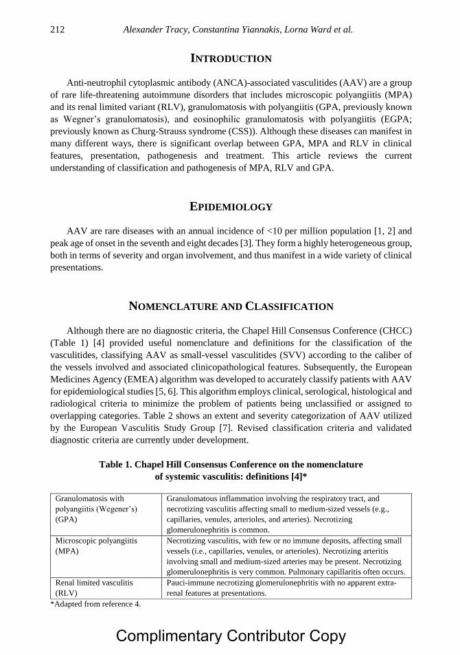
Alexander Tracy, Constantina Yiannakis, Lorna Ward et al. 212
INTRODUCTION
Anti-neutrophil cytoplasmic antibody (ANCA)-associated vasculitides (AAV) are a group
of rare life-threatening autoimmune disorders that includes microscopic polyangiitis (MPA)
and its renal limited variant (RLV), granulomatosis with polyangiitis (GPA, previously known
as Wegner’s granulomatosis), and eosinophilic granulomatosis with polyangiitis (EGPA;
previously known as Churg-Strauss syndrome (CSS)). Although these diseases can manifest in
many different ways, there is significant overlap between GPA, MPA and RLV in clinical
features, presentation, pathogenesis and treatment. This article reviews the current
understanding of classification and pathogenesis of MPA, RLV and GPA.
EPIDEMIOLOGY
AAV are rare diseases with an annual incidence of <10 per million population [1, 2] and
peak age of onset in the seventh and eight decades [3]. They form a highly heterogeneous group,
both in terms of severity and organ involvement, and thus manifest in a wide variety of clinical
presentations.
NOMENCLATURE AND CLASSIFICATION
Although there are no diagnostic criteria, the Chapel Hill Consensus Conference (CHCC)
(Table 1) [4] provided useful nomenclature and definitions for the classification of the
vasculitides, classifying AAV as small-vessel vasculitides (SVV) according to the caliber of
the vessels involved and associated clinicopathological features. Subsequently, the European
Medicines Agency (EMEA) algorithm was developed to accurately classify patients with AAV
for epidemiological studies [5, 6]. This algorithm employs clinical, serological, histological and
radiological criteria to minimize the problem of patients being unclassified or assigned to
overlapping categories. Table 2 shows an extent and severity categorization of AAV utilized
by the European Vasculitis Study Group [7]. Revised classification criteria and validated
diagnostic criteria are currently under development.
Table 1. Chapel Hill Consensus Conference on the nomenclature
of systemic vasculitis: definitions [4]*
Granulomatosis with
polyangiitis (Wegener’s)
(GPA)
Granulomatous inflammation involving the respiratory tract, and
necrotizing vasculitis affecting small to medium-sized vessels (e.g.,
capillaries, venules, arterioles, and arteries). Necrotizing
glomerulonephritis is common.
Microscopic polyangiitis
(MPA)
Necrotizing vasculitis, with few or no immune deposits, affecting small
vessels (i.e., capillaries, venules, or arterioles). Necrotizing arteritis
involving small and medium-sized arteries may be present. Necrotizing
glomerulonephritis is very common. Pulmonary capillaritis often occurs.
Renal limited vasculitis
(RLV)
Pauci-immune necrotizing glomerulonephritis with no apparent extra-
renal features at presentations.
*Adapted from reference 4.
Complimentary Contributor Copy
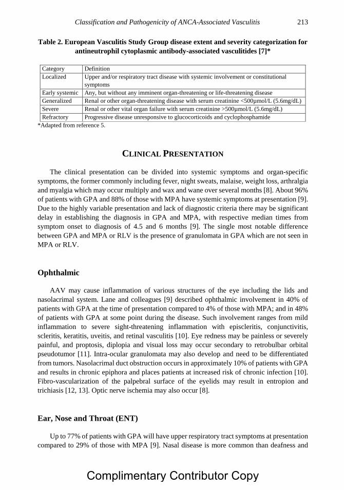
Classification and Pathogenicity of ANCA-Associated Vasculitis 213
Table 2. European Vasculitis Study Group disease extent and severity categorization for
antineutrophil cytoplasmic antibody-associated vasculitides [7]*
Category Definition
Localized Upper and/or respiratory tract disease with systemic involvement or constitutional
symptoms
Early systemic Any, but without any imminent organ-threatening or life-threatening disease
Generalized Renal or other organ-threatening disease with serum creatinine <500µmol/L (5.6mg/dL)
Severe Renal or other vital organ failure with serum creatinine >500µmol/L (5.6mg/dL)
Refractory Progressive disease unresponsive to glucocorticoids and cyclophosphamide
*Adapted from reference 5.
CLINICAL PRESENTATION
The clinical presentation can be divided into systemic symptoms and organ-specific
symptoms, the former commonly including fever, night sweats, malaise, weight loss, arthralgia
and myalgia which may occur multiply and wax and wane over several months [8]. About 96%
of patients with GPA and 88% of those with MPA have systemic symptoms at presentation [9].
Due to the highly variable presentation and lack of diagnostic criteria there may be significant
delay in establishing the diagnosis in GPA and MPA, with respective median times from
symptom onset to diagnosis of 4.5 and 6 months [9]. The single most notable difference
between GPA and MPA or RLV is the presence of granulomata in GPA which are not seen in
MPA or RLV.
Ophthalmic
AAV may cause inflammation of various structures of the eye including the lids and
nasolacrimal system. Lane and colleagues [9] described ophthalmic involvement in 40% of
patients with GPA at the time of presentation compared to 4% of those with MPA; and in 48%
of patients with GPA at some point during the disease. Such involvement ranges from mild
inflammation to severe sight-threatening inflammation with episcleritis, conjunctivitis,
scleritis, keratitis, uveitis, and retinal vasculitis [10]. Eye redness may be painless or severely
painful, and proptosis, diplopia and visual loss may occur secondary to retrobulbar orbital
pseudotumor [11]. Intra-ocular granulomata may also develop and need to be differentiated
from tumors. Nasolacrimal duct obstruction occurs in approximately 10% of patients with GPA
and results in chronic epiphora and places patients at increased risk of chronic infection [10].
Fibro-vascularization of the palpebral surface of the eyelids may result in entropion and
trichiasis [12, 13]. Optic nerve ischemia may also occur [8].
Ear, Nose and Throat (ENT)
Up to 77% of patients with GPA will have upper respiratory tract symptoms at presentation
compared to 29% of those with MPA [9]. Nasal disease is more common than deafness and
Complimentary Contributor Copy
Alexander Tracy, Constantina Yiannakis, Lorna Ward et al. 214
middle ear involvement in both GPA and MPA. More than 90% of patients with GPA develop
ear and upper airway disease [14, 15]. Typical nasal symptoms of GPA include rhinitis, nasal
stuffiness and pain, epistaxis, and brown or bloody crusting. The characteristic “saddle-nose
deformity” is secondary to septal erosion due to chronic nasal inflammation. Patients with GPA
are at increased risk of nasal and sinus infections due to chronic damage which increases the
difficulty in differentiating secondary infection from disease activity.
Middle ear involvement in GPA presents as conductive hearing loss with the commonest
cause being Eustachian tube dysfunction secondary to nasopharyngeal disease [16, 17].
Sensorineural hearing loss is due to inner ear disease which may also be associated with
vestibular dysfunction with some patients presenting with vertigo and balance disturbance [17,
18]. The mechanism of inner ear involvement in GPA is poorly understood when compared to
middle ear involvement [19].
Involvement of the trachea may present with pain, cough, hoarseness, wheezing or stridor,
however it is often asymptomatic at onset. The development of subglottic or bronchial stenosis
can lead to severe dyspnea requiring tracheostomy or reconstruction [11].
Pulmonary
Pulmonary involvement occurred at presentation in 63% of patients with GPA compared
to 29% of those with MPA [9]. Pulmonary manifestations range from new-onset asthma and
‘antibiotic-resistant’ pneumonia to life-threatening alveolar hemorrhage. In those with GPA the
symptoms may be due to single or multiple lung nodules and granulomata, and may be
misdiagnosed as cavitating infectious or neoplastic lesions; mediastinal and hilar
lymphadenopathy may also occur [20]. Pulmonary capillaritis is responsible for lung
hemorrhage and rapidly changing alveolar infiltrates and should be suspected in patients with
falling hemoglobin levels in conjunction with hemoptysis and dyspnea. Those with MPA may
also develop pulmonary interstitial fibrosis [21]. Other indicators of pulmonary involvement
include wheezing, productive or non-productive cough, and exertional dyspnea. [8].
Renal
This is often a severe manifestation of AAV [3] that affects an estimated 77% of patients
with GPA and 92% of those with MPA at presentation [9]. Renal vasculitis presents with
proteinuria, hematuria, and red cell casts due to focal necrotizing glomerulonephritis [22]. It
can lead to rapid deterioration of renal function with eventual dialysis dependency. Hematuria
may be visible or non-visible, while proteinuria is usually insufficient to cause nephrotic
syndrome [8]. Renal biopsy is the most appropriate method of identifying and prognosticating
glomerulonephritis in AAV [23, 24].
Nervous System
AAV can affect the peripheral (PNS) and central nervous system (CNS), as observed in
19% of patients with GPA and 17% of those with MPA at presentation [9]. Involvement of the
Complimentary Contributor Copy
Classification and Pathogenicity of ANCA-Associated Vasculitis 215
PNS usually manifests as motor or sensory deficits in a mononeuritis multiplex or distal sensory
polyneuropathy distribution. While typically painless in the acute stage, pain syndromes
commonly develop as a result of neuro-regeneration. Involvement of the CNS occurs in 8% of
patients with AAV at presentation [15], presenting with weakness, dysphagia, blindness or
other CNS disturbances [25]. Three distinct patterns of CNS involvement have been identified:
during the generalized phase the small and medium sized vessels of the CNS can become
inflamed; secondly, localized granulomatous masses from the upper respiratory tract may
invade the CNS; thirdly, granulomata may develop within CNS structures [26].
Cutaneous
Cutaneous involvement occurs in 42% of GPA and 25% of MPA patients at diagnosis [9].
A wide range of signs are seen that include purpura, papules, urticaria, infiltrative erythema,
infarction and splinter hemorrhage, ulcerated nodules, livedo reticularis, and digital gangrene
[27]. The type of lesion is determined by the size of the blood vessel affected. The most
common manifestation is a palpable purpuric rash beginning along the lower extremities that
may become ulcerated or vesicular. The urticarial rash seen in vasculitis can be differentiated
from allergic non-vasculitic urticaria by the fact that it will usually last for more than one day
and may progress to purpuric lesions [28].
Cardiac
Direct cardiac involvement is uncommon in either GPA or MPA however the former may
be associated with coronary arteritis, pericarditis and valvular disease. Endocarditis can occur
as a result of aortic valvular lesions [29]. MPA may be associated with pericarditis, heart failure
and hypertension however the incidence is very low [30].
Gastrointestinal
Gastrointestinal involvement occurs at presentation in up to 30% of patients with AAV
[14, 31-33]. Gastrointestinal involvement may present with ischemic or vasculitic colitis,
bloody diarrhea, pain and progression to ulceration, perforation and infarction [11, 34].
30-56% of patients with MPA have been found to have GI involvement [35, 36], the commonest
presenting symptom of which was pain, noted in 97% of patients, followed by nausea,
vomiting, diarrhea, melena, and hematemesis [37]. Rarely, other intra-abdominal organs can
be involved such as the gallbladder and pancreas [34].
ETIOPATHOGENESIS
There is evidence for a pathogenic role of ANCA in the etiopathogenesis of inflammation
and organ damage mediated by immune mechanisms. With regards to the innate immune
Complimentary Contributor Copy
Alexander Tracy, Constantina Yiannakis, Lorna Ward et al. 216
system, neutrophils play a pivotal role in the effector phase causing organ and tissue damage.
Complement activation, particularly the alternative pathway, seems to be important in
glomerulonephritis. The adaptive immune system is important in the development of
autoimmunity with the production of autoantibodies against proteinase 3 or myeloperoxidase
by B-cells and there is increasing evidence for a pathogenic role for T-cells. Genetic
susceptibility factors are encoded by certain human leukocyte antigen (HLA) haplotypes [38],
notably HLA-DPB1*0401 alleles are associated with an increased risk for development of GPA
[39], while HLA-DRB1*0901 is associated with MPA [40]. In addition, the DRB1*15
haplotype was associated with anti-PR3 AAV in African American patients [41].
Polymorphisms of genes involved in the control of the immune system and associated with
increased risk of vasculitis include CD226, PTPN22 protein, CTLA-4, IL-10, and TLR9
[42-47], although none are specific for AAV. A recent genome-wide association study
correlated single-nucleotide polymorphisms in HLA-DPB1 and PRTN3 with frequency of
autoreactive T-cells and neutrophil expression of PR3 [48], thus providing a functional link
between genetic risk factors and the immunopathology of AAV.
Copy number variation of FcγR3b and human ß-defensin (DEFB4) may represent further
important mechanisms in AAV susceptibility [42, 49]. Genes expressed by memory T-cells as
well as a CD8+ T-cell transcription signature have been described in both systemic lupus
erythematosus and AAV with associated poorer outcome. This signature is thought to comprise
enriching genes involved in the IL-7 receptor and T-cell receptor signaling pathway [50].
Recent work by Yang and colleagues [51] has addressed the contribution of epigenetic
mechanisms to AAV pathogenesis. Chromatin immunoprecipitation (ChIP) performed on
peripheral blood neutrophils demonstrated an association between specific histone
modifications at the MPO and PRTN3 loci and active AAV. However, it is unclear whether
these histone modifications in themselves play a pathogenic role.
Carriage of the dysfunctional z allele of the α-1-antitrypsin (A1AT) gene is associated with
an increased risk of developing AAV and predicts more severe outcomes [52-57]. Patients
carrying the z allele not only have an increased risk of AAV but also have increased
concentrations of circulating A1AT polymers capable of priming circulating neutrophils for
ANCA-induced activation [58].
Environmental factors may also have a role in disease development. Provocation of an
immune response and inflammation is thought to occur after exposure to silica dust with 22-
46% of patients with AAV having documented exposure to silica prior to disease development.
It is proposed that exposure results in accelerated apoptosis of macrophages and
polymorphonuclear leukocytes, thereby triggering disease development [59, 60].
Infectious triggers have also been implicated in the etiopathogenesis of AAV. A
contributory role of low-grade Staphylococcus aureus infection in the etiopathogenesis of AVV
has been suggested based on the observation that 63% of patients with GPA had chronic nasal
carriage when compared with 20% of control individuals [61]. Moreover, nasal carriage has
been associated with disease relapse [61, 62], and patients who received maintenance co-
trimoxazole treatment were observed to have fewer relapses [63]. A study of bronchioalveolar
lavage fluid (BALF) in GPA patients identified increased levels of infection compared to
controls and demonstrated that BALF was permissive for bacterial growth [64].
Molecular mimicry may also underlie the development of necrotizing and crescentic
glomerulonephritis (NCGN) through infection with fimbriated bacteria, such as Escherichia
coli and Klebsiella pneumonia [65]. The bacterial adhesion protein FimH was found to be
Complimentary Contributor Copy
Classification and Pathogenicity of ANCA-Associated Vasculitis 217
homologous to a major epitope of LAMP-2 which is in turn recognized by anti-LAMP-2
antibodies in pauci-immune NCGN [66, 67] although these results have not been replicated by
other researchers [68].
The development of AAV has been linked to several drug exposures, most commonly
propylthiouracil with a prevalence of ANCA expression ranging from 4% to 46% [69, 70]. D-
penicillamine, hydralazine and minocycline have also been associated with disease.
Sulphasalazine, and other similar drugs, may increase autoantibody production as they induce
apoptosis which in turn increases expression of ANCA antigen targets on the surface of
apoptotic neutrophils [71, 72].
ANCA PATHOGENICITY
Since the discovery of ANCA there has been considerable debate as to whether or not it
has a direct pathogenic role with most authors now accepting that anti-myeloperoxidase (MPO)
antibodies are pathogenic based on the results of clinical, laboratory and animal studies.
Proteinase 3 (PR3) was found to be the target antigen of the cytoplasmic-ANCA indirect
immunofluorescence pattern while MPO is the target antigen of the perinuclear-ANCA pattern,
with the specificity of PR3-ANCA and MPO-ANCA for AAV as high as 98% [73, 74].
Clinical Evidence for ANCA Pathogenicity
The most striking clinical evidence for the pathogenicity of ANCA was the report of a
neonate developing pulmonary hemorrhage and glomerulonephritis caused by transplacental
transfer of ANCA IgG from the mother who had anti-MPO antibody positive MPA [75]. There
has, however, been a lack of clear consistent evidence of a relationship between the serum
ANCA titer and disease activity [76-79]. A meta-analysis of studies investigating the
association between ANCA and disease activity found a rise in the serum ANCA titer and
persistent presence of ANCA was only modestly predictive of future disease relapse, suggesting
that serial ANCA measurements had a limited utility in guiding treatment decisions in
individual patients [80]. It is possible that there are subtle changes in ANCA such as
glycosylation state or change in the predominant subclass that are related to disease activity
and undetected by routine assays [81-84].
Animal Models of AAV
Several murine and rat models of anti-MPO AAV have been created; unfortunately to date
there has not been a suitable animal model for anti-PR3 [85-87]. Xiao and colleagues [88]
immunized MPO-/- mice with purified MPO to generate high avidity anti-MPO antibodies.
Granulomatous inflammation, necrotizing and crescentic glomerulonephritis, and systemic
necrotizing vasculitis developed when mouse splenocytes were transferred to Rag2-/- mice or
when anti-MPO containing IgG was transferred to wild-type mice. Levels of tumor necrosis
alpha (TNF-α) were increased and renal injury enhanced when lipopolysaccharide (LPS) was
Complimentary Contributor Copy
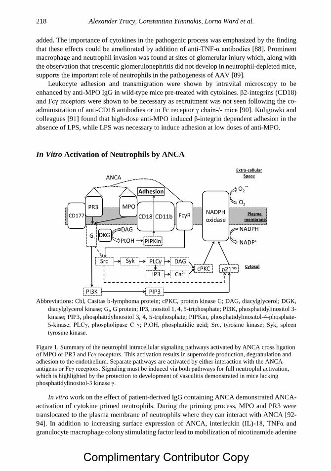
Alexander Tracy, Constantina Yiannakis, Lorna Ward et al. 218
added. The importance of cytokines in the pathogenic process was emphasized by the finding
that these effects could be ameliorated by addition of anti-TNF-α antibodies [88]. Prominent
macrophage and neutrophil invasion was found at sites of glomerular injury which, along with
the observation that crescentic glomerulonephritis did not develop in neutrophil-depleted mice,
supports the important role of neutrophils in the pathogenesis of AAV [89].
Leukocyte adhesion and transmigration were shown by intravital microscopy to be
enhanced by anti-MPO IgG in wild-type mice pre-treated with cytokines. β2-integrins (CD18)
and Fcγ receptors were shown to be necessary as recruitment was not seen following the co-
administration of anti-CD18 antibodies or in Fc receptor γ chain-/- mice [90]. Kuligowki and
colleagues [91] found that high-dose anti-MPO induced β-integrin dependent adhesion in the
absence of LPS, while LPS was necessary to induce adhesion at low doses of anti-MPO.
In Vitro Activation of Neutrophils by ANCA
Abbreviations: Cbl, Casitas b-lymphoma protein; cPKC, protein kinase C; DAG, diacylglycerol; DGK,
diacylglycerol kinase; Gi, G protein; IP3, inositol 1, 4, 5-triphosphate; PI3K, phosphatidylinositol 3-
kinase; PIP3, phosphatidylinositol 3, 4, 5-triphosphate; PIPKin, phosphatidylinositol-4-phosphate-
5-kinase; PLCγ, phospholipase C γ; PtOH, phosphatidic acid; Src, tyrosine kinase; Syk, spleen
tyrosine kinase.
Figure 1. Summary of the neutrophil intracellular signaling pathways activated by ANCA cross ligation
of MPO or PR3 and Fcγ receptors. This activation results in superoxide production, degranulation and
adhesion to the endothelium. Separate pathways are activated by either interaction with the ANCA
antigens or Fcγ receptors. Signaling must be induced via both pathways for full neutrophil activation,
which is highlighted by the protection to development of vasculitis demonstrated in mice lacking
phosphatidylinositol-3 kinase γ.
In vitro work on the effect of patient-derived IgG containing ANCA demonstrated ANCA-
activation of cytokine primed neutrophils. During the priming process, MPO and PR3 were
translocated to the plasma membrane of neutrophils where they can interact with ANCA [92-
94]. In addition to increasing surface expression of ANCA, interleukin (IL)-18, TNFα and
granulocyte macrophage colony stimulating factor lead to mobilization of nicotinamide adenine
ANCA
Plasma membrane
Extra-cellularSpace
Cytosol
Gi
p21ras
Src Syk
IP3
PLCγ
Ca2+
DAGcPKC
DAG
PtOH PIPKin
Adhesion
PIP3PI3K
NADPH
NADP+
O2
O2·-
NADPH oxidase
FcγRCD18 CD11b
MPOPR3
CD177
DKG
Complimentary Contributor Copy
Classification and Pathogenicity of ANCA-Associated Vasculitis 219
dinucleotide phosphate (NADPH) oxidase complex components necessary for superoxide
generation [94-97]. Subsequent crosslinking between ANCA and Fcγ receptors and ANCA
antigens results in degranulation, production of pro-inflammatory cytokines, adhesion to
endothelium and generation of superoxide with multiple signaling pathways implicated [98-
103] (Figure 1). Neutrophil gene expression is also induced by binding of both intact ANCA
and F(ab′)2 portions [104].
Effect of ANCA on Neutrophil-Endothelium Adhesion
Endothelial necrosis is a histological feature of small vessel disease in AAV, with exposure
of the basement membrane leading to platelet aggregation, thrombosis and occlusion [105].
Swelling, necrosis and dehiscence of endothelial cells is an early histological feature and
thought to be the mediated by ANCA activation of cytokine-primed circulating neutrophils.
This results in neutrophil adhesion to and transmigration through endothelium, associated with
neutrophil degranulation and production of superoxide. Adhesion and transmigration of
leukocytes is promoted by the release of chemoattractant proteins by endothelial cells and
upregulation of adhesion molecules. The serum concentrations of soluble factors associated
with this process include vascular endothelial growth factor (VEGF), inter-cellular adhesion
molecule (ICAM)-3, P-selectin, thrombomodulin, angiopoietin-2, matrix metalloproteases 1,
3, and 9; endothelial microparticles and stem cells [106-112].
In vitro studies showed that ANCA stabilized adhesion and promoted transmigration of
neutrophils flowing over endothelial cells [113]. In shear stress models employing human
endothelial cells and healthy donor neutrophils, this interaction depended on the interaction
between neutrophil-expressed CXCR1 and CXCR2, and CD18/CD11b and their ligands on
endothelial cells, [113-115]. An MPO-/- animal model demonstrated stimulation of neutrophil-
endothelial adhesion dependent on the action of CD18 [90].
In activated neutrophils, intracellular F-actin polymerizes making the neutrophil more rigid
and less deformable. This may lead to retention of neutrophils in small diameter capillaries and
increased contact with endothelium, promoting damage leading to cell detachment and lysis
[116, 117]. One study demonstrated reduced CXCR1 and CXCR2 expression on neutrophils
from AAV patients, even those in remission where expression showed an inverse relationship
with serum IL-8 concentrations. Reduced chemokine receptor expression impaired endothelial
transmigration but not adhesion, which may increase contact with endothelium and promote
intra-vascular retention of activated neutrophils [118]. The actions of endothelial cells inhibit
the neutrophil production of superoxide, implying that granule content release may be a more
important pathogenic mechanism in AAV [89-91].
ANCA and Neutrophil Cell Death
Neutrophil apoptosis does not normally result in the release of the intracellular contents,
thus preventing inflammation. ANCA-activated neutrophils were shown to undergo
dysregulated and accelerated apoptosis which may lead to increased inflammation associated
with the release of granule contents, increased surface expression of MPO and PR3, and
macrophage-mediated uptake of ANCA opsonized neutrophils [71, 119, 120].
Complimentary Contributor Copy
Alexander Tracy, Constantina Yiannakis, Lorna Ward et al. 220
More recently, there has been increasing focus on the relationship between ANCA and
neutrophil extracellular trap (NET) formation. NETs are reticular structures released by
activated neutrophils, composed of decondensed chromatin and associated antimicrobial
proteins [121]. Since the first description of their structure in 2004, NETs have been implicated
in the pathogenesis of a range of inflammatory and autoimmune disorders [122].
Kessenbrock and colleagues first demonstrated the presence of NETs in glomeruli from
AAV patients [123], suggesting a potential role in mediating tissue damage. Since then, NETs
were shown to be more prevalent in nerve tissue from patients with MPA neuropathy than other
types of neuropathy [124], and circulating levels of NET remnants were found to be increased
in AAV patients compared to healthy controls [125]. Individual case reports identified NETs
in cutaneous lesions [126] and thrombus [127] from AAV patients. Taken together, these
findings suggest that NETs could play a role in mediating autoimmune damage in multiple
organ systems. Notably, ANCA-induced NETs associate with alternative complement pathway
components C3b and C5b-9 in human serum, with subsequent activation of the alternative
complement pathway in vitro [128]. This provides a possible link between NETosis and
mechanisms of complement-mediated damage in AAV.
Kessenbrock and colleagues also showed that ANCA could stimulate neutrophils to release
NETs [123]. Interestingly, independent work found that neutrophils from AAV patients
exhibited increased NET production compared to neutrophils from healthy controls when
stimulated with ANCA [129]. MPO-ANCA affinity but not titer correlated with increased
levels of NETs in AAV patients [40], and the pro-inflammatory mediator high-mobility group
box 1 (HMGB1) was identified as an additional factor that potentiates ANCA-induced NET
release in vitro [130]. In addition, there is evidence that NET degradation is impaired in serum
from MPA patients [131], which could contribute to both NET-induced tissue damage and
immunogenicity.
Since NETs represent a source of autoantigens PR3 and MPO, it is possible that disordered
NETosis plays a role in the induction of autoimmunity. Sangaletti and colleagues loaded
myeloid dendritic cells (mDCs) with NET components and transferred them into naïve mice
[132]. This induced circulating PR3- and MPO-ANCA and resulted in pulmonary-renal
vasculitis. In contrast, injection of mDCs co-cultured with apoptotic neutrophils resulted in
autoantibody production in the absence of this disease phenotype. This study demonstrated that
NET components are able to induce ANCA-like disease in a murine model, implying that NETs
represent a potential source of autoantigens in AAV. Therefore, NETosis may contribute to a
positive feedback loop in which ANCA induce both NET-mediated tissue damage and the
exposure of NET-derived autoantigens. However, this model does not explain how tolerance
to these autoantigens is broken, and thus far there is limited evidence that this process plays a
pathogenic role in humans.
Cell-Mediated Immunity
Both B- and T-cells have important roles in the pathogenesis of AAV and are important in
the induction of autoimmunity and the development of tissue inflammation. The production of
autoantibodies, implying a loss of self-tolerance to PR3, MPO and possibly LAMP-2, could
occur via several possible mechanisms. The presence of low levels of specific autoantibodies
(SAA) to MPO and PR3 in the healthy population has led to the theory that ANCA form part
Complimentary Contributor Copy
Classification and Pathogenicity of ANCA-Associated Vasculitis 221
of the natural repertoire [133]. One explanation for the non-pathogenic nature of SAA is that
compared to MPO-ANCA derived from patients with AAV, significantly lower titers of SAA
MPO are found with lower avidity suggesting that high affinity autoantibodies underlying the
autoimmune pathology of AAV may result from dysregulation of SAA-producing B-cells.
However, the precise mechanism remains unknown and larger cohort studies are required
[133]. One intriguing suggestion has been the role of complementary peptides in the production
of anti-PR3 ANCA. These peptides are produced by the transcription and translation of the
DNA complementary to the PR3 gene. The peptides so produced generate an anti-
complementary PR3 (cPR3) response which leads to an anti-idiotype response producing anti-
PR3 antibodies [134]. It has also been suggested that cPR3 shows homology with proteins from
staphylococcal bacteria. T-cells specific for cPR3 have also been identified in AAV patients
[135].
T-cells are required to allow IgG subclass switching, a process which has been shown to
be essential in the pathogenesis of AAV [136]. The importance of T-cells in the disease process
is further supported by the presence of high levels of soluble IL-2 receptors and activated CD4+
cells in patient blood and sera, as well as soluble CD4 and CD8 [137]. MPO and PR3 have been
shown to promote the proliferation of autoreactive T-cells [138-140].
Regulatory CD4+ T-cells (Treg) important in the maintenance of self-tolerance have
functional defects in patients with GPA in remission [141, 142]. The percentage of Treg cells
was found to be inversely related to the disease relapse rate in patients with GPA and was
associated with both functional and numerical defects in PR3-specific Treg cells [143].
There has been increasing interest in the role of IL-17 producing helper T-cells (Th17) in
autoimmune disease in which higher proportions of Th17 cells were found in the peripheral
blood of AAV patients compare to controls [144]. Furthermore, in patients with GPA there was
expansion of the effector Th17 population and increased IL-17A production by T-cells
stimulated with aCD3/aCD28 in vitro [145]. MPO-specific Th17 cells were identified in AAV
patients with MPO-ANCA, suggesting a pathogenic role [146]. Mice deficient in IL-17A were
almost completely protected from developing GN in an anti-MPO model of vasculitis [147].
T-cells are prominent at sites of inflammation in AAV particularly in the kidney where
they are the predominant infiltrating cell type [148]. Four major subtypes, Th1, Th2, Th17 and
Treg, were identified in inflamed AAV tissue [149-152]. The presence of interstitial Foxp3-
positive Treg predicted renal survival in patients with MPO ANCA-associated
glomerulonephritis, suggesting a beneficial role in suppression of inflammation [153].
Large populations of T-cells and macrophages have been found in the granulomatous and
vasculitic lesions in AAV and studies have demonstrated the involvement of CD4+ T cells in
AAV disease manifestation, particularly effector memory T cells (TEM) [154]. The theory that
CD4+ cells migrate to sites of inflammation in AAV was supported by the observation of
decreased levels of peripheral blood CD4 TEM in patients with GPA with active disease
compared to those in remission [154]. Moreover, the observation that T-cell depletion is
effective in controlling disease in therapy-resistant patients supports their role in inflammation
[155]. GPA patients in remission were found to have a significantly increased circulating
proportion of Th17 effector memory cells and decreased proportion of Th1 effector memory
cells compared to healthy controls [156]. The degree of this aberrant distribution of TEM subsets
was associated with tendency to relapse and multi-organ disease involvement, providing further
evidence for the pathogenic importance of the Th17 response in AAV.
Complimentary Contributor Copy
Alexander Tracy, Constantina Yiannakis, Lorna Ward et al. 222
One particularly interesting population of Th cells in AAV is the frequently reported
expanded population of CD4+CD28- cells which has been associated with increased disease
severity [154, 157-161]. CD4+CD28- cells have potent effector functions and are a major
source of Th-1 type cytokine secretion, predominantly IFNγ and TNFα and express perforin
and granzyme B [162]. The expansion of this cell population is driven by cytomegalovirus
infection in AAV, and was no difference between AAV patients and controls matched for
CMV-specific IgG seropositivity. Recent evidence showed that prior infection with both
Epstein-Barr virus (EBV) and CMV was associated with increased percentage of peripheral
CD4+CD28- cells compared to either infection alone in the context of GPA [163]. The
expansion of this population in AAV patients was associated with a reduced number of naïve
helper T-cells and an increased risk of infection and mortality [164].
B-cells are responsible for the production of ANCA and are thus vital in the pathogenesis
of AAV. Tissues affected by GPA, such as nasal lesions, contain B-cells [148, 165] and the
proportion of activated B cells is increased in active GPA [158]. That treatment with the B-cell
depleting anti-CD20 monoclonal antibody rituximab has been shown to induce disease
remission and reduce ANCA titers in AAV supports a role for B-cells in the pathogenesis of
the disease [166, 167]. Such B-cells appear to be able to evade the regulatory processes that
ensure tolerance to self-proteins. For example, autoreactive B-cells may persist due to low
surface expression of CD19, resulting in decreased signaling strength and thus allowing them
to evade normal tolerance mechanisms [168].
Further work has examined the role of regulatory B-cells (Breg) in AAV [169], although
this is made challenging by the absence of a definitive Breg phenotype [170]. The
CD19+CD24(hi)CD38(hi) B-cell is one subset that has been demonstrated to possess
regulatory potential [171]. AAV patients with active disease were observed to have
significantly lower proportions of CD19+CD24(hi)CD38(hi) B-cells than healthy controls, and
B-cells from such patients produced less IL-10 when stimulated in vitro [169]. Both the
CD19+CD24(hi)CD38(hi) and IL-10+ B-cell populations increased as patients transitioned
from active disease to remission [169]. Taken together, these findings suggest that active AAV
is associated with deficits in regulatory B-cells.
The selection and maturation of B cells that produce anti-PR3 ANCA may occur in sites
of early granulomatous inflammation as histological examination of granulomata in GPA show
B-cell clusters closely associated with numerous PR3-positive cells [165]. The proteasome
inhibitor bortezomib was studied in a murine model of AAV, noting that bortezomib treatment
was associated with MPO-specific plasma cell depletion, reduced anti-MPO titers, and
prevention of crescentic GN [172]. One case of refractory MPA successfully treated with
bortezomib was recently reported [173]. However, further investigation into timing of
treatment and adverse effect profile is needed before bortezomib can be recommended as a
routine treatment for refractory AAV.
Complement-Mediated Mechanisms
The presence of immune complex deposits in skin tissue of patients with cutaneous
vasculitic lesions, and in renal biopsy tissue in those with ANCA-associated crescentic
glomerulonephritis, suggests a role for complement-mediated immune mechanisms in
development of inflammation and glomerular damage in AAV [174]. Chen and colleagues
Complimentary Contributor Copy
Classification and Pathogenicity of ANCA-Associated Vasculitis 223
[175] demonstrated that approximately 33% of renal biopsy samples from patients with ANCA-
associated NCGN demonstrated C3c expression. Membrane attack complex (MAC), along with
C3d and factors B and P were found in glomeruli and microvasculature of human renal biopsy
tissue [176]. Xiao and colleagues [177] employed renal biopsy samples from patients with
MPO-ANCA-associated pauci-immune NCGN to study the various complement components.
All biopsy samples showed MAC, factor B and C3d. Diseased glomeruli were found to express
C3d and factor B that co-localized with expression of MAC. This, coupled with the observation
that C4d was not detected in either frozen renal sections or paraffin-fixed sections, strongly
suggested that the renal damage associated with AAV was secondary to the alternative pathway
of the complement system [176]. In addition, complement deposition has been associated with
more severe renal disease [175].
Multiple retrospective studies have demonstrated an association between
hypocomplementemia at AAV onset, likely reflecting systemic activation of the complement
cascade, and poor outcomes [178-180]. In the largest of these studies, low serum C3 at disease
onset was an independent predictor of end-stage renal disease and death [179].
In vitro studies indicate that C5a, acting via the neutrophil C5a receptor (C5aR), plays a
role in priming neutrophils for ANCA activation via the alternative pathway [181]. This
suggests that an amplification loop, comprising C5a and neutrophil C5aR, might mediate
ANCA-induced neutrophil activation [181, 182].
Additionally, there is evidence that regulation of the alternative pathway is impaired in
AAV. Complement factor H, a negative regulator of the alternative pathway, was detected at
significantly lower levels in patients with active MPO-AAV compared to patients in remission
and healthy controls [183]. Furthermore, patients with active AAV exhibited deficient factor H
function, shown by reduced regulation of C3b and impaired protection of erythrocytes against
plasma-mediated lysis [184]. These data reinforce the role of the alternative pathway in AAV,
and suggest that impaired negative regulation of this pathway could play a role in its
pathogenesis. It is not yet understood what mechanisms underlie this dysregulation.
Perhaps the most convincing evidence of the role of the complement comes from animal
models where the development of vasculitis is completely blocked by complement depletion
[185]. Xiao and co-workers [182] studied the role of specific complement activation pathways
in the development of NCGN and vasculitis using wild type and knockout mice for the common
pathway component C5, the classic and lectin binding pathway component C4, and the
alternative pathway component Factor B. They showed that C5-/- and Factor B-/- mice, but not
C4-/- mice, were protected from disease development after injection of anti-MPO IgG. Their
findings suggested that stimulation of neutrophils by ANCA caused release of factors that
activate complement via the alternative pathway, initiating an inflammatory amplification loop
that mediated severe necrotizing inflammation. Inhibition of complement factor C5 protected
against anti-MPO IgG-mediated NCGN, with an 80% reduction in glomerular crescent
formation when anti-C5 monoclonal antibodies were administered one day after disease
induction [185].
The use of C5 inhibition to treat AAV has also been tested in patients. In a recent
randomized controlled trial, C5a receptor inhibitor avacopan was non-inferior to high-dose
prednisolone in achieving clinical response in active vasculitis treated with cyclophosphamide
or rituximab [186]. In addition to providing a possible means of reducing glucocorticoid
exposure, this trial provides further evidence for the role of complement in AAV.
Complimentary Contributor Copy
Alexander Tracy, Constantina Yiannakis, Lorna Ward et al. 224
CONCLUSION
The AAV comprising GPA, MPA and RLV are life-threatening diseases affecting small to
medium sized vessels with significant overlap in their non-organ specific and organ specific
features. Current diagnosis of AAV relies on recognition of a clinical picture and supporting
evidence from radiological, immunological and histological investigations. Their highly
variable clinical presentation and absence of diagnostic criteria often leads to a delay in
diagnosis and initiation of effective therapy. The pathogenesis of AAV is complex, involving
both the innate and adaptive immune systems. Significant advances have been made in
understanding the AAV over the past few decades, notably with development of animal models.
Future development of effective treatment approaches and preventative strategies will be
targeted based on improving understanding of pathogenic mechanisms in AAV.
REFERENCES
[1] Watts RA, Gonzalez-Gay MA, Lane SE, Garcia-Porrua C, Bentham G, Scott DG.
Geoepidemiology of systemic vasculitis: comparison of the incidence in two regions of
Europe. Ann Rheum Dis. 2001;60(2):170-2.
[2] Watts R, Lane S, Bentham G, Scott D. Epidemiology of systemic vasculitis. A Ten-Year
Study in the United Kingdom. Arthritis Rheum. 2000;43(2):414-9.
[3] Booth AD, Pusey CD, Jayne DR. Renal vasculitis--an update in 2004. Nephrol Dial
Transplant. 2004;19(8):1964-8.
[4] Jennette J, Falk R, Andrassy K, Bacon P, Churg J, Gross W, et al. Nomenclature of
systemic vasculitides. Proposal of an international consensus conference. Arthritis
Rheum. 1994;37(2):187-92.
[5] Watts R, Lane S, Hanslik T, Hauser T, Hellmich B, Koldingsnes W, et al. Development
and validation of a consensus methodology for the classification of the ANCA-associated
vasculitides and polyarteritis nodosa for epidemiological studies. Ann Rheum Dis.
2007;66(2):222-7.
[6] Liu LJ, Chen M, Yu F, Zhao MH, Wang HY. Evaluation of a new algorithm in
classification of systemic vasculitis. Rheumatology (Oxford). 2008;47(5):708-12.
[7] Jayne D. Update on the European Vasculitis Study Group trials. Curr Opin Rheumatol.
2001; 13:48-55.
[8] Jayne D. The diagnosis of vasculitis. Best practice & research. 2009;23(3):445-53.
[9] Lane SE, Watts RA, Shepstone L, Scott DGI. Primary systemic vasculitis: clinical
features and mortality. QJM. 2005;98(2):97-111.
[10] Pakrou N, Selva D, Leibovitch I. Wegener’s granulomatosis: ophthalmic manifestations
and management. Semin Arthritis Rheum. 2006;35(5):284-92.
[11] Seo P, Stone J. The antineutrophil cytoplasmic antibody-associated vasculitides. Am J
Med. 2004;117(1):39-50.
[12] Robinson MR, Lee SS, Sneller MC, Lerner R, Langford CA, Talar-Williams C, et al.
Tarsal-conjunctival disease associated with Wegener’s granulomatosis. Ophthalmology.
2003;110(9):1770-80.
Complimentary Contributor Copy
Classification and Pathogenicity of ANCA-Associated Vasculitis 225
[13] Jordan DR, Addison DJ. Wegener’s granulomatosis. Eyelid and conjunctival
manifestations as the presenting feature in two individuals. Ophthalmology.
1994;101(3):602-7.
[14] Hoffman G, Kerr G, Leavitt R, Hallahan C, Lebovics R, Travis W, et al. Wegener
granulomatosis: an analysis of 158 patients. Ann Intern Med. 1992;116(6):488-98.
[15] Stone J, Group WsGETR. Limited versus severe Wegener’s granulomatosis: baseline
data on patients in the Wegener’s granulomatosis etanercept trial. Arthritis Rheum.
2003;48(8):2299-309.
[16] Kornblut AD, Wolff SM, Fauci AS. Ear disease in patients with Wegener’s
granulomatosis. Laryngoscope. 1982;92(7 Pt 1):713-7.
[17] Kempf HG. Ear involvement in Wegener’s granulomatosis. Clin Otolaryngol Allied Sci.
1989;14(5):451-6.
[18] Bennett RW, Staker LV. Wegener’s granulomatosis presenting as vertigo. West J Med.
1987;146(3):359-61.
[19] Stone JH, Francis HW. Immune-mediated inner ear disease. Curr Opin Rheumatol.
2000;12(1):32-40.
[20] George TM, Cash JM, Farver C, Sneller M, van Dyke CW, Derus CL, et al. Mediastinal
mass and hilar adenopathy: rare thoracic manifestations of Wegener’s granulomatosis.
Arthritis Rheum. 1997;40(11):1992-7.
[21] Eschun GM, Mink SN, Sharma S. Pulmonary interstitial fibrosis as a presenting
manifestation in perinuclear antineutrophilic cytoplasmic antibody microscopic
polyangiitis. Chest. 2003;123(1):297-301.
[22] Bajema IM, Hagen EC, van der Woude FJ, Bruijn JA. Wegener’s granulomatosis: a meta-
analysis of 349 literary case reports. The Journal of laboratory and clinical medicine.
1997;129(1):17-22.
[23] Berden AE, Ferrario F, Hagen EC, Jayne DR, Jennette JC, Joh K, et al. Histopathologic
classification of ANCA-associated glomerulonephritis. J Am Soc Nephrol. 2010;21(10):
1628-36.
[24] Berden AE, Jones RB, Erasmus DD, Walsh M, Noel LH, Ferrario F, et al. Tubular lesions
predict renal outcome in antineutrophil cytoplasmic antibody-associated
glomerulonephritis after rituximab therapy. J Am Soc Nephrol. 2012;23(2):313-21.
[25] Calabrese LH, Molloy ES, Singhal AB. Primary central nervous system vasculitis:
progress and questions. Ann Neurol. 2007;62(5):430-2.
[26] Holle JU, Gross WL. Neurological involvement in Wegener’s granulomatosis. Curr Opin
Rheumatol. 2011;23(1):7-11.
[27] Keogh KA, Ytterberg SR, Fervenza FC, Carlson KA, Schroeder DR, Specks U.
Rituximab for Refractory Wegener’s Granulomatosis: Report of A Prospective, Open-
Label Pilot Trial. American journal of respiratory and critical care medicine. 2006;
173(2):180-7.
[28] Jennette JC, Falk RJ. Small-vessel vasculitis. N Engl J Med. 1997;337(21):1512-23.
[29] Morelli S, Gurgo Di Castelmenardo AM, Conti F, Sgreccia A, Alessandri C, Bernardo
ML, et al. Cardiac involvement in patients with Wegener’s granulomatosis. Rheumatol
Int. 2000;19(6):209-12.
[30] Knockaert DC. Cardiac involvement in systemic inflammatory diseases. Eur Heart J.
2007;28(15):1797-804.
Complimentary Contributor Copy
Alexander Tracy, Constantina Yiannakis, Lorna Ward et al. 226
[31] Bligny D, Mahr A, Toumelin P, Mouthon L, Guillevin L. Predicting mortality in systemic
Wegener’s granulomatosis: a survival analysis based on 93 patients. Arthritis Rheum.
2004;51(1):83-91.
[32] Aasarod K, Iversen BM, Hammerstrom J, Bostad L, Vatten L, Jorstad S. Wegener’s
granulomatosis: clinical course in 108 patients with renal involvement. Nephrol Dial
Transplant. 2000;15(5):611-8.
[33] Koldingsnes W, Nossent H. Predictors of organ damage and survival in Wegener’s
granulomatosis. Rheumatology. 2002;41(5):572-81.
[34] Morgan M, Savage C. Vasculitis in the gastrointestinal tract. Best Pract Res Clin
Gastroenterol. 2005;19(2):215-33.
[35] Gayraud M, Guillevin L, le Toumelin P, Cohen P, Lhote F, Casassus P, et al. Long-term
followup of polyarteritis nodosa, microscopic polyangiitis, and Churg-Strauss syndrome:
analysis of four prospective trials including 278 patients. Arthritis Rheum.
2001;44(3):666-75.
[36] Lhote F, Cohen P, Guillevin L. Polyarteritis nodosa, microscopic polyangiitis and Churg-
Strauss syndrome. Lupus. 1998;7(4):238-58.
[37] Pagnoux C, Mahr A, Cohen P, Guillevin L. Presentation and outcome of gastrointestinal
involvement in systemic necrotizing vasculitides: analysis of 62 patients with
polyarteritis nodosa, microscopic polyangiitis, Wegener granulomatosis, Churg-Strauss
syndrome, or rheumatoid arthritis-associated vasculitis. Medicine (Baltimore).
2005;84(2):115-28.
[38] Shiina T, Inoko H, Kulski JK. An update of the HLA genomic region, locus information
and disease associations: 2004. Tissue Antigens. 2004;64(6):631-49.
[39] Stassen PM, Cohen-Tervaert JW, Lems SP, Hepkema BG, Kallenberg CG, Stegeman
CA. HLA-DR4, DR13(6) and the ancestral haplotype A1B8DR3 are associated with
ANCA-associated vasculitis and Wegener’s granulomatosis. Rheumatology (Oxford).
2009;48(6):622-5.
[40] Tsuchiya N, Kobayashi S, Kawasaki A, Kyogoku C, Arimura Y, Yoshida M, et al.
Genetic background of Japanese patients with antineutrophil cytoplasmic antibody-
associated vasculitis: association of HLA-DRB1*0901 with microscopic polyangiitis. J
Rheumatol. 2003;30(7):1534-40.
[41] Cao Y, Schmitz JL, Yang J, Hogan SL, Bunch D, Hu Y, et al. DRB1*15 allele is a risk
factor for PR3-ANCA disease in African Americans. J Am Soc Nephrol. 2011;22(6):
1161-7.
[42] Fanciulli M, Norsworthy PJ, Petretto E, Dong R, Harper L, Kamesh L, et al. FCGR3B
copy number variation is associated with susceptibility to systemic, but not organ-
specific, autoimmunity. Nature genetics. 2007;39(6):721-3.
[43] Kamesh L, Heward JM, Williams JM, Gough SC, Chavele KM, Salama A, et al. CT60
and +49 polymorphisms of CTLA 4 are associated with ANCA-positive small vessel
vasculitis. Rheumatology (Oxford). 2009;48(12):1502-5.
[44] Wieczorek S, Hoffjan S, Chan A, Rey L, Harper L, Fricke H, et al. Novel association of
the CD226 (DNAM-1) Gly307Ser polymorphism in Wegener’s granulomatosis and
confirmation for multiple sclerosis in German patients. Genes Immun. 2009;10(6):591-5.
[45] Carr EJ, Niederer HA, Williams J, Harper L, Watts RA, Lyons PA, et al. Confirmation
of the genetic association of CTLA4 and PTPN22 with ANCA-associated vasculitis.
BMC Med Genet. 2009;10:121.
Complimentary Contributor Copy
Classification and Pathogenicity of ANCA-Associated Vasculitis 227
[46] Martorana D, Maritati F, Malerba G, Bonatti F, Alberici F, Oliva E, et al. PTPN22
R620W polymorphism in the ANCA-associated vasculitides. Rheumatology (Oxford).
2012;51(5):805-12.
[47] Rahmattulla C, Mooyaart AL, van Hooven D, Schoones JW, Bruijn JA, Dekkers OM, et
al. Genetic variants in ANCA-associated vasculitis: a meta-analysis. Ann Rheum Dis.
2016;75(9):1687-92.
[48] Merkel PA, Xie G, Monach PA, Ji X, Ciavatta DJ, Byun J, et al. Identification of
Functional and Expression Polymorphisms Associated With Risk for Antineutrophil
Cytoplasmic Autoantibody-Associated Vasculitis. Arthritis Rheumatol. 2017;69(5):
1054-66.
[49] Zhou XJ, Cheng FJ, Lv JC, Luo H, Yu F, Chen M, et al. Higher DEFB4 genomic copy
number in SLE and ANCA-associated small vasculitis. Rheumatology (Oxford). 2012.
[50] McKinney EF, Lyons PA, Carr EJ, Hollis JL, Jayne DR, Willcocks LC, et al. A CD8+ T
cell transcription signature predicts prognosis in autoimmune disease. Nat Med.
2010;16(5):586-91, 1p following 91.
[51] Yang J, Ge H, Poulton CJ, Hogan SL, Hu Y, Jones BE, et al. Histone modification
signature at myeloperoxidase and proteinase 3 in patients with anti-neutrophil
cytoplasmic autoantibody-associated vasculitis. Clin Epigenetics. 2016;8:85.
[52] Baslund B, Szpirt W, Eriksson S, Elzouki AN, Wiik A, Wieslander J, et al. Complexes
between proteinase 3, alpha 1-antitrypsin and proteinase 3 anti-neutrophil cytoplasm
autoantibodies: a comparison between alpha 1-antitrypsin PiZ allele carriers and non-
carriers with Wegener’s granulomatosis. Eur J Clin Invest. 1996;26(9):786-92.
[53] Callea F, Gregorini G, Sinico A, Gonzales G, Bossolasco M, Salvidio G, et al. alpha 1-
Antitrypsin (AAT) deficiency and ANCA-positive systemic vasculitis: genetic and
clinical implications. Eur J Clin Invest. 1997;27(8):696-702.
[54] Elzouki A, Segelmark M, Wieslander J, Eriksson S. Strong link between the alpha 1-
antitrypsin PiZ allele and Wegener’s granulomatosis. J Intern Med. 1994;236(5):543-8.
[55] Esnault VL, Testa A, Audrain M, Roge C, Hamidou M, Barrier JH, et al. Alpha 1-
antitrypsin genetic polymorphism in ANCA-positive systemic vasculitis. Kidney Int.
1993;43(6):1329-32.
[56] Savige JA, Chang L, Cook L, Burdon J, Daskalakis M, Doery J. Alpha 1-antitrypsin
deficiency and anti-proteinase 3 antibodies in anti-neutrophil cytoplasmic antibody
(ANCA)-associated systemic vasculitis. Clin Exp Immunol. 1995;100(2):194-7.
[57] Segelmark M, Elzouki AN, Wieslander J, Eriksson S. The PiZ gene of alpha 1-antitrypsin
as a determinant of outcome in PR3-ANCA-positive vasculitis. Kidney Int.
1995;48(3):844-50.
[58] Morris H, Morgan MD, Wood AM, Smith SW, Ekeowa UI, Herrmann K, et al. ANCA-
associated vasculitis is linked to carriage of the Z allele of alpha antitrypsin and its
polymers. Ann Rheum Dis. 2011;70(10):1851-6.
[59] Hogan SL, Satterly KK, Dooley MA, Nachman PH, Jennette JC, Falk RJ. Silica Exposure
in Anti-Neutrophil Cytoplasmic Autoantibody-Associated Glomerulonephritis and
Lupus Nephritis. J Am Soc Nephrol. 2001;12(1):134-42.
[60] Beaudreuil S, Lasfargues G, Laueriere L, El Ghoul Z, Fourquet F, Longuet C, et al.
Occupational exposure in ANCA-positive patients: a case-control study. Kidney Int.
2005;67(5):1961-6.
Complimentary Contributor Copy
Alexander Tracy, Constantina Yiannakis, Lorna Ward et al. 228
[61] Stegeman CA, Tervaert JW, Sluiter WJ, Manson WL, de Jong PE, Kallenberg CG.
Association of chronic nasal carriage of Staphylococcus aureus and higher relapse rates
in Wegener granulomatosis. Ann Intern Med. 1994;120(1):12-7.
[62] Salmela A, Rasmussen N, Tervaert JWC, Jayne DRW, Ekstrand A. Chronic nasal
Staphylococcus aureus carriage identifies a subset of newly diagnosed granulomatosis
with polyangiitis patients with high relapse rate. Rheumatology (Oxford).
2017;56(6):965-72.
[63] Stegeman CA, Cohen Tervaert JW, de Jong PE, Kallenberg CGM, The Dutch Co-
trimoxazole Wegener Study Group. Trimethoprim-Sulfamethoxazole (Co-Trimoxazole)
for the Prevention of Relapses of Wegener’s Granulomatosis. N Engl J Med.
1996;335(1):16-20.
[64] Richter AG, Stockley RA, Harper L, Thickett DR. Pulmonary infection in Wegener
granulomatosis and idiopathic pulmonary fibrosis. Thorax. 2009;64(8):692-7.
[65] Popa ER, Stegeman CA, Abdulahad WH, van der Meer B, Arends J, Manson WM, et al.
Staphylococcal toxic-shock-syndrome-toxin-1 as a risk factor for disease relapse in
Wegener’s granulomatosis. Rheumatology (Oxford). 2007;46(6):1029-33.
[66] Kain R, Exner M, Brandes R, Ziebermayr R, Cunningham D, Alderson CA, et al.
Molecular mimicry in pauci-immune focal necrotizing glomerulonephritis. Nat Med.
2008;14(10):1088-96.
[67] Kain R, Tadema H, McKinney EF, Benharkou A, Brandes R, Peschel A, et al. High
prevalence of autoantibodies to hLAMP-2 in anti-neutrophil cytoplasmic antibody-
associated vasculitis. J Am Soc Nephrol. 2012;23(3):556-66.
[68] Roth AJ, Brown MC, Smith RN, Badhwar AK, Parente O, Chung H, et al. Anti-LAMP-
2 antibodies are not prevalent in patients with antineutrophil cytoplasmic autoantibody
glomerulonephritis. J Am Soc Nephrol. 2012;23(3):545-55.
[69] Slot MC, Links TP, Stegeman CA, Tervaert JW. Occurrence of antineutrophil
cytoplasmic antibodies and associated vasculitis in patients with hyperthyroidism treated
with antithyroid drugs: A long-term followup study. Arthritis Rheum. 2005;53(1):108-
13.
[70] Zhao MH, Chen M, Gao Y, Wang HY. Propylthiouracil-induced anti-neutrophil
cytoplasmic antibody-associated vasculitis. Kidney Int. 2006;69(8):1477-81.
[71] Gilligan HM, Bredy B, Brady HR, Hebert MJ, Slayter HS, Xu Y, et al. Antineutrophil
cytoplasmic autoantibodies interact with primary granule constituents on the surface of
apoptotic neutrophils in the absence of neutrophil priming. J Exp Med.
1996;184(6):2231-41.
[72] Csernok E, Lamprecht P, Gross WL. Clinical and immunological features of drug-
induced and infection-induced proteinase 3-antineutrophil cytoplasmic antibodies and
myeloperoxidase-antineutrophil cytoplasmic antibodies and vasculitis. Curr Opin
Rheumatol. 2010;22(1):43-8.
[73] Rao JK, Weinberger M, Oddone EZ, Allen NB, Landsman P, Feussner JR. The role of
antineutrophil cytoplasmic antibody (c-ANCA) testing in the diagnosis of Wegener
granulomatosis. A literature review and meta-analysis. Ann Intern Med.
1995;123(12):925-32.
[74] Hagen EC, Daha MR, Hermans J, Andrassy K, Csernok E, Gaskin G, et al. Diagnostic
value of standardized assays for anti-neutrophil cytoplasmic antibodies in idiopathic
Complimentary Contributor Copy
Classification and Pathogenicity of ANCA-Associated Vasculitis 229
systemic vasculitis. EC/BCR Project for ANCA Assay Standardization. Kidney Int.
1998;53(3):743-53.
[75] Bansal PJ, Tobin MC. Neonatal microscopic polyangiitis secondary to transfer of
maternal myeloperoxidase-antineutrophil cytoplasmic antibody resulting in neonatal
pulmonary hemorrhage and renal involvement. Ann Allergy Asthma Immunol.
2004;93(4):398-401.
[76] Marinaki S, Kalsch AI, Grimminger P, Breedijk A, Birck R, Schmitt WH, et al. Persistent
T-cell activation and clinical correlations in patients with ANCA-associated systemic
vasculitis. Nephrol Dial Transplant. 2006;21(7):1825-32.
[77] Falk RJ, Nachman PH, Hogan SL, Jennette JC. ANCA glomerulonephritis and vasculitis:
a Chapel Hill perspective. Semin Nephrol. 2000;20(3):233-43.
[78] Savage CO. ANCA-associated renal vasculitis. Kidney Int. 2001;60(4):1614-27.
[79] Specks U, Fass DN, Finkielman JD, Hummel AM, Viss MA, Litwiller RD, et al.
Functional significance of Asn-linked glycosylation of proteinase 3 for enzymatic
activity, processing, targeting, and recognition by anti-neutrophil cytoplasmic antibodies.
J Biochem. 2007;141(1):101-12.
[80] Tomasson G, Grayson PC, Mahr AD, Lavalley M, Merkel PA. Value of ANCA
measurements during remission to predict a relapse of ANCA-associated vasculitis--a
meta-analysis. Rheumatology (Oxford). 2012;51(1):100-9.
[81] Holland M, Hewins P, Goodall M, Adu D, Jefferis R, Savage C. Anti-neutrophil
cytoplasm antibody IgG subclasses in Wegener’s granulomatosis: a possible pathogenic
role for the IgG4 subclass. Clin Exp Immunol. 2004;138(1):183-92.
[82] Holland M, Yagi H, Takahashi N, Kato K, Savage CO, Goodall DM, et al. Differential
glycosylation of polyclonal IgG, IgG-Fc and IgG-Fab isolated from the sera of patients
with ANCA-associated systemic vasculitis. Biochim Biophys Acta. 2006;1760(4):669-
77.
[83] Holland M, Takada K, Okumoto T, Takahashi N, Kato K, Adu D, et al.
Hypogalactosylation of serum IgG in patients with ANCA-associated systemic vasculitis.
Clin Exp Immunol. 2002;129(1):183-90.
[84] Wuhrer M, Stavenhagen K, Koeleman CA, Selman MH, Harper L, Jacobs BC, et al.
Skewed Fc glycosylation profiles of anti-proteinase 3 immunoglobulin G1 autoantibodies
from granulomatosis with polyangiitis patients show low levels of bisection,
galactosylation, and sialylation. J Proteome Res. 2015;14(4):1657-65.
[85] Pfister H, Ollert M, Frohlich LF, Quintanilla-Martinez L, Colby TV, Specks U, et al.
Antineutrophil cytoplasmic autoantibodies against the murine homolog of proteinase 3
(Wegener autoantigen) are pathogenic in vivo. Blood. 2004;104(5):1411-8.
[86] Relle M, Cash H, Schommers N, Reifenberg K, Galle PR, Schwarting A. PR3 antibodies
do not induce renal pathology in a novel PR3-humanized mouse model for Wegener’s
granulomatosis. Rheumatol Int. 2012.
[87] Schreiber A, Eulenberg-Gustavus C, Bergmann A, Jerke U, Kettritz R. Lessons from a
double-transgenic neutrophil approach to induce antiproteinase 3 antibody-mediated
vasculitis in mice. J Leukoc Biol. 2016;100(6):1443-52.
[88] Xiao H, Heeringa P, Hu P, Liu Z, Zhao M, Aratani Y, et al. Antineutrophil cytoplasmic
autoantibodies specific for myeloperoxidase cause glomerulonephritis and vasculitis in
mice. J Clin Invest. 2002;110(7):955-63.
Complimentary Contributor Copy
Alexander Tracy, Constantina Yiannakis, Lorna Ward et al. 230
[89] Xiao H, Heeringa P, Liu Z, Huugen D, Hu P, Maeda N, et al. The Role of Neutrophils in
the Induction of Glomerulonephritis by Anti-Myeloperoxidase Antibodies. Am J Pathol.
2005;167(1):39-45.
[90] Nolan SL, Kalia N, Nash GB, Kamel D, Heeringa P, Savage CO. Mechanisms of ANCA-
mediated leukocyte-endothelial cell interactions in vivo. J Am Soc Nephrol.
2008;19(5):973-84.
[91] Kuligowski MP, Kwan RY, Lo C, Wong C, James WG, Bourges D, et al.
Antimyeloperoxidase antibodies rapidly induce alpha-4-integrin-dependent glomerular
neutrophil adhesion. Blood. 2009;113(25):6485-94.
[92] Lochman I, Kral V, Lochmanova A, Lupac J, Cebecauer L. ANCA in the diagnosis of
neutrophil-mediated inflammation. Autoimmun Rev. 2011;10(6):295-8.
[93] Tadema H, Abdulahad WH, Lepse N, Stegeman CA, Kallenberg CG, Heeringa P.
Bacterial DNA motifs trigger ANCA production in ANCA-associated vasculitis in
remission. Rheumatology (Oxford). 50(4):689-96.
[94] Hewins P, Morgan M, Holden N, Neil D, Williams J, Savage C, et al. IL-18 is up-
regulated in the kidney and primes neutrophil responsiveness in ANCA-associated
vasculitis. Kidney Int. 2006;69(3):605-15.
[95] Kettritz R, Jennette J, Falk R. Crosslinking of ANCA-antigens stimulates superoxide
release by human neutrophils. J Am Soc Nephrol. 1997;8(3):386-94.
[96] Mulder AH, Heeringa P, Brouwer E, Limburg PC, Kallenberg CG. Activation of
granulocytes by anti-neutrophil cytoplasmic antibodies (ANCA): a Fc gamma RII-
dependent process. Clin Exp Immunol. 1994;98(2):270-8.
[97] Kocher M, Edberg JC, Fleit HB, Kimberly RP. Antineutrophil cytoplasmic antibodies
preferentially engage Fc gammaRIIIb on human neutrophils. J Immunol.
1998;161(12):6909-14.
[98] Harper L, Savage C. Pathogenesis of ANCA-associated systemic vasculitis. J Pathol.
2000; 190:349-59.
[99] Cockwell P, Brooks C, Adu D, Savage C. Interleukin-8: A pathogenetic role in
antineutrophil cytoplasmic autoantibody-associated glomerulonephritis. Kidney Int.
1999;55(3):1125-7.
[100] Williams JM, Savage COS. Characterization of the Regulation and Functional
Consequences of p21ras Activation in Neutrophils by Antineutrophil Cytoplasm
Antibodies. J Am Soc Nephrol. 2005;16(1):90-6.
[101] Hewins P, Williams JM, Wakelam MJ, Savage CO. Activation of Syk in neutrophils by
antineutrophil cytoplasm antibodies occurs via Fcgamma receptors and CD18. J Am Soc
Nephrol. 2004;15(3):796-808.
[102] Ben-Smith A, Dove SK, Martin A, Wakelam MJO, Savage COS. Antineutrophil
cytoplasm autoantibodies from patients with systemic vasculitis activate neutrophils
through distinct signaling cascades: comparison with conventional Fc{gamma} receptor
ligation. Blood. 2001;98(5):1448-55.
[103] Kettritz R, Choi M, Butt W, Rane M, Rolle S, Luft FC, et al. Phosphatidylinositol 3-
Kinase Controls Antineutrophil Cytoplasmic Antibodies--Induced Respiratory Burst in
Human Neutrophils. J Am Soc Nephrol. 2002;13(7):1740-9.
[104] Yang JJ, Preston GA, Alcorta DA, Waga I, Munger WE, Hogan SL, et al. Expression
profile of leukocyte genes activated by anti-neutrophil cytoplasmic autoantibodies
(ANCA). Kidney Int. 2002;62(5):1638-49.
Complimentary Contributor Copy
Classification and Pathogenicity of ANCA-Associated Vasculitis 231
[105] Donald KJ, Edwards RL, McEvoy JD. An ultrastructural study of the pathogenesis of
tissue injury in limited Wegener’s granulomatosis. Pathology. 1976;8(2):161-9.
[106] Monach PA, Kumpers P, Lukasz A, Tomasson G, Specks U, Stone JH, et al. Circulating
angiopoietin-2 as a biomarker in ANCA-associated vasculitis. PLoS One. 7(1):e30197.
[107] Monach PA, Tomasson G, Specks U, Stone JH, Cuthbertson D, Krischer J, et al.
Circulating markers of vascular injury and angiogenesis in antineutrophil cytoplasmic
antibody-associated vasculitis. Arthritis Rheum. 63(12):3988-97.
[108] Kumpers P, Hellpap J, David S, Horn R, Leitolf H, Haller H, et al. Circulating
angiopoietin-2 is a marker and potential mediator of endothelial cell detachment in
ANCA-associated vasculitis with renal involvement. Nephrol Dial Transplant.
2009;24(6):1845-50.
[109] Erdbruegger U, Grossheim M, Hertel B, Wyss K, Kirsch T, Woywodt A, et al. Diagnostic
role of endothelial microparticles in vasculitis. Rheumatology (Oxford).
2008;47(12):1820-5.
[110] de Groot K, Goldberg C, Bahlmann FH, Woywodt A, Haller H, Fliser D, et al. Vascular
endothelial damage and repair in antineutrophil cytoplasmic antibody-associated
vasculitis. Arthritis Rheum. 2007;56(11):3847-53.
[111] Zavada J, Kideryova L, Pytlik R, Hruskova Z, Tesar V. Reduced number of endothelial
progenitor cells is predictive of early relapse in anti-neutrophil cytoplasmic antibody-
associated vasculitis. Rheumatology (Oxford). 2009;48(10):1197-201.
[112] Zavada J, Kideryova L, Pytlik R, Vankova Z, Tesar V. Circulating endothelial progenitor
cells in patients with ANCA-associated vasculitis. Kidney & blood pressure research.
2008;31(4):247-54.
[113] Radford DJ, Luu NT, Hewins P, Nash GB, Savage COS. Antineutrophil cytoplasmic
antibodies stabilize adhesion and promote migration of flowing neutrophils on
endothelial cells. Arthritis Rheum. 2001;44(12):2851-61.
[114] Calderwood JW, Williams JM, Morgan MD, Nash GB, Savage COS. ANCA induces
{beta}2 integrin and CXC chemokine-dependent neutrophil-endothelial cell interactions
that mimic those of highly cytokine-activated endothelium. J Leukoc Biol.
2005;77(1):33-43.
[115] Mayet WJ, Meyer zum Buschenfelde KH. Antibodies to proteinase 3 increase adhesion
of neutrophils to human endothelial cells. Clin Exp Immunol. 1993;94(3):440-6.
[116] Tse W, Nash G, Hewins P, Savage C, Adu D. ANCA-induced neutrophil F-actin
polymerization: implications for microvascular inflammation. Kidney Int.
2005;67(1):130-9.
[117] Savage CO, Pottinger BE, Gaskin G, Pusey CD, Pearson JD. Autoantibodies developing
to myeloperoxidase and proteinase 3 in systemic vasculitis stimulate neutrophil
cytotoxicity toward cultured endothelial cells. Am J Pathol. 1992;141(2):335-42.
[118] Hu N, Westra J, Rutgers A, Doornbos-Van der Meer B, Huitema MG, Stegeman CA, et
al. Decreased CXCR1 and CXCR2 expression on neutrophils in anti-neutrophil
cytoplasmic autoantibody-associated vasculitides potentially increases neutrophil
adhesion and impairs migration. Arthritis Res Ther. 13(6):R201.
[119] Harper L, Ren Y, Savill J, Adu D, Savage COS. Antineutrophil Cytoplasmic Antibodies
Induce Reactive Oxygen-Dependent Dysregulation of Primed Neutrophil Apoptosis and
Clearance by Macrophages. Am J Pathol. 2000;157(1):211-20.
Complimentary Contributor Copy
Alexander Tracy, Constantina Yiannakis, Lorna Ward et al. 232
[120] Harper L, Cockwell P, Adu D, Savage C. Neutrophil priming and apoptosis in anti-
neutrophil cytoplasmic autoantibody-associated vasculitis. Kidney Int. 2001; 59:1729-38.
[121] Brinkmann V, Reichard U, Goosmann C, Fauler B, Uhlemann Y, Weiss DS, et al.
Neutrophil extracellular traps kill bacteria. Science. 2004;303(5663):1532-5.
[122] Papayannopoulos V. Neutrophil extracellular traps in immunity and disease. Nat Rev
Immunol. 2017.
[123] Kessenbrock K, Krumbholz M, Schonermarck U, Back W, Gross WL, Werb Z, et al.
Netting neutrophils in autoimmune small-vessel vasculitis. Nat Med. 2009;15(6):623-5.
[124] Takeuchi H, Kawasaki T, Shigematsu K, Kawamura K, Oka N. Neutrophil extracellular
traps in neuropathy with anti-neutrophil cytoplasmic autoantibody-associated
microscopic polyangiitis. Clin Rheumatol. 2017;36(4):913-7.
[125] Soderberg D, Kurz T, Motamedi A, Hellmark T, Eriksson P, Segelmark M. Increased
levels of neutrophil extracellular trap remnants in the circulation of patients with small
vessel vasculitis, but an inverse correlation to anti-neutrophil cytoplasmic antibodies
during remission. Rheumatology (Oxford). 2015;54(11):2085-94.
[126] Abreu-Velez AM, Smith JG, Jr., Howard MS. Presence of neutrophil extracellular traps
and antineutrophil cytoplasmic antibodies associated with vasculitides. N Am J Med Sci.
2009;1(6):309-13.
[127] Nakazawa D, Tomaru U, Yamamoto C, Jodo S, Ishizu A. Abundant neutrophil
extracellular traps in thrombus of patient with microscopic polyangiitis. Front Immunol.
2012;3:333.
[128] Wang H, Wang C, Zhao MH, Chen M. Neutrophil extracellular traps can activate
alternative complement pathways. Clin Exp Immunol. 2015;181(3):518-27.
[129] Ohlsson SM, Ohlsson S, Soderberg D, Gunnarsson L, Pettersson A, Segelmark M, et al.
Neutrophils from vasculitis patients exhibit an increased propensity for activation by anti-
neutrophil cytoplasmic antibodies. Clin Exp Immunol. 2014;176(3):363-72.
[130] Ma YH, Ma TT, Wang C, Wang H, Chang DY, Chen M, et al. High-mobility group box
1 potentiates antineutrophil cytoplasmic antibody-inducing neutrophil extracellular traps
formation. Arthritis Res Ther. 2016; 18:2.
[131] Nakazawa D, Shida H, Tomaru U, Yoshida M, Nishio S, Atsumi T, et al. Enhanced
formation and disordered regulation of NETs in myeloperoxidase-ANCA-associated
microscopic polyangiitis. J Am Soc Nephrol. 2014;25(5):990-7.
[132] Sangaletti S, Tripodo C, Chiodoni C, Guarnotta C, Cappetti B, Casalini P, et al.
Neutrophil extracellular traps mediate transfer of cytoplasmic neutrophil antigens to
myeloid dendritic cells toward ANCA induction and associated autoimmunity. Blood.
2012;120(15):3007-18.
[133] Xu PC, Cui Z, Chen M, Hellmark T, Zhao MH. Comparison of characteristics of natural
autoantibodies against myeloperoxidase and anti-myeloperoxidase autoantibodies from
patients with microscopic polyangiitis. Rheumatology (Oxford). 2011;50(7):1236-43.
[134] Pendergraft 3rd W, Preston G, Shah R, Tropsha A, Carter CJ, Jennette J, et al.
Autoimmunity is triggered by cPR-3(105-201), a protein complementary to human
autoantigen proteinase-3. Nat Med. 2004;10(1):72-9.
[135] Yang J, Bautz DJ, Lionaki S, Hogan SL, Chin H, Tisch RM, et al. ANCA patients have
T cells responsive to complementary PR-3 antigen. Kidney Int. 2008;74(9):1159-69.
Complimentary Contributor Copy
Classification and Pathogenicity of ANCA-Associated Vasculitis 233
[136] Mellbye OJ, Mollnes TE, Steen LS. IgG subclass distribution and complement activation
ability of autoantibodies to neutrophil cytoplasmic antigens (ANCA). Clin Immunol
Immunopathol. 1994;70(1):32-9.
[137] Schmitt WH, Heesen C, Csernok E, Rautmann A, Gross WL. Elevated serum levels of
soluble interleukin-2 receptor in patients with Wegener’s granulomatosis. Association
with disease activity. Arthritis Rheum. 1992;35(9):1088-96.
[138] Brouwer E, Stegeman CA, Huitema MG, Limburg PC, Kallenberg CG. T cell reactivity
to proteinase 3 and myeloperoxidase in patients with Wegener’s granulomatosis (WG).
Clin Exp Immunol. 1994;98(3):448-53.
[139] Griffith ME, Coulthart A, Pusey CD. T cell responses to myeloperoxidase (MPO) and
proteinase 3 (PR3) in patients with systemic vasculitis. Clin Exp Immunol.
1996;103(2):253-8.
[140] King WJ, Brooks CJ, Holder R, Hughes P, Adu D, Savage CO. T lymphocyte responses
to anti-neutrophil cytoplasmic autoantibody (ANCA) antigens are present in patients with
ANCA-associated systemic vasculitis and persist during disease remission. Clin Exp
Immunol. 1998;112(3):539-46.
[141] Berden AE, Kallenberg CG, Savage CO, Yard BA, Abdulahad WH, de Heer E, et al.
Cellular immunity in Wegener’s granulomatosis: characterizing T lymphocytes. Arthritis
Rheum. 2009;60(6):1578-87.
[142] Abdulahad WH, Stegeman CA, van der Geld YM, Doornbos-van der Meer B, Limburg
PC, Kallenberg CG. Functional defect of circulating regulatory CD4+ T cells in patients
with Wegener’s granulomatosis in remission. Arthritis Rheum. 2007;56(6):2080-91.
[143] Morgan M, Day C, Piper K, Khan N, Harper L, Moss P, et al. Patients with Wegener’s
granulomatosis demonstrate a relative deficiency and functional impairment of T-
regulatory cells. Immunology. 2010;130(1):64-73.
[144] Abdulahad WH, Stegeman CA, Limburg PC, Kallenberg CG. Skewed distribution of
Th17 lymphocytes in patients with Wegener’s granulomatosis in remission. Arthritis
Rheum. 2008;58(7):2196-205.
[145] Szczeklik W, Jakiela B, Wawrzycka-Adamczyk K, Sanak M, Hubalewska-Mazgaj M,
Padjas A, et al. Skewing toward Treg and Th2 responses is a characteristic feature of
sustained remission in ANCA-positive granulomatosis with polyangiitis. Eur J Immunol.
2017;47(4):724-33.
[146] Nogueira E, Hamour S, Sawant D, Henderson S, Mansfield N, Chavele K, et al. Serum
IL-17 and IL-23 levels and autoantigen-specific Th17 cells are elevated in patients with
ANCA-associated vasculitis. Nephrol Dial Transplant. 2010;25(7):2209-19.
[147] Gan PY, Steinmetz OM, Tan DS, O’Sullivan KM, Ooi JD, Iwakura Y, et al. Th17 cells
promote autoimmune anti-myeloperoxidase glomerulonephritis. J Am Soc Nephrol.
2010;21(6):925-31.
[148] Chow FY, Hooke D, Kerr PG. Severe Intestinal Involvement In Wegener’s
Granulomatosis. J Gastroenterol Hepatol. 2003;18(6):749-50.
[149] Velden J, Paust HJ, Hoxha E, Turner JE, Steinmetz OM, Wolf G, et al. Renal IL-17
expression in human ANCA-associated glomerulonephritis. Am J Physiol Renal Physiol.
2012.
[150] Csernok E, Trabandt A, Muller A, Wang G, Moosig F, Paulsen J, et al. Cytokine profiles
in Wegener’s granulomatosis: predominance of type 1 (Th1) in the granulomatous
inflammation. Arthritis Rheum. 1999;42(4):742-50.
Complimentary Contributor Copy
Alexander Tracy, Constantina Yiannakis, Lorna Ward et al. 234
[151] Ohta N. Th1 and Th2 CD4+ T-cells and Tc1 and Tc2 CD8+ T-cells of patients with
Wegener’s Granulomatosis. J Laryngol Otol. 2002;116:605-9.
[152] Balding CEJ. Th2 dominance in nasal mucosa in patients with Wegener’s
Granulomatosis. Clin Exp Immunol. 2001;125:332-9.
[153] Yoshimura J, Fukami K, Koike K, Nagano M, Matsumoto T, Iwatani R, et al. Interstitial
Foxp3-positive T cells may predict renal survival in patients with myeroperoxidase anti-
neutrophil cytoplasmic antibody-associated glomerulonephritis. Clinical and
experimental pharmacology & physiology. 2010;37(9):879-83.
[154] Abdulahad WH, van der Geld YM, Stegeman CA, Kallenberg CG. Persistent expansion
of CD4+ effector memory T cells in Wegener’s granulomatosis. Kidney Int.
2006;70(5):938-47.
[155] Schmitt W, Hagen E, Neumann I, Nowack R, Flores-Suarez L, van der Woude F, et al.
Treatment of refractory Wegener’s granulomatosis with antithymocyte globulin (ATG):
an open study in 15 patients. Kidney Int. 2004;65(4):1440-8.
[156] Lintermans LL, Rutgers A, Stegeman CA, Heeringa P, Abdulahad WH. Chemokine
receptor co-expression reveals aberrantly distributed TH effector memory cells in GPA
patients. Arthritis Res Ther. 2017;19(1):136.
[157] Giscombe R, Nityanand S, Lewin N, Grunewald J, Lefvert A. Expanded T cell
populations in patients with Wegener’s granulomatosis: characteristics and correlates
with disease activity. J Clin Immunol. 1998;18(6):404-13.
[158] Popa ER, Stegeman CA, Bos NA, Kallenberg CG, Tervaert JW. Differential B- and T-
cell activation in Wegener’s granulomatosis. J Allergy Clin Immunol. 1999;103(5
pt1):885-94.
[159] Schlesier M, Kaspar T, Gutfleisch J, Wolff-Vorbeck G, Peter H. Activated CD4+ and
CD8+ T-cell subsets in Wegener’s granulomatosis. Rheumatol Int. 1995;14(5):213-9.
[160] Lamprecht P, Bruhl H, Erdmann A, Holl-Ulrich K, Csernok E, Seitzer U, et al.
Differences in CCR5 expression on peripheral blood CD4+CD28− T-cells and in
granulomatous lesions between localized and generalized Wegener’s granulomatosis
Clin Immunol. 2003;108(1):1-7.
[161] Moosig F, Csernok E, Wang G, Gross W. Costimulatory molecules in Wegener’s
granulomatosis (WG): lack of expression of CD28 and preferential up-regulation of its
ligands B7-1 (CD80) and B7-2 (CD86) on T cells. Clin Exp Immunol. 1998;114(1):113-
8.
[162] Komocsi A, Lamprecht P, Csernok E, Mueller A, Holl-Ulrich K, Seitzer U, et al.
Peripheral blood and granuloma CD4+CD28- T-cells are a major source of interferon
gamma and tumour necrosis factor alpha in Wegener’s Granulomatosis. Am J Pathol.
2002;160(5):1717-24.
[163] Kerstein A, Schuler S, Cabral-Marques O, Fazio J, Hasler R, Muller A, et al.
Environmental factor and inflammation-driven alteration of the total peripheral T-cell
compartment in granulomatosis with polyangiitis. J Autoimmun. 2017;78:79-91.
[164] Morgan MD, Pachnio A, Begum J, Roberts D, Rasmussen N, Neil DA, et al. CD4+CD28-
T-cell expansion in Wegener’s granulomatosis is driven by latent CMV and is associated
with an increased risk of infection and mortality. Arthritis Rheum.
[165] Voswinkel J, Mueller A, Kraemer JA, Lamprecht P, Herlyn K, Holl-Ulrich K, et al. B
lymphocyte maturation in Wegener’s granulomatosis: a comparative analysis of VH
genes from endonasal lesions. Ann Rheum Dis. 2006;65(7):859-64.
Complimentary Contributor Copy
Classification and Pathogenicity of ANCA-Associated Vasculitis 235
[166] Eriksson P. Nine patients with anti-neutrophil cytoplasmic antibody-positive vasculitis
successfully treated with rituximab. J Intern Med. 2005;257(6):540-8.
[167] Ferraro AJ, Day CJ, Drayson MT, Savage CO. Effective therapeutic use of rituximab in
refractory Wegener’s granulomatosis. Nephrol Dial Transplant. 2005;20(3):622-5.
[168] Culton DA, Nicholas MW, Bunch DO, Zhen QL, Kepler TB, Dooley MA, et al. Similar
CD19 dysregulation in two autoantibody-associated autoimmune diseases suggests a
shared mechanism of B-cell tolerance loss. J Clin Immunol. 2007;27(1):53-68.
[169] Aybar LT, McGregor JG, Hogan SL, Hu Y, Mendoza CE, Brant EJ, et al. Reduced
CD5(+) CD24(hi) CD38(hi) and interleukin-10(+) regulatory B cells in active anti-
neutrophil cytoplasmic autoantibody-associated vasculitis permit increased circulating
autoantibodies. Clin Exp Immunol. 2015;180(2):178-88.
[170] Ray A, Dittel BN. Mechanisms of Regulatory B cell Function in Autoimmune and
Inflammatory Diseases beyond IL-10. J Clin Med. 2017;6(1).
[171] Blair PA, Norena LY, Flores-Borja F, Rawlings DJ, Isenberg DA, Ehrenstein MR, et al.
CD19(+)CD24(hi)CD38(hi) B cells exhibit regulatory capacity in healthy individuals but
are functionally impaired in systemic Lupus Erythematosus patients. Immunity.
2010;32(1):129-40.
[172] Bontscho J, Schreiber A, Manz RA, Schneider W, Luft FC, Kettritz R. Myeloperoxidase-
specific plasma cell depletion by bortezomib protects from anti-neutrophil cytoplasmic
autoantibodies-induced glomerulonephritis. J Am Soc Nephrol. 22(2):336-48.
[173] Novikov P, Moiseev S, Bulanov N, Shchegoleva E. Bortezomib in refractory ANCA-
associated vasculitis: a new option? Ann Rheum Dis. 2016;75(1):e9.
[174] Haas M, Eustace JA. Immune complex deposits in ANCA-associated crescentic
glomerulonephritis: a study of 126 cases. Kidney Int. 2004;65(6):2145-52.
[175] Chen M, Xing GQ, Yu F, Liu G, Zhao MH. Complement deposition in renal
histopathology of patients with ANCA-associated pauci-immune glomerulonephritis.
Nephrol Dial Transplant. 2009;24(4):1247-52.
[176] Xing GQ, Chen M, Liu G, Heeringa P, Zhang JJ, Zheng X, et al. Complement activation
is involved in renal damage in human antineutrophil cytoplasmic autoantibody associated
pauci-immune vasculitis. J Clin Immunol. 2009;29(3):282-91.
[177] Xing GQ, Chen M, Liu G, Zheng X, E J, Zhao MH. Differential Deposition of C4d and
MBL in Glomeruli of Patients with ANCA-Negative Pauci-Immune Crescentic
Glomerulonephritis. J Clin Immunol. 2009.
[178] Molad Y, Tovar A, Ofer-Shiber S. Association of low serum complement C3 with
reduced patient and renal survival in antimyeloperoxidase-associated small-vessel
vasculitis. Nephron Clin Pract. 2014;126(1):67-74.
[179] Villacorta J, Diaz-Crespo F, Acevedo M, Cavero T, Guerrero C, Praga M, et al.
Circulating C3 levels predict renal and global outcome in patients with renal vasculitis.
Clin Rheumatol. 2016;35(11):2733-40.
[180] Fukui S, Iwamoto N, Umeda M, Nishino A, Nakashima Y, Koga T, et al. Antineutrophilic
cytoplasmic antibody-associated vasculitis with hypocomplementemia has a higher
incidence of serious organ damage and a poor prognosis. Medicine (Baltimore).
2016;95(37):e4871.
[181] Schreiber A, Xiao H, Jennette JC, Schneider W, Luft FC, Kettritz R. C5a receptor
mediates neutrophil activation and ANCA-induced glomerulonephritis. J Am Soc
Nephrol. 2009;20(2):289-98.
Complimentary Contributor Copy
Alexander Tracy, Constantina Yiannakis, Lorna Ward et al. 236
[182] Xiao H, Schreiber A, Heeringa P, Falk RJ, Jennette JC. Alternative complement pathway
in the pathogenesis of disease mediated by anti-neutrophil cytoplasmic autoantibodies.
Am J Pathol. 2007;170(1):52-64.
[183] Chen SF, Wang FM, Li ZY, Yu F, Zhao MH, Chen M. Plasma complement factor H is
associated with disease activity of patients with ANCA-associated vasculitis. Arthritis
Res Ther. 2015;17:129.
[184] Chen SF, Wang FM, Li ZY, Yu F, Chen M, Zhao MH. The functional activities of
complement factor H are impaired in patients with ANCA-positive vasculitis. Clin
Immunol. 2017; 175:41-50.
[185] Huugen D, van Esch A, Xiao H, Peutz-Kootstra CJ, Buurman WA, Tervaert JW, et al.
Inhibition of complement factor C5 protects against anti-myeloperoxidase antibody-
mediated glomerulonephritis in mice. Kidney Int. 2007;71(7):646-54.
[186] Jayne DRW, Bruchfeld AN, Harper L, Schaier M, Venning MC, Hamilton P, et al.
Randomized Trial of C5a Receptor Inhibitor Avacopan in ANCA-Associated Vasculitis.
J Am Soc Nephrol. 2017;28(9):2756-67.
Complimentary Contributor Copy
In: The Vasculitides. Volume 1 ISBN: 978-1-53615-133-6
Editor: David S. Younger © 2019 Nova Science Publishers, Inc.
Chapter 13
CRYOGLOBULINEMIC VASCULITIS
Clodoveo Ferri1,*, MD, Dilia Giuggioli1, MDi,
Marco Sebastiani1, MD and Michele Colaci2, MD, PhDi 1Reumatologia, Università di Modena e Reggio Emilia,
Policlinico di Modena, Modena, Italy 2Department of Clinical and Experimental Medicine, University of Catania, Italy
ABSTRACT
Cryoglobulinemic vasculitis, also termed mixed cryoglobulinemia syndrome, is a rare
systemic small vessel vasculitis due to the vessel deposition of immune-complexes, mainly
mixed IgG-IgM cryoglobulins. In the majority of cases, it is associated with the hepatitis
C virus infection, immunological, and neoplastic diseases. Cryoglobulinemic vasculitis is
characterized by the classical triad of purpura, weakness, and arthralgia, frequent multiple
organ involvement, and with infrequent late lymphatic and hepatic malignancies. The
etiopathogenesis of cryoglobulinemic vasculitis is not completely understood. However,
hepatitis C viral infection and associated lymphotropism, genetic, and environmental
factors play important roles in B-lymphocyte expansion and cryoglobulin and immune-
complex production. The diagnosis is suggested by clinical evidence of purpura, circulating
mixed cryoglobulinemia and low C4 levels, and pathologically evident leukocytoclastic
vasculitis in skin biopsy lesions. The prognosis is poor in patients with renal disease, liver
failure, malignancy, and/or widespread vasculitis with multiple organ involvement.
Treatment is directed toward eradicating hepatitis C infection employing the new direct-
acting antivirals, immunomodulatory, and immunosuppressant medications as warranted
by the level of clinical severity.
INTRODUCTION
Cryoglobulinemic vasculitis (CV) is a small vessel vasculitis (SVV) due to the vascular
deposition of cryoprecipitable and/or non-cryoprecipitable immune-complexes (IC) and
* Corresponding Author’s Email: [email protected]
Complimentary Contributor Copy
Clodoveo Ferri, Dilia Giuggioli, Marco Sebastiani et al. 238
complement [1]. Cryoglobulinemia and cryoimmunoglobulinemia are interchangeable terms
that refer to the presence in the serum of one (monoclonal cryoglobulinemia) or more
immunoglobulins (Ig) or mixed cryoglobulinemia (MC), which precipitate at temperatures
below 37°C and re-dissolve on rewarming [1].
NOSOLOGY AND CLASSIFICATION
Cryoglobulinemia is classified into three categories according to the Ig composition [1].
Type I cryoglobulinemia consists of singular isotypes or subclass of Ig, whereas types II and
III MC contain ICs composed respectively of polyclonal IgG autoantigens and monoclonal or
polyclonal IgM autoantibodies, the latter of which contain rheumatoid factor (RF) activity.
Type II MC shows a microheterogeneous composition with immunoblotting and two-
dimensional polyacrylamide gel electrophoresis of oligoclonal IgM or a mixture of polyclonal
and monoclonal IgM [1-4]. This serological subset represents an intermediate evolutionary
state between type III and type II MC, so termed type II–III MC. These findings are in
agreement with molecular studies that show the presence of oligoclonal B-lymphocyte
proliferation in liver and bone marrow tissue from patients with MC [1-6]. Whereas
cryoglobulinemia type I is often asymptomatic and usually associated with well-known
hematological disorders [1-6], MC occurs in diverse infectious and systemic disorders and
frequently presents as an isolated laboratory finding without clinical significance. Mixed
cryoglobulinemia syndrome (MCs) and CV are interchangeable terms for the resultant
distinctive SVV related to cryoglobulinemia [1-6].
EPIDEMIOLOGY
Cryoglobulinemic vasculitis is a rare disorder of yet certain prevalence with marked
heterogeneity in its geographical distribution, and increased prevalence in Southern more than
Northern Europe or Northern America. The male to female ratio is 3:1, with the most common
age at onset of disease in the fourth to fifth decades and in older age [1-5]. Because of the
clinical polymorphisms of CV the prevalence of single manifestation such as skin vasculitis,
nephritis, chronic hepatitis, and peripheral neuropathy vary largely among cohorts. The
relationship between hepatitis C virus (HCV) with cryoglobulinemia and CV has been
examined in numerous cohorts [1, 4, 5]. Low levels of circulating MC are detectable in more
than 50% of patients while overt CV develops in approximately 5% of HCV-infected
individuals [1, 8]. Considering the high prevalence of HCV infection worldwide, an increasing
number of patients with HCV-related CV and extrahepatic manifestations are expected to occur
particularly in underdeveloped countries where HCV prevalence is increased relative to the
general population [1, 4]. Essential MCs (EMCs) and CV, so defined as the absence of a
recognized triggering factor that underlie a systemic infectious or specific neoplastic disorder,
occur less frequently in individuals of Southern European descent and in selected geographic
areas where there is a higher prevalence of HCV infection and CV [1, 4].
Complimentary Contributor Copy
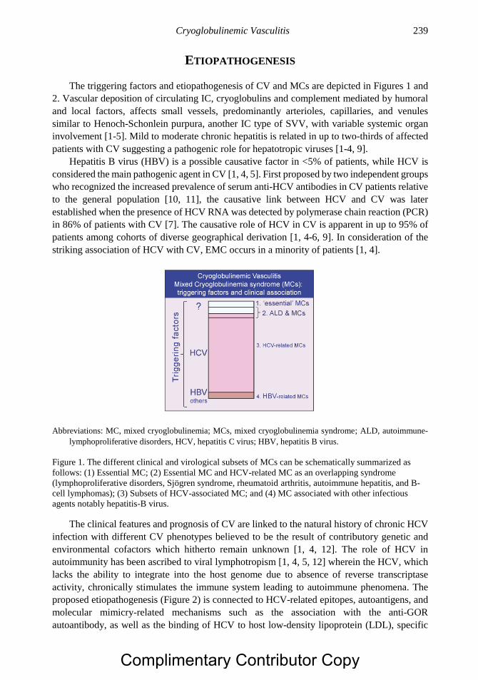
Cryoglobulinemic Vasculitis 239
ETIOPATHOGENESIS
The triggering factors and etiopathogenesis of CV and MCs are depicted in Figures 1 and
2. Vascular deposition of circulating IC, cryoglobulins and complement mediated by humoral
and local factors, affects small vessels, predominantly arterioles, capillaries, and venules
similar to Henoch-Schonlein purpura, another IC type of SVV, with variable systemic organ
involvement [1-5]. Mild to moderate chronic hepatitis is related in up to two-thirds of affected
patients with CV suggesting a pathogenic role for hepatotropic viruses [1-4, 9].
Hepatitis B virus (HBV) is a possible causative factor in <5% of patients, while HCV is
considered the main pathogenic agent in CV [1, 4, 5]. First proposed by two independent groups
who recognized the increased prevalence of serum anti-HCV antibodies in CV patients relative
to the general population [10, 11], the causative link between HCV and CV was later
established when the presence of HCV RNA was detected by polymerase chain reaction (PCR)
in 86% of patients with CV [7]. The causative role of HCV in CV is apparent in up to 95% of
patients among cohorts of diverse geographical derivation [1, 4-6, 9]. In consideration of the
striking association of HCV with CV, EMC occurs in a minority of patients [1, 4].
Abbreviations: MC, mixed cryoglobulinemia; MCs, mixed cryoglobulinemia syndrome; ALD, autoimmune-
lymphoproliferative disorders, HCV, hepatitis C virus; HBV, hepatitis B virus.
Figure 1. The different clinical and virological subsets of MCs can be schematically summarized as
follows: (1) Essential MC; (2) Essential MC and HCV-related MC as an overlapping syndrome
(lymphoproliferative disorders, Sjögren syndrome, rheumatoid arthritis, autoimmune hepatitis, and B-
cell lymphomas); (3) Subsets of HCV-associated MC; and (4) MC associated with other infectious
agents notably hepatitis-B virus.
The clinical features and prognosis of CV are linked to the natural history of chronic HCV
infection with different CV phenotypes believed to be the result of contributory genetic and
environmental cofactors which hitherto remain unknown [1, 4, 12]. The role of HCV in
autoimmunity has been ascribed to viral lymphotropism [1, 4, 5, 12] wherein the HCV, which
lacks the ability to integrate into the host genome due to absence of reverse transcriptase
activity, chronically stimulates the immune system leading to autoimmune phenomena. The
proposed etiopathogenesis (Figure 2) is connected to HCV-related epitopes, autoantigens, and
molecular mimicry-related mechanisms such as the association with the anti-GOR
autoantibody, as well as the binding of HCV to host low-density lipoprotein (LDL), specific
Complimentary Contributor Copy

Clodoveo Ferri, Dilia Giuggioli, Marco Sebastiani et al. 240
antiviral IgG and IgM, RF and cryoglobulins, and monoclonal RF bearing B-cells that express
the WA cross-idiotype. Collectively, these immunological phenomena are the cause and the
consequence of chronic stimulation of B-lymphocytes with expansion of B-cell subpopulations.
HCV-related lymphomagenesis represents an important model of virus-driven malignancy,
similarly to that observed for mucosa-associated lymphoid tissue (MALT) lymphoma of the
stomach related to gastrointestinal Helicobacter (H.) pylori infection [1, 4, 12], two
malignancies characterized by an analogous multistep and multifactorial etiopathogenesis.
Abbreviations: HCV, hepatitis C virus; RF, rheumatoid factor; IC, immune-complex; pSS, primary Sjögren
syndrome; SS, sicca syndrome; PCT, porphyria cutanea tarda; HCC, hepatocellular carcinoma; B-NHL,
B-cell non-Hodgkin lymphoma; CPX, cyclophosphamide; RTX, rituximab; LAC, low antigen-content.
Figure 2. Left: CV is the most frequent extrahepatic manifestation of the so-called HCV syndrome. CV
represents a crossing road between benign and malignant HCV-related complications. Given its
biological characteristics, HCV may be involved in a wide number of autoimmune and
lymphoproliferative disorders. This figure summarizes the main causative factors including, infection,
toxics, genetic and environmental causes potentially involved in the pathogenesis of the complex HCV
syndrome, in particular of CV. These factors, alone or in combination, may trigger two nonexclusive
multistep pathogenic processes, notably the production of benign poly-oligoclonal B-cell proliferation
responsible for organ- and non-organ-specific autoimmune disorders including IC-mediated CV; and
another characterized by different oncogenic alterations, which ultimately leads to malignant
complications. Right: Therapeutic strategies in patients with HCV-related CV and other autoimmune
lymphoproliferative disorders. Following the multistep pathological process leading from HCV
infection to overt CV, etiologic, pathogenic and symptomatic treatments are instituted at different levels.
The interaction between the HCV E2 envelope protein and CD81 molecule, a ubiquitous
tetraspannin present on the surface of B cells, likely represents an important pathogenic step in
the development of HCV-related autoimmune and lymphoproliferative disorders that results in
potent sustained polyclonal stimulation of the B-lymphocytes [1, 4, 12]. The occurrence of
HCV-related autoimmune-lymphoproliferative disorders may also be related to t(14;18)
translocations found in B-cells of HCV-infected patients leading to increased expression of the
Bcl-2 protein, with inhibition of apoptosis and abnormally prolonged B-cell survival [1, 4, 12,
Complimentary Contributor Copy
Cryoglobulinemic Vasculitis 241
13]. Apart from cryoglobulin production with or without overt CV, HCV-driven
lymphoproliferation could explain the pathogenic role of HCV infection in idiopathic B cell
lymphoma [1, 4]. Since the first description in 1994 [14], epidemiological and laboratory
studies have confirmed this association in geographical areas where both HCV-associated CV
and lymphoproliferative disorders occur [1, 4, 12].
The etiopathogenesis of HCV-negative CV [15] remains a challenge to explain apart from
concurrent infectious processes including HBV and concomitant autoimmune rheumatologic
and lymphoproliferative disorders (Figure 1). Patients with human immunodeficiency virus
(HIV) type I infection may also have HCV co-infection. HIV may exert continuous antigenic
stimulation of B-cells leading to MC type III production early in its course, followed by
monoclonal type II MC, and the typical manifestations of CV [1-4, 12].
CLINICAL MANIFESTATIONS
The demographic, clinical-serologic and virologic findings of 270 patients with CV at our
institution are shown in Table 1. The triad of purpura, weakness and arthralgia, the clinical
hallmark of CV and MCs, were frequent seen in addition to chronic hepatitis, type 1
membranoproliferative glomerulonephritis (MPGN), peripheral neuropathy, skin ulcers,
diffuse vasculitis, and in some instances, malignant B-cell non-Hodgkin lymphoma (B-NHL)
and hepatocellular carcinoma [1-5, 9, 12]. Although the prevalence of singular clinical-
serological features of CV in our cohort was comparable among those with type II and III MC,
the reported clinical variability may relate to bias of ascertainment from renal or rheumatology
clinics, and cohorts of geographically diverse origin [1-5, 8].
There are different presenting clinical-serological patterns that vary from mild arthralgia
and sporadic purpura in those with MC alone, to more severe cutaneous and systemic
involvement of classical MCs [1-4]. In all such patients, the disease combines MC with RF
activity, low C4 levels, and the clinical-pathological features of orthostatic purpura due to
leukocytoclastic vasculitis (LCV) with variable multi-organ involvement. Patients chronically
infected with HCV demonstrate asymptomatic serum MC that may precede the clinical onset
of disease by years to decades [1, 4]. Conversely, some patients show typical cryoglobulinemic
manifestations without serum cryoglobulins. The absence of this serological hallmark is not
surprising since cryoglobulins represent only a portion of IC that cryoprecipitate, which may
be transient and vary both among patients and during follow-up in individual patients [1, 4].
Most experts agree that repeated cryoglobulin determinations are necessary for a definite
diagnosis in such patients.
Cutaneous manifestations are the most common clinical feature of CV, with orthostatic
purpura, the pathognomonic feature, varying largely in dimension and diffusion to sporadic
isolated petechiae and severe vasculitic lesions, and complicated by torpid ulcers of the legs
and malleoli [16]. Repeated episodes of purpura lead to stable, sock-like ochraceous
discoloration of the legs [1, 4]. Humoral cofactors and vascular disturbances stemming from
increased cryocrit levels, chronic venous insufficiency and physical stress all contribute to
vasculitic alterations. More intense purpuric manifestations may occur in the afternoon with
prolonged standing and when cryocrit levels are highest, especially in muggy weather
conditions. Arthralgia, non-erosive oligoarthritis, and less frequently frank arthritis may also
Complimentary Contributor Copy
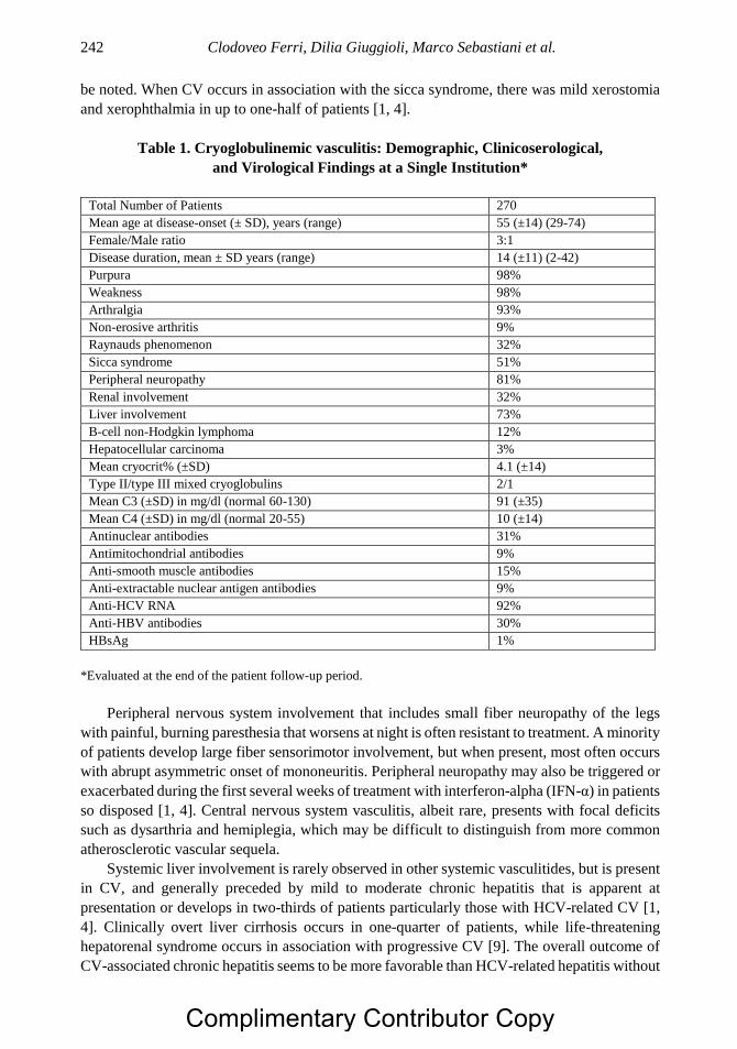
Clodoveo Ferri, Dilia Giuggioli, Marco Sebastiani et al. 242
be noted. When CV occurs in association with the sicca syndrome, there was mild xerostomia
and xerophthalmia in up to one-half of patients [1, 4].
Table 1. Cryoglobulinemic vasculitis: Demographic, Clinicoserological,
and Virological Findings at a Single Institution*
Total Number of Patients 270
Mean age at disease-onset (± SD), years (range) 55 (±14) (29-74)
Female/Male ratio 3:1
Disease duration, mean ± SD years (range) 14 (±11) (2-42)
Purpura 98%
Weakness 98%
Arthralgia 93%
Non-erosive arthritis 9%
Raynauds phenomenon 32%
Sicca syndrome 51%
Peripheral neuropathy 81%
Renal involvement 32%
Liver involvement 73%
B-cell non-Hodgkin lymphoma 12%
Hepatocellular carcinoma 3%
Mean cryocrit% (±SD) 4.1 (±14)
Type II/type III mixed cryoglobulins 2/1
Mean C3 (±SD) in mg/dl (normal 60-130) 91 (±35)
Mean C4 (±SD) in mg/dl (normal 20-55) 10 (±14)
Antinuclear antibodies 31%
Antimitochondrial antibodies 9%
Anti-smooth muscle antibodies 15%
Anti-extractable nuclear antigen antibodies 9%
Anti-HCV RNA 92%
Anti-HBV antibodies 30%
HBsAg 1%
*Evaluated at the end of the patient follow-up period.
Peripheral nervous system involvement that includes small fiber neuropathy of the legs
with painful, burning paresthesia that worsens at night is often resistant to treatment. A minority
of patients develop large fiber sensorimotor involvement, but when present, most often occurs
with abrupt asymmetric onset of mononeuritis. Peripheral neuropathy may also be triggered or
exacerbated during the first several weeks of treatment with interferon-alpha (IFN-α) in patients
so disposed [1, 4]. Central nervous system vasculitis, albeit rare, presents with focal deficits
such as dysarthria and hemiplegia, which may be difficult to distinguish from more common
atherosclerotic vascular sequela.
Systemic liver involvement is rarely observed in other systemic vasculitides, but is present
in CV, and generally preceded by mild to moderate chronic hepatitis that is apparent at
presentation or develops in two-thirds of patients particularly those with HCV-related CV [1,
4]. Clinically overt liver cirrhosis occurs in one-quarter of patients, while life-threatening
hepatorenal syndrome occurs in association with progressive CV [9]. The overall outcome of
CV-associated chronic hepatitis seems to be more favorable than HCV-related hepatitis without
Complimentary Contributor Copy
Cryoglobulinemic Vasculitis 243
MCs, whereas hepatocellular carcinoma is less frequently observed in HCV-related CV
compared to the overall population of HCV-infected individuals. Such differences are difficult
to fully explain, however, the relatively low prevalence of the 1b HCV genotype in those that
develop CV, along with a lower median consumption of alcohol might predict a more benign
clinical course of liver involvement [1, 4]. Immune complex-mediated glomerulonephritis,
most often seen in type 1 MPGN, affects the prognosis and survival of patients with CV due to
hypothesized immune mechanisms [1, 4]. Intestinal vasculitis presents with severe life-
threatening acute abdominal pain necessitating prompt diagnosis and aggressive treatment [1,
4].
Interstitial lung disease characterized by subclinical alveolitis has been described in those
with CV and in association with isolated HCV infection [1, 4]. When present, interstitial lung
disease can be a predisposing condition to pulmonary infections, and in rare instances, to
clinically manifest interstitial lung fibrosis. Capillaritis complicated by severe cough and
hemoptysis, are other rare manifestations of CV-related lung involvement, as well as overt
hyperviscosity syndrome are due to high levels of serum cryoglobulins [1, 4].
A low or undetectable C4 level with a normal C3 fraction occurs in patients with CV
regardless of disease activity. In vitro consumption of hemolytic complement is due to the anti-
complement activity of cryo-Ig [1, 4]. A sudden increase in C4 from low to abnormally high
levels can precede the development of B-NHL [1]. The absence of a clear-cut correlation
between the severity/activity of CV clinical manifestations and the observed cryocrit levels
may be related to the large variability of cryo- and non-cryoprecipitable IC ratio and/or the
intrinsic ability of IC to activate complement, as well as the possible in situ formation of IC
[1].
Endocrine disorders occur more frequently in CV than in the general population including
thyroid disturbances, type II diabetes mellitus, and gonadal dysfunction [1, 12, 17]. The former
include autoimmune thyroiditis, subclinical hypothyroidism, and rarely thyroid cancer.
Reversible hyperthyroidism occurs as a complication of IFN-α treatment [1].
B-NHL, the most common malignancy in association with CV, is often seen late in the
course of MCs [1, 4, 12]. The development of B-NHL may be a consequence of peripheral B-
cell expansion of lymphoid infiltrates in the liver and bone marrow. Such lymphoid infiltrates,
which are considered to be early indolent lymphomas, are indistinguishable from more
advanced B-cell cancers that include chronic lymphocytic leukemia, small lymphocytic
lymphoma, and immunocytoma [1, 4, 12]. While the majority of affected patients remain stable
for years, overt B-NHL is observed in approximately 10% of affected patients most often late
in the course of CV [1, 4-6, 12]. Other neoplastic manifestations, including hepatocellular
carcinoma and papillary thyroid cancer occur in the natural progression of CV [1, 4, 12].
Accordingly, MC may be considered, in part, a pre-neoplastic disorder in which careful clinical
monitoring is recommended even when the severity of MCs is mild.
DIAGNOSIS
There are no standardized methodologies for the measurement of cryoglobulin levels [1,
4] nor have the diagnostic criteria for CV been prospectively validated. In 1966, Meltzer and
Franklin [2] described EMCs in patients with the triad of purpura, weakness, and arthralgia,
Complimentary Contributor Copy
Clodoveo Ferri, Dilia Giuggioli, Marco Sebastiani et al. 244
circulating MC, low C4 levels, LCV of arterioles, capillaries and venules, and multiorgan
involvement without discernible infectious, autoimmune, or systemic involvement [1-6].
Recent classification criteria for CV were developed by a cooperative multicenter study using
a standardized methodology [18]. Successively validated in MC patients referred to experts
from a larger number of countries, these criteria may be usefully employed in epidemiological
and clinical-pathogenic studies, and prospective therapeutic trials [19]. Evidence of MC in the
serum of patients with suspected CV is necessary for a definite classification of the disease [18,
19], taking into account the above-mentioned variability of IC cryoprecipitation needing
repeated cryoglobulin determinations [1].
DIFFERENTIAL DIAGNOSIS
Figure 3 describes aspects of the differential diagnosis of CV which shares a number of
etiopathogenic events with both HCV-related and idiopathic disorders, namely sicca syndrome,
primary Sjögren syndrome (pSS), polyarthritis, classical rheumatoid arthritis, autoimmune
hepatitis, and B-NHL [1, 4, 12]. Therefore, a differential diagnosis with regard to the presence
or absence of HCV infection should be carefully made in all patients with cryoglobulinemia
and/or some autoimmune/neoplastic manifestations; a correct disease classification may
decisively affect the overall clinical therapeutic approach and outcome. The presence of HCV
in the large majority of individuals with CV may nonetheless lead to difficulty in the
differentiation of CV from HCV-infected patients with other extrahepatic manifestations that
characterize the HCV syndrome [12, 20]. Such patients develop slow progression from mild
HCV-associated hepatitis to various extrahepatic manifestations including arthralgia, RF-
positive arthritis, sicca syndrome, Raynaud’s phenomenon, and others to clinically overt
cryoglobulinemic syndrome (Figure 2). Of note, a minority of patients with long-lasting CV
develop a clinically relevant malignancy [1, 20].
Patients with mild CV complain of arthralgia often without synovitis [1, 4, 21, 27] while
those with HCV-associated CV and symmetrical erosive polyarthritis are more likely to have
the syndrome of overlapping CV-rheumatoid arthritis further definable by serum anti-cyclic
citrullinated peptide (anti-CCP) IgG antibodies and RF serology (Figure 3). Sicca syndrome
occurs in up to one-half of patients with CV; however, formal criteria are satisfied in only a
minority of patients [1, 4, 12, 21]. Patients with pSS shares various symptoms with CV
including purpura, xerostomia, xerophthalmia, arthralgia, and laboratory evidence of RF
seropositivity, cryoglobulinemia, and the associated complication of B-cell lymphoma [21].
Histopathological alteration of the salivary glands and the specific autoantibody pattern (anti-
Ro/SSA-La/SSB) of pSS are rarely found in patients with MCs. Moreover, pSS is rarely
complicated by chronic hepatitis, glomerulonephritis, or concomitant HCV infection. In this
regard, HCV infection should be considered an exclusionary criterion for pSS [21]. In rare
cases, and often in subjects without HCV infection, the differential diagnosis may be
challenging. In those instances, it may be more appropriate to classify such patients as CV-pSS
overlap syndrome [1, 4, 12, 21]. This clinical condition shows a rather severe clinical
progression, less frequent Ro/SSA and La/SSB seropositivity, high levels of serum MC,
hypocomplementemia, with frequent autoimmune-vasculitic manifestations, and a higher risk
of lymphoma [4, 21, 27].
Complimentary Contributor Copy
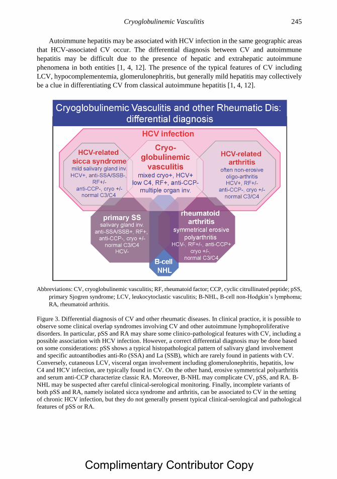
Cryoglobulinemic Vasculitis 245
Autoimmune hepatitis may be associated with HCV infection in the same geographic areas
that HCV-associated CV occur. The differential diagnosis between CV and autoimmune
hepatitis may be difficult due to the presence of hepatic and extrahepatic autoimmune
phenomena in both entities [1, 4, 12]. The presence of the typical features of CV including
LCV, hypocomplementemia, glomerulonephritis, but generally mild hepatitis may collectively
be a clue in differentiating CV from classical autoimmune hepatitis [1, 4, 12].
Abbreviations: CV, cryoglobulinemic vasculitis; RF, rheumatoid factor; CCP, cyclic citrullinated peptide; pSS,
primary Sjogren syndrome; LCV, leukocytoclastic vasculitis; B-NHL, B-cell non-Hodgkin’s lymphoma;
RA, rheumatoid arthritis.
Figure 3. Differential diagnosis of CV and other rheumatic diseases. In clinical practice, it is possible to
observe some clinical overlap syndromes involving CV and other autoimmune lymphoproliferative
disorders. In particular, pSS and RA may share some clinico-pathological features with CV, including a
possible association with HCV infection. However, a correct differential diagnosis may be done based
on some considerations: pSS shows a typical histopathological pattern of salivary gland involvement
and specific autoantibodies anti-Ro (SSA) and La (SSB), which are rarely found in patients with CV.
Conversely, cutaneous LCV, visceral organ involvement including glomerulonephritis, hepatitis, low
C4 and HCV infection, are typically found in CV. On the other hand, erosive symmetrical polyarthritis
and serum anti-CCP characterize classic RA. Moreover, B-NHL may complicate CV, pSS, and RA. B-
NHL may be suspected after careful clinical-serological monitoring. Finally, incomplete variants of
both pSS and RA, namely isolated sicca syndrome and arthritis, can be associated to CV in the setting
of chronic HCV infection, but they do not generally present typical clinical-serological and pathological
features of pSS or RA.
Complimentary Contributor Copy
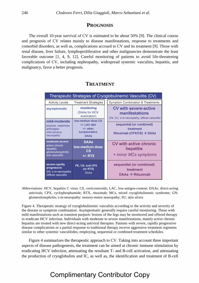
Clodoveo Ferri, Dilia Giuggioli, Marco Sebastiani et al. 246
PROGNOSIS
The overall 10-year survival of CV is estimated to be about 50% [9]. The clinical course
and prognosis of CV relates mainly to disease manifestations, response to treatments and
comorbid disorders, as well as, complications accrued to CV and its treatment [9]. Those with
renal disease, liver failure, lymphoproliferative and other malignancies demonstrate the least
favorable outcome [1, 4, 9, 12]. Careful monitoring of patients to avoid life-threatening
complications of CV, including nephropathy, widespread systemic vasculitis, hepatitis, and
malignancy, favor a better prognosis.
TREATMENT
Abbreviations: HCV, hepatitis C virus; CS, corticosteroids; LAC, low-antigen-content; DAAs: direct-acting
antivirals; CPX, cyclophosphamide; RTX, rituximab; MCs, mixed cryoglobulinemic syndrome; GN:
glomerulonephritis; s-m-neuropathy: sensory-motor neuropathy; SU: skin ulcers
Figure 4. Therapeutic strategy of cryoglobulinemic vasculitis according to the activity and severity of
the disease or symptom combination. Asymptomatic generally require careful monitoring. Those with
mild manifestations such as transient purpuric lesions of the legs may be monitored and offered therapy
to eradicate HCV infection. Individuals with moderate to severe manifestations, mainly active chronic
hepatitis are treated with new direct-acting antiviral therapies. Patients with severe, rapidly progressive
disease complications or a partial response to traditional therapy receive aggressive treatment regimens
similar to other systemic vasculitides; employing, sequential or combined treatment schedules.
Figure 4 summarizes the therapeutic approach to CV. Taking into account three important
aspects of disease pathogenesis, the treatment can be aimed at chronic immune stimulation by
eradicating HCV infection, attenuating the resultant T- and B-cell activation, and attenuating
the production of cryoglobulins and IC, as well as, the identification and treatment of B-cell
Complimentary Contributor Copy
Cryoglobulinemic Vasculitis 247
lymphoproliferation to avert the development of B-NHL. This multistep, multifactorial
etiopathogenesis-oriented approach to therapy targets HCV infection, B-cell proliferation, and
the resultant CV inflammatory involvement. The recent introduction of direct acting antivirals
(DAAs) led to sustained clearance of HCV in the large majority of patients so treated [4, 22-
27], without the frequent side-effects previously observed with IFN-α treatment, such as
peripheral neuropathy, thyroiditis, and rheumatoid-like polyarthritis [1, 4, 12]. Preliminary
studies suggest a role for DAAs in the treatment and eradication of HCV, whereby elevated its
tolerability and efficacy, offers new hope in the management of severe complications [22-27].
However, the variable and unpredictable CV response to the antiviral therapy observed in some
patients, including the paradoxical response with disease exacerbation or new symptom
appearance, indicates that DAAs effect on CV needs to be further evaluated. The immune
system of a single patient may be decisive in the clinical outcome following antiviral treatment.
There is a point of no return in a viral-driven lymphoproliferative disorder that may affect the
overall clinical response after HCV eradication. Like H. pylori-associated gastric lymphomas
[1, 4], HCV-associated CV is more responsive to biologically based treatment of the underlying
infection, early in the he early phase of the illness.
Physicians must choose from among available therapies for CV including low-antigen-
content (LAC) diets, plasma exchange (PE), and immunosuppressant regimens employing
corticosteroids, rituximab, and cyclophosphamide alone or in combination depending upon the
severity and activity of the illness and clinical manifestations [1, 4, 17]. A LAC-diet improves
the clearance of circulating IC by restoring the activity of the reticuloendothelial system, which
becomes overloaded, by circulating cryoglobulins [1, 4, 21]. It reduces the input of alimentary
macromolecules crossing the mucosal barrier of the gut as some foods such as dairy products
and eggs present a potential antigenic activity and might otherwise be pathogenic. Reduction
of the alimentary input of macromolecules directed to mononuclear phagocytic system
improves its function in conditions characterized by abnormal endogenous hyperproduction of
IC responsible for organ damage such as MCs. Combined LAC-diet and 2 to 4 mg per day of
6-methyl-prednisolone may be sufficient to ameliorate mild manifestations of CV such as
sporadic purpura and arthralgia. The limitations of an LAC-diet treatment include poor patient
compliance, which can be improved by tailoring the treatment duration, for example 7 to 14
days of restricted diet followed by 14 to 28 days of free diet. Those with mild-moderate
symptoms including palpable purpura may be particularly sensitive to the small variations in
daily steroid dosage of 1 to 2 mg, and derive benefit from dietary therapy.
Cyclophosphamide and in particular, rituximab are each first-line therapies for EMCs [1,
4, 21, 22]. Rituximab is first-choice treatment based on the results of observational and
controlled studies in seronegative and seropositive HCV patients with CV [1, 4, 21, 22, 27-31].
Traditional and double-filtration PE can reduce the levels of circulating IC and cryoglobulins
[1, 4, 21]. Oral cyclophosphamide at the dose of 50 to 100 mg per day for 2 to 6 weeks during
the tapering of PE sessions, as well as rituximab, reinforces the beneficial effects of PE.
Moreover, the latter can also potentiate the effect of cyclophosphamide [9], and is beneficial in
the treatment of fulminant manifestations of CV such as widespread vasculitis and/or rapidly
progressive MPGN [9].
The treatment of CV should be tailored to individual patients according to the severity of
clinical symptoms, with the most severe vasculitic manifestations treated promptly with
combination corticosteroids, PE, and cyclophosphamide or rituximab [1, 4, 9, 21]. Pilot studies
suggest that sequential or combined antiviral and immunosuppressive treatment is a useful
Complimentary Contributor Copy
Clodoveo Ferri, Dilia Giuggioli, Marco Sebastiani et al. 248
strategy [4, 27, 30, 31] especially in those with major clinical manifestations and partial or
transitory remission after antiviral therapy (Figure 4). Clinically asymptomatic patients may be
monitored without treatment even in the presence of elevated cryocrit levels; in these patients,
an attempt to HCV eradication should be always considered. Careful clinical monitoring of the
disease is mandatory in all cases with particular attention to neoplastic transformation [1, 4].
CONCLUSION
Cryoglobulinemic vasculitis is an important model for the study of autoimmune and
lymphoproliferative diseases mediated by viral infections. Recent advancements in the
understanding of the underlying multifactorial and multistep processes in the etiopathogenesis
of CV have translated into more effective etiological treatment modalities including antivirals
treatments directed at eradication of HCV infection as both a triggering factor and contributor
to the self-perpetuating mechanism of autoimmune disease. In addition, pathogenetic therapies
may be employed to target the variably severe consequences of LCV, systemic SVV,
multiorgan involvement, and malignancies. Future challenges in CV include improvements in
the differential diagnosis with some overlapping disorders, as well as the improvement in
therapeutic strategies tailored to different CV clinical variants in selected patients.
REFERENCES
[1] Ferri C. Mixed cryoglobulinemia. Orphanet J Rare Dis 2008; 3: 25. doi: 10.1186/1750-
1172-3-25.
[2] Meltzer M, Franklin EC, Elias K, et al. Cryoglobulinemia. A clinical and laboratory
study. II. Cryoglobulins with rheumatoid factor activity. Am J Med 1966; 40: 837-856.
[3] Gorevic PD, Kassab HJ, Levo Y, et al. Mixed Cryoglobulinemia: clinical aspects and
long-term follow-up of 40 patients. Am J Med 1980; 69: 287-308.
[4] Ferri C, Sebastiani M, Giuggioli D, et al. Hepatitis C virus syndrome: A constellation of
organ- and non-organ specific autoimmune disorders, B-cell non-Hodgkin's lymphoma,
and cancer. World J Hepatol. 2015; 7:327-343.
[5] Monti G, Galli M, Invernizzi F, et al. and GISC. Italian Group for the Study of
Cryoglobulinaemias. Cryoglobulinaemias: a multi-centre study of the early clinical and
laboratory manifestations of primary and secondary disease. QJM 1995; 88: 115-126.
[6] Dammacco F, Sansonno D, Piccoli C, et al. The lymphoid system in hepatitis C virus
infection: autoimmunity, mixed cryoglobulinemia, and Overt B-cell malignancy. Semin
Liver Dis 2000; 20: 143-157.
[7] Ferri C, Greco F, Longombardo G, et al. Association between hepatitis C virus and mixed
cryoglobulinemia. Clin Exp Rheum 1991; 9: 621-624.
[8] Lunel F, Musset L, Cacoub P, et al. Cryoglobulinemia in chronic liver diseases: role of
hepatitis C virus and liver damage. Gastroenterology 1994; 106: 1291-1300.
[9] Ferri C, Sebastiani M, Giuggioli D, et al. Mixed cryoglobulinemia: demographic, clinical,
and serological features, and survival in 231 patients. Sem Arthritis Rheum 2004; 33: 355-
474.
Complimentary Contributor Copy
Cryoglobulinemic Vasculitis 249
[10] Pascual M, Perrin L, Giostra E, et al. Hepatitis C virus in patients with Cryoglobulinemia
type II. J Inf Dis 1990; 162: 569-570.
[11] Ferri C, Marzo E, Longombardo G, et al. Alpha-interferon in the treatment of mixed
cryoglobulinemia patients. International Cancer Update. Focus on Interferon Alfa 2b.
Cannes, France. November 1-4, 1990. Proc Eur J Cancer 1991, 27: 81-82.
[12] Ferri C, Antonelli A, Mascia MT, et al. HCV-related autoimmune and neoplastic
disorders: the HCV syndrome. Dig Liver Dis 2007; 39 (Suppl 1): S13-S21.
[13] Zignego AL, Ferri C, Giannelli F, et al. Prevalence of BCL-2 rearrangement in hepatitis
C virus-related mixed cryoglobulinemia with or without complicating B-cell lymphoma.
Ann Int Med 2002; 137: 571–580.
[14] Ferri C, Caracciolo F, Zignego AL, et al. Hepatitis C virus infection in patients with non-
Hodgkin’s lymphoma. Br J Haematol 1994; 88:392-394.
[15] Galli M, L. Oreni L, Saccardo F, et al. HCV-unrelated cryoglobulinaemic vasculitis: the
results of a prospective observational study by the Italian Group for the Study of
Cryoglobulinaemias (GISC). Clin Exp Rheumatol 2017; 35 (Suppl. 103): 67-76.
[16] Giuggioli D, Manfredi A, Lumetti F, et al. Cryoglobulinemic vasculitis and skin ulcers.
Our therapeutic strategy and review of the literature. Semin Arthr Rheum 2015; 44: 518-
526.
[17] Antonelli A, Ferri C, Ferrari SM, et al. Immunopathogenesis of HCV-related endocrine
manifestations in chronic hepatitis and mixed cryoglobulinemia. Autoimmun Rev 2008;
8: 18-23.
[18] De Vita S, Soldano F, Isola M, et al. Preliminary classification criteria for the
cryoglobulinaemic vasculitis. Ann Rheum Dis 2011; 70: 1183-1190.
[19] Quartuccio L, Isola M, Corazza L, et al. Validation of the classification criteria for
cryoglobulinaemic vasculitis. Rheumatology. 2014; 53: 2209-2213.
[20] Ferri C, Ramos-Casals M, Zignego AL, et al. on behalf of the ISG-EHCV. International
diagnostic guidelines for patients with HCV-related extrahepatic manifestations. A
multidisciplinary expert statement . Autoimmunity Reviews 2016; 15: 1145–1160
[21] Ferri C, Sebastiani M, Antonelli A, et al. Current treatment of hepatitis C-associated
rheumatic diseases. Arthritis Res Ther 2012; 14:215. doi:10.1186/ar3865.
[22] Pietrogrande M, De Vita S, Zignego AL, et al. Recommendations for the management of
mixed cryoglobulinemia syndrome in hepatitis C virus-infected patients. Autoimmun Rev
2011; 10: 444-454.
[23] Gragnani L, Visentini M, Fognani E, et al. Prospective study of guideline-tailored therapy
with direct- acting antivirals for hepatitis C virus-associated mixed cryoglobulinemia.
Hepatology 2016; 64:1473-1482.
[24] Arcaini L, Besson C, Frigeni M, et al. Interferon-free antiviral treatment in B-cell
lymphoproliferative disorders associated with hepatitis C virus infection. Blood 2016;
128:2527-2532.
[25] Bonacci M, Lens S, Londono MC et al. Virologic, Clinical, and Immune Response
Outcomes of Patients With Hepatitis C Virus-Associated Cryoglobulinemia Treated With
Direct-Acting Antivirals. Clin Gastroenterol Hepatol 2017; 15:575-583.
[26] Saadoun D, Pol S, Ferfar Y, et al. Efficacy and Safety of Sofosbuvir Plus Daclatasvir for
Treatment of HCV- Associated Cryoglobulinemia Vasculitis. Gastroenterology, (2017);
153:49-52.
Complimentary Contributor Copy
Clodoveo Ferri, Dilia Giuggioli, Marco Sebastiani et al. 250
[27] Zignego AL, Ramos-Casals M, Ferri C. et al. on behalf of the ISG-EHCV. International
therapeutic guidelines for patients with HCV-related extrahepatic disorders. A
multidisciplinary expert statement. Autoimmune Review 2017; 16: 523–541.
[28] Ferri C, Cacoub P, Mazzaro C, et al. Treatment with rituximab in patients with mixed
cryoglobulinemia syndrome: Results of multicenter cohort study and review of the
literature. Autoimmune Rev 2011; 11: 48-55.
[29] De Vita S, Quartuccio L, Isola M, et al. A randomised controlled trial of rituximab for
the treatment of severe cryoglobulinemic vasculitis. Arthritis Rheum 2012; 64:843–53.
[30] Terrier B, Saadoun D, Sène D, et al. Efficacy and tolerability of rituximab with or without
PEGylated interferon alfa-2b plus ribavirin in severe hepatitis C virus-related vasculitis:
a long-term follow up study of thirty-two patients. Arthritis Rheum 2009; 60: 2531-2540.
[31] Dammacco F, Tucci FA, Lauletta G, et al. PEGylated interferon-alpha, ribavirin, and
rituximab combined therapy of hepatitis C virus-related mixed cryoglobulinemia: a long-
term study. Blood 2010; 116: 343-353.
Complimentary Contributor Copy
In: The Vasculitides. Volume 1 ISBN: 978-1-53615-133-6
Editor: David S. Younger © 2019 Nova Science Publishers, Inc.
Chapter 14
AORTITIS AND PERIAORTITIS
Eugenia Accorsi Buttini, MD, Chiara Marvisi, MD
and Augusto Vaglio, MD, PhD Nephrology Unit, Parma University Hospital, Parma, Italy
ABSTRACT
Aortitis and periaortitis are inflammatory diseases of the aorta and its branches. They
differ in the extension of inflammation, which is confined to the aortic wall in aortitis and
extends in the periaortic space in periaortitis. Although aortitis occurs as either an
infectious or a non-infectious process, the latter suggests an etiology related to large-vessel
vasculitis. Periaortitis and aortitis are recognizable fibro-inflammatory systemic disorders,
categorized under the rubric of IgG4-related diseases. This chapter reviews the nosology,
pathogenesis, histopathology and clinical phenotype of the different forms of aortitis and
periaortitis, and discusses their diagnosis and management.
Keywords: aortitis, periaortitis, IgG4-related disease, fibrosis, vasculitis, giant cell arteritis,
Takayasu arteritis
ABBREVIATIONS
ANCA anti-neutrophil cytoplasm antibody;
AAV ANCA associated vasculitis;
CRP C-reactive protein;
CT computed tomography;
CTL cytotoxic T lymphocyte;
ECD Erdheim Chester disease;
ESR erythrocyte sedimentation rate; 18F-FDG-PET 18F-fluorodeoxyglucose positron emission tomography;
Corresponding Author’s Email: [email protected].
Complimentary Contributor Copy
Eugenia Accorsi Buttini, Chiara Marvisi and Augusto Vaglio 252
GCA giant cell arteritis;
HLA human leucocyte antigen;
IAAA inflammatory abdominal aortic aneurysm;
IgG4-RD: IgG4-related disease;
IL interleukin;
LVV large vessel vasculitis;
MRI magnetic resonance imaging;
NK Natural killer;
PDGF platelet-derived growth factor;
RPF retroperitoneal fibrosis;
SLE systemic lupus erythematosus;
SUV standardized uptake value;
TA Takayasu arteritis;
TLR: toll-like receptor;
US ultrasonography.
INTRODUCTION
Aortitis and periaortitis are in the spectrum of systemic inflammatory disorders
characterized by chronic inflammation that may be limited to the aortic wall or to the adjacent
periaortic space. Both conditions may arise in the context of a recently recognized clinical-
pathological entity known as an IgG4-related disease (IgG4-RD), characterized by marked
fibrosis and T-lymphocyte and IgG4-positive plasma cell infiltration of various organs.
In 2015, a standardized classification for inflammatory diseases of the aorta was proposed
as a part of a consensus statement issued by the Society for Cardiovascular Pathology and the
Association for European Cardiovascular Pathology [1]. The committee identified three general
categories of inflammatory aortic disease: (a) atherosclerosis, (b) atherosclerosis with excessive
inflammation and (c) aortitis/periaortitis.
Atherosclerosis is a degenerative inflammatory process that starts from the intimal layer of
arteries. It is classified as mild, moderate, or severe based on the medial destruction and the
presence of extracellular lipid deposition with fibrosis. In some cases, the inflammatory
reaction is unusually intense but always due to atherosclerosis. Two major examples of this
phenomenon are atherosclerosis with excessive neutrophilic inflammation and inflammatory
abdominal aortic aneurysms (IAAA).
The terms aortitis and periaortitis are reserved for inflammatory aortic conditions
unattributed to atherosclerosis alone. Aortitis is a pathological term that describes an extensive
inflammatory process that involves the media and/or the intima of the aortic wall.2 The
inflammatory response affects the aorta at any level and extends to its major branches and other
large vessels. Aortitis can be the presenting feature of large vessel vasculitides (LLV), but may
also occur in the absence of clinical symptoms or signs of systemic vasculitis making it an
incidental diagnosis on imaging studies or in histologically studied specimens of surgically
resected aortic tissue [1]. Aortitis rarely develops in association with concomitant
rheumatologic disorders or in the presence of an infectious illness.
Complimentary Contributor Copy
Aortitis and Periaortitis 253
It is recommended to apply the term periaortitis to an inflammatory process that arises from
the adventitia of the aortic wall and extends into the surrounding periaortic space. The ensuing
pathological lesion consists of a fibrous component and a chronic inflammatory infiltrate.
Periaortitis typically involves mainly the lower abdominal aorta and the common iliac arteries
and can arise around either a dilated or non-dilated aorta. The tissue infiltrating the
retroperitoneum frequently encases adjacent structures such as the ureters and the inferior vena
cava [3]. Originally, periaortitis was thought to be confined to the abdominal aorta, although
histopathologic studies provided evidence of simultaneous involvement of other vascular
districts [4]. Computed tomography (CT), magnetic resonance imaging (MRI) and especially 18F-fluorodeoxyglucose positron emission tomography (18F-FDG-PET) imaging identify
vascular inflammation in virtually all large arteries. The thoracic aorta and the epiaortic arteries
are involved in about a third of patients suffering from abdominal periaortitis wherein the
condition is typically asymptomatic [5]. The coronary, renal, mesenteric and coeliac arteries
may rarely be involved. While diverse causes of periaortitis and aortitis may be clearly
distinguishable, the differential diagnosis is often challenging when an affected patient presents
with nonspecific systemic symptoms or fever of unknown origin, and the inflammatory process
diffusely involves the aorta.
In 2001, Hamano and colleagues [6] first described IgG4-RD noting an association
between sclerosing pancreatitis and high serum levels of IgG4+. The recognition of other
involved sites led to the concept of a fibro-inflammatory systemic disease. Large vessels
involved by IgG4-RD [7] present clinically as aortitis or periaortitis. Notwithstanding, the
corresponding lesions are similar throughout the different affected sites.8
CLASSIFICATION AND NOSOLOGY
Aortitis is classified into two main groups, according to the presence or absence of an
infectious source. While infectious aortitis is now rare in developing countries as a result of the
introduction of antibiotics, it nevertheless remains important to differentiate the infectious and
non-infectious entities since the therapeutic strategies significantly differ [2].
Infectious aortitis develops in the course of septicemia or embolization of a septic embolus
as a complication of infective endocarditis [1]. Pre-existing aortic wall damage in the form of
arteriosclerotic lesions or vascular dilatation facilitates infection. Elderly men and immune
compromised patients are populations at the greatest risk for the most common associated
organisms, namely, gram-positive bacteria including Staphylococcus spp, Enterococcus spp
and Streptococcus pneumoniae that typically involve the thoracic aorta; and gram-negative
bacteria, notably Salmonella spp, that affect the abdominal aorta. Syphilitic aortitis develops in
the setting of tertiary syphilis, typically involving the ascending aorta. Treponema pallidum
preselects the vasa vasorum of the aortic wall leading to ischemic injury of the medial layer,
with secondary aneurysmal dilatation. Tuberculous aortitis should be suspected in those with
pulmonary and extrapulmonary tuberculosis in the course of miliary spread or direct extension
of the infectious process from adjacent tissues, such as infected lymph nodes or lung lesions
[2]. In those cases, the thoracic and abdominal aorta are affected with comparable frequency
[1]. Fungal infection-related aortitis occurs in immune-compromised patients or in others
chronically exposed to broad-spectrum antibiotics [1].
Complimentary Contributor Copy
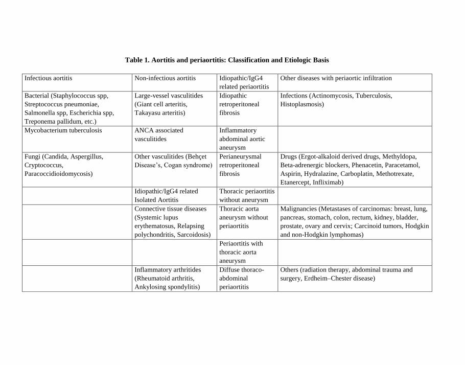
Table 1. Aortitis and periaortitis: Classification and Etiologic Basis
Infectious aortitis Non-infectious aortitis Idiopathic/IgG4
related periaortitis
Other diseases with periaortic infiltration
Bacterial (Staphylococcus spp,
Streptococcus pneumoniae,
Salmonella spp, Escherichia spp,
Treponema pallidum, etc.)
Large-vessel vasculitides
(Giant cell arteritis,
Takayasu arteritis)
Idiopathic
retroperitoneal
fibrosis
Infections (Actinomycosis, Tuberculosis,
Histoplasmosis)
Mycobacterium tuberculosis ANCA associated
vasculitides
Inflammatory
abdominal aortic
aneurysm
Fungi (Candida, Aspergillus,
Cryptococcus,
Paracoccidioidomycosis)
Other vasculitides (Behçet
Disease’s, Cogan syndrome)
Perianeurysmal
retroperitoneal
fibrosis
Drugs (Ergot-alkaloid derived drugs, Methyldopa,
Beta-adrenergic blockers, Phenacetin, Paracetamol,
Aspirin, Hydralazine, Carboplatin, Methotrexate,
Etanercept, Infliximab) Idiopathic/IgG4 related
Isolated Aortitis
Thoracic periaortitis
without aneurysm
Connective tissue diseases
(Systemic lupus
erythematosus, Relapsing
polychondritis, Sarcoidosis)
Thoracic aorta
aneurysm without
periaortitis
Malignancies (Metastases of carcinomas: breast, lung,
pancreas, stomach, colon, rectum, kidney, bladder,
prostate, ovary and cervix; Carcinoid tumors, Hodgkin
and non-Hodgkin lymphomas) Periaortitis with
thoracic aorta
aneurysm
Inflammatory arthritides
(Rheumatoid arthritis,
Ankylosing spondylitis)
Diffuse thoraco-
abdominal
periaortitis
Others (radiation therapy, abdominal trauma and
surgery, Erdheim–Chester disease)
Complimentary Contributor Copy
Aortitis and Periaortitis 255
Non-infectious aortitis associated with LVV, including giant cell arteritis (GCA) and
Takayasu arteritis (TA) represents two large groups of cases [2]. Aortic involvement occurs in
systemic rheumatic disorders. Rheumatoid arthritis vasculitis (RAV), in which aortitis is an
uncommon extra-articular manifestation, may be associated with vascular involvement of
vessels of any size and may appear in any phase of the illness, although typically in the later
stages [9]. The aortic root, the ascending aorta [10] and the branches of the aortic arch [11] can
be involved in ankylosing spondylitis. Aortitis is also described in cases of systemic lupus
erythematosus (SLE), most often manifesting as an aortic aneurysm or dissection [12, 13]; and
in rare cases of relapsing polychondritis [14].
Large vessel involvement occurs in several small vessel vasculitides (SVV). In anti-
neutrophil cytoplasm antibody (ANCA)-associated vasculitides (AAV), aortitis may present
with stenosing arteritis, aneurysmal disease and aortic dissection [15]. Vasculitis involvement
of the vasa vasorum affecting the aortic root is associated with valvular dysfunction in some
cases of Behçet disease [16] Aortitis also occurs in a minority of patients with Cogan syndrome
[17] with severe incipient complications thereof [18]. Aortitis is unusual in sarcoidosis [19].
Cryptic isolated aortitis involving the ascending aorta may be detected in clinically
asymptomatic patients, without evidence of a coexisting rheumatic disease [1].
Periaortitis has to be distinguished from aortitis not only for classification purposes, but
also for its clinical and therapeutic implications. Periaortitis usually affects the infra-renal
portion of the abdominal aorta and the iliac arteries. The aorta is undilated in idiopathic
retroperitoneal fibrosis (RPF) while IAAA and perianeurysmal retroperitoneal fibrosis are the
perianeurysmal forms [3]. Abdominal periaortitis must be differentiated from other diseases
leading to periaortic retroperitoneal infiltration, particularly malignancies, granulomatous
infections, drug-related and radiotherapy-induced conditions, and Erdheim-Chester disease
[20]. In some patients, an idiopathic form of periaortitis involves the thoracic aorta with three
possible patterns: thoracic periaortitis without aneurysm, thoracic aorta aneurysm without
periaortitis, and periaortitis surrounding a thoracic aorta aneurysm.
Both aortitis and periaortitis can arise in the context of IgG4-RD signaling involvement as
a multifocal disease; and in other cases, isolated to the thoracic tract. IgG4-related aortitis
accounts for 75% of thoracic aortitis with a lymphoplasmacytic pattern [21]. Periaortitis is a
common manifestation of IgG4-RD, responsible for up to 50% of periaortitis cases [22]. Table
1 summarizes the main etiologies of aortitis and periaortitis.
MAIN HISTOLOGIC PATTERNS
Aortitis may exhibit four different histological patterns of inflammation, namely
granulomatosus/giant cell, lymphoplasmacytic, mixed inflammatory and suppurative, each
related to different etiologies [1].
The granulomatosus/giant cell pattern is the most common pattern of inflammation in
aortitis. It is characterized by the presence of large activated epithelioid macrophages with or
without giant cells and compact granulomas. An accompanying lymphoplasmacytic infiltrate
is often present. This pattern occurs in GCA- and TA-related aortitis, isolated aortitis, and
aortitis associated with rheumatoid arthritis, ANCA associated vasculitides (AAVs),
sarcoidosis, mycobacterial and fungal infections.
Complimentary Contributor Copy
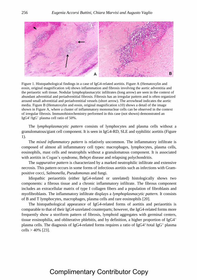
Eugenia Accorsi Buttini, Chiara Marvisi and Augusto Vaglio 256
Figure 1. Histopathological findings in a case of IgG4-related aortitis. Figure A (Hematoxylin and
eosin, original magnification x4) shows inflammation and fibrosis involving the aortic adventitia and
the periaortic soft tissue. Nodular lymphoplasmacytic infiltrates (long arrow) are seen in the context of
abundant adventitial and periadventitial fibrosis. Fibrosis has an irregular pattern and is often organized
around small adventitial and periadventitial vessels (short arrow). The arrowhead indicates the aortic
media. Figure B (Hematoxylin and eosin, original magnification x10) shows a detail of the image
shown in Figure A, where a cluster of inflammatory mononuclear cells can be observed in the context
of irregular fibrosis. Immunohistochemistry performed in this case (not shown) demonstrated an
IgG4+/IgG+ plasma cell ratio of 50%.
The lymphoplasmacytic pattern consists of lymphocytes and plasma cells without a
granulomatous/giant cell component. It is seen in IgG4-RD, SLE and syphilitic aortitis (Figure
1).
The mixed inflammatory pattern is relatively uncommon. The inflammatory infiltrate is
composed of almost all inflammatory cell types: macrophages, lymphocytes, plasma cells,
eosinophils, mast cells and neutrophils without a granulomatous component. It is associated
with aortitis in Cogan’s syndrome, Behçet disease and relapsing polychondritis.
The suppurative pattern is characterized by a marked neutrophilic infiltrate and extensive
necrosis. This pattern occurs in some forms of infectious aortitis such as infections with Gram-
positive cocci, Salmonella, Pseudomonas and fungi.
Idiopathic periaortitis (either IgG4-related or unrelated) histologically shows two
components: a fibrous tissue and a chronic inflammatory infiltrate. The fibrous component
includes an extracellular matrix of type I collagen fibers and a population of fibroblasts and
myofibroblasts. The inflammatory infiltrate displays a lymphoplasmacytic pattern. It consists
of B and T lymphocytes, macrophages, plasma cells and rare eosinophils [20].
The histopathological appearance of IgG4-related forms of aortitis and periaortitis is
comparable to that of their IgG4-unrelated counterparts; however, the IgG4-related forms more
frequently show a storiform pattern of fibrosis, lymphoid aggregates with germinal centers,
tissue eosinophilia, and obliterative phlebitis, and by definition, a higher proportion of IgG4+
plasma cells. The diagnosis of IgG4-related forms requires a ratio of IgG4+/total IgG+ plasma
cells > 40% [23].
Complimentary Contributor Copy
Aortitis and Periaortitis 257
CLINICAL PHENOTYPES
Herein, we describe the clinical phenotype of the most common aortitis and periaortitis
syndromes, with particular emphasis on non-infectious forms, and we highlight the
characteristics of these two conditions in the course of IgG4-RD.
Giant Cell Arteritis
This medium- and large-vessel vasculitis usually affects the aorta and its major branches
with a predilection for the branches of the carotid and vertebral arteries [24]. Defined in the
past as “temporal arteritis,” the term is inadequate to describe GCA because not all patients
have temporal artery involvement and other categories of vasculitis can affect the temporal
arteries. GCA is the most common type of non-infectious arteritis in North America and
Western Europe. It occurs primarily in elderly individuals older than 50 years. Women are
affected two to three times more commonly than men [25].
The typical histological pattern of GCA is a granulomatous inflammatory infiltrate
composed of lymphocytes, macrophages and in 50% of the cases, multinucleated giant cells.25
Inflammation has a focal nature, with “skip” lesions, wherein non-affected arterial segments
intermingle with inflamed tracts. It is located mainly in the intima-media junction of the vessel
wall but it can be present throughout the artery. At the beginning of the disease process, the
intimal layer appears thickened by the inflammatory infiltrate and edema, at later stages by
myofibroblast migration. The media remains well preserved, although focal smooth muscle
necrosis may occur. The adventitia is inflamed in a moderate proportion of cases [26].
The pathogenesis of GCA has not been fully explained but dysregulated interactions
between the vessel wall and the immune system are key to disease development [27]. The allelic
variant of human leucocyte antigen (HLA)-DRB1*04 is the most common genetic association
with GCA. The involvement of class II HLA molecules implies antigen-presentation to CD4+
T cells which then triggers the vasculitic lesion [28]. The involved antigens are still unknown,
both infectious agents and autoantigens have been suspected.
Vascular dendritic cells located at the adventitia-media border of the artery have a critical
role in the initiation of disease. Dendritic cells become activated once their toll-like receptors
(TLRs) are engaged by microbial products and turn into non-tolerogenic cells able to present
potential auto-antigens to T cells; their ability to produce chemokines is thus enhanced, thus
leading to recruitment of T cells and macrophages into the vascular wall through the vasa
vasorum [28]. The activated dendritic cells express CD86, a co-receptor required to trigger T-
cell activation, and produce cytokines, such as interleukin (IL)-6 and IL-12 that guide the
differentiation of T cells into selected functional lineages [27].
IL-6 is a pleiotropic cytokine released also by endothelial cells, vascular smooth muscle
cells, lymphocytes and macrophages and implicated in the production of acute-phase proteins.
IL-6 shifts T-cell differentiation towards the Th17 lineage and blocks the anti-inflammatory
T regulatory cell expansion. Th17 cells produce a plethora of cytokines (IL-17, IL-21, and IL-
22) that recruit macrophages and neutrophils and activate vascular smooth muscle cells,
fibroblasts and endothelial cells.
Complimentary Contributor Copy
Eugenia Accorsi Buttini, Chiara Marvisi and Augusto Vaglio 258
The IL-6/IL-17 cytokine cluster seems to play an important role in early GCA, while the
IL-12/IFN- cytokine cluster appears to drive the tissue remodeling during chronic disease [27].
IL-12 promotes the differentiation of the Th1 lineage, which releases IFN- into the
microenvironment. IFN- amplifies T-cell responses and stimulates macrophage activation.
Macrophages have the potential to induce arterial wall injury through multiple pathways. They
release reactive oxygen intermediates and metalloproteinases that lead to the fragmentation of
the internal elastic lamina. Macrophages produce also platelet-derived growth factor (PDGF)
and vascular endothelial growth factor that are critically involved in intimal layer proliferation.
Myofibroblasts, stimulated by IFN- and PDGF migrate in the intima where they replicate and
secrete extracellular matrix leading to concentric intima hyperplasia [28]. Outgrowth of the
intima is consistently associated with marked neoangiogenesis. The end result is a vascular
occlusion caused by the vessel wall thickening or the formation of arterial aneurysm
(particularly thoracic aorta aneurysm) caused by destruction of the arterial wall [28].
Medium-sized arteries, such as the branches of external and internal carotid arteries and
the vertebral arteries respond with occlusion of the lumen and, consequently, ischemic organ
damage. Clinical symptoms reflect end organ ischemia.
The classic manifestations are headache, scalp tenderness, jaw claudication, blindness,
stroke, transient ischemic attacks and vertigo. Similarly, involvement of the subclavian,
axillary, and proximal brachial arteries leads to arm claudication and absent or asymmetric
pulses [28]. Compared to patients with cranial symptoms, those with aortic arch syndrome tend
to be younger at diagnosis and have a longer duration of symptoms prior to diagnosis [29].
Instead of forming stenotic lesions, the aorta responds with wall destruction. Aortitis most
often occurs in the thoracic aorta while involvement of the branches of the abdominal aorta and
lower extremity arteries is less common. It may produce chest or back pain but it is more
frequently asymptomatic until complications arise. These complications are aneurysm
formation and rupture, dissection and aortic valve insufficiency (Figure 2). In early studies, the
prevalence of aortic involvement in patients with GCA was based on the rate of aneurysm
diagnosed fortuitously or after acute events and autopsy data, remaining under-estimated.
Recently, the large use of imaging techniques (essentially CT, MRI or PET) in the diagnosis
and follow-up of LVVs showed that aortic involvement may be more prevalent than previously
reported, with a frequency ranging from 33%30 to 50% [31-33]. The risk of aortic structural
damage significantly increases in the first 5 years from diagnosis and progresses slowly
afterwards, suggesting that chronic inflammation of the vascular wall is the major determinant
of aortic damage [34].
In almost all patients with GCA, a syndrome of systemic inflammation accompanies
vascular manifestations and presents with nonspecific symptoms including malaise, anorexia,
weight loss, fever and depression. Around 40% of patients with GCA also have polymyalgia
rheumatica, characterized by aching and morning stiffness involving the neck, shoulder and
pelvic girls [25].
Takayasu Arteritis
This disease predominantly affects the aorta and its major branches, along with pulmonary
arteries. In accordance with the Chapel Hill consensus conference nomenclature, TA is a
Complimentary Contributor Copy
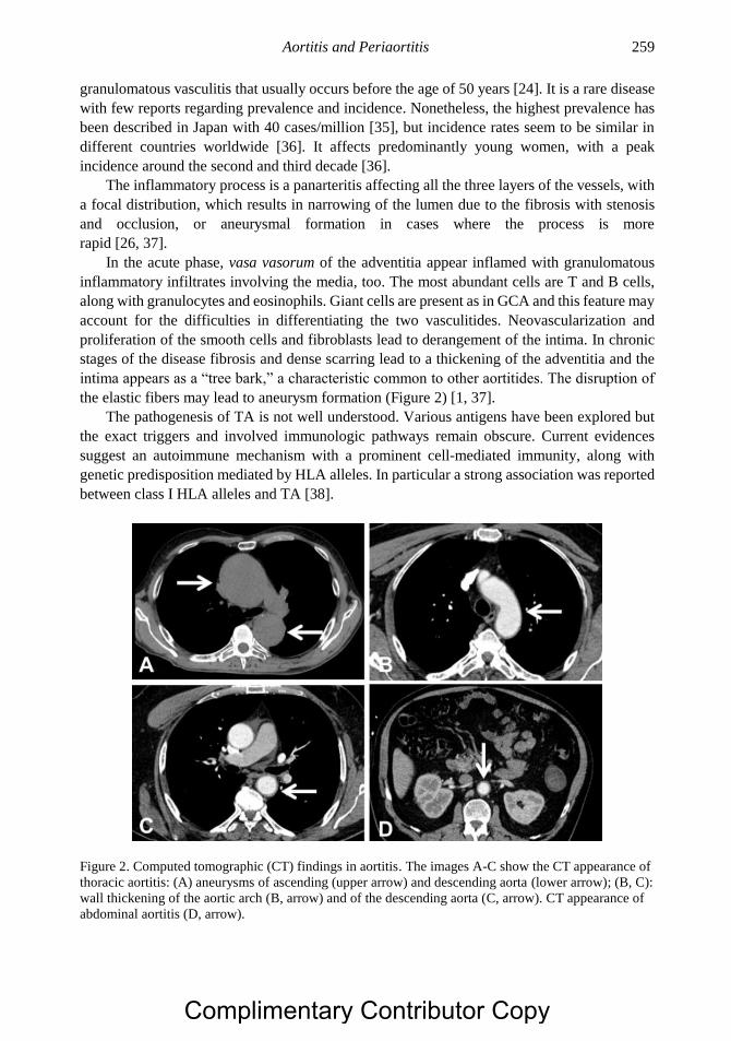
Aortitis and Periaortitis 259
granulomatous vasculitis that usually occurs before the age of 50 years [24]. It is a rare disease
with few reports regarding prevalence and incidence. Nonetheless, the highest prevalence has
been described in Japan with 40 cases/million [35], but incidence rates seem to be similar in
different countries worldwide [36]. It affects predominantly young women, with a peak
incidence around the second and third decade [36].
The inflammatory process is a panarteritis affecting all the three layers of the vessels, with
a focal distribution, which results in narrowing of the lumen due to the fibrosis with stenosis
and occlusion, or aneurysmal formation in cases where the process is more
rapid [26, 37].
In the acute phase, vasa vasorum of the adventitia appear inflamed with granulomatous
inflammatory infiltrates involving the media, too. The most abundant cells are T and B cells,
along with granulocytes and eosinophils. Giant cells are present as in GCA and this feature may
account for the difficulties in differentiating the two vasculitides. Neovascularization and
proliferation of the smooth cells and fibroblasts lead to derangement of the intima. In chronic
stages of the disease fibrosis and dense scarring lead to a thickening of the adventitia and the
intima appears as a “tree bark,” a characteristic common to other aortitides. The disruption of
the elastic fibers may lead to aneurysm formation (Figure 2) [1, 37].
The pathogenesis of TA is not well understood. Various antigens have been explored but
the exact triggers and involved immunologic pathways remain obscure. Current evidences
suggest an autoimmune mechanism with a prominent cell-mediated immunity, along with
genetic predisposition mediated by HLA alleles. In particular a strong association was reported
between class I HLA alleles and TA [38].
Figure 2. Computed tomographic (CT) findings in aortitis. The images A-C show the CT appearance of
thoracic aortitis: (A) aneurysms of ascending (upper arrow) and descending aorta (lower arrow); (B, C):
wall thickening of the aortic arch (B, arrow) and of the descending aorta (C, arrow). CT appearance of
abdominal aortitis (D, arrow).
Complimentary Contributor Copy
Eugenia Accorsi Buttini, Chiara Marvisi and Augusto Vaglio 260
Natural killer (NK) cells and γδ T lymphocytes appear to have a role in inducing apoptosis
of the vascular cells by releasing perforin. These cells are activated by 65-kDa heat shock
protein (HSP) which is overexpressed in aortic samples of patients affected by TA [39]. Similar
to GCA, CD4+ T cells have a leading role particularly in the formation of granulomas. Both
Th1 and Th17 pathways are expanded and correlate with disease activity [40].
The clinical manifestations are variable during the early stages of the disease with anorexia,
fever and malaise. In fibrotic stages (also known as pulseless stages) symptoms are related to
vascular stenosis and aneurysms [41]. Most typical clinical features include: diminished or
absent pulses with limb claudication and discrepancies in blood pressure, multiple vascular
bruits, renovascular hypertension and retinopathy [37]. Complications are related to organ
ischemia and congestive heart failure due to hypertension and valve regurgitation when thoracic
aneurysms are present.
Isolated Aortitis
Isolated aortits is a single-organ vasculitis found primarily in the ascending part of the
thoracic aorta [24]. This condition is not a component of a systemic vasculitis like GCA and
TA, or associated with a systemic disease such as SLE or sarcoidosis. The disorder emerged
with the advent of aortic surgery and histopathologic examination of the aortic surgical
specimens. The introduction of new imaging techniques facilitated recognition of the disease
before development of structural aortic abnormalities requiring surgery.
The epidemiology of isolated aortitis has not been clearly established, but its prevalence is
probably underestimated, as the disease may remain undiagnosed for a long time. The incidence
of isolated aortitis in the population of patients undergoing thoracic aortic surgery is between
3.8% and 4.4% [42-44]. Some studies show a higher incidence in women [42, 43] others in
men [44, 45]. The mean age of patients at diagnosis ranges from 63 to 72 years [42-44]. Unlike
those with TA and GCA, patients with isolated aortitis do not have symptoms related to
systemic illness or extra-aortic arteritis [43].
Most cases of isolated aortitis show a granulomatous/giant cell pattern of inflammation
localized in the medial layer. The inflammatory infiltrate comprises macrophages, giant cells,
lymphocytes, plasma cells and well-formed granulomas replacing irregular areas of medial
destruction. Adventitial inflammation contains minimal mononuclear cell infiltration without
granuloma formation..
Isolated aortitis is identified incidentally during radiological exams or in the repair of the
complications of aortic surgery [46]. Isolated aortitis can lead to thoracic aortic aneurysms,
aortic dissection and aortic valve regurgitation. The aortic aneurysm is certainly the most
frequent complication, so noted in 95% of patients with isolated aortitis and is usually localized
in the ascending tract [42, 43].
Recent studies have evaluated the rate of new aortic events in patients with the diagnosis
of isolated aortitis [47, 48]. Follow-up data indicate that patients with isolated aortitis are at
increased risk for subsequent aortic events, such as new aneurysms and dissections also
involving the descending thoracic aorta or the abdominal aorta. Even in the absence of signs of
systemic vasculitis, aortitis identified in the ascending aorta is associated with an increased
frequency of distal aortic events during long-term follow-up. Patients with isolated aortitis
should be monitored with periodic aortic imaging.
Complimentary Contributor Copy
Aortitis and Periaortitis 261
Though the studies are limited, there is an increasing evidence that patients with aortitis
isolated to aortic ascending segment have actually a smoldering systemic vasculitis that is
subclinical in other districts at the time of presentation and fully manifests at a later time [46].
IgG4-Related Aortitis
Since the first description of IgG4-RD, diverse clinical entities without clear nosology have
been categorized in the spectrum of this systemic fibro-inflammatory disease, including cases
of aortitis, once considered isolated or idiopathic.
IgG4-related aortitis preferentially affects the thoracic aorta and particularly the aortic arch
[21, 49]. It presents with a lymphoplasmacytic pattern and accounts for a significant proportion
of non-infectious thoracic aortitis cases, and about 75% of cases of lymphoplasmacytic thoracic
aortitis [21, 50]. The vasculitic process can also involve the abdominal aorta, along with
medium-sized vessels originating from the aorta, such as the carotid and coronary arteries [7,
51]. Small vessel involvement has also been described, thus supporting the idea that IgG4-RD
may be included in the category of vasculitis of vessels of variable size [52].
IgG4-related aortitis occurs among elderly men aged 60 years or older, in keeping with the
epidemiology of the systemic and other organ-limited forms of IgG4-RD [50].
The inflammatory infiltrate affects predominantly the adventitia with a lesser involvement
of the media (Figure 1), in contrast with other LVV.1 Histological findings are typical and are
the same throughout different lesions, regardless of the involved site. The main features are
storiform fibrosis, obliterative phlebitis, moderate eosinophilia and dense lymphoplasmacytic
infiltrates, with a predominance of T and B cells that occasionally organize into germinal
centers [23]. IgG4-immune staining of the affected tissue is mandatory to have a “definite”
diagnosis of IgG4-related aortitis. However, in cases not undergoing surgery or when the
pathological specimen is unavailable, evidence of aortitis on imaging studies, along with
increased serum IgG4 levels, supports a “possible” diagnosis of this condition [49].
The pathogenesis of IgG4-RD, and consequently of IgG4-related aortitis, remains unclear
although significant advances have been made: IgG4 antibodies does not seem to have a central
role, and their anti-inflammatory characteristics lead to consider them as an attempt to dampen
a primary inflammatory process [53]. The immune mechanisms are probably cell-mediated. A
subpopulation of CD4+ T cells with a cytotoxic phenotype (CTLs) has been found in IgG4-RD
lesions and clonally expanded in the circulation, which appears to orchestrate the pathogenic
mechanisms activated by antigen-presenting B cells [54]. The latter have a pivotal role in
initiating and maintaining the disease, as shown by the efficacy of B cell-depleting therapies,
such as rituximab. The antigens that trigger the immune response are unknown.
IgG4-related aortitis occurs sub-acutely and is often asymptomatic. It may be diagnosed
after an incidental finding on CT (Figure 2) or after pathological examination of surgical
specimens. It can also present with symptoms related to the presence of aneurysms, whereas
end organ ischemia is far less common and appear when medium-size vessels such as the main
aortic branches are also involved [7]. Acute pain may indicate dissection, which has also been
described as a complication of IgG4-related aortitis [55]. Involvement of other organs may be
absent at onset and appear only during follow-up with a metachrounous pattern [56], leading to
difficulties in promptly recognizing IgG4-RD. The most frequent clinical pictures of IgG4-RD
include sclerosing pancreatitis (type 1 autoimmune pancreatitis), Mikulicz disease and RPF,
Complimentary Contributor Copy
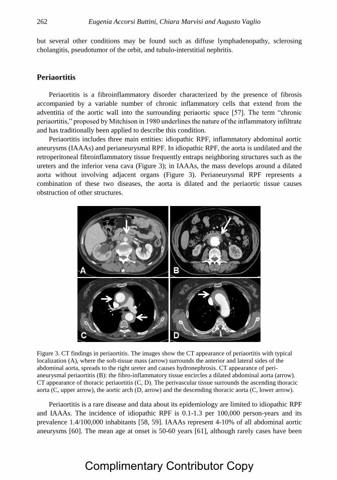
Eugenia Accorsi Buttini, Chiara Marvisi and Augusto Vaglio 262
but several other conditions may be found such as diffuse lymphadenopathy, sclerosing
cholangitis, pseudotumor of the orbit, and tubulo-interstitial nephritis.
Periaortitis
Periaortitis is a fibroinflammatory disorder characterized by the presence of fibrosis
accompanied by a variable number of chronic inflammatory cells that extend from the
adventitia of the aortic wall into the surrounding periaortic space [57]. The term “chronic
periaortitis,” proposed by Mitchison in 1980 underlines the nature of the inflammatory infiltrate
and has traditionally been applied to describe this condition.
Periaortitis includes three main entities: idiopathic RPF, inflammatory abdominal aortic
aneurysms (IAAAs) and perianeurysmal RPF. In idiopathic RPF, the aorta is undilated and the
retroperitoneal fibroinflammatory tissue frequently entraps neighboring structures such as the
ureters and the inferior vena cava (Figure 3); in IAAAs, the mass develops around a dilated
aorta without involving adjacent organs (Figure 3). Perianeurysmal RPF represents a
combination of these two diseases, the aorta is dilated and the periaortic tissue causes
obstruction of other structures.
Figure 3. CT findings in periaortitis. The images show the CT appearance of periaortitis with typical
localization (A), where the soft-tissue mass (arrow) surrounds the anterior and lateral sides of the
abdominal aorta, spreads to the right ureter and causes hydronephrosis. CT appearance of peri-
aneurysmal periaortitis (B): the fibro-inflammatory tissue encircles a dilated abdominal aorta (arrow).
CT appearance of thoracic periaortitis (C, D). The perivascular tissue surrounds the ascending thoracic
aorta (C, upper arrow), the aortic arch (D, arrow) and the descending thoracic aorta (C, lower arrow).
Periaortitis is a rare disease and data about its epidemiology are limited to idiopathic RPF
and IAAAs. The incidence of idiopathic RPF is 0.1-1.3 per 100,000 person-years and its
prevalence 1.4/100,000 inhabitants [58, 59]. IAAAs represent 4-10% of all abdominal aortic
aneurysms [60]. The mean age at onset is 50-60 years [61], although rarely cases have been
Complimentary Contributor Copy
Aortitis and Periaortitis 263
reported in pediatric patients [62]. Men are affected two to three times more often than women
and this ratio is higher in the aneurysmal forms [58].
Pathologic changes in periaortitis involve both the aortic wall and the surrounding soft
tissues. The typical macroscopic appearance of periaortitis is that of a greyish mass infiltrating
the retroperitoneal tissue surrounding the abdominal aorta, the iliac arteries and, in most cases,
the inferior vena cava and the ureters [63]. The mass usually develops between the origin of
the renal arteries and the pelvic brim. In some instances, periaortitis shows atypical
localizations, which might be peri-duodenal, peri-pancreatic, pelvic, presacral, peri-ureteral or
peri-renal and not characterized by involvement of the periaortic space. These cases are thought
to have a different pathogenesis.
Microscopic examination reveals the presence of two components: a fibrous tissue and an
inflammatory infiltrate [20]. The fibrous component comprises fibroblasts that show signs of
activation and transition into myofibroblasts (-smooth muscle actin expression) and produce
an extracellular matrix composed of type I collagen fibers organized in thick irregular bundles.
The inflammatory infiltrate consists of numerous lymphocytes, plasma cells, macrophages and
scattered eosinophils. The inflammatory cells are interspersed within the collagen bundles
(diffuse pattern), but also organized in nodular aggregates, usually around small vessels
(perivascular nodular pattern). These aggregates have a B-cell core surrounded by T cells,
which are predominantly CD4+. In some cases, these lymphoid follicles have the structure of
germinal centers, which is a sign of ectopic lymphoneogenesis, thus proving the presence of a
highly structured immune-mediated/autoimmune response.
The aortic wall shows intimal atherosclerosis, medial thinning and adventitial
inflammation and fibrosis. The composition of the inflammatory infiltrate in the aortic
adventitia is similar to that of the retroperitoneal one. When the pattern is arranged in nodular
aggregates, these are usually centered on the adventitial vasa vasorum which can show signs
of vasculitis [64].
The pathogenesis of periaortitis is still unclear. The theory proposed by Parums and
Mitchinson suggests that it is due to a local inflammatory reaction against antigens localized in
the atherosclerotic plaques of the aorta such as oxidized low-density lipoproteins or ceroid [65,
66]. These antigens would be presented by plaque macrophages to B and T lymphocytes
residing in the adventitia, where they would elicit a fibro-inflammatory response inducing
lymphocyte activation and immunoglobulin production. This theory, however, cannot explain
the presence of constitutional symptoms and the increase in acute phase reactant levels in
patients with periaortitis, and the frequent associations with autoimmune or fibro-inflammatory
diseases involving other organs. Additionally, periaortitis may affect patients without
atherosclerotic lesions and involve vascular territories/segments spared by atherosclerosis [67].
Recently, it has been proposed that periaortitis may actually be a systemic autoimmune disease
rather than the result of a local inflammatory reaction [64]. This theory is supported by
similarities to systemic LLV, such as GCA and TA: the inflammatory process starts in the
adventitial layer leading to perivascular infiltrates surrounding aortic vasa vasorum. Thus, it
cannot be excluded that periarotitis originates as a primary aortitis which subsequently induces
a fibro-inflammatory reaction into the periaortic space [3].
The pathogenesis of the disease is probably multifactorial, recognizing genetic,
environmental and immune-mediated determinants. The role of genetic factors is demonstrated
by the association between periaortitis and the HLA class II allele HLA-DRB1*03, a marker
Complimentary Contributor Copy
Eugenia Accorsi Buttini, Chiara Marvisi and Augusto Vaglio 264
of autoimmune diseases such as SLE, type 1 diabetes, autoimmune thyroiditis and myasthenia
gravis [68].
A more recent study also describes an increased susceptibility to the aneurysmal forms of
periaortitis in patients carrying the 32 polymorphism of the CCR5 gene [69]. CCR5 is
expressed on many immune cells, particularly Th1 cells, and acts by binding to different
chemokines, including RANTES, MIP-1a and MIP-1b. The CCR532 polymorphism creates
a truncated, nonfunctional receptor and probably shifts the immune response toward a Th2
pattern.
Among environmental agents, cigarette smoking and asbestos exposure are significantly
associated with an increased risk of developing the disease. Interestingly, smoking and asbestos
had a multiplicative effect on disease risk, with an odds ratio of 12.04 (95% confidence interval,
4.32 to 38.28) in co-exposed subjects [70]. The role of other environmental or infectious agents
remains elusive.
The immunopathogenic model of periaortitis is based on the hypothesis that the disease is
antigen-driven, although the triggering antigens are still unknown. Based on the available
experimental evidence, it can be postulated that antigen-presenting cells present such
hypothetical antigens to CD4+ T cells within the aortic wall, probably in the adventitia. CD4+
T cells expand, secrete IL-6 [71], which enhances the production of acute-phase proteins and
promotes the activation of B cells and fibroblasts. CD4+ T cells also secrete Th2 cytokines such
as IL-4, IL-10 and IL-13, which induce B-cell expansion and maturation into plasma cells
producing, in a fraction of cases, IgG4. Lymphoid cells also release eotaxin-1 (also known as
CCL11), which drives tissue infiltration by eosinophils and mast cells, whose products induce
fibroblast activation [72]. Once activated, fibroblasts mature into myofibroblasts and secrete
collagen, promoting fibrosis. The pathogenic importance of IL-6 and of B cells was confirmed
in vivo by the efficacy of therapies targeting selectively the IL-6 receptor (tocilizumab) and the
B-cell marker CD20 (rituximab) [71, 73].
The clinical presentation of periaortitis includes two types of manifestations: localized, due
to the compressive effects of the retroperitoneal mass, and systemic, related to the inflammatory
nature of the disease. The more frequent localized symptom, present in about 80% of the
patients, is side, back or abdominal pain. It is usually described as persistent and dull, not
exacerbated by movement or palpation; it transiently responds to nonsteroidal anti-
inflammatory drugs and, in cases of ureteral involvement, it can be colic-like [3]. Ureteral
involvement is the most frequent complication and can be unilateral or bilateral. In cases with
unilateral involvement, ureteral obstruction can also be asymptomatic for a long time and, at
diagnosis, these patients present with renal hypoplasia/atrophy, whose frequency is estimated
to be up to 30%. However, most cases are symptomatic and bilateral involvement usually leads
to acute renal failure. Other urologic manifestations are frequent: they range from testicular
pain, often accompanied by hydrocele and/or varicocele due to spermatic vein encasement by
periaortitis, to retrograde ejaculation and erectile dysfunction [20]. The extrinsic compression
of retroperitoneal lymphatic vessels and veins can be the cause of lower extremity edema and
deep vein thrombosis. Claudication and intestinal ischemia are less common. Systemic
symptoms include fatigue, weight loss, anorexia, sleep disturbances and low-grade fever [3].
Periaortitis can affect not only the lower abdominal aorta and the iliac arteries but also
other vascular segments. This may suggest that in such cases, periaortitis actually represents a
primary, diffuse inflammatory disease of the aorta and its major branches (Figure 3) [5]. In
Complimentary Contributor Copy
Aortitis and Periaortitis 265
1970, Mitchinson described for the first time the presence of mild inflammation and fibrosis in
the adventitial layer of the thoracic aorta in autopsy studies performed on patients with
abdominal periaotitis [4]. Subsequent reports have also documented the involvement of
coronary arteries [57], renal arteries [74], and celiac axis, superior and inferior mesenteric
arteries [75].
A recent study performed in a large cohort of patients showed that, in one third of cases,
periaortitis might also affect the thoracic aorta and the epiaortic arteries [5]. The perivascular
tissue may develop around the ascending aorta, the aortic arch, the descending aorta and the
origin of epiaortic arteries. Three main patterns can be observed: periaortitis with thoracic aorta
aneurysm, thoracic periaortitis without aneurysm and thoracic aorta aneurysm without
periaortitis [5]. Histology of the thoracic tissue is definitely similar to that found in abdominal
periaortitis. The pathological lesions involve the adventitia and the periadventitial tissue and
consist of a fibrous component and a chronic inflammatory infiltrate comprising lymphocytes,
histiocytes, plasma cells, and eosinophils.
The observation that the thoracic aorta is usually spared by atherosclerosis strongly
supports the hypothesis that, when periaortitis develops, it is a primary immune-mediated
process [76]. The clinical presentation of thoracic involvement in periaortitis includes specific
symptoms such as laryngeal nerve paralysis, dry cough, upper limb claudication and
paresthesias; however, in about 85% of cases it is asymptomatic. Interestingly, the comparison
of demographic and clinical characteristics of patients with and without thoracic involvement
revealed that these subgroups were different. Patients with thoracic involvement had a
significantly higher female prevalence, a greater age at disease onset, a higher prevalence of
systemic symptoms and of back or abdominal pain [5].
The female prevalence and the advanced age in patients with thoracic aorta involvement is
a feature shared by GCA, which often affects not only the epiaortic tree but also the thoracic
aorta. Indeed, GCA is characterized by only mild periaortic thickening does not cause
obstructive uropathy, while periaortitis, on the other hand, does not lead to typical GCA-related
cranial symptoms. However, the differential diagnosis between GCA and periaortitis may
become more challenging when patients present with non-specific systemic symptoms,
including fever of unknown origin, and lack clinical manifestations specific for either disease.
Periaortitis may be associated with a large variety of autoimmune conditions. This
observation underlines the relevance of autoimmune mechanisms in the pathogenesis of the
disease. Hashimoto’s thyroiditis is the most commonly associated autoimmune disorder; it may
arise together with periaortitis, but it can also precede or follow periaortitis diagnosis. Overall,
it has been reported that 25% of patients with periaortitis develop hypothyroidism after a
median follow-up of 45 months [77]. Other associated conditions are AAV, particularly
granulomatosis with polyangiitis, membranous nephropathy, SLE, rheumatoid arthritis, uveitis
and psoriasis [3].
IgG4-Related Periaortitis
Periaortitis may also be a manifestation of IgG4-RD. Histological examination is
mandatory to differentiate IgG4-related and –unrelated forms. However, the coexistence of
other clinical conditions belonging to the spectrum of IgG4-RD may help classify periaortitis
as IgG4-related [78].
Complimentary Contributor Copy
Eugenia Accorsi Buttini, Chiara Marvisi and Augusto Vaglio 266
Periaortitis (particularly the abdominal form) is reported among the most frequent
manifestations of IgG4-RD in different studies, even if its prevalence remains quite variable,
ranging from 11% to 30% [79-82]. It appears that environmental factors influence the different
organ involvements, since periaortitis is more frequent in Western cohorts, whereas in Asian
countries extra-vascular lesions are more common [82].
IgG4-related periaortitis may differ from IgG4-unrelated forms in terms of microscopic
findings, whereas the macroscopic ones are very similar. In particular, there were no significant
differences in disease localization and characteristics when CT or MRI findings were compared
[83, 84]. IgG4-related IAAAs (which are considered perianeurysmal forms of periaortitis) are
frequently saccular, as the IgG4-unrelated ones, but the former show a more prominent
thickening of the wall due to the fibrotic tissue which is also adherent to neighboring structures,
leading to an apparently lower risk of rupture [85]. IgG4-related periaortitis is distinguishable
from its IgG4-unrelated counterpart especially for the organization of the fibrotic tissue, which
is storiform in the former, resembling the spokes of a cartwheel. IgG4-unrelated forms usually
do not show obliterative phlebitis, and the inflammatory infiltrate is less dense and not
markedly enriched with IgG4+ plasma cells and eosinophils [83]. However, these differences
may be only slight, and it must be emphasized that the background histologic appearance of
IgG4-related and -unrelated forms is often very similar, thus leading to the concept that these
two entities may represent different ends of the same spectrum.
From a clinical standpoint, there are no substantial differences, except for a male
predominance in IgG4-related forms [86]. The clinical manifestations of both IgG4-related and
-unrelated periaortitis are comparable, with flank and abdominal pain being the most common
presenting symptom. However, it must always be remembered that IgG4-related forms are
more commonly associated with extra-retroperitoneal manifestations of IgG4-RD; this may
help clarify the clinical picture of the disease.
DIAGNOSIS
The differential diagnosis among different forms of inflammatory aortitis and periaortitis
is challenging, and histological examination is often required to have a clear definition of the
disease. The particular site of inflammation makes it difficult to perform biopsies, unless aortic
or retroperitoneal/ureteral surgery is required. Therefore, in most cases the diagnosis is based
on laboratory tests and imaging studies, along with clinical presentation.
Aortitis in Large-Vessel Vasculitides
Both GCA and TA often manifest with typical symptoms, which render them more easily
recognizable than other cases of apparently idiopathic aortitis. Cranial symptoms (e.g., jaw
claudication, scalp tenderness and visual alterations) usually occur in GCA, whereas TA may
manifest with signs and symptoms related to vascular stenosis (e.g., absence of pulses, bruits,
Complimentary Contributor Copy
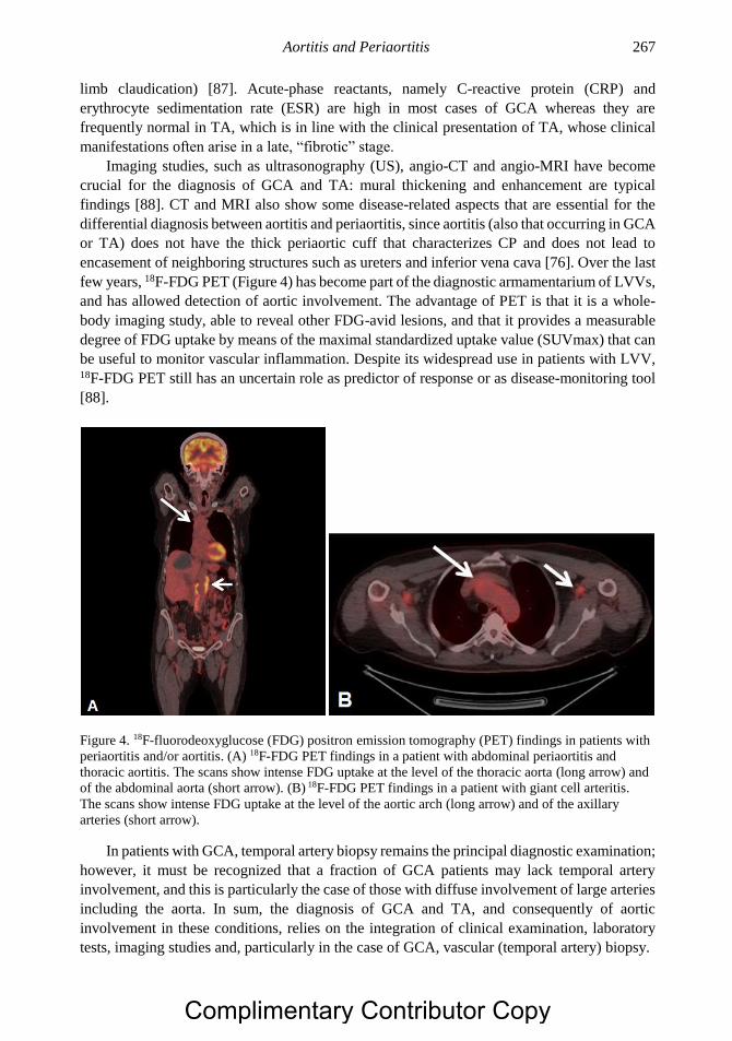
Aortitis and Periaortitis 267
limb claudication) [87]. Acute-phase reactants, namely C-reactive protein (CRP) and
erythrocyte sedimentation rate (ESR) are high in most cases of GCA whereas they are
frequently normal in TA, which is in line with the clinical presentation of TA, whose clinical
manifestations often arise in a late, “fibrotic” stage.
Imaging studies, such as ultrasonography (US), angio-CT and angio-MRI have become
crucial for the diagnosis of GCA and TA: mural thickening and enhancement are typical
findings [88]. CT and MRI also show some disease-related aspects that are essential for the
differential diagnosis between aortitis and periaortitis, since aortitis (also that occurring in GCA
or TA) does not have the thick periaortic cuff that characterizes CP and does not lead to
encasement of neighboring structures such as ureters and inferior vena cava [76]. Over the last
few years, 18F-FDG PET (Figure 4) has become part of the diagnostic armamentarium of LVVs,
and has allowed detection of aortic involvement. The advantage of PET is that it is a whole-
body imaging study, able to reveal other FDG-avid lesions, and that it provides a measurable
degree of FDG uptake by means of the maximal standardized uptake value (SUVmax) that can
be useful to monitor vascular inflammation. Despite its widespread use in patients with LVV, 18F-FDG PET still has an uncertain role as predictor of response or as disease-monitoring tool
[88].
Figure 4. 18F-fluorodeoxyglucose (FDG) positron emission tomography (PET) findings in patients with
periaortitis and/or aortitis. (A) 18F-FDG PET findings in a patient with abdominal periaortitis and
thoracic aortitis. The scans show intense FDG uptake at the level of the thoracic aorta (long arrow) and
of the abdominal aorta (short arrow). (B) 18F-FDG PET findings in a patient with giant cell arteritis.
The scans show intense FDG uptake at the level of the aortic arch (long arrow) and of the axillary
arteries (short arrow).
In patients with GCA, temporal artery biopsy remains the principal diagnostic examination;
however, it must be recognized that a fraction of GCA patients may lack temporal artery
involvement, and this is particularly the case of those with diffuse involvement of large arteries
including the aorta. In sum, the diagnosis of GCA and TA, and consequently of aortic
involvement in these conditions, relies on the integration of clinical examination, laboratory
tests, imaging studies and, particularly in the case of GCA, vascular (temporal artery) biopsy.
Complimentary Contributor Copy
Eugenia Accorsi Buttini, Chiara Marvisi and Augusto Vaglio 268
Isolated Aortitis
Isolated aortitis is by definition a vasculitis confined to the aorta, in the absence of
symptoms related to other systemic diseases [24]. It tends to affect mostly the thoracic aorta in
its ascending part and usually presents as a thoracic aortic aneurysm [42]. It is often an
incidental finding that can be diagnosed in two distinct settings. Traditionally and most
commonly, the disorder is diagnosed pathologically following surgical resection of an aortic
segment for aneurysm or dissection, and the patient is clinically found to have no other signs
or symptoms of vasculitis. Isolated aortitis can also be identified radiologically, most often by
CT or MRI, as an isolated aneurysm or as wall thickening limited to one segment of the aorta
[46]. The absence of a diffuse atherosclerotic disease or of common risk factors for
atherosclerosis should heighten suspicion of isolated aortitis in patients showing the above
abnormalities on CT or MRI.
Once imaging studies disclose an apparently isolated aortitis, and the patient has no
associated rheumatologic diseases, it is mandatory to exclude infectious causes (Table 1);
laboratory tests can be useful in this setting to rule out particularly syphilis and tuberculosis
and, in patients with systemic infections or in immunocompromised subjects, fungal and
bacterial etiologies. Finally, the diagnostic work-up of an isolated aortitis should also consider
the exclusion of IgG4-RD, of which aortitis can also be the sole manifestation. Therefore, in
the absence of a biopsy, testing serum IgG4 levels and searching for other sites potentially
involved by the disease is recommended (see below).
Periaortitis
The diagnosis of periaortitis is usually based on imaging studies and requires histological
confirmation only in a small proportion of patients. Indeed, the clinical signs and symptoms at
disease presentation, as well as the laboratory tests are quite non-specific. Ultrasound is usually
performed at onset and may detect both aneurysmal aortic dilatation and periaortitis as a
hypoechoic periaortic halo. It also allows the detection of hydronephrosis; such US findings
are crucial both at diagnosis and during the follow-up.
On CT, periaortitis appears as a homogeneous, plaque-like tissue, isodense to muscle,
which develops around the anterolateral sides of the abdominal aorta. In the retroperitoneum,
it may encase the ureters, drawing them medially, and also cause inferior vena cava
compression [67]. On MRI, the inflammatory aortic/periaortic thickening and the tissue
surrounding the vessels are seen as hypointense on T1-weighted images, while they are
hyperintense on T2-weighted images during active disease phases, due to the presence of edema
and hypercellularity. Contrast-enhancement, both on CT and MRI, is more pronounced during
the early disease stages [89]. 18F-FDG PET is increasingly used for the diagnosis of periaortitis, although its specificity
is low given that forms of periaortitis secondary to infections or neoplasms may also be FDG-
avid. However, 18F-FDG PET has the advantage of being able to provide whole-body imaging
of metabolically active lesions, thus allowing both the detection of other vascular segments
(Figure 4) involved by periaortitis and of occult neoplastic or infectious foci. In patients with
multifocal fibroinflammatory conditions (e.g., periaortitis and mediastinal fibrosis), 18F-FDG
PET can be positive at different sites [90]. Interestingly, 18F-FDG PET recently proved able to
Complimentary Contributor Copy
Aortitis and Periaortitis 269
predict response to therapy, since metabolically inactive periaortitis is less likely to respond to
glucocorticoid treatment than totally inactive lesions. However, no significant differences in
response to treatment were detected among patients with different degrees of FDG uptake [91].
Periaortic retroperitoneal biopsy is recommended in all cases of difficult interpretation,
especially when there is suspicion of infections or malignancies, and in patients not responsive
to treatment. Patients with atypical presentations usually undergo diagnostic biopsies.
Aortitis and Periaortitis in IgG4-related Disease
As reported above, aortitis and periaortitis may arise in the context of IgG4-RD. In 2008,
criteria were proposed for the diagnosis of IgG4-RD [92]. They include: i) typical organ
involvement (pseudotumoral lesions) with organ swelling and/or dysfunction; ii) histologically
compatible features and immunohistochemical evidence of IgG4+/IgG+ plasma cells >40%
together with IgG4+ plasma cells >10/hpf; iii) serum IgG4 level >135 mg/dl. The diagnosis is
considered to be “definite” when all three criteria are fulfilled, “probable” when i) and ii) are
met, and “possible” when i) and iii) are met and histopathology is either unavailable or non-
diagnostic. These criteria are widely used, even if their specificity and sensitivity still warrant
validation.
It has been reported that in aortic and periaortic tissue, immune staining findings might be
inconsistent with a diagnosis of IgG4-RD, even on a background where the three main
characteristics (storiform fibrosis, obliterative phlebitis and lymphoplasmacytic infiltrate) are
represented [93]. In these cases the diagnosis of IgG4-RD relied upon morphological findings,
along with clinical and imaging data.
Laboratory abnormalities that can be found in IgG4-RD include elevation of acute- phase
reactants, especially in cases with multifocal involvement, and polyclonal
hypergammaglobulinemia. Peripheral eosinophilia and serum IgE increase may be encountered
in about a third of cases. Positive ANCA with specificity for either myeloperoxidase or
proteinase 3 may also occur, indeed overlap forms of IgG4-RD and AAV have recently been
described [94].
The same imaging studies used for the diagnosis and follow-up of aortitis and periaortitis
not associated with other IgG4-related lesions are employed for cases arising in the context of
IgG4-RD. Thus, US, CT or MRI and 18F-FDG PET may all be helpful both at diagnosis and
during the follow-up to detect the major involved sites and to assess their metabolic activity,
although the FDG-avidity of the different IgG4-related lesions varies widely. Typical findings
are tumor-like lesions, which may be diffuse, leading to an enlargement of the involved organ,
or focal. These aspects make IgG4-RD a mimicker of malignancies: in these cases biopsy is the
only way to differentiate them.
Other Diseases with Periaortic Involvement
Periaortic infiltration may be observed in other disorders (Table 1) that should be excluded
before arriving at the diagnosis of periaortitis, whether IgG4-related or not. Unfortunately, an
increase in serum IgG4 has little value in the differential diagnosis between IgG4-related cases
and secondary forms.
Complimentary Contributor Copy
Eugenia Accorsi Buttini, Chiara Marvisi and Augusto Vaglio 270
Malignancies may resemble periaortitis [95] and in these cases imaging studies play a
fundamental role. Indeed, on CT and MRI neoplastic forms appear to be inhomogeneous and
lobulated, more adherent to surrounding organs with no clear cleavage site, and often extend
above the origin of the renal arteries, unlike typical periaortitis [96, 97]. In addition, periaortitis
is usually located anterolateral to the aorta, whereas neoplastic masses are also anterior to the
spine and tend to displace the aorta anteriorly.96,97 Moreover, malignancies may also infiltrate
muscles and erode bones [98].
One large retrospective study evaluated the utility of 18F-FDG PET in differentiating
neoplastic forms from idiopathic/IgG4-related ones [99]. It was reported that malignancies
could have a greater FDG avidity; moreover, PET allows detection of all neoplastic sites, if the
neoplasm itself is FDG-avid.
A rare cause of aortic wall and periaortic involvement is ECD, a non-Langerhans cell
histiocytosis with predilection for long bones, cardiovascular system, central nervous system,
and endocrine glands [100]. Interestingly, ECD can involve both the thoracic and abdominal
aorta, giving rise to an aspect usually reported as “coated aorta.” On imaging ECD should be
suspected when the fibrous tissue surrounds the kidneys, showing the typical finding of “hairy
kidneys” [101]. In these cases, a biopsy is recommended, indeed morphological and immune
staining features are very different in ECD versus idiopathic periaortitis. Typical findings in
ECD include tissue infiltration by CD68+ CD1a- ‘‘foamy’’ histiocytes, along with diffuse
lymphoplasmacytic infiltrates and abundant fibrosis [102].
The conditions (Table 1) that can result in secondary forms of periaortic disease include
drugs, surgical procedures, radiotherapy and trauma [98, 103], that may be excluded by a
careful review of the patient’s medical history.
PRINCIPLES OF TREATMENT
A detailed review of the treatment of all causes of aortitis and periaortitis is beyond the
scope of the present chapter. However, it is important to outline some aspects. The exclusion
of neoplastic, infectious and other proliferative (e.g., ECD) causes of aortic disease have
obvious therapeutic implications since most of the idiopathic forms of aortitis and periaortis
are treated with glucocorticoid and immunosuppressive therapies. It is also important to
carefully differentiate aortitis/periaortitis occurring in the setting either of LVV, systemic
connective tissue or small-vessel vasculitic syndromes, or fibro-inflammatory disorders
including IgG4-RD. LVVs are treated with glucocorticoids and or other conventional
immunosuppressants, and only recently biologic therapies (particularly the anti-IL6 receptor
antibody tocilizumab) have been introduced for the management of GCA [104]. The treatment
of aortitis associated with systemic connective tissue diseases as well as systemic small-vessel
vasculitis varies depending on the underlying condition; generally, the occurrence of aortitis is
considered a severe complication, requiring aggressive immunosuppression. Idiopathic aortitis
and periaortitis, whether isolated or in the context of IgG4-RD, are generally corticosteroid-
sensitive, and therefore corticosteroids should be considered first-line treatment. In relapsing
or difficult-to-treat cases, rituximab has recently proved effective [73, 105]. Aortitis and
periaortitis may lead to aneurysmal dilatation of both the abdominal and thoracic aorta that
Complimentary Contributor Copy
Aortitis and Periaortitis 271
requires evaluation by vascular surgeons as prompt treatment using endovascular or surgical
techniques may prevent life-threatening complications.
CONCLUSION
Inflammatory aortitis and periaortitis are inflammatory diseases of varying etiology, and
recognition of them is critical for optimal management. Imaging studies such as CT, MRI, and 18F-FDG-PET are widely used for their management and follow-up. Diagnostic biopsies are
required in a fraction of cases, with histological patterns that may range from lympho-
plasmacytic to granulomatous and suppurative presentations. The treatment will differ
depending on the cause, with isolated cases and those suffering from systemic immune-
mediated conditions, managed with a combinations of corticosteroids and immunosuppressive
drugs. Surgical evaluation may be required in cases presenting with significant aneurysmal
dilatation, or with less common complications such as dissection and rupture.
REFERENCES
[1] Stone JR, Bruneval P, Angelini A, et al. Consensus statement on surgical pathology of
the aorta from the Society for Cardiovascular Pathology and the Association for European
Cardiovascular Pathology: I. Inflammatory diseases. Cardiovasc Pathol 2015; 24(5):
267-278.
[2] Gornik HL, Creager MA. Aortitis. Circulation 2008; 117(23): 3039-51.
[3] Vaglio A, Salvarani C, Buzio C. Retroperitoneal fibrosis. Lancet 2006; 367(9506): 241-
51.
[4] Mitchinson MJ. The pathology of idiopathic retroperitoneal fibrosis. J Clin Pathol 1970;
23(8): 681-689.
[5] Palmisano A, Urban ML, Corradi D, et al. Chronic periaortitis with thoracic aorta and
epiaortic artery involvement: a systemic large vessel vasculitis? Rheumatology (Oxford)
2015; 54(11): 2004-2009.
[6] Hamano H, Kawa S, Horiuchi A, et al. High serum IgG4 concentrations in patients with
sclerosing pancreatitis. N Engl J Med 2001; 344(10): 732-738.
[7] Perugino CA, Wallace ZS, Meyersohn N, Oliveira G, Stone JR, Stone JH. Large vessel
involvement by IgG4-related disease. Medicine (Baltimore) 2016; 95(28): e3344.
[8] Stone JH, Zen Y, Deshpande V. IgG4-related disease. N Engl J Med 2012; 366(6): 539-
551.
[9] Gravallese EM, Corson JM, Coblyn JS, Pinkus GS, Weinblatt ME. Rheumatoid aortitis:
a rarely recognized but clinically significant entity. Medicine (Baltimore) 1989; 68(2):
95-106.
[10] Kawasuji M, Hetzer R, Oelert H, Stauch G, Borst HG. Aortic valve replacement and
ascending aorta replacement in ankylosing spondylitis: report of three surgical cases and
review of the literature. Thorac Cardiovasc Surg 1982; 30(5): 310-324.
[11] Hull RG, Asherson RA, Rennie JA. Ankylosing spondylitis and an aortic arch syndrome.
Br Heart J 1984; 51(6): 663-665.
Complimentary Contributor Copy
Eugenia Accorsi Buttini, Chiara Marvisi and Augusto Vaglio 272
[12] Kurata A, Kawakami T, Sato J, Sakamoto A, Muramatsu T, Nakabayashi K. Aortic
aneurysms in systemic lupus erythematosus: a meta-analysis of 35 cases in the literature
and two different pathogeneses. Cardiovasc Pathol 2011; 20(1): e1-7.
[13] Silvestri V, Simonte G. Aortic Pathology in Systemic Lupus Erythematosus: A Case
Report and Review of Literature. Ann Vasc Surg 2017; 43: 312 e5- e12.
[14] Selim AG, Fulford LG, Mohiaddin RH, Sheppard MN. Active aortitis in relapsing
polychondritis. J Clin Pathol 2001; 54(11): 890-892.
[15] Chirinos JA, Tamariz LJ, Lopes G, et al. Large vessel involvement in ANCA-associated
vasculitides: report of a case and review of the literature. Clin Rheumatol 2004; 23(2):
152-159.
[16] Calamia KT, Schirmer M, Melikoglu M. Major vessel involvement in Behcet's disease:
an update. Curr Opin Rheumatol 2011; 23(1): 24-31.
[17] Haynes BF, Kaiser-Kupfer MI, Mason P, Fauci AS. Cogan syndrome: studies in thirteen
patients, long-term follow-up, and a review of the literature. Medicine (Baltimore) 1980;
59(6): 426-441.
[18] Singer O. Cogan and Behcet syndromes. Rheum Dis Clin North Am 2015; 41(1): 75-91,
viii.
[19] Weiler V, Redtenbacher S, Bancher C, Fischer MB, Smolen JS. Concurrence of
sarcoidosis and aortitis: case report and review of the literature. Ann Rheum Dis 2000;
59(11): 850-853.
[20] Vaglio A, Maritati F. Idiopathic Retroperitoneal Fibrosis. J Am Soc Nephrol 2016; 27(7):
1880-1889.
[21] Stone JH, Khosroshahi A, Deshpande V, Stone JR. IgG4-related systemic disease
accounts for a significant proportion of thoracic lymphoplasmacytic aortitis cases.
Arthritis Care Res (Hoboken) 2010; 62(3): 316-322.
[22] Zen Y, Kasashima S, Inoue D. Retroperitoneal and aortic manifestations of
immunoglobulin G4-related disease. Semin Diagn Pathol 2012; 29(4): 212-218.
[23] Deshpande V, Zen Y, Chan JK, et al. Consensus statement on the pathology of IgG4-
related disease. Mod Pathol 2012; 25(9): 1181-92.
[24] Jennette JC, Falk RJ, Bacon PA, et al. 2012 revised International Chapel Hill Consensus
Conference Nomenclature of Vasculitides. Arthritis Rheum 2013; 65(1): 1-11.
[25] Salvarani C, Cantini F, Hunder GG. Polymyalgia rheumatica and giant-cell arteritis.
Lancet 2008; 372(9634): 234-245.
[26] Gravanis MB. Giant cell arteritis and Takayasu aortitis: morphologic, pathogenetic and
etiologic factors. Int J Cardiol 2000; 75 Suppl 1: S21-33; discussion S5-6.
[27] Weyand CM, Goronzy JJ. Immune mechanisms in medium and large-vessel vasculitis.
Nat Rev Rheumatol 2013; 9(12): 731-740.
[28] Weyand CM, Goronzy JJ. Medium- and large-vessel vasculitis. N Engl J Med 2003;
349(2): 160-169.
[29] Muratore F, Kermani TA, Crowson CS, et al. Large-vessel giant cell arteritis: a cohort
study. Rheumatology (Oxford) 2015; 54(3): 463-470.
[30] Marie I, Proux A, Duhaut P, et al. Long-term follow-up of aortic involvement in giant
cell arteritis: a series of 48 patients. Medicine (Baltimore) 2009; 88(3): 182-192.
[31] Henes JC, Muller M, Krieger J, et al. [18F] FDG-PET/CT as a new and sensitive imaging
method for the diagnosis of large vessel vasculitis. Clin Exp Rheumatol 2008; 26(3 Suppl
49): S47-52.
Complimentary Contributor Copy
Aortitis and Periaortitis 273
[32] Walter MA, Melzer RA, Schindler C, Muller-Brand J, Tyndall A, Nitzsche EU. The value
of [18F]FDG-PET in the diagnosis of large-vessel vasculitis and the assessment of
activity and extent of disease. Eur J Nucl Med Mol Imaging 2005; 32(6): 674-681.
[33] Bossert M, Prati C, Balblanc JC, Lohse A, Wendling D. Aortic involvement in giant cell
arteritis: current data. Joint Bone Spine 2011; 78(3): 246-251.
[34] Garcia-Martinez A, Arguis P, Prieto-Gonzalez S, et al. Prospective long term follow-up
of a cohort of patients with giant cell arteritis screened for aortic structural damage
(aneurysm or dilatation). Ann Rheum Dis 2014; 73(10): 1826-1832.
[35] Toshihiko N. Current status of large and small vessel vasculitis in Japan. Int J Cardiol
1996; 54 Suppl: S91-98.
[36] Richards BL, March L, Gabriel SE. Epidemiology of large-vessel vasculidities. Best
Pract Res Clin Rheumatol 2010; 24(6): 871-883.
[37] Johnston SL, Lock RJ, Gompels MM. Takayasu arteritis: a review. J Clin Pathol 2002;
55(7): 481-486.
[38] Saruhan-Direskeneli G, Hughes T, Aksu K, et al. Identification of multiple genetic
susceptibility loci in Takayasu arteritis. Am J Hum Genet 2013; 93(2): 298-305.
[39] Seko Y, Minota S, Kawasaki A, et al. Perforin-secreting killer cell infiltration and
expression of a 65-kD heat-shock protein in aortic tissue of patients with Takayasu's
arteritis. J Clin Invest 1994; 93(2): 750-758.
[40] Saadoun D, Garrido M, Comarmond C, et al. Th1 and Th17 cytokines drive inflammation
in Takayasu arteritis. Arthritis Rheumatol 2015; 67(5): 1353-1360.
[41] Kim ESH, Beckman J. Takayasu arteritis: challenges in diagnosis and management.
Heart 2017.
[42] Rojo-Leyva F, Ratliff NB, Cosgrove DM, 3rd, Hoffman GS. Study of 52 patients with
idiopathic aortitis from a cohort of 1,204 surgical cases. Arthritis Rheum 2000; 43(4):
901-907.
[43] Miller DV, Isotalo PA, Weyand CM, Edwards WD, Aubry MC, Tazelaar HD. Surgical
pathology of noninfectious ascending aortitis: a study of 45 cases with emphasis on an
isolated variant. Am J Surg Pathol 2006; 30(9): 1150-1158.
[44] Schmidt J, Sunesen K, Kornum JB, Duhaut P, Thomsen RW. Predictors for
pathologically confirmed aortitis after resection of the ascending aorta: a 12-year Danish
nationwide population-based cross-sectional study. Arthritis Res Ther 2011; 13(3): R87.
[45] Talarico R, Boiardi L, Pipitone N, et al. Isolated aortitis versus giant cell arteritis: are
they really two sides of the same coin? Clin Exp Rheumatol 2014; 32(3 Suppl 82): S55-
58.
[46] Cinar I, Wang H, Stone JR. Clinically isolated aortitis: pitfalls, progress, and possibilities.
Cardiovasc Pathol 2017; 29: 23-32.
[47] Wang H, Smith RN, Spooner AE, et al. Giant cell aortitis of the ascending aorta without
signs or symptoms of systemic vasculitis is associated with elevated risk of distal aortic
events. Arthritis Rheum 2012; 64(1): 317-319.
[48] Espitia O, Samson M, Le Gallou T, et al. Comparison of idiopathic (isolated) aortitis and
giant cell arteritis-related aortitis. A French retrospective multicenter study of 117
patients. Autoimmun Rev 2016; 15(6): 571-576.
[49] Stone JR. Aortitis, periaortitis, and retroperitoneal fibrosis, as manifestations of IgG4-
related systemic disease. Curr Opin Rheumatol 2011; 23(1): 88-94.
Complimentary Contributor Copy
Eugenia Accorsi Buttini, Chiara Marvisi and Augusto Vaglio 274
[50] Koo BS, Koh YW, Hong S, et al. Frequency of immunoglobulin G4-related aortitis in
cases with aortic resection and their clinical characteristics compared to other aortitises.
Int J Rheum Dis 2014; 17(4): 420-424.
[51] Kasashima S, Zen Y, Kawashima A, et al. Inflammatory abdominal aortic aneurysm:
close relationship to IgG4-related periaortitis. Am J Surg Pathol 2008; 32(2): 197-204.
[52] Alba MA, Milisenda J, Fernandez S, et al. Small-vessel vasculitis with prominent IgG4
positive plasma cell infiltrates as potential part of the spectrum of IgG4-related disease:
a case report. Clin Exp Rheumatol 2015; 33(2 Suppl 89): S-138-141.
[53] Stone JH. IgG4-related disease: pathophysiologic insights drive emerging treatment
approaches. Clin Exp Rheumatol 2016; 34(4 Suppl 98): 66-68.
[54] Mattoo H, Mahajan VS, Maehara T, et al. Clonal expansion of CD4(+) cytotoxic T
lymphocytes in patients with IgG4-related disease. J Allergy Clin Immunol 2016; 138(3):
825-838.
[55] Uchida T, Hamasaki A, Kuroda Y, et al. Immunoglobulin G Subclass 4-Related
Lymphoplasmacytic Thoracic Aortitis in a Patient with Acute Type A Aortic Dissection.
Ann Thorac Cardiovasc Surg 2017.
[56] Kamisawa T, Zenimoto M, Obayashi T. [IgG4-related sclerosing disease]. Rinsho Byori
2009; 57(11): 1113-1119.
[57] Mitchinson MJ, Wight DG, Arno J, Milstein BB. Chronic coronary periarteritis in two
patients with chronic periaortitis. J Clin Pathol 1984; 37(1): 32-36.
[58] Uibu T, Oksa P, Auvinen A, et al. Asbestos exposure as a risk factor for retroperitoneal
fibrosis. Lancet 2004; 363(9419): 1422-1426.
[59] van Bommel EF, Jansen I, Hendriksz TR, Aarnoudse AL. Idiopathic retroperitoneal
fibrosis: prospective evaluation of incidence and clinicoradiologic presentation. Medicine
(Baltimore) 2009; 88(4): 193-201.
[60] von Fritschen U, Malzfeld E, Clasen A, Kortmann H. Inflammatory abdominal aortic
aneurysm: A postoperative course of retroperitoneal fibrosis. J Vasc Surg 1999; 30(6):
1090-1098.
[61] Vaglio A, Palmisano A, Alberici F, et al. Prednisone versus tamoxifen in patients with
idiopathic retroperitoneal fibrosis: an open-label randomised controlled trial. Lancet
2011; 378(9788): 338-346.
[62] Miller OF, Smith LJ, Ferrara EX, McAleer IM, Kaplan GW. Presentation of idiopathic
retroperitoneal fibrosis in the pediatric population. J Pediatr Surg 2003; 38(11): 1685-
1688.
[63] Corradi D, Maestri R, Palmisano A, et al. Idiopathic retroperitoneal fibrosis:
clinicopathologic features and differential diagnosis. Kidney Int 2007; 72(6): 742-753.
[64] Vaglio A, Corradi D, Manenti L, Ferretti S, Garini G, Buzio C. Evidence of autoimmunity
in chronic periaortitis: a prospective study. Am J Med 2003; 114(6): 454-462.
[65] Parums DV. The spectrum of chronic periaortitis. Histopathology 1990; 16(5): 423-31.
[66] Mitchinson MJ. Chronic periaortitis and periarteritis. Histopathology 1984; 8(4): 589-
600.
[67] Vaglio A, Palmisano A, Corradi D, Salvarani C, Buzio C. Retroperitoneal fibrosis:
evolving concepts. Rheum Dis Clin North Am 2007; 33(4): 803-17, vi-vii.
[68] Martorana D, Vaglio A, Greco P, et al. Chronic periaortitis and HLA-DRB1*03: another
clue to an autoimmune origin. Arthritis Rheum 2006; 55(1): 126-30.
Complimentary Contributor Copy
Aortitis and Periaortitis 275
[69] Boiardi L, Vaglio A, Nicoli D, et al. CC chemokine receptor 5 polymorphism in chronic
periaortitis. Rheumatology (Oxford) 2011; 50(6): 1025-1032.
[70] Goldoni M, Bonini S, Urban ML, et al. Asbestos and smoking as risk factors for
idiopathic retroperitoneal fibrosis: a case-control study. Ann Intern Med 2014; 161(3):
181-188.
[71] Vaglio A, Catanoso MG, Spaggiari L, et al. Interleukin-6 as an inflammatory mediator
and target of therapy in chronic periaortitis. Arthritis Rheum 2013; 65(9): 2469-2475.
[72] Mangieri D, Corradi D, Martorana D, et al. Eotaxin/CCL11 in idiopathic retroperitoneal
fibrosis. Nephrol Dial Transplant 2012; 27(10): 3875-3884.
[73] Maritati F, Corradi D, Versari A, et al. Rituximab therapy for chronic periaortitis. Ann
Rheum Dis 2012; 71(7): 1262-1264.
[74] Scheel PJ, Jr., Feeley N. Retroperitoneal fibrosis: the clinical, laboratory, and
radiographic presentation. Medicine (Baltimore) 2009; 88(4): 202-207.
[75] Salvarani C, Calamia KT, Matteson EL, et al. Vasculitis of the gastrointestinal tract in
chronic periaortitis. Medicine (Baltimore) 2011; 90(1): 28-39.
[76] Vaglio A, Pipitone N, Salvarani C. Chronic periaortitis: a large-vessel vasculitis? Curr
Opin Rheumatol 2011; 23(1): 1-6.
[77] Ceresini G, Urban ML, Corradi D, et al. Association between idiopathic retroperitoneal
fibrosis and autoimmune thyroiditis: a case-control study. Autoimmun Rev 2015; 14(1):
16-22.
[78] Rossi GM, Rocco R, Accorsi Buttini E, Marvisi C, Vaglio A. Idiopathic retroperitoneal
fibrosis and its overlap with IgG4-related disease. Intern Emerg Med 2017; 12(3): 287-
299.
[79] Campochiaro C, Ramirez GA, Bozzolo EP, et al. IgG4-related disease in Italy: clinical
features and outcomes of a large cohort of patients. Scand J Rheumatol 2016; 45(2): 135-
145.
[80] Inoue D, Yoshida K, Yoneda N, et al. IgG4-related disease: dataset of 235 consecutive
patients. Medicine (Baltimore) 2015; 94(15): e680.
[81] Wallace ZS, Deshpande V, Mattoo H, et al. IgG4-Related Disease: Clinical and
Laboratory Features in One Hundred Twenty-Five Patients. Arthritis Rheumatol 2015;
67(9): 2466-2475.
[82] Zen Y, Nakanuma Y. IgG4-related disease: a cross-sectional study of 114 cases. Am J
Surg Pathol 2010; 34(12): 1812-1819.
[83] Khosroshahi A, Carruthers MN, Stone JH, et al. Rethinking Ormond's disease:
"idiopathic" retroperitoneal fibrosis in the era of IgG4-related disease. Medicine
(Baltimore) 2013; 92(2): 82-91.
[84] Koo BS, Koh YW, Hong S, et al. Clinicopathologic characteristics of IgG4-related
retroperitoneal fibrosis among patients initially diagnosed as having idiopathic
retroperitoneal fibrosis. Mod Rheumatol 2015; 25(2): 194-198.
[85] Kasashima S, Zen Y. IgG4-related Inflammatory Abdominal Aortic Aneurysm,
Spectrum of IgG4-related Chronic Periaortitis. Ann Vasc Dis 2010; 3(3): 182-189.
[86] Zen Y, Onodera M, Inoue D, et al. Retroperitoneal fibrosis: a clinicopathologic study
with respect to immunoglobulin G4. Am J Surg Pathol 2009; 33(12): 1833-1839.
[87] Weyand CM, Goronzy JJ. Giant-cell arteritis and polymyalgia rheumatica. N Engl J Med
2014; 371(17): 1653.
Complimentary Contributor Copy
Eugenia Accorsi Buttini, Chiara Marvisi and Augusto Vaglio 276
[88] Pipitone N, Versari A, Salvarani C. Role of imaging studies in the diagnosis and follow-
up of large-vessel vasculitis: an update. Rheumatology (Oxford) 2008; 47(4): 403-408.
[89] Pipitone N, Ghinoi A, Versari A, Vaglio A, Palmisano A, Salvarani C. Images in
cardiovascular medicine. Chronic periaortitis. Circulation 2008; 118(11): 1214-1216.
[90] Fernandez-Lopez R, Lojo JA, Acevedo-Banez I, Gonzalez-Leon R, Borrego-Dorado I.
(18)F-FDG PET/CT in patients with idiopathic retroperitoneal and mediastinal fibrosis.
Eur J Nucl Med Mol Imaging 2016; 43(9): 1739-1740.
[91] Accorsi Buttini E, Maritati F, Vaglio A. [(18)F]-Fluorodeoxyglucose Positron Emission
Tomography and Response to Therapy in Idiopathic Retroperitoneal Fibrosis. Eur Urol
2018; 73(1): 145-146.
[92] Umehara H, Okazaki K, Nakamura T, et al. Current approach to the diagnosis of IgG4-
related disease - Combination of comprehensive diagnostic and organ-specific criteria.
Mod Rheumatol 2017; 27(3): 381-391.
[93] Corradi D, Nicastro M, Vaglio A. Immunoglobulin G4-related disease: some missing
pieces in a still unsolved complex puzzle. Cardiovasc Pathol 2016; 25(2): 90-92.
[94] Danlos FX, Rossi GM, Blockmans D, et al. Antineutrophil cytoplasmic antibody-
associated vasculitides and IgG4-related disease: A new overlap syndrome. Autoimmun
Rev 2017; 16(10): 1036-1043.
[95] Lipton S, Schwab P. Neoplasm mimics of rheumatologic presentations: sialadenitis,
ocular masquerade syndromes, retroperitoneal fibrosis, and regional pain syndromes.
Rheum Dis Clin North Am 2011; 37(4): 623-637.
[96] Cronin CG, Lohan DG, Blake MA, Roche C, McCarthy P, Murphy JM. Retroperitoneal
fibrosis: a review of clinical features and imaging findings. AJR Am J Roentgenol 2008;
191(2): 423-431.
[97] Urban ML, Palmisano A, Nicastro M, Corradi D, Buzio C, Vaglio A. Idiopathic and
secondary forms of retroperitoneal fibrosis: a diagnostic approach. Rev Med Interne
2015; 36(1): 15-21.
[98] Mirault T, Lambert M, Puech P, et al. Malignant retroperitoneal fibrosis: MRI
characteristics in 50 patients. Medicine (Baltimore) 2012; 91(5): 242-250.
[99] Wang Y, Guan Z, Gao D, et al. The value of (18)F-FDG PET/CT in the distinction
between retroperitoneal fibrosis and its malignant mimics. Semin Arthritis Rheum 2018;
47(4): 593-600.
[100] Gianfreda D, Musetti C, Nicastro M, et al. Erdheim-Chester Disease as a Mimic of IgG4-
Related Disease: A Case Report and a Review of a Single-Center Cohort. Medicine
(Baltimore) 2016; 95(21): e3625.
[101] Palmisano A, Vaglio A. Chronic periaortitis: a fibro-inflammatory disorder. Best Pract
Res Clin Rheumatol 2009; 23(3): 339-353.
[102] Diamond EL, Dagna L, Hyman DM, et al. Consensus guidelines for the diagnosis and
clinical management of Erdheim-Chester disease. Blood 2014; 124(4): 483-492.
[103] Alberti C. Drug-induced retroperitoneal fibrosis: short aetiopathogenetic note, from the
past times of ergot-derivatives large use to currently applied bio-pharmacology. G Chir
2015; 36(4): 187-191.
[104] Stone JH, Klearman M, Collinson N. Trial of Tocilizumab in Giant-Cell Arteritis. N Engl
J Med 2017; 377(15): 1494-1495.
[105] Carruthers MN, Topazian MD, Khosroshahi A, et al. Rituximab for IgG4-related disease:
a prospective, open-label trial. Ann Rheum Dis 2015; 74(6): 1171-1177.
Complimentary Contributor Copy
In: The Vasculitides. Volume 1 ISBN: 978-1-53615-133-6
Editor: David S. Younger © 2019 Nova Science Publishers, Inc.
Chapter 15
SYSTEMIC VASCULITIS AND THE LUNG
Christian Pagnoux1,*, MD, MS, MPH, Noura Mustapha1, MD
and Gerard P. Cox2, MB 1Mount Sinai Hospital, Division of Rheumatology – Vasculitis Clinic,
Toronto; Ontario, Canada 2Division of Respirology, McMaster University,
St. Joseph’s Healthcare, Hamilton, Ontario, Canada
ABSTRACT
The lungs are affected by systemic vasculitides with differing frequency,
characteristics, and severity, depending on the specific vasculitis syndrome. Alveolar
hemorrhage, nodules, interstitial disease and airway stenoses are the most common lung
manifestations of antineutrophil cytoplasm antibody-associated vasculitides. Pulmonary
vascular involvement occurs in Behçet disease and Takayasu arteritis, where it manifests
as pulmonary artery aneurysm, thrombosis and stenosis. Lung involvement can occur along
with other systemic features of vasculitis syndromes but can also be isolated, which leads
to diagnostic and therapeutic challenges. Lung infection, pulmonary edema due to cardiac
failure or renal impairment, cancer, and drug-induced lung toxicity are potential mimickers
of pulmonary involvement in vasculitis. Once a diagnosis has been made, the management
of lung disease in patients with vasculitis includes systemic and local therapies.
Keywords: vasculitis, lung, ANCA, AAV
INTRODUCTION
The lungs are affected by systemic vasculitides with differing frequency, characteristics
and severity, which vary by type of vasculitis. There can be involvement of the airways, lung
parenchyma, intra-parenchymal proximal pulmonary arteries and veins, capillary vessels or
pleura. These manifestations can be clinically silent or life-threatening, as with diffuse alveolar
* Corresponding Author’s Email: [email protected].
Complimentary Contributor Copy
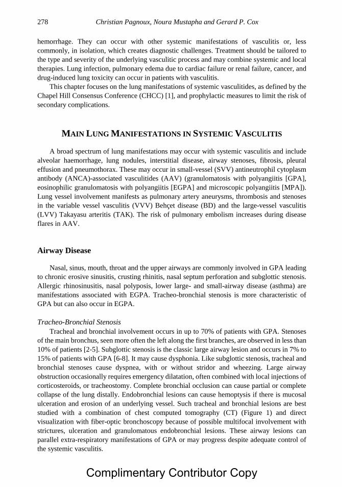
Christian Pagnoux, Noura Mustapha and Gerard P. Cox 278
hemorrhage. They can occur with other systemic manifestations of vasculitis or, less
commonly, in isolation, which creates diagnostic challenges. Treatment should be tailored to
the type and severity of the underlying vasculitic process and may combine systemic and local
therapies. Lung infection, pulmonary edema due to cardiac failure or renal failure, cancer, and
drug-induced lung toxicity can occur in patients with vasculitis.
This chapter focuses on the lung manifestations of systemic vasculitides, as defined by the
Chapel Hill Consensus Conference (CHCC) [1], and prophylactic measures to limit the risk of
secondary complications.
MAIN LUNG MANIFESTATIONS IN SYSTEMIC VASCULITIS
A broad spectrum of lung manifestations may occur with systemic vasculitis and include
alveolar haemorrhage, lung nodules, interstitial disease, airway stenoses, fibrosis, pleural
effusion and pneumothorax. These may occur in small-vessel (SVV) antineutrophil cytoplasm
antibody (ANCA)-associated vasculitides (AAV) (granulomatosis with polyangiitis [GPA],
eosinophilic granulomatosis with polyangiitis [EGPA] and microscopic polyangiitis [MPA]).
Lung vessel involvement manifests as pulmonary artery aneurysms, thrombosis and stenoses
in the variable vessel vasculitis (VVV) Behçet disease (BD) and the large-vessel vasculitis
(LVV) Takayasu arteritis (TAK). The risk of pulmonary embolism increases during disease
flares in AAV.
Airway Disease
Nasal, sinus, mouth, throat and the upper airways are commonly involved in GPA leading
to chronic erosive sinusitis, crusting rhinitis, nasal septum perforation and subglottic stenosis.
Allergic rhinosinusitis, nasal polyposis, lower large- and small-airway disease (asthma) are
manifestations associated with EGPA. Tracheo-bronchial stenosis is more characteristic of
GPA but can also occur in EGPA.
Tracheo-Bronchial Stenosis
Tracheal and bronchial involvement occurs in up to 70% of patients with GPA. Stenoses
of the main bronchus, seen more often the left along the first branches, are observed in less than
10% of patients [2-5]. Subglottic stenosis is the classic large airway lesion and occurs in 7% to
15% of patients with GPA [6-8]. It may cause dysphonia. Like subglottic stenosis, tracheal and
bronchial stenoses cause dyspnea, with or without stridor and wheezing. Large airway
obstruction occasionally requires emergency dilatation, often combined with local injections of
corticosteroids, or tracheostomy. Complete bronchial occlusion can cause partial or complete
collapse of the lung distally. Endobronchial lesions can cause hemoptysis if there is mucosal
ulceration and erosion of an underlying vessel. Such tracheal and bronchial lesions are best
studied with a combination of chest computed tomography (CT) (Figure 1) and direct
visualization with fiber-optic bronchoscopy because of possible multifocal involvement with
strictures, ulceration and granulomatous endobronchial lesions. These airway lesions can
parallel extra-respiratory manifestations of GPA or may progress despite adequate control of
the systemic vasculitis.
Complimentary Contributor Copy
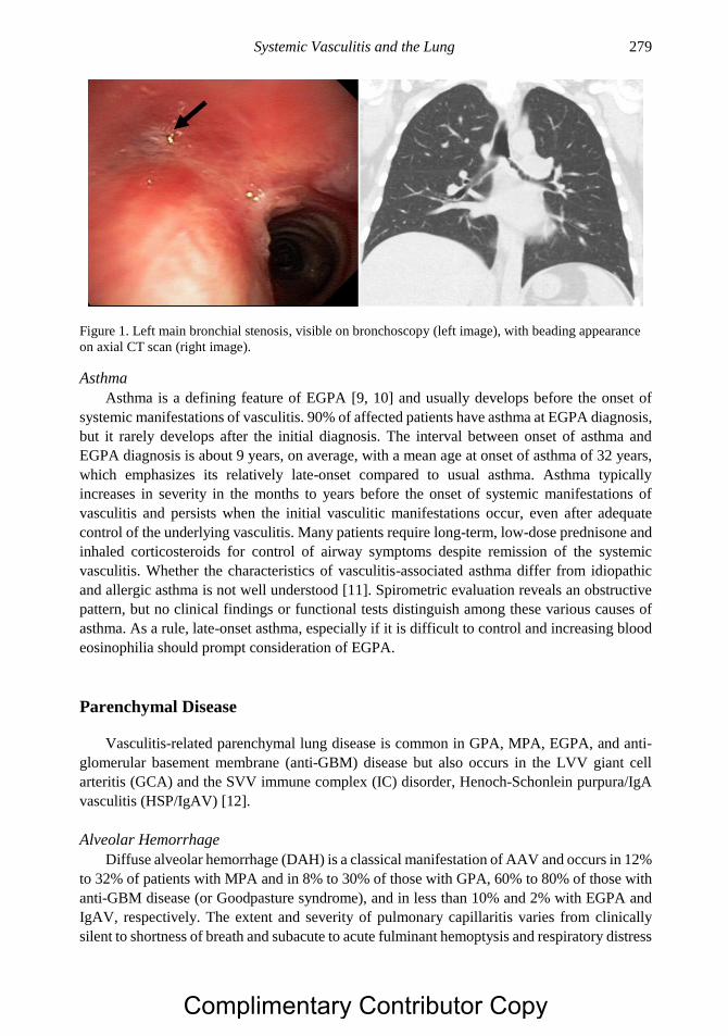
Systemic Vasculitis and the Lung 279
Figure 1. Left main bronchial stenosis, visible on bronchoscopy (left image), with beading appearance
on axial CT scan (right image).
Asthma
Asthma is a defining feature of EGPA [9, 10] and usually develops before the onset of
systemic manifestations of vasculitis. 90% of affected patients have asthma at EGPA diagnosis,
but it rarely develops after the initial diagnosis. The interval between onset of asthma and
EGPA diagnosis is about 9 years, on average, with a mean age at onset of asthma of 32 years,
which emphasizes its relatively late-onset compared to usual asthma. Asthma typically
increases in severity in the months to years before the onset of systemic manifestations of
vasculitis and persists when the initial vasculitic manifestations occur, even after adequate
control of the underlying vasculitis. Many patients require long-term, low-dose prednisone and
inhaled corticosteroids for control of airway symptoms despite remission of the systemic
vasculitis. Whether the characteristics of vasculitis-associated asthma differ from idiopathic
and allergic asthma is not well understood [11]. Spirometric evaluation reveals an obstructive
pattern, but no clinical findings or functional tests distinguish among these various causes of
asthma. As a rule, late-onset asthma, especially if it is difficult to control and increasing blood
eosinophilia should prompt consideration of EGPA.
Parenchymal Disease
Vasculitis-related parenchymal lung disease is common in GPA, MPA, EGPA, and anti-
glomerular basement membrane (anti-GBM) disease but also occurs in the LVV giant cell
arteritis (GCA) and the SVV immune complex (IC) disorder, Henoch-Schonlein purpura/IgA
vasculitis (HSP/IgAV) [12].
Alveolar Hemorrhage
Diffuse alveolar hemorrhage (DAH) is a classical manifestation of AAV and occurs in 12%
to 32% of patients with MPA and in 8% to 30% of those with GPA, 60% to 80% of those with
anti-GBM disease (or Goodpasture syndrome), and in less than 10% and 2% with EGPA and
IgAV, respectively. The extent and severity of pulmonary capillaritis varies from clinically
silent to shortness of breath and subacute to acute fulminant hemoptysis and respiratory distress
Complimentary Contributor Copy
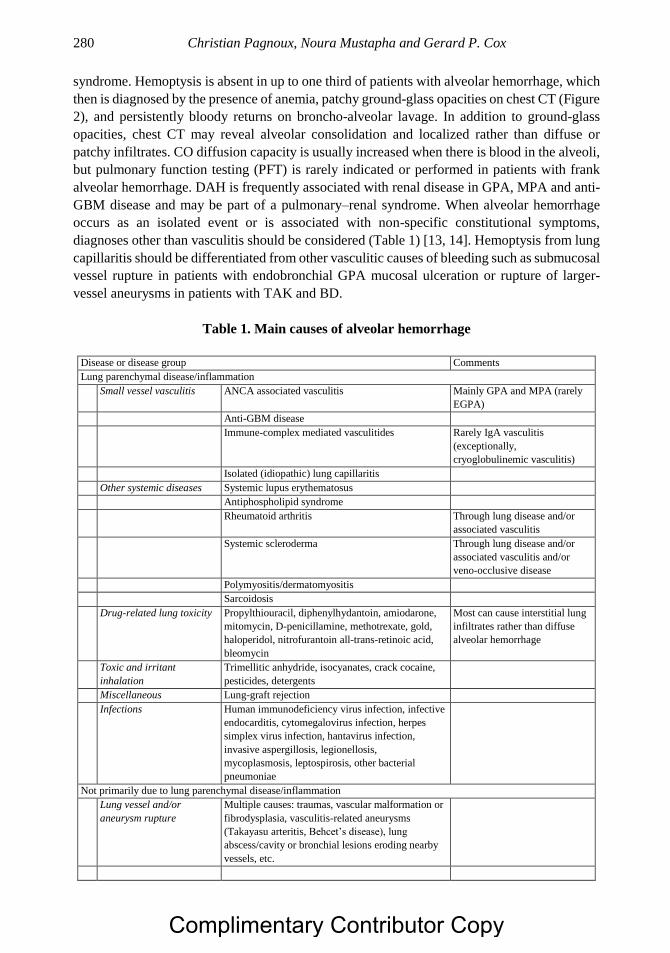
Christian Pagnoux, Noura Mustapha and Gerard P. Cox 280
syndrome. Hemoptysis is absent in up to one third of patients with alveolar hemorrhage, which
then is diagnosed by the presence of anemia, patchy ground-glass opacities on chest CT (Figure
2), and persistently bloody returns on broncho-alveolar lavage. In addition to ground-glass
opacities, chest CT may reveal alveolar consolidation and localized rather than diffuse or
patchy infiltrates. CO diffusion capacity is usually increased when there is blood in the alveoli,
but pulmonary function testing (PFT) is rarely indicated or performed in patients with frank
alveolar hemorrhage. DAH is frequently associated with renal disease in GPA, MPA and anti-
GBM disease and may be part of a pulmonary–renal syndrome. When alveolar hemorrhage
occurs as an isolated event or is associated with non-specific constitutional symptoms,
diagnoses other than vasculitis should be considered (Table 1) [13, 14]. Hemoptysis from lung
capillaritis should be differentiated from other vasculitic causes of bleeding such as submucosal
vessel rupture in patients with endobronchial GPA mucosal ulceration or rupture of larger-
vessel aneurysms in patients with TAK and BD.
Table 1. Main causes of alveolar hemorrhage
Disease or disease group Comments
Lung parenchymal disease/inflammation
Small vessel vasculitis ANCA associated vasculitis Mainly GPA and MPA (rarely
EGPA)
Anti-GBM disease
Immune-complex mediated vasculitides Rarely IgA vasculitis
(exceptionally,
cryoglobulinemic vasculitis)
Isolated (idiopathic) lung capillaritis
Other systemic diseases Systemic lupus erythematosus
Antiphospholipid syndrome
Rheumatoid arthritis Through lung disease and/or
associated vasculitis
Systemic scleroderma Through lung disease and/or
associated vasculitis and/or
veno-occlusive disease
Polymyositis/dermatomyositis
Sarcoidosis
Drug-related lung toxicity Propylthiouracil, diphenylhydantoin, amiodarone,
mitomycin, D-penicillamine, methotrexate, gold,
haloperidol, nitrofurantoin all-trans-retinoic acid,
bleomycin
Most can cause interstitial lung
infiltrates rather than diffuse
alveolar hemorrhage
Toxic and irritant
inhalation
Trimellitic anhydride, isocyanates, crack cocaine,
pesticides, detergents
Miscellaneous Lung-graft rejection
Infections Human immunodeficiency virus infection, infective
endocarditis, cytomegalovirus infection, herpes
simplex virus infection, hantavirus infection,
invasive aspergillosis, legionellosis,
mycoplasmosis, leptospirosis, other bacterial
pneumoniae
Not primarily due to lung parenchymal disease/inflammation
Lung vessel and/or
aneurysm rupture
Multiple causes: traumas, vascular malformation or
fibrodysplasia, vasculitis-related aneurysms
(Takayasu arteritis, Behcet’s disease), lung
abscess/cavity or bronchial lesions eroding nearby
vessels, etc.
Complimentary Contributor Copy
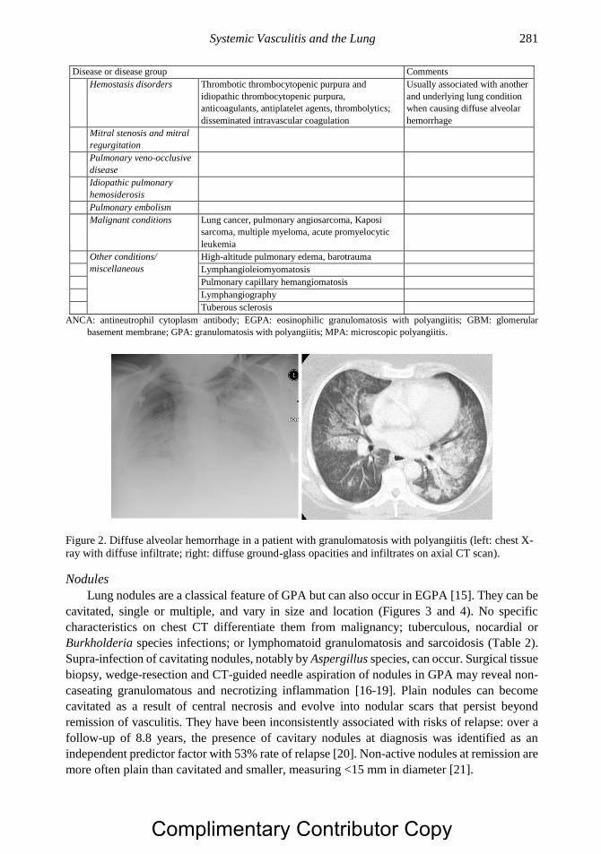
Systemic Vasculitis and the Lung 281
Disease or disease group Comments
Hemostasis disorders Thrombotic thrombocytopenic purpura and
idiopathic thrombocytopenic purpura,
anticoagulants, antiplatelet agents, thrombolytics;
disseminated intravascular coagulation
Usually associated with another
and underlying lung condition
when causing diffuse alveolar
hemorrhage
Mitral stenosis and mitral
regurgitation
Pulmonary veno-occlusive
disease
Idiopathic pulmonary
hemosiderosis
Pulmonary embolism
Malignant conditions Lung cancer, pulmonary angiosarcoma, Kaposi
sarcoma, multiple myeloma, acute promyelocytic
leukemia
Other conditions/
miscellaneous
High-altitude pulmonary edema, barotrauma
Lymphangioleiomyomatosis
Pulmonary capillary hemangiomatosis
Lymphangiography
Tuberous sclerosis
ANCA: antineutrophil cytoplasm antibody; EGPA: eosinophilic granulomatosis with polyangiitis; GBM: glomerular
basement membrane; GPA: granulomatosis with polyangiitis; MPA: microscopic polyangiitis.
Figure 2. Diffuse alveolar hemorrhage in a patient with granulomatosis with polyangiitis (left: chest X-
ray with diffuse infiltrate; right: diffuse ground-glass opacities and infiltrates on axial CT scan).
Nodules
Lung nodules are a classical feature of GPA but can also occur in EGPA [15]. They can be
cavitated, single or multiple, and vary in size and location (Figures 3 and 4). No specific
characteristics on chest CT differentiate them from malignancy; tuberculous, nocardial or
Burkholderia species infections; or lymphomatoid granulomatosis and sarcoidosis (Table 2).
Supra-infection of cavitating nodules, notably by Aspergillus species, can occur. Surgical tissue
biopsy, wedge-resection and CT-guided needle aspiration of nodules in GPA may reveal non-
caseating granulomatous and necrotizing inflammation [16-19]. Plain nodules can become
cavitated as a result of central necrosis and evolve into nodular scars that persist beyond
remission of vasculitis. They have been inconsistently associated with risks of relapse: over a
follow-up of 8.8 years, the presence of cavitary nodules at diagnosis was identified as an
independent predictor factor with 53% rate of relapse [20]. Non-active nodules at remission are
more often plain than cavitated and smaller, measuring <15 mm in diameter [21].
Complimentary Contributor Copy
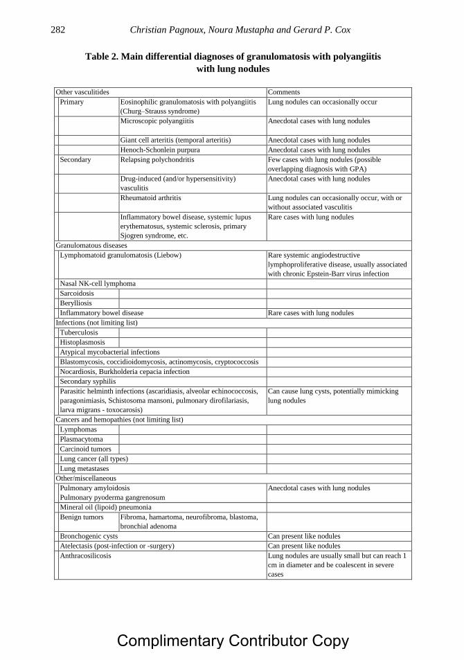
Christian Pagnoux, Noura Mustapha and Gerard P. Cox 282
Table 2. Main differential diagnoses of granulomatosis with polyangiitis
with lung nodules
Other vasculitides Comments
Primary Eosinophilic granulomatosis with polyangiitis
(Churg–Strauss syndrome)
Lung nodules can occasionally occur
Microscopic polyangiitis
Anecdotal cases with lung nodules
Giant cell arteritis (temporal arteritis) Anecdotal cases with lung nodules
Henoch-Schonlein purpura Anecdotal cases with lung nodules
Secondary Relapsing polychondritis Few cases with lung nodules (possible
overlapping diagnosis with GPA)
Drug-induced (and/or hypersensitivity)
vasculitis
Anecdotal cases with lung nodules
Rheumatoid arthritis Lung nodules can occasionally occur, with or
without associated vasculitis
Inflammatory bowel disease, systemic lupus
erythematosus, systemic sclerosis, primary
Sjogren syndrome, etc.
Rare cases with lung nodules
Granulomatous diseases
Lymphomatoid granulomatosis (Liebow) Rare systemic angiodestructive
lymphoproliferative disease, usually associated
with chronic Epstein-Barr virus infection
Nasal NK-cell lymphoma
Sarcoidosis
Berylliosis
Inflammatory bowel disease Rare cases with lung nodules
Infections (not limiting list)
Tuberculosis
Histoplasmosis
Atypical mycobacterial infections
Blastomycosis, coccidioidomycosis, actinomycosis, cryptococcosis
Nocardiosis, Burkholderia cepacia infection
Secondary syphilis
Parasitic helminth infections (ascaridiasis, alveolar echinococcosis,
paragonimiasis, Schistosoma mansoni, pulmonary dirofilariasis,
larva migrans - toxocarosis)
Can cause lung cysts, potentially mimicking
lung nodules
Cancers and hemopathies (not limiting list)
Lymphomas
Plasmacytoma
Carcinoid tumors
Lung cancer (all types)
Lung metastases
Other/miscellaneous
Pulmonary amyloidosis
Pulmonary pyoderma gangrenosum
Anecdotal cases with lung nodules
Mineral oil (lipoid) pneumonia
Benign tumors Fibroma, hamartoma, neurofibroma, blastoma,
bronchial adenoma
Bronchogenic cysts Can present like nodules
Atelectasis (post-infection or -surgery) Can present like nodules
Anthracosilicosis Lung nodules are usually small but can reach 1
cm in diameter and be coalescent in severe
cases
Complimentary Contributor Copy
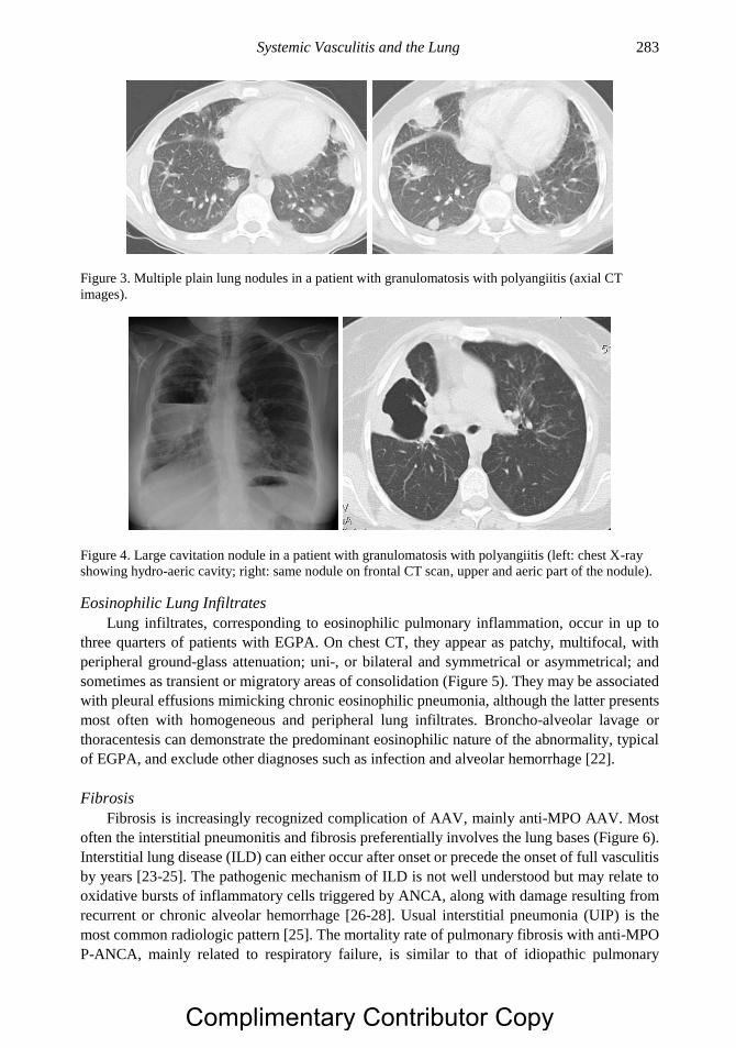
Systemic Vasculitis and the Lung 283
Figure 3. Multiple plain lung nodules in a patient with granulomatosis with polyangiitis (axial CT
images).
Figure 4. Large cavitation nodule in a patient with granulomatosis with polyangiitis (left: chest X-ray
showing hydro-aeric cavity; right: same nodule on frontal CT scan, upper and aeric part of the nodule).
Eosinophilic Lung Infiltrates
Lung infiltrates, corresponding to eosinophilic pulmonary inflammation, occur in up to
three quarters of patients with EGPA. On chest CT, they appear as patchy, multifocal, with
peripheral ground-glass attenuation; uni-, or bilateral and symmetrical or asymmetrical; and
sometimes as transient or migratory areas of consolidation (Figure 5). They may be associated
with pleural effusions mimicking chronic eosinophilic pneumonia, although the latter presents
most often with homogeneous and peripheral lung infiltrates. Broncho-alveolar lavage or
thoracentesis can demonstrate the predominant eosinophilic nature of the abnormality, typical
of EGPA, and exclude other diagnoses such as infection and alveolar hemorrhage [22].
Fibrosis
Fibrosis is increasingly recognized complication of AAV, mainly anti-MPO AAV. Most
often the interstitial pneumonitis and fibrosis preferentially involves the lung bases (Figure 6).
Interstitial lung disease (ILD) can either occur after onset or precede the onset of full vasculitis
by years [23-25]. The pathogenic mechanism of ILD is not well understood but may relate to
oxidative bursts of inflammatory cells triggered by ANCA, along with damage resulting from
recurrent or chronic alveolar hemorrhage [26-28]. Usual interstitial pneumonia (UIP) is the
most common radiologic pattern [25]. The mortality rate of pulmonary fibrosis with anti-MPO
P-ANCA, mainly related to respiratory failure, is similar to that of idiopathic pulmonary
Complimentary Contributor Copy
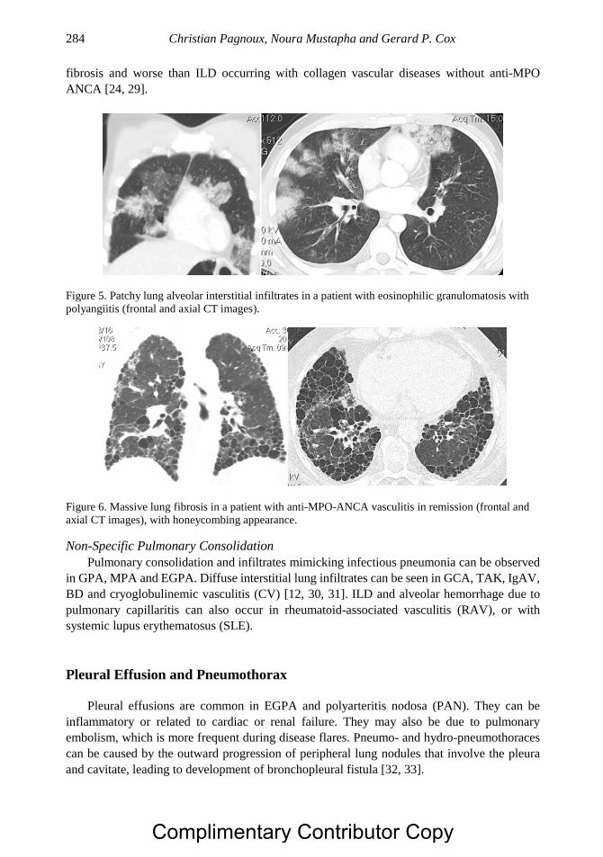
Christian Pagnoux, Noura Mustapha and Gerard P. Cox 284
fibrosis and worse than ILD occurring with collagen vascular diseases without anti-MPO
ANCA [24, 29].
Figure 5. Patchy lung alveolar interstitial infiltrates in a patient with eosinophilic granulomatosis with
polyangiitis (frontal and axial CT images).
Figure 6. Massive lung fibrosis in a patient with anti-MPO-ANCA vasculitis in remission (frontal and
axial CT images), with honeycombing appearance.
Non-Specific Pulmonary Consolidation
Pulmonary consolidation and infiltrates mimicking infectious pneumonia can be observed
in GPA, MPA and EGPA. Diffuse interstitial lung infiltrates can be seen in GCA, TAK, IgAV,
BD and cryoglobulinemic vasculitis (CV) [12, 30, 31]. ILD and alveolar hemorrhage due to
pulmonary capillaritis can also occur in rheumatoid-associated vasculitis (RAV), or with
systemic lupus erythematosus (SLE).
Pleural Effusion and Pneumothorax
Pleural effusions are common in EGPA and polyarteritis nodosa (PAN). They can be
inflammatory or related to cardiac or renal failure. They may also be due to pulmonary
embolism, which is more frequent during disease flares. Pneumo- and hydro-pneumothoraces
can be caused by the outward progression of peripheral lung nodules that involve the pleura
and cavitate, leading to development of bronchopleural fistula [32, 33].
Complimentary Contributor Copy
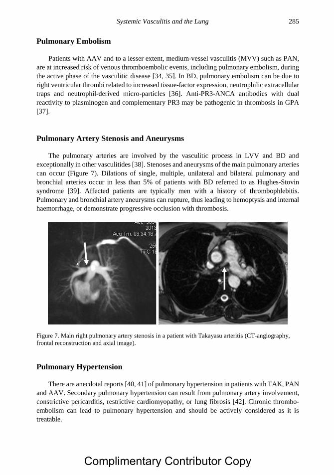
Systemic Vasculitis and the Lung 285
Pulmonary Embolism
Patients with AAV and to a lesser extent, medium-vessel vasculitis (MVV) such as PAN,
are at increased risk of venous thromboembolic events, including pulmonary embolism, during
the active phase of the vasculitic disease [34, 35]. In BD, pulmonary embolism can be due to
right ventricular thrombi related to increased tissue-factor expression, neutrophilic extracellular
traps and neutrophil-derived micro-particles [36]. Anti-PR3-ANCA antibodies with dual
reactivity to plasminogen and complementary PR3 may be pathogenic in thrombosis in GPA
[37].
Pulmonary Artery Stenosis and Aneurysms
The pulmonary arteries are involved by the vasculitic process in LVV and BD and
exceptionally in other vasculitides [38]. Stenoses and aneurysms of the main pulmonary arteries
can occur (Figure 7). Dilations of single, multiple, unilateral and bilateral pulmonary and
bronchial arteries occur in less than 5% of patients with BD referred to as Hughes-Stovin
syndrome [39]. Affected patients are typically men with a history of thrombophlebitis.
Pulmonary and bronchial artery aneurysms can rupture, thus leading to hemoptysis and internal
haemorrhage, or demonstrate progressive occlusion with thrombosis.
Figure 7. Main right pulmonary artery stenosis in a patient with Takayasu arteritis (CT-angiography,
frontal reconstruction and axial image).
Pulmonary Hypertension
There are anecdotal reports [40, 41] of pulmonary hypertension in patients with TAK, PAN
and AAV. Secondary pulmonary hypertension can result from pulmonary artery involvement,
constrictive pericarditis, restrictive cardiomyopathy, or lung fibrosis [42]. Chronic thrombo-
embolism can lead to pulmonary hypertension and should be actively considered as it is
treatable.
Complimentary Contributor Copy
Christian Pagnoux, Noura Mustapha and Gerard P. Cox 286
Phrenic Palsy
Unilateral phrenic palsy due to mono-neuritis multiplex can occur in GPA and EGPA [43,
44]. However, it can be co-incidental, related to pre-existing conditions including trauma, or
due to an unknown and unrelated cause.
LARGE-VESSEL VASCULITIDES
Parenchymal lung manifestations occur so rarely in LVV that when present, should prompt
review of the diagnosis of vasculitis or consideration of a co-existing condition. However,
involvement of pulmonary arteries can be an expected feature of LVV, especially in TAK.
Giant Cell Arteritis
One-quarter of patients with GCA have a dry, non-productive cough. Less common
manifestations include alveolar hemorrhage, T-CD4+ lymphocytic alveolitis, pleural effusion
and thickening, multiple pulmonary infarctions, lung nodules and ILD [45-48].
Takayasu Arteritis
TAK can involve the pulmonary arteries, although less frequently than the aorta or other
arteries with stenoses, with or without dilations (Figure 7). The involvement can be associated
pulmonary hypertension, lung infarction, arterial rupture and hemorrhage, regional hypo-
perfusion, and lung ventilation and perfusion mismatching suggestive of pulmonary embolism.
There may be lung nodules, parenchymal consolidation, sub-pleural reticulo-linear changes,
pleural effusion and thickening, pulmonary-renal syndrome, and ILD [49-56].
MEDIUM-VESSEL VASCULITIDES
Pulmonary artery and less often parenchymal involvement can occur in MVV.
Polyarteritis Nodosa
The lung parenchyma is not typically affected in PAN and the occurrence of capillaritis
with alveolar hemorrhage should raise doubt about the diagnosis of PAN or question the
possibility of an underlying genetic disease such as STING-associated vasculopathy [57]. Prior
to the 1994 CHCC [58], patients presenting purpura, hemoptysis, and pulmonary hemorrhage
that preceded the explosive phase of systemic necrotizing vasculitis affecting the kidney and
lungs with rapidly progressive glomerulonephritis and pulmonary capillaritis were considered
to have a microscopic form of PAN. This condition was later considered a separate category of
Complimentary Contributor Copy
Systemic Vasculitis and the Lung 287
SVV, termed MPA [59]. Pulmonary edema and pleural effusion in PAN are usually due to renal
and/or cardiac insufficiency. Patients with PAN can present with interstitial pneumonia, lung
nodules and cavitary lesions. Although clinical and angiographic bronchial artery involvement
is considered uncommon, seven of ten patients were found to have bronchial arteritis on post-
mortem examination [60-62], so this manifestation of vasculitis may be under-recognized.
Kawasaki Disease
Children with Kawasaki disease (KD) often develop respiratory symptoms due to upper-
respiratory tract inflammation. Affected children may present with pneumonia, and up to 15%
show lung consolidation on chest radiography within 10 days of illness onset. Other
radiographic abnormalities include peri-bronchial cuffing, pleural effusion, atelectasis and air
trapping [63]. The incidence of coronary artery lesions is greater in patients with abnormal
chest radiographs. Interstitial micro-nodular infiltrates and larger inflammatory pulmonary
nodules can occur [64-68].
SMALL-VESSEL VASCULITIDES
Parenchymal and airway disease occur more commonly in SVV than MVV and LVV.
Alveolar hemorrhage is the hallmark of GPA, MPA and EGPA; lung nodules and bronchial
stenosis are more characteristic of GPA, and asthma is most consistent with EGPA [69].
Granulomatosis with Polyangiitis
Overall, 70% to 100% of patients with GPA show lung involvement, with clinical
manifestations ranging from mild cough, dyspnea, chest pain, and intermittent hemoptysis to
acute respiratory distress syndrome and massive alveolar hemorrhage. In 6% of patients, lung
involvement remains asymptomatic, especially with lung nodules. Tracheobronchial
involvement, including subglottic stenosis, ulcerating tracheo-bronchitis and sometimes
endobronchial stenoses, can be seen in 17% of patients [70]. Subglottic involvement seems to
be more common in the pediatric-onset GPA compared with the adult population, occurring in
up to 41% of cases [71]. Although not specific, lung nodules are among the most characteristic
involvement, and are present in 40% to 66% of patients with GPA, occurring as ≤ 10 in number,
unilateral, bilateral, and single or multiple lesions [20]. Half of them may cavitate, especially
when larger than 2 cm [72]. The differential diagnosis of lung nodules remains wide, and should
include primary and metastatic tumors, sarcoidosis, tuberculosis and fungal infections (Table
2). Alveolar hemorrhage, the most feared manifestation, is usually secondary to capillaritis and
is noted in 18% to 30% of patients with GPA. It can occur at disease onset or later in the course
signaling relapse of the disease [20, 73]. It may appear as ground-glass opacities surrounding
pulmonary nodules. In all, 30% to 50% of patients show other pulmonary infiltrates or lung
consolidations, and 9% to 28% show pleural effusion. Spontaneous pneumothorax and
empyema can occur, sometimes related to nodule cavitation and rupture. Pulmonary embolism
Complimentary Contributor Copy
Christian Pagnoux, Noura Mustapha and Gerard P. Cox 288
may indicate active GPA and systemic inflammation. All patients with acute presentation of
GPA should undergo chest CT/HRCT, because small nodules, alveolar hemorrhage and other
lesions may be missed on chest radiography. Even when the diagnosis of GPA is clinically
apparent, broncho-alveolar lavage should be considered to exclude concurrent infection and
clinically silent alveolar hemorrhage. Surgical lung biopsies, targeting nodules and
consolidated lesions, have a diagnostic yield of 76% to 91% [3, 74]. Transbronchial biopsies
are seldom positive, unless performed on a macroscopically abnormal lesion. Transbronchial
cryobiopsy and video-assisted thoracoscopic biopsy are other possible diagnostic options [75].
Microscopic Polyangiitis
Alveolar hemorrhage and pulmonary–renal syndrome can be observed in MPA [59, 76,
77]. Arteritis with intimal thickening and focal destruction of the internal elastic lamina and
sub-intimal fibrous scarring of the media are typical histological features in diagnostic tissue
biopsy specimens. Diffuse alveolar damage and pulmonary fibrosis (Figure 6) can complicate
MPA, mainly in patients with anti-MPO P-ANCA MPA. In patients with established idiopathic
pulmonary fibrosis, MPO-ANCA positivity may be present in up to 5%. Seroconversion from
negative to positive anti-MPO during follow up can be found in up to 15% of patients [78, 79].
The risk of developing systemic vasculitis over time is greater in MPO-ANCA positive patients
than those with negative ANCA, and can be as high as 22% [78]. Additionally, pulmonary
fibrosis and vasculitis may have independent outcomes, with progression of the fibrosis despite
treatment and sustained good control of the vasculitis [24-26, 29]. There is little data on the
role of immune suppressing therapies and anti-fibrotic therapies such as pirfenidone and
nintedanib in MPO-positive IPF and MPA with pulmonary fibrosis.
Eosinophilic Granulomatosis with Polyangiitis
This rare pulmonary and systemic SVV with an annual incidence of 0.5 to 6.8 per million
and a prevalence of 10.7 to 14 per million, develops through three prototypical successive
phases. The prodromal phase begins with asthma and allergic manifestations, followed by
tissue eosinophilia affecting visceral organs such as the lung and myocardium, and later
development of vasculitic features primarily affecting the skin and peripheral nerves. The
preponderance of Th2 lymphocytes and eosinophils suggests their participation in the
pathogenesis of EGPA. However, ANCA production is noted in up to 40% of patients, which
suggests a role in amplifying or facilitating the development of vasculitic lesions. Asthma is a
predominant feature in EGPA, present in up to 91% of cases at diagnosis [9]. Lung infiltrates
were reported in 39% of patients in the largest series (and up to 72% in a smaller previous
series) [9, 80], presenting as multifocal peripheral consolidation in two-thirds of patients, with
unilateral or bilateral, and symmetrical or asymmetrical appearance [81]. A centri-lobular
perivascular dense pattern with scattered centri-lobular nodules measuring 5 mm within
ground-glass opacities is often found. Multiple large nodules may occur, with occasional
cavitation; however, alveolar haemorrhage is rare in EGPA, occurring in less than 5%. Non-
compressive mediastinal or hilar adenopathy may also be detected on chest CT. Pleural
Complimentary Contributor Copy
Systemic Vasculitis and the Lung 289
effusions are present in 9% to 30% of patients and are typically exudative and rich in
eosinophils [9].
Anti–Glomerular Basement Membrane Disease
Anti-GBM disease, which was included in the revised 2012 CHCC [1] under the SVV
category of IC diseases, typically affects young people, aged 15 to 35 years. Isolated
glomerulonephritis occurs in 20% to 40% of patients and is combined with alveolar hemorrhage
in 60% to 70% of patients. Isolated alveolar hemorrhage is a rare but described presenting
feature of anti-GBM. Alveolar hemorrhage is strongly associated with smoking [82, 83]. Up to
one third of patients with anti-GBM disease become ANCA positive at some time during their
illness. Such patients have intermediate prognosis with worse renal prognosis than those with
isolated AAV.
Other Small-Vessel Vasculitides
Alveolar hemorrhage occurs in less than 5% of patients with IgAV [30] and mixed CV.
Other lung manifestations may be due to co-incident human immunodeficiency virus (HIV) or
hepatitis C virus (HCV) infection, or a lymphoproliferative disorder.
VARIABLE VESSEL VASCULITIDES
Behçet Disease
Lung manifestations occur in less than 10% of patients with BD [84-89]. Pulmonary artery
involvement is most common after aortic involvement in young men with aneurysms and
thrombosis, with or without associated aneurysm or vascular stenosis [90, 91]. Clinically, it can
remain silent, or present with hemoptysis. Other symptoms of pulmonary artery involvement
are cough, fever, pleuritic chest pain and dyspnea [86]. Patients should be systematically
screened for aneurysms, which can be seen on chest radiographs, but are best visualized by
chest CT and magnetic resonance imaging (MRI). They can be multiple, unilateral or bilateral,
with varying diameter, usually 1 to 7 cm. Pulmonary artery involvement is associated with poor
prognosis, with mortality exceeding 25% after 7 years of follow-up. Aneurysms can rupture,
causing catastrophic hemorrhage or lead to thrombosis with later pulmonary hypertension and
lung infarction. Most patients have concomitant extra-pulmonary venous thrombi and
thrombophlebitis (Hughes-Stovin Syndrome). A pulmonary aneurysm justifies treating with
cyclophosphamide and high-dose corticosteroids. Consensus is lacking on the role of
anticoagulant and antiplatelet therapies as they may be associated with massive hemorrhage
and mortality risk. Surgery, embolization, and endovascular procedures should be considered
in combination with systemic therapy because of the complications associated with active
vasculitis. Atelectasis, nodular, or reticular opacities and cavities can occur, corresponding to
foci of pulmonary hemorrhage, infarction, and diffuse alveolar hemorrhage due to SVV.
Complimentary Contributor Copy
Christian Pagnoux, Noura Mustapha and Gerard P. Cox 290
Secondary organizing pneumonia, pleural nodules or effusions resulting from vasculitis of the
pleura or thrombosis of the superior vena cava, ulcerative lesions of the trachea and/or proximal
bronchia as well as mediastinal lymphadenopathy can also occur. Pulmonary embolism in BD
is rare and most often due to local pulmonary artery thrombosis rather than embolic peripheral
venous thrombosis.
Other Vasculitides
Lung manifestations occur in patients with lupus vasculitis, RAV, sarcoidosis, systemic
scleroderma, HCV infection, and illicit use of substances such as cocaine [92-96]. Alveolar
hemorrhage due to capillaritis in SLE is similar to what is observed in MPA. Different
pathogenic mechanisms may be involved in the development of ILD and lung nodules in RAV.
Cocaine and levamisole-tainted cocaine induce lung complications including a GPA-like
syndrome with skin, nasal sinus involvement and lung-nodules rather than alveolar
hemorrhage. ANCA-seropositivity can be present as well as ELISA findings of specificity to
PR3 and various patterns on indirect immunofluorescence [97, 98].
TREATMENT AND PROGNOSIS
Before initiating therapy for pulmonary manifestations, the treating physician should be
certain that other conditions, especially infections, cancers, and drug toxicity, are not
mimicking pulmonary vasculitis. These conditions may co-exist with active vasculitis, such as
supra- or opportunistic infection of a lung cavity with Mycobacteria and Aspergillus species or
cancer leading to paraneoplastic syndrome [99, 100]. Treatment should be individualized to the
vasculitis identified, its severity and the type of organ involvement. Although lung
manifestations in AAV do not necessarily confer poorer prognosis, alveolar hemorrhage and
tracheo-bronchial stenoses warrant aggressive management to avert excess morbidity and
mortality. It has been demonstrated that rituximab was as effective for induction therapy as
cyclophosphamide in diffuse alveolar hemorrhage in the rituximab in ANCA-Associated
Vasculitis (RAVE) trial, although patients requiring mechanical ventilation were excluded from
this trial [101]. Conversely, the preliminary results of the large, controlled PEXIVAS study
[102] suggested that plasma exchange did not improve survival in patients with severe diffuse
alveolar hemorrhage, as opposed to reports from a few small case series or case-control studies
[103]. Systemic treatment is often disappointing in subglottic and bronchial stenoses.
Endoscopic dilations with bougies, balloons and repeated local injection of corticosteroids with
or without mitomycin application may be effective. Tracheal stents and surgery should be
considered only in refractory disease. Patients with GPA and lung nodules, without any other
threatening systemic manifestations or major organ involvement, can be managed with less
toxic drugs such as methotrexate. The optimal treatment strategy for lung fibrosis and ILD
remains to be determined.
Consensus is lacking on the optimal clinical and laboratory monitoring of patients with
vasculitis and lung manifestations, but repeated chest radiographs and PFTs at 3 to 6-month
intervals earlier in the disease seem reasonable. Repeat CT, because of radiation concerns,
Complimentary Contributor Copy
Systemic Vasculitis and the Lung 291
should be reserved to resolve uncertainties or to evaluate response to treatment, complications
or new manifestations of disease. The intensity of follow-up would vary, depending on the
severity of the lung lesions and the impairment of function as well as the presence of subglottic
and bronchial stenotic lesions which require direct visualization at bronchoscopy.
CONCLUSION
Lung manifestations are common in several vasculitides and can present various features,
some of them potentially life-threatening. After exclusion of conditions mimicking vasculitis,
treatment with systemic and local therapies can be initiated with the expectation of
improvement. In the longer term, complications of treatment can be as troublesome as
complications of the disease. Identification of effective, less toxic and optimal combinations of
therapies for the most severely affected patients, and those with chronic lung complications
such as anti-MPO-ANCA–associated pulmonary fibrosis, and others with AAV will be
clarified by emerging evidence from clinical trials.
REFERENCES
[1] Jennette JC, Falk RJ, Bacon PA, Basu N, Cid MC, Ferrario F et al. 2012 revised
International Chapel Hill Consensus Conference Nomenclature of Vasculitides. Arthritis
Rheum 2013, 65(1):1-11.
[2] Holle JU, Gross WL, Latza U, Nolle B, Ambrosch P, Heller M et al. Improved outcome
in 445 patients with Wegener’s granulomatosis in a German vasculitis center over four
decades. Arthritis Rheum 2011, 63(1):257-266.
[3] Hoffman GS, Kerr GS, Leavitt RY, Hallahan CW, Lebovics RS, Travis WD et al.
Wegener granulomatosis: an analysis of 158 patients. Ann Intern Med 1992, 116(6):488-
498.
[4] Hervier B, Pagnoux C, Renaudin K, Masseau A, Pottier P, Planchon B et al.
[Endobronchial stenosis in Wegener’s granulomatosis]. Rev Med Interne 2006,
27(6):453-457.
[5] Polychronopoulos VS, Prakash UB, Golbin JM, Edell ES, Specks U: Airway
involvement in Wegener’s granulomatosis. Rheum Dis Clin North Am 2007, 33(4):755-
775, VI.
[6] Pagnoux C, Wolter NE: Vasculitis of the upper airways. Swiss Med Wkly 2012,
142:w13541.
[7] Langford CA, Sneller MC, Hallahan CW, Hoffman GS, Kammerer WA, Talar-Williams
C et al. Clinical features and therapeutic management of subglottic stenosis in patients
with Wegener’s granulomatosis. Arthritis Rheum 1996, 39(10):1754-1760.
[8] Lebovics RS, Hoffman GS, Leavitt RY, Kerr GS, Travis WD, Kammerer W et al. The
management of subglottic stenosis in patients with Wegener’s granulomatosis.
Laryngoscope 1992, 102(12 Pt 1):1341-1345.
[9] Comarmond C, Pagnoux C, Khellaf M, Cordier JF, Hamidou M, Viallard JF et al.
Eosinophilic granulomatosis with polyangiitis (Churg-Strauss): clinical characteristics
Complimentary Contributor Copy
Christian Pagnoux, Noura Mustapha and Gerard P. Cox 292
and long-term followup of the 383 patients enrolled in the French Vasculitis Study Group
cohort. Arthritis Rheum 2013, 65(1):270-281.
[10] Moosig F, Bremer JP, Hellmich B, Holle JU, Holl-Ulrich K, Laudien M et al. A vasculitis
centre based management strategy leads to improved outcome in eosinophilic
granulomatosis and polyangiitis (Churg-Strauss, EGPA): monocentric experiences in 150
patients. Annals of the rheumatic diseases 2013, 72(6):1011-1017.
[11] Cottin V, Khouatra C, Dubost R, Glerant JC, Cordier JF: Persistent airflow obstruction
in asthma of patients with Churg-Strauss syndrome and long-term follow-up. Allergy
2009, 64(4):589-595.
[12] Manganelli P, Fietta P, Carotti M, Pesci A, Salaffi F: Respiratory system involvement in
systemic vasculitides. Clinical and experimental rheumatology 2006, 24(2 Suppl
41):S48-59.
[13] Cordier JF, Cottin V: Alveolar hemorrhage in vasculitis: primary and secondary. Semin
Respir Crit Care Med 2011, 32(3):310-321.
[14] Ioachimescu OC, Stoller JK: Diffuse alveolar hemorrhage: diagnosing it and finding the
cause. Cleve Clin J Med 2008, 75(4):258, 260, 264-255 passim.
[15] Cordier JF, Valeyre D, Guillevin L, Loire R, Brechot JM: Pulmonary Wegener’s
granulomatosis. A clinical and imaging study of 77 cases. Chest 1990, 97(4):906-912.
[16] Mark EJ, Flieder DB, Matsubara O: Treated Wegener’s granulomatosis: distinctive
pathological findings in the lungs of 20 patients and what they tell us about the natural
history of the disease. Hum Pathol 1997, 28(4):450-458.
[17] Duna GF, Galperin C, Hoffman GS: Wegener’s granulomatosis. Rheum Dis Clin North
Am 1995, 21(4):949-986.
[18] Travis WD, Hoffman GS, Leavitt RY, Pass HI, Fauci AS: Surgical pathology of the lung
in Wegener’s granulomatosis. Review of 87 open lung biopsies from 67 patients. Am J
Surg Pathol 1991, 15(4):315-333.
[19] Schnabel A, Holl-Ulrich K, Dalhoff K, Reuter M, Gross WL: Efficacy of transbronchial
biopsy in pulmonary vaculitides. European Respiratory Journal 1997, 10(12):2738-
2743.
[20] Russell BA, Mohan S, Chahal R, Carette S, Pagnoux C, Canadian Vasculitis N:
Prognostic significance of cavitary lung nodules in granulomatosis with polyangiitis - A
clinical and imaging study of 225 patients. Arthritis care & research 2017.
[21] Komocsi A, Reuter M, Heller M, Murakozi H, Gross WL, Schnabel A: Active disease
and residual damage in treated Wegener’s granulomatosis: an observational study using
pulmonary high-resolution computed tomography. Eur Radiol 2003, 13(1):36-42.
[22] Wallaert B, Gosset P, Prin L, Bart F, Marquette CH, Tonnel AB: Bronchoalveolar lavage
in allergic granulomatosis and angiitis. Eur Respir J 1993, 6(3):413-417.
[23] Tzelepis GE, Kokosi M, Tzioufas A, Toya SP, Boki KA, Zormpala A et al. Prevalence
and outcome of pulmonary fibrosis in microscopic polyangiitis. Eur Respir J 2010,
36(1):116-121.
[24] Comarmond C, Crestani B, Tazi A, Hervier B, Adam-Marchand S, Nunes H et al.
Pulmonary fibrosis in antineutrophil cytoplasmic antibodies (ANCA)-associated
vasculitis: a series of 49 patients and review of the literature. Medicine (Baltimore) 2014,
93(24):340-349.
[25] Alba MA, Flores-Suarez LF, Henderson AG, Xiao H, Hu P, Nachman PH et al. Interstital
lung disease in ANCA vasculitis. Autoimmun Rev 2017, 16(7):722-729.
Complimentary Contributor Copy
Systemic Vasculitis and the Lung 293
[26] Hervier B, Pagnoux C, Agard C, Haroche J, Amoura Z, Guillevin L et al. Pulmonary
fibrosis associated with ANCA-positive vasculitides. Retrospective study of 12 cases and
review of the literature. Ann Rheum Dis 2009, 68(3):404-407.
[27] Yamada H: ANCA: associated lung fibrosis. Semin Respir Crit Care Med 2011,
32(3):322-327.
[28] Foulon G, Delaval P, Valeyre D, Wallaert B, Debray MP, Brauner M et al. ANCA-
associated lung fibrosis: analysis of 17 patients. Respir Med 2008, 102(10):1392-1398.
[29] Homma S, Matsushita H, Nakata K: Pulmonary fibrosis in myeloperoxidase
antineutrophil cytoplasmic antibody-associated vasculitides. Respirology 2004,
9(2):190-196.
[30] Nadrous HF, Yu AC, Specks U, Ryu JH: Pulmonary involvement in Henoch-Schonlein
purpura. Mayo Clinic proceedings 2004, 79(9):1151-1157.
[31] Karam GH, Fulmer JD: Giant cell arteritis presenting as interstitial lung disease. Chest
1982, 82(6):781-784.
[32] Ates A, Karaaslan Y, Yildiz G, Gunesen O, Han S: A case of Wegener’s granulomatosis
complicated with hydropneumothorax. J Clin Rheumatol 2006, 12(5):264-265.
[33] Epstein DM, Gefter WB, Miller WT, Gohel V, Bonavita JA: Spontaneous pneumothorax:
an uncommon manifestation of Wegener granulomatosis. Radiology 1980, 135(2):327-
328.
[34] Allenbach Y, Seror R, Pagnoux C, Teixeira L, Guilpain P, Guillevin L et al. High
frequency of venous thromboembolic events in Churg-Strauss syndrome, Wegener’s
granulomatosis and microscopic polyangiitis but not polyarteritis nodosa: a systematic
retrospective study on 1130 patients. Ann Rheum Dis 2009, 68(4):564-567.
[35] Merkel PA, Lo GH, Holbrook JT, Tibbs AK, Allen NB, Davis JC, Jr. et al. Brief
communication: high incidence of venous thrombotic events among patients with
Wegener granulomatosis: the Wegener’s Clinical Occurrence of Thrombosis (WeCLOT)
Study. Ann Intern Med 2005, 142(8):620-626.
[36] Kambas K, Chrysanthopoulou A, Vassilopoulos D, Apostolidou E, Skendros P, Girod A
et al. Tissue factor expression in neutrophil extracellular traps and neutrophil derived
microparticles in antineutrophil cytoplasmic antibody associated vasculitis may promote
thromboinflammation and the thrombophilic state associated with the disease. Ann
Rheum Dis 2014, 73(10):1854-1863.
[37] Bautz DJ, Preston GA, Lionaki S, Hewins P, Wolberg AS, Yang JJ et al. Antibodies with
dual reactivity to plasminogen and complementary PR3 in PR3-ANCA vasculitis.
Journal of the American Society of Nephrology : JASN 2008, 19(12):2421-2429.
[38] Le Thi Huong DL, Goes MJ, Cluzel P, Piette JC, Godeau P: Pulmonary artery stenosis
and periarteritis nodosa. Scand J Rheumatol 1996, 25(4):266-267.
[39] Bilgin G, Sungur G, Kucukterzi V: Systemic and pulmonary screening of patients with
Behcet’s disease during periodic follow-up. Respir Med 2013, 107(3):466-471.
[40] Comparato E, Toso M: [A case of polyarteritis nodosa associated with pulmonary
hypertension]. Minerva Med 1977, 68(42):2963-2970.
[41] Launay D, Souza R, Guillevin L, Hachulla E, Pouchot J, Simonneau G et al. Pulmonary
arterial hypertension in ANCA-associated vasculitis. Sarcoidosis Vasc Diffuse Lung Dis
2006, 23(3):223-228.
[42] Grant SC, Levy RD, Venning MC, Ward C, Brooks NH: Wegener’s granulomatosis and
the heart. Br Heart J 1994, 71(1):82-86.
Complimentary Contributor Copy
Christian Pagnoux, Noura Mustapha and Gerard P. Cox 294
[43] Sharma A, Bambery P, Bettadapura Shamanna S, Wanchu A, Singh S: Phrenic nerve
palsy in a patient of Churg Strauss syndrome and mononeuritis multiplex. Clin
Rheumatol 2008, 27(1):137-139.
[44] Armani M, Spinazzi M, Andrigo C, Fassina A, Mantovan M, Tavolato B: Severe
dysphagia in lower cranial nerve involvement as the initial symptom of Wegener’s
granulomatosis. J Neurol Sci 2007, 263(1-2):187-190.
[45] Kramer MR, Melzer E, Nesher G, Sonnenblick M: Pulmonary manifestations of temporal
arteritis. Eur J Respir Dis 1987, 71(5):430-433.
[46] Huong Dle T, Andreu MR, Duhaut P, Godeau P, Piette JC: Intra-alveolar haemorrhage
in temporal arteritis. Ann Rheum Dis 2003, 62(2):189-190.
[47] Carassou P, Aletti M, Cinquetti G, Banal F, Landais C, Graffin B et al. [Respiratory
manifestations of giant cell arteritis: 8 cases and review of the literature]. Presse Med
2010, 39(9):e188-196.
[48] Machado EB, Michet CJ, Ballard DJ, Hunder GG, Beard CM, Chu CP et al. Trends in
incidence and clinical presentation of temporal arteritis in Olmsted County, Minnesota,
1950-1985. Arthritis Rheum 1988, 31(6):745-749.
[49] Yang CD, Teng JL, Gu YY, Chen SL: Takayasu’s arteritis presenting with bilateral
pulmonary granulomatosis. Clin Rheumatol 2007, 26(4):612-614.
[50] Savage BJ, Gupta RK, Angle J, Okusa MD: Takayasu arteritis presenting as a pulmonary-
renal syndrome. Am J Med Sci 2003, 325(5):275-281.
[51] Haque U, Hellmann D, Traill T, Venbrux A, Stone J: Takayasu’s arteritis involving
proximal pulmonary arteries and mimicking thromboembolic disease. The Journal of
rheumatology 1999, 26(2):450-453.
[52] Nakabayashi K, Kurata N, Nangi N, Miyake H, Nagasawa T: Pulmonary artery
involvement as first manifestation in three cases of Takayasu arteritis. International
journal of cardiology 1996, 54 Suppl: S177-183.
[53] Kakkar N, Vasishta RK, Banerjee AK, Singh S, Kumar L: Pulmonary capillary
haemangiomatosis as a cause of pulmonary hypertension in Takayasu’s aortoarteritis.
Respiration 1997, 64(5):381-383.
[54] Takahashi K, Honda M, Furuse M, Yanagisawa M, Saitoh K: CT findings of pulmonary
parenchyma in Takayasu arteritis. J Comput Assist Tomogr 1996, 20(5):742-748.
[55] Ogawa Y, Hayashi K, Sakamoto I, Matsunaga N: Pulmonary arterial lesions in Takayasu
arteritis: relationship of inflammatory activity to scintigraphic findings and sequential
changes. Ann Nucl Med 1996, 10(2):219-223.
[56] Suzuki Y: Ventilation/perfusion mismatch of one lung in a patient with Takayasu’s
arteritis. Clin Nucl Med 1986, 11(10):733-734.
[57] Liu Y, Jesus AA, Marrero B, Yang D, Ramsey SE, Sanchez GAM et al. Activated STING
in a vascular and pulmonary syndrome. N Engl J Med 2014, 371(6):507-518.
[58] Jennette JC, Falk RJ, Andrassy K, Bacon PA, Churg J, Gross WL et al. Nomenclature of
Systemic Vasculitides. Arthritis & Rheumatism 1994, 37(2):187-192.
[59] Guillevin L, Durand-Gasselin B, Cevallos R, Gayraud M, Lhote F, Callard P et al.
Microscopic polyangiitis: clinical and laboratory findings in eighty-five patients.
Arthritis Rheum 1999, 42(3):421-430.
[60] Matsumoto T, Homma S, Okada M, Kuwabara N, Kira S, Hoshi T et al. The lung in
polyarteritis nodosa: a pathologic study of 10 cases. Hum Pathol 1993, 24(7):717-724.
Complimentary Contributor Copy
Systemic Vasculitis and the Lung 295
[61] Lee YJ, Park SS, Kim SY, Lee JY, Koo HK, Yoon HI: A case of systemic polyarteritis
nodosa involving bronchial artery. Sarcoidosis Vasc Diffuse Lung Dis 2010, 27(2):164-
168.
[62] Menon Y, Singh R, Cuchacovich R, Espinoza LR: Pulmonary involvement in hepatitis
B-related polyarteritis nodosa. Chest 2002, 122(4):1497-1498.
[63] Umezawa T, Saji T, Matsuo N, Odagiri K: Chest x-ray findings in the acute phase of
Kawasaki disease. Pediatr Radiol 1989, 20(1-2):48-51.
[64] De Maddi F, Cinelli R, Rigante D, Mazzarella G, Siani P: Lung parenchymal
consolidation as an uncommon presentation and cause of delayed diagnosis in atypical
Kawasaki syndrome. Rheumatol Int 2009, 29(11):1373-1376.
[65] Sengler C, Gaedicke G, Wahn U, Keitzer R: Pulmonary symptoms in Kawasaki disease.
Pediatr Infect Dis J 2004, 23(8):782-784.
[66] Uziel Y, Hashkes PJ, Kassem E, Gottesman G, Wolach B: “Unresolving pneumonia” as
the main manifestation of atypical Kawasaki disease. Arch Dis Child 2003, 88(10):940-
942.
[67] Freeman AF, Crawford SE, Finn LS, Lopez-Andreu JA, Ferrando-Monleon S, Perez-
Tamarit D et al. Inflammatory pulmonary nodules in Kawasaki disease. Pediatr
Pulmonol 2003, 36(2):102-106.
[68] Voynow JA, Schanberg L, Sporn T, Kredich D: Pulmonary complications associated with
Kawasaki disease. J Pediatr 2002, 140(6):786-787.
[69] Flores-Suarez LF, Alba MA, Mateos-Toledo H, Ruiz N: Pulmonary Involvement in
Systemic Vasculitis. Curr Rheumatol Rep 2017, 19(9):56.
[70] Daum TE, Specks U, Colby TV, Edell ES, Brutinel MW, Prakash UB et al.
Tracheobronchial involvement in Wegener’s granulomatosis. Am J Respir Crit Care Med
1995, 151(2 Pt 1):522-526.
[71] Cabral DA, Uribe AG, Benseler S, O’Neil KM, Hashkes PJ, Higgins G et al.
Classification, presentation, and initial treatment of Wegener’s granulomatosis in
childhood. Arthritis Rheum 2009, 60(11):3413-3424.
[72] Mahmoud S, Ghosh S, Farver C, Lempel J, Azok J, Renapurkar RD: Pulmonary
Vasculitis: Spectrum of Imaging Appearances. Radiol Clin North Am 2016, 54(6):1097-
1118.
[73] Terrier B, Dechartres A, Deligny C, Godmer P, Charles P, Hayem G et al.
Granulomatosis with polyangiitis according to geographic origin and ethnicity: clinical-
biological presentation and outcome in a French population. Rheumatology (Oxford)
2017, 56(3):445-450.
[74] Mark EJ, Matsubara O, Tan-Liu NS, Fienberg R: The pulmonary biopsy in the early
diagnosis of Wegener’s (pathergic) granulomatosis: a study based on 35 open lung
biopsies. Hum Pathol 1988, 19(9):1065-1071.
[75] Iftikhar IH, Alghothani L, Sardi A, Berkowitz D, Musani AI: Transbronchial Lung
Cryobiopsy and Video-assisted Thoracoscopic Lung Biopsy in the Diagnosis of Diffuse
Parenchymal Lung Disease. A Meta-analysis of Diagnostic Test Accuracy. Ann Am
Thorac Soc 2017, 14(7):1197-1211.
[76] Lauque D, Cadranel J, Lazor R, Pourrat J, Ronco P, Guillevin L et al. Microscopic
polyangiitis with alveolar hemorrhage. A study of 29 cases and review of the literature.
Groupe d’Etudes et de Recherche sur les Maladies “Orphelines” Pulmonaires
(GERM”O”P). Medicine 2000, 79(4):222-233.
Complimentary Contributor Copy
Christian Pagnoux, Noura Mustapha and Gerard P. Cox 296
[77] Collins CE, Quismorio FP, Jr.: Pulmonary involvement in microscopic polyangiitis. Curr
Opin Pulm Med 2005, 11(5):447-451.
[78] Kagiyama N, Takayanagi N, Kanauchi T, Ishiguro T, Yanagisawa T, Sugita Y:
Antineutrophil cytoplasmic antibody-positive conversion and microscopic polyangiitis
development in patients with idiopathic pulmonary fibrosis. BMJ Open Respir Res 2015,
2(1):e000058.
[79] Ando M, Miyazaki E, Ishii T, Mukai Y, Yamasue M, Fujisaki H et al. Incidence of
myeloperoxidase anti-neutrophil cytoplasmic antibody positivity and microscopic
polyangitis in the course of idiopathic pulmonary fibrosis. Respir Med 2013, 107(4):608-
615.
[80] Lanham JG, Elkon KB, Pusey CD, Hughes GR: Systemic vasculitis with asthma and
eosinophilia: a clinical approach to the Churg-Strauss syndrome. Medicine 1984,
63(2):65-81.
[81] Choi YH, Im JG, Han BK, Kim JH, Lee KY, Myoung NH: Thoracic manifestation of
Churg-Strauss syndrome: radiologic and clinical findings. Chest 2000, 117(1):117-124.
[82] Herody M, Duvic C, Noel LH, Nedelec G, Grunfeld JP: Cigarette smoking and other
inhaled toxins in anti-GBM disease. Contrib Nephrol 2000, 130:94-102.
[83] Donaghy M, Rees AJ: Cigarette smoking and lung haemorrhage in glomerulonephritis
caused by autoantibodies to glomerular basement membrane. Lancet 1983,
2(8364):1390-1393.
[84] Uzun O, Erkan L, Akpolat I, Findik S, Atici AG, Akpolat T: Pulmonary involvement in
Behcet’s disease. Respiration 2008, 75(3):310-321.
[85] Uzun O, Akpolat T, Erkan L: Pulmonary vasculitis in behcet disease: a cumulative
analysis. Chest 2005, 127(6):2243-2253.
[86] Seyahi E, Melikoglu M, Akman C, Hamuryudan V, Ozer H, Hatemi G et al. Pulmonary
artery involvement and associated lung disease in Behcet disease: a series of 47 patients.
Medicine 2012, 91(1):35-48.
[87] Hamzaoui A, Hamzaoui K: [Pulmonary complications of Behcet’s disease and
Takayasu’s arteritis]. Rev Mal Respir 2005, 22(6 Pt 1):999-1019.
[88] Erkan F, Gul A, Tasali E: Pulmonary manifestations of Behcet’s disease. Thorax 2001,
56(7):572-578.
[89] Ceylan N, Bayraktaroglu S, Erturk SM, Savas R, Alper H: Pulmonary and vascular
manifestations of Behcet disease: imaging findings. AJR Am J Roentgenol 2010,
194(2):W158-164.
[90] Erkan D, Yazici Y, Sanders A, Trost D, Yazici H: Is Hughes-Stovin syndrome Behcet’s
disease? Clinical and experimental rheumatology 2004, 22(4 Suppl 34):S64-68.
[91] Erkan F, Kiyan E, Tunaci A: Pulmonary complications of Behcet’s disease. Clin Chest
Med 2002, 23(2):493-503.
[92] Schneider F, Gruden J, Tazelaar HD, Leslie KO: Pleuropulmonary pathology in patients
with rheumatic disease. Arch Pathol Lab Med 2012, 136(10):1242-1252.
[93] Carmier D, Marchand-Adam S, Diot P, Diot E: Respiratory involvement in systemic
lupus erythematosus. Rev Mal Respir 2010, 27(8):e66-78.
[94] Marigliano B, Soriano A, Margiotta D, Vadacca M, Afeltra A: Lung involvement in
connective tissue diseases: a comprehensive review and a focus on rheumatoid arthritis.
Autoimmun Rev 2013, 12(11):1076-1084.
Complimentary Contributor Copy
Systemic Vasculitis and the Lung 297
[95] Derrett-Smith EC, Nihtyanova SI, Harvey J, Salama AD, Denton CP: Revisiting ANCA-
associated vasculitis in systemic sclerosis: clinical, serological and immunogenetic
factors. Rheumatology (Oxford) 2013, 52(10):1824-1831.
[96] Quemeneur T, Mouthon L, Cacoub P, Meyer O, Michon-Pasturel U, Vanhille P et al.
Systemic vasculitis during the course of systemic sclerosis: report of 12 cases and review
of the literature. Medicine 2013, 92(1):1-9.
[97] Restrepo CS, Carrillo JA, Martinez S, Ojeda P, Rivera AL, Hatta A: Pulmonary
complications from cocaine and cocaine-based substances: imaging manifestations.
Radiographics 2007, 27(4):941-956.
[98] Specks U: The growing complexity of the pathology associated with cocaine use. J Clin
Rheumatol 2011, 17(4):167-168.
[99] Solans-Laque R, Bosch-Gil JA, Perez-Bocanegra C, Selva-O’Callaghan A, Simeon-
Aznar CP, Vilardell-Tarres M: Paraneoplastic vasculitis in patients with solid tumors:
report of 15 cases. The Journal of rheumatology 2008, 35(2):294-304.
[100] Chemouny JM, Pagnoux C, Caudwell V, Karras A, Borie R, Guillevin L et al. ANCA-
associated diseases and lung carcinomas: a five-case series. Clin Nephrol 2014,
81(2):132-137.
[101] Stone JH, Merkel PA, Spiera R, Seo P, Langford CA, Hoffman GS et al. Rituximab
versus cyclophosphamide for ANCA-associated vasculitis. N Engl J Med 2010, 363(3):
221-232.
[102] Walsh M, Merkel PA, Peh CA, Szpirt W, Guillevin L, Pusey CD et al. Plasma exchange
and glucocorticoid dosing in the treatment of anti-neutrophil cytoplasm antibody
associated vasculitis (PEXIVAS): protocol for a randomized controlled trial. Trials 2013,
14:73.
[103] Uechi E, Okada M, Fushimi K: Effect of plasma exchange on in-hospital mortality in
patients with pulmonary hemorrhage secondary to antineutrophil cytoplasmic antibody-
associated vasculitis: A propensity-matched analysis using a nationwide administrative
database. PloS one 2018, 13(4):e0196009.
Complimentary Contributor Copy

Complimentary Contributor Copy
In: The Vasculitides. Volume 1 ISBN: 978-1-53615-133-6
Editor: David S. Younger © 2019 Nova Science Publishers, Inc.
Chapter 16
SYSTEMIC VASCULITIS AND THE KIDNEY:
ANCA-ASSOCIATED VASCULITIS AND
GLOMERULONEPHRITIS
Koyal Jain1,2,, MD, Manish Saha1,2, MD and Ronald J. Falk1-3, MD 1University of North Carolina (UNC) Kidney Center, Chapel Hill, NC, US
2Division of Nephrology and Hypertension, Department of Medicine,
UNC-Chapel Hill, Chapel Hill, NC, US 3Department of Pathology and Laboratory Medicine, UNC-Chapel Hill,
Chapel Hill, NC, US
ABSTRACT
Systemic vasculitides affecting small- and medium-sized vessels are particularly likely
to involve the kidneys. These vasculitides include microscopic polyangiitis,
granulomatosis with polyangiitis, and eosinophilic granulomatosis with polyangiitis, IgA
vasculitis, polyarteritis nodosa, and cryoglobulinemic vasculitis. The most common
culprits of kidney injury in adults are microscopic polyangiitis, granulomatosis with
polyangiitis, and eosinophilic granulomatosis with polyangiitis, which predominantly
affect small vessels and are typically associated with the presence of anti-neutrophil
cytoplasmic autoantibodies. For this reason, we focus here on anti-neutrophil cytoplasmic
autoantibody-associated vasculitides. Over the past three decades, tremendous progress has
been made through investigative efforts to understand etiologic, pathogenetic and clinical
underpinnings of these disorders. We highlight notable findings that represent key
breakthroughs in the field and proceed to the clinical presentation, diagnosis, and an up-to-
date discussion of studies related to clinical management for inducing and maintaining
remission. Finally, we review key prognostic indicators of renal outcomes and predictors
of treatment resistance and relapse. Although these diseases have been transformed from
ones that were usually fatal to ones that are now chronic with potential for long-term and
symptom-free remission, many challenges remain to decrease diagnostic delays, increase
our understanding of molecular mechanisms of genetic and environmental risk factors for
Correspondence Author’s Email: [email protected].
Complimentary Contributor Copy
Koyal Jain, Manish Saha and Ronald J. Falk 300
disease, and further refine and personalize treatment strategies that maintain remission and
minimize treatment-related morbidity.
Keywords: ANCA, vasculitis, kidney
INTRODUCTION
Many forms of systemic vasculitis directly affect the kidneys, and resultant injury can lead
to chronic kidney disease and even end-stage kidney disease. Vasculitides affecting small- and
medium-sized vessels are particularly likely to involve the kidneys. They include polyarteritis
nodosa (PAN), IgA vasculitis (IgAV, formerly Henoch-Schönlein purpura), microscopic
polyangiitis (MPA), granulomatosis with polyangiitis (GPA, formerly Wegener
granulomatosis), eosinophilic granulomatosis with polyangiitis (EGPA, formerly Churg-
Strauss syndrome), and cryoglobulinemic vasculitis. The most common causes of kidney injury
among this group are IgAV in children; and MPA, GPA, and EGPA in adults. MPA, GPA, and
EGPA affect predominantly small vessels, and are often, although not always, associated with
the presence of antineutrophil cytoplasmic autoantibodies (ANCA). Given their predilection
for kidney involvement, further discussion in this chapter will focus primarily on these ANCA-
associated vasculitides (AAV).
EPIDEMIOLOGY
Most data on the incidence and prevalence of AAV derive from studies in Caucasian
populations of European descent, although there are also emerging data from Asia and parts of
the Middle East. Disproportionate reporting in Caucasians may reflect case ascertainment bias,
availability of diagnostic resources, or high prevalence of competing diagnoses, for example,
tuberculosis in the case of granulomatous disease, rather than true difference. However, data
from multi-ethnic populations suggest that there are true differences in disease incidence based
on geographical location, ethnicity, or race, or a combination thereof. As an example, a study
of a multi-ethnic population in a Paris suburb, 28% of who were of non-European ancestry,
found that the overall prevalence of AAV, as well as PAN, in Europeans was twice that
observed in non-Europeans [1]. Similar ethnic differences are seen in New Zealand, where
Europeans appear to have a much higher annual incidence of AAV of 60.2 cases per million
individuals than the indigenous Māori population with 34.2 cases per million individuals [2].
The annual incidence in Pacific Peoples in New Zealand of 17.26 cases per million individuals
was also lower than that of Europeans.
The average incidence of AAV in Japan appears to be similar to that in the United Kingdom
(UK), with 22.6 and 21.8 cases per million individuals annually, respectively, [3] however,
with notable differences. In Japan, the vast majority (83%) of patients has MPA and is MPO-
ANCA positive. Conversely, 66% of patients in the UK have GPA, and a similar number are
proteinase 3 (PR3)-ANCA positive. Interestingly, kidney involvement was noted to be less
common in patients with GPA in Japan than in the UK. AAV is also recognized in China [4]
and India [5], as well as regions of the Middle East [6], but there are no data regarding incidence
Complimentary Contributor Copy
Systemic Vasculitis and the Kidney 301
and prevalence in these areas. In a study on distribution of ANCA types, MPO-ANCA was
more common than PR3-ANCA in the Chinese patients (OR 6.8, CI 2.6-17.8) [7]. Though
AAV is uncommon in Africans, there are rare case reports of AAV from Africa [8]. African
Americans are more often MPO-ANCA positive (71% vs. 54%, p = 0.01) and younger at
diagnosis (52 versus 57 years), when compared to Caucasians [9]. However, treatment
response, ESRD, renal relapse, and death were similar between the two groups [9]. It is
important to note that while PAN, a medium-vessel vasculitis, is not classified as an AAV; it
has historically been included in most of these data. Collectively, there is believed to be an
overall incidence of approximately 10 to 20 cases per million individuals annually in Caucasian
patients of European descent [10]. The prevalence of AAV is approximately 46 to 184 cases
per million individuals [11].
Strikingly, multiple examples exist of differences in incidence of AAV that appear to be
dependent upon geographic milieu. For example, in northern Saskatchewan, Canada, a 2.69
odds ratio (OR) of developing AAV was reported in a rural versus urban setting [12]. A study
from the Australian Capital Territory of New South Wales found the annual incidence of MPA
to be significantly higher in rural versus urban areas (13.9 vs 1.6 cases per million individuals),
with a similar trend seen in the incidence of GPA [13].
There are also multiple reports that demonstrate differences in incidence relative to latitude.
A north-to-south positive gradient was observed in the Southern Hemisphere, and a south-to-
north positive gradient is seen in the Northern Hemisphere. In other words, incidence of AAV
appears to correlate with latitudinal equatorial proximity. This association was apparent in one
study from New Zealand [2], located in the Southern Hemisphere, where an increase in
incidence of GPA was noted moving from north to south. A second study comparing incidence
of AAV in two regions of Europe, including Norwich, UK, latitude 52°N; and Lugo, Spain,
43°N, both in the Northern Hemisphere, found a higher incidence of GPA in Norwich, the more
northerly latitude, than in Lugo, even though the overall incidence of AAV was nearly identical
[14]. Interestingly, no such difference has been noted in MPA or EGPA. Similar observations
regarding incidence relative to latitude have been reported elsewhere [15]. The incidence of
AAV is similar in males and females, and the average age of disease-onset varies somewhat
within populations, but occurs predominantly in the sixth and seventh decades of life. The
relationship of disease to this demographic may be somewhat skewed since, when evaluated
retrospectively, a number of patients arguably hade evidence of disease years prior to diagnosis.
Diagnostic delays are well-substantiated in the literature [16, 17]. Furthermore, most studies of
disease do not include pediatric patients, and it is important to remain cognizant that AAV can
occur at any age [18-20].
PATHOGENESIS
Since the initial discovery of AAV and glomerulonephritis [21], incredible strides have
been made in understanding the etiology and pathogenesis of these diseases. From a
fundamental point of view, for autoimmunity to occur, there needs to be a break in self-
tolerance to autoantigens. The three major phases of autoimmunity in a susceptible individual
include initiation, propagation and resolution [22]. Genetic susceptibility and environmental
factors including many infections are common initial triggers. Molecular mimicry and
Complimentary Contributor Copy
Koyal Jain, Manish Saha and Ronald J. Falk 302
autoantigen complementary to a bacterial peptide are some of the mechanisms responsible for
origination of ANCA production. HLA can bind peptides, from self- or non-self-antigens and
present it to T cells for initiation of immune activation. Among other genetics associations, a
strong link has been found between HLA genetic regions and AAV [24].
Along with continuous antigen exposure, a defect in regulatory machinery of immune
response, T regulatory cells (Tregs) and B regulatory cells results in propagation of the disease
process. Although the mechanism still needs to be defined, multiple studies have shown
involvement of persistently activated T cell effector arm of the adaptive immune system [23].
A balance between Tregs/effector cells is essential to prevent autoimmunity. FOXP3 is a major
transcriptional factor for CD4+ Tregs; in active AAV, these Tregs, due to use of a splice variant
of FOXP3, are unable to suppress effector CD4+ T cells, thus playing an important role in the
propagation of autoimmunity [25]. Also, B cells with regulatory function (with high CD5
expression) are decreased in active AAV and increased during remission [26].
Various in vitro observations have established that ANCA can bind to cytokine-primed
neutrophils, resulting in its degranulation, respiratory burst and efflux of the proteinases. In
addition, upon activation, neutrophils can release proinflammatory cytokines, resulting in
augmentation of inflammatory cascade. Both MPO and PR3 are cytoplasmic antigens, priming
with cytokine results in its expression on cell surfaces. ANCA bind to FcR expressed on
neutrophil and F(ab)2 segment binds to MPO/PR3 antigen on the cell surface leading to
neutrophil activation [27-29].
The pathogenicity of IgG ANCA comes from different observations, clinical, in vitro and
from animal models. The majority of patients with pauciimmune necrotizing crescentic
glomerulonephritis have positive ANCA serology, but its titer does not always correlate with
disease severity, remission or relapse. The first true clinical record depicting the pathogenicity
comes from a case of transplacental passive transfer of anti-MPO IgG from a mother with MPA
to her neonate, resulting in pulmonary hemorrhage and renal dysfunction [30]. Both clinical
hints and in vitro results are only suggestive, but to prove the pathogenicity of IgG ANCA, one
strives to fulfill Koch’s postulates as established by our group in an animal model: (1)
immunization of MPO knockout mice MPO-/- with purified mouse MPO in complete Freund’s
adjuvant resulting in development of anti-MPO antibody; (2) anti-MPO splenocytes containing
B and T lymphocytes; (3) injection anti-MPO IgG causes pauci-immune crescentic
glomerulonephritis in immune deficient Rag2-/- mice and also wild type mice; and (4) adoptive
transfer of anti-MPO splenocytes into Rag2-/- resulted in a severe phenotype of vasculitis in a
dose-dependent manner [31]. This demonstrates that anti-MPO antibody itself is sufficient to
cause disease, but the severe phenotype seen in the adoptive transfer model is possibly due to
synergistic effects of anti-MPO T lymphocytes. Studies on bone marrow transplantation
approach showed that bone marrow-derived cells, neutrophils and monocytes were necessary
and sufficient for induction of disease by anti-MPO antibody [32]. Systemic administration of
bacterial LPS in an animal model caused enhanced circulating TNF-alpha resulting in severe
phenotype of crescentic glomerulonephritis induced by IgG MPO [33]. In addition, due to
unknown factors, there is evidence of activation of alternate complement pathway, priming of
neutrophils by C5a by binding to C5aR results in augmentation of inflammatory pathways [34].
The observations from in vivo and in vitro studies stems the therapeutic tree in AAV, from use
of cyclophosphamide, which suppresses both B cell and T cell effector function, to more
specific B cell therapy with anti-CD20 monoclonal antibody.
Complimentary Contributor Copy
Systemic Vasculitis and the Kidney 303
Silica
Noted geographical differences in incidence may imply that environmental and other geo-
locational triggers play a role in disease development. Silica has been implicated as one such
trigger. The first account of a possible association of silica with autoimmune disease was a
1914 report of systemic sclerosis occurring in Scottish stonemasons [35]. There have since been
a number of studies correlating silica exposure with the development of other autoimmune
diseases, particularly rheumatoid arthritis (RA) [36], but also systemic lupus erythematosus
(SLE) [37-39]. Data regarding the latter are conflicting [40], but differences in results may
reflect dissimilarities in study methodology and patient characteristics. Data on the contribution
of silica to the development of AAV have been inconsistent. A study of 2,288 patients from
Sweden found no statistically significant association between an occupation and development
of GPA, although borderline associations were seen in miners, paper workers, bakers, and
animal keepers [41]. However, a meta-analysis of six case-control studies examining silica
exposure and risk of AAV found an overall significant summary effect of silica exposure
(summary OR 2.56; 95% confidence interval [CI] 1.51-4.36) with development of AAV [42].
This summary OR was similar to the OR seen in studies looking exclusively at either MPA
(OR 3.95, 95% CI 1.89-8.24) or GPA (OR 3.56, 95% CI 1.85-8.82).
Difficulties in comparing studies like these have been due largely to study heterogeneity,
with studies using different methods for ascertaining silica exposure. Negative studies have
been quite large numbering up to 2,288 patients, whereas positive studies have generally been
small, ranging from 16 to 129 patients, with variable inclusion criteria. For example, one study
included only men [43], while others differed in the age range of patients included. All but one
study [44] included only patients with glomerulonephritis, with or without other organ system
involvement. Determination of ANCA seropositivity by immunofluorescence (IF) enzyme-
linked immunosorbant assay (ELISA) or a combination thereof varies in the literature with one
study including 3 patients who were ANCA seronegative [45]. The duration of exposure to
potential silica-containing compounds, as well as the time from exposure to clinically-evident
disease has varied. However, the degree of silica exposure, which included both duration and
intensity of exposure, was found to be significant in a case control study of biopsy proven
glomerulonephritis caused by AAV [46]. High lifetime silica exposure was associated with
development of AAV when compared to none (OR 1.9, 95% CI 1.0-3.5, P = 0.05). No study of
silica exposure included patients with pulmonary- or otolaryngologic-limited disease, even
though exposure is likely through inhalation of crystalline particulates. However, in terms of
exposure risk, the study by Hogan and coworkers [40] did not find significant differences in
patients with and without pulmonary involvement. The geographical region of either study
could have contributed to the variable results.
Despite data inconsistencies, silica is generally accepted as having a potential role in the
development and perpetuation of AAV. Mechanisms by which silica may promote
autoimmunity have not yet been fully elucidated. One study of silica-exposed workers in Brazil
[47] found increased levels of soluble interleukin-2 receptor (sIL-2R), thought to reflect
increased immune activation in these individuals. However, investigators did not specifically
consider the role of silica in autoimmunity. Another study of slate miners demonstrated
increased and chronic T-cell activation in patients with silicosis either with or without
autoimmune disease, including one with pauci-immune glomerulonephritis [48], and found
proliferation of peripheral blood mononuclear cells (PBMC) to be 45% higher in cell cultures
Complimentary Contributor Copy
Koyal Jain, Manish Saha and Ronald J. Falk 304
of silica-exposed patients than in healthy controls. Patients had elevated levels of both pro-
inflammatory (interleukin [IL]-1β, IL-6, tumor necrosis factor [TNF]-α, interferon [IFN]-γ) and
anti-inflammatory (IL-10, tumor growth factor [TGF]-γ) cytokines. Chronic T-cell activation
was observed in cultured PBMC of silicosis patients in Japan [49]. In particular, activation of
T responder cells (Tresp) resulted in later entry into the peripheral CD4+25+ population,
whereas activation of FoxP3+ regulatory T-cells (Treg) led to more rapid and higher levels of
apoptosis. The net effect was reduced inhibitory function. Silica may also contribute to immune
dysregulation by increasing oxidative stress, as suggested by a study of denim sandblasting
workers with silicosis in Turkey [50]. Patients had a 1.4-fold higher level of superoxide
dismutase (SOD), a possible marker for increased reactive oxygen species generation in these
patients [50]. Another study showed decreased L-selectin expression in workers with >16 years
of occupational silica exposure, and associations with inflammatory and oxidative stress
biomarkers [51]. More recently, neutrophil extracellular traps (NETs) have been implicated in
the pathogenesis of AAV [52, 53]. These NETs are involved in trapping silica and have been
postulated to trigger autoantibody production by exposing neoantigens [54].
Taken together, these data suggest a somewhat generalized disruption in immune
homeostasis in silica-exposed individuals. However, studies to date have not specifically
addressed the mechanisms by which silica may cause or promote disease in AAV. By
extrapolation, silica is more likely to have an adjuvant effect in patients with predisposition to
AAV rather than to be a primary trigger.
Infection
Wegener considered infection as a potential factor in the cause of GPA in the first half of
the 20th century [55]. Since then Staphylococcus (S.) aureus has received particular attention.
The first study comparing the rates of GPA relapse with carriage of S. aureus found that 36 of
57 (63%) patients were chronic carriers of S. aureus, and analysis demonstrated an adjusted
relative risk (RR) for relapse of 7.16 (95% CI 1.63-31.50) independent of other factors [56]. A
2008 study of Polish patients with limited disease [57] found that 17 of 28 (60%) patients were
chronic nasal carriers of S. aureus, independently conferring a relative hazard risk (HR) for
relapse of 4.56 (CI 2.45-7.65). A third study compared nasal carriage of S. aureus in patients
with GPA to those with rheumatoid arthritis (RA) and chronic rhinosinusitis with nasal polyps
(CRS), as well as to hospital staff and subjects without regular exposure to a hospital
environment, noting that 72% of patients with GPA had S. aureus nasal colonization, versus
28% of those with CRS, and 25% of patients without regular hospital exposure [58]. Patients
with RA and hospital workers had S. aureus carriage rates of 46% and 58% respectively, but
the differences were statistically insignificant. Colonized GPA patients had significantly higher
rates of endoscopically-proven endonasal activity, with more frequent upper respiratory tract
involvement at first presentation, and with significantly higher relapse rates compared to
uncolonized patients. The use of prophylactic antibiotics, such as prophylactic
trimethoprim/sulfamethoxazole, to reduce the relapse risk by decreasing carrier rates is
controversial [59-61]. In a 2017 study of AAV [59], the relapse rates for GPA were higher for
chronic nasal carriers of S. aureus, and prophylactic trimethoprim/
sulfamethoxazole reduced the number of carriers (OR 0.19, 95% CI 0.04-0.91, P = 0.04).
Complimentary Contributor Copy
Systemic Vasculitis and the Kidney 305
However, in this study, prophylactic trimethoprim/sulfamethoxazole itself did not reduce the
relapse risk (OR 0.71, 95% CI 0.36-1.41, P = 0.33).
The mechanism(s) by which infection may incite disease has not been fully elucidated, but
early investigations have yielded intriguing possibilities. Molecular mimicry, which leads to
cross-reactivity of antibodies to both pathogen-derived and self-antigens with substantial
structural homology, has been implicated as one of the etiopathogenic factors in AAV. Such
cross-reactivity has been observed in several other diseases; streptococcal M proteins and
cardiac myosin, as well as other proteins, cross-react in rheumatic fever [62, 63];
Campylobacter jejuni epitopes and peripheral gangliosides cross-react in Guillain-Barré
syndrome [64]; and Epstein Barr virus epitopes and Mycobacterium avium paratuberculosis
cross-react with myelin basic protein in multiple sclerosis [65]. A peptide or protein
homologous to complementary PR3 (cPR3), transcribed and translated from the antisense
strand of PRTN3 DNA, is thought to be an inciting antigen in a subset of patients with PR3-
ANCA disease. In those who are predisposed, antibodies are generated to cPR3. Anti-idiotypic
antibodies, or antibodies to anti-cPR3 antibodies, are then produced that are reactive to PR3
[66, 67]. Complementary PR3 shows homology with proteins of multiple microorganisms,
including two proteins found in S. aureus, thus suggesting a role for molecular mimicry in the
pathogenesis of AAV. While PR3-ANCA and MPO-ANCA patients may have the same type
of S. aureus as the general population, they do have different distributions of clonal complexes
[68]. Given recent reports of S. aureus proteins binding MPO and/or PR3 antigens, it is
postulated [61, 69] that this bacterial protein-autoantigen complex is then internalized and
processed by B-cells and then presented to T-cells. This could account for antibody production
by B-cells and role of T-cells in ANCA.
Although it remains highly controversial, lysosome-associated membrane protein 2
(LAMP-2) has been identified as a possible antigenic target in patients with AAV [70]. The
P41-49 epitope of LAMP-2 reportedly has significant homology with an epitope of FimH, having
in common 8 of 9 amino acids [71], but these findings were not subsequently confirmed [72].
Illicit and Prescription Drugs
Both illicit and prescription drugs have been implicated in the cause of AAV, but the
etiopathogenic mechanisms area not well understood. The ant-helminthic agent levamisole,
sometimes found in cocaine, is the most recognized drug cause of vasculitis. Affected patients
[73, 74] are typically MPO-ANCA positive, but may have dual positivity with MPO- and PR3-
ANCA [73, 74], and typically present with cutaneous disease. While cutaneous disease in
idiopathic AAV typically manifests as recurring crops of purpuric lower extremity lesions, the
lesions associated with levamisole are more widespread and variable in character, with necrotic
lesions involving the ears, and bullous purpura in any location. Full-thickness, large necrotic
lesions are suggestive of levamisole exposure. Affected patients can have arthralgia,
particularly of the large joints, and constitutional symptoms. Severe disease with isolated
glomerulonephritis and pulmonary-renal syndrome occurs. Patients with signs and symptoms
of AAV should be questioned regarding illicit drug use, particularly when dual MPO-ANCA
and PR3-ANCA serology is present, and urine toxicology screen for the presence of cocaine
and levamisole should be obtained.
Complimentary Contributor Copy
Koyal Jain, Manish Saha and Ronald J. Falk 306
The commonest prescription drug offenders include hydralazine and propylthiouracil
(PTU), although penicillamine, minocycline, sulfasalazine, allopurinol, carbimazole,
methimazole, and gold have all been implicated [75, 76]. Unlike levamisole-induced disease,
the presentation of other drug-induced forms of disease mirrors idiopathic disease, with rapidly
progressive glomerulonephritis being especially common. Serologically, patients generally
have MPO antibodies at high titers, and dual positivity to MPO and PR3 is rare. Affected
patients tend to be positive for other autoantibodies, including antinuclear antibody (ANA),
anti-double-stranded DNA (anti-dsDNA), anti-histone, anti-lactoferrin antibodies, and other
forms of ANCA, specifically anti-human neutrophil elastase (HNE). Propylthiouracil typically
causes a pauci-immune necrotizing and crescentic glomerulo-nephritis, but other organ systems
may be involved. While patients with PTU-induced disease tend to have high titers of MPO-
ANCA, the ANA is not usually positive. In both illicit and prescription drug-induced AAV,
cessation of the causative agent is imperative, and management is similar to that of the
idiopathic forms of disease, employing immunosuppressive medication, as well as
plasmapheresis and hemodialysis as indicated by disease severity.
Genetic Associations
The ability to sequence DNA from humans is becoming more and more facile, and
investigators have taken full advantage of this to understand the genetic underpinnings of AAV,
uncovering disease associations with both MHC and non-MHC genes [24]. Genome-wide
association studies (GWAS) of patients and healthy controls of European descent revealed an
association of PR3-ANCA positive AAV with HLA-DP, SERPINA1 (encoding α-1 antitrypsin),
and PRTN3 (encoding PR3), whereas MPO-ANCA positive disease was associated with HLA-
DQ [77, 78]. The largest risk was seen with single‐nucleotide polymorphism (SNP) variants
rs141530233 and rs1042169 at the HLA–DPB1 locus [78]. Among non-MHC genes, an
association were demonstrated with CTLA4, which encodes a T-cell surface molecule (CTLA-
4) that down-regulates T-cell activation [77]. Two CTLA4 single nucleotide polymorphisms
(SNP), +49 G [51-53] and CT60 [77, 79, 80], have been implicated in several studies. All of
the studies identifying disease association with the CTLA4 SNP were performed in European
cohorts comprising only Caucasian patients. No such association with CTLA4 was found in
Japanese patients with AAV [81]. The SNP rs3087243 of the CTLA4 gene [80], which encodes
an inhibitory regulator of T-cell activation, and rs2476601 of the PTPN22 gene [80, 82, 83],
which encodes a regulator of cytoplasmic tyrosine kinase and T-cell function, are both
associated with AAV in Caucasian European cohorts [80, 81, 83]. Mutations in these genes
have been implicated in other autoimmune diseases, suggesting a predisposition to general
immune dysregulation [80, 82].
Racial and geographic variability may reflect differences in genetic predisposition. AAV
has been rarely described in Africa [8], and it is notably rare in African Americans. An
association of the HLA-DRB1*15 alleles with PR3-ANCA positive disease has been found,
conferring a 73.3-fold higher risk in African American patients than in community-based
controls [84]. These alleles were also associated with PR3-ANCA positive disease in
Caucasians, but with an odds ratio of only 2.2. The DRB1*1501 allelic variant, which is of
Complimentary Contributor Copy
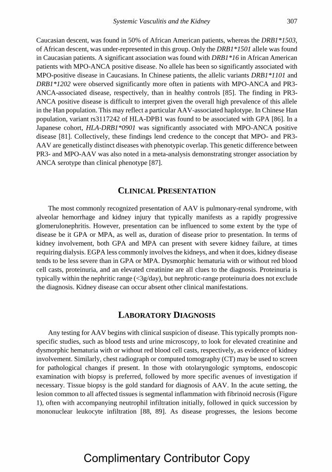
Systemic Vasculitis and the Kidney 307
Caucasian descent, was found in 50% of African American patients, whereas the DRB1*1503,
of African descent, was under-represented in this group. Only the DRB1*1501 allele was found
in Caucasian patients. A significant association was found with DRB1*16 in African American
patients with MPO-ANCA positive disease. No allele has been so significantly associated with
MPO-positive disease in Caucasians. In Chinese patients, the allelic variants DRB1*1101 and
DRB1*1202 were observed significantly more often in patients with MPO-ANCA and PR3-
ANCA-associated disease, respectively, than in healthy controls [85]. The finding in PR3-
ANCA positive disease is difficult to interpret given the overall high prevalence of this allele
in the Han population. This may reflect a particular AAV-associated haplotype. In Chinese Han
population, variant rs3117242 of HLA-DPB1 was found to be associated with GPA [86]. In a
Japanese cohort, HLA-DRB1*0901 was significantly associated with MPO-ANCA positive
disease [81]. Collectively, these findings lend credence to the concept that MPO- and PR3-
AAV are genetically distinct diseases with phenotypic overlap. This genetic difference between
PR3- and MPO-AAV was also noted in a meta-analysis demonstrating stronger association by
ANCA serotype than clinical phenotype [87].
CLINICAL PRESENTATION
The most commonly recognized presentation of AAV is pulmonary-renal syndrome, with
alveolar hemorrhage and kidney injury that typically manifests as a rapidly progressive
glomerulonephritis. However, presentation can be influenced to some extent by the type of
disease be it GPA or MPA, as well as, duration of disease prior to presentation. In terms of
kidney involvement, both GPA and MPA can present with severe kidney failure, at times
requiring dialysis. EGPA less commonly involves the kidneys, and when it does, kidney disease
tends to be less severe than in GPA or MPA. Dysmorphic hematuria with or without red blood
cell casts, proteinuria, and an elevated creatinine are all clues to the diagnosis. Proteinuria is
typically within the nephritic range (<3g/day), but nephrotic-range proteinuria does not exclude
the diagnosis. Kidney disease can occur absent other clinical manifestations.
LABORATORY DIAGNOSIS
Any testing for AAV begins with clinical suspicion of disease. This typically prompts non-
specific studies, such as blood tests and urine microscopy, to look for elevated creatinine and
dysmorphic hematuria with or without red blood cell casts, respectively, as evidence of kidney
involvement. Similarly, chest radiograph or computed tomography (CT) may be used to screen
for pathological changes if present. In those with otolaryngologic symptoms, endoscopic
examination with biopsy is preferred, followed by more specific avenues of investigation if
necessary. Tissue biopsy is the gold standard for diagnosis of AAV. In the acute setting, the
lesion common to all affected tissues is segmental inflammation with fibrinoid necrosis (Figure
1), often with accompanying neutrophil infiltration initially, followed in quick succession by
mononuclear leukocyte infiltration [88, 89]. As disease progresses, the lesions become
Complimentary Contributor Copy
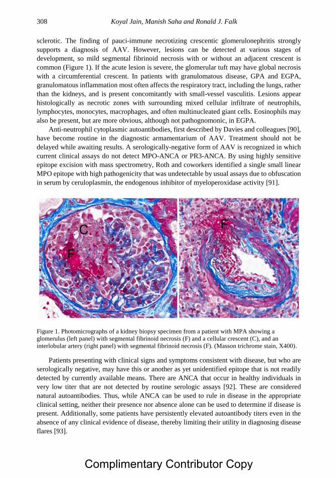
Koyal Jain, Manish Saha and Ronald J. Falk 308
sclerotic. The finding of pauci-immune necrotizing crescentic glomerulonephritis strongly
supports a diagnosis of AAV. However, lesions can be detected at various stages of
development, so mild segmental fibrinoid necrosis with or without an adjacent crescent is
common (Figure 1). If the acute lesion is severe, the glomerular tuft may have global necrosis
with a circumferential crescent. In patients with granulomatous disease, GPA and EGPA,
granulomatous inflammation most often affects the respiratory tract, including the lungs, rather
than the kidneys, and is present concomitantly with small-vessel vasculitis. Lesions appear
histologically as necrotic zones with surrounding mixed cellular infiltrate of neutrophils,
lymphocytes, monocytes, macrophages, and often multinucleated giant cells. Eosinophils may
also be present, but are more obvious, although not pathognomonic, in EGPA.
Anti-neutrophil cytoplasmic autoantibodies, first described by Davies and colleagues [90],
have become routine in the diagnostic armamentarium of AAV. Treatment should not be
delayed while awaiting results. A serologically-negative form of AAV is recognized in which
current clinical assays do not detect MPO-ANCA or PR3-ANCA. By using highly sensitive
epitope excision with mass spectrometry, Roth and coworkers identified a single small linear
MPO epitope with high pathogenicity that was undetectable by usual assays due to obfuscation
in serum by ceruloplasmin, the endogenous inhibitor of myeloperoxidase activity [91].
Figure 1. Photomicrographs of a kidney biopsy specimen from a patient with MPA showing a
glomerulus (left panel) with segmental fibrinoid necrosis (F) and a cellular crescent (C), and an
interlobular artery (right panel) with segmental fibrinoid necrosis (F). (Masson trichrome stain, X400).
Patients presenting with clinical signs and symptoms consistent with disease, but who are
serologically negative, may have this or another as yet unidentified epitope that is not readily
detected by currently available means. There are ANCA that occur in healthy individuals in
very low titer that are not detected by routine serologic assays [92]. These are considered
natural autoantibodies. Thus, while ANCA can be used to rule in disease in the appropriate
clinical setting, neither their presence nor absence alone can be used to determine if disease is
present. Additionally, some patients have persistently elevated autoantibody titers even in the
absence of any clinical evidence of disease, thereby limiting their utility in diagnosing disease
flares [93].
Complimentary Contributor Copy
Systemic Vasculitis and the Kidney 309
MANAGEMENT
Treatment of AAV can be considered in three phases: remission induction, remission
maintenance, and treatment of relapse. Over the past decade, there has been an explosion of
investigation into identifying ideal induction and maintenance strategies for patients with AAV.
Cyclophosphamide with corticosteroids has been used historically as induction therapy for
AAV. Cyclophosphamide, an oxazaphosphorine that is chemically related to the nitrogen
mustard family of alkylating agents [94], is the mainstay of AAV treatment. Multiple animal
models have demonstrated its immunosuppressant activity apart from its antimitotic or
cytotoxic characteristic [95, 96].
Although there is no randomized trial studying the optimum dose with glucocorticoids, the
question of oral versus pulse intravenous (IV) cyclophosphamide to reduce the cumulative
exposure to cyclophosphamide has been looked at. The CYCLOPS trial compared pulse iv
versus daily oral cyclophosphamide in 149 patients with AAV and found no difference in time
to remission, but a lower rate of leukopenia in the pulse group (HR 0.41, CI 0.23-0.71) [97].
Though the relapse rate was higher in the IV group in a long term follow up study, the mortality
and renal function was similar [98]. The cumulative dose of cyclophosphamide should be kept
in mind when choosing a regimen.
More recently, great strides have been made using rituximab-induced B-cell depletion for
induction of remission. Rituximab, a chimeric murine/human monoclonal antibody that
depletes B-cells by binding CD20 and inducing antibody-dependent cellular cytotoxicity
(ADCC), was reported to be non-inferior to cyclophosphamide for induction therapy in GPA
and MPA in short- [99, 100] and long-term [101] follow-up. Rituximab is approved by the
Food and Drug Administration and European Medicines Agency for induction therapy in
patients with GPA and MPA.
Plasmapheresis has been used in AAV patients with diffuse alveolar hemorrhage and
severe renal disease [102, 103]. The MEPEX trial randomized 137 patients with AAV and
serum creatinine > 5.8 mg/dL to receive plasma exchange or intravenous prednisolone, in
addition to cyclophosphamide and oral prednisolone. Plasma exchange reduced risk of end
stage kidney disease at 3 and 12 months [103]. A randomized controlled trial (PEXIVAS) of
plasma exchange and glucocorticoid dosing was conducted in 704 AAV by Walsh and
colleagues, the preliminary unpublished results of which were presented at the 2018 meeting
of the European Renal Association – European Dialysis and Transplant Association (ERA-
EDTA) suggesting that plasma exchange was not beneficial in preventing death or progression
to ESRD.
B-cell depletion to durably maintain remission is a logical next step, and there are
numerous case reports, case series, and retrospective studies that suggest efficacy of this
strategy [104-106]. The Maintenance of Remission using Rituximab in Systemic ANCA-
associated Vasculitis trial (MAINRITSAN), compared low dose rituximab with azathioprine
for remission maintenance in 115 AAV patients. Although rituximab was superior to
azathioprine in preventing relapse, azathioprine was tapered and stopped earlier, which limits
the results of the study [107]. Currently, the RITAZAREM trial (NCT01697267) in the United
States is underway to also address this question of remission maintenance.
Complete depletion of peripheral B-cells cannot continue indefinitely if toxicities
associated with immunosuppression are to be avoided. Targeted depletion of only B-cells with
Complimentary Contributor Copy
Koyal Jain, Manish Saha and Ronald J. Falk 310
the future capacity to produce pathogenic ANCA would be ideal. To this end, there are data
suggesting that patients with active disease have a lower percentage of CD5+ B-cells compared
to patients in remission, and that rituximab-exposed patients who have a robust reconstitution
of CD5+ B-cells after therapy require less immunosuppression [108].
Novel treatment strategies aimed at other components of the inflammatory process, such
as alternative complement pathway activation, are also needed. Alternative complement
activation pathway products in kidney tissue (Bb) and urine (Bb, C3a, C5a, and soluble C5b-
9) of patients with active disease correlate with severity of kidney injury [109]. In the mouse
model of MPO-ANCA glomerulonephritis, blockade of the C5a receptor (C5aR) using an oral
antagonist ameliorates disease in mice expressing human C5aR [110]. At present, a randomized
phase 3 clinical trial (ADVOCATE) is underway to evaluate the safety and efficacy of C5aR
blockade to induce remission in patients with ANCA vasculitis treated concomitantly with
rituximab or cyclophosphamide/azathioprine (NCT02994927).
Mepolizumab is an anti-interleukin-5 monoclonal antibody that reduces eosinophilic
inflammation, and has been successfully used to prevent exacerbations of eosinophilic asthma
[111]. In a study of 136 relapsing or refractory EGPA, patients were treated with mepolizumab
versus placebo [112]. Mepolizumab treatment was associated with a higher rate of remission
(OR 16.74, CI 3.61-77.56) at 36 and 48 weeks. The Food and Drug Administration recently
approved Mepolizumab for the treatment of EGPA in 2017.
There are several alternative drug options for maintenance immunosuppression, including
azathioprine, mycophenolate mofetil, methotrexate, and leflunomide. Controlled trials of these
various options are limited. A meta-analysis looking at the comparative efficacy of
azathioprine, mycophenolate mofetil, methotrexate, and leflunomide found a 55% probability
that leflunomide is superior to the other three agents for remission maintenance [113].
However, true head-to-head comparisons are lacking. Therefore, provider preference, patient
tolerance, cost, and risk of drug-specific adverse effects all play a role in which medication is
selected.
OUTCOMES
Although AAV is associated with high mortality, the introduction of immunosuppressive
medications has improved the survival over time [114]. The objective now is to prevent relapse
and preserve organ function.
The presence of kidney disease and its severity at presentation have long been recognized
as important prognostic indicators of renal outcomes in patients with ANCA-associated
vasculitis [115, 116]. A recent study investigating predictors of patient and renal survival in a
Dutch cohort of 273 consecutive patients with ANCA vasculitis followed for almost twenty
years [117] found that overall survival was better in patients without kidney involvement
(hazard ratio [HR] 0.55, CI 0.33-0.92, P = 0.02). Renal survival was worse in MPO-ANCA
positive patients (HR 2.1, CI 1.11-3.8, P = 0.01).
A study of 350 patients with AAV evaluated predictors of treatment resistance and relapse
[118]. Altogether, 88% of those enrolled had kidney involvement at or near the time of
diagnosis and followed longitudinally for a median of 49 months. Female or black patients and
those with severe kidney disease appeared to be more resistant to initial therapy. Increased
Complimentary Contributor Copy
Systemic Vasculitis and the Kidney 311
relapse risk was associated with the presence of lung (HR 1.71, CI 1.04-2.81) or upper airway
disease (HR 1.73, CI 1.04-2.88) and PR3-ANCA positivity (HR 1.87, CI 1.11-3.14). Most
importantly, however, severity of histopathologic findings at diagnosis does not predict patients
for whom treatment would be futile [119]. While the presence of kidney disease is associated
with poor renal outcomes [115, 116], lack of renal involvement is associated with increased
risk of relapse [114]. More recently, a high neutrophil to lymphocyte ratio (NLR>5.9) has been
shown to predict more frequent relapse in AAV during follow-up [120].
Another controversy is routine monitoring of serum autoantibodies to predict relapses.
Presence of PR3-ANCA at the time of remission maintenance has been associated with high
rates of relapse [121]. In a recent study of 181 MPO-ANCA positive Japanese patients, the
reappearance of MPO-ANCA, after negative conversion, was associated with relapse (OR 26.2,
CI 8.2-101) [122]. However, 12-29% patients do not relapse despite reappearance of MPO-
ANCA [93, 122, 123]. Thus, serial monitoring of ANCA may predict relapse, but given the
high proportion of patients that do not relapse, it should not be used to guide therapy.
CONCLUSION
Upon hearing the first case reports of patients with GPA [55], Ludwig Aschoff, a renowned
German pathologist, described the disease as “a new and very special disease” [124]. Over the
past three decades, tremendous progress, more than could have been imagined as recently as
the late 20th century, has been made through investigative efforts to understand the etiologies,
pathogenesis and clinical management of AAV and glomerulonephritis. They have been
transformed from diseases that were usually fatal to diseases that are chronic and manageable,
with the potential for long-term, symptom-free remission. Nevertheless, many challenges
remain. Ideally, investigations will focus on increasing the understanding of molecular
mechanisms of genetic and environmental risk factors for disease, decreasing diagnostic delays
to lessen the impact of disease on patient’s health, and further defining and personalizing
treatment strategies that will induce and maintain remission while minimizing occurrence of
relapse. Future pursuits, built upon the foundation of discoveries to date, may well lead to a
cure.
REFERENCES
[1] Mahr A, Guillevin L, Poissonnet M, et al. Prevalences of polyarteritis nodosa,
microscopic polyangiitis, Wegener’rs granulomatosis, and Churg-Strauss syndrome in a
French urban multiethnic population in 2000: a capture-recapture estimate. Arthritis
Rheum. 2004; 51: 92-99.
[2] de Zoysa JR. Vasculitis in New Zealand. Clin Exp Nephrol. 2013; 17: 712-714.
[3] Fujimoto S, Watts RA, Kobayashi S, et al. Comparison of the epidemiology of anti-
neutrophil cytoplasmic antibody-associated vasculitis between Japan and the U.K.
Rheumatology(Oxford). 2011; 50: 1916-1920.
[4] Liu LJ, Chen M, Yu F, et al. Evaluation of a new algorithm in classification of systemic
vasculitis. Rheumatology (Oxford). 2008; 47: 708-712.
Complimentary Contributor Copy
Koyal Jain, Manish Saha and Ronald J. Falk 312
[5] Kumar A, Pandhi A, Menon A, et al. Wegener’rs granulomatosis in India: clinical
features, treatment and outcome of twenty-five patients. The Indian journal of chest
diseases & allied sciences. 2001; 43: 197-204.
[6] el Reshaid K, Kapoor MM, el Reshaid W, et al. The spectrum of renal disease associated
with microscopic polyangiitis and classic polyarteritis nodosa in Kuwait. Nephrol Dial
Transplant. 1997; 12: 1874-1882.
[7] Pearce FA, Craven A, Merkel PA, et al. Global ethnic and geographic differences in the
clinical presentations of anti-neutrophil cytoplasm antibody-associated vasculitis.
Rheumatology (Oxford). 2017; 56: 1962-1969.
[8] Ndongo S, Diallo S, Tiendrebeogo J, et al. [Systemic vasculitis: study of 27 cases in
Senegal]. Medecine tropicale: revue du Corps de sante colonial. 2010; 70: 264-266.
[9] Geetha D, Poulton CJ, Hu Y, et al. Clinical characteristics and outcome of pauci-immune
glomerulonephritis in African Americans. Semin Arthritis Rheum. 2014; 43: 778-783.
[10] Ntatsaki E, Watts RA, Scott DG. Epidemiology of ANCA-associated vasculitis. Rheum
Dis Clin North Am. 2010; 36: 447-461.
[11] Watts RA, Mahr A, Mohammad AJ, et al. Classification, epidemiology and clinical
subgrouping of antineutrophil cytoplasmic antibody (ANCA)-associated vasculitis.
Nephrol Dial Transplant. 2015; 30 Suppl 1: i14-22.
[12] Anderson K, Klassen J, Stewart SA, et al. Does geographic location affect incidence of
ANCA-associated renal vasculitis in northern Saskatchewan, Canada? Rheumatology
(Oxford). 2013; 52: 1840-1844.
[13] Ormerod AS, Cook MC. Epidemiology of primary systemic vasculitis in the Australian
Capital Territory and south-eastern New South Wales. Intern Med J. 2008; 38: 816-823.
[14] Watts RA, Gonzalez-Gay MA, Lane SE, et al. Geoepidemiology of systemic vasculitis:
comparison of the incidence in two regions of Europe. Ann Rheum Dis. 2001; 60: 170-
172.
[15] Gibson A, Stamp LK, Chapman PT, et al. The epidemiology of Wegener’rs
granulomatosis and microscopic polyangiitis in a Southern Hemisphere region.
Rheumatology (Oxford). 2006; 45: 624-628.
[16] Poulton CJ, Nachman PH, Hu Y, et al. Pathways to renal biopsy and diagnosis among
patients with ANCA small-vessel vasculitis. Clin Exp Rheumatol. 2013; 31: S32-S37.
[17] Sokolowska B, Szczeklik W, Mastalerz L, et al. Effect of delayed diagnosis on disease
course and management of Churg-Strauss syndrome: a retrospective study. Clin
Rheumatol. 2013; 32: 349-354.
[18] Canter D, Muscal E, Nanda K, et al. Younger age and severity of renal presentation
distinguishes microscopic polyangiitis from granulomatosis with polyangiitis in
children: An ARChiVe Study. Arthritis Rheumatol. 2014; 66: S15-S16.
[19] Yin XL, Zou MS, Zhang Y, et al. Twenty-three-year review of disease patterns from
renal biopsies: an experience from a pediatric renal center. J Nephrol. 2013; 26: 699-
707.
[20] Fina A, Dubus JC, Tran A, et al. Eosinophilic granulomatosis with polyangiitis in
children: Data from the French RespiRare(R) cohort. Pediatric pulmonology. 2018.
[21] Falk RJ, Jennette JC. Anti-neutrophil cytoplasmic autoantibodies with specificity for
myeloperoxidase in patients with systemic vasculitis and idiopathic necrotizing and
crescentic glomerulonephritis. N Engl J Med. 1988; 318: 1651-1657.
Complimentary Contributor Copy
Systemic Vasculitis and the Kidney 313
[22] Rosenblum MD, Remedios KA, Abbas AK. Mechanisms of human autoimmunity. J Clin
Invest. 2015; 125: 2228-2233.
[23] von Borstel A, Sanders JS, Rutgers A, et al. Cellular immune regulation in the
pathogenesis of ANCA-associated vasculitides. Autoimmun Rev. 2018; 17: 413-421.
[24] Lyons PA, Rayner TF, Trivedi S, et al. Genetically distinct subsets within ANCA-
associated vasculitis. N Engl J Med. 2012; 367: 214-223.
[25] Free ME, Bunch DO, McGregor JA, et al. Patients with antineutrophil cytoplasmic
antibody-associated vasculitis have defective treg cell function exacerbated by the
presence of a suppression-resistant effector cell population. Arthritis Rheum. 2013; 65:
1922-1933.
[26] Bunch DO, Mendoza CE, Aybar LT, et al. Gleaning relapse risk from B cell phenotype:
decreased CD5+ B cells portend a shorter time to relapse after B cell depletion in patients
with ANCA-associated vasculitis. Ann Rheum Dis. 2015; 74: 1784-1786.
[27] Jennette JC, Falk RJ, Gasim AH. Pathogenesis of antineutrophil cytoplasmic
autoantibody vasculitis. Curr Opin Nephrol Hypertens. 2011; 20: 263-270.
[28] Falk RJ, Terrell RS, Charles LA, et al. Anti-neutrophil cytoplasmic autoantibodies
induce neutrophils to degranulate and produce oxygen radicals in vitro. Proc Natl Acad
Sci USA. 1990; 87: 4115-4119.
[29] Porges AJ, Redecha PB, Kimberly WT, et al. Anti-neutrophil cytoplasmic antibodies
engage and activate human neutrophils via Fc gamma RIIa. J Immunol. 1994; 153: 1271-
1280.
[30] Schlieben DJ, Korbet SM, Kimura RE, et al. Pulmonary-renal syndrome in a newborn
with placental transmission of ANCAs. Am J Kidney Dis. 2005; 45: 758-761.
[31] Xiao H, Heeringa P, Hu P, et al. Antineutrophil cytoplasmic autoantibodies specific for
myeloperoxidase cause glomerulonephritis and vasculitis in mice. J Clin Invest. 2002;
110: 955-963.
[32] Schreiber A, Xiao H, Falk RJ, et al. Bone marrow-derived cells are sufficient and
necessary targets to mediate glomerulonephritis and vasculitis induced by anti-
myeloperoxidase antibodies. J Am Soc Nephrol. 2006; 17: 3355-3364.
[33] Huugen D, Xiao H, van EA, et al. Aggravation of anti-myeloperoxidase antibody-
induced glomerulonephritis by bacterial lipopolysaccharide: role of tumor necrosis
factor-alpha. Am J Pathol. 2005; 167: 47-58.
[34] Schreiber A, Xiao H, Jennette JC, et al. C5a receptor mediates neutrophil activation and
ANCA-induced glomerulonephritis. J Am Soc Nephrol. 2009; 20: 289-298.
[35] Bramwell B. Sclerodermia, Diffuse. Transactions Medico-Chirurgical Society of
Edinburgh. 1914; 33: 152-170.
[36] Miller FW, Alfredsson L, Costenbader KH, et al. Epidemiology of environmental
exposures and human autoimmune diseases: findings from a National Institute of
Environmental Health Sciences Expert Panel Workshop. J Autoimmun. 2012; 39: 259-
271.
[37] Parks CG, Cooper GS, Nylander-French LA, et al. Occupational exposure to crystalline
silica and risk of systemic lupus erythematosus: a population-based, case-control study
in the southeastern United States. Arthritis Rheum. 2002; 46: 1840-1850.
[38] Finckh A, Cooper GS, Chibnik LB, et al. Occupational silica and solvent exposures and
risk of systemic lupus erythematosus in urban women. Arthritis Rheum. 2006; 54: 3648-
3654.
Complimentary Contributor Copy
Koyal Jain, Manish Saha and Ronald J. Falk 314
[39] Cooper GS, Wither J, Bernatsky S, et al. Occupational and environmental exposures and
risk of systemic lupus erythematosus: silica, sunlight, solvents. Rheumatology (Oxford).
2010; 49: 2172-2180.
[40] Hogan SL, Satterly KK, Dooley MA, et al. Silica exposure in anti-neutrophil cytoplasmic
autoantibody-associated glomerulonephritis and lupus nephritis. J Am Soc Nephrol.
2001; 12: 134-142.
[41] Knight A, Sandin S, Askling J. Occupational risk factors for Wegener’rs granulomatosis:
a case-control study. Ann Rheum Dis. 2010; 69: 737-740.
[42] Gomez-Puerta JA, Gedmintas L, Costenbader KH. The association between silica
exposure and development of ANCA-associated vasculitis: systematic review and meta-
analysis. Autoimmun Rev. 2013; 12: 1129-1135.
[43] Gregorini G, Ferioli A, Donato F, et al. Association between silica exposure and
necrotizing crescentic glomerulonephritis with p-ANCA and anti-MPO antibodies: a
hospital- based case-control study. Adv Exp Med Biol. 1993; 336: 435-440.
[44] Lane SE, Watts RA, Bentham G, et al. Are environmental factors important in primary
systemic vasculitis?: A case-control study. Arthritis Rheum. 2003; 48: 814-823.
[45] Nuyts GD, Van Vlem E, De Vos A, et al. Wegener granulomatosis is associated to
exposure to silicon compounds: a case-control study [see comments] [published erratum
appears in Nephrol Dial Transplant 1995 Nov;10(11):2168]. Nephrol Dial Transplant.
1995; 10: 1162-1165.
[46] Hogan SL, Cooper GS, Savitz DA, et al. Association of silica exposure with anti-
neutrophil cytoplasmic autoantibody small-vessel vasculitis: a population-based, case-
control study. Clin J Am Soc Nephrol. 2007; 2: 290-299.
[47] Rocha-Parise M, Santos LM, Damoiseaux JG, et al. Lymphocyte activation in silica-
exposed workers. International journal of hygiene and environmental health. 2014; 217:
586-591.
[48] Subra JF, Renier G, Reboul P, et al. Lymphopenia in occupational pulmonary silicosis
with or without autoimmune disease. Clin Exp Immunol. 2001; 126: 540-544.
[49] Lee S, Hayashi H, Maeda M, et al. Environmental factors producing autoimmune
dysregulation--chronic activation of T cells caused by silica exposure. Immunobiology.
2012; 217: 743-748.
[50] Palabiyik SS, Girgin G, Tutkun E, et al. Immunomodulation and oxidative stress in
denim sandblasting workers: changes caused by silica exposure. Arhiv za higijenu rada
i toksikologiju. 2013; 64: 431-437.
[51] Nardi J, Nascimento S, Goethel G, et al. Inflammatory and oxidative stress parameters
as potential early biomarkers for silicosis. Clinica chimica acta; international journal of
clinical chemistry. 2018; 484: 305-313.
[52] Jennette JC, Falk RJ. Pathogenesis of antineutrophil cytoplasmic autoantibody-mediated
disease. Nature reviews Rheumatology. 2014; 10: 463-473.
[53] Soderberg D, Segelmark M. Neutrophil Extracellular Traps in ANCA-Associated
Vasculitis. Frontiers in immunology. 2016; 7: 256.
[54] Li Y, Cao X, Liu Y, et al. Neutrophil Extracellular Traps Formation and Aggregation
Orchestrate Induction and Resolution of Sterile Crystal-Mediated Inflammation.
Frontiers in immunology. 2018; 9: 1559.
[55] Wegener F. On generalised septic vessel diseases. By Friedrich Wegener, 1937
(translation) [classical article]. Thorax. 1987; 42: 918-919.
Complimentary Contributor Copy
Systemic Vasculitis and the Kidney 315
[56] Stegeman CA, Tervaert JW, Sluiter WJ, et al. Association of chronic nasal carriage of
Staphylococcus aureus and higher relapse rates in Wegener granulomatosis [see
comments]. Ann Intern Med. 1994; 120: 12-17.
[57] Zycinska K, Wardyn KA, Zielonka TM, et al. Chronic crusting, nasal carriage of
Staphylococcus aureus and relapse rate in pulmonary Wegener’rs granulomatosis.
Journal of physiology and pharmacology: An official journal of the Polish Physiological
Society. 2008; 59 Suppl 6: 825-831.
[58] Laudien M, Gadola SD, Podschun R, et al. Nasal carriage of Staphylococcus aureus and
endonasal activity in Wegener s granulomatosis as compared to rheumatoid arthritis and
chronic Rhinosinusitis with nasal polyps. Clin Exp Rheumatol. 2010; 28: 51-55.
[59] Salmela A, Rasmussen N, Tervaert JWC, et al. Chronic nasal Staphylococcus aureus
carriage identifies a subset of newly diagnosed granulomatosis with polyangiitis patients
with high relapse rate. Rheumatology (Oxford). 2017; 56: 965-972.
[60] Stegeman CA, Tervaert JW, De Jong PE, et al. Trimethoprim-sulfamethoxazole (co-
trimoxazole) for the prevention of relapses of Wegener’rs granulomatosis. Dutch Co-
Trimoxazole Wegener Study Group. N Engl J Med. 1996; 335: 16-20.
[61] Cohen Tervaert JW. Trimethoprim-sulfamethoxazole and antineutrophil cytoplasmic
antibodies-associated vasculitis. Curr Opin Rheumatol. 2018; 30: 388-394.
[62] Rejholec V, Wagner V. Response by antibodies to tissue antigens in the course of
rheumatic fever. Ann Rheum Dis. 1956; 15: 364-372.
[63] El-Demellawy M, El-Ridi R, Guirguis NI, et al. Preferential recognition of human
myocardial antigens by T lymphocytes from rheumatic heart disease patients. Infect
Immun. 1997; 65: 2197-2205.
[64] Wakerley BR, Yuki N. Infectious and noninfectious triggers in Guillain-Barre syndrome.
Expert review of clinical immunology. 2013; 9: 627-639.
[65] Mameli G, Cossu D, Cocco E, et al. Epstein-Barr virus and Mycobacterium avium subsp.
paratuberculosis peptides are cross recognized by anti-myelin basic protein antibodies in
multiple sclerosis patients. J Neuroimmunol. 2014; 270: 51-55.
[66] Pendergraft WF, Preston GA, Shah RR, et al. Autoimmunity is triggered by cPR-3(105-
201), a protein complementary to human autoantigen proteinase-3. Nat Med. 2004; 10:
72-79.
[67] Pendergraft WF, III, Pressler BM, Jennette JC, et al. Autoantigen complementarity: a
new theory implicating complementary proteins as initiators of autoimmune disease. J
Mol Med. 2005; 83: 12-25.
[68] Glasner C, de Goffau MC, van Timmeren MM, et al. Genetic loci of Staphylococcus
aureus associated with anti-neutrophil cytoplasmic autoantibody (ANCA)-associated
vasculitides. Sci Rep. 2017; 7: 12211.
[69] Oliveira DBG. Linked help from bacterial proteins drives autoantibody production in
small vessel vasculitis. Med Hypotheses. 2018; 112: 24-26.
[70] Kain R, Matsui K, Exner M, et al. A novel class of autoantigens of anti-neutrophil
cytoplasmic antibodies in necrotizing and crescentic glomerulonephritis: the lysosomal
membrane glycoprotein h-lamp-2 in neutrophil granulocytes and a related membrane
protein in glomerular endothelial cells. J Exp Med. 1995; 181: 585-597.
[71] Kain R, Exner M, Brandes R, et al. Molecular mimicry in pauci-immune focal
necrotizing glomerulonephritis. Nat Med. 2008; 14: 1088-1096.
Complimentary Contributor Copy
Koyal Jain, Manish Saha and Ronald J. Falk 316
[72] Roth AJ, Brown MC, Smith RN, et al. Anti-LAMP-2 antibodies are not prevalent in
patients with antineutrophil cytoplasmic autoantibody glomerulonephritis. J Am Soc
Nephrol. 2012; 23: 545-555.
[73] McGrath MM, Isakova T, Rennke HG, et al. Contaminated cocaine and antineutrophil
cytoplasmic antibody-associated disease. Clin J Am Soc Nephrol. 2011; 6: 2799-2805.
[74] Pearson T, Bremmer M, Cohen J, et al. Vasculopathy related to cocaine adulterated with
levamisole: A review of the literature. Dermatology online journal. 2012; 18: 1.
[75] Pendergraft WF, III, Niles JL. Trojan horses: drug culprits associated with antineutrophil
cytoplasmic autoantibody (ANCA) vasculitis. Curr Opin Rheumatol. 2014; 26: 42-49.
[76] Hogan JJ, Markowitz GS, Radhakrishnan J. Drug-induced glomerular disease: immune-
mediated injury. Clin J Am Soc Nephrol. 2015; 10: 1300-1310.
[77] Kamesh L, Heward JM, Williams JM, et al. CT60 and +49 polymorphisms of CTLA 4
are associated with ANCA-positive small vessel vasculitis. Rheumatology (Oxford).
2009; 48: 1502-1505.
[78] Merkel PA, Xie G, Monach PA, et al. Identification of Functional and Expression
Polymorphisms Associated With Risk for Antineutrophil Cytoplasmic Autoantibody-
Associated Vasculitis. Arthritis Rheumatol. 2017; 69: 1054-1066.
[79] Lee YH, Choi SJ, Ji JD, et al. CTLA-4 and TNF-alpha promoter-308 A/G
polymorphisms and ANCA-associated vasculitis susceptibility: a meta-analysis.
Molecular biology reports. 2012; 39: 319-326.
[80] Carr EJ, Niederer HA, Williams J, et al. Confirmation of the genetic association of
CTLA4 and PTPN22 with ANCA-associated vasculitis. BMC Med Genet. 2009; 10: 121.
[81] Tsuchiya N, Kobayashi S, Kawasaki A, et al. Genetic background of Japanese patients
with antineutrophil cytoplasmic antibody-associated vasculitis: association of HLA-
DRB1*0901 with microscopic polyangiitis. J Rheumatol. 2003; 30: 1534-1540.
[82] Jagiello P, Aries P, Arning L, et al. The PTPN22 620W allele is a risk factor for
Wegener’rs granulomatosis. Arthritis Rheum. 2005; 52: 4039-4043.
[83] Cao Y, Liu K, Tian Z, et al. PTPN22 R620W Polymorphism and ANCA Disease Risk
in White Populations: A Metaanalysis. J Rheumatol. 2015; 42: 292-299.
[84] Cao Y, Schmitz JL, Yang J, et al. DRB1*15 Allele Is a Risk Factor for PR3-ANCA
Disease in African Americans. J Am Soc Nephrol. 2011; 22: 1161-1167.
[85] Luo H, Chen M, Yang R, et al. The association of HLA-DRB1 alleles with antineutrophil
cytoplasmic antibody-associated systemic vasculitis in Chinese patients. Hum Immunol.
2011; 72: 422-425.
[86] Wu Z, Wu Q, Xu J, et al. HLA-DPB1 variant rs3117242 is associated with anti-
neutrophil cytoplasmic antibody-associated vasculitides in a Han Chinese population. Int
J Rheum Dis. 2017; 20: 1009-1015.
[87] Rahmattulla C, Mooyaart AL, van Hooven D, et al. Genetic variants in ANCA-associated
vasculitis: a meta-analysis. Ann Rheum Dis. 2016; 75: 1687-1692.
[88] Jennette JC, Falk RJ. The pathology of vasculitis involving the kidney. Am J Kidney Dis.
1994; 24: 130-141.
[89] Vizjak A, Rott T, Koselj-Kajtna M, et al. Histologic and immunohistologic study and
clinical presentation of ANCA-associated glomerulonephritis with correlation to ANCA
antigen specificity. Am J Kidney Dis. 2003; 41: 539-549.
Complimentary Contributor Copy
Systemic Vasculitis and the Kidney 317
[90] Davies DJ, Moran JE, Niall JF, et al. Segmental necrotising glomerulonephritis with
antineutrophil antibody: possible arbovirus aetiology? Br Med J (Clin Res Ed). 1982;
285: 606.
[91] Roth AJ, Ooi JD, Hess JJ, et al. Epitope specificity determines pathogenicity and
detectability in ANCA-associated vasculitis. J Clin Invest. 2013; 123: 1773-1783.
[92] Cui Z, Zhao MH, Segelmark M, et al. Natural autoantibodies to myeloperoxidase,
proteinase 3, and the glomerular basement membrane are present in normal individuals.
Kidney Int. 2010; 78: 590-597.
[93] Thai LH, Charles P, Resche-Rigon M, et al. Are anti-proteinase-3 ANCA a useful marker
of granulomatosis with polyangiitis (Wegener’rs) relapses? Results of a retrospective
study on 126 patients. Autoimmun Rev. 2014; 13: 313-318.
[94] Zhang J, Tian Q, Yung Chan S, et al. Metabolism and transport of oxazaphosphorines
and the clinical implications. Drug metabolism reviews. 2005; 37: 611-703.
[95] Casey TP. Immunosuppression by cyclophosphamide in NZB X NZW mice with lupus
nephritis. Blood. 1968; 32: 436-444.
[96] Many A, Schwartz RS. On the mechanism of immunological tolerance in
cyclophosphamide-treated mice. Clin Exp Immunol. 1970; 6: 87-99.
[97] de Groot K, Harper L, Jayne DR, et al. Pulse versus daily oral cyclophosphamide for
induction of remission in antineutrophil cytoplasmic antibody-associated vasculitis: a
randomized trial. Ann Intern Med. 2009; 150: 670-680.
[98] Harper L, Morgan MD, Walsh M, et al. Pulse versus daily oral cyclophosphamide for
induction of remission in ANCA-associated vasculitis: long-term follow-up. Ann Rheum
Dis. 2012; 71: 955-960.
[99] Jones RB, Tervaert JW, Hauser T, et al. Rituximab versus cyclophosphamide in ANCA-
associated renal vasculitis. N Engl J Med. 2010; 363: 211-220.
[100] Stone JH, Merkel PA, Spiera R, et al. Rituximab versus cyclophosphamide for ANCA-
associated vasculitis. N Engl J Med. 2010; 363: 221-232.
[101] Specks U, Merkel PA, Seo P, et al. Efficacy of remission-induction regimens for ANCA-
associated vasculitis. N Engl J Med. 2013; 369: 417-427.
[102] Klemmer PJ, Chalermskulrat W, Reif MS, et al. Plasmapheresis therapy for diffuse
alveolar hemorrhage in patients with small-vessel vasculitis. Am J Kidney Dis. 2003; 42:
1149-1153.
[103] Jayne DR, Gaskin G, Rasmussen N, et al. Randomized trial of plasma exchange or high-
dosage methylprednisolone as adjunctive therapy for severe renal vasculitis. J Am Soc
Nephrol. 2007; 18: 2180-2188.
[104] Smith RM, Jones RB, Guerry MJ, et al. Rituximab for remission maintenance in
relapsing antineutrophil cytoplasmic antibody-associated vasculitis. Arthritis Rheum.
2012; 64: 3760-3769.
[105] Charles P, Neel A, Tieulie N, et al. Rituximab for induction and maintenance treatment
of ANCA-associated vasculitides: a multicentre retrospective study on 80 patients.
Rheumatology (Oxford). 2014; 53: 532-539.
[106] Pendergraft WF, III, Cortazar FB, Wenger J, et al. Long-term maintenance therapy using
rituximab-induced continuous B-cell depletion in patients with ANCA vasculitis. Clin J
Am Soc Nephrol. 2014; 9: 736-744.
Complimentary Contributor Copy
Koyal Jain, Manish Saha and Ronald J. Falk 318
[107] Guillevin L, Pagnoux C, Karras A, et al. Rituximab versus azathioprine for maintenance
in ANCA-associated vasculitis. N Engl J Med. 2014; 371: 1771-1780.
[108] Bunch DO, McGregor JG, Khandoobhai NB, et al. Decreased CD5+ B cells in active
ANCA vasculitis and relapse after rituximab. Clin J Am Soc Nephrol. 2013; 8: 382-391.
[109] Gou SJ, Yuan J, Wang C, et al. Alternative complement pathway activation products in
urine and kidneys of patients with ANCA-associated GN. Clin J Am Soc Nephrol. 2013;
8: 1884-1891.
[110] Xiao H, Dairaghi DJ, Powers JP, et al. C5a Receptor (CD88) Blockade Protects against
MPO-ANCA GN. J Am Soc Nephrol. 2014; 25: 225-231.
[111] Ortega HG, Liu MC, Pavord ID, et al. Mepolizumab treatment in patients with severe
eosinophilic asthma. N Engl J Med. 2014; 371: 1198-1207.
[112] Wechsler ME, Akuthota P, Jayne D, et al. Mepolizumab or Placebo for Eosinophilic
Granulomatosis with Polyangiitis. N Engl J Med. 2017; 376: 1921-1932.
[113] Hazlewood GS, Metzler C, Tomlinson GA, et al. Non-biologic remission maintenance
therapy in adult patients with ANCA-associated vasculitis: a systematic review and
network meta-analysis. Joint Bone Spine. 2014; 81: 337-341.
[114] Mukhtyar C, Flossmann O, Hellmich B, et al. Outcomes from studies of Antineutrophil
Cytoplasm Antibody Associated Vasculitis: a systematic review by the EULAR
Systemic Vasculitis Task Force. Ann Rheum Dis. 2007.
[115] Hogan SL, Nachman PH, Wilkman AS, et al. Prognostic markers in patients with
antineutrophil cytoplasmic autoantibody-associated microscopic polyangiitis and
glomerulonephritis. J Am Soc Nephrol. 1996; 7: 23-32.
[116] Watanabe K, Tani Y, Kimura H, et al. Clinical outcomes of Japanese MPO-ANCA-
related nephritis: significance of initial renal death for survival. Intern Med. 2012; 51:
1969-1976.
[117] de Joode AA, Sanders JS, Stegeman CA. Renal survival in proteinase 3 and
myeloperoxidase ANCA-associated systemic vasculitis. Clin J Am Soc Nephrol. 2013;
8: 1709-1717.
[118] Hogan SL, Falk RJ, Chin H, et al. Predictors of relapse and treatment resistance in
antineutrophil cytoplasmic antibody-associated small-vessel vasculitis. Ann Intern Med.
2005; 143: 621-631.
[119] Lee T, Gasim A, Derebail VK, et al. Predictors of Treatment Outcomes in ANCA-
Associated Vasculitis with Severe Kidney Failure. Clin J Am Soc Nephrol. 2014; 9: 905-
913.
[120] Ahn SS, Jung SM, Song JJ, et al. Neutrophil to lymphocyte ratio at diagnosis can estimate
vasculitis activity and poor prognosis in patients with ANCA-associated vasculitis: a
retrospective study. BMC Nephrol. 2018; 19: 187.
[121] Slot MC, Tervaert JW, Boomsma MM, et al. Positive classic antineutrophil cytoplasmic
antibody (C-ANCA) titer at switch to azathioprine therapy associated with relapse in
proteinase 3-related vasculitis. Arthritis Rheum. 2004; 51: 269-273.
[122] Watanabe H, Sada KE, Matsumoto Y, et al. Association between reappearance of
myeloperoxidase-antineutrophil cytoplasmic antibody and relapse in antineutrophil
cytoplasmic antibody-associated vasculitis: Subgroup analysis of nationwide prospective
cohort studies. Arthritis Rheumatol. 2018.
Complimentary Contributor Copy

Systemic Vasculitis and the Kidney 319
[123] Boomsma MM, Stegeman CA, van der Leij MJ, et al. Prediction of relapses in
Wegener’rs granulomatosis by measurement of antineutrophil cytoplasmic antibody
levels: a prospective study. Arthritis Rheum. 2000; 43: 2025-2033.
[124] Socias R, James DG, Pozniak A. Wegener and Wegener’rs granulomatosis. Thorax.
1987; 42: 920-921.
Complimentary Contributor Copy

Complimentary Contributor Copy
In: The Vasculitides. Volume 1 ISBN: 978-1-53615-133-6
Editor: David S. Younger © 2019 Nova Science Publishers, Inc.
Chapter 17
ANTI-GLOMERULAR BASEMENT
MEMBRANE DISEASE
Stephen P. McAdoo*, PhD and Charles D. Pusey, MD Centre for Inflammatory Disease,
Department of Medicine, Imperial College London, London, UK
ABSTRACT
Anti-glomerular basement membrane (anti-GBM) disease is a rare small vessel vasculitis
that typically presents with rapidly progressive glomerulonephritis, often in combination with
diffuse alveolar haemorrhage. It is characterised by the presence of directly pathogenic
autoantibodies directed against antigens expressed in type IV collagen in glomerular and
alveolar basement membranes. Treatment aims to rapidly remove these antibodies, and to
suppress ongoing antibody production, with a combination of plasmapheresis,
cyclophosphamide and prednisolone. When started promptly, the majority of patients respond
successfully to treatment, though recovery from severe renal dysfunction presenting with a
requirement for dialysis is uncommon. In contrast to other forms of glomerulonephritis and
vasculitis, relapse in anti-GBM disease is exceedingly rare, as is recurrent disease after renal
transplantation. Despite its rarity, anti-GBM has been extensively studied and now serves as
model autoimmune disorder, with recent studies describing novel mechanisms of HLA-
associated immune susceptibility, epidemiologic clustering of disease suggesting the
importance of environmental exposures, and a potential role for injurious T cell responses in
inciting disease
Keywords: ANCA-associated vasculitis, basement membrane, antibodies
INTRODUCTION
Anti-glomerular basement membrane (GBM) disease is a rare small vessel vasculitis that
usually presents with rapidly progressive glomerulonephritis, diffuse alveolar hemorrhage, or
* Corresponding Author’s E-mail: [email protected]
Complimentary Contributor Copy
Stephen P. McAdoo and Charles D. Pusey 322
both. It is typified by the presence of autoantibodies directed against antigens intrinsic to the
glomerular and alveolar basement membranes, which can be detected in serum or deposited in
tissue, and it is therefore classified an immune-complex small vessel vasculitis in the Revised
International Chapel Hill Consensus Conference Nomenclature of Vasculitides [1]. The
Consensus acknowledges the relative misnomer of anti-glomerular basement membrane
disease, given the frequent involvement of alveolar basement membranes, though recognizes
the widely accepted use of anti-GBM disease to describe this condition regardless of lung
involvement. The Consensus also recognizes the phasing out of eponymous terminology in the
classification of vasculitides, though the term ‘Goodpasture Disease’ is still often used to
describe this condition, a reference dating back to a case of necrotizing glomerulonephritis and
lung hemorrhage in 1919, that was at the time attributed to atypical influenza infection [2]. It
was not until the 1970s, however, and the explosion in immunological laboratory methods, that
the disease was fully described [3], following the demonstration of deposited anti-GBM
antibodies in renal tissue [4], their detection in circulation [5], and ultimately their pathogenic
potential upon passive transfer to non-human primates [6]. Treatment with plasmapheresis and
immunosuppression, to rapidly remove these pathogenic autoantibodies and to prevent their
ongoing production, was introduced a few years later [7], and remains the mainstay of current
treatment approaches, though novel agents are under investigation. Despite its rarity, anti-GBM
disease has served as a model for the study of autoimmunity, and it is often regarded as a
prototypic antibody-mediated disease. However, the recent recognition of a role for cellular
immune effectors in experimental models, studies of the molecular basis of genetic
susceptibility to disease, and the description of atypical clinical presentations, highlight that the
immuno-pathogenic mechanisms underlying anti-GBM are not yet fully understood, and
require further investigation to improve treatment strategies and clinical outcomes.
EPIDEMIOLOGY
Anti-GBM disease is rare, with an estimated incidence of 1-2 per million population
(pmp)/year (yr) in European populations, based on single-center biopsy- or serology-based
series. A recent study from Ireland was the first to define a nationwide disease incidence, by
identifying all cases over a decade via reference immunology laboratories and a national
pathology database, reporting a rate of 1.64 pmp/yr [8]. The disease is well recognized in Asian
populations [9-12], though is thought to be rarer in African populations [13].
Single-center series suggest that 15-20% of pulmonary-renal syndromes are caused by anti-
GBM disease [14-16], and large renal biopsy series show that it accounts for 10-15% of all
cases of crescentic glomerulonephritis [17], though it appears to be a rare cause of end-stage
renal disease (ESRD) in both adults and children [18, 19]. Anti-GBM disease has a bimodal
age distribution, with a peak incidences in the 3rd decade, where a slight male preponderance
and presentation with both kidney and lung disease are observed, and in the
6-7th decades, where presentation with isolated kidney disease is more common [20-22].
Complimentary Contributor Copy
Anti-Glomerular Basement Membrane Disease 323
GENETIC ASSOCIATIONS
The precise etiology of anti-GBM disease remains obscure, though it is likely that
environmental triggers act in genetically susceptible individuals to induce disease onset. Anti-
GBM disease has strong HLA-gene associations, with approximately 80% of patients inheriting
an HLA-DR2 haplotype [23]. A hierarchy of associations with particular DRB1 alleles has been
identified, some positively associated with disease, such as DRB1*1501, an observation that
has been replicated in both Caucasian and Asian populations [23-25], and some conferring a
dominant-negative protective effect (e.g., DRB1*01). It has previously been suggested that the
dominant-negative effect might be attributed to the higher affinity of the latter allele for binding
peptides from the target autoantigen (thus preventing their favorable presentation on MHC
molecules) [23], though a recent series of studies using mice transgenic for human HLA
molecules (and lacking murine MHC Class II) suggest novel mechanisms of HLA-disease
association. These experiments confirmed epidemiologic observations in humans: HLA-DR15
transgenic mice are susceptible to the induction of experimental anti-GBM disease, whereas
mice expressing either HLA-DR1 or both HLA-DR15 and -DR1 are resistant [26, 27]. These
studies suggest that presentation of the immunodominant T-cell epitope in the distinct binding
registries of HLA-DR15 and –DR1 can differentially induce conventional and tolerogenic T cell
responses, respectively, thus accounting for the dominant protective effect of HLA-DR1 via
induction of antigen-specific regulatory T cells, even when co-inherited with the HLA-DR15
susceptibility allele.
In contrast to other antibody, associated glomerular diseases, such as anti-neutrophil
cytoplasmic antibody (ANCA)-associated vasculitis and membranous nephropathy, where
genome-wide association studies have identified polymorphisms in the target autoantigen
associated with disease susceptibility, a small study did not identify any polymorphisms in
COL4A3, the gene encoding the anti-GBM disease autoantigen, related to disease
predisposition [28]. This observation may require validation in a larger cohort of patients,
however, before polymorphisms in COL4A3, or other collagen genes, can be definitively
excluded from playing a role in disease pathogenesis.
ENVIRONMENTAL ASSOCIATIONS
Goodpasture’s original description of pulmonary-renal syndrome occurred during an
influenza outbreak in 1919, and other small series of anti-GBM disease have likewise been
described in association with influenza infection [29, 30]. Other reports have described seasonal
variation and ‘outbreaks’ of anti-GBM disease [20, 21], and a recent Chinese study identified
a high proportion of patients with prodromal respiratory tract infection at the time of anti-GBM
disease diagnosis [31]. These observations suggest that those environmental triggers such as
infection may contribute to disease onset, a hypothesis that is further supported by the formal
identification of both spatial and temporal clustering of cases in the recent Irish study [8]. A
number of mechanisms may account for this association with infection – studies in
experimental animal models, for example, suggest auto-reactivity to basement membrane
antigens may arise through a process of ‘molecular mimicry’ or idiotype anti-idiotype
interactions following a primary response to microbial or other foreign peptides [32, 33]. Of
Complimentary Contributor Copy
Stephen P. McAdoo and Charles D. Pusey 324
note, excess reactivity to microbial peptides has been described in patients with anti-GBM
disease [34]. It is also possible that usually quiescent autoreactive lymphocytes undergo
‘bystander activation’ because of systemic inflammatory responses, or that pulmonary
inflammation at times of infection may release usually sequestered alveolar basement
membrane antigens to immune detection.
The release of sequestered antigens may also account for the association of pulmonary
irritant exposure and the development of lung hemorrhage in anti-GBM disease, which occurs
in nearly all patients who smoke but less frequently in non-smokers [35]. Hydrocarbon
exposure has likewise been implicated in disease onset [36, 37], and there are case reports of
anti-GBM disease after use of inhaled recreational drugs including cocaine and amphetamine
[38-40]. It has been suggested that pulmonary irritants may increase capillary permeability,
thus predisposing to alveolar bleeding, or that they may modify or expose sequestered basement
membrane antigens to immune detection, resulting in disease enhancement.
IMMUNOPATHOGENESIS
The Autoantigen
The collagen IV family consists of six genetically distinct α-chains (α1-6) that trimerize
with each other to make specific triple-helical protomers: α1α1α2, α3α4α5, and α5α5α6. The
expression of the α3α4α5 protomer is almost exclusively restricted to the glomerular and
alveolar basement membranes (with the α1α1α2 protomer being most abundantly expressed
elsewhere), and the clinical pattern of renal-pulmonary disease thus reflects the restricted
expression of this antigen to these locations. In the GBM, these α3α4α5 protomers polymerize
end-to-end via their C-terminal non-collagenous domains (NC1) to form hexameric NC1
structures. Specialized sulfilimine bonds cross-linking opposing trimeric NC1 domains
reinforce the quaternary structure of this hexamer. It has recently been shown that the
generation of these crosslinks, between methionine and lysine residues in the non-collagenous
domains, is dependent on the activity of peroxidasin, a peroxidase enzyme that generates
hydrobromous acid, a critical intermediate for crosslink formation [41, 42]. The α3α4α5
protomers likewise associate via their N-terminal 7S domains to complete a lattice-like network
that is essential for glomerular structure and function. The primary target of the autoimmune
response in anti-GBM disease was first identified as a 27kDa protein in collagenase-solubilized
GBM preparations, and subsequently shown to be the non-collagenase domain of the α3 chain:
α3(IV)NC1 [43, 44]. Immunization with either collagenase-solubilized or recombinant forms
of this protein from various species induces disease in a number of animal models, confirming
the universal antigenicity of this protein.
HUMORAL IMMUNITY
Anti-GBM disease is regarded as a prototypic antibody-mediated condition, following the
seminal observation of disease induction following passive transfer of autoantibody from
humans to Squirrel monkeys [6]. All patients with typical anti-GBM disease have antibodies
Complimentary Contributor Copy
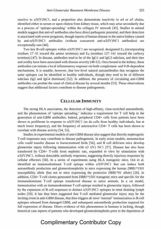
Anti-Glomerular Basement Membrane Disease 325
reactive to α3(IV)NC1, and a proportion also demonstrate reactivity to α4 or α5 chains,
identified either in serum or upon elution from kidney tissue, which may arise secondarily due
to a process of ‘epitope-spreading’ within the collagen IV network [45]. Studies in animal
models suggest that anti-α5 antibodies also have direct pathogenic potential, and their detection
is associated with worse prognosis, though reports of human disease in the native kidney caused
by anti-α5(IV)NC1 antibodies (without concurrent anti-α3(IV)NC1 antibodies) are
exceptionally rare [46].
Two key B-cell epitopes within α3(IV)NC1 are recognized, designated EA (incorporating
residues 17–31 toward the amino terminus) and EB (residues 127–141 toward the carboxy
terminus) [47]. In disease, antibodies tend to be of the IgG1 and IgG3 subclass, and their titer
and avidity have been associated with disease severity [48-51]. Once bound in the kidney, these
antibodies can initiate a local inflammatory response via both complement- and FcR-dependent
mechanisms. It is notable, however, that low-level natural autoantibodies that recognize the
same epitopes can be identified in healthy individuals, though they tend to be of different
subclass (Ig2 and IgG4 dominant) [52]. In addition, the presence of circulating anti-GBM
antibodies can predate the onset of clinical disease by several months [53]. These observations
suggest that additional factors contribute to disease pathogenesis.
CELLULAR IMMUNITY
The strong HLA association, the detection of high-affinity, class-switched autoantibody,
and the phenomenon of ‘epitope spreading,’ indicates a requirement for T cell help in the
generation of anti-GBM antibodies. Indeed, peripheral CD4+ cells from patients have been
shown to proliferate in response to α3(IV)NC1 (as do cells from healthy individuals, but at
much lower frequency), and the frequency of autoreactive CD4+ T cells has been shown to
correlate with disease activity [54, 55].
Studies in experimental models of anti-GBM disease also suggest that directly nephrogenic
T-cell responses may contribute to disease pathogenesis. In early avian models, mononuclear
cells could transfer disease to bursectomised birds [56], and B cell deficient mice develop
glomerular injury following immunization with α3 (IV) NC1 [57]. Disease has also been
transferred by CD4+ T-cells from nephritic rats, expanded in vitro by stimulation with
α3(IV)NC1, without detectable antibody responses, suggesting directly injurious responses by
cellular effectors [58]. In a series of experiments using HLA transgenic mice, Ooi et al.
identified an immunodominant T-cell epitope within α3(IV)NC1 that can induce both
autoantibody production and glomerulonephritis in mice expressing the human DRB1*1501
susceptibility allele (but not in mice expressing the protective DRB1*01 allele) [26]. In
addition, CD4+ T-cell clones generated from DRB1*1501 transgenic mice and specific for the
immunodominant T-cell epitope transferred disease to naïve animals. In a rat model,
immunization with an immunodominant T-cell epitope resulted in glomerular injury, followed
by the expansion of B cell responses to distinct α3(IV)NC1 epitopes in renal draining lymph
nodes [59]. It has thus been suggested that T-cell mediated glomerular injury may be the
inciting event in anti-GBM disease, that then triggers de novo ‘internal’ immunization to B-cell
epitopes released from damaged GBM, and subsequent autoantibody production required for
full expression of disease. Direct evidence of this phenomenon in humans is lacking, though
historical case reports of patients who developed glomerulonephritis prior to the deposition of
Complimentary Contributor Copy
Stephen P. McAdoo and Charles D. Pusey 326
anti-GBM antibodies, and the detection of an early T-cell infiltrate in pathology studies [60,
61], suggests that cell-mediated injury may occur in at least a subset of patients.
TOLERANCE AND AUTOIMMUNITY
α3 (IV) NC1 is expressed in human thymus [55, 62], though the finding of natural
autoantibodies and autoreactive T cells in healthy individuals suggests central tolerance to the
antigen is incomplete. However, additional immune ‘check-points’ must be broken for disease
to develop. Of note, the B cell epitopes within native α3(IV)NC1 are cryptic, being sequestered
within the quaternary structure of the hexameric NC1 structure stabilized by sulfilamine cross-
links, such that they remain protected from immune surveillance in normal circumstances [63].
It has been shown that patient-derived anti-GBM antibodies do not bind cross-linked α3α4α5
hexamers until they are dissociated, and it is proposed that the resistance of some mouse strains
to experimental anti-GBM disease is due to extensive cross-linking in murine GBM [64]. Thus
a process of ‘conformational transformation’ of the cross-linked α3α4α5 hexamer is thought to
be critical to disease pathogenesis, by exposing hidden epitopes to immune detection, and
allowing the binding of pathogenic autoantibodies [45]. This may explain the association of
anti-GBM disease with other pathologies that physically disrupt or modify the structure of the
glomerular (e.g., ANCA vasculitis, lithotripsy) or alveolar (e.g., smoking) basement
membranes, and of the induction of humoral responses following T cell mediated glomerular
injury.
Relapses are rare in anti-GBM disease, suggesting that immune tolerance can be re-instated
as disease resolves. This is supported by the finding of an expanding population of CD25+
antigen-specific T cells in patients after their acute presentation, that may suppress responses
to α3(IV)NC1 [65].
CLINICAL PRESENTATION AND DIAGNOSIS
The majority of patients present with rapidly progressive glomerulonephritis (RPGN). As
such, a short prodrome of non-specific constitutional symptoms is typical, culminating with
overt features of oliguria, fluid overload and uremia. Approximately half of patients require
hemodialysis at the point of initial presentation [22]. Loin pain, attributable to distension of the
renal capsule, and visible hematuria are recognized. Urine microscopy may identify
dysmorphic erythrocytes or red blood cells casts. Proteinuria is usually in the sub-nephrotic
range (<3g/day), perhaps reflecting a severe reduction in glomerular blood flow that limits the
rate at which protein filtration can occur.
Approximately 40-60% of patients have concurrent alveolar hemorrhage, which is more
common in young male patients, and in current smokers [35]. It may present with cough,
dyspnea, and hemoptysis, or be apparent radiographically. A disproportionately severe iron-
deficient anemia should also alert to the possibility of covert underlying alveolar bleeding. A
small proportion of patients (<10%) may present with isolated pulmonary disease in the absence
of renal involvement. In one small series, it was found that 11% of patients with lung
hemorrhage required artificial ventilation during their treatment for anti-GBM disease [66].
Complimentary Contributor Copy
Anti-Glomerular Basement Membrane Disease 327
The presence of extra-renal/pulmonary manifestations is uncommon in anti-GBM disease,
and may suggest an alternative cause of pulmonary-renal syndrome, or that the patient has co-
existing ANCA positivity.
Central to the diagnosis of anti-GBM disease either is the identification of anti-GBM
antibodies, in serum or deposited in tissue, along with pathological features of crescentic
glomerulonephritis, with or without evidence of alveolar hemorrhage.
SEROLOGIC TESTING
Circulating anti-GBM antibodies were first described using indirect immunofluorescence
techniques on normal human or primate kidney [5]. In current practice, a variety of commercial
immunoassays is available, which are accepted to have high sensitivity and specificity [67].
These may use native human or animal GBM isolates, or more commonly recombinant
α3(IV)NC1 preparations. It is recognized that between 5-10% of patients with demonstrable
anti-GBM antibody on renal biopsy are negative by conventional serum assays. As summarized
in a recent commentary by Glassock [68], this may be due to:
Intrinsic sensitivity of the chosen assay
Isotope or subclass of antibody not being detectable (e.g., for example, rare cases of
IgA or IgG4 mediated anti-GBM disease are described)
Antibody disappearance prior to clinical resolution
The ‘immunological sink’ – high affinity antibodies rapidly bind tissue and are
removed from circulation, with only low affinity antibodies remaining,
Absence of relevant epitopes in antigens used in the detection assay
T-lymphocyte, complement or other mediators causing tissue damage rather than a
predominantly antibody mediated process
A proportion of patients who have demonstrable deposition of IgG on the GBM by
immunofluorescence, but who are negative for circulating antibodies by these conventional
techniques, may be positive when tested by highly sensitive biosensor assay [69]. These cases
demonstrate that serological tests, though useful for rapid diagnoses, should not be the sole
means of excluding a diagnosis of anti-GBM disease, and reinforces the importance of
performing a renal biopsy when possible.
DEPOSITED ANTIBODY
Direct immunofluorescence for immunoglobulin on frozen kidney tissue has high
sensitivity for detecting deposited antibodies, and is the gold standard for diagnosis of anti-
GBM disease, typically showing a strong linear ribbon-like appearance (Figure 1A). Other
causes of linear fluorescence should be excluded (including diabetes, monoclonal
paraproteinemia, lupus nephritis, and rarely fibrillary glomerulonephritis). In addition, it should
be noted that fluorescence might be negative or unclear in cases with severe glomerular
inflammation, where the underlying architecture is so disrupted that the linear pattern may not
Complimentary Contributor Copy
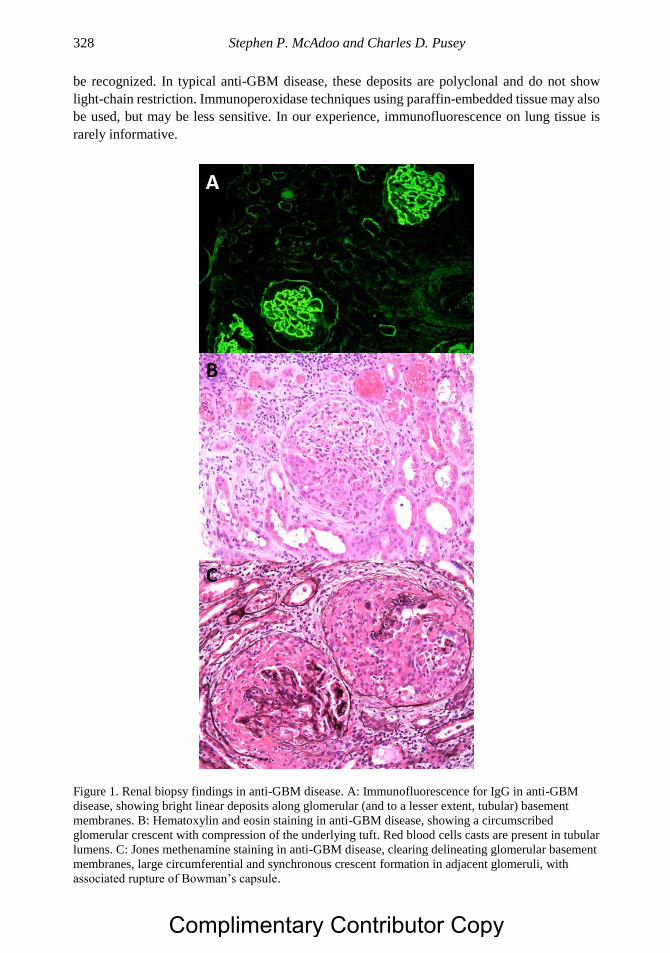
Stephen P. McAdoo and Charles D. Pusey 328
be recognized. In typical anti-GBM disease, these deposits are polyclonal and do not show
light-chain restriction. Immunoperoxidase techniques using paraffin-embedded tissue may also
be used, but may be less sensitive. In our experience, immunofluorescence on lung tissue is
rarely informative.
Figure 1. Renal biopsy findings in anti-GBM disease. A: Immunofluorescence for IgG in anti-GBM
disease, showing bright linear deposits along glomerular (and to a lesser extent, tubular) basement
membranes. B: Hematoxylin and eosin staining in anti-GBM disease, showing a circumscribed
glomerular crescent with compression of the underlying tuft. Red blood cells casts are present in tubular
lumens. C: Jones methenamine staining in anti-GBM disease, clearing delineating glomerular basement
membranes, large circumferential and synchronous crescent formation in adjacent glomeruli, with
associated rupture of Bowman’s capsule.
Complimentary Contributor Copy
Anti-Glomerular Basement Membrane Disease 329
Conventional direct immunofluorescence techniques will identify all IgG subclasses,
though will not differentiate the antigenic target of the kidney-bound antibody, and so may
detect anti-α3 (IV) NCI, anti-α5(IV)NC1, or other antibodies recognizing GBM components.
Antibodies to non-collagen chain antigens, such as nidogen (entactin), have been identified in
historical case series [70, 71], though their significance is not well characterized. In addition to
detecting deposited anti-GBM antibody, immunofluorescence may demonstrate the presence
of complement components, in particular C3 and C1q, along the GBM [21]. A proportion of
patients may also demonstrate immunoglobulin or complement deposition along tubular
basement membranes.
RENAL BIOPSY FINDINGS
Almost all patients with anti-GBM disease will have evidence of crescent formation on
kidney biopsy (Figure 1B), and in 80% of patients more than 50% of glomeruli will be affected.
The average proportion of affected glomeruli is approximately 75% [17, 72]. The proportion
of crescents observed in the biopsy correlates strongly with the degree of renal impairment at
presentation [21, 22]. These crescents will typically be of uniform age (Figure 1C), in contrast
to other causes of RPGN, where a mixture of cellular, fibrocellular and fibrous crescents may
be seen. Asynchronous crescent formation, or the presence of necrotizing vasculitis in larger
blood vessels on renal biopsy, should thus alert to the possibility of a concomitant ANCA-
mediated process. Given the acuity of disease onset, interstitial fibrosis and tubular atrophy are
uncommon in anti-GBM disease (unless there is pre-existing kidney pathology) though
interstitial inflammation may be observed. Electron-dense deposits are not seen in isolated anti-
GBM disease, though electron microscopy is necessary to exclude concomitant glomerular
pathologies, such as membranous glomerulonephritis, and may identify other diseases that may
cause linear fluorescence (such as fibrillary GN and diabetic GBM thickening).
DIAGNOSIS OF ALVEOLAR HEMORRHAGE
There are no uniform diagnostic criteria for the diagnosis of diffuse alveolar hemorrhage,
and diagnosis is often made on a combination of clinical, radiological and laboratory findings.
This may account for the variable frequency of lung hemorrhage described in clinical series of
anti-GBM disease.
Plain chest radiography may demonstrate non-specific diffuse opacification patterns with
occasional predilection for the midzones and apical and costophrenic sparing [73]. High-
resolution computed tomography may show ground-glass opacifications due to alveolar filling
with blood, which may progress to frank consolidation, again with peripheral sparing. As
hemorrhage is resorbed in the pulmonary interstitium, later imaging may show reticular or
nodular appearances [74].
Acute alveolar hemorrhage may be indicated by the finding of hemorrhagic broncho-
alveolar lavage fluid, classically with increasing blood content on successive washes. After 2-
3 days, alveolar macrophages convert hemoglobin to hemosiderin, and these hemosiderin-laden
cells may persist in the lung for several weeks, being evident on Perls staining of broncho-
Complimentary Contributor Copy
Stephen P. McAdoo and Charles D. Pusey 330
alveolar samples [75]. Threshold proportions of 20-30% hemosiderin-laden cells of the total
macrophage count have been suggested to be strongly indicative of diffuse alveolar
hemorrhage.
An increase in diffusing capacity of the lung for carbon monoxide (KCO) has been reported
in alveolar hemorrhage in anti-GBM disease, being attributed to increased CO uptake by intra-
alveolar erythrocytes [76]. However, this finding may not be present in all cases [66], perhaps
due to ventilation-perfusion mismatch in a proportion, such that measurement of KCO may
have useful positive, but not negative, predictive value.
Lung biopsy is rarely performed in cases of alveolar hemorrhage in anti-GBM disease.
When undertaken, it is likely to show alveolar lumens filled with erythrocytes and hemosiderin-
laden cells. Pulmonary capillaritis may be present, with features of fibrinoid necrosis of
capillary walls and inflammation and edema of the alveolar interstitium [77].
TREATMENT
The combination of immunosuppression and plasmapheresis to treat anti-GBM disease was
first described in 1976 [7], and it remains the core recommendation of the latest KDIGO
guideline for treating anti-GBM GN [78]. We have reproduced a recommended treatment
schedule (Table 1).
Plasmapheresis aims to rapidly remove directly pathogenic anti-GBM antibodies from the
circulation, and its use is supported by observational studies that suggest improved renal and
patient survival compared to historical or contemporaneous cohorts treated with
immunosuppression alone [22, 79, 80], and one small randomized trial [81]. The American
Society for Apheresis (ASFA) includes anti-GBM disease in its recommendations for
therapeutic plasmapheresis (Grade 1B, 1C) [82]. Immunoadsorption is an alternative form of
extra-corporeal therapy that may be more efficient than plasma exchange for the removal of
pathogenic autoantibody (though conversely it may not remove pro-inflammatory or pro-
coagulant factors). In small series, it appears to have comparable outcomes to plasma exchange
therapy [83, 84].
In addition to plasma exchange therapy, immunosuppression and corticosteroid treatment
are required to prevent ongoing autoantibody production to reduce renal and pulmonary
inflammation. Daily oral cyclophosphamide and oral prednisolone are the most commonly used
agents, forming the basis of nearly all published experience in anti-GBM disease. An
uncontrolled retrospective analysis suggests that daily oral cyclophosphamide may be superior
to pulsed intravenous treatment [85], in contrast to AAV. The use of other immunosuppressive
therapies is less well described [86-88]. Rituximab is an effective treatment for a number of
other autoantibody associated glomerular diseases, though published experience in anti-GBM
disease is limited to approximately 20 cases [89-98], where it has most commonly been used
as adjunctive or second-line therapy in resistant disease. It appears to be associated with
immunological response, though clinical outcomes are variable. At present, there is insufficient
evidence to recommend its use first-line in the treatment of anti-GBM disease; however, it may
be considered where there are compelling contra-indications to cyclophosphamide therapy or
as adjunctive treatment in severe disease. Rituximab may also be considered for expediting
antibody clearance in patients awaiting renal transplantation who remain seropositive.
Complimentary Contributor Copy
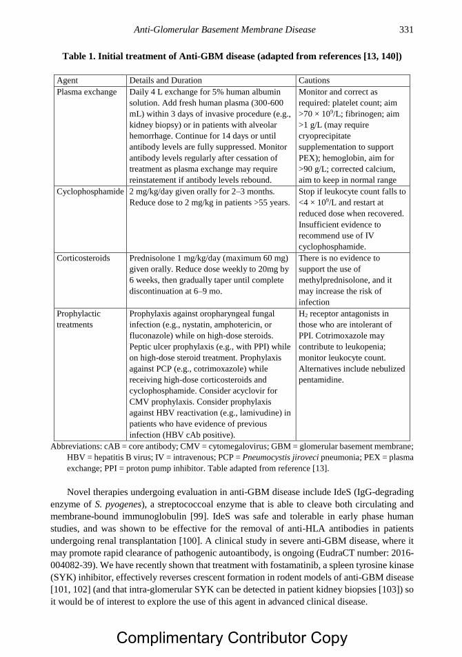
Anti-Glomerular Basement Membrane Disease 331
Table 1. Initial treatment of Anti-GBM disease (adapted from references [13, 140])
Agent Details and Duration Cautions
Plasma exchange Daily 4 L exchange for 5% human albumin
solution. Add fresh human plasma (300-600
mL) within 3 days of invasive procedure (e.g.,
kidney biopsy) or in patients with alveolar
hemorrhage. Continue for 14 days or until
antibody levels are fully suppressed. Monitor
antibody levels regularly after cessation of
treatment as plasma exchange may require
reinstatement if antibody levels rebound.
Monitor and correct as
required: platelet count; aim
>70 × 109/L; fibrinogen; aim
>1 g/L (may require
cryoprecipitate
supplementation to support
PEX); hemoglobin, aim for
>90 g/L; corrected calcium,
aim to keep in normal range
Cyclophosphamide 2 mg/kg/day given orally for 2–3 months.
Reduce dose to 2 mg/kg in patients >55 years.
Stop if leukocyte count falls to
<4 × 10⁹/L and restart at
reduced dose when recovered.
Insufficient evidence to
recommend use of IV
cyclophosphamide.
Corticosteroids Prednisolone 1 mg/kg/day (maximum 60 mg)
given orally. Reduce dose weekly to 20mg by
6 weeks, then gradually taper until complete
discontinuation at 6–9 mo.
There is no evidence to
support the use of
methylprednisolone, and it
may increase the risk of
infection
Prophylactic
treatments
Prophylaxis against oropharyngeal fungal
infection (e.g., nystatin, amphotericin, or
fluconazole) while on high-dose steroids.
Peptic ulcer prophylaxis (e.g., with PPI) while
on high-dose steroid treatment. Prophylaxis
against PCP (e.g., cotrimoxazole) while
receiving high-dose corticosteroids and
cyclophosphamide. Consider acyclovir for
CMV prophylaxis. Consider prophylaxis
against HBV reactivation (e.g., lamivudine) in
patients who have evidence of previous
infection (HBV cAb positive).
H2 receptor antagonists in
those who are intolerant of
PPI. Cotrimoxazole may
contribute to leukopenia;
monitor leukocyte count.
Alternatives include nebulized
pentamidine.
Abbreviations: cAB = core antibody; CMV = cytomegalovirus; GBM = glomerular basement membrane;
HBV = hepatitis B virus; IV = intravenous; PCP = Pneumocystis jiroveci pneumonia; PEX = plasma
exchange; PPI = proton pump inhibitor. Table adapted from reference [13].
Novel therapies undergoing evaluation in anti-GBM disease include IdeS (IgG-degrading
enzyme of S. pyogenes), a streptococcoal enzyme that is able to cleave both circulating and
membrane-bound immunoglobulin [99]. IdeS was safe and tolerable in early phase human
studies, and was shown to be effective for the removal of anti-HLA antibodies in patients
undergoing renal transplantation [100]. A clinical study in severe anti-GBM disease, where it
may promote rapid clearance of pathogenic autoantibody, is ongoing (EudraCT number: 2016-
004082-39). We have recently shown that treatment with fostamatinib, a spleen tyrosine kinase
(SYK) inhibitor, effectively reverses crescent formation in rodent models of anti-GBM disease
[101, 102] (and that intra-glomerular SYK can be detected in patient kidney biopsies [103]) so
it would be of interest to explore the use of this agent in advanced clinical disease.
Complimentary Contributor Copy
Stephen P. McAdoo and Charles D. Pusey 332
OUTCOME AND PROGNOSIS
Diffuse alveolar hemorrhage secondary to anti-GBM disease appears to be highly
responsive to treatment, with 90-100% of cases responding to immunosuppression and
plasmapheresis [22, 66]. Data on long-term respiratory outcomes, however, are scarce. One
small study suggested that patients who had lung hemorrhage have significantly reduced KCO
compared to controls without lung involvement [104]. However, a subsequent larger series
suggested that long-term respiratory sequela after lung hemorrhage in anti-GBM disease are
not common [66]. This is in contrast to AAV, where interstitial lung disease is increasingly
recognized as an important long-term complication, especially in patients positive for MPO-
ANCA, suggesting that distinct mechanisms of pulmonary injury in these diseases [105, 106].
Renal outcomes in anti-GBM disease may be more variable. Long-term follow up of the
largest cohort of patients (n = 71) all treated with the combination of plasma exchange,
cyclophosphamide and corticosteroids, suggests that this approach is effective in preserving
independent kidney function in the majority of patients, including those who present with
severe kidney dysfunction, provided they do not require dialysis at presentation [22]. In patients
presenting with creatinine values <500umol/L, renal survival was 95% and 94% at 1- and 5-
years respectively. In patients presenting with creatinine >500umol/L, but not requiring
immediate dialysis, renal survival was 82% and 50% at the same respective time-points. In
patients presenting with an initial requirement for dialysis, however, renal recovery occurred
in only 8% at 1 year. Other reports have described similarly low levels of renal recovery in
patients presenting with dialysis-dependent kidney failure, with the highest rate of
approximately 20% recovery in one series [107].
Predictors of poor renal outcome include severe renal dysfunction at diagnosis, the
proportion of glomeruli affected by crescents, and oliguria at presentation [21, 22, 108]. A
recent worldwide, multi-center study recruited 123 cases of renal biopsy-proven anti-GBM
glomerulonephritis, making it the largest histopathological study in anti-GBM disease to date
[109]. It confirmed previous observations of more favorable outcomes in patients presenting
with creatinine of <500µmol/L. Independent predictors of ESRD were dialysis-requirement at
presentation, reduced proportion of normal glomeruli, and increased interstitial infiltrate on
kidney biopsy. Of note, no patient with 100% crescents or >50% sclerotic glomeruli recovered
renal function, and so withholding treatment may be considered in these cases if lung
hemorrhage is not present. Rare cases of renal recovery despite adverse pathologic findings
[110, 111], however, highlights the need to consider all cases for treatment, with specific
attention to other features that might predict renal recovery on biopsy (such as concomitant
acute tubular injury). A short trial of early treatment may be considered, and rapidly tapered if
there is no evidence of renal recovery within 2-4 weeks. In addition, the potential benefit of a
period of immunosuppression to expedite autoantibody clearance, thus allowing earlier kidney
transplantation, should be considered in suitable patients.
Relapse is rare in anti-GBM disease, occurring in fewer than 3% of patients [22]. It is
usually associated with ongoing exposure to pulmonary irritants such as cigarette smoke or
hydrocarbons [112, 113], and avoidance of these precipitants is an essential part of long-term
management of these cases. We recommend repeat kidney biopsy in cases of relapse with
kidney involvement, in order to secure an accurate diagnosis and to exclude concomitant
Complimentary Contributor Copy
Anti-Glomerular Basement Membrane Disease 333
pathologies such as AAV and membranous nephropathy. In confirmed cases, standard re-
treatment with cytotoxic medications and corticosteroids is usually indicated.
The long-term outcome of patients who progress to ESRD due to anti-GBM GN appears
to comparable to patients with ESRD of other causes, whether they remain on dialysis or
undergo kidney transplantation [18, 114]. It should be noted, however, that kidney
transplantation performed in the presence of anti-GBM antibodies results in a high likelihood
of disease recurrence in the allograft, at frequencies of up to 50% in historical series [115], and
that a period of at least six months sustained seronegativity is advised prior to undertaking
transplantation in patients who have reached ESRD due to anti-GBM disease [78]. Under these
circumstances, and with current immunosuppressive regimens, recurrent disease is rare [18].
VARIANT PRESENTATIONS OF ANTI-GBM DISEASE
Double-Positive Anti-GBM and ANCA-Associated GN
In some series, almost half of patients with anti-GBM disease have detectable ANCA
(usually recognizing myeloperoxidase, MPO), and up to 10% of patients with ANCA have
circulating anti-GBM antibodies [116-118]. This phenomenon was first reported within a few
years of the first description of ANCA [116, 119], and while it is clear that the autoantibodies
are antigenically distinct [120], the mechanism of this association is not understood. It has been
shown that ANCA may be detected before the onset of anti-GBM disease, suggesting that
ANCA-induced glomerular inflammation may be a trigger for the development of an anti-GBM
response, perhaps by modifying or exposing usually sequestered disease epitopes in GBM [53].
These ‘double-positive’ patients appear to have a hybrid clinical phenotype, experiencing
the early morbidity and mortality of anti-GBM disease, with severe kidney and lung disease at
presentation, requiring aggressive immunosuppressive therapy and plasma exchange [121].
There is a suggestion they more be more responsive to treatment, with increased rates of
recovery from dialysis. During long-term follow-up, however, they relapse at a frequency
comparable to patients with AAV, suggesting they warrant more careful long-term follow up
and maintenance immunosuppression, unlike patients with single-positive anti-GBM disease.
Anti-GBM Disease Associated with Membranous Nephropathy
There are several reports of anti-GBM disease associated with membranous nephropathy,
occurring as a preceding, simultaneous, or succeeding diagnosis [122, 123]. As with the
ANCA-association, it is postulated that disruption of glomerular architecture by one disease
reveals hidden epitopes that allow the second process to occur. A rapid decline in kidney
function in a patient with known membranous nephropathy should raise suspicion of the
development of superimposed crescentic nephritis or anti-GBM disease, and re-biopsy is
recommended. We suggest that such cases are treated initially as for anti-GBM disease, though
how they should be managed in the long-term is not clear. The authors of a recent case report
suggest that rituximab may be a useful agent to treat both pathologies simultaneously [92].
Complimentary Contributor Copy
Stephen P. McAdoo and Charles D. Pusey 334
Post-Transplant Anti-GBM Disease in Alport Syndrome
After kidney transplantation for Alport syndrome, recipients may develop anti-GBM
antibodies as an alloimmune response to the neo-antigens contained in ‘normal’ α3, α4 or α5
chains expressed in the kidney allograft. In classical X-linked Alport syndrome (caused by
mutations in the COL4A5 gene encoding the α5 collagen chain) these antibodies do not
recognize the individual EA and EB epitopes of the α3 chain recognized by sera from typical
Goodpasture patients, but rather a distinct, composite epitope on the α5 chain, that is not
sequestered within the native hexamer of the Goodpasture antigen [45]. It should be noted that
commercially available anti-GBM assays, which are optimized to detect reactivity to the
α3(IV)NC1 antigen, might fail to detect circulating antibodies in this setting. Anti-GBM
antibodies may be detected in 5-10% of Alport patients following transplantation, though the
development of overt glomerulonephritis in the allograft is less frequent (perhaps owing to the
effects of maintenance immunosuppression). When glomerulonephritis develops, however, it
usually occurs early and carries a high risk of graft loss [124, 125]. Repeated transplantation in
this setting almost invariably leads to more aggressive disease recurrence and rapid graft loss,
and is undertaken at very high risk [126]. Individuals with large COL4A5 gene deletions are at
increased risk of post-transplant anti-GBM disease, and recent guidelines encourage the use of
genetic testing to inform discussions regarding the risk of de novo anti-GBM disease after
transplantation [127].
‘ATYPICAL’ ANTI-GBM DISEASE
‘Atypical Anti-GBM disease’ is a term that has been used variably to describe cases with
classical linear IgG staining on renal biopsy, but uncharacteristic clinicopathologic features. A
large series (n = 20) of ‘atypical’ cases was recently reported by Nasr and colleagues – all had
bright linear GBM staining for immunoglobulin on kidney biopsy, though none had detectable
α3(IV)NC1 antibodies by commercial ELISA, and none had lung hemorrhage [128]. Most had
had only mild renal insufficiency, though all had proliferative glomerular changes on renal
biopsy, but without diffuse crescent formation. Treatment was with heterogeneous
immunosuppression (and generally did not include plasma exchange), and both patient and
renal survival at one year were favorable, at 93% and 85%, respectively. Of note, GBM staining
was IgM dominant in two cases, and IgA dominant in another. In addition, half of the patients
had restriction for either kappa or lambda lights on immunofluorescence testing, though none
had glomerular capillary wall deposits on electron microscopy (that would be expected in
proliferative GN with monoclonal immunoglobulin deposits; PGNMID), nor did they have
identified features of an underlying plasma cell dyscrasia. A number of other case reports and
small series have similarly described ‘atypical’ cases, with similarly indolent renal disease and
seronegativity by conventional assay. One recent report describes a case of florid anti-GBM
disease with kappa-light chain restriction on GBM staining in association with a circulating
paraprotein [129], thus suggesting that anti-GBM disease may occur within the spectrum of
‘monoclonal gammopathy of renal significance.’ A number of mechanisms for the unusual
pattern of renal injury in these cases have been proposed, including differences in antibody
Complimentary Contributor Copy
Anti-Glomerular Basement Membrane Disease 335
isotype or subclasses, varied antigenic targets, and differential pathogenicity in antibodies
related to their ability to fix complement or recruit inflammatory cells [130].
ANTI-GBM DISEASE AFTER ALEMTUZUMAB THERAPY
Secondary autoimmune phenomena after alemtuzumab, an anti-CD52 lymphocyte
depleting treatment for relapsing-remitting multiple sclerosis (MS), are common [131].
Autoimmune thyroid disease occurred in up to 30% of patients in controlled trials, and there
are case reports of both membranous nephropathy and anti-GBM disease following treatment
[132, 133]. It has been proposed that T-cell reconstitution after alemtuzumab is driven largely
by homeostatic expansion of cells that have escaped deletion (rather than by thymopoesis),
resulting in a T-cell pool that is enriched for autoreactive cells [134]. Anti-GBM disease has
not been reported when alemtuzumab is used for other indications other than MS (e.g., renal
transplantation), perhaps due to the use of maintenance immunosuppression, or due to the
shared HLA susceptibilities in MS and anti-GBM disease. Careful monitoring of patients for
renal pathology after treatment with alemtuzumab is needed in order to facilitate early detection
and treatment [135]. We suggest conventional treatment for anti-GBM disease occurring in this
setting.
ISOLATED PULMONARY INVOLVEMENT IN ANTI-GBM DISEASE
Presentation with isolated or predominant pulmonary involvement in anti-GBM disease is
recognized, though uncommon, being estimated to occur in <10% of patients in larger series.
Such cases are not extensively characterized, perhaps reflecting publication bias from renal
centers, though there are small case series [136, 137]. Some patients may have mild urinary
abnormalities and minor proliferative changes on renal biopsy, but with preserved excretory
renal function, while others may have no clinical or histological evidence of renal of renal
inflammation. Renal biopsy, however, may still reveal linear deposits of immunoglobulin,
including in those patients who are negative by serological assay. A small series from Sweden
recently described four young female patients who presented with severe alveolar hemorrhage
and favorable renal outcome, who were seronegative for circulating anti-GBM antibodies by
conventional assay [138]. They were, however, found to have circulating IgG4 anti-GBM
antibodies by dedicated ELISA, and this was confirmed on kidney biopsy. Together, these
findings suggest that clinical presentation in anti-GBM disease may be influenced by
differences in antibody subclass or antigen target, and highlight the need to consider variant
anti-GBM disease in cases of ‘idiopathic’ pulmonary hemorrhage.
CONCLUSION
Despite its rarity, anti-GBM disease has been extensively studied, and it now serves as a
model autoimmune disorder: it has a well-characterized autoantigen with defined epitopes; it
demonstrates the directly pathogenic potential of specific autoantibodies; T-cell responses
Complimentary Contributor Copy
Stephen P. McAdoo and Charles D. Pusey 336
contribute to disease pathogenesis, both as orchestrators of the immune response and as
effectors of tissue injury; and recent work sheds light on the molecular mechanisms via which
HLA inheritance may confer autoimmune susceptibility. However, variant and atypical
presentations demonstrate that anti-GBM disease cannot be regarded as a ‘one-dimensional’
entity [139], and that the mechanisms which induce the aberrant adaptive immune response
directed to self-antigen are not fully understood and may not be common to all patients. Further
insights into these inciting events and, given the extreme rarity of relapses, the mechanisms via
which immune tolerance is reinstated as disease resolves, may have important therapeutic
implications not just in anti-GBM disease, but also for a spectrum of autoimmune disorders in
future. The abrupt clinical presentation of life-threatening lung hemorrhage and rapidly
progressively glomerulonephritis, however, renders the disease of equal import to physicians
as to immunologists, as early recognition and prompt initiation of immunosuppression and
plasmapheresis is necessary to preserve kidney function and prevent mortality. Current
treatment strategies, when introduced early enough, appear to be effective, though improved
prognostic and therapeutic approaches are required for those presenting with advanced renal
failure.
REFERENCES
[1] Jennette JC, Falk RJ, Bacon PA, et al. 2012 revised International Chapel Hill Consensus
Conference Nomenclature of Vasculitides. Arthritis and rheumatism. 2013;65(1):1-11.
[2] Goodpasture E. The significance of certain pulmonary lesions in relation to the etiology
of influenza. Am J Med Sci. 1919;158:863-870.
[3] Wilson CB, Dixon FJ. Anti-glomerular basement membrane antibody-induced
glomerulonephritis. Kidney international. 1973;3(2):74-89.
[4] Scheer R, Grossman M. Immune aspects of the glomerulonephritis associated with
pulmonary haemorrhage. Annals of internal medicine. 1964;60(6):1009-1021.
[5] McPhaul JJ, Jr., Dixon FJ. The presence of anti-glomerular basement membrane
antibodies in peripheral blood. Journal of immunology. 1969;103(6):1168-1175.
[6] Lerner RA, Glassock RJ, Dixon FJ. The role of anti-glomerular basement membrane
antibody in the pathogenesis of human glomerulonephritis. The Journal of experimental
medicine. 1967;126(6):989-1004.
[7] Lockwood CM, Rees AJ, Pearson TA, Evans DJ, Peters DK, Wilson CB.
Immunosuppression and plasma-exchange in the treatment of Goodpasture’s syndrome.
Lancet. 1976;1(7962):711-715.
[8] Canney M, O’Hara PV, McEvoy CM, et al. Spatial and Temporal Clustering of Anti-
Glomerular Basement Membrane Disease. Clinical journal of the American Society of
Nephrology: CJASN. 2016;11(8):1392-1399.
[9] McPhaul JJ, Jr., Mullins JD. Glomerulonephritis mediated by antibody to glomerular
basement membrane. Immunological, clinical, and histopathological characteristics. The
Journal of clinical investigation. 1976;57(2):351-361.
[10] Taylor DM, Yehia M, Simpson IJ, Thein H, Chang Y, de Zoysa JR. Anti-glomerular
basement membrane disease in Auckland. Intern Med J. 2012;42(6):672-676.
Complimentary Contributor Copy
Anti-Glomerular Basement Membrane Disease 337
[11] Hirayama K, Yamagata K, Kobayashi M, Koyama A. Anti-glomerular basement
membrane antibody disease in Japan: part of the nationwide rapidly progressive
glomerulonephritis survey in Japan. Clinical and experimental nephrology.
2008;12(5):339-347.
[12] Li FK, Tse KC, Lam MF, et al. Incidence and outcome of antiglomerular basement
membrane disease in Chinese. Nephrology. 2004;9(2):100-104.
[13] Pusey CD. Anti-glomerular basement membrane disease. Kidney international.
2003;64(4):1535-1550.
[14] Saxena R, Bygren P, Arvastson B, Wieslander J. Circulating autoantibodies as
serological markers in the differential diagnosis of pulmonary renal syndrome. J Intern
Med. 1995;238(2):143-152.
[15] Niles JL, Bottinger EP, Saurina GR, et al. The syndrome of lung hemorrhage and
nephritis is usually an ANCA-associated condition. Arch Intern Med. 1996;156(4):440-
445.
[16] Gallagher H, Kwan JT, Jayne DR. Pulmonary renal syndrome: a 4-year, single-center
experience. American journal of kidney diseases: the official journal of the National
Kidney Foundation. 2002;39(1):42-47.
[17] Jennette JC. Rapidly progressive crescentic glomerulonephritis. Kidney international.
2003;63(3):1164-1177.
[18] Tang W, McDonald SP, Hawley CM, et al. Anti-glomerular basement membrane
antibody disease is an uncommon cause of end-stage renal disease. Kidney international.
2013;83(3):503-510.
[19] Saran R, Robinson B, Abbott KC, et al. US Renal Data System 2017 Annual Data Report:
Epidemiology of Kidney Disease in the United States. American journal of kidney
diseases: the official journal of the National Kidney Foundation. 2018;71(3S1):A7.
[20] Savage CO, Pusey CD, Bowman C, Rees AJ, Lockwood CM. Antiglomerular basement
membrane antibody mediated disease in the British Isles 1980-4. British medical journal.
1986;292(6516):301-304.
[21] Fischer EG, Lager DJ. Anti-glomerular basement membrane glomerulonephritis: a
morphologic study of 80 cases. American journal of clinical pathology. 2006;125(3):445-
450.
[22] Levy JB, Turner AN, Rees AJ, Pusey CD. Long-term outcome of anti-glomerular
basement membrane antibody disease treated with plasma exchange and
immunosuppression. Annals of internal medicine. 2001;134(11):1033-1042.
[23] Fisher M, Pusey CD, Vaughan RW, Rees AJ. Susceptibility to anti-glomerular basement
membrane disease is strongly associated with HLA-DRB1 genes. Kidney international.
1997;51(1):222-229.
[24] Yang R, Cui Z, Zhao J, Zhao MH. The role of HLA-DRB1 alleles on susceptibility of
Chinese patients with anti-GBM disease. Clinical immunology. 2009;133(2):245-250.
[25] Kitagawa W, Imai H, Komatsuda A, et al. The HLA-DRB1*1501 allele is prevalent
among Japanese patients with anti-glomerular basement membrane antibody-mediated
disease. Nephrology, dialysis, transplantation: official publication of the European
Dialysis and Transplant Association - European Renal Association. 2008;23(10):3126-
3129.
Complimentary Contributor Copy
Stephen P. McAdoo and Charles D. Pusey 338
[26] Ooi JD, Chang J, O’Sullivan KM, et al. The HLA-DRB1*15:01-restricted Goodpasture’s
T cell epitope induces GN. Journal of the American Society of Nephrology: JASN.
2013;24(3):419-431.
[27] Ooi JD, Petersen J, Tan YH, et al. Dominant protection from HLA-linked autoimmunity
by antigen-specific regulatory T cells. Nature. 2017;545(7653):243-247.
[28] Persson U, Hertz JM, Carlsson M, et al. Patients with Goodpasture’s disease have two
normal COL4A3 alleles encoding the NC1 domain of the type IV collagen alpha 3 chain.
Nephrology, dialysis, transplantation: official publication of the European Dialysis and
Transplant Association - European Renal Association. 2004;19(8):2030-2035.
[29] Perez GO, Bjornsson S, Ross AH, Aamato J, Rothfield N. A mini-epidemic of
Goodpasture’s syndrome clinical and immunological studies. Nephron. 1974;13(2):161-
173.
[30] Wilson CB, Smith RC. Goodpasture’s syndrome associated with influenza A2 virus
infection. Annals of internal medicine. 1972;76(1):91-94.
[31] Gu QH, Xie LJ, Jia XY, et al. Fever and prodromal infections in anti-glomerular
basement membrane disease. Nephrology. 2017.
[32] Arends J, Wu J, Borillo J, et al. T cell epitope mimicry in antiglomerular basement
membrane disease. Journal of immunology. 2006;176(2):1252-1258.
[33] Reynolds J, Preston GA, Pressler BM, et al. Autoimmunity to the alpha 3 chain of type
IV collagen in glomerulonephritis is triggered by ‘autoantigen complementarity.’ Journal
of autoimmunity. 2015;59:8-18.
[34] Li JN, Jia X, Wang Y, et al. Plasma from patients with anti-glomerular basement
membrane disease could recognize microbial peptides. PloS one. 2017;12(4):e0174553.
[35] Donaghy M, Rees AJ. Cigarette smoking and lung haemorrhage in glomerulonephritis
caused by autoantibodies to glomerular basement membrane. Lancet.
1983;2(8364):1390-1393.
[36] Bombassei GJ, Kaplan AA. The association between hydrocarbon exposure and anti-
glomerular basement membrane antibody-mediated disease (Goodpasture’s syndrome).
American journal of industrial medicine. 1992;21(2):141-153.
[37] D’Apice AJ, Kincaid-Smith P, Becker GH, Loughhead MG, Freeman JW, Sands JM.
Goodpasture’s syndrome in identical twins. Annals of internal medicine. 1978;88(1):61-
62.
[38] Garcia-Rostan y Perez GM, Garcia Bragado F, Puras Gil AM. Pulmonary hemorrhage
and antiglomerular basement membrane antibody-mediated glomerulonephritis after
exposure to smoked cocaine (crack): a case report and review of the literature. Pathol Int.
1997;47(10):692-697.
[39] Peces R, Navascues RA, Baltar J, Seco M, Alvarez J. Antiglomerular basement
membrane antibody-mediated glomerulonephritis after intranasal cocaine use. Nephron.
1999;81(4):434-438.
[40] De Rosa N, Glanville A. ECMO-dependent respiratory failure after snorting speed
associated with anti-GBM antibodies. Respirol Case Rep. 2015;3(4):138-140.
[41] Bhave G, Cummings CF, Vanacore RM, et al. Peroxidasin forms sulfilimine chemical
bonds using hypohalous acids in tissue genesis. Nat Chem Biol. 2012;8(9):784-790.
[42] McCall AS, Cummings CF, Bhave G, Vanacore R, Page-McCaw A, Hudson BG.
Bromine is an essential trace element for assembly of collagen IV scaffolds in tissue
development and architecture. Cell. 2014;157(6):1380-1392.
Complimentary Contributor Copy
Anti-Glomerular Basement Membrane Disease 339
[43] Saus J, Wieslander J, Langeveld JP, Quinones S, Hudson BG. Identification of the
Goodpasture antigen as the alpha 3(IV) chain of collagen IV. The Journal of biological
chemistry. 1988;263(26):13374-13380.
[44] Turner N, Mason PJ, Brown R, et al. Molecular cloning of the human Goodpasture
antigen demonstrates it to be the alpha 3 chain of type IV collagen. The Journal of clinical
investigation. 1992;89(2):592-601.
[45] Pedchenko V, Bondar O, Fogo AB, et al. Molecular architecture of the Goodpasture
autoantigen in anti-GBM nephritis. The New England journal of medicine.
2010;363(4):343-354.
[46] Cui Z, Zhao MH, Jia XY, et al. Antibodies to alpha5 chain of collagen IV are pathogenic
in Goodpasture’s disease. Journal of autoimmunity. 2016;70:1-11.
[47] Netzer KO, Leinonen A, Boutaud A, et al. The goodpasture autoantigen. Mapping the
major conformational epitope(s) of alpha3(IV) collagen to residues 17-31 and 127-141
of the NC1 domain. The Journal of biological chemistry. 1999;274(16):11267-11274.
[48] Zhao J, Yan Y, Cui Z, Yang R, Zhao MH. The immunoglobulin G subclass distribution
of anti-GBM autoantibodies against rHalpha3(IV)NC1 is associated with disease
severity. Human immunology. 2009;70(6):425-429.
[49] Segelmark M, Butkowski R, Wieslander J. Antigen restriction and IgG subclasses among
anti-GBM autoantibodies. Nephrology, dialysis, transplantation: official publication of
the European Dialysis and Transplant Association - European Renal Association.
1990;5(12):991-996.
[50] Cui Z, Zhao MH. Avidity of anti-glomerular basement membrane autoantibodies was
associated with disease severity. Clinical immunology. 2005;116(1):77-82.
[51] Segelmark M, Hellmark T, Wieslander J. The prognostic significance in Goodpasture’s
disease of specificity, titre and affinity of anti-glomerular-basement-membrane
antibodies. Nephron. Clinical practice. 2003;94(3):c59-68.
[52] Cui Z, Zhao MH, Segelmark M, Hellmark T. Natural autoantibodies to myeloperoxidase,
proteinase 3, and the glomerular basement membrane are present in normal individuals.
Kidney international. 2010;78(6):590-597.
[53] Olson SW, Arbogast CB, Baker TP, et al. Asymptomatic autoantibodies associate with
future anti-glomerular basement membrane disease. Journal of the American Society of
Nephrology: JASN. 2011;22(10):1946-1952.
[54] Zou J, Hannier S, Cairns LS, et al. Healthy individuals have Goodpasture autoantigen-
reactive T cells. Journal of the American Society of Nephrology: JASN. 2008;19(2):396-
404.
[55] Salama AD, Chaudhry AN, Ryan JJ, et al. In Goodpasture’s disease, CD4(+) T cells
escape thymic deletion and are reactive with the autoantigen alpha3(IV)NC1. Journal of
the American Society of Nephrology: JASN. 2001;12(9):1908-1915.
[56] Bolton WK, Tucker FL, Sturgill BC. New avian model of experimental
glomerulonephritis consistent with mediation by cellular immunity. Nonhumorally
mediated glomerulonephritis in chickens. The Journal of clinical investigation.
1984;73(5):1263-1276.
[57] Dean EG, Wilson GR, Li M, et al. Experimental autoimmune Goodpasture’s disease: a
pathogenetic role for both effector cells and antibody in injury. Kidney international.
2005;67(2):566-575.
Complimentary Contributor Copy
Stephen P. McAdoo and Charles D. Pusey 340
[58] Wu J, Hicks J, Borillo J, Glass WF, 2nd, Lou YH. CD4(+) T cells specific to a glomerular
basement membrane antigen mediate glomerulonephritis. The Journal of clinical
investigation. 2002;109(4):517-524.
[59] Robertson J, Wu J, Arends J, et al. Activation of glomerular basement membrane-specific
B cells in the renal draining lymph node after T cell-mediated glomerular injury. Journal
of the American Society of Nephrology: JASN. 2005;16(11):3256-3263.
[60] Bolton WK, Innes DJ, Jr., Sturgill BC, Kaiser DL. T-cells and macrophages in rapidly
progressive glomerulonephritis: clinicopathologic correlations. Kidney international.
1987;32(6):869-876.
[61] Nolasco FE, Cameron JS, Hartley B, Coelho A, Hildreth G, Reuben R. Intraglomerular
T cells and monocytes in nephritis: study with monoclonal antibodies. Kidney
international. 1987;31(5):1160-1166.
[62] Wong D, Phelps RG, Turner AN. The Goodpasture antigen is expressed in the human
thymus. Kidney international. 2001;60(5):1777-1783.
[63] Vanacore RM, Ham AJ, Cartailler JP, et al. A role for collagen IV cross-links in
conferring immune privilege to the Goodpasture autoantigen: structural basis for the
crypticity of B cell epitopes. The Journal of biological chemistry. 2008;283(33):22737-
22748.
[64] Luo W, Wang XP, Kashtan CE, Borza DB. Alport alloantibodies but not Goodpasture
autoantibodies induce murine glomerulonephritis: protection by quinary crosslinks
locking cryptic alpha3(IV) collagen autoepitopes in vivo. Journal of immunology.
2010;185(6):3520-3528.
[65] Salama AD, Chaudhry AN, Holthaus KA, et al. Regulation by CD25+ lymphocytes of
autoantigen-specific T-cell responses in Goodpasture’s (anti-GBM) disease. Kidney
international. 2003;64(5):1685-1694.
[66] Lazor R, Bigay-Game L, Cottin V, et al. Alveolar hemorrhage in anti-basement
membrane antibody disease: a series of 28 cases. Medicine (Baltimore). 2007;86(3):181-
193.
[67] Sinico RA, Radice A, Corace C, Sabadini E, Bollini B. Anti-glomerular basement
membrane antibodies in the diagnosis of Goodpasture syndrome: a comparison of
different assays. Nephrology, dialysis, transplantation: official publication of the
European Dialysis and Transplant Association - European Renal Association.
2006;21(2):397-401.
[68] Glassock RJ. Atypical anti-glomerular basement membrane disease: lessons learned. Clin
Kidney J. 2016;9(5):653-656.
[69] Salama AD, Dougan T, Levy JB, et al. Goodpasture’s disease in the absence of
circulating anti-glomerular basement membrane antibodies as detected by standard
techniques. American journal of kidney diseases: the official journal of the National
Kidney Foundation. 2002;39(6):1162-1167.
[70] Bygren P, Cederholm B, Heinegard D, Wieslander J. Non-Goodpasture anti-GBM
antibodies in patients with glomerulonephritis. Nephrology, dialysis, transplantation:
official publication of the European Dialysis and Transplant Association - European
Renal Association. 1989;4(4):254-261.
[71] Saxena R, Bygren P, Butkowski R, Wieslander J. Entactin: a possible auto-antigen in the
pathogenesis of non-Goodpasture anti-GBM nephritis. Kidney international.
1990;38(2):263-272.
Complimentary Contributor Copy
Anti-Glomerular Basement Membrane Disease 341
[72] Jennette JC, Thomas DB. Crescentic glomerulonephritis. Nephrology, dialysis,
transplantation: official publication of the European Dialysis and Transplant
Association - European Renal Association. 2001;16 Suppl 6:80-82.
[73] Cortese G, Nicali R, Placido R, Gariazzo G, Anro P. Radiological aspects of diffuse
alveolar haemorrhage. Radiol Med. 2008;113(1):16-28.
[74] Serra G, Brun AL, Toledano D, Cluzel P, Grenier PA. Thoracic involvement in systemic
primary vasculitis: radiological patterns and follow-up. JBR-BTR. 2014;97(2):57-68.
[75] Colby TV, Fukuoka J, Ewaskow SP, Helmers R, Leslie KO. Pathologic approach to
pulmonary hemorrhage. Annals of diagnostic pathology. 2001;5(5):309-319.
[76] Ewan PW, Jones HA, Rhodes CG, Hughes JM. Detection of intrapulmonary hemorrhage
with carbon monoxide uptake. Application in goodpasture’s syndrome. The New England
journal of medicine. 1976;295(25):1391-1396.
[77] Lombard CM, Colby TV, Elliott CG. Surgical pathology of the lung in anti-basement
membrane antibody-associated Goodpasture’s syndrome. Hum Pathol. 1989;20(5):445-
451.
[78] Kidney Disease: Improving Global Outcomes (KDIGO) Glomerulonephritis Work
Group. KDIGO Clinical Practice Guideline for Glomerulonephritis. Kidney Int., Suppl.
2012;2:139-274.
[79] Simpson IJ, Doak PB, Williams LC, et al. Plasma exchange in Goodpasture’s syndrome.
American journal of nephrology. 1982;2(6):301-311.
[80] Cui Z, Zhao J, Jia XY, et al. Anti-glomerular basement membrane disease: outcomes of
different therapeutic regimens in a large single-center Chinese cohort study. Medicine
(Baltimore). 2011;90(5):303-311.
[81] Johnson JP, Moore J, Jr., Austin HA, 3rd, Balow JE, Antonovych TT, Wilson CB.
Therapy of anti-glomerular basement membrane antibody disease: analysis of prognostic
significance of clinical, pathologic and treatment factors. Medicine (Baltimore).
1985;64(4):219-227.
[82] Schwartz J, Padmanabhan A, Aqui N, et al. Guidelines on the Use of Therapeutic
Apheresis in Clinical Practice-Evidence-Based Approach from the Writing Committee
of the American Society for Apheresis: The Seventh Special Issue. J Clin Apher.
2016;31(3):149-162.
[83] Biesenbach P, Kain R, Derfler K, et al. Long-term outcome of anti-glomerular basement
membrane antibody disease treated with immunoadsorption. PloS one.
2014;9(7):e103568.
[84] Zhang YY, Tang Z, Chen DM, Gong DH, Ji DX, Liu ZH. Comparison of double filtration
plasmapheresis with immunoadsorption therapy in patients with anti-glomerular
basement membrane nephritis. BMC Nephrol. 2014;15:128.
[85] Huart A, Josse AG, Chauveau D, et al. Outcomes of patients with Goodpasture syndrome:
A nationwide cohort-based study from the French Society of Hemapheresis. Journal of
autoimmunity. 2016;73:24-29.
[86] Mori M, Nwaogwugwu U, Akers GR, McGill RL. Anti-glomerular basement membrane
disease treated with mycophenolate mofetil, corticosteroids, and plasmapheresis. Clinical
nephrology. 2013;80(1):67-71.
[87] Garcia-Canton C, Toledo A, Palomar R, et al. Goodpasture’s syndrome treated with
mycophenolate mofetil. Nephrology, dialysis, transplantation: official publication of the
Complimentary Contributor Copy
Stephen P. McAdoo and Charles D. Pusey 342
European Dialysis and Transplant Association - European Renal Association.
2000;15(6):920-922.
[88] Kiykim AA, Horoz M, Gok E. Successful treatment of resistant antiglomerular basement
membrane antibody positivity with mycophenolic acid. Intern Med. 2010;49(6):577-580.
[89] Mutsaers P, Selten H, van Dam B. Additional antibody suppression from rituximab added
to conventional therapy in severe, refractory anti-GBM nephritis. NDT plus.
2010;3(4):421-422.
[90] Shah Y, Mohiuddin A, Sluman C, et al. Rituximab in anti-glomerular basement
membrane disease. QJM: monthly journal of the Association of Physicians.
2012;105(2):195-197.
[91] Syeda UA, Singer NG, Magrey M. Anti-glomerular basement membrane antibody
disease treated with rituximab: A case-based review. Semin Arthritis Rheum.
2013;42(6):567-572.
[92] Bandak G, Jones BA, Li J, Yee J, Umanath K. Rituximab for the treatment of refractory
simultaneous anti-glomerular basement membrane (anti-GBM) and membranous
nephropathy. Clin Kidney J. 2014;7(1):53-56.
[93] Narayanan M, Casimiro I, Pichler R. A unique way to treat Goodpasture’s disease. BMJ
Case Rep. 2014;2014.
[94] Touzot M, Poisson J, Faguer S, et al. Rituximab in anti-GBM disease: A retrospective
study of 8 patients. Journal of autoimmunity. 2015;60:74-79.
[95] Huang J, Wu L, Huang X, et al. Successful Treatment of Dual-Positive Anti-
Myeloperoxidase and Anti-Glomerular Basement Membrane Antibody Vasculitis with
Pulmonary-Renal Syndrome. Case Rep Nephrol Dial. 2016;6(1):1-7.
[96] Arzoo K, Sadeghi S, Liebman HA. Treatment of refractory antibody mediated
autoimmune disorders with an anti-CD20 monoclonal antibody (rituximab). Annals of
the rheumatic diseases. 2002;61(10):922-924.
[97] Wechsler E, Yang T, Jordan SC, Vo A, Nast CC. Anti-glomerular basement membrane
disease in an HIV-infected patient. Nat Clin Pract Nephrol. 2008;4(3):167-171.
[98] Schless B, Yildirim S, Beha D, Keller F, Czock D. Rituximab in two cases of
Goodpasture’s syndrome. NDT plus. 2009;2(3):225-227.
[99] Yang R, Otten MA, Hellmark T, et al. Successful treatment of experimental
glomerulonephritis with IdeS and EndoS, IgG-degrading streptococcal enzymes.
Nephrology, dialysis, transplantation: official publication of the European Dialysis and
Transplant Association - European Renal Association. 2010;25(8):2479-2486.
[100] Jordan SC, Lorant T, Choi J, et al. IgG Endopeptidase in Highly Sensitized Patients
Undergoing Transplantation. The New England journal of medicine. 2017;377(5):442-
453.
[101] McAdoo SP, Reynolds J, Bhangal G, et al. Spleen tyrosine kinase inhibition attenuates
autoantibody production and reverses experimental autoimmune GN. Journal of the
American Society of Nephrology: JASN. 2014;25(10):2291-2302.
[102] Smith J, Syed A, Bhangal G, et al. Treatment With a Spleen Tyrosine Kinase Inhibitor
Reduced Inflammation and Protected Kidney Function in Experimental Renal Allograft
Rejection. Transplantation. 2014;98:27-27.
[103] McAdoo SP, Bhangal G, Page T, Cook HT, Pusey CD, Tam FW. Correlation of disease
activity in proliferative glomerulonephritis with glomerular spleen tyrosine kinase
expression. Kidney international. 2015;88(1):52-60.
Complimentary Contributor Copy
Anti-Glomerular Basement Membrane Disease 343
[104] Conlon PJ, Jr., Walshe JJ, Daly C, et al. Antiglomerular basement membrane disease: the
long-term pulmonary outcome. American journal of kidney diseases: the official journal
of the National Kidney Foundation. 1994;23(6):794-796.
[105] Arulkumaran N, Periselneris N, Gaskin G, et al. Interstitial lung disease and ANCA-
associated vasculitis: a retrospective observational cohort study. Rheumatology.
2011;50(11):2035-2043.
[106] Alba MA, Flores-Suarez LF, Henderson AG, et al. Interstital lung disease in ANCA
vasculitis. Autoimmun Rev. 2017;16(7):722-729.
[107] Walker RG, Scheinkestel C, Becker GJ, Owen JE, Dowling JP, Kincaid-Smith P. Clinical
and morphological aspects of the management of crescentic anti-glomerular basement
membrane antibody (anti-GBM) nephritis/Goodpasture’s syndrome. Q J Med.
1985;54(213):75-89.
[108] Alchi B, Griffiths M, Sivalingam M, Jayne D, Farrington K. Predictors of renal and
patient outcomes in anti-GBM disease: clinicopathologic analysis of a two-centre cohort.
Nephrology, dialysis, transplantation: official publication of the European Dialysis and
Transplant Association - European Renal Association. 2015;30(5):814-821.
[109] van Daalen EE, Jennette JC, McAdoo SP, et al. Predicting Outcome in Patients with Anti-
GBM Glomerulonephritis. Clinical journal of the American Society of Nephrology:
CJASN. 2018;13(1):63-72.
[110] Laczika K, Knapp S, Derfler K, Soleiman A, Horl WH, Druml W. Immunoadsorption in
Goodpasture’s syndrome. American journal of kidney diseases: the official journal of the
National Kidney Foundation. 2000;36(2):392-395.
[111] Lemahieu W, Ombelet S, Lerut E, Jamar S, Sprangers B. Reversal of Dialysis-Dependent
Anti-Glomerular Basement Membrane Disease Using Plasma Exchange,
Glucocorticosteroids, and Rituximab. Kidney Int Rep. 2018;3(5):1229-1232.
[112] Liu P, Waheed S, Boujelbane L, Maursetter LJ. Multiple recurrences of anti-glomerular
basement membrane disease with variable antibody detection: can the laboratory be
trusted? Clin Kidney J. 2016;9(5):657-660.
[113] Gu B, Magil AB, Barbour SJ. Frequently relapsing anti-glomerular basement membrane
antibody disease with changing clinical phenotype and antibody characteristics over time.
Clin Kidney J. 2016;9(5):661-664.
[114] Briggs JD, Jones E. Renal transplantation for uncommon diseases. Scientific Advisory
Board of the ERA-EDTA Registry. European Renal Association-European Dialysis and
Transplant Association. Nephrology, dialysis, transplantation: official publication of the
European Dialysis and Transplant Association - European Renal Association.
1999;14(3):570-575.
[115] Choy BY, Chan TM, Lai KN. Recurrent glomerulonephritis after kidney transplantation.
American journal of transplantation: official journal of the American Society of
Transplantation and the American Society of Transplant Surgeons. 2006;6(11):2535-
2542.
[116] Jayne DR, Marshall PD, Jones SJ, Lockwood CM. Autoantibodies to GBM and
neutrophil cytoplasm in rapidly progressive glomerulonephritis. Kidney international.
1990;37(3):965-970.
[117] Levy JB, Hammad T, Coulthart A, Dougan T, Pusey CD. Clinical features and outcome
of patients with both ANCA and anti-GBM antibodies. Kidney international.
2004;66(4):1535-1540.
Complimentary Contributor Copy
Stephen P. McAdoo and Charles D. Pusey 344
[118] Rutgers A, Slot M, van Paassen P, van Breda Vriesman P, Heeringa P, Tervaert JW.
Coexistence of anti-glomerular basement membrane antibodies and myeloperoxidase-
ANCAs in crescentic glomerulonephritis. American journal of kidney diseases: the
official journal of the National Kidney Foundation. 2005;46(2):253-262.
[119] O’Donoghue DJ, Short CD, Brenchley PE, Lawler W, Ballardie FW. Sequential
development of systemic vasculitis with anti-neutrophil cytoplasmic antibodies
complicating anti-glomerular basement membrane disease. Clinical nephrology.
1989;32(6):251-255.
[120] Short AK, Esnault VL, Lockwood CM. Anti-neutrophil cytoplasm antibodies and anti-
glomerular basement membrane antibodies: two coexisting distinct autoreactivities
detectable in patients with rapidly progressive glomerulonephritis. American journal of
kidney diseases: the official journal of the National Kidney Foundation. 1995;26(3):439-
445.
[121] McAdoo SP, Tanna A, Hruskova A, et al. Patients double-seropositive for ANCA and
anti-GBM antibodies have varied renal survival, frequency of relapse, and outcomes
compared to single-seropositive patients. Kidney international. 2017;92(3):693-702.
[122] Basford AW, Lewis J, Dwyer JP, Fogo AB. Membranous nephropathy with crescents.
Journal of the American Society of Nephrology: JASN. 2011;22(10):1804-1808.
[123] Jia XY, Hu SY, Chen JL, et al. The clinical and immunological features of patients with
combined anti-glomerular basement membrane disease and membranous nephropathy.
Kidney international. 2014;85(4):945-952.
[124] Byrne MC, Budisavljevic MN, Fan Z, Self SE, Ploth DW. Renal transplant in patients
with Alport’s syndrome. American journal of kidney diseases: the official journal of the
National Kidney Foundation. 2002;39(4):769-775.
[125] Kashtan CE. Renal transplantation in patients with Alport syndrome. Pediatr Transplant.
2006;10(6):651-657.
[126] Browne G, Brown PA, Tomson CR, et al. Retransplantation in Alport post-transplant
anti-GBM disease. Kidney international. 2004;65(2):675-681.
[127] Savige J, Gregory M, Gross O, Kashtan C, Ding J, Flinter F. Expert guidelines for the
management of Alport syndrome and thin basement membrane nephropathy. Journal of
the American Society of Nephrology: JASN. 2013;24(3):364-375.
[128] Nasr SH, Collins AB, Alexander MP, et al. The clinicopathologic characteristics and
outcome of atypical anti-glomerular basement membrane nephritis. Kidney international.
2016;89(4):897-908.
[129] Vankalakunti M, Nada R, Kumar A, Patro K, Ramakrishnan S, Rangarajan D. Circulating
Monoclonal IgG1-kappa Antibodies Causing Anti-glomerular Basement Membrane
Nephritis. Indian J Nephrol. 2017;27(4):327-329.
[130] Rosales IA, Colvin RB. Glomerular disease with idiopathic linear immunoglobulin
deposition: a rose by any other name would be atypical. Kidney international.
2016;89(4):750-752.
[131] Costelloe L, Jones J, Coles A. Secondary autoimmune diseases following alemtuzumab
therapy for multiple sclerosis. Expert Rev Neurother. 2012;12(3):335-341.
[132] Clatworthy MR, Wallin EF, Jayne DR. Anti-glomerular basement membrane disease
after alemtuzumab. The New England Journal of Medicine. 2008;359(7):768-769.
Complimentary Contributor Copy
Anti-Glomerular Basement Membrane Disease 345
[133] Meyer D, Coles A, Oyuela P, Purvis A, Margolin DH. Case report of anti-glomerular
basement membrane disease following alemtuzumab treatment of relapsing-remitting
multiple sclerosis. Mult Scler Relat Disord. 2013;2(1):60-63.
[134] Jones JL, Thompson SA, Loh P, et al. Human autoimmunity after lymphocyte depletion
is caused by homeostatic T-cell proliferation. Proceedings of the National Academy of
Sciences of the United States of America. 2013;110(50):20200-20205.
[135] Sprangers B, Decoo D, Dive D, Lysandropoulos A, Vanopdenbosch L, Bovy C.
Management of adverse renal events related to alemtuzumab treatment in multiple
sclerosis: a Belgian consensus. Acta Neurol Belg. 2017.
[136] Ang C, Savige J, Dawborn J, et al. Anti-glomerular basement membrane (GBM)-
antibody-mediated disease with normal renal function. Nephrology, dialysis,
transplantation: official publication of the European Dialysis and Transplant
Association - European Renal Association. 1998;13(4):935-939.
[137] Cui Z, Zhao MH, Singh AK, Wang HY. Antiglomerular basement membrane disease
with normal renal function. Kidney International. 2007;72(11):1403-1408.
[138] Ohlsson S, Herlitz H, Lundberg S, et al. Circulating anti-glomerular basement membrane
antibodies with predominance of subclass IgG4 and false-negative immunoassay test
results in anti-glomerular basement membrane disease. American Journal of Kidney
Diseases: The official journal of the National Kidney Foundation. 2014;63(2):289-293.
[139] Canney M, Little MA. ANCA in anti-GBM disease: moving beyond a one-dimensional
clinical phenotype. Kidney international. 2017;92(3):544-546.
[140] McAdoo SP, Pusey CD. Anti-Glomerular Basement Membrane Disease. Clinical journal
of the American Society of Nephrology: CJASN. 2017;12(7):1162-1172.
Complimentary Contributor Copy

Complimentary Contributor Copy
In: The Vasculitides. Volume 1 ISBN: 978-1-53615-133-6
Editor: David S. Younger © 2019 Nova Science Publishers, Inc.
Chapter 18
SYSTEMIC VASCULITIS OF
THE GASTROINTESTINAL TRACT
Daniel Geh, Dimitrios Chanouzas, PhD, MB, ChB, MSc
and Matthew David Morgan*, MB, ChB, PhD Renal Immunobiology, Centre for Translational Inflammation Research,
Institute of Clinical Sciences, College of Medical and Dental Sciences,
University of Birmingham, Birmingham, UK
ABSTRACT
Systemic vasculitides are a cause of vascular inflammation throughout the body
including the gastrointestinal tract. The nature of gastrointestinal (GI) involvement
depends on the size and location of the affected vessels. Involvement of large vessels
typically leads to downstream ischemia and tissue infarction. Medium vessel involvement
can cause hemorrhage from ruptured aneurysms, downstream ischemia and tissue
infarction. Small vessel involvement typically leads to ulceration, patchy infarction and
bleeding. Severe GI involvement in systemic vasculitis confers a worse prognosis.
Management of the underlying vasculitic process includes some form of
immunosuppression however surgical intervention to manage intra-abdominal pathology
caused by the vasculitic process may also be necessary. Revascularization procedures may
be needed to manage aneurysm formation and to rescue ischemic tissue. This chapter
reviews GI involvement in primary and secondary vasculitic diseases and outlines their
management. Consideration will be given to GI tract complications due to
immunosuppressive therapy for systemic vasculitis.
Keywords: systemic, vasculitis, gastrointestinal, autoimmunity
* Corresponding Author’s Email: [email protected].
Complimentary Contributor Copy
Daniel Geh, Dimitrios Chanouzas and Matthew David Morgan 348
INTRODUCTION
The hallmark of vasculitis is inflammation in the blood vessels seen histologically as
fibrinoid necrosis or in some cases granulomatous inflammation. Inflammation and occlusion
of the vessel lumen of muscular arteries leads to downstream tissue ischemia and necrosis while
focal involvement of the vessel wall can result in aneurysm formation and vessel rupture with
catastrophic bleeding [1]. Vasculitis affects any part of the body depending on the blood vessel
or vascular bed involved. In some vasculitides, gastrointestinal tract (GIT) involvement is
common. In considering how different vasculitides can affect the GIT it is important to revisit
the classification of vasculitis.
Vasculitis can be classified as primary or secondary in nature, and localized or systemic in
its distribution and organ involvement. The 2012 Chapel Hill Consensus Conference (CHCC)
[2] provides a useful nosology and definitions of the vasculitides based upon the caliber of the
vessel involved and the associated clinicopathological features (Table 1). Primary systemic
vasculitides occur as a primary process, whereas secondary vasculitides are secondarily
associated with another underlying condition such as connective tissue disease, hypersensitivity
reactions to drugs or infection. Localized or single organ vasculitides manifest symptoms
confined to a single organ or to the skin whereas systemic vasculitides are characterized by
constitutional symptoms and multiorgan involvement.
Overall, the extent and distribution of GIT involvement by the underlying vasculitic
process, whether primary or secondary, depends mainly upon the size and location of affected
vessels. A vasculitic cause for a given GIT disturbance should be suspected when unusual sites
such as the stomach, duodenum, and rectum are involved by mesenteric ischemia along with
systemic features or multi-organ involvement, particularly in young patients [3]. In a recent
series reporting endoscopically confirmed upper GI involvement amongst 148 patients with
vasculitis, IgA vasculitis was the most prevalent underlying vasculitic process (57%) followed
by Takayasu arteritis (TA) (14%), microscopic polyangitis (MPA) (10%) and polyarteritis
nodosa (PAN) (7%) [4]. Notably, patients with Bechet disease (BD) were excluded from this
study.
Table 1. Classification of Vasculitis*
Primary Secondary
Large vessel Giant cell arteritis
Takayasu arteritis
Medium vessel Polyarteritis nodosa
Kawasaki disease
Small vessel Henoch-Schonlein purpura Systemic lupus erythematosus
Granulomatosis with polyangiitis Rheumatoid vasculitis
Microscopic polyangiitis Cryoglobulinemia
Eosinophilic granulomatosis with polyangiitis Other (drugs, infections, malignancy)
*Adapted from reference 13.
Acute mesenteric ischemia and tissue infarction secondary to vasculitis are well recognized
[5-10]. Although relatively rare, vasculitic GIT involvement leading to acute abdominal
pathology is potentially catastrophic and it is therefore important to promptly diagnose it in the
appropriate clinical setting. Mesenteric vasculitis accounts for less than 5% of all patients with
Complimentary Contributor Copy
Systemic Vasculitis of the Gastrointestinal Tract 349
chronic mesenteric ischemia [11], while other GIT manifestations of vasculitis include
ulceration, submucosal oedema, hemorrhage, paralytic ileus, bowel obstruction and perforation
[12].
This chapter will consider both primary and secondary vasculitides first giving a brief
outline of the diseases followed by specific GIT involvement with consideration of the
investigations, management and prognostic implications of GIT involvement. Treatment-
related GIT morbidity will also be considered.
PRIMARY SYSTEMIC VASCULITIS
Large Vessel Vasculitides
Vasculitic involvement of large vessels leads to symptoms similar to mesenteric ischemia
and may be difficult to differentiate from atherosclerotic disease when systemic features are
absent or not apparent.
Takayasu Arteritis
Epidemiology
Takayasu arteritis involves large arteries and is pathologically characterized by
granulomatous inflammation of the aorta and its major branches [13]. The disease, which has
a female preponderance with a predominant age at onset of 10 to 40 years [14, 15], is rare but
has an increased incidence in Asian, Mexican and Japanese populations [16].
Classification and Diagnostic Criteria
Table 2. The American College of Rheumatology 1990 Criteria for
the Classification of Takayasu Arteritis*
Criteria Definition
Age at disease onset <40 years Development of symptoms or findings related to Takayasu arteritis at age
<40 years
Claudication of extremities Development and worsening of fatigue and discomfort in muscles of one or more
extremity while in use, especially the upper extremities
Decreased brachial artery pulse Decreased pulsation of one or both of brachial arteries
BP difference >10mmHg Difference of >10mmHg in systolic blood pressure between arms
Bruit over subclavian arteries or
aorta
Bruit audible on auscultation over one or both subclavian arteries or abdominal
aorta
Arteriogram abnormality Arteriographic narrowing or occlusion of the entire aorta, its primary branches,
or large arteries in the proximal upper or lower extremities, not due to
arteriosclerosis, fibromuscular dysplasia or similar causes; changes usually
focal or segmental
For purposes of classification, a patient shall be said to have Takayasu arteritis if at least three of these six criteria are
present. The presence of any three or more criteria yields a sensitivity of 90.5% and a specificity of 97.8%.
*Adapted from reference 15.
Complimentary Contributor Copy
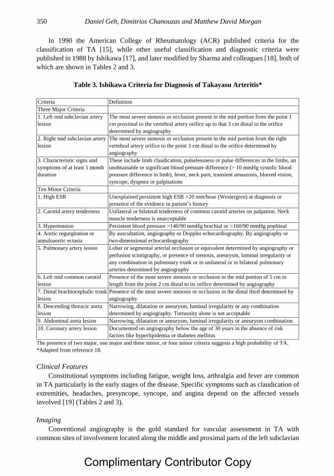
Daniel Geh, Dimitrios Chanouzas and Matthew David Morgan 350
In 1990 the American College of Rheumatology (ACR) published criteria for the
classification of TA [15], while other useful classification and diagnostic criteria were
published in 1988 by Ishikawa [17], and later modified by Sharma and colleagues [18], both of
which are shown in Tables 2 and 3.
Table 3. Ishikawa Criteria for Diagnosis of Takayasu Arteritis*
Criteria Definition
Three Major Criteria
1. Left mid subclavian artery
lesion
The most severe stenosis or occlusion present in the mid portion from the point 1
cm proximal to the vertebral artery orifice up to that 3 cm distal to the orifice
determined by angiography
2. Right mid subclavian artery
lesion
The most severe stenosis or occlusion present in the mid portion from the right
vertebral artery orifice to the point 3 cm distal to the orifice determined by
angiography
3. Characteristic signs and
symptoms of at least 1 month
duration
These include limb claudication, pulselessness or pulse differences in the limbs, an
unobtainable or significant blood pressure difference (> 10 mmHg systolic blood
pressure difference in limb), fever, neck pain, transient amaurosis, blurred vision,
syncope, dyspnea or palpitations
Ten Minor Criteria
1. High ESR Unexplained persistent high ESR >20 mm/hour (Westergren) at diagnosis or
presence of the evidence in patient’s history
2. Carotid artery tenderness Unilateral or bilateral tenderness of common carotid arteries on palpation. Neck
muscle tenderness is unacceptable
3. Hypertension Persistent blood pressure >140/90 mmHg brachial or >160/90 mmHg popliteal
4. Aortic regurgitation or
annuloaortic ectasia
By auscultation, angiography or Doppler echocardiography. By angiography or
two-dimensional echocardiography
5. Pulmonary artery lesion Lobar or segmental arterial occlusion or equivalent determined by angiography or
perfusion scintigraphy, or presence of stenosis, aneurysm, luminal irregularity or
any combination in pulmonary trunk or in unilateral or in bilateral pulmonary
arteries determined by angiography
6. Left mid common carotid
lesion
Presence of the most severe stenosis or occlusion in the mid portion of 5 cm in
length from the point 2 cm distal to its orifice determined by angiography
7. Distal brachiocephalic trunk
lesion
Presence of the most severe stenosis or occlusion in the distal third determined by
angiography
8. Descending thoracic aorta
lesion
Narrowing, dilatation or aneurysm, luminal irregularity or any combination
determined by angiography. Tortuosity alone is not acceptable
9. Abdominal aorta lesion Narrowing, dilatation or aneurysm, luminal irregularity or aneurysm combination
10. Coronary artery lesion Documented on angiography below the age of 30 years in the absence of risk
factors like hyperlipidemia or diabetes mellitus
The presence of two major, one major and three minor, or four minor criteria suggests a high probability of TA.
*Adapted from reference 18.
Clinical Features
Constitutional symptoms including fatigue, weight loss, arthralgia and fever are common
in TA particularly in the early stages of the disease. Specific symptoms such as claudication of
extremities, headaches, presyncope, syncope, and angina depend on the affected vessels
involved [19] (Tables 2 and 3).
Imaging
Conventional angiography is the gold standard for vascular assessment in TA with
common sites of involvement located along the middle and proximal parts of the left subclavian
Complimentary Contributor Copy
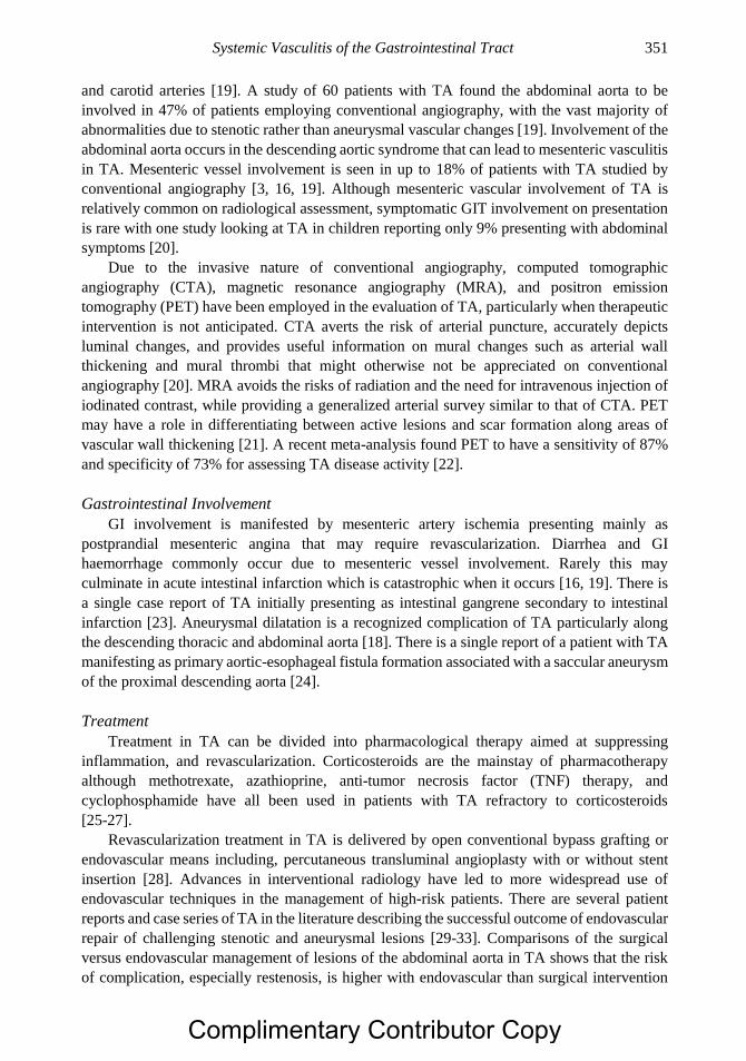
Systemic Vasculitis of the Gastrointestinal Tract 351
and carotid arteries [19]. A study of 60 patients with TA found the abdominal aorta to be
involved in 47% of patients employing conventional angiography, with the vast majority of
abnormalities due to stenotic rather than aneurysmal vascular changes [19]. Involvement of the
abdominal aorta occurs in the descending aortic syndrome that can lead to mesenteric vasculitis
in TA. Mesenteric vessel involvement is seen in up to 18% of patients with TA studied by
conventional angiography [3, 16, 19]. Although mesenteric vascular involvement of TA is
relatively common on radiological assessment, symptomatic GIT involvement on presentation
is rare with one study looking at TA in children reporting only 9% presenting with abdominal
symptoms [20].
Due to the invasive nature of conventional angiography, computed tomographic
angiography (CTA), magnetic resonance angiography (MRA), and positron emission
tomography (PET) have been employed in the evaluation of TA, particularly when therapeutic
intervention is not anticipated. CTA averts the risk of arterial puncture, accurately depicts
luminal changes, and provides useful information on mural changes such as arterial wall
thickening and mural thrombi that might otherwise not be appreciated on conventional
angiography [20]. MRA avoids the risks of radiation and the need for intravenous injection of
iodinated contrast, while providing a generalized arterial survey similar to that of CTA. PET
may have a role in differentiating between active lesions and scar formation along areas of
vascular wall thickening [21]. A recent meta-analysis found PET to have a sensitivity of 87%
and specificity of 73% for assessing TA disease activity [22].
Gastrointestinal Involvement
GI involvement is manifested by mesenteric artery ischemia presenting mainly as
postprandial mesenteric angina that may require revascularization. Diarrhea and GI
haemorrhage commonly occur due to mesenteric vessel involvement. Rarely this may
culminate in acute intestinal infarction which is catastrophic when it occurs [16, 19]. There is
a single case report of TA initially presenting as intestinal gangrene secondary to intestinal
infarction [23]. Aneurysmal dilatation is a recognized complication of TA particularly along
the descending thoracic and abdominal aorta [18]. There is a single report of a patient with TA
manifesting as primary aortic-esophageal fistula formation associated with a saccular aneurysm
of the proximal descending aorta [24].
Treatment
Treatment in TA can be divided into pharmacological therapy aimed at suppressing
inflammation, and revascularization. Corticosteroids are the mainstay of pharmacotherapy
although methotrexate, azathioprine, anti-tumor necrosis factor (TNF) therapy, and
cyclophosphamide have all been used in patients with TA refractory to corticosteroids
[25-27].
Revascularization treatment in TA is delivered by open conventional bypass grafting or
endovascular means including, percutaneous transluminal angioplasty with or without stent
insertion [28]. Advances in interventional radiology have led to more widespread use of
endovascular techniques in the management of high-risk patients. There are several patient
reports and case series of TA in the literature describing the successful outcome of endovascular
repair of challenging stenotic and aneurysmal lesions [29-33]. Comparisons of the surgical
versus endovascular management of lesions of the abdominal aorta in TA shows that the risk
of complication, especially restenosis, is higher with endovascular than surgical intervention
Complimentary Contributor Copy
Daniel Geh, Dimitrios Chanouzas and Matthew David Morgan 352
[34, 35]. A multicenter retrospective study found that over a median follow up of 6.5 years the
risk of complications following endovascular repair was 50% compared to 37.5% after surgical
repair [35]. Multivariable analysis revealed that patients with TA and evidence of systemic
inflammation at the time of revascularisation, as defined by an erythrocyte sedimentation rate
(ESR) > 30 mm/hour and C-reactive protein (CRP) level > 6 mg/L, were 7-fold more likely to
develop post-procedural complications including restenosis.
Giant Cell Arteritis
Epidemiology and Classification
Giant cell arteritis (GCA) is characterized by granulomatous inflammation that
predominantly affects the thoracic aorta and its branches. The age of onset is greater than 50
years with an increased incidence in those 70 to 80 years of age, and a higher incidence in
Northern Europeans and North Americans of Scandinavian descent compared to other ethnic
groups [36]. The 1990 ACR criteria are shown in Table 4. It should be noted that these criteria
were intended for the classification not the diagnosis of GCA [37].
Table 4. The American College of Rheumatology 1990 Criteria for the Classification
of Giant Cell Arteritis*
Criteria Definition
Age at disease onset >50 years Development of symptoms beginning at age 50 years or older
New headache New onset of or new type of localized pain in the head
Temporal artery abnormality Temporal artery tenderness on palpation or decreased pulsation, unrelated to
arteriosclerosis of cervical arteries
Elevated ESR ESR > 50mm in the 1st hour by the Westergren method
Abnormal artery biopsy Biopsy specimen with artery showing vasculitis characterized by a
predominance of mononuclear cell infiltration or granulomatous inflammation,
usually with multinucleated giant cells
For the purposes of classification at least 3 criteria must be fulfilled; sensitivity 93.5%, specificity 91.2%.
*Adapted from reference 37.
Clinical Features
The commonest manifestation of GCA is headache followed by scalp tenderness, jaw
claudication and visual disturbances; constitutional symptoms such as fever, fatigue and weight
loss are also prominent [5, 36]. Approximately 20% of patients with GCA develop clinical
manifestations reflecting large vessel involvement including aneurysmal and stenotic lesions
of the abdominal aorta which in turn can lead to GI sequelae [36].
Gastrointestinal Involvement
GCA can affect both the liver and the bowel. Asymptomatic involvement of the liver, as
manifested by elevated serum alkaline phosphatase and transaminase levels, is reported in up
to 50% of patients with GCA [38]. The mechanism leading to liver enzyme derangement is not
fully understood however when liver biopsy has been conducted in cases of symptomatic liver
involvement, this has demonstrated granulomatous inflammation of medium-sized arterioles
within the portal tracts [39]. Whilst clinically evident mesenteric involvement is uncommon,
Complimentary Contributor Copy
Systemic Vasculitis of the Gastrointestinal Tract 353
occult radiographic manifestations are noted on arteriography in 18% of patients with GCA [5,
40, 41].
In total there have been 11 reported cases of GCA with bowel involvement, mainly in the
form of mesenteric ischemia. Of 11 patients with bowel involvement in GCA, nine were
biopsy-proven [5, 8, 42-49], of whom four presented with abdominal symptoms in the absence
of symptoms of temporal arteritis. Temporal artery biopsy was positive in only two of the four
patients suggesting a negative temporal artery biopsy may not be able to reliably exclude GCA
involving the mesenteric vasculature [5].
Treatment
The mainstay of treatment in GCA is high dose corticosteroids to induce remission.
Methotrexate is recommended as an adjunctive therapy, while TNF- and interleukin (IL)-6
inhibitors may have a role in the treatment of relapsing or refractory disease [36, 50-52]. The
IL-6 inhibitor tocilizumab has shown particular promise with a recent phase three randomized
controlled trial demonstrating significant improvement in sustained remission rates and steroid
sparing effects in both newly diagnosed and relapsing GCA [52]. For occlusive disease surgical
revascularization is indicated. Comparison of the outcomes of 15 patients undergoing open
revascularization for occlusive mesenteric vasculitis secondary to TA, PAN, GCA and
indeterminate vasculitis compared to 163 patients undergoing open operations for
atherosclerotic disease [11] showed that both groups had similar freedom from mesenteric
symptoms and primary graft patency. In this study 8 of the 15 patients with vasculitic disease
had active inflammation at the time of intervention and by the end of follow up (mean follow-
up 41 months) 14 of 15 patients remained asymptomatic suggesting that surgical
revascularization even during active disease is an effective treatment for occlusive mesenteric
vasculitis.
MEDIUM VESSEL VASCULITIDES
Ischemia and infarction in dependent organs associated with bleeding in the GIT and within
the abdominal cavity, and aneurysm formation occurs in the context of vasculitic involvement
affecting medium size vessels typified by polyarteritis nodosa (PAN) and Kawasaki disease
(KD).
Polyarteritis Nodosa
Epidemiology
This systemic necrotizing vasculitis is characterized by fibrinoid necrosis that typically
involves medium sized arteries [53]. It typically affects middle-age and older individuals with
a peak onset in the 6th decade of life. Hepatitis B virus (HBV) infection is associated in up to
one third of patients with PAN and is an important consideration with regards to treatment and
prognosis [54].
Complimentary Contributor Copy
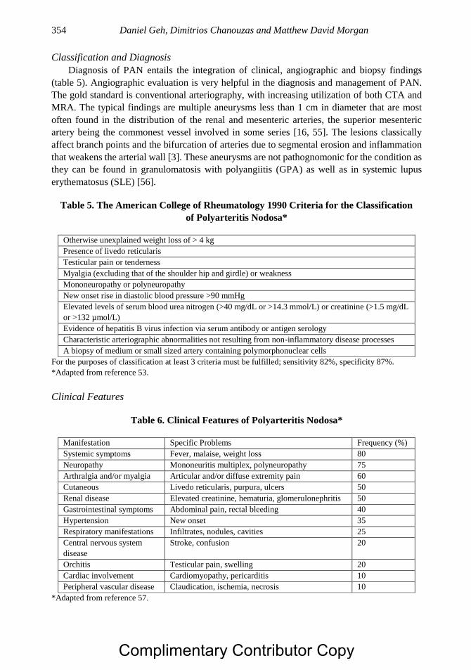
Daniel Geh, Dimitrios Chanouzas and Matthew David Morgan 354
Classification and Diagnosis
Diagnosis of PAN entails the integration of clinical, angiographic and biopsy findings
(table 5). Angiographic evaluation is very helpful in the diagnosis and management of PAN.
The gold standard is conventional arteriography, with increasing utilization of both CTA and
MRA. The typical findings are multiple aneurysms less than 1 cm in diameter that are most
often found in the distribution of the renal and mesenteric arteries, the superior mesenteric
artery being the commonest vessel involved in some series [16, 55]. The lesions classically
affect branch points and the bifurcation of arteries due to segmental erosion and inflammation
that weakens the arterial wall [3]. These aneurysms are not pathognomonic for the condition as
they can be found in granulomatosis with polyangiitis (GPA) as well as in systemic lupus
erythematosus (SLE) [56].
Table 5. The American College of Rheumatology 1990 Criteria for the Classification
of Polyarteritis Nodosa*
Otherwise unexplained weight loss of > 4 kg
Presence of livedo reticularis
Testicular pain or tenderness
Myalgia (excluding that of the shoulder hip and girdle) or weakness
Mononeuropathy or polyneuropathy
New onset rise in diastolic blood pressure >90 mmHg
Elevated levels of serum blood urea nitrogen (>40 mg/dL or >14.3 mmol/L) or creatinine (>1.5 mg/dL
or >132 µmol/L)
Evidence of hepatitis B virus infection via serum antibody or antigen serology
Characteristic arteriographic abnormalities not resulting from non-inflammatory disease processes
A biopsy of medium or small sized artery containing polymorphonuclear cells
For the purposes of classification at least 3 criteria must be fulfilled; sensitivity 82%, specificity 87%.
*Adapted from reference 53.
Clinical Features
Table 6. Clinical Features of Polyarteritis Nodosa*
Manifestation Specific Problems Frequency (%)
Systemic symptoms Fever, malaise, weight loss 80
Neuropathy Mononeuritis multiplex, polyneuropathy 75
Arthralgia and/or myalgia Articular and/or diffuse extremity pain 60
Cutaneous Livedo reticularis, purpura, ulcers 50
Renal disease Elevated creatinine, hematuria, glomerulonephritis 50
Gastrointestinal symptoms Abdominal pain, rectal bleeding 40
Hypertension New onset 35
Respiratory manifestations Infiltrates, nodules, cavities 25
Central nervous system
disease
Stroke, confusion 20
Orchitis Testicular pain, swelling 20
Cardiac involvement Cardiomyopathy, pericarditis 10
Peripheral vascular disease Claudication, ischemia, necrosis 10
*Adapted from reference 57.
Complimentary Contributor Copy
Systemic Vasculitis of the Gastrointestinal Tract 355
Clinical features of PAN include constitutional symptoms such as malaise, weight loss and
fever as well as specific symptoms and signs reflecting the extent of multi-system involvement
[57] (Table 6).
Gastrointestinal Involvement
GI involvement occurs in 14 to 65% of patients with PAN affecting both the bowel and
hepatobiliary system [55]. Abdominal pain is the most commonly reported GI symptom in
PAN. Bowel manifestations of PAN include ulceration, obstruction, infarction, and perforation
and hemorrhage that can affect the stomach, small and large bowel and the appendix with the
small bowel being most commonly affected [56-58]. Bowel involvement can arise from stenotic
lesions that lead to bowel wall ischemia and aneurysm formation that results in hemorrhage.
Necrosis of the bowel wall with the potential for subsequent perforation can develop if systemic
inflammation gives rise to transmural ischemia and confers a poor prognosis [55]. Liver
involvement occurs in 16 to 56% of patients and can manifest as fibrinoid necrosis of the liver,
hepatic artery aneurysms, liver infarction or hepatolithiasis [55, 59, 60]. Small vessel vasculitis
of the arteries that supply the bile ducts and gall bladder can lead to intrahepatic sclerosing
cholangitis, bile duct stricture, cholecystitis, and hemobilia. Pancreatic involvement can also
occur in the form of pancreatitis, pseudocysts and pancreatic insufficiency [55]. Hepatobiliary
involvement is potentially life threatening with death due to rupture of the hepatic artery
reported [61].
A retrospective analysis of 54 patients with PAN reported GI involvement in 24 (44%)
with two-thirds noting abdominal pain at presentation, while all others had abdominal pain at
some point in their illness. Of the 24 patients with GIT involvement at presentation 13 (54%)
developed an acute abdomen secondary to bowel infarction, perforation, acute cholecystitis,
gall bladder infarction, aneurysmal rupture of hepatic, splenic or renal arteries, or bleeding from
perforated gastric ulcers [62]. Multiple aneurysm formation is a defining feature of PAN and is
noted in 50 to 70% of those with involvement of the GIT with the hepatic artery affected in 50
to 60%; splenic artery in 45%; and pancreatic artery in 25 to 35% [62-64]. Massive internal
hemorrhage due to rupture of an aneurysm within the intestine is rare but nonetheless carries a
high mortality of up to 75% [64-67].
Treatment
The mainstay of treatment for mild PAN is corticosteroids. However GI involvement
constitutes severe disease, which has been shown to have a better outcome when treated with a
combination of cyclophosphamide and corticosteroids [68]. PAN secondary to HBV infection
is treated with two weeks of corticosteroids followed by antiviral treatment combined with
plasma exchange. Those so treated with antiviral therapy do not experience HBV relapses once
viral replication has stopped and seroconversion has been achieved [54].
Occlusive mesenteric disease can be managed successfully with open revascularization
[11]. Percutaneous endovascular trans-catheter embolization is increasing used to deal with
ruptured visceral artery aneurysms associated with PAN [69]. If tissue necrosis or gangrene
occurs due to infarction then definitive surgical management in the form of resection is
indicated [70].
Complimentary Contributor Copy
Daniel Geh, Dimitrios Chanouzas and Matthew David Morgan 356
Kawasaki Disease
Epidemiology and Clinical Features
Kawasaki disease (KD) is one of the commonest childhood vasculitides with a particularly
high incidence and prevalence in East Asia. Most patients have disease onset before 5 years of
age with a peak incidence at 30 months [71]. The diagnosis is confirmed by the presence of
fever for more than five days, and the presence of four of the following five diagnostic criteria:
bilateral conjunctival injection; changes of the mucous membranes of the upper respiratory
tract including injected pharynx, fissured lips or strawberry tongue; polymorphous rash; limb
changes including peripheral edema, erythema, or periungual desquamation; and cervical
adenopathy. Untreated KD is a self-limiting disease that lasts for 10 to 12 days; however,
coronary artery aneurysms develop in 20% to 25% of patients with significant associated
morbidity and mortality [71].
Gastrointestinal Involvement
GI involvement is relatively uncommon but tends to involve the gallbladder and the liver
leading to acute non-calculous distension of the gallbladder (gallbladder hydrops), hepatic
dysfunction with elevated serum transaminase concentrations and febrile cholestatic jaundice
[73-72]. Presentation with an acute abdomen was reported in 4.6% of children with KD [73]
due to vasculitic appendicitis, paralytic ileus, or hemorrhagic duodenitis. KD may be an
important cause of febrile cholestatic jaundice with up to 21% of febrile cholestatic cases
overall being attributed to KD in a recent series [74]. A high index of suspicion for KD should
therefore be maintained for children presenting with febrile cholestatic jaundice.
Treatment
Treatment of KD with aspirin and intravenous immunoglobulin (IVIg) reduces the
incidence of coronary artery complications compared with aspirin alone [75]. There are no
studies that have investigated the GI outcomes or GI response to treatment in KD. Interestingly,
elevated bilirubin and gamma-glutamyl transpeptidase (GT) have been associated with non-
responsiveness, defined by persistent fever, to IVIg treatment [76].
SMALL VESSEL VASCULITIDES
Involvement of small vessels by vasculitis can lead to ulceration, erosions, strictures and
occasionally perforation.
IgA Vasculitis/Henoch-Schönlein Purpura
Epidemiology and Clinical Features
IgA vasculitis/Henoch-Schönlein purpura (IgAV/HSP) is a small vessel vasculitis
characterized by a leukocytoclastic vasculitis and deposition of IgA-containing immune
complexes. It is the commonest form of systemic vasculitis in children with a peak incidence
at four to six years of age, although adult patients have been described [77]. It is usually self-
Complimentary Contributor Copy
Systemic Vasculitis of the Gastrointestinal Tract 357
limiting and characterized by the tetrad of palpable purpura in the absence of thrombocytopenia
or coagulopathy; arthritis or arthralgia; abdominal pain; and renal involvement. The disease
follows a more severe course when it presents in adults with a higher incidence of renal
impairment and end-stage renal disease [78].
Diagnosis
Although childhood IgAV/HSP is typically clinically diagnosed, adults generally undergo
skin or renal biopsy for tissue confirmation. Imaging can be helpful in assessing the GI
manifestations of IgAV/HSP especially ultrasonography for the detection of increased bowel
wall thickness, hematomas, and intussusception [79].
Gastrointestinal Involvement
GI involvement occurs in 50% of children with IgAV/HSP and can affect the hepatobiliary
system or the bowel in a number of ways [16]. Bowel manifestations include; intussusception
[80, 81], intestinal ischemia and infarction, intestinal perforation, gastrointestinal hemorrhage
and purpura [82-88] esophageal necrosis [89], ileitis and ileal stricture [90], fistulae formation
and acute appendicitis [91]. Hepatobiliary features include; pancreatitis [92] and hydrops of the
gallbladder [93]. Functional GIT disorders such as irritable bowel syndrome are reported to
have a higher prevalence in children with IgAV/HSP despite resolution of acute flares and are
associated with increased steroid use [94]. GI symptoms tend to occur within eight days of the
appearance of the rash although they may precede the rash. Abdominal pain in seen in 60-70%
of children with IgAV/HSP and is thought to be caused by submucosal bleeding and oedema
[95]. A 2001 Spanish study [96] noted that 16.7% of affected children presented with
abdominal pain as the solitary symptom; 7.6% presented with abdominal pain and a rash, and
2.6% with abdominal pain, rash and joint pains. Altogether, 70.5% of patients progressed to
mesenteric ischemia, and 30.8% had evidence of GI bleeding. The commonest GI manifestation
in childhood IgAV/HSP was intussusception. Although the prognosis of IgAV/HSP in children
is good, adult onset IgAV/HSP often has a less favorable outcome [78]. In adults severe GI
involvement, manifested by abdominal pain and bleeding [97-99], can be associated with a fatal
outcome [100-102].
Predictive Markers in Gastrointestinal IgAV/HSP
Due to the high frequency of GI complications in IgAV/HSP and its potentially serious
complications predictive markers have been sought. Serum amyloid A and faecal calprotectin
have both been demonstrated to be significantly increased in GIT IgAV/HSP compared to non
GIT IgAV/HSP [103, 104]. In a study of 70 patients with IgAV/HSP compared to controls
serum amyloid A levels were significantly increased in cases of IgAV/HSP with GIT
involvement but were not increased in renal or joint involvement [103]. Faecal calprotectin is
a heterodimer of calcium binding proteins and elevated stool levels are associated with GI
inflammation [105]. Faecal calprotectin is a useful screening tool in the diagnostic workup of
Complimentary Contributor Copy
Daniel Geh, Dimitrios Chanouzas and Matthew David Morgan 358
inflammatory bowel disease [106]. In a study of 66 children with IgAV/HSP faecal calprotectin
levels were found to be significantly different between groups with and without GIT
involvement as well as being significantly different between groups with and without renal
involvement [104].
Hematological ratios such as neutrophil-lymphocyte ratio and platelet-lymphocyte ratio
have been associated with GIT IgAV/HSP and have been suggested as screening tools for GI
involvement [107, 108].
Personalised medicine and genomics may be another approach to risk stratification of GIT
IgAV/HSP. A Chinese study has reported an association between platelet-activating factor
acetyl-hydrolase gene polymorphisms and GI bleeding [109]. None of these predictive markers
are in routine use and further prospective studies with larger numbers are needed to confirm
these findings before they could be used in routine clinical practice.
Treatment
Management of IgAV/HSP is largely supportive with administration of non-steroidal anti-
inflammatory drugs and corticosteroids for severe abdominal pain and joint involvement. There
is no convincing evidence to suggest that early use of corticosteroids reduces the risk of renal
involvement or acute GI complications [110]. Plasma exchange, IVIg, mycophenolate mofetil
(MMF) and single dose cyclophosphamide have all been reported in the management of GIT
IgAV/HSP [111-114]. In one case series of eight children with severe GIT IgAV/HSP
involvement six responded to IVIg with two having a further relapse both of which responded
to a second dose of IVIg [113]. Plasma exchange has also been reported to improve disease
control in a case series [111]. The management of intussusception in children with IgAV/HSP
often requires surgical correction [115].
ANCA-Associated Vasculitis
Epidemiology and Classification
The antineutrophil cytoplasmic antibody (ANCA)-associated vasculitides (AAV) are a
group of autoimmune inflammatory conditions comprising GPA, microscopic polyangiitis
(MPA), and eosinophilic granulomatosis with polyangiitis (EGPA) that predominantly affect
small- to medium-sized blood vessels.
The individual annual incidence of the AAV is 10 to 20 per million population with a peak
age of onset in the seventh and eighth decades [118]. Table 7 lists classification criteria for the
AAV [13].
Clinical Features
The clinical features of AAV include constitutional symptoms including malaise, joint
pains and weight loss, and other symptoms and signs depending on the extent of systemic
involvement. The upper and lower airways, skin, peripheral and central nervous system, as well
as the kidneys, are typically involved organ systems [119].
Complimentary Contributor Copy
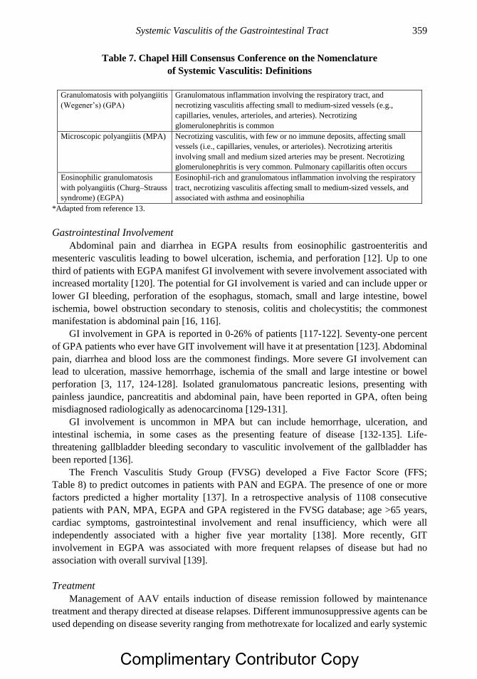
Systemic Vasculitis of the Gastrointestinal Tract 359
Table 7. Chapel Hill Consensus Conference on the Nomenclature
of Systemic Vasculitis: Definitions
Granulomatosis with polyangiitis
(Wegener’s) (GPA)
Granulomatous inflammation involving the respiratory tract, and
necrotizing vasculitis affecting small to medium-sized vessels (e.g.,
capillaries, venules, arterioles, and arteries). Necrotizing
glomerulonephritis is common
Microscopic polyangiitis (MPA) Necrotizing vasculitis, with few or no immune deposits, affecting small
vessels (i.e., capillaries, venules, or arterioles). Necrotizing arteritis
involving small and medium sized arteries may be present. Necrotizing
glomerulonephritis is very common. Pulmonary capillaritis often occurs
Eosinophilic granulomatosis
with polyangiitis (Churg–Strauss
syndrome) (EGPA)
Eosinophil-rich and granulomatous inflammation involving the respiratory
tract, necrotizing vasculitis affecting small to medium-sized vessels, and
associated with asthma and eosinophilia
*Adapted from reference 13.
Gastrointestinal Involvement
Abdominal pain and diarrhea in EGPA results from eosinophilic gastroenteritis and
mesenteric vasculitis leading to bowel ulceration, ischemia, and perforation [12]. Up to one
third of patients with EGPA manifest GI involvement with severe involvement associated with
increased mortality [120]. The potential for GI involvement is varied and can include upper or
lower GI bleeding, perforation of the esophagus, stomach, small and large intestine, bowel
ischemia, bowel obstruction secondary to stenosis, colitis and cholecystitis; the commonest
manifestation is abdominal pain [16, 116].
GI involvement in GPA is reported in 0-26% of patients [117-122]. Seventy-one percent
of GPA patients who ever have GIT involvement will have it at presentation [123]. Abdominal
pain, diarrhea and blood loss are the commonest findings. More severe GI involvement can
lead to ulceration, massive hemorrhage, ischemia of the small and large intestine or bowel
perforation [3, 117, 124-128]. Isolated granulomatous pancreatic lesions, presenting with
painless jaundice, pancreatitis and abdominal pain, have been reported in GPA, often being
misdiagnosed radiologically as adenocarcinoma [129-131].
GI involvement is uncommon in MPA but can include hemorrhage, ulceration, and
intestinal ischemia, in some cases as the presenting feature of disease [132-135]. Life-
threatening gallbladder bleeding secondary to vasculitic involvement of the gallbladder has
been reported [136].
The French Vasculitis Study Group (FVSG) developed a Five Factor Score (FFS;
Table 8) to predict outcomes in patients with PAN and EGPA. The presence of one or more
factors predicted a higher mortality [137]. In a retrospective analysis of 1108 consecutive
patients with PAN, MPA, EGPA and GPA registered in the FVSG database; age >65 years,
cardiac symptoms, gastrointestinal involvement and renal insufficiency, which were all
independently associated with a higher five year mortality [138]. More recently, GIT
involvement in EGPA was associated with more frequent relapses of disease but had no
association with overall survival [139].
Treatment
Management of AAV entails induction of disease remission followed by maintenance
treatment and therapy directed at disease relapses. Different immunosuppressive agents can be
used depending on disease severity ranging from methotrexate for localized and early systemic
Complimentary Contributor Copy
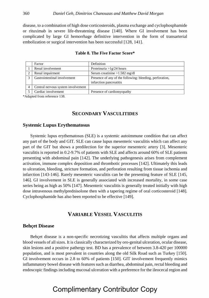
Daniel Geh, Dimitrios Chanouzas and Matthew David Morgan 360
disease, to a combination of high dose corticosteroids, plasma exchange and cyclophosphamide
or rituximab in severe life-threatening disease [140]. Where GI involvement has been
complicated by large GI hemorrhage definitive intervention in the form of transarterial
embolization or surgical intervention has been successful [128, 141].
Table 8. The Five Factor Score*
Factor Definition
1 Renal involvement Proteinuria >1g/24 hours
2 Renal impairment Serum creatinine >1.582 mg/dl
3 Gastrointestinal involvement Presence of any of the following: bleeding, perforation,
infarction pancreatitis
4 Central nervous system involvement
5 Cardiac involvement Presence of cardiomyopathy
*Adapted from reference 138.
SECONDARY VASCULITIDES
Systemic Lupus Erythematosus
Systemic lupus erythematosus (SLE) is a systemic autoimmune condition that can affect
any part of the body and GIT. SLE can cause lupus mesenteric vasculitis which can affect any
part of the GIT but shows a predilection for the superior mesenteric artery [3]. Mesenteric
vasculitis is reported in 0.2-9.7% of patients with SLE and affects around 60% of SLE patients
presenting with abdominal pain [142]. The underlying pathogenesis arises from complement
activation, immune complex deposition and thrombotic processes [142]. Ultimately this leads
to ulceration, bleeding, stricture formation, and perforation resulting from tissue ischemia and
infarction [143-146]. Rarely mesenteric vasculitis can be the presenting feature of SLE [145,
146]. GI involvement in SLE is generally associated with increased mortality, in some case
series being as high as 50% [147]. Mesenteric vasculitis is generally treated initially with high
dose intravenous methylprednisolone then with a tapering regime of oral corticosteroid [148].
Cyclophosphamide has also been reported to be effective [149].
VARIABLE VESSEL VASCULITIS
Behçet Disease
Behçet disease is a non-specific necrotizing vasculitis that affects multiple organs and
blood vessels of all sizes. It is classically characterized by oro-genital ulceration, ocular disease,
skin lesions and a positive pathergy test. BD has a prevalence of between 3.8-420 per 100000
population, and is most prevalent in countries along the old Silk Road such as Turkey [150].
GI involvement occurs in 2.8 to 60% of patients [150]. GIT involvement frequently mimics
inflammatory bowel disease with features such as diarrhea, abdominal pain, rectal bleeding and
endoscopic findings including mucosal ulceration with a preference for the ileocecal region and
Complimentary Contributor Copy
Systemic Vasculitis of the Gastrointestinal Tract 361
esophagus [12]. The GIT features of BD arise from large vessel vasculitis resulting in ischemia
and infarction or from neutrophilic phlebitis leading to mucosal ulceration [151, 152].
Ulceration is often deep and can penetrate through the bowel wall leading to perforation,
hemorrhage and fistula formation [153-155]. The most common hepatic manifestation of BD
is Budd-Chiari syndrome, seen in 1.3-3.2% of patients, which can result in portal hypertension,
ascites, varices and liver failure [150]. It is associated with significant mortality, one study
reporting a mean survival of 10.4 months [156]. Other rare GIT complications of BD include
carotid-esophageal fistula, aortic pseudoaneurysm rupture into the colon and pancreatitis [157-
159].
Disease Monitoring
Intestinal BD can be monitored clinically with the Disease Activity Index for Intestinal BD
(DAIBD), categorizing activity as quiescent, mild, moderate and severe [160]. Fecal
Calprotectin has been shown to be raised in intestinal BD and correlate with disease severity;
this could potentially become a predictive marker for intestinal BD [161].
Treatment
There is no universally accepted treatment regime, however 5-ASA and sulfasalazine have
been the mainstay of treatment for mild to moderate disease and corticosteroids for severe
disease or to induce remission [162, 163]. Azathioprine, anti-TNF therapy, thalidomide and
methotrexate have also been used successfully in intestinal BD [164-166]. For Budd-Chiari
Syndrome the mainstay of management is cyclophosphamide and corticosteroids whilst
anticoagulation remains controversial [167]. Surgical intervention is generally used for
uncontrolled GI bleeding and perforation [168]. Endoscopic intervention has been used for
ulcer related GI bleeding [169]. Large vessel involvement can be treated via open surgical
techniques or via the endovascular approach [170].
Other Secondary Vasculitides
GI complications occur in other autoimmune conditions such as rheumatoid arthritis (RA)
and mixed connective tissue disease (MCTD) due to secondary vasculitis. In patients with RA
vasculitis, up to 38% of cases have involvement of the mesenteric vessels leading rarely to
ischemic ulceration and bowel perforation [171]. Secondary vasculitis leading to GI
involvement in MCTD is rare and occurs in the form of small and large bowel ulceration and
perforation [172, 173] as well as fatal hemorrhage [174]. Vasculitic GI manifestations occur in
patients with Hepatitis C virus infection, as well as non-infectious mixed cryoglobulinemic
vasculitis, ranging from abdominal pain to GI bleeding, infarction, duodenitis, hepatic and
mesenteric vasculitis and perforation. GI perforation is associated with increased mortality
[175-177]. In addition, GI involvement has been described in vasculitis secondary to the drugs
propylthiouracil [178], hydroxyurea [179] and cocaine [180], or infection associated with
cytomegalovirus (CMV) [181].
Urticarial vasculitis (UV) is a rare small vessel vasculitis thought to be secondary to
immune complex deposition into the blood vessel wall. Clinical features include urticarial rash,
angioedema and tissue ischemia. The etiology of UV is usually idiopathic however; it can also
Complimentary Contributor Copy
Daniel Geh, Dimitrios Chanouzas and Matthew David Morgan 362
be secondary to autoimmune disease, paraneoplastic disease, drug reactions and infections
[182]. UV secondary to infection has been reported to present as circumferential duodenitis and
ulceration secondary to duodenal ischemia [183].
Treatment-Related GI Side Effects
As the treatment of vasculitis often involves immunosuppression with drugs such as
corticosteroids, cyclophosphamide, azathioprine, methotrexate and biological therapies such as
rituximab, treatment-related morbidity of the GI tract must be considered. Reactivation of CMV
can lead to GI inflammation in the context of immunosuppression. Symptoms of abdominal
pain, diarrhea and bleeding, as well as endoscopic signs of erythema, nodules and ulceration,
are non-specific but similar to those of vasculitis [12]. It is very important to secure the
diagnosis with biopsy specimens and peripheral blood CMV viral load. CMV disease usually
responds to anti-viral therapy. Candida involvement of the oral cavity and the esophagus is a
recognized complication of high-dose corticosteroid treatment. British Society of
Rheumatology Guidelines for the management of ANCA-associated vasculitis recommend that
anti-fungal prophylaxis should be considered in patients receiving immunosuppressive therapy
[140, 179]. However, according to a recent meta-analysis there is no clear evidence that the use
of topical anti-fungal preparations such as nystatin is effective in preventing fungal colonization
and infection in immunosuppressed patients [184]. Finally, corticosteroids and NSAID can lead
to gastroduodenal ulceration as well as small and large intestine enteropathy [12].
CONCLUSION
GI involvement in vasculitis largely reflects the size of the blood vessel involved. Large
vessel involvement may lead to stenotic lesions manifesting as ischemia or infarction of large,
but often unusual segments of the GI tract. Medium vessel involvement is typified by
aneurysmal as well as stenotic disease that can produce ischemia or infarction and bleeding
whilst small vessel disease can present with abdominal pain, patchy ulceration, infarction and
bleeding as well as intestinal perforation. Hepatobiliary and pancreatic involvement can occur
and represent common GIT manifestations in some vasculitides. Treatment is aimed at
controlling the underlying vasculitic process with immunosuppression although open surgical
or endovascular therapy is important in the management of mesenteric vasculitis. Severe
gastrointestinal involvement in vasculitis is associated with a higher mortality.
REFERENCES
[1] Watts RA, Scott DG. Recent developments in the classification and assessment of
vasculitis. Best Pract Res Clin Rheumatol. 2009;23(3):429-43.
[2] Jennette JC. Overview of the 2012 revised International Chapel Hill Consensus
Conference nomenclature of vasculitides. Clin Exp Nephrol. 2013;17(5):603-6.
Complimentary Contributor Copy
Systemic Vasculitis of the Gastrointestinal Tract 363
[3] Ha HK, Lee SH, Rha SE, Kim JH, Byun JY, Lim HK, et al. Radiologic features of
vasculitis involving the gastrointestinal tract. Radiographics. 2000;20(3):779-94.
[4] Gong EJ, Kim do H, Chun JH, Ahn JY, Choi KS, Jung KW, et al. Endoscopic Findings
of Upper Gastrointestinal Involvement in Primary Vasculitis. Gut Liver. 2016;10(4):542-
8.
[5] Annamalai A, Francis ML, Ranatunga SK, Resch DS. Giant cell arteritis presenting as
small bowel infarction. J Gen Intern Med. 2007;22(1):140-4.
[6] Simon S, Schittko G, Bosenberg H, Holl-Ulrich K, Schwarz-Eywill M. [Fulminant course
of a Takayasu’s arteritis and rare mesenteric arterial maninfestion]. Z Rheumatol.
2006;65(6):520, 2-6.
[7] Abdulwahab A, Almoallim H, Khan N. Isolated eosinophilic mesenteric vasculitis with
extensive thrombosis and splenic infarction in a 13-year-old boy. Clin Rheumatol.
2007;26(2):254-7.
[8] Trimble MA, Weisz MA. Infarction of the sigmoid colon secondary to giant cell arteritis.
Rheumatology (Oxford). 2002;41(1):108-10.
[9] Mocan H, Mocan MC, Sen Y, Kuzey G, Civiloglu C. Fatal polyarteritis nodosa with
massive mesenteric necrosis in a child. Clin Rheumatol. 1999;18(1):88-90.
[10] Edwards WH, Jr., Martin RS, 3rd, Edwards WH, Sr., Mulherin JL, Jr. Surviving
gastrointestinal infarction due to polyarteritis nodosa: a rare event. Am Surg.
1992;58(3):167-72.
[11] Rits Y, Oderich GS, Bower TC, Miller DV, Cooper L, Ricotta JJ, 2nd, et al. Interventions
for mesenteric vasculitis. J Vasc Surg. 2010;51(2):392-400 e2.
[12] Hokama A, Kishimoto K, Ihama Y, Kobashigawa C, Nakamoto M, Hirata T, et al.
Endoscopic and radiographic features of gastrointestinal involvement in vasculitis. World
J Gastrointest Endosc. 2012;4(3):50-6.
[13] Jennette J, Falk R, Andrassy K, Bacon P, Churg J, Gross W, et al. Nomenclature of
systemic vasculitides. Proposal of an international consensus conference. Arthritis
Rheum. 1994;37(2):187-92.
[14] Lupi-Herrera E, Sanchez-Torres G, Marcushamer J, Mispireta J, Horwitz S, Vela J.
Takayasu’s arteritis. Clinical study of 107 cases. American Heart Journal. 1977;
93(1):94-103.
[15] Arend W, Michel B, Bloch D, Hunder G, Calabrese L, Edworthy S, et al. The American
College of Rheumatology 1990 criteria for the classification of Takayasu arteritis.
Arthritis and Rheumatism. 1990;33(8):1129-34.
[16] Morgan MD, Savage CO. Vasculitis in the gastrointestinal tract. Best Pract Res Clin
Gastroenterol. 2005;19(2):215-33.
[17] Ishikawa K. Diagnostic approach and proposed criteria for the clinical diagnosis of
Takayasu’s arteriopathy. Journal of the American College of Cardiology. 1988;12(4):
964-72.
[18] Sharma BK, Jain S, Suri S, Numano F. Diagnostic criteria for Takayasu arteritis. Int J
Cardiol. 1996; 54 Suppl: S141-7.
[19] Kerr GS, Hallahan CW, Giordano J, Leavitt RY, Fauci AS, Rottem M, et al. Takayasu
Arteritis. Ann Intern Med. 1994;120(11):919-29.
[20] Yamada I, Nakagawa T, Himeno Y, Numano F, Shibuya H. Takayasu arteritis: evaluation
of the thoracic aorta with CT angiography. Radiology. 1998;209(1):103-9.
Complimentary Contributor Copy
Daniel Geh, Dimitrios Chanouzas and Matthew David Morgan 364
[21] Kissin EY, Merkel PA. Diagnostic imaging in Takayasu arteritis. Curr Opin Rheumatol.
2004;16(1):31-7.
[22] Soussan M, Nicolas P, Schramm C, Katsahian S, Pop G, Fain O, et al. Management of
large-vessel vasculitis with FDG-PET: a systematic literature review and meta-analysis.
Medicine (Baltimore). 2015; 94(14):e622.
[23] Misra DP, Krishnan N, Gochhait D, Emmanuel D, Negi VS. Takayasu arteritis (TA) first
presenting with intestinal ischemia: a case report and review of gastrointestinal tract
involvement (ischemic and non-ischemic) associated with TA. Rheumatol Int.
2017;37(1):169-75.
[24] Reddi A, Chetty R. Primary aorto-esophageal fistula due to Takayasu’s aortitis.
Cardiovasc Pathol. 2003;12(2):112-4.
[25] Hoffman G, Leavitt R, Kerr G, Rottem M, Sneller M, Fauci A. Treatment of
glucocorticoid-resistant or relapsing Takayasu arteritis with methotrexate. Arthritis and
Rheumatism. 1994;37(4):578-82.
[26] Valsakumar A, Valappil U, Jorapur V, Garg N, Nityanand S, Sinha N. Role of
immunosuppressive therapy on clinical, immunological, and angiographic outcome in
active Takayasu’s arteritis. Journal of Rheumatology. 2003;30(8):1793-8.
[27] Hoffman GS, Merkel PA, Brasington RD, Lenschow DJ, Liang P. Anti-tumor necrosis
factor therapy in patients with difficult to treat Takayasu arteritis. Arthritis Rheum.
2004;50(7):2296-304.
[28] Liang P, Hoffman GS. Advances in the medical and surgical treatment of Takayasu
arteritis. Curr Opin Rheumatol. 2005;17(1):16-24.
[29] Angiletta D, Marinazzo D, Guido G, Fullone M, Pulli R, Regina G. Eight-year follow-up
of endovascular repair of a brachiocephalic trunk aneurysm due to Takayasu’s arteritis.
J Vasc Surg. 2012;56(2):504-7.
[30] Lee BB, Laredo J, Neville R, Villavicencio JL. Endovascular management of takayasu
arteritis: is it a durable option? Vascular. 2009;17(3):138-46.
[31] Hu J, Huang H, Zhang X, Li G, Liu Q, Wu M, et al. Stent placement for treatment of long
segment (>/=80 mm) carotid artery stenosis in patients with Takayasu disease. J Vasc
Interv Radiol. 2012;23(11):1473-7.
[32] Bonilla-Abadia F, Echeverri AF, Carbonell JP, Canas CA. Multiple endovascular stent-
graft implantations in a patient with aortic thoracic and abdominal aneurysms due
Takayasu arteritis. Rheumatol Int. 2014;34(5):723-5.
[33] Agarwal G, Vats HS, Raval AN, Yevzlin AS, Chan MR, Gimelli G. Chronic total
occlusion and successful drug-eluting stent placement in Takayasu arteritis-induced renal
artery stenosis. Clin Med Res. 2013;11(4):233-6.
[34] Liang P, Tan-Ong M, Hoffman G. Takayasu’s arteritis: vascular interventions and
outcomes. Journal of Rheumatology. 2004;31(1):102-6.
[35] Saadoun D, Lambert M, Mirault T, Resche-Rigon M, Koskas F, Cluzel P, et al.
Retrospective analysis of surgery versus endovascular intervention in Takayasu arteritis:
a multicenter experience. Circulation. 2012;125(6):813-9.
[36] Salvarani C, Pipitone N, Versari A, Hunder GG. Clinical features of polymyalgia
rheumatica and giant cell arteritis. Nat Rev Rheumatol. 2012;8(9):509-21.
[37] Hunder G, Bloch D, Michel B, Stevens M, Arend W, Calabrese L, et al. The American
College of Rheumatology 1990 Criteria for the Classification of Giant Cell Arteritis.
Arthritis and Rheumatism. 1990;33(8):1122-8.
Complimentary Contributor Copy
Systemic Vasculitis of the Gastrointestinal Tract 365
[38] Kyle V. Laboratory investigations including liver in polymyalgia rheumatica/giant cell
arteritis. Baillieres Clin Rheumatol. 1991;5(3):475-84.
[39] Lee S, Childerhouse A, Moss K. Gastrointestinal symptoms and granulomatous vasculitis
involving the liver in giant cell arteritis: a case report and review of the literature.
Rheumatology (Oxford). 2011;50(12):2316-7.
[40] Scola CJ, Li C, Upchurch KS. Mesenteric involvement in giant cell arteritis. An
underrecognized complication? Analysis of a case series with clinicoanatomic
correlation. Medicine (Baltimore). 2008;87(1):45-51.
[41] Grayson PC, Maksimowicz-McKinnon K, Clark TM, Tomasson G, Cuthbertson D,
Carette S, et al. Distribution of arterial lesions in Takayasu’s arteritis and giant cell
arteritis. Ann Rheum Dis. 2012;71(8):1329-34.
[42] Stenwig JT. Intestinal gangrene due to giant-cell arteritis. Report of a case. J Oslo City
Hosp. 1976;26(5):49-55.
[43] Smith JA, O’Sullivan M, Gough J, Williams BD. Small-intestinal perforation secondary
to localized giant-cell arteritis of the mesenteric vessels. Br J Rheumatol.
1988;27(3):236-8.
[44] Srigley JR, Gardiner GW. Giant cell arteritis with small bowel infarction. A case report
and review of the literature. Am J Gastroenterol. 1980;73(2):157-61.
[45] Krant JD, Ross JM. Extracranial giant cell arteritis restricted to the small bowel. Arthritis
Rheum. 1992;35(5):603-4.
[46] Evans DJ, Wilkins MJ, Wazir JF, Rosin D. Extracranial giant cell arteritis. J R Coll Surg
Edinb. 1998;43(3):207-8.
[47] Tsuyuoka R, Takahashi T, Shinoda E, Taniguchi Y, Nishibe K, Takeuchi E, et al.
Intestinal perforation in temporal arteritis, associated with paroxysmal nocturnal
hemoglobinuria. Intern Med. 1996;35(2):159-61.
[48] Phelan MJ, Kok K, Burrow C, Thompson RN. Small bowel infarction in association with
giant cell arteritis. Br J Rheumatol. 1993;32(1):63-5.
[49] Ljungstrom K, Strandberg O, Sandstedt B. Infarction of the small bowel caused by giant
cell arteritis. Case report. Acta chirurgica Scandinavica. 1989;155(6-7):361-3.
[50] Cantini F, Niccoli L, Salvarani C, Padula A, Olivieri I. Treatment of longstanding active
giant cell arteritis with infliximab: report of four cases. Arthritis Rheum.
2001;44(12):2933-5.
[51] Seitz M, Reichenbach S, Bonel HM, Adler S, Wermelinger F, Villiger PM. Rapid
induction of remission in large vessel vasculitis by IL-6 blockade. A case series. Swiss
Med Wkly. 2011; 141:w13156.
[52] Stone JH, Tuckwell K, Dimonaco S, Klearman M, Aringer M, Blockmans D, et al. Trial
of Tocilizumab in Giant-Cell Arteritis. N Engl J Med. 2017;377(4):317-28.
[53] Pagnoux C, Seror R, Henegar C, Mahr A, Cohen P, Le Guern V, et al. Clinical features
and outcomes in 348 patients with polyarteritis nodosa: a systematic retrospective study
of patients diagnosed between 1963 and 2005 and entered into the French Vasculitis
Study Group Database. Arthritis Rheum. 2010;62(2):616-26.
[54] Guillevin L, Mahr A, Callard P, Godmer P, Pagnoux C, Leray E, et al. Hepatitis B virus-
associated polyarteritis nodosa: clinical characteristics, outcome, and impact of treatment
in 115 patients. Medicine (Baltimore). 2005;84(5):313-22.
[55] Ebert EC, Hagspiel KD, Nagar M, Schlesinger N. Gastrointestinal involvement in
polyarteritis nodosa. Clin Gastroenterol Hepatol. 2008;6(9):960-6.
Complimentary Contributor Copy
Daniel Geh, Dimitrios Chanouzas and Matthew David Morgan 366
[56] Travers RL, Allison DJ, Brettle RP, Hughes GR. Polyarteritis nodosa: a clinical and
angiographic analysis of 17 cases. Semin Arthritis Rheum. 1979;8(3):184-99.
[57] Pagnoux C, Mahr A, Cohen P, Guillevin L. Presentation and outcome of gastrointestinal
involvement in systemic necrotizing vasculitides: analysis of 62 patients with
polyarteritis nodosa, microscopic polyangiitis, Wegener granulomatosis, Churg-Strauss
syndrome, or rheumatoid arthritis-associated vasculitis. Medicine (Baltimore).
2005;84(2):115-28.
[58] Mowrey FH, Lundberg EA. The clinical manifestations of essential polyanglitis
(periarteritis nodosa), with emphasis on the hepatic manifestations. Ann Intern Med.
1954;40(6):1145-64.
[59] Guma M, Lorenzo-Zuniga V, Olive A, Perendreu J, Bechini J, Domenech E, et al. Occult
liver involvement by polyarteritis nodosa. Clin Rheumatol. 2002;21(2):184-6.
[60] Choi HL, Sung RH, Kang MH, Jeon HJ, Yun HY, Jang LC, et al. Polyarteritis nodosa
presented as a dilatation of the intrahepatic bile duct. Ann Surg Treat Res.
2014;87(5):273-5.
[61] Kanai R, Nakamura M, Tomisato K, Fukuhara T, Kondo A, Nakamura S, et al.
Cholangitis as an initial manifestation of polyarteritis nodosa. Intern Med.
2014;53(20):2307-12.
[62] Levine SM, Hellmann DB, Stone JH. Gastrointestinal involvement in polyarteritis
nodosa (1986-2000): Presentation and outcomes in 24 patients. The American Journal of
Medicine. 2002;112(5):386-91.
[63] Siebels M, Andrassy K, Noldge G, Sanwald R. [Spontaneous splenic and renal rupture
in polyarteritis nodosa]. Aktuelle Radiol. 1998;8(5):236-8.
[64] Parent BA, Cho SW, Buck DG, Nalesnik MA, Gamblin TC. Spontaneous rupture of
hepatic artery aneurysm associated with polyarteritis nodosa. Am Surg. 2010;76(12):
1416-9.
[65] Kendirli T, Yuksel S, Oral M, Unal N, Tulunay M, Dilek US, et al. Fatal polyarteritis
nodosa with gastrointestinal involvement in a child. Pediatr Emerg Care. 2006;
22(12):810-2.
[66] Tanabe J, Abe T, Okada N, Hayashi T, Akamatsu H, Tanimura H, et al. Massive
gastrointestinal bleeding in a patient with polyarteritis nodosa. J Gastroenterol.
2004;39(1):86-8.
[67] Vaidya GN, Khorasani-Zadeh A, John S. Polyarteritis nodosa presenting as profuse
gastrointestinal bleeding. BMJ Case Rep. 2014;2014.
[68] Leib ES, Restivo C, Paulus HE. Immunosuppressive and corticosteroid therapy of
polyarteritis nodosa. Am J Med. 1979;67(6):941-7.
[69] Koc O, Ozbek O, Gumus S, Demir A. Endovascular management of massive
gastrointestinal bleeding associated with polyarteritis nodosa. J Vasc Interv Radiol.
2009;20(2):277-9.
[70] Jagwani AV, Fathi NQ, Jailani RF, Zakaria AD. Rare and unusual case of polyarteritis
nodosa involving the gastrointestinal tract leading to bowel gangrene. BMJ Case Rep.
2017;2017.
[71] Burns JC, Glode MP. Kawasaki syndrome. Lancet. 2004;364(9433):533-44.
[72] Eladawy M, Dominguez SR, Anderson MS, Glode MP. Kawasaki disease and the
pediatric gastroenterologist: a diagnostic challenge. J Pediatr Gastroenterol Nutr.
2013;56(3):297-9.
Complimentary Contributor Copy
Systemic Vasculitis of the Gastrointestinal Tract 367
[73] Zulian F, Falcini F, Zancan L, Martini G, Secchieri S, Luzzatto C, et al. Acute surgical
abdomen as presenting manifestation of Kawasaki disease. The Journal of Pediatrics.
2003;142(6):731-5.
[74] Taddio A, Pellegrin MC, Centenari C, Filippeschi IP, Ventura A, Maggiore G. Acute
febrile cholestatic jaundice in children: keep in mind Kawasaki disease. J Pediatr
Gastroenterol Nutr. 2012;55(4):380-3.
[75] Newburger J, Takahashi M, Beiser A, Burns J, Bastian J, Chung K, et al. A single
intravenous infusion of gamma globulin as compared with four infusions in the treatment
of acute Kawasaki syndrome. New England Journal of Medicine. 1991;324 (23):1633-9.
[76] Fukunishi M, Kikkawa M, Hamana K, Onodera T, Matsuzaki K, Matsumoto Y, et al.
Prediction of non-responsiveness to intravenous high-dose gamma-globulin therapy in
patients with Kawasaki disease at onset. J Pediatr. 2000;137(2):172-6.
[77] Gardner-Medwin JM, Dolezalova P, Cummins C, Southwood TR. Incidence of Henoch-
Schonlein purpura, Kawasaki disease, and rare vasculitides in children of different ethnic
origins. The Lancet. 2002;360(9341):1197-202.
[78] Pillebout E, Thervet E, Hill G, Alberti C, Vanhille P, Nochy D. Henoch-Schonlein
Purpura in Adults: Outcome and Prognostic Factors. J Am Soc Nephrol. 2002;13(5):
1271-8.
[79] Choong C, Beasley S. Intra-abdominal manifestations of Henoch-Schonlein purpura.
Journal of Paediatrics and Child Health. 1998;349(5):405-9.
[80] Jasic M, Subat-Dezulovic M, Nikolic H, Jonjic N, Manestar K, Dezulovic M. Henoch-
Schonlein purpura complicated by appendicitis, intussusception and ureteritis. Coll
Antropol. 2011;35(1):197-201.
[81] Huang WK, Fan PC, Chen YC, Tian YC, Kang SC, Hsueh S, et al. Henoch-Schonlein
purpura with intussusception in an adult. Am J Med Sci. 2012;344(4):337-9.
[82] Nakamura S, Hisamatsu T, Kikuchi J, Adachi M, Yamagishi Y, Imaeda H, et al. A case
of IgA-related enteropathy complicated with gastrointestinal bleeding and progressive
IgA nephropathy: a possible variant Henoch-Schonlein purpura? Intern Med.
2010;49(16):1755-61.
[83] Cristescu DA, Yuvienco C, Schwartz S. Fatal Henoch-Schonlein purpura in an adult with
Dieulafoy lesions. J Clin Rheumatol. 2012;18(5):253-6.
[84] Rathore M, Shrivastava R, Goyal R, Radotra BD, Thapa BR. Henoch Schonlein purpura
presenting as duodenal ulcer and gastric outlet obstruction. Indian J Pediatr.
2014;81(2):189-90.
[85] Waelkens B, Van Moerkercke W, Lerut E, Sprangers B. Henoch-Schonlein Purpura
complicated with major gastro-intestinal hemorrhage. Acta Gastroenterol Belg.
2014;77(4):379-82.
[86] Yamazaki T, Akimoto T, Iwazu Y, Sugase T, Takeshima E, Numata A, et al. Henoch-
Schonlein purpura complicated with severe gastrointestinal bleeding. CEN Case Rep.
2015;4(1):106-11.
[87] Shah R, Ramakrishnan M, Vollmar A, Harrell A, Van Trump R, Masoud A. Henoch-
Schonlein Purpura Presenting as Severe Gastrointestinal and Renal Involvement with
Mixed Outcomes in an Adult Patient. Cureus. 2017;9(3):e1088.
[88] Naruse G, Shimada K. Images in clinical medicine. Cutaneous and gastrointestinal
purpura. N Engl J Med. 2013;369(19):1843.
Complimentary Contributor Copy
Daniel Geh, Dimitrios Chanouzas and Matthew David Morgan 368
[89] Iorio N, Bernstein GR, Malik Z, Schey R. Acute Esophageal Necrosis Presenting With
Henoch-Schonlein Purpura. ACG Case Rep J. 2015;3(1):17-9.
[90] Fan Z, Tian X, Pan J, Li Y, Zhang Y, Jing H. Terminal ileitis induced by Henoch-
Schonlein purpura that presented as acute appendicitis: a case report. Medicine
(Baltimore). 2015;94(5):e492.
[91] Semeena N, Adlekha S. Henoch-schonlein purpura associated with gangrenous
appendicitis: a case report. Malays J Med Sci. 2014;21(2):71-3.
[92] Branski D, Gross V, Gross-Kieselstein E, Roll D, Abrahamov A. Pancreatitis as a
complication of Henoch--Schonlein purpura. J Pediatr Gastroenterol Nutr. 1982;1
(2):275-6.
[93] McCrindle BW, Wood RA, Nussbaum AR. Henoch-Schonlein syndrome. Unusual
manifestations with hydrops of the gallbladder. Clin Pediatr (Phila). 1988;27(5):254-6.
[94] Saps M, Dhroove G, Chogle A. Henoch-Schonlein purpura leads to functional
gastrointestinal disorders. Dig Dis Sci. 2011;56(6):1789-93.
[95] Koster MJ, Warrington KJ. Vasculitis of the mesenteric circulation. Best Pract Res Clin
Gastroenterol. 2017;31(1):85-96.
[96] Calvino M, Llorca J, Garcia-Porrua C, Fernandez-Iglesias J, Rodriguez-Ledo P,
Gonzalez-Gay M. Henoch-Schonlein purpura in children from northwestern Spain: a 20-
year epidemiologic and clinical study. Medicine (Baltimore). 2001;80(5):279-90.
[97] Mills J, Michel B, Bloch D, Calabrese L, Hunder G, Arend W, et al. The American
College of Rheumatology 1990 Criteria for the Classification of Henoch-Schönlein
Purpura. Arthritis and Rheumatism. 1990;33(8):1114-21.
[98] Garcia-Porrua C, Gonzalez-Gay M. Comparative clinical and epidemiological study of
hypersensitivity vasculitis versus Henoch-Schonlein purpura in adults. Seminars in
Arthritis and Rheumatism. 1999;28(6):404-12.
[99] Blanco R, Martinez-Taboada V, Rodriguez-Valverde V, Garcia-Fuentes M, Gonzalez-
Gay M. Henoch-Schonlein purpura in adulthood and childhood: two different
expressions of the same syndrome. Arthritis and Rheumatism. 1997;40(5):859-64.
[100] Carmichael P, Brun E, Jayawardene S, Abdulkadir A, O’Donnell PJ. A fatal case of
bowel and cardiac involvement in Henoch-Schonlein purpura. Nephrol Dial Transplant.
2002;17(3):497-9.
[101] Oshikata C, Tsurikisawa N, Takigawa M, Omori T, Sugano S, Tsuburai T, et al. An adult
patient with Henoch-Schonlein purpura and non-occlusive mesenteric ischemia. BMC
Res Notes. 2013;6:26.
[102] Oshima K, Iino N, Sasaki N, Takahashi N, Tanabe Y, Ito S, et al. Adult onset
anaphylactoid purpura with severe gastrointestinal involvement. Intern Med. 2003;42
(5):436-42.
[103] He X, Zhao Y, Li Y, Kang S, Ding Y, Luan J, et al. Serum amyloid A levels associated
with gastrointestinal manifestations in Henoch-Schonlein purpura. Inflammation.
2012;35(4):1251-5.
[104] Kanik A, Baran M, Ince FD, Cebeci O, Bozkurt M, Cavusoglu D, et al. Faecal
calprotectin levels in children with Henoch-Schonlein purpura: is this a new marker for
gastrointestinal involvement? Eur J Gastroenterol Hepatol. 2015;27(3):254-8.
[105] Roseth AG, Schmidt PN, Fagerhol MK. Correlation between faecal excretion of indium-
111-labelled granulocytes and calprotectin, a granulocyte marker protein, in patients with
inflammatory bowel disease. Scand J Gastroenterol. 1999;34(1):50-4.
Complimentary Contributor Copy
Systemic Vasculitis of the Gastrointestinal Tract 369
[106] van Rheenen PF, Van de Vijver E, Fidler V. Faecal calprotectin for screening of patients
with suspected inflammatory bowel disease: diagnostic meta-analysis. BMJ.
2010;341:c3369.
[107] Gayret OB, Erol M, Tekin Nacaroglu H. The Relationship of Neutrophil-Lymphocyte
Ratio and Platelet-Lymphocyte Ratio with Gastrointestinal Bleeding in Henoch-
Schonlein Purpura. Iran J Pediatr. 2016;26(5):e8191.
[108] Makay B, Gucenmez OA, Duman M, Unsal E. The relationship of neutrophil-to-
lymphocyte ratio with gastrointestinal bleeding in Henoch-Schonlein purpura. Rheumatol
Int. 2014;34(9):1323-7.
[109] Wang BX, Mei H, Peng HM, Gao Y, Ding Y. [Association between platelet-activating
factor acetylhydrolase gene polymorphisms and gastrointestinal bleeding in children with
Henoch-Schonlein purpura]. Zhongguo Dang Dai Er Ke Za Zhi. 2017;19(4):385-8.
[110] Huber A, King J, McLaine P, Klassen T, Pothos M. A randomized, placebo-controlled
trial of prednisone in early Henoch Schonlein Purpura2004.
[111] Basaran O, Cakar N, Uncu N, Celikel BA, Kara A, Cayci FS, et al. Plasma exchange
therapy for severe gastrointestinal involvement of Henoch Schonlein purpura in children.
Clin Exp Rheumatol. 2015; 33(2 Suppl 89): S-176-80.
[112] Chou T, Louissant VR, Adams A, Gurkan S, Chefitz D, Weller AS, et al. Successful
Treatment of Henoch-Schonlein Purpura With Recurrent Gastrointestinal Involvement
With Mycophenolate Mofetil: A Brief Report. Clin Pediatr (Phila). 2015;54(9):900-3.
[113] Cherqaoui B, Chausset A, Stephan JL, Merlin E. Intravenous immunoglobulins for severe
gastrointestinal involvement in pediatric Henoch-Schonlein purpura: A French
retrospective study. Arch Pediatr. 2016;23(6):584-90.
[114] Uluca U, Ece A, Sen V, Yel S, Tan I, Karabel D. Management of intestinal bleeding with
single-dose cyclophosphamide in Henoch-Schonlein purpura. Pediatr Int.
2015;57(3):498-500.
[115] Schwab J, Benya E, Lin R, Majd K. Contrast enema in children with Henoch-Schonlein
purpura. J Pediatr Surg. 2005;40(8):1221-3.
[116] Vaglio A, Corradi D, Ronda N, Garini G, Buzio C. Large Bowel Obstruction Heralding
Churg-Strauss Syndrome. Am J Gastroenterology. 2004;99(3):562-3.
[117] Akbulut S. Multiple ileal perforations in a patient with Wegener’s granulomatosis: a case
report and literature review. J Gastrointest Surg. 2012;16(4):857-62.
[118] Bligny D, Mahr A, Toumelin P, Mouthon L, Guillevin L. Predicting mortality in systemic
Wegener’s granulomatosis: a survival analysis based on 93 patients. Arthritis Rheum.
2004;51(1):83-91.
[119] Hoffman G, Kerr G, Leavitt R, Hallahan C, Lebovics R, Travis W, et al. Wegener
granulomatosis: an analysis of 158 patients. Ann Intern Med. 1992;116(6):488-98.
[120] Aasarod K, Iversen BM, Hammerstrom J, Bostad L, Vatten L, Jorstad S. Wegener’s
granulomatosis: clinical course in 108 patients with renal involvement. Nephrol Dial
Transplant. 2000;15(5):611-8.
[121] Koldingsnes W, Nossent H. Predictors of organ damage and survival in Wegener’s
granulomatosis. Rheumatology. 2002;41(5):572-81.
[122] Masiak A, Zdrojewski L, Zdrojewski Z, Bullo-Piontecka B, Rutkowski B.
Gastrointestinal tract involvement in granulomatosis with polyangiitis. Prz
Gastroenterol. 2016;11(4):270-5.
Complimentary Contributor Copy
Daniel Geh, Dimitrios Chanouzas and Matthew David Morgan 370
[123] Latus J, Koetter I, Fritz P, Kimmel M, Biegger D, Ott G, et al. Gastrointestinal
involvement in granulomatosis with polyangiitis and microscopic polyangiitis:
histological features and outcome. Int J Rheum Dis. 2014;17(4):412-9.
[124] Samim M, Pronk A, Verheijen PM. Intestinal perforation as an early complication in
Wegener’s granulomatosis. World J Gastrointest Surg. 2010;2(5):169-71.
[125] Yildirim AC, Kocak E, Yildiz P, Yildiz M, Karakayali AS, Kaptanoglu B, et al. Multiple
intestinal perforation in a patient with Wegener’s granulomatosis: a case report and
review of the literature. Gastroenterol Clin Biol. 2010;34(12):712-5.
[126] Strivens RL, Bateman A, Arden NK, Edwards CJ. Intestinal perforation and jejunal
haemorrhage due to Wegener’s granulomatosis. Clin Exp Rheumatol. 2005;23(1):124.
[127] Tavakkoli H, Zobeiri M, Salesi M, Sanei MH. Upper Gastrointestinal Bleeding as the
First Manifestation of Wegener’s Granulomatosis. Middle East J Dig Dis. 2016;8(3):
235-9.
[128] Kocak G, Kocak E, Huddam B, Azak A, Voyvoda N, Bulut M, et al. Successful surgical
treatment of massive lower gastrointestinal bleeding in a patient with Wegener’s
granulomatosis. Am Surg. 2012;78(9):E416-7.
[129] Kontis E, Papalexopoulou N, Zen Y, Prachalias AA. Isolated primary pancreatic
Wegener’s granulomatosis: report of two cases. JOP. 2014;15(4):403-6.
[130] O’Neil K, Jones D, Lawson J. Wegener’s granulomatosis masquerading as pancreatic
carcinoma. Digestive diseases and sciences. 1992;37(5):702-4.
[131] Valerieva Y, Golemanov B, Tzolova N, Mitova R. Pancreatic mass as an initial
presentation of severe Wegener’s granulomatosis. Ann Gastroenterol. 2013;26(3):267-9.
[132] Ueda S, Matsumoto M, Ahn T, Adachi S, Oku K, Takagi M, et al. Microscopic
polyangiitis complicated with massive intestinal bleeding. J Gastroenterol. 2001;
36(4):264-70.
[133] Tsai CN, Chang CM, Chuang CH, Jin YT, Liu MF, Wang CR. Extended colonic
ulcerations in a patient with microscopic polyangiitis. Ann Rheum Dis. 2004;63(11)
:1521-2.
[134] Passam FH, Diamantis ID, Perisinaki G, Saridaki Z, Kritikos H, Georgopoulos D, et al.
Intestinal ischemia as the first manifestation of vasculitis. Semin Arthritis Rheum.
2004;34(1):431-41.
[135] Ito Y, Tanaka A, Sugiura Y, Sezaki R. An autopsy case of intraabdominal hemorrhage in
microscopic polyangiitis. Intern Med. 2011;50(14):1501-2.
[136] Kitaguchi D, Kurata M, Shimomura O, Oda T, Ohkohchi N. Gallbladder bleeding
associated with microscopic polyangiitis: a case report. J Surg Case Rep. 2017;2017(8)
:rjx159.
[137] Guillevin L, Lhote F, Gayraud M, Cohen P, Jarrousse B, Lortholary O, et al. Prognostic
Factors in Polyarteritis Nodosa and Churg-Strauss Syndrome: A Prospective Study in
342 Patients. Medicine (Baltimore). 1996;75(1):17-28.
[138] Guillevin L, Pagnoux C, Seror R, Mahr A, Mouthon L, Le Toumelin P. The Five-Factor
Score revisited: assessment of prognoses of systemic necrotizing vasculitides based on
the French Vasculitis Study Group (FVSG) cohort. Medicine (Baltimore). 2011;90(1):19-
27.
[139] Tsurikisawa N, Oshikata C, Kinoshita A, Tsuburai T, Saito H. Longterm Prognosis of
121 Patients with Eosinophilic Granulomatosis with Polyangiitis in Japan. J Rheumatol.
2017;44(8):1206-15.
Complimentary Contributor Copy
Systemic Vasculitis of the Gastrointestinal Tract 371
[140] Ntatsaki E, Carruthers D, Chakravarty K, D’Cruz D, Harper L, Jayne D, et al. BSR and
BHPR guideline for the management of adults with ANCA-associated vasculitis.
Rheumatology (Oxford). 2014;53(12):2306-9.
[141] Li L, Li X, Fu P, Liu F. Recurrent gastrointestinal bleeding with ANCA associated
glomerulonephritis successfully treated by transarterial embolization. Pak J Med Sci.
2013;29(6):1465-7.
[142] Ju JH, Min JK, Jung CK, Oh SN, Kwok SK, Kang KY, et al. Lupus mesenteric vasculitis
can cause acute abdominal pain in patients with SLE. Nat Rev Rheumatol. 2009;5(5):273-
81.
[143] Ebert EC, Hagspiel KD. Gastrointestinal and hepatic manifestations of systemic lupus
erythematosus. J Clin Gastroenterol. 2011;45(5):436-41.
[144] Iannella I, Candela S, Di Libero L, Argano F, Tartaglia E, Candela G. Ischemic necrosis
with sigmoid perforation in a patient with Systemic Lupus Erythematosus (SLE): case
report. G Chir. 2012;33(3):77-80.
[145] Chattopadhyay P, Abby Philips C, Dhua D, Saha S. Systemic lupus erythematosus
presenting as ischaemic proctitis. Lupus. 2011;20(6):653-5.
[146] Nozari N, Divsalar P. Systemic lupus erythematosus presenting with a fatal intestinal
vasculitis: a case report. Middle East J Dig Dis. 2014;6(3):162-4.
[147] Zizic TM, Classen JN, Stevens MB. Acute abdominal complications of systemic lupus
erythematosus and polyarteritis nodosa. Am J Med. 1982;73(4):525-31.
[148] Sultan SM, Ioannou Y, Isenberg DA. A review of gastrointestinal manifestations of
systemic lupus erythematosus. Rheumatology (Oxford). 1999;38(10):917-32.
[149] Grimbacher B, Huber M, von Kempis J, Kalden P, Uhl M, Kohler G, et al. Successful
treatment of gastrointestinal vasculitis due to systemic lupus erythematosus with
intravenous pulse cyclophosphamide: a clinical case report and review of the literature.
Br J Rheumatol. 1998;37(9):1023-8.
[150] Skef W, Hamilton MJ, Arayssi T. Gastrointestinal Behcet’s disease: a review. World J
Gastroenterol. 2015;21(13):3801-12.
[151] Vaiopoulos AG, Sfikakis PP, Kanakis MA, Vaiopoulos G, Kaklamanis PG.
Gastrointestinal manifestations of Behcet’s disease: advances in evaluation and
management. Clin Exp Rheumatol. 2014; 32(4 Suppl 84):S140-8.
[152] Chin AB, Kumar AS. Behcet colitis. Clin Colon Rectal Surg. 2015;28(2):99-102.
[153] Kasahara Y, Tanaka S, Nishino M, Umemura H, Shiraha S, Kuyama T. Intestinal
involvement in Behcet’s disease: review of 136 surgical cases in the Japanese literature.
Dis Colon Rectum. 1981;24(2):103-6.
[154] Lee JY, Park JY, Kim EH, Tak MJ, Lee ES. Simultaneous involvement of nervous and
gastrointestinal systems in Behcet’s disease. Ann Dermatol. 2012;24(2):225-7.
[155] Bes C, Dagli U, Yilmaz F, Soy M. Massive lower gastrointestinal bleeding associated
with solitary rectal ulcer in a patient with Behcet’s disease. Reumatismo. 2015;67(2):82-
4.
[156] Bayraktar Y, Balkanci F, Bayraktar M, Calguneri M. Budd-Chiari syndrome: a common
complication of Behcet’s disease. Am J Gastroenterol. 1997;92(5):858-62.
[157] Kimura N, Takada K, Murata K, Kohsaka H, Miyasaka N. A case of Behcet’s disease
complicated by carotid-oesophageal fistula. Rheumatology (Oxford). 2014;53(1):196-8.
[158] Lee SL, Ku YM, Won Y. Spontaneous aortic pseudoaneurysm rupture into the sigmoid
colon in Behcet’s disease patient. World J Gastroenterol. 2015;21(46):13201-4.
Complimentary Contributor Copy
Daniel Geh, Dimitrios Chanouzas and Matthew David Morgan 372
[159] Backmund M, Schomerus P. Acute pancreatitis and pericardial effusion in Behcet’s
disease. Gastroenterology. 1999;117(1):286.
[160] Cheon JH, Han DS, Park JY, Ye BD, Jung SA, Park YS, et al. Development, validation,
and responsiveness of a novel disease activity index for intestinal Behcet’s disease.
Inflamm Bowel Dis. 2011;17(2):605-13.
[161] Ozseker B, Sahin C, Ozseker HS, Efe SC, Kav T, Bayraktar Y. The Role of Fecal
Calprotectin in Evaluating Intestinal Involvement of Behcet’s Disease. Dis Markers.
2016; 2016:5423043.
[162] Jung YS, Hong SP, Kim TI, Kim WH, Cheon JH. Long-term clinical outcomes and
factors predictive of relapse after 5-aminosalicylate or sulfasalazine therapy in patients
with intestinal Behcet disease. J Clin Gastroenterol. 2012; 46(5):e38-45.
[163] Hisamatsu T, Naganuma M, Matsuoka K, Kanai T. Diagnosis and management of
intestinal Behcet’s disease. Clin J Gastroenterol. 2014;7(3):205-12.
[164] Tanida S, Inoue N, Kobayashi K, Naganuma M, Hirai F, Iizuka B, et al. Adalimumab for
the treatment of Japanese patients with intestinal Behcet’s disease. Clin Gastroenterol
Hepatol. 2015;13(5):940-8 e3.
[165] Hatemi I, Hatemi G, Pamuk ON, Erzin Y, Celik AF. TNF-alpha antagonists and
thalidomide for the management of gastrointestinal Behcet’s syndrome refractory to the
conventional treatment modalities: a case series and review of the literature. Clin Exp
Rheumatol. 2015;33(6 Suppl 94):S129-37.
[166] Iwata S, Saito K, Yamaoka K, Tsujimura S, Nawata M, Hanami K, et al. Efficacy of
combination therapy of anti-TNF-alpha antibody infliximab and methotrexate in
refractory entero-Behcet’s disease. Mod Rheumatol. 2011;21(2):184-91.
[167] Hatemi G, Silman A, Bang D, Bodaghi B, Chamberlain AM, Gul A, et al. EULAR
recommendations for the management of Behcet disease. Ann Rheum Dis. 2008;67
(12):1656-62.
[168] Jung YS, Yoon JY, Lee JH, Jeon SM, Hong SP, Kim TI, et al. Prognostic factors and
long-term clinical outcomes for surgical patients with intestinal Behcet’s disease.
Inflamm Bowel Dis. 2011;17(7):1594-602.
[169] Nishiyama N, Mori H, Kobara H, Rafiq K, Fujihara S, Ayaki M, et al. Endoscopic
management with over-the-scope clips for intestinal bleeding of Behcet’s disease.
Gastrointest Endosc. 2015;81(5):1275-6.
[170] Calamia KT, Schirmer M, Melikoglu M. Major vessel involvement in Behcet’s disease:
an update. Curr Opin Rheumatol. 2011;23(1):24-31.
[171] Ebert EC, Hagspiel KD. Gastrointestinal and hepatic manifestations of rheumatoid
arthritis. Dig Dis Sci. 2011;56(2):295-302.
[172] Maeda N, Abe S, Yoshizawa T, Ogawa K, Sekigawa I, Iida N, et al. Low-dose
cyclosporin for multiple colonic ulcers associated with mixed connective tissue disease.
Clin Rheumatol. 1999;18(5):410-3.
[173] Marshall JB, Kretschmar JM, Gerhardt DC, Winship DH, Winn D, Treadwell EL, et al.
Gastrointestinal manifestations of mixed connective tissue disease. Gastroenterology.
1990;98(5 Pt 1):1232-8.
[174] Cooke CL, Lurie HI. Case report: fatal gastrointestinal hemorrhage in mixed connective
tissue disease. Arthritis Rheum. 1977;20(7):1421-7.
Complimentary Contributor Copy
Systemic Vasculitis of the Gastrointestinal Tract 373
[175] Terrier B, Carrat F, Krastinova E, Marie I, Launay D, Lacraz A, et al. Prognostic factors
of survival in patients with non-infectious mixed cryoglobulinaemia vasculitis: data from
242 cases included in the CryoVas survey. Ann Rheum Dis. 2013;72(3):374-80.
[176] Calle Toro JS, Davalos DM, Charry JD, Arrunategi AM, Tobon G. Hepatic and
Mesenteric Vasculitis as Presenting Manifestation of Mixed Cryoglobulinemia Related
to Chronic Hepatitis C Virus Infection in a Female Patient. J Clin Rheumatol.
2016;22(4):212-4.
[177] Berera SR, Gomez A, Dholaria K, Arosemena LR, Ladino-Avellaneda MA, Barisoni L,
et al. A Rare Case of Hepatitis C-Associated Cryoglobulinemic Duodenal Vasculitis.
ACG Case Rep J. 2016; 3(4):e134.
[178] Gaburri PD, Assis RV, Ferreira LE. Intestinal ulcers due to drug-induced perinuclear
anti-neutrophilic cytoplasmic antibody-positive vasculitis and antiphospholipid
syndrome. Clin Gastroenterol Hepatol. 2005; 3(4):A28.
[179] Lapraik C, Watts R, Bacon P, Carruthers D, Chakravarty K, D’Cruz D, et al. BSR and
BHPR guidelines for the management of adults with ANCA associated vasculitis.
Rheumatology (Oxford). 2007;46(10):1615-6.
[180] Ogunbameru A, Jandali M, Issa A, Quwatli W, Woodlock T, Choudhry W. Acute
pancreatitis as initial presentation of cocaine-induced vasculitis: a case report. JOP.
2015;16(2):192-4.
[181] Omori K, Hasegawa K, Ogawa M, Hisada S, Kanai N, Shibata N, et al. Small-bowel
hemorrhage caused by cytomegalovirus vasculitis following fulminant hepatitis. J
Gastroenterol. 2002;37(11):954-60.
[182] Venzor J, Lee WL, Huston DP. Urticarial vasculitis. Clin Rev Allergy Immunol.
2002;23(2):201-16.
[183] Wong U, Yfantis H, Xie G. Urticarial Vasculitis-Associated Intestinal Ischemia. Case
Rep Gastrointest Med. 2016; 2016:8603679.
[184] Gotzsche PC, Johansen HK. Nystatin prophylaxis and treatment in severely
immunodepressed patients. The Cochrane database of systematic reviews. 2014(9):
CD002033.
Complimentary Contributor Copy

Complimentary Contributor Copy
In: The Vasculitides. Volume 1 ISBN: 978-1-53615-133-6
Editor: David S. Younger © 2019 Nova Science Publishers, Inc.
Chapter 19
RHEUMATOID ARTHRITIS VASCULITIS
Elana J. Bernstein, MD, MSc and Robert F. Spiera*, MD The Scleroderma, Vasculitis and Myositis Center, Department of Medicine,
Division of Rheumatology, The Hospital for Special Surgery, New York, NY, US
ABSTRACT
Rheumatoid arthritis is a systemic autoimmune disease characterized by inflammatory
synovitis. The presence ok fever, weight loss, digital infarcts, deep cutaneous ulcers,
mononeuritis multiplex, peripheral gangrene, and other extra-articular complications
suggest rheumatoid vasculitis. It is confirmed by the presence of necrotizing arteritis in a
biopsy of involved skin, nerve or muscle tissue. Accurately recognized and properly treated
with immunosuppressant and biological therapies, the morbidity and damage related to
rheumatoid vasculitis can be mitigated. Thus, it is important to consider the diagnosis in
any patient with RA in whom features emerge that could suggest the presence of arteritis.
This chapter considers the epidemiology, clinical and laboratory manifestations,
histopathology, and management of rheumatoid vasculitis.
Keywords: rheumatoid, vasculitis, classification, clinical, pathology, treatment
INTRODUCTION
Rheumatoid arthritis (RA) is a systemic autoimmune disease characterized by
inflammatory synovitis. An estimated 1,293,000 adults ≥18 year of age in the United States
have RA representing about 0.6% of the adult population. Rheumatoid vasculitis (RV) is
suggested by the presence fever, weight loss, digital infarcts, deep cutaneous ulcers,
mononeuritis multiplex, peripheral gangrene, and other extra-articular complications extra-
articular (ExRA) complications, including necrotizing vasculitis, the initial histopathologic
manifestations of which are generally found in a biopsy of skin, nerve and muscle tissue in the
majority of affected patients. While not strikingly different from classical or definite RA, those
* Corresponding Author’s E-mail: [email protected].
Complimentary Contributor Copy
Elana J. Bernstein and Robert F. Spiera 376
with RV have a reduced survival compared to the general population that is immediately
evident and continues for up to six years [1, 2]. Vasculitis is a particularly significant ExRA
manifestation, as it typically requires an escalation of immunosuppressive therapy at a time
when patients may have already accrued significant disease-related tissue damage and therapy-
related complications. It is important to consider RV because unrecognized and therefore
untreated, the morbidity of this RA feature is substantial. This chapter discusses the
epidemiology, clinical manifestations, histopathology, and treatment of RV.
HISTORICAL ASPECTS
The earliest reported patients with RA were children described in 1951 by Pirani and
Bennett [3] with particularly severe RA progressing from childhood. The first patient was ill
for four decades with numerous exacerbations and rare remissions with progressive limitation
of the joints and a diagnosis of Still’s disease that emerged into chronic disseminated RA before
he died. Postmortem examination was notable for severe deforming RA and ankylosis, with
left cerebellar hemorrhage, atrophy of bone, skin and muscle; healed glomerulonephritis and
marked inflammatory spinal arachnoiditis with splenic follicular necrosis. The second patient
presented at age 22 years after many years of disability unable to walk bedridden when he died.
Postmortem examination was notable for severe deforming RA and ankylosis with widespread
polyarteritis nodosa (PAN). A third patient developed progressive joint deformities from age
17 months before fatal pneumonia. Postmortem examination was notable for RA involving all
joints and chronic aortitis. In the same year, Sokoloff and colleagues [4] reported arteritis in
small muscular arteries of muscle biopsy tissue in 5 of 57 (10%) adults with RA, the
histopathology of which included, mononuclear and polymorphonuclear cell adventitial
infiltration, slight necrosis, endothelial cell injury, and secondary elastic involvement without
vascular thrombosis or aneurysm formation, and frequent cardiac complication manifesting as
pericarditis. The incidence of arteritis is estimated to occur overall in about 8.8% of patients
with RA. These observations were followed by 10 additional patient descriptions in 1957 by
Sokoloff and Bunim [5] of arteritis in patients with RA. The age range was 30 to 65 years (mean
47 years), at presentation, with a duration of arthritis of 6 months to 30 years (mean 9 years).
The diagnosis of RV was established at postmortem examination in five patients and by biopsy
of muscle, subcutaneous nodules, and synovial tissue antemortem in five patients. Postmortem
examination showed widespread arteritis in three patients, with isolated involvement of the
testis in one patient, and the sciatic nerve and coronary artery in another patient. Among the
five patients who were diagnosed antemortem, arteritis was detected in muscle biopsy tissue in
two patients, in early subcutaneous nodules in two others, and in synovial tissue of another
patient. In contrast to the earlier study conducted by the same author of vasculitic muscle biopsy
lesions in RA [4], the pathological features of the subsequent 10 patients [5] resembled PAN.
Two additional patients with RV were mentioned after publication of the latter study by the
same authors [5], both with necrotizing arteritis, in which one was associated with arteritis of
the pancreas, and another with isolated peripheral nerve vasculitis (PNV).
Complimentary Contributor Copy
Rheumatoid Arthritis Vasculitis 377
TERMINOLOGY AND NOSOLOGY
In 2010, the American College of Rheumatology (ACR), and the European League Against
Rheumatism Collaborative Initiative jointly published RA classification criteria [6] that
replaced the 1987 Revised ACR criteria for RA [7], defining definite RA by the presence of
synovitis in at least one joint, the absence of a better alternative diagnosis, and achievement of
a threshold score from the individual scores in four domains in involved joints, in addition to a
compatible symptom duration, serological abnormalities, and increased acute-phase responses.
This represented a focus upon features associated with earlier disease that was associated with
persistent and erosive disease rather than late-stage features thereof. The Revised 2012 Chapel
Hill Consensus Conference (CHCC) [8] provides a useful nosology for the vasculitides,
categorizing RV as a vasculitis associated with systemic disease. Scott and Bacon [9] described
the response to early treatment with intravenous cyclophosphamide and methylprednisolone
compared to conventional drug regimens, defining eligible patients as those meeting one or
more criteria of the following criteria including, MNM, peripheral gangrene, biopsy evidence
of acute necrotizing arteritis plus systemic disease manifested by fever and weight loss; and
deep cutaneous ulcers or active ExRA disease if associated with typical digital infarcts or
biopsy tissue evidence of vasculitis (Table 1). Overall, the combination of intravenous
cyclophosphamide and methylprednisolone resulted in more frequent healing of vasculitic
lesions including leg ulcers and neuropathy, and a lower mortality than did other conventional
treatments.
Table 1. Scott and Bacon Criteria for RV1
Mononeuritis multiplex or peripheral neuropathy
Peripheral gangrene
Biopsy evidence of acute necrotizing arteritis plus systemic illness such as fever or weight
loss
Deep cutaneous ulcers or active extra-articular disease (pleurisy, pericarditis, scleritis) if
associated with typical digital infarcts or biopsy evidence of vasculitis 1[9].
EPIDEMIOLOGY
In a population-based study of patients living within the Norwich Health Authority in the
United Kingdom from 1988 to 1994, the overall annual incidence rate of RV was 12.5 per
million (95% CI 8.5 to 17.7) [10]. In a retrospective cohort study of hospitalized RA patients
in Malmo, Sweden, from 1990 to 1994, the annual incidence rate of RV was calculated to be
7.1 per million (95% CI 2.7 to 16.1) [11]. This annual incidence rate may underestimate the
true incidence rate, as not all patients with RV are hospitalized. There are conflicting data as to
whether the incidence of RV has changed or remained stable over recent decades. However,
the preponderance of data does suggest that the incidence of RV may in fact be decreasing,
perhaps due to improvements in therapy for RA. In an incident RA cohort in Olmsted County,
Minnesota, from 1955 to 1994 [12], the 10-year cumulative incidence rate of severe RV,
Complimentary Contributor Copy
Elana J. Bernstein and Robert F. Spiera 378
defined as major cutaneous vasculitis and neuropathy due to vasculitis, remained stable by
decade of RA diagnosis (2.1% for 1955-1964, 3.5% for 1965-1974, 2.5% for 1975-1984, 4.3%
for 1985-1994, p-value = 0.72). However, looking at more recent data from Olmsted County,
Minnesota [13], the 10-year cumulative incidence of RV decreased from 3.6% in the 1985-
1994 RA cohort to 0.6% in the 1995-2007 RA cohort on (p = 0.011). In a retrospective study
of the prevalence of RV among US veterans [14], the prevalence of RV decreased significantly
between 2000 and 2001 for both inpatient and outpatient US veterans. The prevalence
decreased by 31% (41/1000 RA cases to 28/1000 RA cases, p < 0.003) among ambulatory US
veterans and by 53% (32/1000 RA cases to 15/1000 RA cases, p < 0.001) among hospitalized
US veterans. Moreover, an extension of the Norwich Health Authority studies from 1988 to
1994 [10] to 2002 showed a decline in the annual incidence rate of RV over the period 1988 to
2002 [15]. The annual incidence rate of RV in 1988-1992 was 11.6 per million (95% confidence
interval [CI] 7.4 to 17.0); in 1993-1997 it was 8.9 per million (95% CI 5.3 to 13.6); and in 1998-
2002 it was 3.6 per million (95% CI 1.6 to 7.1) [15]. The overall annual incidence rate of RV
during the time period 1988 to 2002 in the former Norwich Health Authority in the UK was 7.9
per million (95% CI 5.9 to 10.4) [15]. Data from a population-based database of hospitalizations
in California compiled by the California Office of Statewide Health Planning and Development
indicated that the risk of hospitalization for RV was 33% lower in the period 1998 to 2001 than
1983 to 1987 (adjusted rate ratio 0.67, 95% CI 0.61–0.74; p = value < 0.0001). Adjusting for
age, sex, and ethnicity, the rate decreased from 170 per 100,000 persons with RA in 1983 to 99
per 100,000 persons with RA in 2001 [16]. A plausible explanation for the declining incidence
of RV include more aggressive early therapy and improved therapeutic armamentarium
including the use of biologics for treating RA in general,. Conversely, there have been rare case
reports of vasculitis occurring in the context of therapy with TNF inhibitors, although the
magnitude of that complication of anti- TNF therapy in patients with RA is very low [17–21].
Patients who develop RV typically have longer-standing RA lasting 10 years or more [22,
23]. In a retrospective study of 50 patients with RV, the mean duration or RA prior to the onset
of RV was 13.6 years [24]. Although RA more frequently affects women than men [22], men
are more likely to develop RV than women with RA [10, 23]. In a population-based study of
patients living within the Norwich Health Authority in the United Kingdom from 1988 to 1994,
the annual incidence rate of RV in men was 15.8 per million (95% CI 9.5 to 24.7) compared to
9.4 per million (95% CI 4.8 to 16.4) in women [10]. Patients with erosive, seropositive disease
are also more likely to develop RV than those without destructive disease who are seronegative
[23, 25]. Moreover, the presence of certain other extra-articular manifestations of RA, such as
rheumatoid nodules and Felty syndrome, has been associated with the development of RV [23,
25, 26]. Smoking is a major risk factor for later development of both RA in general and RV in
particular [27–29]. In a case-control study of 45 RA patients with RV and 211 RA patients
without RV, a significantly greater percentage of RV patients were current smokers (p-value <
0.02) [28]. In a case-control study of patients with severe RA with and without ExRA from the
Mayo Clinic in Rochester, Minnesota, and 3 RA cohorts in Sweden, smoking was an
independent predictor of RV in a multivariate mode [29].
An intriguing observation from a large case-controlled study of RV reported from the Mayo
clinic suggested that hydroxychloroquine and low dose aspirin use in patients with RA might
be protective against the development of RV, perhaps related to the benefits of those
interventions with regard to vasculopathy and atherosclerosis [30].
Complimentary Contributor Copy
Rheumatoid Arthritis Vasculitis 379
Genetic factors likely also contribute to the development of RV. In a meta-analysis of
individual patient data from 1,568 patients with RA, 129 of whom had RV, those with RV were
significantly more likely than others without RV to have a “double dose” of the shared epitope:
HLA-DRB1*0401/*0401 (OR 6.2, 95% CI 1.01–37.9), *0401/*0404 (odds ratio [OR] 4.1, 95%
CI 1.1–16.2), and *0101/*0401 (OR 4.0, 95% CI 1.4–11.6) [31]. In a case-control study of 46
patients with RV and 178 RA without ExRA from the Mayo Clinic and Sweden, the HLA-C3
allele was positively associated with RV (allele frequency 0.411 in RV patients versus 0.199 in
RA controls without ExRA (p-value < 0.001) [29].
CLINICAL PRESENTATION
As mentioned, RV often presents relatively late in the course of the disease and can occur
at a time when articular disease activity is not high. Rheumatoid vasculitis can manifest in a
variety of ways including, deep cutaneous ulcers [32], digital gangrene [33], nail fold infarcts
[34], MNM [35], and scleritis [36, 37]. Systemic vasculitis commonly extends beyond the
peripheral nervous system (PNS) and skin, to internal organs including, the stomach, heart,
intestine, pancreas, kidney, and gallbladder [22, 38–40]. A postmortem series of 81 Japanese
patients with RA showed histologically evident necrotizing arteritis in 25 (30.8%) patients;
however, there was no mention of frequency of clinically significant antemortem vasculitic
disease. Therefore, the true prevalence of clinically significant arteritis in this cohort might
have been significantly less than the 30.8% stated.
Scott and colleagues [24] reported the clinical features of 50 patients with RV based
clinically on the development of deep cutaneous ulceration, acute peripheral neuropathy,
mononeuritis or MNM; peripheral gangrene or severe systemic disease in the presence of
typical digital or nail fold infarcts. So defined, the clinical manifestations of RV in the 50
patients included cutaneous features of digit infarcts, ulcerations, purpura, and gangrene noted
in 44 (88%) patients; rheumatoid nodules in 43 (86%); systemic features of weight loss,
hepatomegaly and splenomegaly in 41 (82%); sensory or motor neuropathy in 21 (42%); and
cardiac involvement. This included pericarditis, arrhythmia, aortic incompetence, and
myocardial infarction in 17 (34%); pulmonary involvement including fibrosing alveolitis,
pleurisy, effusions and lung nodules in 17 (34%); renal involvement due to amyloid, chronic
renal failure, proteinuria and hematuria in 12 (24%); ophthalmic manifestations of scleritis in
seven (14%); and gastrointestinal features in five (10%). An open study by Scott and Bacon9
of 45 RV patients analyzed treatment with intravenous cyclophosphamide plus methyl-
prednisolone given (21 patients) versus conventional drug regimens including oral
azathioprine, prednisolone, D-penicillamine, chlorambucil, oral cyclophosphamide,
prostaglandin infusion, and intramuscular methotrexate, in whom the diagnosis of vasculitis
was noted in 57% of the former group versus 46% of the latter. In this cohort of severe RV
receiving intermittent bolus intravenous cyclophosphamide plus methylprednisolone versus
other modalities, rheumatoid nodules were seen respectively in 90% and 71% of patients, nail
infarcts in 78% and 70% of cases, neuropathy in 52% and 25%, leg ulcers in 43% and 33%,
cardiac involvement in 34% and 18%, peripheral gangrene in 14% and 8%, and ophthalmic
manifestations in 14% and 12%. There was more severe initial disease, a higher incidence of
Complimentary Contributor Copy
Elana J. Bernstein and Robert F. Spiera 380
neuropathy, and frequent evidence of necrotizing arteritis on biopsy in the former group
compared to the latter.
Puéchal and colleagues [41] studied RV among 32 patients defined by the RA and
histologically proven necrotizing vasculitis in cutaneous nerve or muscle biopsy tissue
specimens respectively in 4 (14%) and 10 (36%) of patients, and together in 14 (50%). The
clinical presentation of their patients included 5 (14%) patients with mononeuritis, 18 (51%)
patients with MNM, and 12 (34%) patients with distal symmetrical sensory or sensorimotor
neuropathy. Cutaneous lesions were seen in 12 (38%) patients including, purpura in nine, ulcers
in six, nail-edge infarcts in four, livedo in four, gangrene in two, and one patient each with
maculopapular rash or bullous rash. Cardiac involvement was evidenced in four (12.5%)
patients including, pericarditis, and fatal rupture of a small mitral valve, congestive heart
failure, and myocardial infarction. Eye involvement was noted in two (6%) patients including
scleritis and scleromalacia. Life threatening gastrointestinal involvement was noted in 2 (6%)
patients due to small bowel perforation and necrosis by endarteritis, accompanied by acute
abdomen with sepsis and gangrenous toes and cutaneous ulcers, despite treatment with high-
dose corticosteroids and cyclophosphamide therapy,. Altogether, among the 32 patients, all had
evidence of PNV that was accompanied by cutaneous vasculitic involvement in 12 patients, 4
of whom had cardiac involvement, and 2 with concomitant vasculitic involvements of nerve,
skin, and eye. Four patients had PNV and kidney involvement, and two others had lung
involvement.
Vollerstein and colleagues [1] studied 52 patients with RV at the Mayo Clinic from 1974
to 1981 who developed clinical vasculitis evidenced by classic ischemic skin lesions, MNM,
or a positive tissue biopsy in comparison to population controls. The initial manifestation of
vasculitis was seen in skin in 26 (50%) patients; nerve in 20 (38%) patients, or both in 3 (6%)
patients. Serositis, pulmonary or renal manifestations were present each in six patients (12%);
and three patients (6%) had eye findings. Mononeuritis presented in 2 (4%) patients; while
mononeuritis multiplex involved two nerves in 9 (12%) patients; three nerves in 5 (10%)
patients, and four nerves altogether in four (13%) patients. More than 90% of tissue biopsy
specimens revealed vascular necrosis and inflammation.
Despite advances in therapy of RA in general, the morbidity and even mortality of RV
remains high. A study from the UK reported a mortality rate as high as 60% at 5 years in
patients diagnosed with RV despite aggressive therapy usually with cyclophosphamide and
high dose corticosteroids. Organ damage and infection were the leading causes of death in this
cohort [42].
SEROLOGICAL STUDIES
Among 50 patients with RV described by Scott and colleagues [24], IgM rheumatoid factor
(RF) was positive in 94% of patients, the C1q assay was abnormal in 65% of patients, serum
antinuclear antibody (ANA) titers were raised in 59% of patients; immune complex assay was
positive in 50% of patients overall; with abnormally reduced serum C3 and C4 complement
levels respectively in only 0.73% and 0.2% of patients. Serological findings were not
mentioned in a report by Scott and Bacon [9] of 45 patients with RV treated with
cyclophosphamide and methylprednisolone or other regimens.
Complimentary Contributor Copy
Rheumatoid Arthritis Vasculitis 381
In the series of 32 patients with RV described by Puéchal and colleagues [41], in whom
vasculitis was obtained in cutaneous nerve or muscle biopsy tissue specimens, articular erosions
were noted in all patients, RF seropositivity was demonstrated in 97% of patients, ANA
antibody elevation was noted in 27%, decreased C4 complement titers in 62%, circulating
immune complexes in 66%, and cryoglobulins in 13% of patients so studied. At onset of the
disease, the erythrocyte sedimentation rate (ESR) was a mean of 80 mm/hour. Hepatitis B
surface antigen was not detected in any of the 25 sera so studied, however one patient tested
positive for hepatitis B surface antibody; there was no data available for hepatitis C
seropositivity.
In the series of 52 patients with RV summarized by Vollerstein and colleagues [1], the ESR
was elevated in 44 of 51 (86%) patients; low titers of ANA were noted in 15 of 43 (35%)
patients; cyroglobulins in 1 of 28 (4%) patients; immune complexes in 3 of 8 (38%) patients;
and C3 and C4 complement was abnormally reduced in 1 of 31 (3%) and 7 of 29 (24%) patients
so studied. There is no data on the serological evaluation of the subjects reported by Scott and
Bacon [9].
Anti-cyclic citrullinated peptide autoantibodies (anti-CCP) are commonly found at high
titers in those with RV, but their absence does not preclude the diagnosis [43]. Anti-neutrophil
cytoplasmic antibodies (ANCA) are detectable in greater than a third of patients, generally in
a perinuclear (pANCA) pattern. More specific enzyme immunoassays however typically do not
reveal the presence of anti-proteinase 3 (PR3) or anti-myeloperoxidase (MPO) antibodies
[30, 44].
HISTOPATHOLOGY
RV primarily affects small- and medium-sized vessels [23, 45]. Small-vessel disease may
histologically resemble a hypersensitivity vasculitis, whereas medium-vessel disease typically
resembles PAN [46]. A given patient with RV may have demonstrable vasculitic involvement
of both small- and medium-vessels, with histopathological features of vascular necrosis,
occlusion, and tissue ischemia. In a series of 50 patients described by Scott and colleagues [24],
vasculitis was noted in 28 patients including leukocytoclastic vasculitis (LCV) of dermal and
sub-dermal capillaries was seen in 16 of 19 skin biopsies performed for cutaneous rashes; and
necrotizing vasculitis involving small arteries was noted from rectal biopsy tissue in 8 patients,
renal biopsy tissue in 2 patients, ovarian and appendix specimens in one patient, and in the
coronary artery at postmortem examination in another patient.
Among fifteen patients who died with RV, postmortem examination in seven patients
showed active vasculitis in five, three of which showed systemic vasculitis; and one each with
vasculitis limited to the coronary arteries, peripheral nerves and muscle, or skin; and inactive
vasculitis in another patient. One remaining patient failed to show vasculitis at postmortem
examination. In the later series by Scott and Bacon [9], the clinical diagnosis of RV was
confirmed histologically in 57% of the patients treated with cyclophosphamide and
methylprednisolone compared to 46% of those treated with other conventional therapies.
Biopsy tissue specimens of 10 skin rashes showed LCV capillaritis among four patient
receiving cyclophosphamide and methylprednisolone, as did six patients receiving other
treatments. Deep rectal biopsy showed necrotizing arteritis in nine of 18 (50%), patients who
Complimentary Contributor Copy
Elana J. Bernstein and Robert F. Spiera 382
received cyclophosphamide and methylprednisolone compared to four of 17 (24%) patients in
the group receiving other treatments.
In the series of 32 patients with RV and PNS, vasculitis described by Puéchal [41],
vasculitis was found in nerve biopsy tissue with the same frequency among those with sensory
or predominantly sensory neuropathy as among others with predominantly motor findings
(67% versus 64%). However, vasculitic nerve lesions were no more frequent in the patients
with MNM than those with distal symmetrical sensory or sensorimotor neuropathy (59% versus
78%). Among 44 nerve fascicles from 28 nerve specimens, Wallerian degeneration was noted
in three-quarters of fibers compared to segmental demyelination in 1% of fascicles. There was
a close correlation between the severity of the clinical neuropathy and degree of axonal loss or
fiber degeneration.
Vollerstein and colleagues [1] summarized a series of 52 patients with RV defined by
classical ischemic skin lesions including ulceration, gangrene, purpura or petechiae, in the
absence of significant atherosclerosis; MNM, or a positive biopsy tissue specimen. Altogether,
26 patients underwent biopsies of skin, nerve or other tissues that showed vascular necrosis and
inflammation I more than 90% of specimens. Vasculitis was noted in 17 of 25 (68%) skin
biopsy tissues, and in 5 of 25 (25%) nerve biopsy tissues specimens, in addition to one patient
each with histologic evidence of vasculitis of the kidney, gastrointestinal tract, and muscle
tissues; rectal biopsy tissue specimens were not sampled for vasculitis.
PROGNOSIS
Using actuarial methods and then current ARA criteria for RA [47], Vollerstein and
colleagues1 noted decreased survival of patients with RV when compared to age- and sex-
matched Upper Middle Western general population. The clinical factors that predict decreased
survival in RV using an univariate proportional-hazards model in older patients includes failure
to receive previous non-steroidal anti-inflammatory drug (NSAID) therapy, previous
administration of cytotoxic immunosuppressive agents, a higher dosage of corticosteroids at
diagnosis, the decision to continue or initiate corticosteroids, and an abnormal urinary sediment
(p < 0.05). Increasing referral distance, concomitant comorbid disease, hypogammaglobinemia,
and hypoalbuminemia also confer a trend toward decreased survival. The authors did not
demonstrate a statistically significant association between decreased survival and sex, type of
skin lesion, MNM or number of extremities involved, a positive biopsy, initial manifestation
of vasculitis, duration of RA, activity of RA, ExRA manifestations or subcutaneous nodules,
previous treatment, presence of concentration of RF, C3 and C4 levels, anemia, thrombocytosis,
ESR, and abnormal chest radiograph.
Voskuyl and colleagues [48] studied the mortality of 61 RV patients in comparison to 244
RA controls. The unadjusted risk of death (HR) in RV patients compared with RA controls was
1.65 (95% CI 1.05-2.58). After adjustment for prognostic factors, the HR was reduced to 1.26
(95% CI 0.79-2.01), mainly due to removal of the effects of age and sex. There was no excess
mortality seen in RV patients with severe organ involvement when compared with RV patients
without severe organ involvement, although the former patients were treated more often with
Complimentary Contributor Copy
Rheumatoid Arthritis Vasculitis 383
cytostatic and immunosuppressive drugs. Infection was the main cause of death in RV patients
and cardiovascular disease in the RA controls. Vasculitis was reported as the cause of death in
only one RV patient. After allowance for general risk factors such as age and sex, there
remained only a slight excess mortality in RV patients compared with RA controls.
TREATMENT
Isolated nail fold lesions tend to be benign and typically do not require an escalation of
immunosuppressive therapy [34].
Cyclophosphamide
Cyclophosphamide in combination with high dose corticosteroids is the mainstay of
therapy for RV [49]. An open label study compared intravenous cyclophosphamide plus
intravenous methylprednisolone in 21 patients, with a variety of other treating modalities
including azathioprine, prednisolone, D-penicillamine, chlorambucil, oral cyclophosphamide,
prostaglandin infusion and intramuscular methotrexate in 24 patients, noting that those
receiving intravenous cyclophosphamide plus intravenous methylprednisolone were more
likely to experience healing or significant improvement in nail infarcts, leg ulcers, sensory
neuropathy, and mononeuritis [9]. Patients receiving intravenous cyclophosphamide plus
intravenous methylprednisolone were also less likely to have relapses of their RV or to suffer
serious complications such as limb amputation or death. Patients receiving intravenous
cyclophosphamide plus intravenous methylprednisolone did better than those receiving the
other medications despite more serious disease at baseline. In a case series of 16 RA patients
with necrotizing scleritis or peripheral ulcerative keratitis refractory to aggressive therapy with
topical and systemic steroids and NSAID, 8 (50%) patients ultimately responded to
cyclophosphamide, 6 (38%) eventually responded to methotrexate, and 1 (6%) patient
eventually responded to cyclosporine A [37]. In addition to medical therapy, 9 (56%) patients
with peripheral ulcerative keratitis also required surgical therapy, including ulcer debridement,
conjunctival resection, application of cyanoacrylate tissue adhesive, and tarsorrhaphy. Abel and
colleagues [50] reported a series of 5 patients with RV who clinically improved following
treatment with oral cyclophosphamide. There are patients in whom cyclophosphamide was
ineffective and those with contraindications to this particular therapy. In such instances,
treatment with other medications, such as tumor necrosis factor (TNF) inhibitors [45, 51] and
rituximab [52, 53], has been attempted. However, evidence to date is limited to case reports
and case series.
Rituximab
Given its proven efficacy of rituximab in the treatment RA and diverse small vessel
vasculitides (SVV) including ANCA associated vasculitis and cryoglobulimemic vasculitis,
rituximab is an appealing candidate biologic for the treatment of RV. A retrospective study of
Complimentary Contributor Copy
Elana J. Bernstein and Robert F. Spiera 384
17 patients with RV from the Autoimmunity and Rituximab Registry reported that 12 (71%)
patients who achieved a complete response, while 4 (23%) patients showed a partial response
after six months of treatment with rituximab [54]. Twelve months after therapy, 14 (82%)
patients achieved sustained complete responses. Hellmann and colleagues [52] report two
patients with RV-associated cutaneous ulcers refractory to corticosteroids and disease
modifying anti-rheumatic drug therapy but successfully treated with rituximab. Maher and
Wilson [53] reported a patient with RV-associated foot drop and sural nerve necrotizing
vasculitis in who foot drop improved following multiple courses of rituximab therapy.
Tumor Necrosis Factor Inhibitors
Unger and colleagues [51] reported three patients with RV who developed treatment-
limiting adverse effects or a poor clinical response to cyclophosphamide and a later beneficial
response to infliximab. Puéchal and colleagues [45] performed a retrospective study of nine
patients with RV treated with etanercept or infliximab after failing to respond to treatment with
a mean cumulative dose of 8.4 g (range 4 to 15 g) of cyclophosphamide over a mean of 6.2
months (range 3 to 10 months) and high dose corticosteroids. Of the nine patients, 5 developed
a complete remission and experienced partial remission after a mean of 28.6 weeks of therapy.
Two patients withdrew due to adverse effects and one patient never achieved remission. Two
of the patients who responded to TNF inhibitory therapy developed four cutaneous relapses. In
2 patients with RV studied in an open-label pilot study of infliximab [55], both of whom were
previously refractory to cyclophosphamide, one demonstrated complete responsiveness to
infliximab and another showed a partial response. Garcia-Porrua and Gonzalez-Gay [56]
reported a patient with refractory RV-associated MNM and sural nerve biopsy tissue specimen
that revealed necrotizing vasculitis with fibrinoid necrosis in whom foot drop improved after
treatment with methotrexate and infliximab. Van der Bijl and colleagues [57] described a
patient with RV characterized by fibular mononeuropathy in who foot drop improved following
the addition of infliximab to the regimen of methotrexate.
Azathioprine
Azathioprine was evaluated in RV in a small randomized, double blind, placebo-controlled
study of 15 patients. No differences between the azathioprine-treated and the placebo-treated
groups at a mean duration of 27 weeks of therapy [58] were recognized. In a randomized non-
blinded trial in 19 patients with RV limited to cutaneous involvement eight patients were treated
with azathioprine and prednisone versus continuation of the existing therapeutic regimen in 11
patients, all taking non-steroidal anti-inflammatory drugs including hydroxychloroquine (one
patient), aurothioglucose or gold thioglucose, (two patients) and one each of penicillamine, and
one sulfasalazine. Although patients treated with azathioprine and prednisone show greater
improvement at 3 months, there appeared to be no significant difference in clinical or laboratory
parameters between the two groups at 18 months, [50, 59].
Complimentary Contributor Copy
Rheumatoid Arthritis Vasculitis 385
Other Biologics
There are case reports suggesting that tocilizumab, an anti-interleukin (IL)-6 strategy
proven effective in giant cell arteritis (GCA), a large vessel vasculitis may be of value in
patients with refractory RV [60–62]. Successful treatment of RV with abatacept, a co-
stimulatory molecule blocker has been described, and abatacept has been of interest in the
treatment of granulomatosis with polyangiitis (GPA), another SVV. Not surprisingly however,
given the rarity of the condition and the more common use of cyclophosphamide and now
rituximab in patients with severe disease, our understanding of the role of some of these newer
biologics in the treatment of RV remains limited.
REFERENCES
[1] Vollertsen RS, Conn DL, Ballard DJ, Ilstrup DM, Kazmar RE, Silverfield JC.
Rheumatoid vasculitis: survival and associated risk factors. Medicine (Baltimore).
1986;65(6):365-375. http://www.ncbi.nlm.nih.gov/pubmed/3784899.
[2] Rasker JJ, Cosh JA. Cause and age at death in a prospective study of 100 patients with
rheumatoid arthritis. Ann Rheum Dis. 1981;40(2):115-120. http://www.ncbi.nlm.nih.
gov/pubmed/7224683.
[3] Pirani Cl, Bennett GA. Rheumatoid arthritis; a report of three cases progressing from
childhood and emphasizing certain systemic manifestations. Bull Hosp Joint Dis.
1951;12(2):335-367.
[4] Sokoloff L, Wilens Sl, Bunim JJ. Arteritis of striated muscle in rheumatoid arthritis. Am
J Pathol. 27(1):157-173. http://www.ncbi.nlm.nih.gov/pubmed/14799620.
[5] Sokoloff L, Bunim JJ. Vascular lesions in rheumatoid arthritis. J Chronic Dis.
1957;5(6):668-687. doi:10.1016/0021-9681(57)90075-9.
[6] Aletaha D, Neogi T, Silman AJ, et al. 2010 Rheumatoid arthritis classification criteria:
an American College of Rheumatology/European League Against Rheumatism
collaborative initiative. Ann Rheum Dis. 2010;69(9):1580-1588. doi:10.1136/ard.2010.
138461.
[7] Arnett FC, Edworthy SM, Bloch DA, et al. The american rheumatism association 1987
revised criteria for the classification of rheumatoid arthritis. Arthritis Rheum.
1988;31(3):315-324. doi:10.1002/art.1780310302.
[8] Jennette JC, Falk RJ, Bacon PA, et al. 2012 Revised International Chapel Hill Consensus
Conference Nomenclature of Vasculitides. Arthritis Rheum. 2013;65(1):1-11.
doi:10.1002/art.37715.
[9] Scott DGI, Bacon PA. Intravenous cyclophosphamide plus methylprednisolone in
treatment of systemic rheumatoid vasculitis. Am J Med. 1984;76(3):377-384. doi:
https://doi.org/10.1016/0002-9343(84)90654-5.
[10] Watts RA, Carruthers DM, Symmons DPM, Scott DGI. The Incidence of Rheumatoid
Vasculitis in the Norwich Health Authority. Rheumatology. 1994;33(9):832-833.
doi:10.1093/rheumatology/33.9.832.
[11] Turesson C, Jacobsson L, Bergstrom U. Extra-articular rheumatoid arthritis: prevalence
and mortality. Rheumatology (Oxford). 1999;38(7):668-674.
Complimentary Contributor Copy
Elana J. Bernstein and Robert F. Spiera 386
[12] Turesson C, McClelland RL, Christianson TJH, Matteson EL. No decrease over time in
the incidence of vasculitis or other extra-articular manifestations in rheumatoid arthritis
- Results from a community based study. In: Annals of the Rheumatic Diseases. Vol 63.
BMJ Publishing Group; 2004:172.
[13] Myasoedova E, Crowson Cs, Turesson C, Gabriel Se, Matteson El. Incidence of
Extraarticular Rheumatoid Arthritis in Olmsted County, Minnesota, in 1995-2007 Versus
1985-1994: A Population-based Study. J Rheumatol. 2011;38(6): 983-989.
doi:10.3899/jrheum.101133.
[14] Bartels C, Bell C, Rosenthal A, Shinki K. Decline in rheumatoid vasculitis prevalence
among US veterans: A retrospective cross-sectional study. Arthritis Rheum. 2009;
60(9):2553-2557. doi:10.1002/art.24775.
[15] Watts RA, Mooney J, Lane SE, Scott DGI. Rheumatoid vasculitis: becoming extinct?
Rheumatology (Oxford). 2004;43(7):920-923. doi:10.1093/rheumatology/keh210.
[16] Ward MM. Decreases in rates of hospitalizations for manifestations of severe rheumatoid
arthritis, 1983-2001. Arthritis Rheum. 2004;50(4):1122-1131. doi:10.1002/art.20158.
[17] Jarrett SJ, Cunnane G, Conaghan PG, et al. Anti-tumor necrosis factor-alpha therapy-
induced vasculitis: case series. J Rheumatol. 2003;30(10):2287-2291. http://www.
ncbi.nlm.nih.gov/pubmed/14528531.
[18] Mohan N, Edwards ET, Cupps TR, et al. Leukocytoclastic vasculitis associated with
tumor necrosis factor-alpha blocking agents. J Rheumatol. 2004;31(10):1955-1958.
http://www.ncbi.nlm.nih.gov/pubmed/15468359.
[19] Ramos-Casals M, Roberto-Perez-Alvarez, Diaz-Lagares C, Cuadrado M-J, Khamashta
MA. Autoimmune diseases induced by biological agents. Autoimmun Rev.
2010;9(3):188-193. doi:10.1016/j.autrev.2009.10.003.
[20] Saint Marcoux B, De Bandt M. Vasculitides induced by TNFα antagonists: a study in 39
patients in France. Jt Bone Spine. 2006;73(6):710-713. doi:10.1016/j.jbspin.
2006.02.010.
[21] Sokumbi O, Wetter DA, Makol A, Warrington KJ. Vasculitis Associated With Tumor
Necrosis Factor-α Inhibitors. Mayo Clin Proc. 2012;87(8):739-745. doi:10.1016/j.
mayocp.2012.04.011.
[22] Turesson C, Matteson EL. Extra-articular features of rheumatoid arthritis and systemic
involvement. In: Hochberg MC, Smolen JS, Weinblatt ME, eds. Rheumatology. 5th ed.
Philadelphia: Mosby/Elsevier; 2011:839-848. https://books.google.com/books?id=9bR
EAQAACAAJ.
[23] Turesson C, Matteson EL. Vasculitis in rheumatoid arthritis. Curr Opin Rheumatol.
2009;21(1):35-40. doi:10.1097/BOR.0b013e32831c5303.
[24] Scott DGI, Scott DGI, Bacon PA, Tribe CR. Systemic rheumatoid vasculitis: A clinical
and laboratory study of 50 cases. Med. 1981;60(4):288-297.
[25] Voskuyl AE, Zwinderman AH, Westedt ML, Vandenbroucke JP, Breedveld FC, Hazes
JM. Factors associated with the development of vasculitis in rheumatoid arthritis: results
of a case-control study. Ann Rheum Dis. 1996;55(3):190-192. http://www.
ncbi.nlm.nih.gov/pubmed/8712883.
[26] Turesson C, McClelland RL, Christianson TJH, Matteson EL. Clustering of extra-
articular disease manifestations in patients with rheumatoid arthritis. In: Arthritis and
Rheumatism. Vol 50. J.B. Lippincott Co; 2004:S174-S174.
Complimentary Contributor Copy
Rheumatoid Arthritis Vasculitis 387
[27] Silman AJ, Newman J, Macgregor AJ. Cigarette smoking increases the risk of
rheumatoid arthritis: Results from a nationwide study of disease-discordant twins.
Arthritis Rheum. 1996;39(5):732-735. doi:10.1002/art.1780390504.
[28] Struthers GR, Scott DL, Delamere JP, Sheppeard H, Kitt M. Smoking and rheumatoid
vasculitis. Rheumatol Int. 1981;1(3):145-146. doi:10.1007/BF00541260.
[29] Turesson C, Schaid DJ, Weyand CM, Jacobsson L. Association of HLA-C3 and smoking
with vasculitis in patients with rheumatoid arthritis. Arthritis Rheum. 2006;54(9):2776.
[30] Makol A, Crowson CS, Wetter DA, Sokumbi O, Matteson EL, Warrington KJ. Vasculitis
associated with rheumatoid arthritis: a case-control study. Rheumatology.
2014;53(5):890-899. doi:10.1093/rheumatology/ket475.
[31] Gorman JD, David-Vaudey E, Pai M, Lum RF. Particular HLA-DRB1 shared epitope
genotypes are strongly associated with rheumatoid vasculitis. Arthritis Rheum.
2004;50(11):3476-3484. doi:10.1002/art.20588.
[32] Wilkinson M, Kirk J. Leg Ulcers Complicating Rheumatoid Arthritis. Scott Med J.
1965;10(4):175-182. doi:10.1177/003693306501000405.
[33] Breedveld FC. 6 Vasculitis associated with connective tissue disease. Baillieres Clin
Rheumatol. 1997;11(2):315-334. doi:10.1016/S0950-3579(97)80048-9.
[34] Watts RA, Carruthers DM, Scott DG. Isolated nail fold vasculitis in rheumatoid arthritis.
Ann Rheum Dis. 1995;54(11):927-929. http://www.ncbi.nlm.nih.gov/pubmed/
7492244.
[35] Agarwal V, Singh R, Wiclaf, et al. A clinical, electrophysiological, and pathological
study of neuropathy in rheumatoid arthritis. Clin Rheumatol. 2008;27(7):841-844.
doi:10.1007/s10067-007-0804-x.
[36] Foster CS, Forstot SL, Wilson LA. Mortality rate in rheumatoid arthritis patients
developing necrotizing scleritis or peripheral ulcerative keratitis. Effects of systemic
immunosuppression. Ophthalmology. 1984;91(10):1253-1263.
[37] Messmer EM, Foster CS. Destructive corneal and scleral disease associated with
rheumatoid arthritis. Medical and surgical management. Cornea. 1995;14(4):408-417.
http://www.ncbi.nlm.nih.gov/pubmed/7671613.
[38] Turesson C, Jacobsson LTH. Epidemiology of extra-articular manifestations in
rheumatoid arthritis. Scand J Rheumatol. 2004;33(2):65-72. http://www.ncbi.nlm.nih.
gov/pubmed/15163106.
[39] Harper L, Cockwell P, Howie AJ, et al. Focal segmental necrotizing glomerulonephritis
in rheumatoid arthritis. QJM. 1997;90(2):125-132. http://www.ncbi.nlm.nih.gov/
pubmed/9068803.
[40] Pagnoux C, Mahr A, Cohen P, Guillevin L. Presentation and outcome of gastrointestinal
involvement in systemic necrotizing vasculitides: analysis of 62 patients with
polyarteritis nodosa, microscopic polyangiitis, Wegener granulomatosis, Churg-Strauss
syndrome, or rheumatoid arthritis-associated. Medicine (Baltimore). 2005;84(2):115-
128. http://www.ncbi.nlm.nih.gov/pubmed/15758841.
[41] Puéchal X, Said G, Hilliquin P, et al. Peripheral neuropathy with necrotizing vasculitis
in rheumatoid arthritis. A clinicopathologic and prognostic study of thirty-two patients.
Arthritis Rheum. 1995;38(11):1618-1629. http://www.ncbi.nlm.nih.gov/pubmed/
7488283.
Complimentary Contributor Copy
Elana J. Bernstein and Robert F. Spiera 388
[42] Ntatsaki E, Mooney J, Scott DGI, Watts RA. Systemic rheumatoid vasculitis in the era
of modern immunosuppressive therapy. Rheumatology. 2014;53(1):145-152. doi:10.
1093/rheumatology/ket326.
[43] Laskaria K, Ahmadi-Simab K, Lamken M, Csernok E, Gross WL, Hellmich B. Are anti-
cyclic citrullinated peptide autoantibodies seromarkers for rheumatoid vasculitis in a
cohort of patients with systemic vasculitis? Ann Rheum Dis. 2010;69(2):469-471.
doi:10.1136/ard.2009.110411.
[44] Coremans IE, Hagen EC, Daha MR, et al. Antilactoferrin antibodies in patients with
rheumatoid arthritis are associated with vasculitis. Arthritis Rheum. 1992;35(12):1466-
1475. http://www.ncbi.nlm.nih.gov/pubmed/1282009.
[45] Puechal X, Miceli-Richard C, Mejjad O, et al. Anti-tumour necrosis factor treatment in
patients with refractory systemic vasculitis associated with rheumatoid arthritis. Ann
Rheum Dis. 2008;67(6):880-884. doi:10.1136/ard.2007.081679.
[46] Ball J. Rheumatoid arthritis and polyarteritis nodosa. Ann Rheum Dis. 1954;13(4):277-
290. http://www.ncbi.nlm.nih.gov/pubmed/13229234.
[47] Ropes Mw, Bennett Ga, Cobb S, Jacox R, Jessar Ra. 1958 Revision of diagnostic criteria
for rheumatoid arthritis. Bull Rheum Dis. 1958;9(4):175-176. http://www.ncbi.
nlm.nih.gov/pubmed/13596783.
[48] Voskuyl AE, Zwinderman AH, Westedt ML, Vandenbroucke JP, Breedveld FC, Hazes
JM. The mortality of rheumatoid vasculitis compared with rheumatoid arthritis. Arthritis
Rheum. 1996;39(2):266-271. http://www.ncbi.nlm.nih.gov/pubmed/8849377.
[49] Turesson C, Matteson EL. Management of extra-articular disease manifestations in
rheumatoid arthritis. Curr Opin Rheumatol. 2004;16(3):206-211. http://www.ncbi.
nlm.nih.gov/pubmed/15103246.
[50] Abel T, Andrews BS, Cunningham PH, Brunner CM, Davis JS, Horwitz DA. Rheumatoid
vasculitis: effect of cyclophosphamide on the clinical course and levels of circulating
immune complexes. Ann Intern Med. 1980;93(3):407-413. http://www.ncbi.
nlm.nih.gov/pubmed/7436157.
[51] Unger L, Kayser M, Nüsslein HG. Successful treatment of severe rheumatoid vasculitis
by infliximab. Ann Rheum Dis. 2003;62(6):587 LP-588. http://ard.bmj.com/content/
62/6/587.abstract.
[52] Hellmann M, Jung N, Owczarczyk K, Hallek M, Rubbert A. Successful treatment of
rheumatoid vasculitis-associated cutaneous ulcers using rituximab in two patients with
rheumatoid arthritis. Rheumatology. 2008;47(6):929-930. http://dx.doi.org/10.1093/
rheumatology/ken129.
[53] Maher L, Wilson JG. Successful treatment of rheumatoid vasculitis-associated foot drop
with rituximab. Rheumatology (Oxford). 2006;45(11):1450.
[54] Puéchal X, Gottenberg JE, Berthelot JM, et al. Rituximab therapy for systemic vasculitis
associated with rheumatoid arthritis: Results from the Autoimmunity and Rituximab
Registry. Arthritis Care Res (Hoboken). 2012;64(3):331-339. doi:10.1002/acr.20689.
[55] Bartolucci P, Ramanoelina J, Cohen P, et al. Efficacy of the anti-TNF-alpha antibody
infliximab against refractory systemic vasculitides: an open pilot study on 10 patients.
Rheumatology (Oxford). 2002;41(10):1126-1132. http://www.ncbi.nlm.nih.gov/
pubmed/12364631.
[56] Garcia-Porrua C, González-Gay MA. Successful treatment of refractory mononeuritis
multiplex secondary to rheumatoid arthritis with the anti-tumour necrosis factor alpha
Complimentary Contributor Copy
Rheumatoid Arthritis Vasculitis 389
monoclonal antibody infliximab. Rheumatology (Oxford). 2002;41(2):234-235.
http://www.ncbi.nlm.nih.gov/pubmed/11886980.
[57] van der Bijl AE, Allaart CF, Van Vugt J, Van Duinen S, Breedveld FC. Rheumatoid
vasculitis treated with infliximab. J Rheumatol. 2005;32(8):1607-1609. http://www.
ncbi.nlm.nih.gov/pubmed/16078342.
[58] Nicholls A, Snaith ML, Maini RN, Scott JT. Proceedings: Controlled trial of azathioprine
in rheumatoid vasculitis. Ann Rheum Dis. 1973;32(6):589-591. http://www.ncbi.
nlm.nih.gov/pubmed/4586509.
[59] Heurkens AH, Westedt ML, Breedveld FC. Prednisone plus azathioprine treatment in
patients with rheumatoid arthritis complicated by vasculitis. Arch Intern Med.
1991;151(11):2249-2254. http://www.ncbi.nlm.nih.gov/pubmed/1953230.
[60] Iijima T, Suwabe T, Sumida K, et al. Tocilizumab improves systemic rheumatoid
vasculitis with necrotizing crescentic glomerulonephritis. Mod Rheumatol.
2015;25(1):138-142. doi:10.3109/14397595.2013.874748.
[61] Sumida K, Ubara Y, Takemoto F, Takaichi K. Successful treatment with humanised anti-
interleukin 6 receptor antibody for multidrug-refractory and anti-tumour necrosis factor-
resistant systemic rheumatoid vasculitis. Clin Exp Rheumatol. 29(1 Suppl 64):S133.
http://www.ncbi.nlm.nih.gov/pubmed/21385558.
[62] Yoshida S, Takeuchi T, Sawaki H, Imai T, Makino S, Hanafusa T. Successful treatment
with tocilizumab of pericarditis associated with rheumatoid arthritis. Mod Rheumatol.
2014;24(4):677-680. doi:10.3109/14397595.2013.874733.
Complimentary Contributor Copy

Complimentary Contributor Copy
In: The Vasculitides. Volume 1 ISBN: 978-1-53615-133-6
Editor: David S. Younger © 2019 Nova Science Publishers, Inc.
Chapter 20
DERMATOLOGIC ASPECTS OF
SYSTEMIC VASCULITIS
Rami N. Al-Rohil1, MBBS, Ruben Peredo-Wende2, MD
and J. Andrew Carlson3,*, MD 1Department of Pathology, Microbiology and Immunology,
Vanderbilt University Medical Center, US 2Department of Internal Medicine, Division of Rheumatology Chief,
Albany Medical College, US 3Department of Pathology, Division of Dermatology and Dermatopathology,
Albany Medical College, US
ABSTRACT
Systemic and localized vasculitis affects the skin and subcutis due to their large
vascular bed, as well as, hemodynamic factors such as stasis in lower extremities, and
environmental influences as occurs in cold exposure. The initial cutaneous manifestations
of vasculitides include diverse and dynamic patterns of discoloration, swelling,
hemorrhage, and necrosis. One-half of affected patients present with localized, self-limited
disease to the skin without any known trigger or associated systemic disease, known as
idiopathic cutaneous leukocytoclastic vasculitis. Cutaneous vasculitis manifests as
urticaria, erythema, petechiae, purpura, purpuric papules, hemorrhagic vesicles and bullae,
nodules, livedo racemosa, deep punched-out ulcers and digital gangrene. Skin biopsy and
dermatopathology contribute relevant information however; they require correlation with
the clinical history, physical exam and laboratory findings in order to reach an accurate
diagnosis in a given affected patient.
* Corresponding Author’s E-mail: [email protected].
Complimentary Contributor Copy
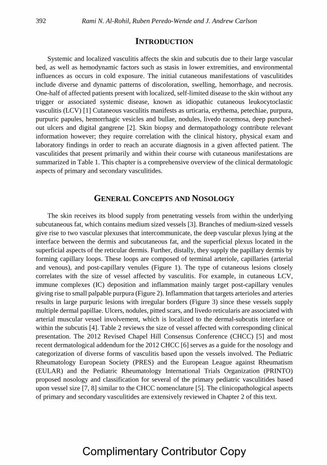
Rami N. Al-Rohil, Ruben Peredo-Wende and J. Andrew Carlson 392
INTRODUCTION
Systemic and localized vasculitis affects the skin and subcutis due to their large vascular
bed, as well as hemodynamic factors such as stasis in lower extremities, and environmental
influences as occurs in cold exposure. The initial cutaneous manifestations of vasculitides
include diverse and dynamic patterns of discoloration, swelling, hemorrhage, and necrosis.
One-half of affected patients present with localized, self-limited disease to the skin without any
trigger or associated systemic disease, known as idiopathic cutaneous leukocytoclastic
vasculitis (LCV) [1] Cutaneous vasculitis manifests as urticaria, erythema, petechiae, purpura,
purpuric papules, hemorrhagic vesicles and bullae, nodules, livedo racemosa, deep punched-
out ulcers and digital gangrene [2]. Skin biopsy and dermatopathology contribute relevant
information however; they require correlation with the clinical history, physical exam and
laboratory findings in order to reach an accurate diagnosis in a given affected patient. The
vasculitides that present primarily and within their course with cutaneous manifestations are
summarized in Table 1. This chapter is a comprehensive overview of the clinical dermatologic
aspects of primary and secondary vasculitides.
GENERAL CONCEPTS AND NOSOLOGY
The skin receives its blood supply from penetrating vessels from within the underlying
subcutaneous fat, which contains medium sized vessels [3]. Branches of medium-sized vessels
give rise to two vascular plexuses that intercommunicate, the deep vascular plexus lying at the
interface between the dermis and subcutaneous fat, and the superficial plexus located in the
superficial aspects of the reticular dermis. Further, distally, they supply the papillary dermis by
forming capillary loops. These loops are composed of terminal arteriole, capillaries (arterial
and venous), and post-capillary venules (Figure 1). The type of cutaneous lesions closely
correlates with the size of vessel affected by vasculitis. For example, in cutaneous LCV,
immune complexes (IC) deposition and inflammation mainly target post-capillary venules
giving rise to small palpable purpura (Figure 2). Inflammation that targets arterioles and arteries
results in large purpuric lesions with irregular borders (Figure 3) since these vessels supply
multiple dermal papillae. Ulcers, nodules, pitted scars, and livedo reticularis are associated with
arterial muscular vessel involvement, which is localized to the dermal-subcutis interface or
within the subcutis [4]. Table 2 reviews the size of vessel affected with corresponding clinical
presentation. The 2012 Revised Chapel Hill Consensus Conference (CHCC) [5] and most
recent dermatological addendum for the 2012 CHCC [6] serves as a guide for the nosology and
categorization of diverse forms of vasculitis based upon the vessels involved. The Pediatric
Rheumatology European Society (PRES) and the European League against Rheumatism
(EULAR) and the Pediatric Rheumatology International Trials Organization (PRINTO)
proposed nosology and classification for several of the primary pediatric vasculitides based
upon vessel size [7, 8] similar to the CHCC nomenclature [5]. The clinicopathological aspects
of primary and secondary vasculitides are extensively reviewed in Chapter 2 of this text.
Complimentary Contributor Copy
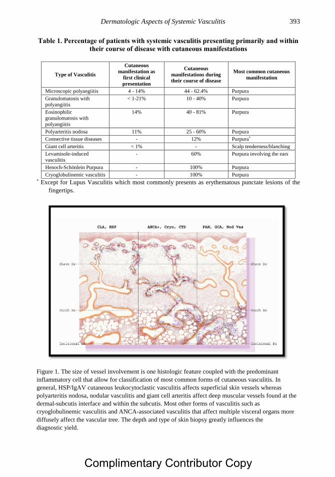
Dermatologic Aspects of Systemic Vasculitis 393
Table 1. Percentage of patients with systemic vasculitis presenting primarily and within
their course of disease with cutaneous manifestations
Type of Vasculitis
Cutaneous
manifestation as
first clinical
presentation
Cutaneous
manifestations during
their course of disease
Most common cutaneous
manifestation
Microscopic polyangiitis 4 - 14% 44 - 62.4% Purpura
Granulomatosis with polyangiitis
< 1-21% 10 - 40% Purpura
Eosinophilic granulomatosis with
polyangiitis
14% 40 - 81% Purpura
Polyarteritis nodosa 11% 25 - 60% Purpura
Connective tissue diseases - 12% Purpura*
Giant cell arteritis < 1% - Scalp tenderness/blanching
Levamisole-induced
vasculitis
- 60% Purpura involving the ears
Henoch-Schönlein Purpura - 100% Purpura
Cryoglobulinemic vasculitis - 100% Purpura * Except for Lupus Vasculitis which most commonly presents as erythematous punctate lesions of the
fingertips.
Figure 1. The size of vessel involvement is one histologic feature coupled with the predominant
inflammatory cell that allow for classification of most common forms of cutaneous vasculitis. In
general, HSP/IgAV cutaneous leukocytoclastic vasculitis affects superficial skin vessels whereas
polyarteritis nodosa, nodular vasculitis and giant cell arteritis affect deep muscular vessels found at the
dermal-subcutis interface and within the subcutis. Most other forms of vasculitis such as
cryoglobulinemic vasculitis and ANCA-associated vasculitis that affect multiple visceral organs more
diffusely affect the vascular tree. The depth and type of skin biopsy greatly influences the
diagnostic yield.
Complimentary Contributor Copy
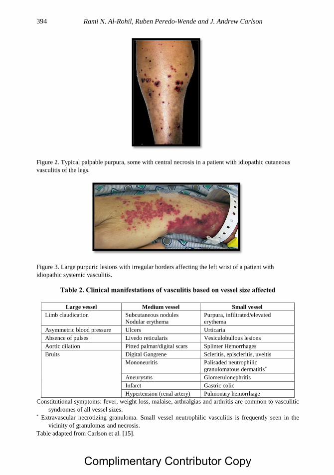
Rami N. Al-Rohil, Ruben Peredo-Wende and J. Andrew Carlson 394
Figure 2. Typical palpable purpura, some with central necrosis in a patient with idiopathic cutaneous
vasculitis of the legs.
Figure 3. Large purpuric lesions with irregular borders affecting the left wrist of a patient with
idiopathic systemic vasculitis.
Table 2. Clinical manifestations of vasculitis based on vessel size affected
Large vessel Medium vessel Small vessel
Limb claudication Subcutaneous nodules
Nodular erythema
Purpura, infiltrated/elevated
erythema
Asymmetric blood pressure Ulcers Urticaria
Absence of pulses Livedo reticularis Vesiculobullous lesions
Aortic dilation Pitted palmar/digital scars Splinter Hemorrhages
Bruits Digital Gangrene Scleritis, episcleritis, uveitis
Mononeuritis Palisaded neutrophilic
granulomatous dermatitis*
Aneurysms Glomerulonephritis
Infarct Gastric colic
Hypertension (renal artery) Pulmonary hemorrhage
Constitutional symptoms: fever, weight loss, malaise, arthralgias and arthritis are common to vasculitic
syndromes of all vessel sizes. * Extravascular necrotizing granuloma. Small vessel neutrophilic vasculitis is frequently seen in the
vicinity of granulomas and necrosis.
Table adapted from Carlson et al. [15].
Complimentary Contributor Copy
Dermatologic Aspects of Systemic Vasculitis 395
EPIDEMIOLOGY
The incidence of cutaneous vasculitis ranges from 15.4 to 29.7 cases per million per year,
affecting adults more than children of all ages, with slight female predominance, and up to 90%
of children diagnosed with Henoch–Schönlein purpura (HSP/ (IgA vasculitis [IgAV) [1]. The
mean age at presentation of adult vasculitis is 47 years, while amongst children the mean age
at onset is 7 years [1, 4]. The onset of vasculitis after exposure to a trigger such as a drug or
infection is 7 to 10 days. A mean interval of 6 months occurs between the onset of symptoms
and signs of systemic disease and the onset of secondary cutaneous vasculitis. Almost one-half
of patients presenting with cutaneous vasculitis have disease localized to the skin without an
attributable cause, trigger or associated systemic disease, referred to as idiopathic LCV [1]. The
remainder of localized cutaneous vasculitis cases are attributed to recent infection and drug
ingestion. LCV that results from drugs or infection is termed hypersensitivity or allergic
vasculitis. The antinuclear cytoplasmic antibody (ANCA)-associated vasculitides (AAV)
vasculitis syndromes include eosinophilic granulomatosis with polyangiitis (EGPA)
(previously known as Churg-Strauss Syndrome [CSS]), microscopic polyangiitis (MPA) and
granulomatosis with polyangiitis (GPA) (previously known as Wegner granulomatosis [WG])
may first present with cutaneous vasculitis [9]. The onset of cutaneous vasculitis may be an
indication of secondary vasculitis in association with connective tissue disease (CTD) such as
systemic lupus erythematosus (SLE) vasculitis (LV) [10], and rheumatoid arthritis (RA) related
vasculitis (RAV) [11].
The evolution of cutaneous vasculitis occurs in three phases, (a) single acute, self-limited
episode that resolve in ≤ 6 months in association with drug exposure or an infectious trigger so
noted in 60% of patients; (b) relapsing disease with symptom-free periods usually found in
patients with HSP/IgAV and cryoglobulinemic vasculitis (CV) so noted in 20% of patients; and
(c) a chronic, unremitting disease most often associated with primary systemic vasculitides and
secondary vasculitides in association with CTD, CV, or malignancy altogether in about 20% of
patients [1]. The duration of vasculitis ranges from 1 week to 318 months, with mean and
median durations of 28 months and 3.7 months, respectively. Less than 20% of cutaneous
vasculitis cases have extracutaneous or visceral vasculitis. Fatal disease occurs in less than 7%
of patients [1, 12].
ETIOPATHOGENESIS
A variety of injurious processes can lead to identical responses in the vessel wall resulting
in the activation of neutrophils, abnormal neutrophil diapedesis and fibrinoid necrosis, the
hallmarks of the vasculitides [1, 13]. Other morphological patterns of vasculitis include
lymphocytic endarteritis and endarteritis obliterans of transplant vascular rejection, which are
not generally associated with abundant fibrin deposits and destruction of the vessel wall with
loss of the elastic lamina [14, 15]. Adding to the difficulties in the histological evaluation of
vasculitides are that the characteristics of the initial insult may be lost during the process of
repair leading to the transformation from active, acute inflammatory lesions into older, often
sclerotic lesions wherein T-cells and macrophages predominate and neovascularization
transpire to compensate for the ischemic insult. Nonetheless, the type of inflammatory cell
Complimentary Contributor Copy
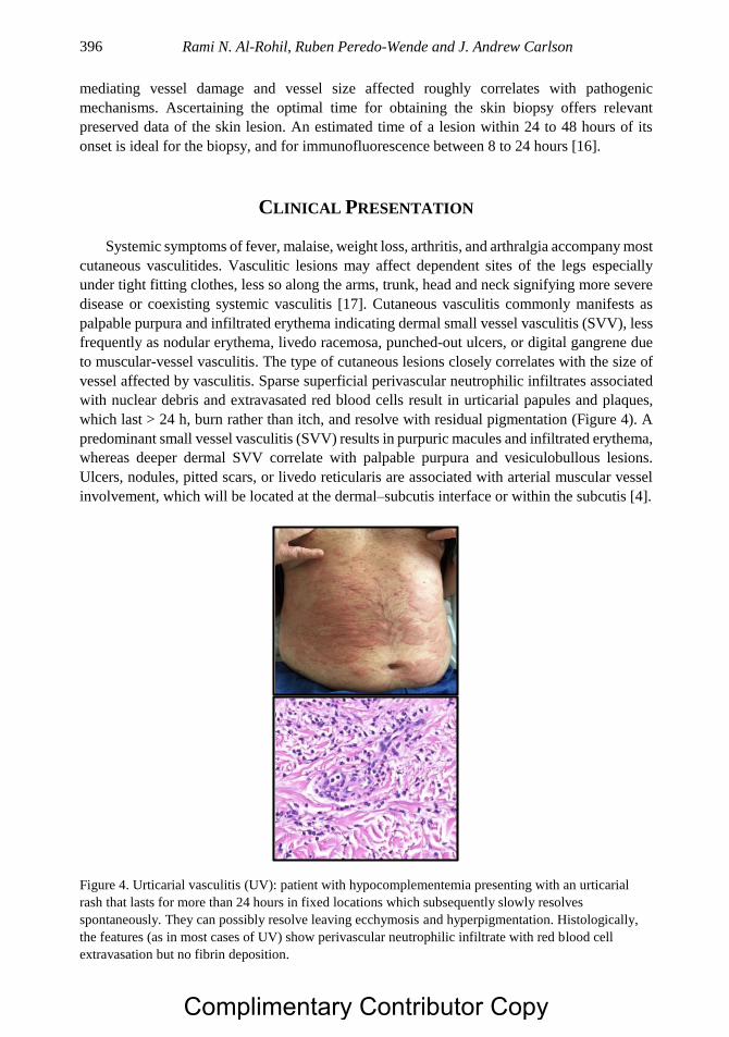
Rami N. Al-Rohil, Ruben Peredo-Wende and J. Andrew Carlson 396
mediating vessel damage and vessel size affected roughly correlates with pathogenic
mechanisms. Ascertaining the optimal time for obtaining the skin biopsy offers relevant
preserved data of the skin lesion. An estimated time of a lesion within 24 to 48 hours of its
onset is ideal for the biopsy, and for immunofluorescence between 8 to 24 hours [16].
CLINICAL PRESENTATION
Systemic symptoms of fever, malaise, weight loss, arthritis, and arthralgia accompany most
cutaneous vasculitides. Vasculitic lesions may affect dependent sites of the legs especially
under tight fitting clothes, less so along the arms, trunk, head and neck signifying more severe
disease or coexisting systemic vasculitis [17]. Cutaneous vasculitis commonly manifests as
palpable purpura and infiltrated erythema indicating dermal small vessel vasculitis (SVV), less
frequently as nodular erythema, livedo racemosa, punched-out ulcers, or digital gangrene due
to muscular-vessel vasculitis. The type of cutaneous lesions closely correlates with the size of
vessel affected by vasculitis. Sparse superficial perivascular neutrophilic infiltrates associated
with nuclear debris and extravasated red blood cells result in urticarial papules and plaques,
which last > 24 h, burn rather than itch, and resolve with residual pigmentation (Figure 4). A
predominant small vessel vasculitis (SVV) results in purpuric macules and infiltrated erythema,
whereas deeper dermal SVV correlate with palpable purpura and vesiculobullous lesions.
Ulcers, nodules, pitted scars, or livedo reticularis are associated with arterial muscular vessel
involvement, which will be located at the dermal–subcutis interface or within the subcutis [4].
Figure 4. Urticarial vasculitis (UV): patient with hypocomplementemia presenting with an urticarial
rash that lasts for more than 24 hours in fixed locations which subsequently slowly resolves
spontaneously. They can possibly resolve leaving ecchymosis and hyperpigmentation. Histologically,
the features (as in most cases of UV) show perivascular neutrophilic infiltrate with red blood cell
extravasation but no fibrin deposition.
Complimentary Contributor Copy
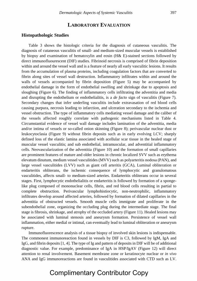
Dermatologic Aspects of Systemic Vasculitis 397
LABORATORY EVALUATION
Histopathologic Studies
Table 3 shows the histologic criteria for the diagnosis of cutaneous vasculitis. The
diagnosis of cutaneous vasculitis of small- and medium-sized muscular vessels is established
by biopsy and examination of hematoxylin and eosin (H& E)-stained sections followed by
direct immunofluourescent (DIF) studies. Fibrinoid necrosis is comprised of fibrin deposition
within and around the vessel wall and is a feature of nearly all early vasculitic lesions. It results
from the accumulation of plasma proteins, including coagulation factors that are converted to
fibrin along sites of vessel wall destruction. Inflammatory infiltrates within and around the
walls of vessels accompanied by fibrin deposition (Figure 5) may be accompanied by
endothelial damage in the form of endothelial swelling and shrinkage due to apoptosis and
sloughing (Figure 6). The finding of inflammatory cells infiltrating the adventitia and media
and disrupting the endothelium or endothelialitis, is a de facto sign of vasculitis (Figure 7).
Secondary changes that infer underling vasculitis include extravasation of red blood cells
causing purpura, necrosis leading to infarction, and ulceration secondary to the ischemia and
vessel obstruction. The type of inflammatory cells mediating vessel damage and the caliber of
the vessels affected roughly correlate with pathogenic mechanisms listed in Table 4.
Circumstantial evidence of vessel wall damage includes lamination of the adventitia, media
and/or intima of vessels or so-called onion skinning (Figure 8); perivascular nuclear dust or
leukocytoclasia (Figure 9) without fibrin deposits such as in early evolving LCV; sharply
defined loss of the elastic lamina associated with acellular scar tissue in the healed stage of
muscular vessel vasculitis; and sub endothelial, intramuscular, and adventitial inflammatory
cells. Neovascularization of the adventitia (Figure 10) and the formation of small capillaries
are prominent features of mature and older lesions in chronic localized SVV such as erythema
elevatum dinutum, medium vessel vasculitides (MVV) such as polyarteritis nodosa (PAN), and
large vessel vasculitides (LVV) such as giant cell arteritis (GCA), Luminal obliteration or
endarteritis obliterans, the ischemic consequence of lymphocytic and granulomatous
vasculitides, affects small- to medium-sized arteries. Endarteritis obliterans occur in several
stages. First, lymphocytic endothelialitis or endarteritis is followed by formation of a sponge-
like plug composed of mononuclear cells, fibrin, and red blood cells resulting in partial to
complete obstruction. Perivascular lymphohistiocytic, non-neutrophilic, inflammatory
infiltrates develop around affected arteries, followed by formation of dilated capillaries in the
adventitia of obstructed vessels. Smooth muscle cells immigrate and proliferate in the
subendothelial zone, organizing the occluding plug during the intermediate stage. The final
stage is fibrosis, shrinkage, and atrophy of the occluded artery (Figure 11). Healed lesions may
be associated with luminal stenosis and aneurysm formation. Persistence of vessel wall
inflammation, either medial or intimal, can eventually lead to luminal obliteration or aneurysm
rupture.
Immunofluorescence analysis of a tissue biopsy of involved skin lesions is indispensable.
The commonest immunoreaction found in vessels by DIF is C3, followed by IgM, IgA and
IgG, and fibrin deposits [1, 4]. The type of Ig and pattern of deposits in DIF will be of additional
diagnostic value. For example, predominance of IgA in HSP/IgAV (Figure 12) will direct
attention to renal involvement. Basement membrane zone or keratinocyte nuclear or in vivo
ANA and IgG immunoreactions are found in vasculitides associated with CTD such as LV.
Complimentary Contributor Copy
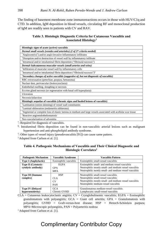
Rami N. Al-Rohil, Ruben Peredo-Wende and J. Andrew Carlson 398
The finding of basement membrane zone immunoreactions occurs in those with HUV/C1q and
CTD. In addition, IgM deposition in blood vessels, circulating RF and monoclonal production
of IgM are readily seen in patients with CV and RAV.
Table 3. Histologic Diagnostic Criteria for Cutaneous Vasculitis and
Associated Histology1
Histologic signs of acute (active) vasculitis
Dermal small vessels (venules and arterioles) (2 of 3* criteria needed) *Angiocentric(†) and/or angio-invasive inflammatory infiltrates *Disruption and/or destruction of vessel wall by inflammatory infiltrate *Intramural and/or intraluminal fibrin deposition (“fibrinoid necrosis”)
Dermal-Subcutaneous muscular vessels (small arteries and veins) *Infiltration of muscular vessel wall by inflammatory cells *Intramural and/or intraluminal fibrin deposition (“fibrinoid necrosis”)§ †Secondary changes of active vasculitis (suggestive of, but not diagnostic of vasculitis)
RBC extravasation (petechiae, purpura, hematoma)
Nuclear dust, perivascular (leukocytoclasia)
Endothelial swelling, sloughing or necrosis
Eccrine gland necrosis (or regeneration with basal cell hyperplasia)
Ulceration
Necrosis/infarction
Histologic sequelae of vasculitis (chronic signs and healed lesions of vasculitis) †Lamination (onion-skinning) of vessel wall constituents †Luminal obliteration (endarteritis obliterans) *Segmental or complete loss of elastic lamina in medium and large vessels associated with acellular scar tissue †Reactive angioendotheliomatosis
Neo-vascularization of adventitia *: Required for diagnosis of vasculitis. §: Intraluminal fibrin deposition can be found in non-vasculitic arterial lesions such as malignant
hypertension and anti-phospholipid antibody syndrome. †: Other types of vessel injury (pseudovasculitis [91]) can cause same pattern. 1 Adapted from Carlson et al. [1].
Table 4. Pathogenic Mechanisms of Vasculitis and Their Clinical Diagnostic and
Histologic Correlates1
Pathogenic Mechanism Vasculitic Syndrome Vasculitis Pattern
Type I (Anphylactic) Eosinophilic vasculitis Eosinophilic small-vessel vasculitis
Type II (Cytotoxic-
cytolytic antibody)
EGPA
GPA
MPA
Eosinophilic small- and medium vessel vasculitis Neutrophilic mostly small- and medium vessel vasculitis
Neutrophilic mostly small- and medium vessel vasculitis
Type III (Immune
complex)
HSP
CLA
CV
PAN
Neutrophilic small-vessel vasculitis Neutrophilic small-vessel vasculitis
Neutrophilic mostly small- and medium vessel vasculitis
Neutrophilic medium-vessel vasculitis
Type IV (Delayed
hypersensitivity)
GCA Chronic GVHD
Granulomatous medium-vessel vasculitis Lymphocytic small-vessel vasculitis
CLA = Cutaneous leukocytoclastic angiitis; CV = Cryoglobulinemic vasculitis; EGPA = Eosinophilic
granulomatosis with polyangiitis; GCA = Giant cell arteritis; GPA = Granulomatosis with
polyangiitis; GVHD = Graft-versus-host disease; HSP = Henoch-Schӧnlein purpura;
MPA=Microscopic polyangiitis, PAN = Polyarteritis nodosa 1 Adapted from Carlson et al. [1].
Complimentary Contributor Copy
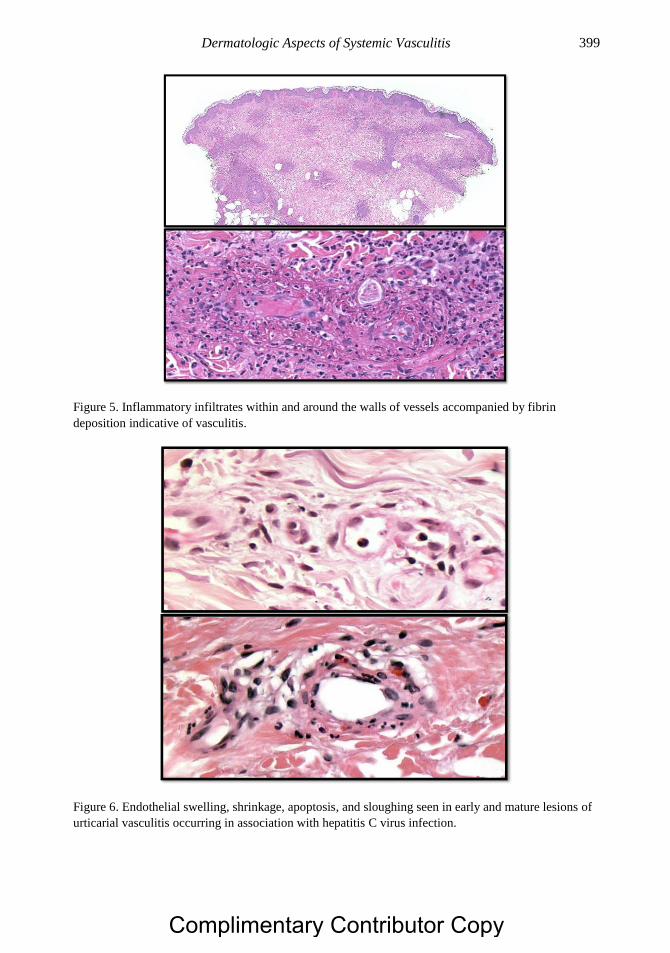
Dermatologic Aspects of Systemic Vasculitis 399
Figure 5. Inflammatory infiltrates within and around the walls of vessels accompanied by fibrin
deposition indicative of vasculitis.
Figure 6. Endothelial swelling, shrinkage, apoptosis, and sloughing seen in early and mature lesions of
urticarial vasculitis occurring in association with hepatitis C virus infection.
Complimentary Contributor Copy
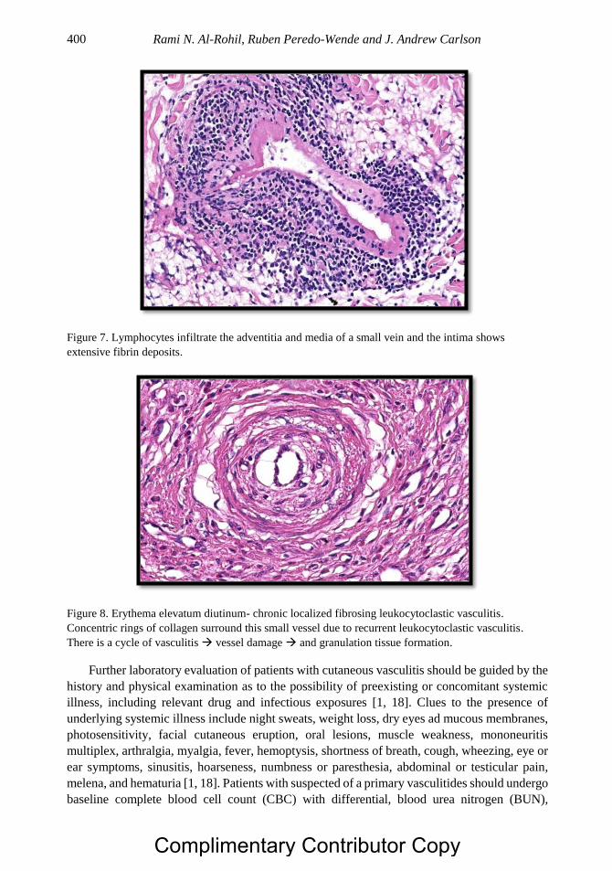
Rami N. Al-Rohil, Ruben Peredo-Wende and J. Andrew Carlson 400
Figure 7. Lymphocytes infiltrate the adventitia and media of a small vein and the intima shows
extensive fibrin deposits.
Figure 8. Erythema elevatum diutinum- chronic localized fibrosing leukocytoclastic vasculitis.
Concentric rings of collagen surround this small vessel due to recurrent leukocytoclastic vasculitis.
There is a cycle of vasculitis vessel damage and granulation tissue formation.
Further laboratory evaluation of patients with cutaneous vasculitis should be guided by the
history and physical examination as to the possibility of preexisting or concomitant systemic
illness, including relevant drug and infectious exposures [1, 18]. Clues to the presence of
underlying systemic illness include night sweats, weight loss, dry eyes ad mucous membranes,
photosensitivity, facial cutaneous eruption, oral lesions, muscle weakness, mononeuritis
multiplex, arthralgia, myalgia, fever, hemoptysis, shortness of breath, cough, wheezing, eye or
ear symptoms, sinusitis, hoarseness, numbness or paresthesia, abdominal or testicular pain,
melena, and hematuria [1, 18]. Patients with suspected of a primary vasculitides should undergo
baseline complete blood cell count (CBC) with differential, blood urea nitrogen (BUN),
Complimentary Contributor Copy
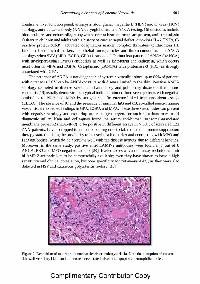
Dermatologic Aspects of Systemic Vasculitis 401
creatinine, liver function panel, urinalysis, stool guaiac, hepatitis B (HBV) and C virus (HCV)
serology, antinuclear antibody (ANA), cryoglobulins, and ANCA testing. Other studies include
blood cultures and echocardiography when fever or heart murmurs are present, anti-streptolysin
O titers in children and adults with a history of cardiac septal defect; cytokines IL-6, TNFα, C-
reactive protein (CRP), activated coagulation marker complex thrombin–antithrombin III,
functional endothelial markers endothelial microparticles and thrombomodulin; and ANCA
serology when SVV (MPA, EGPA, GPA) is suspected. Perinuclear pattern of ANCA (pANCA)
with myeloperoxidase (MPO) antibodies as well as lactoferrin and cathepsin, which occurs
most often in MPA and EGPA. Cytoplasmic (cANCA) with proteinase-3 (PR3) is strongly
associated with GPA.
The presence of ANCA is not diagnostic of systemic vasculitis since up to 60% of patients
with cutaneous LCV can be ANCA-positive with disease limited to the skin. Positive ANCA
serology so noted in diverse systemic inflammatory and pulmonary disorders that mimic
vasculitis [19] usually demonstrates atypical indirect immunofluorescent patterns with negative
antibodies to PR-3 and MPO by antigen specific enzyme-linked immunosorbent assays
(ELISA). The absence of IC and the presence of minimal IgG and C3, so-called pauci-immune
vasculitis, are expected findings in GPA, EGPA and MPA. These three vasculitides can present
with negative serology and exploring other antigen targets for such situations may be of
diagnostic utility. Kain and colleagues found the serum anti-human lysosomal-associated
membrane protein-2 (hLAMP-2) to be positive in different assays in > 80% of untreated 122
AVV patients. Levels dropped to almost becoming undetectable once the immunosuppression
therapy started, raising the possibility to be used as a biomarker and contrasting with MPO and
PR3 antibodies, which do no correlate well with the disease activity due to different kinetics.
Moreover, in the same study, positive anti-hLAMP-2 antibodies were found in 7 out of 8
ANCA, PR3 and MPO negative patients [20]. Inadequacies of current assay techniques limit
hLAMP-2 antibody kits to be commercially available; even they have shown to have a high
sensitivity and clinical correlation, but poor specificity for cutaneous AAV, as they were also
detected in HSP and cutaneous polyarteritis nodosa [21].
Figure 9. Deposition of neutrophilic nuclear debris or leukocytoclasia. Note the disruption of the small
thin wall vessel by fibrin and numerous degenerated adventitial apoptotic neutrophilic nuclei.
Complimentary Contributor Copy
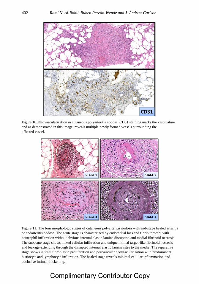
Rami N. Al-Rohil, Ruben Peredo-Wende and J. Andrew Carlson 402
Figure 10. Neovascularization in cutaneous polyarteritis nodosa. CD31 staining marks the vasculature
and as demonstrated in this image, reveals multiple newly formed vessels surrounding the
affected vessel.
Figure 11. The four morphologic stages of cutaneous polyarteritis nodosa with end-stage healed arteritis
or endarteritis nodosa. The acute stage is characterized by endothelial loss and fibrin thrombi with
neutrophil infiltration without obvious internal elastic lamina disruption and medial fibrinoid necrosis.
The subacute stage shows mixed cellular infiltration and unique intimal target-like fibrinoid necrosis
and leakage extending through the disrupted internal elastic lamina sites to the media. The reparative
stage shows intimal fibroblastic proliferation and perivascular neovascularization with predominant
histiocyte and lymphocyte infiltration. The healed stage reveals minimal cellular inflammation and
occlusive intimal thickening.
CD31
STAGE 1 STAGE 2
STAGE 3 STAGE 4
Complimentary Contributor Copy
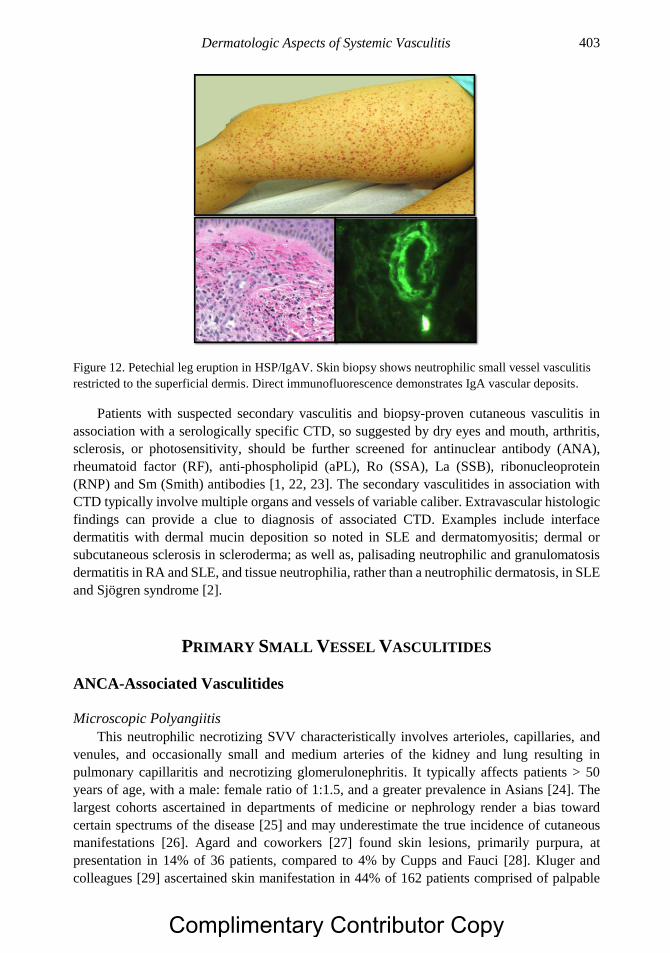
Dermatologic Aspects of Systemic Vasculitis 403
Figure 12. Petechial leg eruption in HSP/IgAV. Skin biopsy shows neutrophilic small vessel vasculitis
restricted to the superficial dermis. Direct immunofluorescence demonstrates IgA vascular deposits.
Patients with suspected secondary vasculitis and biopsy-proven cutaneous vasculitis in
association with a serologically specific CTD, so suggested by dry eyes and mouth, arthritis,
sclerosis, or photosensitivity, should be further screened for antinuclear antibody (ANA),
rheumatoid factor (RF), anti-phospholipid (aPL), Ro (SSA), La (SSB), ribonucleoprotein
(RNP) and Sm (Smith) antibodies [1, 22, 23]. The secondary vasculitides in association with
CTD typically involve multiple organs and vessels of variable caliber. Extravascular histologic
findings can provide a clue to diagnosis of associated CTD. Examples include interface
dermatitis with dermal mucin deposition so noted in SLE and dermatomyositis; dermal or
subcutaneous sclerosis in scleroderma; as well as, palisading neutrophilic and granulomatosis
dermatitis in RA and SLE, and tissue neutrophilia, rather than a neutrophilic dermatosis, in SLE
and Sjögren syndrome [2].
PRIMARY SMALL VESSEL VASCULITIDES
ANCA-Associated Vasculitides
Microscopic Polyangiitis
This neutrophilic necrotizing SVV characteristically involves arterioles, capillaries, and
venules, and occasionally small and medium arteries of the kidney and lung resulting in
pulmonary capillaritis and necrotizing glomerulonephritis. It typically affects patients > 50
years of age, with a male: female ratio of 1:1.5, and a greater prevalence in Asians [24]. The
largest cohorts ascertained in departments of medicine or nephrology render a bias toward
certain spectrums of the disease [25] and may underestimate the true incidence of cutaneous
manifestations [26]. Agard and coworkers [27] found skin lesions, primarily purpura, at
presentation in 14% of 36 patients, compared to 4% by Cupps and Fauci [28]. Kluger and
colleagues [29] ascertained skin manifestation in 44% of 162 patients comprised of palpable
Complimentary Contributor Copy
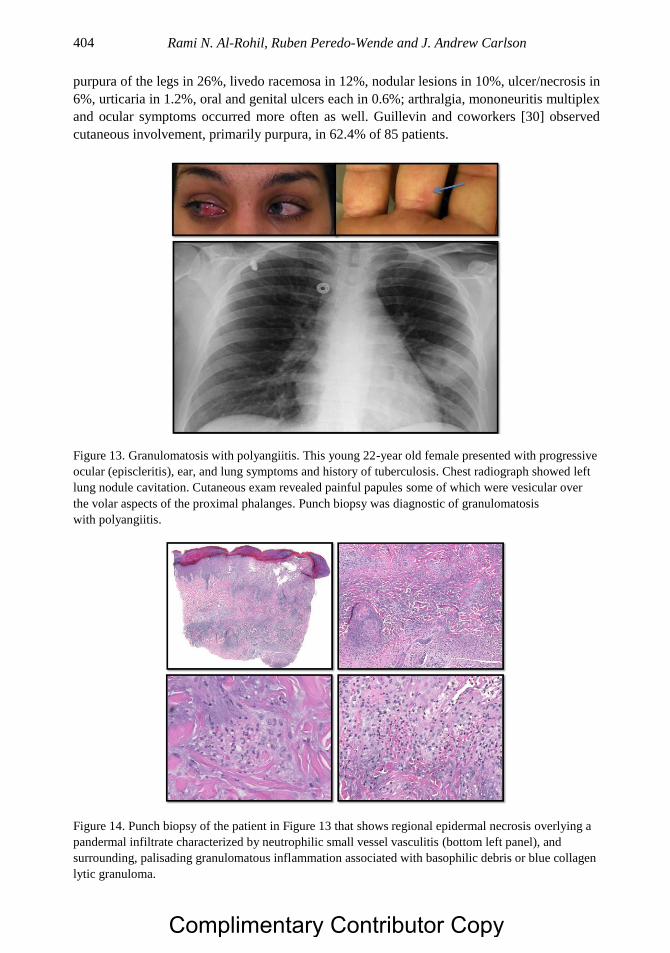
Rami N. Al-Rohil, Ruben Peredo-Wende and J. Andrew Carlson 404
purpura of the legs in 26%, livedo racemosa in 12%, nodular lesions in 10%, ulcer/necrosis in
6%, urticaria in 1.2%, oral and genital ulcers each in 0.6%; arthralgia, mononeuritis multiplex
and ocular symptoms occurred more often as well. Guillevin and coworkers [30] observed
cutaneous involvement, primarily purpura, in 62.4% of 85 patients.
Figure 13. Granulomatosis with polyangiitis. This young 22-year old female presented with progressive
ocular (episcleritis), ear, and lung symptoms and history of tuberculosis. Chest radiograph showed left
lung nodule cavitation. Cutaneous exam revealed painful papules some of which were vesicular over
the volar aspects of the proximal phalanges. Punch biopsy was diagnostic of granulomatosis
with polyangiitis.
Figure 14. Punch biopsy of the patient in Figure 13 that shows regional epidermal necrosis overlying a
pandermal infiltrate characterized by neutrophilic small vessel vasculitis (bottom left panel), and
surrounding, palisading granulomatous inflammation associated with basophilic debris or blue collagen
lytic granuloma.
Complimentary Contributor Copy
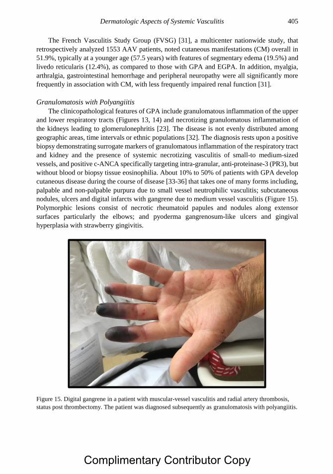
Dermatologic Aspects of Systemic Vasculitis 405
The French Vasculitis Study Group (FVSG) [31], a multicenter nationwide study, that
retrospectively analyzed 1553 AAV patients, noted cutaneous manifestations (CM) overall in
51.9%, typically at a younger age (57.5 years) with features of segmentary edema (19.5%) and
livedo reticularis (12.4%), as compared to those with GPA and EGPA. In addition, myalgia,
arthralgia, gastrointestinal hemorrhage and peripheral neuropathy were all significantly more
frequently in association with CM, with less frequently impaired renal function [31].
Granulomatosis with Polyangiitis
The clinicopathological features of GPA include granulomatous inflammation of the upper
and lower respiratory tracts (Figures 13, 14) and necrotizing granulomatous inflammation of
the kidneys leading to glomerulonephritis [23]. The disease is not evenly distributed among
geographic areas, time intervals or ethnic populations [32]. The diagnosis rests upon a positive
biopsy demonstrating surrogate markers of granulomatous inflammation of the respiratory tract
and kidney and the presence of systemic necrotizing vasculitis of small-to medium-sized
vessels, and positive c-ANCA specifically targeting intra-granular, anti-proteinase-3 (PR3), but
without blood or biopsy tissue eosinophilia. About 10% to 50% of patients with GPA develop
cutaneous disease during the course of disease [33-36] that takes one of many forms including,
palpable and non-palpable purpura due to small vessel neutrophilic vasculitis; subcutaneous
nodules, ulcers and digital infarcts with gangrene due to medium vessel vasculitis (Figure 15).
Polymorphic lesions consist of necrotic rheumatoid papules and nodules along extensor
surfaces particularly the elbows; and pyoderma gangrenosum-like ulcers and gingival
hyperplasia with strawberry gingivitis.
Figure 15. Digital gangrene in a patient with muscular-vessel vasculitis and radial artery thrombosis,
status post thrombectomy. The patient was diagnosed subsequently as granulomatosis with polyangiitis.
Complimentary Contributor Copy
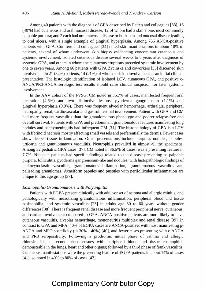
Rami N. Al-Rohil, Ruben Peredo-Wende and J. Andrew Carlson 406
Among 40 patients with the diagnosis of GPA described by Patten and colleagues [33], 16
(40%) had cutaneous and oral mucosal disease, 12 of whom had a skin alone, most commonly
palpable purpura; and 2 each had oral mucosal disease or both skin and mucosal disease leading
to oral ulcers, with a single example of gingival hyperplasia. Among 766 ANCA-positive
patients with GPA, Comfere and colleagues [34] noted skin manifestations in about 10% of
patients, several of whom underwent skin biopsy evidencing concomitant cutaneous and
systemic involvement, isolated cutaneous disease several weeks to 8 years after diagnosis of
systemic GPA, and others in whom the cutaneous eruptions preceded systemic involvement by
one to seven years. Among 66 patients with GPA Zycinska and coworkers [35] discerned skin
involvement in 21 (32%) patients, 14 (21%) of whom had skin involvement as an initial clinical
presentation. The histologic identification of isolated LCV, cutaneous GPA, and positive c-
ANCA/PR3-ANCA serologic test results should raise clinical suspicion for later systemic
involvement.
In the AAV cohort of the FVSG, CM noted in 36.7% of cases, manifested frequent oral
ulceration (4.6%) and two distinctive lesions: pyoderma gangrenosum (1.1%) and
gingival hyperplasia (0.9%). There was frequent alveolar hemorrhage, arthralgia, peripheral
neuropathy, renal, cardiovascular and gastrointestinal involvement. Patient with GPA and CM
had more frequent vasculitis than the granulomatous phenotype and poorer relapse-free and
overall survival. Patients with GPA and predominant granulomatous features manifesting lung
nodules and pachymeningitis had infrequent CM [31]. The histopathology of GPA is a LCV
with fibrinoid necrosis mostly affecting small vessels and preferentially the dermis. Fewer cases
show deeper tissue inflammation. Other presentations include purpura, nodules, papules,
urticaria and granulomatous vasculitis. Neutrophils prevailed in almost all the specimens.
Among 52 pediatric GPA cases [37], CM noted in 36.5% of cases, was a presenting feature in
7.7%. Nineteen patients had specific findings related to the disease presenting as palpable
purpura, folliculitis, pyoderma gangrenosum-like and nodules, with histopathologic findings of
leukocytoclastic vasculitis, granulomatous inflammation, granulomatous vasculitis and
palisading granulomas. Acneiform papules and pustules with perifollicular inflammation are
unique to this age group [37].
Eosinophilic-Granulomatosis with Polyangiitis
Patients with EGPA present clinically with adult-onset of asthma and allergic rhinitis, and
pathologically with necrotizing granulomatous inflammation, peripheral blood and tissue
eosinophilia, and systemic vasculitis [23] in adults age 30 to 60 years without gender
differences [38]. There is frequent renal disease and more frequent peripheral nerve, cutaneous,
and cardiac involvement compared to GPA. ANCA-positive patients are more likely to have
cutaneous vasculitis, alveolar hemorrhage, mononeuritis multiplex and renal disease [39]. In
contrast to GPA and MPA, 40% of EGPA cases are ANCA-positive, with most manifesting p-
ANCA and MPO specificity (in 30% - 40%) [40], and fewer cases presenting with c-ANCA
and PR3 seropositivity. Following a prodromic initial phase of asthma and allergic
rhinosinusitis, a second phase ensues with peripheral blood and tissue eosinophilia
demonstrable in the lungs, heart and other organs; followed by a third phase of frank vasculitis.
Cutaneous manifestations were the presenting feature of EGPA patients in about 14% of cases
[41], so noted in 40% to 80% of cases [42].
Complimentary Contributor Copy
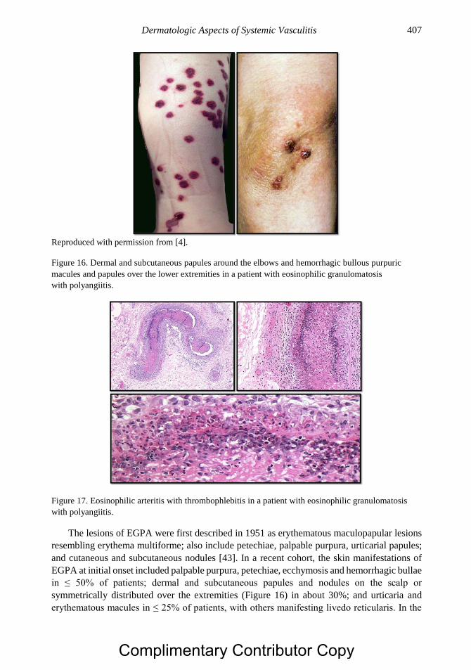
Dermatologic Aspects of Systemic Vasculitis 407
Reproduced with permission from [4].
Figure 16. Dermal and subcutaneous papules around the elbows and hemorrhagic bullous purpuric
macules and papules over the lower extremities in a patient with eosinophilic granulomatosis
with polyangiitis.
Figure 17. Eosinophilic arteritis with thrombophlebitis in a patient with eosinophilic granulomatosis
with polyangiitis.
The lesions of EGPA were first described in 1951 as erythematous maculopapular lesions
resembling erythema multiforme; also include petechiae, palpable purpura, urticarial papules;
and cutaneous and subcutaneous nodules [43]. In a recent cohort, the skin manifestations of
EGPA at initial onset included palpable purpura, petechiae, ecchymosis and hemorrhagic bullae
in ≤ 50% of patients; dermal and subcutaneous papules and nodules on the scalp or
symmetrically distributed over the extremities (Figure 16) in about 30%; and urticaria and
erythematous macules in ≤ 25% of patients, with others manifesting livedo reticularis. In the
Complimentary Contributor Copy
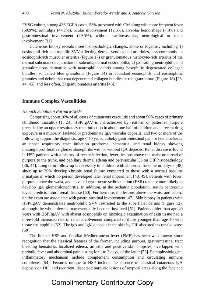
Rami N. Al-Rohil, Ruben Peredo-Wende and J. Andrew Carlson 408
FVSG cohort, among 436 EGPA cases, 53% presented with CM along with more frequent fever
(50.9%), arthralgia (44.1%), ocular involvement (12.5%), alveolar hemorrhage (7.8%) and
gastrointestinal involvement (29.5%), without cardiovascular, neurological or renal
involvement [31].
Cutaneous biopsy reveals three histopathologic changes, alone or together, including 1)
eosinophil-rich neutrophilic SVV affecting dermal venules and arterioles, less commonly an
eosinophil-rich muscular arteritis (Figure 17) or granulomatous histiocyte-rich arteritis of the
dermal subcutaneous junction or subcutis; dermal eosinophilia; 2) palisading neutrophilic and
granulomatous dermatitis with neutrophilic debris among basophilic degenerated collagen
bundles, so called blue granuloma (Figure 14) or abundant eosinophils and eosinophilic
granules and debris that coat degenerated collagen bundles or red granulomas (Figure 18) [23,
44, 45]; and less often, 3) granulomatous arteritis [45].
Immune Complex Vasculitides
Henoch Schönlein Purpura/IgAV
Comprising about 10% of all cases of cutaneous vasculitis and about 90% cases of primary
childhood vasculitis [1, 23], HSP/IgAV is characterized by retiform or patterned purpura
preceded by an upper respiratory tract infection in about one-half of children and a recent drug
exposure in a minority. Isolated or predominate IgA vascular deposits, and two or more of the
following support the diagnosis: age ≤ 20 years, colicky gastrointestinal pain or hematochezia,
an upper respiratory tract infection prodrome, hematuria, and renal biopsy showing
mesangioproliferative glomerulonephritis with or without IgA deposits. Renal disease is found
in HSP patients with a history of recent infection, fever, lesions above the waist or spread of
purpura to the trunk, and papillary dermal edema and perivascular C3 on DIF histopathology
[46, 47]. Long term follow-up is necessary in children with abnormal baseline urinalysis [48]
since up to 20% develop chronic renal failure compared to those with a normal baseline
urinalysis in which no person developed later renal impairment [48, 49]. Patients with fever,
purpura above the waist, and elevated erythrocyte sedimentation (ESR) rate are more likely to
develop IgA glomerulonephritis. In addition, in the pediatric population, serum pentraxin3
levels predicts future renal disease [50]. Furthermore, the lesions above the waist and edema
on the exam are associated with gastrointestinal involvement [47]. Skin biopsy in patients with
HSP/IgAV demonstrates neutrophilic SVV restricted to the superficial dermis (Figure 12),
although the whole dermis may eventually become involved [51]. Patients older than age 40
years with HSP/IgAV with absent eosinophils on histologic examination of skin tissue had a
three-fold increased risk of renal involvement compared to those younger than age 40 with
tissue eosinophilia [52]. The IgA and IgM deposits in the skin by DIF also predicts renal disease
[50].
The link of HSP and familial Mediterranean fever (FMF) has been well known since
recognition that the classical features of the former, including purpura, gastrointestinal tract
bleeding hematuria, localized edema, arthritis and positive skin biopsies; overlapped with
periodic fever and abdominal pain lasting for 1 to 3 days, of the latter [53]. Pathophysiological
inflammatory mechanism include complement consumption and circulating immune
complexes [54]. Features unique to HSP include the absence of classical cutaneous IgA
deposits on DIF, and recurrent, dispersed purpuric lesions of atypical areas along the face and
Complimentary Contributor Copy
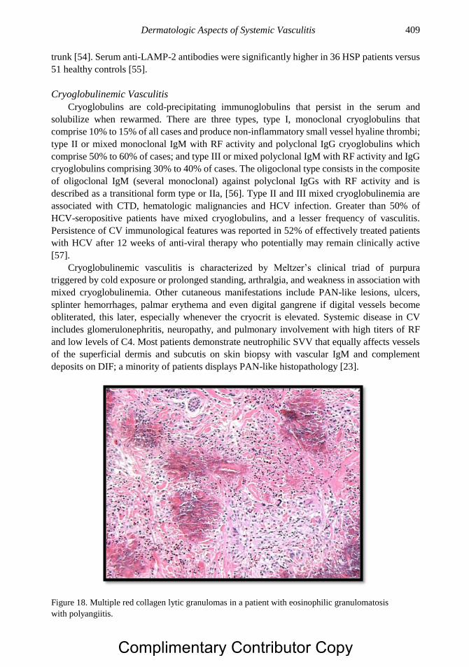
Dermatologic Aspects of Systemic Vasculitis 409
trunk [54]. Serum anti-LAMP-2 antibodies were significantly higher in 36 HSP patients versus
51 healthy controls [55].
Cryoglobulinemic Vasculitis
Cryoglobulins are cold-precipitating immunoglobulins that persist in the serum and
solubilize when rewarmed. There are three types, type I, monoclonal cryoglobulins that
comprise 10% to 15% of all cases and produce non-inflammatory small vessel hyaline thrombi;
type II or mixed monoclonal IgM with RF activity and polyclonal IgG cryoglobulins which
comprise 50% to 60% of cases; and type III or mixed polyclonal IgM with RF activity and IgG
cryoglobulins comprising 30% to 40% of cases. The oligoclonal type consists in the composite
of oligoclonal IgM (several monoclonal) against polyclonal IgGs with RF activity and is
described as a transitional form type or IIa, [56]. Type II and III mixed cryoglobulinemia are
associated with CTD, hematologic malignancies and HCV infection. Greater than 50% of
HCV-seropositive patients have mixed cryoglobulins, and a lesser frequency of vasculitis.
Persistence of CV immunological features was reported in 52% of effectively treated patients
with HCV after 12 weeks of anti-viral therapy who potentially may remain clinically active
[57].
Cryoglobulinemic vasculitis is characterized by Meltzer’s clinical triad of purpura
triggered by cold exposure or prolonged standing, arthralgia, and weakness in association with
mixed cryoglobulinemia. Other cutaneous manifestations include PAN-like lesions, ulcers,
splinter hemorrhages, palmar erythema and even digital gangrene if digital vessels become
obliterated, this later, especially whenever the cryocrit is elevated. Systemic disease in CV
includes glomerulonephritis, neuropathy, and pulmonary involvement with high titers of RF
and low levels of C4. Most patients demonstrate neutrophilic SVV that equally affects vessels
of the superficial dermis and subcutis on skin biopsy with vascular IgM and complement
deposits on DIF; a minority of patients displays PAN-like histopathology [23].
Figure 18. Multiple red collagen lytic granulomas in a patient with eosinophilic granulomatosis
with polyangiitis.
Complimentary Contributor Copy
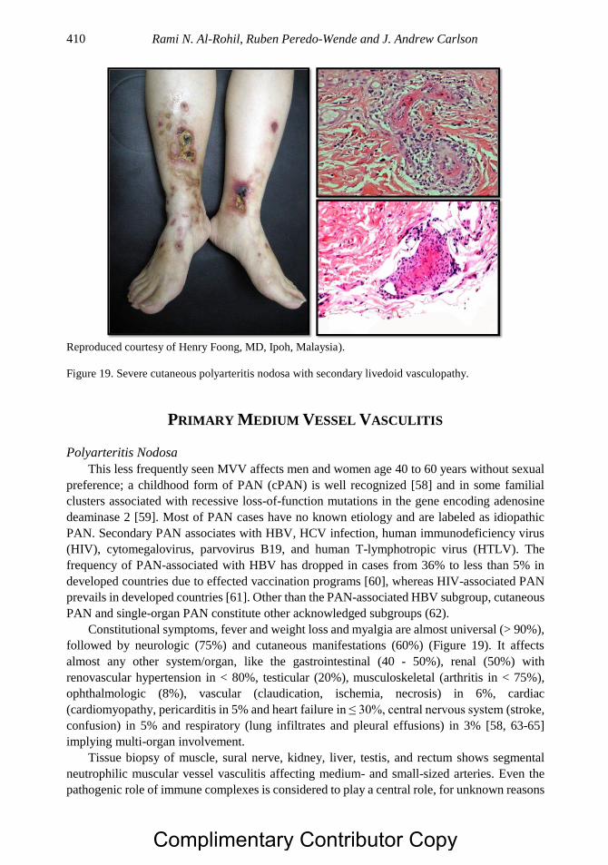
Rami N. Al-Rohil, Ruben Peredo-Wende and J. Andrew Carlson 410
Reproduced courtesy of Henry Foong, MD, Ipoh, Malaysia).
Figure 19. Severe cutaneous polyarteritis nodosa with secondary livedoid vasculopathy.
PRIMARY MEDIUM VESSEL VASCULITIS
Polyarteritis Nodosa
This less frequently seen MVV affects men and women age 40 to 60 years without sexual
preference; a childhood form of PAN (cPAN) is well recognized [58] and in some familial
clusters associated with recessive loss-of-function mutations in the gene encoding adenosine
deaminase 2 [59]. Most of PAN cases have no known etiology and are labeled as idiopathic
PAN. Secondary PAN associates with HBV, HCV infection, human immunodeficiency virus
(HIV), cytomegalovirus, parvovirus B19, and human T-lymphotropic virus (HTLV). The
frequency of PAN-associated with HBV has dropped in cases from 36% to less than 5% in
developed countries due to effected vaccination programs [60], whereas HIV-associated PAN
prevails in developed countries [61]. Other than the PAN-associated HBV subgroup, cutaneous
PAN and single-organ PAN constitute other acknowledged subgroups (62).
Constitutional symptoms, fever and weight loss and myalgia are almost universal (> 90%),
followed by neurologic (75%) and cutaneous manifestations (60%) (Figure 19). It affects
almost any other system/organ, like the gastrointestinal (40 - 50%), renal (50%) with
renovascular hypertension in < 80%, testicular (20%), musculoskeletal (arthritis in < 75%),
ophthalmologic (8%), vascular (claudication, ischemia, necrosis) in 6%, cardiac
(cardiomyopathy, pericarditis in 5% and heart failure in ≤ 30%, central nervous system (stroke,
confusion) in 5% and respiratory (lung infiltrates and pleural effusions) in 3% [58, 63-65]
implying multi-organ involvement.
Tissue biopsy of muscle, sural nerve, kidney, liver, testis, and rectum shows segmental
neutrophilic muscular vessel vasculitis affecting medium- and small-sized arteries. Even the
pathogenic role of immune complexes is considered to play a central role, for unknown reasons
Complimentary Contributor Copy
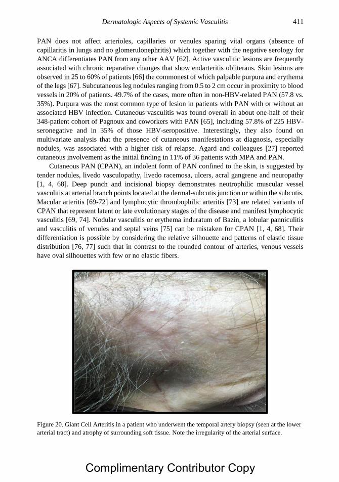
Dermatologic Aspects of Systemic Vasculitis 411
PAN does not affect arterioles, capillaries or venules sparing vital organs (absence of
capillaritis in lungs and no glomerulonephritis) which together with the negative serology for
ANCA differentiates PAN from any other AAV [62]. Active vasculitic lesions are frequently
associated with chronic reparative changes that show endarteritis obliterans. Skin lesions are
observed in 25 to 60% of patients [66] the commonest of which palpable purpura and erythema
of the legs [67]. Subcutaneous leg nodules ranging from 0.5 to 2 cm occur in proximity to blood
vessels in 20% of patients. 49.7% of the cases, more often in non-HBV-related PAN (57.8 vs.
35%). Purpura was the most common type of lesion in patients with PAN with or without an
associated HBV infection. Cutaneous vasculitis was found overall in about one-half of their
348-patient cohort of Pagnoux and coworkers with PAN [65], including 57.8% of 225 HBV-
seronegative and in 35% of those HBV-seropositive. Interestingly, they also found on
multivariate analysis that the presence of cutaneous manifestations at diagnosis, especially
nodules, was associated with a higher risk of relapse. Agard and colleagues [27] reported
cutaneous involvement as the initial finding in 11% of 36 patients with MPA and PAN.
Cutaneous PAN (CPAN), an indolent form of PAN confined to the skin, is suggested by
tender nodules, livedo vasculopathy, livedo racemosa, ulcers, acral gangrene and neuropathy
[1, 4, 68]. Deep punch and incisional biopsy demonstrates neutrophilic muscular vessel
vasculitis at arterial branch points located at the dermal-subcutis junction or within the subcutis.
Macular arteritis [69-72] and lymphocytic thrombophilic arteritis [73] are related variants of
CPAN that represent latent or late evolutionary stages of the disease and manifest lymphocytic
vasculitis [69, 74]. Nodular vasculitis or erythema induratum of Bazin, a lobular panniculitis
and vasculitis of venules and septal veins [75] can be mistaken for CPAN [1, 4, 68]. Their
differentiation is possible by considering the relative silhouette and patterns of elastic tissue
distribution [76, 77] such that in contrast to the rounded contour of arteries, venous vessels
have oval silhouettes with few or no elastic fibers.
Figure 20. Giant Cell Arteritis in a patient who underwent the temporal artery biopsy (seen at the lower
arterial tract) and atrophy of surrounding soft tissue. Note the irregularity of the arterial surface.
Complimentary Contributor Copy
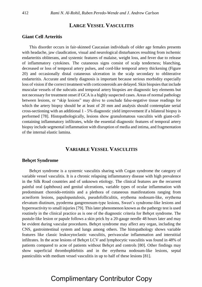
Rami N. Al-Rohil, Ruben Peredo-Wende and J. Andrew Carlson 412
LARGE VESSEL VASCULITIS
Giant Cell Arteritis
This disorder occurs in fair-skinned Caucasian individuals of older age females presents
with headache, jaw claudication, visual and neurological disturbances resulting from ischemic
endarteritis obliterans, and systemic features of malaise, weight loss, and fever due to release
of inflammatory cytokines. The cutaneous signs consist of scalp tenderness; blanching,
decreased or loss of temporal artery pulses, and cord-like temporal artery thickening (Figure
20) and occasionally distal cutaneous ulceration in the scalp secondary to obliterative
endarteritis. Accurate and timely diagnosis is important because serious morbidity especially
loss of vision if the correct treatment with corticosteroids are delayed. Skin biopsies that include
muscular vessels of the subcutis and temporal artery biopsies are diagnostic key elements but
not necessary for treatment onset if GCA is a highly suspected cases. Areas of normal pathology
between lesions, or “skip lesions” may drive to conclude false-negative tissue readings for
which the artery biopsy should be at least of 20 mm and analysis should contemplate serial
cross-sectioning with an additional 1 - 5% diagnostic yield improvement if a bilateral biopsy is
performed [78]. Histopathologically, lesions show granulomatous vasculitis with giant-cell-
containing inflammatory infiltrates, while the essential diagnostic features of temporal artery
biopsy include segmental inflammation with disruption of media and intima, and fragmentation
of the internal elastic lamina.
VARIABLE VESSEL VASCULITIS
Behçet Syndrome
Behçet syndrome is a systemic vasculitis sharing with Cogan syndrome the category of
variable vessel vasculitis. It is a chronic relapsing inflammatory disease with high prevalence
in the Silk Road countries and of unknown etiology. The clinical features are the recurrent
painful oral (aphthous) and genital ulcerations, variable types of ocular inflammation with
predominant choroido-retinitis and a plethora of cutaneous manifestations ranging from
acneiform lesions, papulopustulosis, pseudofolliculitis, erythema nodosum-like, erythema
elevatum diutinum, pyoderma gangrenosum-type lesions, Sweet’s syndrome-like lesions and
hyperreactivity to small injuries [79]. This later phenomenon known as the pathergy test is used
routinely in the clinical practice as is one of the diagnostic criteria for Behçet syndrome. The
pustule-like lesion or papule follows a skin prick by a 20-gauge needle 48 hours later and may
be evident during vascular procedures. Behçet syndrome may affect any organ, including the
CNS, gastrointestinal system and lungs among others. The histopathology shows variable
features like classic leukocytoclastic vasculitis, perivascular inflammation and interstitial
infiltrates. In the acne lesions of Behçet LCV and lymphocytic vasculitis was found in 48% of
patients compared to acne of patients without Behçet and controls [80]. Other findings may
show superficial thrombophlebitis and in the erythema nodosum-like lesions, septal
panniculitis with medium vessel vasculitis in up to half of these lesions [81].
Complimentary Contributor Copy
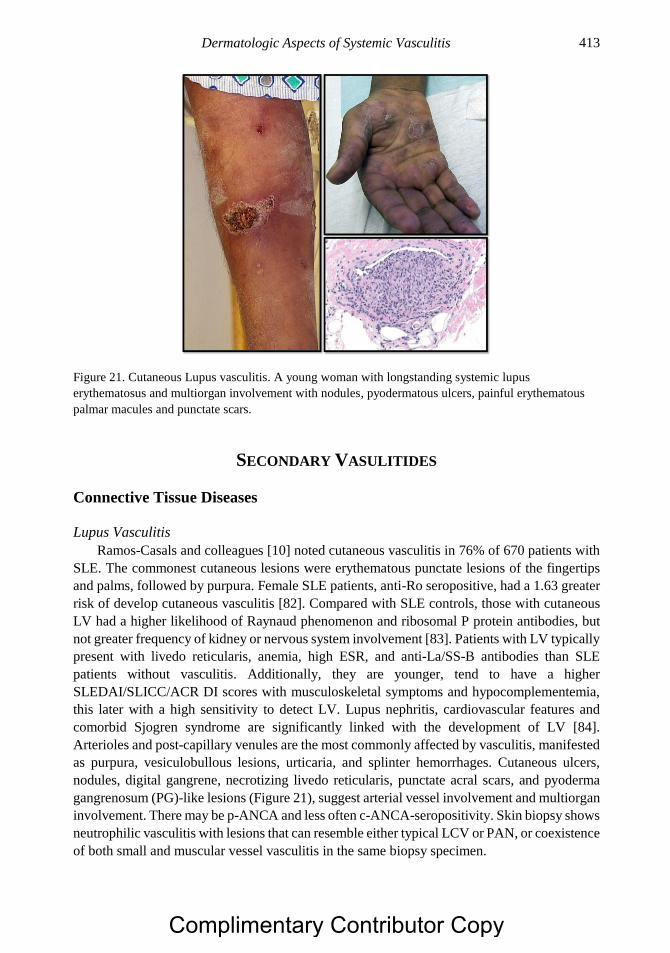
Dermatologic Aspects of Systemic Vasculitis 413
Figure 21. Cutaneous Lupus vasculitis. A young woman with longstanding systemic lupus
erythematosus and multiorgan involvement with nodules, pyodermatous ulcers, painful erythematous
palmar macules and punctate scars.
SECONDARY VASULITIDES
Connective Tissue Diseases
Lupus Vasculitis
Ramos-Casals and colleagues [10] noted cutaneous vasculitis in 76% of 670 patients with
SLE. The commonest cutaneous lesions were erythematous punctate lesions of the fingertips
and palms, followed by purpura. Female SLE patients, anti-Ro seropositive, had a 1.63 greater
risk of develop cutaneous vasculitis [82]. Compared with SLE controls, those with cutaneous
LV had a higher likelihood of Raynaud phenomenon and ribosomal P protein antibodies, but
not greater frequency of kidney or nervous system involvement [83]. Patients with LV typically
present with livedo reticularis, anemia, high ESR, and anti-La/SS-B antibodies than SLE
patients without vasculitis. Additionally, they are younger, tend to have a higher
SLEDAI/SLICC/ACR DI scores with musculoskeletal symptoms and hypocomplementemia,
this later with a high sensitivity to detect LV. Lupus nephritis, cardiovascular features and
comorbid Sjogren syndrome are significantly linked with the development of LV [84].
Arterioles and post-capillary venules are the most commonly affected by vasculitis, manifested
as purpura, vesiculobullous lesions, urticaria, and splinter hemorrhages. Cutaneous ulcers,
nodules, digital gangrene, necrotizing livedo reticularis, punctate acral scars, and pyoderma
gangrenosum (PG)-like lesions (Figure 21), suggest arterial vessel involvement and multiorgan
involvement. There may be p-ANCA and less often c-ANCA-seropositivity. Skin biopsy shows
neutrophilic vasculitis with lesions that can resemble either typical LCV or PAN, or coexistence
of both small and muscular vessel vasculitis in the same biopsy specimen.
Complimentary Contributor Copy
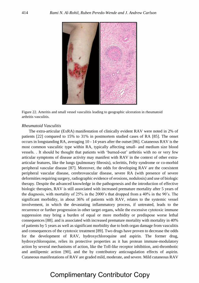
Rami N. Al-Rohil, Ruben Peredo-Wende and J. Andrew Carlson 414
Figure 22. Arteritis and small vessel vasculitis leading to geographic ulceration in rheumatoid
arthritis vasculitis.
Rheumatoid Vasculitis
The extra-articular (ExRA) manifestation of clinically evident RAV were noted in 2% of
patients [22] compared to 15% to 31% in postmortem studied cases of RA [85]. The onset
occurs in longstanding RA, averaging 10 - 14 years after the outset [86]. Cutaneous RAV is the
most common vasculitic type within RA, typically affecting small- and medium size blood
vessels. . It should be thought that patients with ‘burned-out’ arthritis with no or very few
articular symptoms of disease activity may manifest with RAV in the context of other extra-
articular features, like the lungs (pulmonary fibrosis), sclerittis, Felty syndrome or co-morbid
peripheral vascular disease [87]. Moreover, the odds for developing RAV are the coexistent
peripheral vascular disease, cerebrovascular disease, severe RA (with presence of severe
deformities requiring surgery, radiographic evidence of erosions, nodulosis) and use of biologic
therapy. Despite the advanced knowledge in the pathogenesis and the introduction of effective
biologic therapies, RAV is still associated with increased premature mortality after 5 years of
the diagnosis, with mortality of 25% in the 2000’s that dropped from a 40% in the 90’s. The
significant morbidity, in about 36% of patients with RAV, relates to the systemic vessel
involvement, in which the devastating inflammatory process, if untreated, leads to the
recurrence or further progression in other target organs, while the excessive cytotoxic immune
suppression may bring a burden of equal or more morbidity or predispose worse lethal
consequences [88]. and is associated with increased premature mortality with mortality in 40%
of patients by 5 years as well as significant morbidity due to both organ damage from vasculitis
and consequences of the cytotoxic treatment [89]. Two drugs have proven to decrease the odds
for the development of RAV, hydroxychloroquine and aspirin. The former drug,
hydroxychloroquine, relies its protective properties as it has protean immune-modulatory
action by several mechanisms of action, like the Toll-like receptor inhibition, anti-thrombotic
and antilipemic action [90], and the by contributory anticoagulation effects of aspirin.
Cutaneous manifestations of RAV are graded mild, moderate, and severe. Mild cutaneous RAV
Complimentary Contributor Copy
Dermatologic Aspects of Systemic Vasculitis 415
presents with nail fold and palmar telangiectasia, thrombosis, minute digital ulceration,
petechiae, and livedo reticularis. A well-known presentation are Bywater lesions consisting in
periungual infarctions in long-term RA carrying without systemic implications requiring no
escalade in the immune suppression [91]. Moderate cases manifest palpable purpura. Severely
affected patients present with digital gangrene, nail-fold infarcts, and large cutaneous ulcers
(Figure 22). The histopathology of RAV mimics that of polyarteritis nodosa without forming
microaneurysms [92]. To define the vasculitic process, three layers of vessel need to be
involved, distinguishing this from perivascular infiltrates without invasive features commonly
seen in RA. Neutrophilic proliferation as well as, lymphocytic, and plasmacytic cells infiltrate
all layers of the small arteries [93]. Patients with RA and cutaneous LCV alone without RAV,
have a better prognosis than those with other ExRA involvement [22]. High acute phase
reactants in a RA patient with established disease are the only proving biomarkers contrasting
with the rheumatoid factor and anti-citrullinated cyclic peptide antibodies which titers are
minimally or not clinically meaningful in RAV. Conversely, the absence of RF and anti-CCP
antibodies have an excellent negative predictive value for RAV [94]. Some patients may
express a positive anti-nuclear antibody or an anti-perinuclear antibody (atypical), other than
then PR3 or MPO antigens suggestive of the underlying vasculitic process targeting other
antigens in neutrophil granules. Anti-lactoferrin antibodies have been reported in the latter
circumstance [95]. Minocycline-induced cutaneous PAN was reported and associated with the
chronic treatment for acne treatment improving after discontinuation of the drug [96].
Drug-Induced Cutaneous Vasculitis
Hypersensitivity vasculitis due to adverse drug reactions manifests as superficial dermal
neutrophilic or lymphocytic SVV on skin biopsy, and represents about 20% of cases of
cutaneous vasculitis [1, 4, 68, 97]. Tissue eosinophilia is an indicator of drug-induced cutaneous
SVV [98].
Tumor Necrosis Factor-α
Tumor necrosis factor-α (TNF-α) inhibitors employed in the treatment of autoimmune and
rheumatic diseases [99] were the reported cause of cutaneous vasculitis in 8 patients so treated
for 2 to 72 months [100], including four patients with RA, three patients with ulcerative colitis,
and one patient with Crohn disease. The commonest presenting manifestation of the patients
was palpable purpura, followed by ulcerated lesions, erythematous macules and blisters. After
discontinuation of anti-TNF-α, none had recurrent vasculitis. Appearance of ANCA titers in
patients under anti-TNF-α therapy should prompt excluding AAV overlap, for which anti-TNF-
α is not efficacious, and that requires switching to disease specific therapy [101].
Levamisole
Levamisole was originally introduced as an anthelmintic agent and later employed in
Behçet disease and rheumatoid arthritis for immunosuppression. It has been employed in colon
cancer enhancing the immunity by potentiating the T-cell mediated immune response. More
recently, Levamisole was added to cocaine to potentiate the stimulant effects as it has dopamine
agonistic effects provoking a synergistic effect with cocaine [102]. Affected patients have
Complimentary Contributor Copy
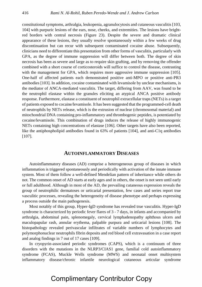
Rami N. Al-Rohil, Ruben Peredo-Wende and J. Andrew Carlson 416
constitutional symptoms, arthralgia, leukopenia, agranulocytosis and cutaneous vasculitis [103,
104] with purpuric lesions of the ears, nose, cheeks, and extremities. The lesions have bright-
red borders with central necrosis (Figure 23). Despite the severe and dramatic clinical
appearance of these lesions, they usually resolve spontaneously within a few weeks of drug
discontinuation but can recur with subsequent contaminated cocaine abuse. Subsequently,
clinicians need to differentiate this presentation from other forms of vasculitis, particularly with
GPA, as the degree of immune suppression will differ between both. The degree of skin
necrosis has been as severe and large as to require skin grafting, and by removing the offender
combined with a short course of corticosteroids will suffice to control the disease, contrasting
with the management for GPA, which requires more aggressive immune suppression [105].
One-half of affected patients each demonstrated positive anti-MPO or positive anti-PR3
antibodies [103]. In addition, cocaine contaminated with levamisole by unclear mechanisms, is
the mediator of ANCA-mediated vasculitis. The target, differing from AAV, was found to be
the neutrophil elastase within the granules eliciting an atypical ANCA positive antibody
response. Furthermore, elastase a constituent of neutrophil extracellular traps (NETs) is a target
of patients exposed to cocaine/levamisole. It has been suggested that the programmed-cell death
of neutrophils by NETs release, which is the extrusion of nuclear (chromosomal material) and
mitochondrial DNA containing pro-inflammatory and thrombogenic peptides, is potentiated by
cocaine/levamisole. This combination of drugs induces the release of highly immunogenic
NETs containing high concentrations of elastase [106]. Other targets have also been reported,
like the antiphospholipid antibodies found in 63% of patients [104], and anti-C1q antibodies
[107].
AUTOINFLAMMATORY DISEASES
Autoinflammatory diseases (AD) comprise a heterogeneous group of diseases in which
inflammation is triggered spontaneously and periodically with activation of the innate immune
system. Most of them follow a well-defined Mendelian pattern of inheritance while others do
not. The common onset of AD starts at early ages and in others, the onset is not seen until early
or full adulthood. Although in most of the AD, the prevailing cutaneous expression reveals the
group of neutrophilic dermatoses or urticarial presentation, few cases and series report true
vasculitic processes, revealing the heterogeneity of disease phenotype and perhaps expressing
a process outside the main pathogenesis.
Most notably of this group, Hyper-IgD syndrome has revealed true vasculitis. Hyper-IgD
syndrome is characterized by periodic fever flares of 3 - 7 days, in infants and accompanied by
arthralgia, abdominal pain, splenomegaly, cervical lymphadenopathy aphthous ulcers and
maculopapular rash, annular erythema, palpable purpura and urticarial lesions [108]. The
histopathology revealed perivascular infiltrates of variable numbers of lymphocytes and
polymorphonuclear neutrophils fibrin deposits and red blood cell extravasation in a case report
and analog findings in 7 out of 17 cases [109].
In cryopyrin-associated periodic syndromes (CAPS), which is a continuum of three
disorders with the mutations in the NLRP3/CIAS1 gene, familial cold autoinflammatory
syndrome (FCAS), Muckle Wells syndrome (MWS) and neonatal onset multisystem
inflammatory disease/chronic infantile neurological cutaneous articular syndrome
Complimentary Contributor Copy
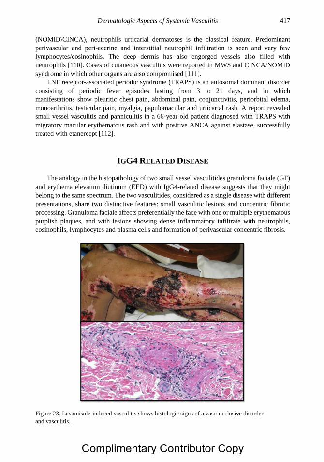
Dermatologic Aspects of Systemic Vasculitis 417
(NOMID\CINCA), neutrophils urticarial dermatoses is the classical feature. Predominant
perivascular and peri-eccrine and interstitial neutrophil infiltration is seen and very few
lymphocytes/eosinophils. The deep dermis has also engorged vessels also filled with
neutrophils [110]. Cases of cutaneous vasculitis were reported in MWS and CINCA/NOMID
syndrome in which other organs are also compromised [111].
TNF receptor-associated periodic syndrome (TRAPS) is an autosomal dominant disorder
consisting of periodic fever episodes lasting from 3 to 21 days, and in which
manifestations show pleuritic chest pain, abdominal pain, conjunctivitis, periorbital edema,
monoarthritis, testicular pain, myalgia, papulomacular and urticarial rash. A report revealed
small vessel vasculitis and panniculitis in a 66-year old patient diagnosed with TRAPS with
migratory macular erythematous rash and with positive ANCA against elastase, successfully
treated with etanercept [112].
IGG4 RELATED DISEASE
The analogy in the histopathology of two small vessel vasculitides granuloma faciale (GF)
and erythema elevatum diutinum (EED) with IgG4-related disease suggests that they might
belong to the same spectrum. The two vasculitides, considered as a single disease with different
presentations, share two distinctive features: small vasculitic lesions and concentric fibrotic
processing. Granuloma faciale affects preferentially the face with one or multiple erythematous
purplish plaques, and with lesions showing dense inflammatory infiltrate with neutrophils,
eosinophils, lymphocytes and plasma cells and formation of perivascular concentric fibrosis.
Figure 23. Levamisole-induced vasculitis shows histologic signs of a vaso-occlusive disorder
and vasculitis.
Complimentary Contributor Copy
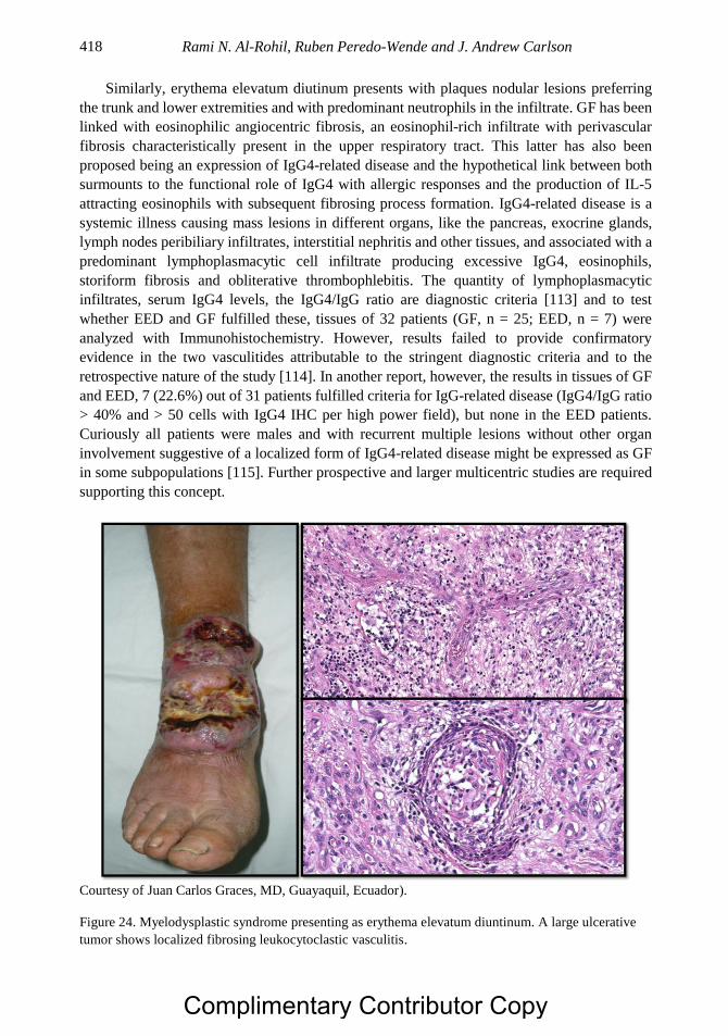
Rami N. Al-Rohil, Ruben Peredo-Wende and J. Andrew Carlson 418
Similarly, erythema elevatum diutinum presents with plaques nodular lesions preferring
the trunk and lower extremities and with predominant neutrophils in the infiltrate. GF has been
linked with eosinophilic angiocentric fibrosis, an eosinophil-rich infiltrate with perivascular
fibrosis characteristically present in the upper respiratory tract. This latter has also been
proposed being an expression of IgG4-related disease and the hypothetical link between both
surmounts to the functional role of IgG4 with allergic responses and the production of IL-5
attracting eosinophils with subsequent fibrosing process formation. IgG4-related disease is a
systemic illness causing mass lesions in different organs, like the pancreas, exocrine glands,
lymph nodes peribiliary infiltrates, interstitial nephritis and other tissues, and associated with a
predominant lymphoplasmacytic cell infiltrate producing excessive IgG4, eosinophils,
storiform fibrosis and obliterative thrombophlebitis. The quantity of lymphoplasmacytic
infiltrates, serum IgG4 levels, the IgG4/IgG ratio are diagnostic criteria [113] and to test
whether EED and GF fulfilled these, tissues of 32 patients (GF, n = 25; EED, n = 7) were
analyzed with Immunohistochemistry. However, results failed to provide confirmatory
evidence in the two vasculitides attributable to the stringent diagnostic criteria and to the
retrospective nature of the study [114]. In another report, however, the results in tissues of GF
and EED, 7 (22.6%) out of 31 patients fulfilled criteria for IgG-related disease (IgG4/IgG ratio
> 40% and > 50 cells with IgG4 IHC per high power field), but none in the EED patients.
Curiously all patients were males and with recurrent multiple lesions without other organ
involvement suggestive of a localized form of IgG4-related disease might be expressed as GF
in some subpopulations [115]. Further prospective and larger multicentric studies are required
supporting this concept.
Courtesy of Juan Carlos Graces, MD, Guayaquil, Ecuador).
Figure 24. Myelodysplastic syndrome presenting as erythema elevatum diuntinum. A large ulcerative
tumor shows localized fibrosing leukocytoclastic vasculitis.
Complimentary Contributor Copy
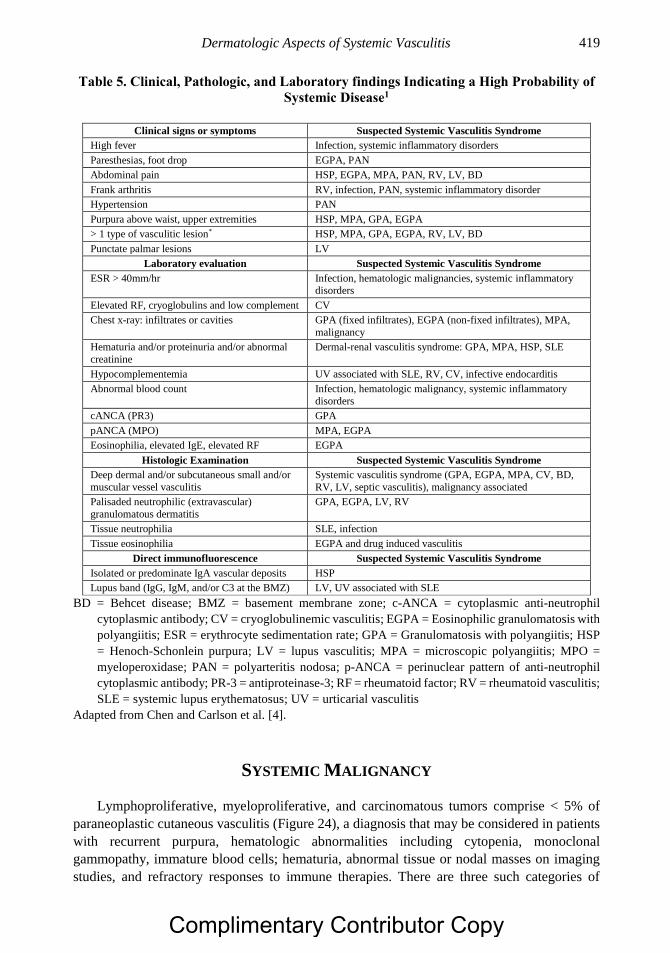
Dermatologic Aspects of Systemic Vasculitis 419
Table 5. Clinical, Pathologic, and Laboratory findings Indicating a High Probability of
Systemic Disease1
Clinical signs or symptoms Suspected Systemic Vasculitis Syndrome
High fever Infection, systemic inflammatory disorders
Paresthesias, foot drop EGPA, PAN
Abdominal pain HSP, EGPA, MPA, PAN, RV, LV, BD
Frank arthritis RV, infection, PAN, systemic inflammatory disorder
Hypertension PAN
Purpura above waist, upper extremities HSP, MPA, GPA, EGPA
> 1 type of vasculitic lesion* HSP, MPA, GPA, EGPA, RV, LV, BD
Punctate palmar lesions LV
Laboratory evaluation Suspected Systemic Vasculitis Syndrome
ESR > 40mm/hr Infection, hematologic malignancies, systemic inflammatory
disorders
Elevated RF, cryoglobulins and low complement CV
Chest x-ray: infiltrates or cavities GPA (fixed infiltrates), EGPA (non-fixed infiltrates), MPA,
malignancy
Hematuria and/or proteinuria and/or abnormal
creatinine
Dermal-renal vasculitis syndrome: GPA, MPA, HSP, SLE
Hypocomplementemia UV associated with SLE, RV, CV, infective endocarditis
Abnormal blood count Infection, hematologic malignancy, systemic inflammatory
disorders
cANCA (PR3) GPA
pANCA (MPO) MPA, EGPA
Eosinophilia, elevated IgE, elevated RF EGPA
Histologic Examination Suspected Systemic Vasculitis Syndrome
Deep dermal and/or subcutaneous small and/or
muscular vessel vasculitis
Systemic vasculitis syndrome (GPA, EGPA, MPA, CV, BD,
RV, LV, septic vasculitis), malignancy associated
Palisaded neutrophilic (extravascular)
granulomatous dermatitis
GPA, EGPA, LV, RV
Tissue neutrophilia SLE, infection
Tissue eosinophilia EGPA and drug induced vasculitis
Direct immunofluorescence Suspected Systemic Vasculitis Syndrome
Isolated or predominate IgA vascular deposits HSP
Lupus band (IgG, IgM, and/or C3 at the BMZ) LV, UV associated with SLE
BD = Behcet disease; BMZ = basement membrane zone; c-ANCA = cytoplasmic anti-neutrophil
cytoplasmic antibody; CV = cryoglobulinemic vasculitis; EGPA = Eosinophilic granulomatosis with
polyangiitis; ESR = erythrocyte sedimentation rate; GPA = Granulomatosis with polyangiitis; HSP
= Henoch-Schonlein purpura; LV = lupus vasculitis; MPA = microscopic polyangiitis; MPO =
myeloperoxidase; PAN = polyarteritis nodosa; p-ANCA = perinuclear pattern of anti-neutrophil
cytoplasmic antibody; PR-3 = antiproteinase-3; RF = rheumatoid factor; RV = rheumatoid vasculitis;
SLE = systemic lupus erythematosus; UV = urticarial vasculitis
Adapted from Chen and Carlson et al. [4].
SYSTEMIC MALIGNANCY
Lymphoproliferative, myeloproliferative, and carcinomatous tumors comprise < 5% of
paraneoplastic cutaneous vasculitis (Figure 24), a diagnosis that may be considered in patients
with recurrent purpura, hematologic abnormalities including cytopenia, monoclonal
gammopathy, immature blood cells; hematuria, abnormal tissue or nodal masses on imaging
studies, and refractory responses to immune therapies. There are three such categories of
Complimentary Contributor Copy
Rami N. Al-Rohil, Ruben Peredo-Wende and J. Andrew Carlson 420
patients including, those with true paraneoplastic vasculitic syndromes wherein the vasculitis
improves with extirpation or treatment of the tumor; vasculitis masquerading as malignancy
such as lung masses in GPA; and malignancy masquerading as vasculitides as in emboli from
an atrial myxoma and superficial migratory thrombophlebitis with pancreatic cancer. Most
paraneoplastic cutaneous vasculitic syndromes are the result of a paraproteinemia secondary to
lymphoproliferative disorders, including cryoglobulinemia in association with lymphocytic
lymphoma and Waldenström macroglobulinemia.
PROGNOSIS
There are clinical and histologic predictors for the prognosis of cutaneous vasculitis that
suggest the likelihood of concomitant or later systemic vasculitis [1, 18, 116] as shown in Table
5. The distinction between localized cutaneous and systemic vasculitis is important since the
former carries a relatively favorable outcome, while the latter conveys the likelihood of
permanent organ damage, increased morbidity and mortality. About 20% to 40% of patient
with cutaneous vasculitis have concomitant limited systemic vasculitis notably kidney such as
so called renal-dermal vasculitis while the likelihood of chronicity and systemic progression is
enhanced when there is coexisting CTD, cryoglobulinemia, frank ulceration [117], arthralgia
[19], more than one form of cutaneous vasculitic lesion such as ulceration and palpable purpura,
putative muscular and SVV, normal serum IgA levels [17], paresthesia, fever, painless lesions
[19] and cutaneous necrosis [118]. Histologically, the severity of vessel injury in cutaneous
vasculitis correlates with the clinical severity and course [117, 119]. Others have noted
histopathological changes of LCV were not predictive of extracutaneous involvement [120].
Lesional IgA deposits by DIF predict proteinuria and subsequent renal involvement [121]. Up
to 60% of patients presenting with cutaneous vasculitis on skin biopsy have SVV restricted to
the dermis while the remainder have deep dermal and pannicular SVV and muscular vessel
involvement [122].
THERAPY
Therapy depends upon the nature and severity of the vasculitis. Mild hypersensitivity due
to drug reactions should be treated with discontinuation of the offending agents,
antihistamines for urticaria-associated pruritus, and a short course of corticosteroids in more
severe cases. Simple observation may be adequate for mild cases and transient lesions of
HSP/IgAV-related purpura. Non-steroidal anti-inflammatory agents, colchicine,
antihistamines, and dapsone may be employed in chronic cutaneous vasculitis without
recognizable cause and in selected patients prior to administration of corticosteroids and
cytotoxic drugs. Rituximab has equivalent remission-induction efficacy as cyclophosphamide
in AAV each in conjunction with corticosteroids [123]. Morphologic alternations of the vessel
wall lumina and perivascular areas may be useful in treatment strategy. Healed arteritis with
intimal thickening due to luminal occlusion should suggest the need for anticoagulation and
vascular dilating agents, whereas pathologically confirmed acute and subacute arteritis
Complimentary Contributor Copy
Dermatologic Aspects of Systemic Vasculitis 421
generally warrants combination immunosuppressive therapy to suppress ongoing vascular
inflammation and tissue damage.
REFERENCES
[1] Carlson, JA; Ng, BT; Chen, KR. Cutaneous vasculitis update: diagnostic criteria,
classification, epidemiology, etiology, pathogenesis, evaluation and prognosis. Am J
Dermatopathol., 2005, 27(6), 504-28.
[2] Carlson, JA. The histological assessment of cutaneous vasculitis. Histopathology., 2010,
56(1), 3-23.
[3] Braverman, IM. The cutaneous microcirculation. J Investig Dermatol Symp Proc., 2000,
5(1), 3-9.
[4] Chen, KR; Carlson, JA. Clinical approach to cutaneous vasculitis. Am J Clin Dermatol.,
2008, 9(2), 71-92.
[5] Jennette, JC; Falk, RJ; Bacon, PA; Basu, N; Cid, MC; Ferrario, F; et al. 2012 revised
International Chapel Hill Consensus Conference Nomenclature of Vasculitides. Arthritis
Rheum., 2013, 65(1), 1-11.
[6] Sunderkotter, CH; Zelger, B; Chen, KR; Requena, L; Piette, W; Carlson, JA; et al.
Dermatological Addendum to the 2012 International Chapel Hill Consensus Conference
Nomenclature of Vasculitides. Arthritis Rheumatol., 2017.
[7] Ruperto, N; Ozen, S; Pistorio, A; Dolezalova, P; Brogan, P; Cabral, DA; et al.
EULAR/PRINTO/PRES criteria for Henoch-Schonlein purpura, childhood polyarteritis
nodosa, childhood Wegener granulomatosis and childhood Takayasu arteritis: Ankara
2008. Part I: Overall methodology and clinical characterisation. Ann Rheum Dis., 2010,
69(5), 790-7.
[8] Ozen, S; Pistorio, A; Iusan, SM; Bakkaloglu, A; Herlin, T; Brik, R; et al.
EULAR/PRINTO/PRES criteria for Henoch-Schonlein purpura, childhood polyarteritis
nodosa, childhood Wegener granulomatosis and childhood Takayasu arteritis: Ankara
2008. Part II: Final classification criteria. Ann Rheum Dis., 2010, 69(5), 798-806.
[9] Lane, SE; Watts, R; Scott, DG. Epidemiology of systemic vasculitis. Curr Rheumatol
Rep., 2005, 7(4), 270-5.
[10] Ramos-Casals, M; Nardi, N; Lagrutta, M; Brito-Zeron, P; Bove, A; Delgado, G; et al.
Vasculitis in systemic lupus erythematosus: prevalence and clinical characteristics in 670
patients. Medicine (Baltimore)., 2006, 85(2), 95-104.
[11] Bartels, CM; Bridges, AJ. Rheumatoid vasculitis: vanishing menace or target for new
treatments? Curr Rheumatol Rep., 2010, 12(6), 414-9.
[12] Tai, YJ; Chong, AH; Williams, RA; Cumming, S; Kelly, RI. Retrospective analysis of
adult patients with cutaneous leukocytoclastic vasculitis. Australas J Dermatol., 2006,
47(2), 92-6.
[13] Sunderkotter, C; Seeliger, S; Schonlau, F; Roth, J; Hallmann, R; Luger, TA; et al.
Different pathways leading to cutaneous leukocytoclastic vasculitis in mice. Exp
Dermatol., 2001, 10(6), 391-404.
Complimentary Contributor Copy
Rami N. Al-Rohil, Ruben Peredo-Wende and J. Andrew Carlson 422
[14] Haas, M; Kraus, ES; Samaniego-Picota, M; Racusen, LC; Ni, W; Eustace, JA. Acute
renal allograft rejection with intimal arteritis: histologic predictors of response to therapy
and graft survival. Kidney Int., 2002, 61(4), 1516-26.
[15] Mihatsch, MJ; Nickeleit, V; Gudat, F. Morphologic criteria of chronic renal allograft
rejection. Transplant Proc., 1999, 31(1-2), 1295-7.
[16] Demirkesen, C. Approach to cutaneous vasculitides with special emphasis on small
vessel vasculitis: histopathology and direct immunofluorescence. Curr Opin Rheumatol.,
2017, 29(1), 39-44.
[17] Ioannidou, DJ; Krasagakis, K; Daphnis, EK; Perakis, KE; Sotsiou, F; Tosca, AD.
Cutaneous small vessel vasculitis: an entity with frequent renal involvement. Arch
Dermatol., 2002, 138(3), 412-4.
[18] Carlson, JA; Cavaliere, LF; Grant-Kels, JM. Cutaneous vasculitis: diagnosis and
management. Clin Dermatol., 2006, 24(5), 414-29.
[19] Sais, G; Vidaller, A; Jucgla, A; Servitje, O; Condom, E; Peyri, J. Prognostic factors in
leukocytoclastic vasculitis: a clinicopathologic study of 160 patients. Arch Dermatol.,
1998, 134(3), 309-15.
[20] Kain, R; Tadema, H; McKinney, EF; Benharkou, A; Brandes, R; Peschel, A; et al. High
prevalence of autoantibodies to hLAMP-2 in anti-neutrophil cytoplasmic antibody-
associated vasculitis. J Am Soc Nephrol., 2012, 23(3), 556-66.
[21] Kain, R. L29. Relevance of anti-LAMP-2 in vasculitis: why the controversy. Presse
Med., 2013, 42(4 Pt 2), 584-8.
[22] Chen, KR; Toyohara, A; Suzuki, A; Miyakawa, S. Clinical and histopathological
spectrum of cutaneous vasculitis in rheumatoid arthritis. Br J Dermatol., 2002, 147(5),
905-13.
[23] Carlson, JA; Chen, KR. Cutaneous vasculitis update: small vessel neutrophilic vasculitis
syndromes. Am J Dermatopathol., 2006, 28(6), 486-506.
[24] Watts, RA; Scott, DG; Jayne, DR; Ito-Ihara, T; Muso, E; Fujimoto, S; et al. Renal
vasculitis in Japan and the UK--are there differences in epidemiology and clinical
phenotype? Nephrology, dialysis, transplantation: official publication of the European
Dialysis and Transplant Association - European Renal Association., 2008, 23(12), 3928-
31.
[25] Savage, CO; Winearls, CG; Evans, DJ; Rees, AJ; Lockwood, CM. Microscopic
polyarteritis: presentation, pathology and prognosis. Q J Med., 1985, 56(220), 467-83.
[26] Kawakami, T; Soma, Y; Saito, C; Ogawa, H; Nagahuchi, Y; Okazaki, T; et al. Cutaneous
manifestations in patients with microscopic polyangiitis: two case reports and a
minireview. Acta Derm Venereol., 2006, 86(2), 144-7.
[27] Agard, C; Mouthon, L; Mahr, A; Guillevin, L. Microscopic polyangiitis and polyarteritis
nodosa: how and when do they start? Arthritis Rheum., 2003, 49(5),
709-15.
[28] Cupps, TR; Fauci, AS. The vasculitides. Major Probl Intern Med., 1981, 21, 1-211.
[29] Kluger, N; Pagnoux, C; Guillevin, L; Frances, C; French Vasculitis Study, G.
Comparison of cutaneous manifestations in systemic polyarteritis nodosa and
microscopic polyangiitis. Br J Dermatol., 2008, 159(3), 615-20.
[30] Guillevin, L; Durand-Gasselin, B; Cevallos, R; Gayraud, M; Lhote, F; Callard, P; et al.
Microscopic polyangiitis: clinical and laboratory findings in eighty-five patients.
Arthritis Rheum., 1999, 42(3), 421-30.
Complimentary Contributor Copy
Dermatologic Aspects of Systemic Vasculitis 423
[31] Frumholtz, L; Laurent-Roussel, S; Aumaitre, O; Maurier, F; Le Guenno, G; Carlotti, A;
et al. Clinical and pathological significance of cutaneous manifestations in ANCA-
associated vasculitides. Autoimmun Rev., 2017, 16(11), 1138-46.
[32] Mahr, AD; Neogi, T; Merkel, PA. Epidemiology of Wegener’s granulomatosis: Lessons
from descriptive studies and analyses of genetic and environmental risk determinants.
Clin Exp Rheumatol., 2006, 24(2 Suppl 41), S82-91.
[33] Patten, SF; Tomecki, KJ. Wegener’s granulomatosis: cutaneous and oral mucosal
disease. J Am Acad Dermatol., 1993, 28(5 Pt 1), 710-8.
[34] Comfere, NI; Macaron, NC; Gibson, LE. Cutaneous manifestations of Wegener’s
granulomatosis: a clinicopathologic study of 17 patients and correlation to antineutrophil
cytoplasmic antibody status. J Cutan Pathol., 2007, 34(10), 739-47.
[35] Zycinska, K; Wardyn, K; Zielonka, TM; Nitsch-Osuch, A; Smolarczyk, R. Cutaneous
changes: an initial manifestation of pulmonary Wegener’s granulomatosis. Adv Exp Med
Biol., 2013, 755, 307-10.
[36] Frances, C; Du, LT; Piette, JC; Saada, V; Boisnic, S; Wechsler, B; et al. Wegener’s
granulomatosis. Dermatological manifestations in 75 cases with clinicopathologic
correlation. Arch Dermatol., 1994, 130(7), 861-7.
[37] Wright, AC; Gibson, LE; Davis, DM. Cutaneous manifestations of pediatric
granulomatosis with polyangiitis: a clinicopathologic and immunopathologic analysis. J
Am Acad Dermatol., 2015, 72(5), 859-67.
[38] Pagnoux, C; Guilpain, P; Guillevin, L. Churg-Strauss syndrome. Curr Opin Rheumatol.,
2007, 19(1), 25-32.
[39] Kallenberg, CG. Antineutrophil cytoplasmic autoantibody-associated small-vessel
vasculitis. Curr Opin Rheumatol., 2007, 19(1), 17-24.
[40] Baldini, C; Talarico, R; Della Rossa, A; Bombardieri, S. Clinical manifestations and
treatment of Churg-Strauss syndrome. Rheum Dis Clin North Am., 2010, 36(3), 527-43.
[41] Davis, MD; Daoud, MS; McEvoy, MT; Su, WP. Cutaneous manifestations of Churg-
Strauss syndrome: a clinicopathologic correlation. J Am Acad Dermatol., 1997, 37(2 Pt
1), 199-203.
[42] Noth, I; Strek, ME; Leff, AR. Churg-Strauss syndrome. Lancet., 2003, 361(9357), 587-
94.
[43] Strauss, L; Churg, J; Zak, FG. Cutaneous lesions of allergic granulomatosis, a
histopathologic study. J Invest Dermatol., 1951, 17(6), 349-59.
[44] Chen, KR; Ohata, Y; Sakurai, M; Nakayama, H. Churg-Strauss syndrome: report of a
case without preexisting asthma. J Dermatol., 1992, 19(1), 40-7.
[45] Chen, KR; Sakamoto, M; Ikemoto, K; Abe, R; Shimizu, H. Granulomatous arteritis in
cutaneous lesions of Churg-Strauss syndrome. J Cutan Pathol., 2007, 34(4), 330-7.
[46] Tancrede-Bohin, E; Ochonisky, S; Vignon-Pennamen, MD; Flageul, B; Morel, P;
Rybojad, M. Schonlein-Henoch purpura in adult patients. Predictive factors for IgA
glomerulonephritis in a retrospective study of 57 cases. Arch Dermatol., 1997, 133(4),
438-42.
[47] Johnson, EF; Lehman, JS; Wetter, DA; Lohse, CM; Tollefson, MM. Henoch-Schonlein
purpura and systemic disease in children: retrospective study of clinical findings,
histopathology and direct immunofluorescence in 34 paediatric patients. Br J Dermatol.,
2015, 172(5), 1358-63.
Complimentary Contributor Copy
Rami N. Al-Rohil, Ruben Peredo-Wende and J. Andrew Carlson 424
[48] Narchi, H. Risk of long term renal impairment and duration of follow up recommended
for Henoch-Schonlein purpura with normal or minimal urinary findings: a systematic
review. Arch Dis Child., 2005, 90(9), 916-20.
[49] Goldstein, AR; White, RH; Akuse, R; Chantler, C. Long-term follow-up of childhood
Henoch-Schonlein nephritis. Lancet., 1992, 339(8788), 280-2.
[50] Yuksel, S; Caglar, M; Evrengul, H; Becerir, T; Tepeli, E; Ergin, A; et al. Could serum
pentraxin 3 levels and IgM deposition in skin biopsies predict subsequent renal
involvement in children with Henoch-Schonlein purpura? Pediatr Nephrol., 2015, 30(6),
969-74.
[51] Magro, CM; Crowson, AN. A clinical and histologic study of 37 cases of
immunoglobulin A-associated vasculitis. Am J Dermatopathol., 1999, 21(3), 234-40.
[52] Poterucha, TJ; Wetter, DA; Gibson, LE; Camilleri, MJ; Lohse, CM. Histopathology and
correlates of systemic disease in adult Henoch-Schonlein purpura: a retrospective study
of microscopic and clinical findings in 68 patients at Mayo Clinic. J Am Acad Dermatol.,
2013, 68(3), 420-4 e3.
[53] Flatau, E; Kohn, D; Schiller, D; Lurie, M; Levy, E. Schonlein-Henoch syndrome in
patients with familial Mediterranean fever. Arthritis Rheum., 1982, 25(1), 42-7.
[54] Ben-Chetrit, E; Yazici, H. Non-thrombocytopenic purpura in familial Mediterranean
fever-comorbidity with Henoch-Schonlein purpura or an additional rare manifestation of
familial Mediterranean fever? Rheumatology (Oxford)., 2016, 55(7), 1153-8.
[55] Takeuchi, S; Kimura, S; Soma, Y; Waki, M; Yamaguchi, M; Nakazawa, D; et al.
Lysosomal-associated membrane protein-2 plays an important role in the pathogenesis
of primary cutaneous vasculitis. Rheumatology (Oxford)., 2013, 52(9), 1592-8.
[56] Tissot, JD; Hochstrasser, DF; Spertini, F; Schifferli, JA; Schneider, P. Pattern variations
of polyclonal and monoclonal immunoglobulins of different isotypes analyzed by high-
resolution two-dimensional electrophoresis. Electrophoresis., 1993, 14(3), 227-34.
[57] Bonacci, M; Lens, S; Londono, MC; Marino, Z; Cid, MC; Ramos-Casals, M; et al.
Virologic, Clinical, and Immune Response Outcomes of Patients With Hepatitis C Virus-
Associated Cryoglobulinemia Treated With Direct-Acting Antivirals. Clin Gastroenterol
Hepatol., 2017, 15(4), 575-83 e1.
[58] Lhote, F; Cohen, P; Guillevin, L. Polyarteritis nodosa, microscopic polyangiitis and
Churg-Strauss syndrome. Lupus., 1998, 7(4), 238-58.
[59] Wu, W; Chaer, RA. Nonarteriosclerotic vascular disease. Surg Clin North Am., 2013,
93(4), 833-75, viii.
[60] Trepo, C; Guillevin, L. Polyarteritis nodosa and extrahepatic manifestations of HBV
infection: the case against autoimmune intervention in pathogenesis. J Autoimmun., 2001,
16(3), 269-74.
[61] Kallenberg, CG; Tadema, H. Vasculitis and infections: contribution to the issue of
autoimmunity reviews devoted to “autoimmunity and infection”. Autoimmun Rev., 2008,
8(1), 29-32.
[62] Forbess, L; Bannykh, S. Polyarteritis nodosa. Rheum Dis Clin North Am., 2015, 41(1),
33-46, vii.
[63] Bonsib, SM. Polyarteritis nodosa. Semin Diagn Pathol., 2001, 18(1), 14-23.
[64] Coblyn, JS; McCluskey, RT. Case records of the Massachusetts General Hospital.
Weekly clinicopathological exercises. Case 3-2003. A 36-year-old man with renal
Complimentary Contributor Copy
Dermatologic Aspects of Systemic Vasculitis 425
failure, hypertension, and neurologic abnormalities. N Engl J Med., 2003, 348(4), 333-
42.
[65] Pagnoux, C; Seror, R; Henegar, C; Mahr, A; Cohen, P; Le Guern, V; et al. Clinical
features and outcomes in 348 patients with polyarteritis nodosa: a systematic
retrospective study of patients diagnosed between 1963 and 2005 and entered into the
French Vasculitis Study Group Database. Arthritis Rheum., 2010, 62(2), 616-26.
[66] Cohen, RD; Conn, DL; Ilstrup, DM. Clinical features, prognosis, and response to
treatment in polyarteritis. Mayo Clin Proc., 1980, 55(3), 146-55.
[67] Decleva, I; Marzano, AV; Barbareschi, M; Berti, E. Cutaneous manifestations in
systemic vasculitis. Clin Rev Allergy Immunol., 1997, 15(1), 5-20.
[68] Carlson, JA; Chen, KR. Cutaneous vasculitis update: neutrophilic muscular vessel and
eosinophilic, granulomatous, and lymphocytic vasculitis syndromes. Am J
Dermatopathol., 2007, 29(1), 32-43.
[69] Al-Daraji, W; Gregory, AN; Carlson, JA. “Macular arteritis”, a latent form of cutaneous
polyarteritis nodosa? Am J Dermatopathol., 2008, 30(2), 145-9.
[70] Fein, H; Sheth, AP; Mutasim, DF. Cutaneous arteritis presenting with hyperpigmented
macules: macular arteritis. J Am Acad Dermatol., 2003, 49(3), 519-22.
[71] Sadahira, C; Yoshida, T; Matsuoka, Y; Takai, I; Noda, M; Kubota, Y. Macular arteritis
in Japanese patients. J Am Acad Dermatol., 2005, 52(2), 364-6.
[72] Saleh, Z; Mutasim, DF. Macular lymphocytic arteritis: a unique benign cutaneous
arteritis, mediated by lymphocytes and appearing as macules. J Cutan Pathol., 2009,
36(12), 1269-74.
[73] Lee, JS; Kossard, S; McGrath, MA. Lymphocytic thrombophilic arteritis: a newly
described medium-sized vessel arteritis of the skin. Arch Dermatol., 2008, 144(9), 1175-
82.
[74] Macarenco, RS; Galan, A; Simoni, PM; Macarenco, AC; Tintle, SJ; Rose, R; et al.
Cutaneous lymphocytic thrombophilic (macular) arteritis: a distinct entity or an indolent
(reparative) stage of cutaneous polyarteritis nodosa? Report of 2 cases of cutaneous
arteritis and review of the literature. Am J Dermatopathol., 2013, 35(2), 213-9.
[75] Segura, S; Pujol, RM; Trindade, F; Requena, L. Vasculitis in erythema induratum of
Bazin: a histopathologic study of 101 biopsy specimens from 86 patients. J Am Acad
Dermatol., 2008, 59(5), 839-51.
[76] Dalton, SR; Fillman, EP; Ferringer, T; Tyler, W; Elston, DM. Smooth muscle pattern is
more reliable than the presence or absence of an internal elastic lamina in distinguishing
an artery from a vein. J Cutan Pathol., 2006, 33(3), 216-9.
[77] Yus, ES; Simon, P. About the histopathology of erythema induratum-nodular vasculitis.
Am J Dermatopathol., 1999, 21(3), 301-6.
[78] Nordborg, E; Nordborg, C. Giant cell arteritis: strategies in diagnosis and treatment. Curr
Opin Rheumatol., 2004, 16(1), 25-30.
[79] Hatemi, G; Yazici, Y; Yazici, H. Behcet’s syndrome. Rheum Dis Clin North Am., 2013,
39(2), 245-61.
[80] Kalkan, G; Karadag, AS; Astarci, HM; Akbay, G; Ustun, H; Eksioglu, M. A
histopathological approach: when papulopustular lesions should be in the diagnostic
criteria of Behcet’s disease? J Eur Acad Dermatol Venereol., 2009, 23(9), 1056-60.
Complimentary Contributor Copy
Rami N. Al-Rohil, Ruben Peredo-Wende and J. Andrew Carlson 426
[81] Demirkesen, C; Tuzuner, N; Mat, C; Senocak, M; Buyukbabani, N; Tuzun, Y; et al.
Clinicopathologic evaluation of nodular cutaneous lesions of Behcet syndrome. Am J
Clin Pathol., 2001, 116(3), 341-6.
[82] Fukuda, MV; Lo, SC; de Almeida, CS; Shinjo, SK. Anti-Ro antibody and cutaneous
vasculitis in systemic lupus erythematosus. Clin Rheumatol., 2009, 28(3), 301-4.
[83] Shinjo, SK; Bonfa, E. Cutaneous vasculitis in systemic lupus erythematosus: association
with anti-ribosomal P protein antibody and Raynaud phenomenon. Clin Rheumatol.,
2011, 30(2), 173-7.
[84] Gheita, TA; Abaza, NM; Sayed, S; El-Azkalany, GS; Fishawy, HS; Eissa, AH.
Cutaneous vasculitis in systemic lupus erythematosus patients: potential key players and
implications. Lupus., 2017, 961203317739134.
[85] Genta, MS; Genta, RM; Gabay, C. Systemic rheumatoid vasculitis: a review. Semin
Arthritis Rheum., 2006, 36(2), 88-98.
[86] Scott, DG; Bacon, PA. Intravenous cyclophosphamide plus methylprednisolone in
treatment of systemic rheumatoid vasculitis. Am J Med., 1984, 76(3), 377-84.
[87] Makol, A; Matteson, EL; Warrington, KJ. Rheumatoid vasculitis: an update. Curr Opin
Rheumatol., 2015, 27(1), 63-70.
[88] Foster, CS. Ocular manifestations of the potentially lethal rheumatologic and vasculitic
disorders. Journal francais d’ophtalmologie., 2013, 36(6), 526-32.
[89] Puechal, X; Said, G; Hilliquin, P; Coste, J; Job-Deslandre, C; Lacroix, C; et al. Peripheral
neuropathy with necrotizing vasculitis in rheumatoid arthritis. A clinicopathologic and
prognostic study of thirty-two patients. Arthritis Rheum., 1995, 38(11), 1618-29.
[90] Wallace, DJ; Gudsoorkar, VS; Weisman, MH; Venuturupalli, SR. New insights into
mechanisms of therapeutic effects of antimalarial agents in SLE. Nat Rev Rheumatol.,
2012, 8(9), 522-33.
[91] Price-Forbes, AN; Watts, RA; Lane, SE; et al. Do we need to treat isolated nailfold
vasculitis (NFV) in rheumatoid arthritis (RA) more aggressively? Abstract 848,
American College of Rheumatology annual scientific meeting, October 2002.
[92] Pagnoux, C; Mahr, A; Cohen, P; Guillevin, L. Presentation and outcome of
gastrointestinal involvement in systemic necrotizing vasculitides: analysis of 62 patients
with polyarteritis nodosa, microscopic polyangiitis, Wegener granulomatosis, Churg-
Strauss syndrome, or rheumatoid arthritis-associated vasculitis. Medicine (Baltimore).,
2005, 84(2), 115-28.
[93] Radic, M; Martinovic Kaliterna, D; Radic, J. Overview of vasculitis and vasculopathy in
rheumatoid arthritis--something to think about. Clin Rheumatol., 2013, 32(7), 937-42.
[94] Makol, A; Crowson, CS; Wetter, DA; Sokumbi, O; Matteson, EL; Warrington, KJ.
Vasculitis associated with rheumatoid arthritis: a case-control study. Rheumatology
(Oxford)., 2014, 53(5), 890-9.
[95] Coremans, IE; Hagen, EC; Daha, MR; van der Woude, FJ; van der Voort, EA; Kleijburg-
van der Keur, C; et al. Antilactoferrin antibodies in patients with rheumatoid arthritis are
associated with vasculitis. Arthritis Rheum., 1992, 35(12), 1466-75.
[96] Culver, B; Itkin, A; Pischel, K. Case report and review of minocycline-induced cutaneous
polyarteritis nodosa. Arthritis Rheum., 2005, 53(3), 468-70.
[97] Lee, JS; Loh, TH; Seow, SC; Tan, SH. Prolonged urticaria with purpura: the spectrum of
clinical and histopathologic features in a prospective series of 22 patients exhibiting the
clinical features of urticarial vasculitis. J Am Acad Dermatol., 2007, 56(6), 994-1005.
Complimentary Contributor Copy
Dermatologic Aspects of Systemic Vasculitis 427
[98] Bahrami, S; Malone, JC; Webb, KG; Callen, JP. Tissue eosinophilia as an indicator of
drug-induced cutaneous small-vessel vasculitis. Arch Dermatol., 2006, 142(2), 155-61.
[99] Keystone, EC; Ware, CF. Tumor necrosis factor and anti-tumor necrosis factor therapies.
J Rheumatol Suppl., 2010, 85, 27-39.
[100] Sokumbi, O; Wetter, DA; Makol, A; Warrington, KJ. Vasculitis associated with tumor
necrosis factor-alpha inhibitors. Mayo Clin Proc., 2012, 87(8), 739-45.
[101] Reitblat, T; Reitblat, O. Appearance of ANCA - associated vasculitis under Tumor
necrosis factor-alpha inhibitors treatment. Am J Case Rep., 2013, 14, 80-2.
[102] Chang, A; Osterloh, J; Thomas, J. Levamisole: a dangerous new cocaine adulterant. Clin
Pharmacol Ther., 2010, 88(3), 408-11.
[103] McGrath, MM; Isakova, T; Rennke, HG; Mottola, AM; Laliberte, KA; Niles, JL.
Contaminated cocaine and antineutrophil cytoplasmic antibody-associated disease. Clin
J Am Soc Nephrol., 2011, 6(12), 2799-805.
[104] Gross, RL; Brucker, J; Bahce-Altuntas, A; Abadi, MA; Lipoff, J; Kotlyar, D; et al. A
novel cutaneous vasculitis syndrome induced by levamisole-contaminated cocaine. Clin
Rheumatol., 2011, 30(10), 1385-92.
[105] Ullrich, K; Koval, R; Koval, E; Bapoje, S; Hirsh, JM. Five consecutive cases of a
cutaneous vasculopathy in users of levamisole-adulterated cocaine. J Clin Rheumatol.,
2011, 17(4), 193-6.
[106] Lood, C; Hughes, GC. Neutrophil extracellular traps as a potential source of autoantigen
in cocaine-associated autoimmunity. Rheumatology (Oxford)., 2017, 56(4), 638-43.
[107] Gomez-Puerta, JA; Munoz, CH; Vanegas-Garcia, AL; Urrego, T; Vasquez, G; Gonzalez
LA. Anti C1q antibodies. A promising biomarker for cocaine-levamisole induced
vasculitis. Reumatol Clin., 2017.
[108] Drenth, JP; Boom, BW; Toonstra, J; Van der Meer, JW. Cutaneous manifestations and
histologic findings in the hyperimmunoglobulinemia D syndrome. International Hyper
IgD Study Group. Arch Dermatol., 1994, 130(1), 59-65.
[109] Boom, BW; Daha, MR; Vermeer, BJ; van der Meer, JW. IgD immune complex vasculitis
in a patient with hyperimmunoglobulinemia D and periodic fever. Arch Dermatol., 1990,
126(12), 1621-4.
[110] Kolivras, A; Theunis, A; Ferster, A; Lipsker, D; Sass, U; Dussart, A; et al. Cryopyrin-
associated periodic syndrome: an autoinflammatory disease manifested as neutrophilic
urticarial dermatosis with additional perieccrine involvement. J Cutan Pathol., 2011,
38(2), 202-8.
[111] Ebrahimi-Fakhari, D; Wahlster, L; Mackensen, F; Blank, N. Clinical manifestations and
longterm followup of a patient with CINCA/NOMID syndrome. J Rheumatol., 2010,
37(10), 2196-7.
[112] Lamprecht, P; Moosig, F; Adam-Klages, S; Mrowietz, U; Csernok, E; Kirrstetter, M; et
al. Small vessel vasculitis and relapsing panniculitis in tumour necrosis factor receptor
associated periodic syndrome (TRAPS). Ann Rheum Dis., 2004, 63(11), 1518-20.
[113] Deshpande, V; Zen, Y; Chan, JK; Yi, EE; Sato, Y; Yoshino, T; et al. Consensus statement
on the pathology of IgG4-related disease. Mod Pathol., 2012, 25(9), 1181-92.
[114] Kavand, S; Lehman, JS; Gibson, LE. Granuloma Faciale and Erythema Elevatum
Diutinum in Relation to Immunoglobulin G4-Related Disease: An Appraisal of 32 Cases.
Am J Clin Pathol., 2016, 145(3), 401-6.
Complimentary Contributor Copy
Rami N. Al-Rohil, Ruben Peredo-Wende and J. Andrew Carlson 428
[115] Cesinaro, AM; Lonardi, S; Facchetti, F. Granuloma faciale: a cutaneous lesion sharing
features with IgG4-associated sclerosing diseases. Am J Surg Pathol., 2013, 37(1), 66-
73.
[116] Russell, JP; Gibson, LE. Primary cutaneous small vessel vasculitis: approach to diagnosis
and treatment. Int J Dermatol., 2006, 45(1), 3-13.
[117] Ratnam, KV; Boon, YH; Pang, BK. Idiopathic hypersensitivity vasculitis:
clinicopathologic correlation of 61 cases. Int J Dermatol., 1995, 34(11), 786-9.
[118] Watts, RA; Jolliffe, VA; Grattan, CE; Elliott, J; Lockwood, M; Scott, DG. Cutaneous
vasculitis in a defined population--clinical and epidemiological associations. J
Rheumatol., 1998, 25(5), 920-4.
[119] Hodge, SJ; Callen, JP; Ekenstam, E. Cutaneous leukocytoclastic vasculitis: correlation
of histopathological changes with clinical severity and course. J Cutan Pathol., 1987,
14(5), 279-84.
[120] Cribier, B; Couilliet, D; Meyer, P; Grosshans, E. The severity of histopathological
changes of leukocytoclastic vasculitis is not predictive of extracutaneous involvement.
Am J Dermatopathol., 1999, 21(6), 532-6.
[121] Barnadas, MA; Perez, E; Gich, I; Llobet, JM; Ballarin, J; Calero, F; et al. Diagnostic,
prognostic and pathogenic value of the direct immunofluorescence test in cutaneous
leukocytoclastic vasculitis. Int J Dermatol., 2004, 43(1), 19-26.
[122] Sais, G; Vidaller, A; Servitje, O; Jucgla, A; Peyri, J. Leukocytoclastic vasculitis and
common variable immunodeficiency: successful treatment with intravenous immune
globulin. J Allergy Clin Immunol., 1996, 98(1), 232-3.
[123] Geetha, D; Kallenberg, C; Stone, JH; Salama, AD; Appel, GB; Duna, G; et al. Current
therapy of granulomatosis with polyangiitis and microscopic polyangiitis: the role of
rituximab. J Nephrol., 2015, 28(1), 17-27.
Complimentary Contributor Copy
ABOUT THE EDITOR
David S. Younger, MD, MPH, MS Department of Neurology, Division of Neuro-Epidemiology
New York University School of Medicine;
For the past three decades, the author, David S. Younger MD, MPH, MS has been a
clinician, educator, and public health researcher. Dr. Younger is an authority in the use of
immunotherapy to treat childhood and adult neuro-immunological disorders. He edits Motor
Disorders 3rd Edition, The Vasculitides, Human Lyme Neuroborreliosis, Vasculitis of the
Nervous System, and Global and Domestic Public Health and Neuroepidemiology; and the
forthcoming book, The Autoimmune Brain. He has authored more than 250 peer-reviewed
articles, book chapters, and abstracts. In addition to a clinical teaching and practice, he is a
doctoral candidate in City University of New York School of Public Health, Department of
Health Policy and Management.
Dr. Younger obtained his MD degree from Columbia College of Physicians and Surgeons
P&S in 1981, afterward entering internship and residencies in Internal Medicine at Montefiore
Hospital, and neurology at the Columbia University Neurological Institute. He continued his
training in three postdoctoral fellowships: epilepsy and EEG, clinical neuromuscular disease,
and clinical trials investigation at the Neurological Institute, mentored by “Bud” Rowland,
Chairman of the Department. He laid the groundwork for his later academic career at his alma
mater as an Assistant Professor until he moved to New York University becoming Clinical
Associate Professor of Neurology, and the first Chief of the Division of Neuromuscular
Disease.
Frustrated by the lack of connection with larger populations of disease risks and public
health policy reform, he entered the New York University College of Global Public Health
Master’s degree program in Public Health, followed by s Master’s degree program in
Epidemiology at the Columbia Mailman School of Public Health. Urged further by colleagues
and family, Dr. Younger embarked on a doctoral degree program in Health Policy and
Management at City University of New York, School of Public Health focusing his thesis on
the opioid epidemic and health and legislative policy reform of medical cannabis. He leaves
New York University this year after 25 years of service, to join the Neuroscience teaching
faculty of the City University of New York, School of Medicine.
Dr. Younger brings to the second edition of The Vasculitides, his typical editorial talent in
assembling expert authors and cutting-edge chapters, notwithstanding in-depth topics of
neurological vasculitis. His website: http://www.davidsyounger.com provides a useful toolbox
of on-line resources.
Complimentary Contributor Copy

Complimentary Contributor Copy
LIST OF CONTRIBUTORS
Rami N. Al-Rohil, MBBS
Department of Pathology
Vanderbilt University Medical Center
Nashville, TN, USA
Dermatologic Aspects of Systemic Vasculitis (Chapter 20)
Elana J. Bernstein, MD, MSc
Division of Rheumatology
New York Presbyterian Hospital, Columbia University
New York, NY, USA
Rheumatoid Arthritis Vasculitis (Chapter 19)
Eugenia Accorsi Buttini, MD
Nephrology Unit
Parma University Hospital
Parma, Italy
Aortitis and Periaortitis (Chapter 14)
J. Andrew Carlson, MD
Department of Pathology
Division of Dermatology and Dermatopathology
Albany Medical College, MC-81
47 New Scotland Ave
Albany, NY, USA
Dermatologic Aspects of Systemic Vasculitis (Chapter 20)
F. David Carmona, PhD
Departamento de Genética e Instituto de Biotecnología
Universidad de Granada
Granada, Spain
Genetic Aspects of Vasculitis (Chapter 8)
Complimentary Contributor Copy
List of Contributors 432
Min Chen, MD, PhD
Renal Division of the Department of Medicine, Peking University First Hospital
Institute of Nephrology, Peking University
Key Laboratory of Renal Disease, Ministry of Health of China
Beijing, 100034, P.R. China
Complement Factors In ANCA-Associated Vasculitis (Chapter 6)
Michele Colaci, MD, PhD
Department of Medical and Surgical Sciences for Children and Adults
Section of Rheumatology
University of Modena and Reggio Emilia
Modena, Italy
Cryoglobulinemic Vasculitis (Chapter 13)
Gerard P. Cox, MB, FRCPCI, FRCPC
Division of Respirology
McMaster University
St. Joseph's Healthcare
Hamilton, Ontario, Canada
Systemic Vasculitis and the Lung (Chapter 15)
Elena Csernok, PhD
Department of Rheumatology
University of Schleswig-Holstein, Campus Lübeck
Lübeck, Germany
Detection Techniques and Clinical Relevance of ANCA Testing (Chapter 11)
Ronald J. Falk, MD
University of North Carolina Kidney Center
Division of Nephrology and Hypertension
Chapel Hill, NC, USA
Systemic Vasculitis and the Kidney: ANCA-Associated Vasculitis and Glomerulonephritis
(Chapter 16)
Nomenclature and Pathologic Features of Vasculitides (Chapter 9)
Clodoveo Ferri, MD
Department of Medical and Surgical Sciences for Children and Adults
Section of Rheumatology
University of Modena and Reggio Emilia
Modena, Italy
Cryoglobulinemic Vasculitis (Chapter 13)
Complimentary Contributor Copy
List of Contributors 433
Adil H. M. Gasim, MD
Division of Nephropathology
Department of Pathology and Laboratory Medicine
The University of North Carolina School of Medicine
Chapel Hill, NC, USA.
Nomenclature and Pathologic Features of Vasculitides (Chapter 9)
Daniel Geh
Renal Immunobiology, Centre for Translational Inflammation Research
Institute of Clinical Sciences, College of Medical and Dental Sciences
University of Birmingham
Birmingham, UK
Systemic Vasculitis of the Gastrointestinal Tract (Chapter 18)
Dilia Giuggioli, MD
Department of Medical and Surgical Sciences for Children and Adults
Section of Rheumatology
University of Modena and Reggio Emilia
Modena, Italy
Cryoglobulinemic Vasculitis (Chapter 13)
Miguel A. González-Gay, MD, PhD
Department of Rheumatology
Hospital Universitatio Marqués de Valdecilla, IDIVAL
University of Cantabria
Santander, Spain
Genetic Aspects of Vasculitis (Chapter 8)
Bernhard Hellmich, MD
Klinik für Innere Medizin
Rheumatologie und Immunologie, Medius Kliniken Kirchheim
Kirchheim, Germany
Detection Techniques and Clinical Relevance of ANCA Testing (Chapter 11)
Akihiro Ishizu MD, PhD
Faculty of Health Sciences
Hokkaido University
Sapporo, Hokkaido, Japan
Neutrophilic Cell Pathobiology in the Vasculitides (Chapter 5)
Complimentary Contributor Copy
List of Contributors 434
Koyal Jain, MD, FASN
University of North Carolina Kidney Center
Division of Nephrology and Hypertension
Chapel Hill, NC, USA
Systemic Vasculitis and the Kidney: ANCA-Associated Vasculitis and Glomerulonephritis
(Chapter 16)
J. Charles Jennette, M.D.
Kenneth M. Brinkhous Distinguished Professor and Chair Department of
Pathology and Laboratory Medicine School of Medicine Chief of Pathology and
Laboratory Medicine Services, UNC Hospitals Executive Director, UNC
Nephropathology Division University of North Carolina at Chapel Hill
Chapel Hill, NC, USA
Nomenclature and Pathologic Features of Vasculitides (Chapter 9)
Mark A. Little, FRCP, PhD
Trinity Health Kidney Centre, Tallaght Hospital
Irish Centre for Vascular Biology, University of Dublin, Dublin, Ireland
Experimental Autoimmune Vasculitis: Insights into Human Vasculitis Using Animal Models
(Chapter 7)
Juliane Mahrhold
Klinik für Innere Medizin
Rheumatologie und Immunologie, Medius
Kliniken Kirchheim
Akademisches Lehrkrankenhaus der Universität Tübingen
Eugenstr. 3, 73230 Kirchheim unter Teck, Germany
Detection Techniques and Clinical Relevance of ANCA Testing (Chapter 11)
Ana Márquez, PhD
Systemic Autoimmune Disease Unit
Instituto de Investigación Biosanitaria de Granada
Instituto de Parasitología y Biomedicina ‘López-Neyra’, CSIC
Granada, Spain
Genetic Aspects of Vasculitis (Chapter 8)
Javier Martín, MD, PhD
Instituto de Parasitología y Biomedicina ‘López-Neyra’, CSIC
Granada, Spain
Genetic Aspects of Vasculitis (Chapter 8)
Complimentary Contributor Copy
List of Contributors 435
Chiara Marvisi, MD
The Nephrology Unit
Parma University Hospital
Parma, Italy
Aortitis and Periaortitis (Chapter 14)
Eric L. Matteson, MD, MPH
Division of Rheumatology, Department of Medicine
Division of Epidemiology, Department of Health Sciences Research
Mayo Clinic College of Medicine
Rochester, MN, USA
History and Background of Vasculitis (Chapter 1)
Stephen P. McAdoo, MBBS PhD
Renal and Vascular Inflammation Section
Department of Medicine
Imperial College London, London, UK
Anti-Glomerular Basement Membrane Disease (Chapter 17)
Matthew David Morgan MB ChB, PhD, FRCP, PGCert, FHEA
Renal Unit, University Hospitals Birmingham NHS Foundation Trust
Institute of Clinical Sciences, College of Medical and Dental Sciences,
University of Birmingham
Birmingham, UK.
Classification and Pathogenicity of ANCA-Associated Vasculitis (Chapter 12)
Systemic Vasculitis of the Gastrointestinal Tract (Chapter 18)
Noura Mustapha, MD
Department of Rheumatology
University of Toronto
Toronto; Ontario, Canada
Systemic Vasculitis and the Lung (Chapter 15)
Seza Ozen, MD
Department of Pediatric Rheumatology
Department of Pediatric Pathology
Hacettepe University School of Medicine
Ankara, Turkey
Classification of Pediatric Vasculitides (Chapter 10)
Complimentary Contributor Copy
List of Contributors 436
Christian Pagnoux, MD, MSc, MPH
Division of Rheumatology
Vasculitis Clinic
University of Toronto
Toronto, Ontario, Canada
Systemic Vasculitis and the Lung (Chapter 15)
Ruben Peredo-Wende, MD
Department of Internal Medicine
Chief, Division of Rheumatology
Albany Medical College
Albany, NY, USA
Dermatologic Aspects of Systemic Vasculitis (Chapter 20)
Charles D. Pusey, MD
Department of Medicine
Renal and Vascular Inflammation Section
Imperial College London
London, United Kingdom
Foreword
Anti-Glomerular Basement Membrane Disease (Chapter 17)
Joanna Robson, MBBS, BSc (Hons), PhD, MRCP
Faculty of Health and Applied Sciences, University of the West of England
University of Bristol, School of Clinical Science
Bristol, UK
Epidemiology of Primary Systemic Vasculitis (Chapter 3)
Health Related Quality of Life and Its Measurement in the Vasculitides (Chapter 4)
Manish Saha, MD
University of North Carolina Kidney Center
Division of Nephrology and Hypertension
Department of Medicine
Chapel Hill, NC, USA
Systemic Vasculitis and the Kidney: ANCA-Associated Vasculitis and Glomerulonephritis
(Chapter 16)
Alan D. Salama, MBBS, MA, PhD, FRCP
UCL Centre for Nephrology
Royal Free Hospital
London, UK
Experimental Autoimmune Vasculitis: Insights into Human Vasculitis Using Animal Models
(Chapter 7)
Complimentary Contributor Copy
List of Contributors 437
Marco Sebastiani, MD
Reumatologia
Università di Modena e Reggio Emilia
Policlinico di Modena
Via del Pozzo, 71, 41122
Modena, Italy
Cryoglobulinemic Vasculitis (Chapter 13)
Robert F. Spiera, MD
Division of Rheumatology
The Hospital for Special Surgery
New York, NY, USA
Rheumatoid Arthritis Vasculitis (Chapter 19)
Alexander Tracy, BA BM, BCh, MRCP
Department of Rheumatology
Sandwell and West Birmingham Hospitals NHS Trust
Birmingham, UK
Classification and Pathogenicity of ANCA-Associated Vasculitis (Chapter 12)
Augusto Vaglio, MD, PhD
Department of Biomedical Experimental and Clinical Sciences, University of Firenze
Nephrology and Dialysis Unit, Meyer Children’s University Hospital
Firenze, Italy
Aortitis and Periaortitis (Chapter 14)
Chen Wang, MD, PhD
Renal Division of the Department of Medicine, Peking University First Hospital
Institute of Nephrology, Peking University
Key Laboratory of Renal Disease, Ministry of Health of China
Beijing, 100034, P.R. China
Complement Factors In ANCA-Associated Vasculitis (Chapter 6)
Lorna A. Ward, MD
University of Birmingham
Birmingham, UK
Classification and Pathogenicity of ANCA-Associated Vasculitis (Chapter 12)
Complimentary Contributor Copy
List of Contributors 438
Richard A. Watts, MD, MPH
Norwich Medical School, University of East Anglia,
Norwich, UK
Department of Rheumatology, Ipswich Hospital NHS Trust
Ipswich, UK
Foreword
Epidemiology of Primary Systemic Vasculitis (Chapter 3)
Health Related Quality of Life and its Measurement in the Vasculitides (Chapter 4)
Constantina Yiannakis, MB, ChB, MRCS
Department of Otolaryngology
Greater Glasgow and Clyde NHS Trust
Scotland, United Kingdom
Classification and Pathogenicity of ANCA-Associated Vasculitis (Chapter 12)
David S. Younger, MD, MPH, MS
Department of Neurology, Division of Neuro-Epidemiology
New York University School of Medicine;
Doctoral Candidate, Department of Health Policy and Management
School of Public Health, City University of New York,
New York, New York, USA
Preface
Overview of Primary and Secondary Vasculitides (Chapter 2)
Complimentary Contributor Copy
INDEX
A
AAV, vii, 10, 19, 23, 26, 27, 44, 53, 54, 63, 65, 69,
70, 71, 72, 73, 81, 82, 83, 84, 87, 89, 95, 96, 97,
98, 99, 103, 104, 107, 108, 109, 110, 111, 112,
113, 114, 115, 125, 126, 127, 128, 129, 130, 131,
143, 144, 145, 148, 149, 150, 175, 179, 180, 181,
182, 183, 184, 185, 186, 189, 194, 197, 203, 204,
206, 207, 208, 211, 212, 213, 214, 215, 216, 217,
219, 220, 221, 222, 223, 224, 251, 255, 265, 269,
277, 278, 279, 283, 285, 289, 290, 291, 300, 301,
302, 303, 304, 305, 306, 307, 308, 309, 310, 311,
330, 332, 333, 358, 359, 395, 401, 405, 406, 411,
415, 416, 420
acid, 32, 106, 137, 144, 218, 280, 324, 342
acne, 412, 415
adaptive immune response, 117, 130, 336
adenopathy, 225, 288, 356
adenosine, 196, 410
adhesion, 140, 145, 154, 165, 166, 216, 218, 219,
230, 231
adjunctive therapy, 118, 126, 317, 353
adjustment, 382
adulthood, 84, 86, 368, 416
adults, 19, 25, 31, 40, 56, 60, 69, 73, 77, 90, 136,
193, 194, 196, 197, 198, 299, 300, 322, 357, 368,
371, 373, 375, 376, 395, 401, 406
adventitia, 4, 5, 7, 22, 30, 36, 37, 38, 40, 45, 183,
253, 256, 257, 259, 261, 262, 263, 264, 265, 397,
398, 400
adverse effects, 82, 85, 310, 384
adverse event, 91, 97, 115, 126
aggregation, 136, 137, 143, 151
aggressive therapy, 380, 383
agranulocytosis, 100, 416
AIDS, 20, 40, 41, 58, 60
airflow obstruction, 292
airways, 73, 277, 358
albumin, 115, 331
algorithm, 52, 77, 200, 203, 212, 224, 311
alkaline phosphatase, 352
allele, 71, 76, 137, 140, 141, 142, 144, 146, 147,
148, 152, 156, 160, 161, 165, 216, 226, 227, 263,
307, 316, 323, 325, 337, 379
allergic asthma, 279
allergic reaction, 47
alveolar hemorrhage, 277, 287, 288, 289, 290, 292,
340
alveolar macrophage, 329
alveolitis, 243, 286, 379
amaurosis, 350
American Heart Association, 195, 200
amino, 77, 137, 143, 144, 147, 153, 305, 325
anaphylactoid purpura, 8, 27, 28, 54, 368
anastomosis, 7
ANCA specificity, vii, 143, 181, 207, 208, 209
ANCA, complement, renal, vasculitides, 103
ANCA-associated vasculitides, 52, 77, 92, 99, 126,
203, 204, 209, 224, 227, 272, 300, 313, 317, 423
ANCA-associated vasculitis, vii, 13, 15, 19, 53, 54,
71, 72, 76, 84, 87, 88, 89, 90, 95, 100, 101, 114,
118, 120, 122, 123, 125, 133, 134, 159, 160, 161,
175, 179, 197, 203, 209, 226, 227, 229, 230, 231,
233, 235, 236, 293, 297, 310, 312, 313, 314, 316,
317, 318, 321, 343, 362, 371, 393
ANCA-cytokine sequence, 95, 96, 99
anemia, 280, 326, 382, 413
aneurysm, 7, 8, 20, 23, 46, 50, 60, 61, 143, 176, 187,
193, 195, 198, 252, 254, 255, 258, 259, 260, 265,
268, 273, 274, 277, 280, 289, 347, 348, 350, 351,
353, 355, 364, 366, 376, 397
angina, 73, 350, 351
angioedema, 29, 361
angiogenesis, 138, 231
Complimentary Contributor Copy
Index 440
angiography, 10, 21, 22, 37, 38, 39, 42, 43, 44, 45,
155, 196, 200, 285, 350, 351, 363
animal, v, vii, 108, 109, 113, 121, 125, 126, 130,
132, 133, 217, 219, 223, 224, 302, 303, 309, 323,
324, 325, 327, 433, 436
ankylosing spondylitis, 85, 90, 255, 271
anorexia, 20, 38, 258, 260, 264
anticardiolipin, 33, 56
anticoagulation, 30, 361, 414, 420
anti-GBM disease, vii, 180, 184, 185, 279, 280, 289,
296, 321, 322, 323, 324, 325, 326, 327, 328, 329,
330, 331, 332, 333, 334, 335, 337, 342, 343, 344,
345
antigen, 8, 23, 24, 32, 33, 40, 73, 95, 104, 106, 132,
137, 144, 145, 152, 155, 158, 166, 203, 204, 206,
207, 208, 209, 217, 232, 240, 242, 246, 247, 252,
257, 261, 264, 302, 305, 316, 323, 324, 326, 334,
335, 336, 338, 339, 340, 354, 381, 401
antigenicity, 324
antigen-presenting cells (APCs), 106, 264
antigens, 33, 53, 99, 103, 104, 108, 109, 111, 112,
118, 130, 143, 146, 151, 152, 153, 155, 158, 160,
166, 184, 207, 208, 211, 218, 219, 226, 230, 232,
233, 257, 259, 261, 263, 264, 302, 305, 315, 321,
322, 323, 324, 327, 329, 334, 415
anti-glomerular basement membrane disease, 129,
171, 322, 337, 338, 339, 340, 342, 343, 344, 345
antihistamines, 420
anti-inflammatory agents, 420
antiphospholipid antibodies, 33, 57, 416
antiphospholipid syndrome, 124, 373
antiviral therapy, 248, 355
anxiety, 85, 92
aorta, 6, 18, 20, 30, 35, 46, 60, 65, 82, 84, 138, 173,
174, 176, 177, 179, 187, 197, 198, 251, 252, 253,
254, 255, 257, 258, 259, 260, 261, 262, 263, 264,
265, 267, 268, 270, 271, 273, 286, 349, 350, 351,
352, 363
aortitis, vi, ix, 13, 18, 19, 30, 46, 60, 61, 174, 175,
176, 177, 187, 188, 189, 251, 252, 253, 254, 255,
256, 257, 258, 259, 260, 261, 263, 266, 267, 268,
269, 270, 271, 272, 273, 274, 364, 376, 429, 433,
437
aphthous ulcers, 55, 172, 187, 190, 416
apoptosis, 96, 142, 160, 216, 217, 219, 232, 240,
260, 304, 397, 399
appendicitis, 356, 357, 367, 368
arterial hypertension, 293
arteries, 4, 5, 6, 7, 11, 13, 17, 18, 19, 20, 22, 23, 24,
25, 28, 29, 35, 36, 37, 38, 39, 40, 42, 48, 59, 82,
86, 171, 173, 174, 176, 179, 180, 182, 184, 185,
187, 188, 190, 196, 198, 212, 252, 253, 255, 257,
258, 263, 264, 265, 267, 270, 285, 286, 348, 349,
352, 353, 354, 355, 359, 376, 381, 392, 397, 398,
403, 410, 411, 415
arteriography, 353, 354
arterioles, 5, 18, 24, 25, 27, 33, 35, 44, 48, 82, 179,
180, 181, 182, 184, 185, 186, 187, 212, 239, 244,
352, 359, 392, 398, 403, 408, 411
arteriosclerosis, 349, 352
arthralgia, 23, 24, 26, 28, 29, 31, 73, 86, 173, 195,
196, 213, 237, 241, 243, 244, 247, 305, 350, 357,
396, 400, 404, 405, 406, 408, 409, 416, 420
arthritis, 18, 26, 28, 29, 31, 35, 55, 57, 73, 152, 156,
186, 195, 196, 241, 242, 244, 245, 254, 255, 280,
282, 357, 375, 376, 385, 388, 394, 396, 403, 408,
410, 414, 419
aspergillosis, 99, 280
aspiration, 49, 281
aspiration pneumonia, 49
assessment, vii, 21, 26, 46, 81, 83, 86, 89, 92, 115,
273, 350, 362, 370, 421
asthma, 6, 24, 25, 182, 214, 229, 278, 279, 287, 288,
292, 296, 310, 318, 359, 406, 423
asymptomatic, 214, 238, 241, 248, 253, 255, 258,
261, 264, 265, 287, 353
atherosclerosis, 42, 157, 252, 263, 265, 268, 378,
382
atrophy, 110, 264, 329, 376, 397, 411
autoantigens, 144, 184, 211, 220, 238, 257, 301, 315
autoimmune disease, 41, 72, 78, 103, 104, 111, 131,
138, 143, 145, 147, 148, 149, 186, 192, 194, 221,
227, 235, 248, 263, 264, 303, 306, 313, 314, 315,
344, 362, 375
autoimmune hepatitis, 206, 239, 244, 245
autoimmunity, vii, 46, 58, 76, 99, 109, 113, 118,
121, 127, 128, 133, 138, 145, 153, 161, 164, 216,
220, 226, 232, 239, 248, 249, 274, 301, 302, 303,
313, 315, 322, 326, 338, 339, 341, 342, 345, 347,
384, 388, 424, 427
autopsy, 12, 49, 51, 258, 265, 370
autosomal dominant, 417
Azathioprine, 361, 384
B
B cell depletion, 313
bacteria, 96, 99, 106, 139, 216, 221, 232, 253
barotrauma, 281
basal ganglia, 38, 59
basement membrane, 73, 129, 171, 175, 180, 184,
191, 219, 279, 281, 296, 317, 321, 323, 324, 326,
328, 329, 331, 336, 337, 338, 339, 340, 341, 342,
343, 344, 345, 398, 419
Behçet disease (BD), 19, 74, 82, 147, 174, 175, 187,
278
Complimentary Contributor Copy
Index 441
Behçet disease and Cogan syndrome., 171
Beijing, 103, 430, 437
benign, 6, 7, 23, 32, 240, 243, 383, 425
bilateral, 30, 38, 43, 195, 264, 283, 285, 287, 288,
289, 294, 350, 356, 412
biomarkers, ix, 157, 180, 304, 314, 415
biopsy, 20, 22, 24, 25, 26, 28, 30, 34, 39, 42, 43, 44,
45, 46, 47, 48, 49, 65, 75, 154, 155, 185, 186,
194, 195, 196, 197, 207, 214, 222, 237, 267, 268,
269, 270, 281, 288, 292, 295, 303, 307, 308, 312,
322, 327, 328, 329, 330, 331, 332, 333, 334, 335,
352, 353, 354, 357, 362, 375, 376, 377, 380, 381,
382, 391, 392, 393, 396, 397, 403, 404, 405, 406,
408, 409, 410, 411, 412, 413, 415, 420, 425
bleeding, 280, 324, 326, 347, 348, 353, 354, 355,
357, 359, 360, 361, 362, 369, 370, 372, 408
blindness, 7, 20, 22, 35, 50, 85, 215, 258
blood, 7, 8, 9, 10, 12, 17, 27, 33, 35, 38, 39, 41, 44,
47, 54, 69, 97, 126, 135, 147, 182, 193, 196, 197,
198, 215, 221, 231, 234, 260, 279, 280, 307, 326,
328, 329, 348, 350, 354, 358, 359, 360, 361, 362,
392, 394, 396, 397, 398, 400, 405, 406, 411, 414,
416,419
blood cultures, 401
blood flow, 17, 38, 326
blood pressure, 9, 147, 197, 198, 231, 260, 350, 394
blood urea nitrogen, 196, 354, 400
blood vessels, 7, 10, 17, 33, 35, 38, 47, 126, 135,
193, 329, 348, 358, 360, 398, 411, 414
blood-brain barrier, 44
bone, 7, 108, 128, 238, 243, 302, 376
bowel, 5, 35, 73, 282, 352, 353, 355, 357, 359, 361,
363, 365, 366, 368, 373, 380
bowel perforation, 35, 359, 361, 380
brain, 29, 33, 35, 36, 37, 38, 39, 41, 43, 44, 45, 48,
49, 58, 60, 172
broncho-alveolar lavage, 280, 283, 288, 329
Budd-Chiari syndrome, 361, 371
C
calcium, 142, 143, 331, 357
caliber, 4, 17, 19, 38, 39, 41, 42, 81, 82, 136, 212,
348, 397, 403
cancer, 9, 248, 277, 278, 281, 282, 290
capillary, 27, 180, 184, 185, 186, 277, 281, 294, 324,
330, 334, 392, 413
carbon monoxide, 330, 341
cardiac involvement, 215, 368, 379, 380, 406
cardiomyopathy, 24, 25, 360, 410
cardiovascular disease, 141, 157, 383
cardiovascular system, 270
carotid arteries, 7, 258, 350, 351
categorization, 18, 172, 173, 212, 213, 392
Caucasians, 141, 193, 300, 301, 306
causal relationship, 190
causation, 64
cell biology, 116
cell death, 96, 97, 100, 416
cell differentiation, 148, 150, 257
cell surface, 95, 108, 111, 139, 143, 148, 302, 306
central nervous system, 9, 12, 19, 58, 60, 86, 175,
187, 188, 192, 214, 225, 270, 358, 410
central nervous system (CNS), 19, 187, 214
cerebral amyloid angiopathy, 43
cerebral arteries, 40, 42, 59
cerebrovascular disease, 414
challenges, 19, 64, 65, 90, 194, 203, 248, 273, 277,
278, 299, 311
Chapel Hill Consensus Conference 2012, 172
chemical, 153, 338
chemical bonds, 338
chemiluminescence, 206
chemokine receptor, 158, 165, 219, 275
chemokines, 142, 148, 257, 264
chest radiography, 287, 288, 329
childhood, ix, 8, 19, 22, 27, 30, 40, 41, 44, 50, 51,
52, 54, 57, 63, 67, 73, 136, 151, 163, 190, 193,
194, 195, 196, 197, 198, 199, 200, 295, 356, 357,
368, 376, 385, 408, 410, 421, 424
cholecystitis, 355, 359
chromosome, 129, 136, 140, 142, 145, 159, 160, 161
chronic granulomatous disease, 96
chronic inflammatory cells, 23, 262
chronic lymphocytic leukemia, 243
chronic renal failure, 379, 408
chronic venous insufficiency, 241
cigarette smoking, 264
circulation, 110, 128, 155, 232, 261, 322, 327, 330,
368
classification, vi, vii, 3, 9, 10, 14, 15, 18, 19, 23, 25,
26, 28, 34, 47, 50, 51, 52, 53, 54, 56, 57, 61, 63,
64, 65, 69, 70, 73, 76, 77, 78, 79, 82, 136, 151,
172, 173, 191, 193, 194, 195, 196, 197, 198, 199,
200, 201, 204, 208, 211, 212, 224, 225, 238, 244,
249, 252,253, 254, 255, 295, 311, 312, 322, 348,
349, 350, 352, 354, 358, 362, 363, 364, 368, 375,
377, 385, 392, 393, 421, 430, 432, 434, 436, 437
claudication, 7, 46, 84, 85, 197, 198, 258, 260, 265,
266, 350, 352, 394, 410, 412
cleavage, 104, 105, 106, 114, 115, 270
clinical diagnosis, 171, 190, 208, 363, 381
clinical presentation, ix, 17, 201, 212, 213, 224, 264,
265, 266, 267, 294, 299, 312, 316, 322, 335, 336,
380, 392, 393, 406
clinical syndrome, 7, 39, 207, 211
Complimentary Contributor Copy
Index 442
clinical trials, vii, ix, 15, 19, 24, 81, 82, 86, 88, 90,
92, 114, 291
clustering, 151, 321, 323
coagulopathy, 357
cocaine, 206, 280, 290, 297, 305, 316, 324, 338, 361,
373, 415, 427
collaboration, 82, 83, 137, 149
collagen, 32, 49, 56, 73, 116, 138, 153, 184, 186,
256, 263, 264, 284, 321, 323, 324, 325, 329, 334,
338, 339, 340, 400, 404, 408, 409
collateral, 105
collateral damage, 105
colon cancer, 415
colonization, 304, 362
common immunopathological pathways, 149, 150
common molecular pathways, 149
complement, v, vii, ix, 28, 32, 35, 39, 47, 50, 53,
103, 104, 105, 106, 107, 109, 110, 111, 112, 113,
114, 115, 116, 117, 118, 119, 120, 121, 122, 123,
124, 126, 127, 132, 133, 140, 142, 145, 162, 180,
184, 185, 187, 211, 216, 220, 222, 223, 232, 233,
235, 236, 237,239, 243, 302, 310, 318, 325, 327,
329, 335, 360, 380, 381, 408, 409, 419, 430, 437
complement factor H, 105, 120, 122, 236
complications, 20, 21, 22, 32, 34, 85, 151, 155, 163,
187, 198, 240, 245, 246, 255, 258, 260, 271, 278,
289, 290, 291, 295, 296, 297, 347, 352, 356, 357,
358, 361, 371, 375, 383
compression, 264, 268, 328
computed tomography, 21, 197, 251, 278, 292, 307,
329
conference, 52, 77, 150, 171, 172, 190, 194, 200,
224, 259, 363
congestive heart failure, 260, 380
conjunctivitis, 195, 213, 417
connective tissue, 19, 23, 25, 31, 47, 49, 74, 206,
270, 296, 348, 361, 372, 387, 395
consensus, 28, 29, 47, 52, 61, 77, 91, 136, 150, 151,
171, 172, 190, 194, 199, 200, 203, 204, 206, 208,
224, 252, 259, 345, 363
consolidation, 280, 283, 284, 286, 287, 288, 295, 329
controversial, 111, 121, 141, 146, 207, 304, 305, 361
coronary arteries, 5, 179, 195, 261, 265, 381
coronary artery aneurysms, 159, 356
correlation, 33, 58, 110, 150, 232, 243, 316, 365,
382, 391, 392, 401, 423, 428
corticosteroid therapy, 30, 34, 36, 57, 138, 366
corticosteroids, 22, 26, 29, 30, 32, 34, 36, 43, 44, 46,
47, 48, 49, 246, 247, 270, 271, 278, 279, 289,
290, 309, 331, 332, 333, 341, 351, 353, 355, 358,
360, 361, 362, 380, 382, 383, 384, 412, 416, 420
cough, 26, 214, 243, 265, 286, 287, 289, 326, 400
cranial nerve, 25, 29, 36, 48, 294
creatinine, 110, 111, 113, 115, 196, 213, 307, 309,
332, 354, 360, 401, 419
crescentic glomerulonephritis, 53, 99, 103, 104, 108,
119, 120, 127, 128, 130, 133, 183, 204, 216, 217,
222, 235, 302, 308, 312, 314, 315, 322, 327, 337,
344, 389
cross-phenotype meta-analyses, 149
cross-sectional study, 75, 76, 273, 275, 386
cryoglobulinemic vasculitis, 19, 171, 173, 175, 182,
185, 186, 237, 245, 246, 250, 280, 284, 299, 300,
361, 393, 395, 419
cryptococcosis, 282
cyclophosphamide, 10, 24, 26, 27, 30, 32, 34, 35, 36,
43, 44, 49, 52, 54, 57, 82, 88, 115, 213, 223, 240,
246, 247, 289, 290, 297, 302, 309, 310, 317, 321,
330, 331, 332, 351, 355, 358, 360, 361, 362, 369,
371, 377, 379, 380, 381, 383, 384, 385, 388, 420,
426
cyclosporine, 383
cytokines, 47, 95, 106, 138, 139, 140, 141, 148, 153,
165, 218, 219, 257, 264, 273, 302, 304, 401, 412
cytomegalovirus, 211, 222, 280, 331, 361, 373, 410
cytoplasm, 52, 89, 108, 112, 118, 126, 134, 160, 208,
209, 211, 227, 229, 230, 251, 255, 277, 278, 281,
297, 312, 343, 344
D
database, 19, 28, 48, 142, 199, 297, 322, 359, 373,
378
debridement, 383
deficiency, 20, 76, 109, 111, 128, 160, 161, 196,
227, 233
Deficiency of Adenosine deaminase 2 (DADA2),
190
degradation, 97, 98, 99, 100, 133, 220
dehiscence, 177, 179, 219
dementia, 32, 37, 41, 42, 60
dendritic cell, 99, 106, 109, 118, 220, 232, 257
deposition, 28, 54, 73, 99, 106, 109, 110, 114, 119,
127, 128, 132, 184, 194, 195, 223, 235, 237, 239,
252, 325, 327, 329, 344, 356, 360, 361, 392, 396,
397, 398, 399, 403, 424
dermatitis, 394, 403, 408, 419
dermatology, 50, 164, 165, 166
dermatomyositis, 280, 403
dermis, 392, 403, 406, 408, 409, 417, 420
destruction, 21, 105, 252, 258, 260, 288, 395, 397,
398
detectable, 74, 205, 238, 325, 327, 333, 334, 344,
381
Complimentary Contributor Copy
Index 443
detection, 24, 45, 48, 53, 105, 203, 206, 207, 209,
267, 268, 270, 322, 324, 325, 326, 327, 335, 343,
357
developed countries, 195, 410
diabetes, 47, 48, 243, 327, 350
diagnostic criteria, 9, 29, 30, 52, 60, 172, 173, 192,
194, 199, 212, 213, 224, 243, 329, 350, 356, 388,
412, 418, 421, 425
dialysis, 83, 118, 119, 120, 160, 163, 214, 307, 321,
332, 333, 337, 338, 339, 340, 341, 342, 343, 345,
422
diarrhea, 8, 215, 359, 360, 362
diastolic blood pressure, 196, 354
differential diagnosis, 17, 186, 187, 198, 244, 245,
248, 253, 265, 266, 267, 269, 274, 287, 337
disability, 43, 49, 88, 90, 92, 376
disease activity, 34, 49, 53, 62, 82, 86, 88, 92, 111,
112, 113, 120, 121, 156, 207, 208, 214, 217, 233,
234, 236, 243, 260, 325, 342, 351, 372, 379, 401,
414
disseminated intravascular coagulation, 281
distribution, 28, 70, 73, 75, 97, 144, 152, 157, 172,
174, 188, 190, 195, 215, 221, 233, 238, 259, 301,
322, 339, 348, 354, 411
dosage, 32, 35, 118, 247, 317, 382
down-regulation, 106, 110
drug abuser, 41
drug reactions, 186, 362, 415, 420
drug side effects, 50
drugs, 10, 34, 35, 47, 48, 49, 75, 184, 206, 217, 228,
254, 270, 290, 324, 348, 361, 362, 395, 414, 416,
420
duodenitis, 356, 361, 362
dysarthria, 29, 242
dysphagia, 215, 294
dyspnea, 31, 214, 278, 287, 289, 326, 350
E
edema, 179, 187, 257, 264, 268, 287, 330, 356, 405,
408, 417
electron microscopy, 329, 334
emboli, 8, 49, 56, 253, 289, 355, 360, 371, 420
embolism, 281, 285, 287, 290
embolization, 253, 289, 355, 360, 371
encephalitis, ix, 41, 59
encephalopathy, ix
encoding, 137, 139, 140, 141, 142, 143, 148, 207,
306, 323, 334, 338, 410
end stage renal disease (ESRD), 111
endocrine, 249, 270
endothelial cells, 40, 53, 111, 112, 113, 121, 139,
140, 219, 231, 257, 315
endothelium, 122, 218, 219, 231, 397
end-stage renal disease, 223, 322, 337, 357
England, 63, 76, 77, 78, 79, 81, 122, 124, 435
environmental factors, 76, 98, 237, 266, 301, 314
environmental influences, 391
enzyme, 23, 138, 139, 141, 147, 163, 205, 209, 303,
324, 331, 352, 381, 401
enzyme immunoassay, 205, 209, 381
enzyme-linked immunosorbent assay, 401
eosinophilia, 6, 25, 182, 256, 261, 269, 279, 288,
296, 359, 405, 406, 408, 415, 419, 427
eosinophilic granuloma, 10, 19, 69, 82, 104, 171,
183, 212, 278, 281, 284, 292, 299, 300, 358, 395,
407, 409
eosinophilic granulomatosis with polyangiitis
(Churg-Strauss), 171, 183
eosinophilic pneumonia, 182, 283
eosinophils, 22, 25, 30, 44, 47, 182, 183, 256, 259,
263, 264, 265, 266, 288, 408, 417, 418
eosinophlic granulomatosis with polyangiitis
(EGPA), 143
epidemiologic, 69, 77, 196, 321, 323, 368
epidemiology, v, ix, 3, 50, 57, 63, 64, 65, 67, 74, 75,
76, 77, 78, 79, 91, 198, 200, 201, 212, 224, 238,
260, 261, 262, 273, 300, 311, 312, 313, 322, 337,
349, 352, 353, 356, 358, 375, 376, 377, 387, 395,
421, 422, 423, 433, 434, 435, 437
episcleritis, 213, 394, 404
epitopes, 305, 325, 326, 327, 333, 334, 335, 340
Erdheim Chester disease, 251
erosion, 34, 214, 278, 354
erythema multiforme, 407
erythrocyte sedimentation rate, 110, 251, 267, 352,
381, 419
etanercept, 225, 384, 417
ethnic groups, 71, 79, 136, 352
etiology, 6, 19, 26, 38, 60, 68, 125, 135, 136, 171,
172, 173, 175, 190, 194, 196, 198, 251, 271, 301,
323, 336, 361, 410, 412, 421
evidence, vii, 5, 19, 20, 22, 23, 26, 30, 33, 34, 35, 37,
39, 40, 41, 42, 44, 47, 48, 49, 59, 78, 103, 104,
105, 108, 110, 127, 129, 131, 138, 139, 145, 146,
147, 159, 160, 180, 181, 182, 196, 209, 211, 215,
217, 220, 221, 222, 223, 224, 237, 244, 253, 255,
261, 264, 269, 291, 301, 302, 307, 308, 325, 327,
329, 330, 331, 332, 335, 350, 352, 357, 358, 362,
377, 380, 382, 383, 397, 414, 418
evolution, 3, 395
exclusion, 45, 179, 182, 188, 268, 270, 291
excretion, 368
exercises, 424
Complimentary Contributor Copy
Index 444
exposure, 34, 48, 76, 79, 172, 205, 216, 219, 220,
223, 227, 264, 274, 302, 303, 304, 305, 309, 313,
314, 324, 332, 338, 391, 392, 395, 408, 409
extracellular matrix, 139, 256, 258, 263
extravasation, 396, 397, 398, 416
F
false negative, 20
families, 10, 142, 151
family studies, 136
female rat, 238, 403
fever, 4, 20, 23, 24, 25, 26, 28, 31, 34, 37, 40, 48, 84,
146, 163, 195, 213, 253, 258, 260, 264, 265, 289,
350, 352, 356, 375, 377, 394, 396, 400, 408, 410,
412, 416, 417, 419, 420, 424, 427
fibers, 97, 176, 256, 259, 263, 382, 411
fibrin, 37, 38, 176, 177, 395, 396, 397, 398, 399,
400, 401, 402, 416
fibrinoid necrosis, 22, 32, 35, 37, 39, 47, 100, 175,
176, 177, 178, 179, 189, 307, 308, 330, 348, 353,
355, 384, 395, 398, 402, 406
fibroblasts, 256, 257, 259, 263, 264
fibrosis, 23, 38, 39, 46, 47, 60, 72, 110, 214, 225,
228, 243, 251, 252, 254, 255, 256, 259, 261, 262,
263, 264, 265, 268, 269, 270, 271, 272, 273, 274,
275, 276, 278, 283, 284, 285, 288, 290, 291, 292,
293, 296, 329, 397, 414, 417, 418
fibrous tissue, 256, 263, 270
fluid, 111, 216, 326, 329
fluorescence, 104, 204, 205, 327, 329
Food and Drug Administration, 10, 82, 309, 310
Ford, 118
formation, 21, 23, 29, 30, 33, 35, 45, 46, 96, 97, 98,
99, 100, 101, 104, 105, 106, 107, 108, 112, 114,
116, 118, 122, 131, 132, 138, 143, 147, 150, 157,
159, 176, 187, 207, 220, 223, 232, 243, 258, 259,
260, 324, 328, 329, 331, 334, 347, 348, 351, 353,
355, 357, 360, 361, 376, 397, 400, 417, 418
foundations, 3, 27
France, ix, 138, 249, 386, 432, 435
freedom, 353
fulminant hepatitis, 373
fungal infection, 96, 255, 287, 331
G
gait, 39
gallbladder, 215, 356, 357, 359, 368, 379
gamma globulin, 367
gangrene, 13, 34, 215, 351, 355, 365, 366, 375, 377,
379, 380, 382, 391, 392, 396, 405, 409, 411, 413,
415
gastric outlet obstruction, 367
gastroenterologist, 366
gastrointestinal, vi, 24, 25, 28, 35, 126, 132, 181,
186, 187, 206, 215, 226, 240, 275, 347, 348, 351,
352, 354, 355, 356, 357, 359, 360, 362, 363, 364,
365, 366, 367, 368, 369, 370, 371, 372, 379, 380,
382, 387, 405, 406, 408, 410, 412, 426, 431, 434
genes, 10, 15, 129, 136, 138, 139, 140, 141, 142,
143, 145, 146, 148, 149, 150, 152, 154, 155, 156,
158, 159, 160, 161, 207, 216, 230, 234, 306, 323,
337
genetic background, 67, 72, 127, 129, 131, 133, 145
genetic component, 76, 135, 136, 137, 141, 143, 145,
150, 152, 167, 207
genetic differences, 71, 75, 129
genetic factors, 26, 135, 136, 138, 263
genetic landscape, 149, 150
genetic predisposition, 259, 306
genetics, 10, 50, 143, 147, 151, 153, 155, 156, 157,
158, 159, 160, 161, 164, 166, 226, 302
genome, 65, 135, 136, 138, 139, 143, 148, 157, 158,
159, 160, 162, 207, 216, 323
genome-wide association studies (GWAS), 65, 136
genotype, 31, 158, 162, 164, 243
Germany, 4, 10, 71, 77, 78, 124, 138, 430, 432, 433
GI bleeding, 357, 358, 359, 361
giant cell arteritis, vii, 3, 7, 10, 18, 21, 50, 51, 63, 64,
65, 67, 76, 77, 78, 82, 85, 90, 91, 137, 149, 151,
152, 153, 154, 155, 171, 174, 175, 177, 251, 252,
254, 255, 267, 272, 273, 279, 282, 293, 294, 348,
352, 363, 364, 365, 385, 393, 397, 398, 425
glomerulonephritis, 5, 6, 25, 27, 28, 31, 73, 107, 108,
109, 110, 117, 118, 119, 120, 123, 126, 127, 128,
129, 130, 131, 133, 134, 173, 178, 179, 180, 182,
184, 185, 186, 191, 197, 211, 212, 214, 216, 217,
218, 221, 225, 228, 229, 230, 233, 234, 235, 236,
243, 244, 245, 246, 289, 296, 301, 302, 303, 305,
310, 311, 312, 313, 314, 316, 317, 318, 321, 325,
327, 332, 334, 336, 337, 338, 339, 340, 341, 342,
343, 354, 359, 371, 376, 405, 408, 409, 411, 423
glomerulus, 113, 129, 308
glucocorticoid, 10, 82, 84, 91, 153, 223, 269, 270,
297, 309, 364
glucose, 10
glutamic acid, 48
glutathione, 148
glycosylation, 217, 229
granules, 96, 106, 126, 408, 415, 416
granulomas, 7, 181, 255, 260, 394, 406, 408, 409
Complimentary Contributor Copy
Index 445
granulomatosis with polyangiitis (GPA), 19, 82, 143,
172, 194, 203, 354, 385, 395
granulomatosis with polyangiitis (Wegener), 171
growth, 154, 158, 166, 216, 252, 258
guidelines, 44, 47, 48, 61, 91, 200, 206, 249, 250,
276, 334, 344, 373
Guillain-Barre syndrome, 315
H
H. influenza meningitis, 58
haplotypes, 154, 155, 158, 163, 216
Hashimoto, ix, 265
HBV, 10, 24, 69, 179, 190, 239, 241, 242, 331, 353,
355, 401, 410, 411, 424
HBV infection, 69, 355, 411, 424
HBV-associated vasculitis and HCV-associated CV,
190
headache, 7, 20, 24, 29, 31, 32, 33, 37, 38, 39, 40,
42, 44, 85, 258, 352, 412
health, x, 63, 64, 74, 81, 85, 87, 88, 91, 92, 105, 113,
200, 311, 314
health care system, 63, 64, 74, 87
hematopoietic stem cells, 131
hematuria, 27, 73, 186, 195, 214, 307, 326, 354, 379,
400, 408, 419
hemiparesis, 29, 35, 39, 40, 59
hemiplegia, 37, 40, 59, 242
hemodialysis, 306, 326
hemoptysis, 6, 24, 25, 26, 214, 243, 278, 279, 285,
286, 287, 289, 326, 400
hemorrhage, 24, 38, 47, 59, 73, 131, 178, 181, 184,
186, 188, 204, 214, 215, 217, 229, 277, 278, 279,
280, 281, 283, 284, 286, 287, 288, 289, 290, 292,
295, 297, 302, 307, 309, 317, 321, 324, 326, 327,
329, 330, 331, 332, 334, 335, 336, 337, 338, 340,
341, 347, 355,357, 359, 360, 361, 367, 370, 372,
373, 376, 391, 392, 394, 405, 406, 408
Henoch-Schonlein purpura, 151, 161, 162, 163, 239,
279, 282, 293, 348, 367, 368, 369, 419, 421, 423,
424
hepatitis, 9, 10, 23, 31, 43, 52, 56, 74, 172, 179, 185,
196, 237, 238, 239, 240, 241, 242, 244, 245, 246,
248, 249, 250, 289, 295, 331, 354, 381, 399, 401
heterogeneity, 20, 130, 192, 238, 303, 416
high density lipoprotein, 141
high throughput genotyping, 150
highly active antiretroviral therapy, 41
histological examination, 46, 222, 266
history, v, 3, 10, 11, 13, 29, 37, 51, 56, 61, 72, 239,
270, 285, 292, 350, 391, 392, 400, 404, 408, 433
HIV, 18, 20, 36, 38, 41, 43, 58, 60, 241, 289, 342,
410
HIV/AIDS, 18, 36, 38, 41
HLA, 67, 71, 75, 77, 79, 136, 137, 138, 140, 141,
144, 145, 146, 147, 148, 150, 151, 152, 153, 154,
155, 156, 158, 159, 160, 162, 164, 165, 167, 207,
216, 226, 252, 257, 259, 263, 274, 302, 306, 316,
321, 323, 325, 331, 335, 336, 337, 338, 379, 387
HLA antibodies, 331
HLA antigens, 153, 155, 158, 160
HLA class II, 137, 141, 146, 150, 151, 152, 160, 263
HLA-B*5201, 140
HLA-B51, 147, 165
HLA-DQα1, 137
homeostasis, 100, 304
hospitalization, 76, 378
host, 17, 105, 111, 398
Hughes-Stovin syndrome, 285, 296
human, 20, 28, 41, 97, 105, 108, 109, 111, 112, 113,
119, 125, 126, 127, 128, 129, 130, 131, 132, 134,
135, 136, 140, 151, 153, 155, 156, 159, 216, 219,
220, 223, 230, 231, 232, 233, 235, 241, 252, 257,
289, 306, 309, 310, 313, 315, 322, 323, 325, 326,
327, 331, 336, 339, 340, 401, 410
human immunodeficiency virus, 20, 241, 289, 410
human leukocyte antigen, 136, 153, 216
human neutrophil elastase, 306
human neutrophils, 97, 112, 230, 313
humoral immunity, 33, 131
hydralazine-associated microscopic polyangiitis, 190
hydronephrosis, 46, 262, 268
hydrops, 356, 357, 368
hypergammaglobulinemia, 269
hyperplasia, 22, 36, 47, 258, 398, 405, 406
hypersensitivity, 8, 9, 27, 29, 128, 129, 132, 282,
348, 368, 381, 395, 398, 420, 428
hypertension, 22, 23, 24, 29, 124, 196, 198, 215,
260, 285, 410, 425
hypocomplementemic urticarial vasculitis (anti-C1q
vasculitis), 171
hypothesis, 40, 149, 264, 265, 323
I
identical twins, 338
identification, 20, 28, 64, 66, 70, 79, 136, 138, 142,
150, 204, 323, 327, 406
idiopathic, 3, 4, 6, 9, 10, 11, 46, 53, 60, 116, 210,
228, 241, 244, 255, 261, 262, 266, 270, 271, 273,
274, 275, 276, 279, 280, 281, 283, 288, 296, 305,
306, 312, 335, 344, 361, 391, 392, 394, 395, 410
idiopathic thrombocytopenic purpura, 281
IgA vasculitis/Henoch-Schönlein purpura
(IgAV/HSP), 145, 356
Complimentary Contributor Copy
Index 446
IgG4-related disease, 19, 61, 174, 251, 252, 271,
272, 274, 275, 276, 417, 418, 427
IL12B, 140, 150, 155, 156
IL-13, 264
IL-17, 128, 133, 153, 221, 233, 257, 258
illicit drug use, 305
image, 10, 205, 256, 279, 285, 402
Immune Complex Small Vessel Vasculitis, 73, 184
immune regulation, 145, 313
immune response, 127, 128, 131, 132, 134, 138, 139,
140, 141, 216, 261, 264, 302, 336, 415
immune system, 72, 97, 104, 116, 117, 131, 132,
134, 139, 145, 194, 211, 216, 224, 257, 302, 416
immune-mediated conditions, 149, 271
immunity, 34, 104, 106, 116, 117, 128, 130, 131,
156, 157, 158, 232, 233, 259, 415
immunization, 127, 128, 131, 132, 302, 325
Immunochip, 76, 137, 138, 139, 140, 147, 148, 149,
150, 153, 164, 167
immunodeficiency, 41, 60, 131, 280, 428
immunofluorescence, 104, 203, 205, 209, 217, 290,
303, 327, 329, 334, 396, 403, 419, 422, 423, 428
immunogenetics, 137, 156, 161, 165, 166
immunoglobulin, 49, 54, 62, 109, 139, 140, 143,
145, 148, 161, 171, 180, 184, 185, 190, 198, 229,
263, 272, 274, 275, 327, 329, 331, 334, 335, 339,
344, 356, 424
immunohistochemistry, 113
immunomodulatory, 106, 139, 148, 237
immunosuppression, 31, 32, 270, 309, 310, 322, 330,
332, 333, 334, 335, 336, 337, 347, 362, 387, 401,
415
immunosuppressive therapies, 126, 270, 330
in vitro, 97, 98, 103, 104, 106, 108, 109, 111, 112,
157, 220, 221, 222, 302, 313, 325
in vivo, 47, 97, 98, 103, 104, 108, 109, 111, 112,
117, 118, 132, 134, 229, 230, 264, 302, 340, 397
incidence, 32, 34, 44, 46, 57, 65, 66, 67, 68, 69, 70,
71, 72, 73, 74, 75, 76, 77, 78, 100, 115, 119, 122,
142, 191, 193, 194, 195, 196, 197, 199, 212, 215,
224, 235, 259, 260, 262, 274, 287, 288, 293, 294,
300, 301, 303, 312, 322, 349, 352, 356, 358, 376,
377, 378, 379, 386, 395, 403
India, 68, 78, 155, 300, 312
individuals, 33, 40, 47, 74, 75, 143, 162, 194, 196,
216, 225, 235, 238, 243, 244, 257, 300, 301, 303,
304, 308, 317, 323, 325, 326, 339, 353, 412
indolent, 243, 334, 411, 425
induction, vii, 27, 31, 36, 43, 44, 47, 53, 54, 96, 99,
106, 108, 109, 112, 115, 117, 118, 124, 127, 128,
130, 131, 133, 181, 220, 223, 232, 290, 302, 309,
317, 323, 324, 326, 359, 365, 420
infarction, 37, 38, 40, 43, 59, 215, 286, 289, 347,
351, 353, 355, 357, 360, 361, 362, 363, 365, 397,
398
infection, 4, 6, 8, 9, 19, 23, 31, 32, 36, 39, 40, 41, 47,
58, 59, 60, 69, 74, 126, 127, 128, 161, 179, 185,
196, 211, 213, 214, 216, 222, 228, 234, 237, 238,
240, 241, 243, 244, 245, 246, 248, 253, 277, 278,
280, 281, 282, 283, 288, 289, 290, 304, 305, 322,
323, 331, 348, 353, 361, 362, 380, 395, 408, 409,
410, 419, 424
infectious agents, 239, 257, 264
inferior vena cava, 253, 262, 263, 267, 268
inflammatory abdominal aortic aneurysms, 252, 262
inflammatory bowel disease, 47, 86, 206, 209, 358,
360, 368, 369
inflammatory cells, 21, 30, 33, 36, 38, 41, 46, 48,
114, 142, 144, 178, 263, 283, 335, 397, 398
inflammatory disease, 106, 144, 182, 189, 204, 225,
251, 252, 261, 263, 264, 271, 354, 412, 416
inflammatory responses, 106, 111, 140, 145, 324
infliximab, 58, 365, 372, 384, 388, 389
influenza, 58, 322, 323, 338
inhibition, 50, 109, 113, 114, 115, 124, 223, 240,
342, 414
initiation, 41, 47, 111, 126, 128, 224, 257, 301, 336
injections, 54, 278
injury, 38, 95, 96, 107, 109, 111, 112, 119, 122, 123,
127, 128, 131, 172, 176, 184, 211, 217, 231, 253,
258, 299, 300, 307, 310, 316, 325, 326, 332, 334,
336, 339, 340, 376, 398, 420
innate immunity, 96, 100, 105, 106, 116, 123, 139,
142, 146, 148, 194
inner ear, 214, 225
insertion, 144, 163, 351
integration, 267, 354
integrin, 145, 218, 230, 231
intercellular adhesion molecule, 139
interface, 392, 393, 396, 403
interferon, 106, 139, 154, 157, 166, 234, 242, 249,
250, 304
interferon gamma, 139, 234
interstitial lung disease, 243, 283, 293, 332, 343
interstitial nephritis, 262, 418
interstitial pneumonia, 283, 287
intervention, 114, 351, 353, 360, 361, 364, 424
intestinal perforation, 357, 362, 365, 370
intima, 4, 7, 9, 22, 36, 38, 40, 176, 183, 252, 257,
258, 259, 397, 400, 412
intracranial pressure, 37
intussusception, 27, 357, 358, 367
iodinated contrast, 351
Ireland, 125, 138, 322, 433
iritis, 8, 14
Complimentary Contributor Copy
Index 447
iron, 326
irradiation, 108, 128
irritable bowel disease, 204
irritable bowel syndrome, 357
ischemia, 17, 114, 124, 176, 213, 258, 260, 261, 264,
347, 348, 349, 351, 353, 354, 355, 357, 359, 360,
361, 362, 364, 368, 370, 381, 397, 410
ischemia-reperfusion injury, 114
isolation, 278
J
Japan, ix, 8, 14, 51, 55, 64, 65, 66, 67, 68, 71, 73, 76,
77, 78, 79, 95, 155, 158, 193, 195, 200, 259, 273,
300, 304, 311, 337, 370, 422, 432
jaundice, 46, 61, 356, 359, 367
joint pain, 8, 357, 358
joints, 26, 86, 305, 376, 377
K
Kaposi sarcoma, 281
Kawasaki disease, 8, 18, 63, 64, 65, 67, 68, 76, 77,
78, 79, 82, 141, 149, 157, 158, 159, 171, 173,
175, 177, 178, 191, 193, 200, 287, 295, 348, 353,
356, 366, 367
kidney, vi, 5, 12, 24, 25, 26, 52, 53, 79, 90, 100, 103,
110, 112, 116, 118, 119, 120, 125, 128, 129, 132,
133, 160, 182, 186, 191, 208, 209, 221, 227, 228,
229, 230, 231, 232, 234, 235, 236, 254, 274, 286,
299, 300, 307, 308, 309, 310, 313, 316, 317, 318,
322, 325, 327, 329, 331, 332, 333, 334, 335, 336,
337, 339, 340, 341, 342, 343, 344, 345, 379, 380,
382, 403, 405, 410, 413, 420, 422, 431, 432, 433,
435
kidney failure, 90, 307, 332
kidney transplantation, 332, 333, 334, 343
Korea, 52, 68, 92, 155
Kussmaul, 3, 4, 5, 6, 9, 12
Kuwait, 65, 312
L
laboratory tests, 173, 190, 266, 267, 268
lactic acid level, 57
lactoferrin, 306, 401, 415
large vessel vasculitis, vii, 6, 21, 65, 91, 150, 171,
173, 174, 197, 252, 271, 272, 361, 365, 385
lead, 38, 84, 105, 112, 115, 142, 150, 172, 214, 218,
219, 241, 244, 258, 259, 260, 261, 265, 267, 270,
285, 289, 300, 311, 351, 352, 355, 356, 359, 362,
395, 397
legs, 4, 22, 28, 37, 48, 49, 241, 242, 246, 394, 396,
404, 411
lesions, 6, 8, 20, 22, 24, 25, 26, 28, 29, 32, 33, 34,
37, 40, 43, 46, 47, 48, 57, 77, 86, 99, 101, 110,
115, 125, 130, 131, 138, 141, 153, 158, 159, 172,
175, 179, 182, 184, 185, 186, 187, 190, 191, 192,
196, 207, 214, 215, 220, 221, 222, 225, 234, 237,
241, 246, 253, 257, 258, 261, 263, 265, 266, 267,
268, 269, 278, 280, 287, 288, 290, 291, 294, 305,
307, 336, 351, 352, 354, 355, 359, 360, 362, 365,
367, 376, 377, 380, 382, 383, 385, 392, 393, 394,
395, 396, 397, 398, 399, 403, 405, 406, 407, 408,
409, 411, 412, 413, 415, 416, 417, 418, 419, 420,
423, 425, 426
lethargy, 29, 36, 42, 49
leukocytes, 41, 119, 176, 185, 216, 219
leukopenia, 309, 331, 416
ligand, 113, 142, 143, 158
light, 33, 46, 105, 139, 141, 184, 328, 334, 336
lipid peroxidation, 141
lipoproteins, 141, 147
Listeria monocytogenes, 57
lithotripsy, 326
liver, 4, 97, 204, 237, 238, 242, 243, 246, 248, 352,
355, 356, 361, 365, 366, 401, 410
loci, 129, 134, 135, 136, 137, 140, 141, 142, 143,
144, 147, 148, 149, 155, 157, 158, 164, 216, 273,
315
locus, 129, 137, 138, 140, 144, 146, 152, 153, 156,
157, 160, 162, 164, 167, 226, 306
low-density lipoprotein, 263
lumen, 21, 23, 37, 45, 46, 185, 258, 259, 348
lung, vi, 24, 25, 26, 57, 73, 112, 131, 172, 181, 183,
184, 189, 191, 208, 214, 243, 253, 254, 277, 278,
279, 280, 281, 282, 283, 284, 285, 286, 287, 288,
289, 290, 291, 292, 293, 294, 295, 296, 297, 311,
322, 324, 326, 328, 329, 330, 332, 333, 334, 336,
337, 338, 341, 343, 379, 380, 403, 404, 406, 410,
420, 430, 434
lupus, 18, 32, 33, 56, 57, 100, 114, 124, 165, 173,
179, 186, 189, 206, 254, 280, 290, 314, 317, 327,
348, 360, 371, 419
lymph, 38, 179, 195, 214, 253, 262, 290, 325, 340,
416, 418
lymphadenopathy, 195, 214, 262, 290, 416
lymphocytes, 22, 30, 36, 44, 161, 174, 176, 233, 240,
256, 257, 260, 263, 265, 288, 302, 308, 324, 340,
416, 417, 425
lymphoid, 14, 132, 138, 143, 243, 248, 256, 263
lymphoma, 31, 32, 40, 43, 47, 60, 218, 240, 241,
242, 243, 244, 248, 249, 282, 420
lysis, 116, 176, 219, 223
Complimentary Contributor Copy
Index 448
M
macrophages, 30, 41, 44, 47, 108, 110, 128, 174,
176, 178, 182, 183, 216, 221, 255, 256, 257, 260,
263, 308, 340, 395
magnetic resonance, 10, 21, 196, 198, 252, 253, 289,
351
magnitude, 378
majority, 19, 30, 33, 47, 74, 97, 110, 125, 237, 243,
244, 300, 302, 321, 326, 332, 351, 375
malaise, 20, 39, 213, 258, 260, 354, 358, 394, 396,
412
malignancy, 48, 206, 237, 243, 244, 246, 248, 281,
348, 395, 419, 420
management, ix, 10, 15, 20, 42, 44, 47, 48, 61, 90,
91, 114, 126, 173, 195, 200, 204, 207, 224, 249,
251, 270, 271, 273, 276, 277, 290, 291, 292, 299,
306, 311, 312, 332, 343, 344, 347, 349, 351, 354,
355, 358, 361, 362, 364, 366, 371, 372, 373, 375,
387, 416, 422
marrow, 117, 128, 133, 302, 313
mass, 37, 60, 111, 225, 262, 263, 264, 308, 370, 418
matrix, 97, 100, 139, 155, 159, 166, 178, 219
measurement, 81, 83, 88, 243, 319, 330
media, 4, 5, 7, 21, 22, 23, 30, 35, 37, 40, 45, 46, 47,
176, 177, 179, 183, 252, 256, 257, 259, 261, 288,
397, 400, 402, 412
median, 27, 65, 74, 213, 243, 265, 310, 352, 395
mediation, 107, 121, 184, 339
medical, x, 3, 4, 5, 85, 89, 156, 159, 161, 164, 165,
166, 167, 270, 337, 364, 383
medicine, x, 3, 4, 78, 116, 117, 118, 120, 121, 122,
123, 151, 152, 156, 159, 160, 166, 225, 276, 336,
337, 338, 339, 341, 342, 358, 367, 403
medium vessel vasculitis, 171, 173, 174, 398, 405,
412
membranes, 105, 184, 321, 322, 328
membranoproliferative glomerulonephritis, 28, 241
membranous nephropathy, 265, 323, 333, 335, 342,
344
memory, 29, 216, 221, 234
meningitis, 18, 19, 36, 37, 58
mental status change, 40
mesenteric vessels, 361, 365
meta-analysis, 28, 74, 76, 77, 90, 158, 161, 164, 205,
209, 217, 225, 227, 228, 229, 272, 303, 307, 310,
314, 316, 318, 351, 362, 364, 369, 379
methodology, 19, 50, 74, 148, 151, 199, 203, 224,
244, 303, 421
methylprednisolone, 27, 35, 49, 57, 108, 118, 317,
331, 360, 377, 379, 380, 381, 383, 385, 426
mice, 98, 108, 109, 116, 117, 118, 126, 127, 128,
130, 131, 132, 133, 134, 157, 217, 218, 220, 223,
229, 236, 302, 310, 313, 317, 323, 325, 421
microcirculation, 421
microorganisms, 96, 126, 305
microparticles, 106, 112, 113, 115, 123, 219, 231,
293, 401
microscopic polyangiitis, 12, 19, 51, 52, 54, 63, 64,
69, 76, 77, 78, 79, 82, 100, 101, 104, 120, 143,
171, 172, 194, 199, 203, 204, 212, 225, 226, 229,
232, 278, 281, 292, 293, 296, 299, 300, 311, 312,
316, 318, 358, 366, 370, 387, 395, 419, 422, 424,
426, 428
microscopy, 4, 182, 184, 218, 307, 326
mimicry, 132, 216, 228, 301, 305, 315, 338
minocycline-associated vasculitis, 190
mitochondrial DNA, 416
models, v, vii, 98, 109, 111, 112, 113, 125, 126, 127,
128, 130, 131, 132, 133, 217, 219, 223, 224, 302,
309, 322, 323, 324, 325, 331, 433, 436
molecules, 105, 112, 126, 130, 137, 139, 140, 141,
142, 165, 219, 234, 257, 323
monoclonal antibody, 109, 114, 115, 222, 302, 309,
310, 342, 389
mononeuritis multiplex, 4, 5, 23, 24, 31, 34, 49, 215,
294, 375, 380, 389, 400, 404, 406
monozygotic twins, 136
morbidity, ix, 22, 79, 126, 195, 211, 290, 300, 333,
349, 356, 362, 375, 376, 380, 412, 414, 420
mortality, ix, 22, 26, 34, 35, 43, 44, 78, 126, 179,
195, 222, 224, 226, 234, 283, 289, 290, 297, 309,
310, 333, 336, 355, 356, 359, 360, 361, 362, 369,
377, 380, 382, 386, 388, 414, 420
mortality rate, 43, 283, 380
mucin, 403
mucocutaneous lymph node syndrome, 8, 14, 22, 51,
158, 179
mucous membrane, 356, 400
multiple sclerosis, 78, 121, 226, 305, 315, 344, 345
multivariate analysis, 44, 411
muscle biopsy, 22, 24, 48, 376, 380, 381
musculoskeletal, 85, 86, 410, 413
mutations, 146, 163, 190, 334, 410, 416
myalgia, 23, 24, 31, 35, 196, 213, 354, 400, 405,
410, 417
mycobacterial infection, 282
myelin basic protein, 305, 315
myeloperoxidase, 53, 95, 99, 100, 101, 104, 117,
118, 119, 121, 122, 123, 126, 133, 134, 139, 143,
155, 172, 191, 203, 204, 209, 216, 217, 227, 228,
229, 230, 231, 232, 233, 235, 236, 269, 293, 296,
308, 312, 313, 317, 318, 333, 339, 342, 344, 381,
401, 419
Complimentary Contributor Copy
Index 449
myeloperoxidase-specific anti-neutrophil
cytoplasmic autoantibodies (MPO-ANCA), 172
myocardial infarction, 49, 179, 379, 380
myofibroblasts, 41, 256, 263, 264
N
nasal polyp, 278, 304, 315
National Academy of Sciences, 79, 121, 122, 345
National Institutes of Health, 25
necrosis, 8, 17, 21, 22, 23, 31, 32, 33, 34, 35, 36, 37,
39, 45, 47, 49, 73, 96, 100, 127, 175, 176, 177,
178, 179, 182, 183, 184, 185, 186, 189, 217, 219,
234, 256, 257, 281, 307, 308, 330, 348, 353, 354,
355, 357, 363, 371, 376, 380, 381, 382, 384, 388,
389, 391, 392, 394, 395, 397, 398, 402, 404, 406,
410, 415, 416, 420, 427
necrotizing glomerulonephritis, 5, 6, 13, 128, 132,
134, 204, 208, 212, 214, 228, 315, 322, 387, 403
necrotizing vasculitis, 5, 6, 23, 24, 25, 28, 29, 43, 57,
108, 180, 181, 182, 191, 192, 196, 200, 204, 212,
217, 286, 329, 353, 359, 360, 375, 380, 381, 384,
388, 405, 426
nematode, 5
neoplasm, 270
neovascularization, 395, 402
nephritis, 5, 6, 8, 28, 54, 79, 100, 114, 124, 127, 129,
130, 146, 147, 162, 163, 195, 200, 238, 306, 314,
317, 318, 327, 333, 337, 339, 340, 341, 342, 343,
344, 413, 424
nephropathy, 27, 54, 114, 115, 124, 162, 186, 192,
246, 333, 344, 367
nerve, 18, 22, 23, 25, 34, 47, 48, 49, 61, 213, 220,
265, 294, 375, 376, 380, 381, 382, 384, 406, 410
nerve biopsy, 23, 48, 49, 382, 384
nervous system, ix, 14, 17, 20, 29, 32, 45, 50, 51, 55,
57, 58, 59, 60, 83, 148, 192, 242, 354, 360, 413
Netherlands, 138, 143
NETs-ANCA vicious cycle, 95, 98, 99
neuritis, 286
neurobiology, 41
neurofibroma, 282
neuropathy, 9, 14, 23, 24, 31, 35, 41, 47, 48, 49, 57,
61, 220, 232, 242, 246, 377, 378, 379, 380, 382,
383, 387, 388, 409, 411, 426
neutrophil extracellular traps (NETs), vii, 95, 96,
108, 304, 416
neutrophils, 23, 41, 53, 95, 96, 97, 98, 99, 100, 103,
104, 107, 108, 110, 111, 112, 113, 115, 118, 120,
121, 122, 123, 127, 131, 133, 144, 175, 178, 179,
182, 183, 184, 203, 205, 208, 211, 216, 217, 218,
219, 220, 223, 228, 230, 231, 232, 256, 257, 302,
308, 313, 395, 416, 417, 418
New England, 59, 116, 120, 123, 152, 156, 158, 159,
339, 341, 342, 344, 367
nicotinamide, 218
nitric oxide, 139, 147, 155, 163, 166
nodules, 5, 6, 23, 24, 26, 29, 49, 181, 196, 214, 215,
277, 278, 281, 282, 283, 284, 286, 287, 288, 290,
292, 295, 354, 362, 376, 378, 379, 382, 391, 392,
394, 396, 405, 406, 407, 411, 413
nomenclature, v, vii, 9, 14, 17, 19, 50, 52, 56, 77, 88,
116, 136, 150, 171, 172, 175, 187, 190, 192, 193,
194, 196, 199, 200, 212, 224, 259, 272, 291, 294,
322, 336, 359, 362, 363, 385, 392, 421, 431
non-Hodgkin’s lymphoma, 245, 249
non-steroidal anti-inflammatory drugs, 34, 358, 384
North America, 51, 139, 140, 145, 156, 182, 194,
257, 352
O
obstruction, 39, 213, 262, 264, 278, 355, 397
obstructive uropathy, 265
occlusion, 7, 17, 33, 38, 40, 56, 58, 84, 175, 193,
219, 258, 259, 278, 285, 348, 349, 350, 364, 381,
420
oedema, 357
oesophageal, 371
oligomerization, 106
optic nerve, 29
oral cavity, 195, 362
oral lesion, 400
organ, x, 9, 19, 25, 26, 29, 31, 32, 34, 36, 44, 47, 48,
81, 86, 119, 125, 128, 171, 172, 174, 181, 188,
190, 192, 193, 196, 207, 211, 212, 213, 215, 220,
221, 224, 226, 235, 237, 239, 240, 241, 245, 247,
248, 258, 260, 261, 266, 269, 276, 290, 303, 306,
310, 348, 358, 369, 382, 410, 412, 414, 418, 420
osteoclastogenesis, 145
overlap, vii, 73, 126, 173, 207, 212, 224, 244, 245,
269, 275, 276, 307, 415
oxidative stress, 139, 304, 314
P
paediatric patients, 201, 423
pain, 4, 5, 8, 24, 28, 30, 31, 37, 46, 48, 49, 83, 84,
85, 88, 194, 195, 196, 214, 215, 243, 261, 264,
265, 266, 276, 287, 326, 350, 352, 354, 355, 357,
358, 359, 360, 361, 362, 371, 400, 408, 416, 417,
419
palpation, 264, 350, 352
PAN, 4, 5, 8, 9, 10, 18, 19, 22, 23, 24, 25, 33, 35, 48,
69, 70, 82, 175, 176, 179, 190, 194, 196, 200,
Complimentary Contributor Copy
Index 450
284, 285, 286, 300, 301, 348, 353, 354, 355, 359,
376, 381, 397, 398, 409, 410, 411, 413, 415, 419
pancreatitis, 46, 60, 61, 253, 261, 271, 355, 357, 359,
360, 361, 372, 373
paraneoplastic syndrome, 290
paraneoplastic vasculitis, 190
parenchyma, 5, 46, 277, 286, 294
pathogenesis, vii, ix, 3, 6, 8, 10, 11, 12, 13, 26, 48,
53, 56, 59, 76, 95, 98, 99, 101, 103, 104, 107,
108, 109, 111, 112, 113, 114, 118, 125, 131, 133,
134, 138, 139, 147, 151, 161, 164, 172, 173, 180,
186, 191, 192, 200, 207, 211, 212, 216, 218, 220,
221, 222, 223,224, 230, 231, 236, 240, 246, 251,
257, 259, 261, 263, 265, 288, 301, 304, 305, 311,
313, 314, 323, 325, 326, 336, 340, 360, 414, 416,
421, 424
pathogenicity, vi, 126, 134, 211, 217, 302, 308, 317,
335, 434, 436, 437
pathology, ix, 4, 5, 6, 9, 11, 13, 28, 47, 49, 52, 61,
118, 123, 124, 130, 134, 142, 171, 172, 186, 191,
221, 229, 231, 252, 271, 272, 273, 292, 296, 297,
299, 316, 322, 326, 329, 335, 337, 341, 347, 348,
375, 391, 412, 422, 427, 429, 431, 434
pathophysiology, ix, 61, 136, 137, 144, 147
pathway, 96, 100, 103, 104, 105, 106, 107, 109, 110,
111, 112, 113, 115, 118, 119, 122, 123, 124, 126,
128, 133, 138, 142, 143, 148, 184, 187, 211, 216,
220, 223, 236, 302, 310, 318
pattern recognition, 107, 159
pediatric, vi, 19, 23, 27, 29, 44, 45, 52, 55, 77, 124,
136, 158, 159, 193, 194, 195, 196, 197, 198, 200,
263, 274, 287, 301, 312, 366, 369, 392, 406, 408,
423, 434
peptide, 118, 244, 245, 302, 305, 381, 388, 415
perforation, 215, 278, 355, 356, 359, 360, 361, 365,
370, 371
periaortitis, vi, 60, 251, 252, 253, 254, 255, 256, 257,
262, 263, 264, 265, 266, 267, 268, 269, 270, 271,
273, 274, 275, 276, 429, 433, 437
pericardial effusion, 372
pericarditis, 24, 215, 354, 376, 377, 379, 380, 389,
410
peripheral blood, 211, 216, 221, 234, 303, 336, 362,
406
peripheral neuropathy, 23, 24, 25, 31, 32, 35, 48, 49,
61, 196, 238, 241, 377, 379, 405, 406
peripheral vascular disease, 414
permeability, 187, 324
petechiae, 31, 241, 382, 391, 392, 398, 407, 415
pharmacology, 234, 276, 315
pharmacotherapy, 351
phenotype, 26, 71, 125, 126, 130, 135, 136, 146,
149, 167, 172, 181, 208, 220, 222, 251, 257, 261,
302, 307, 313, 333, 343, 345, 406, 416, 422
phlebitis, 37, 47, 187, 256, 261, 266, 269, 361
phosphate, 112, 121, 218, 219
photosensitivity, 400, 403
physicians, 10, 28, 69, 81, 82, 83, 336
placebo, 92, 115, 310, 369, 384
plasma cells, 22, 30, 44, 46, 47, 60, 256, 260, 263,
264, 265, 266, 269, 417
plasma levels, 110, 112
plasma membrane, 96, 144, 218
plasma proteins, 104, 397
plasmapheresis, 29, 306, 321, 322, 330, 332, 336,
341
plasminogen, 138, 153, 285, 293
platelet aggregation, 219
platelets, 97, 105, 113, 116, 145
pleiotropy, 150
pleura, 277, 284, 290
pleural effusion, 278, 283, 286, 287, 410
pleuritic chest pain, 289, 417
pneumonia, 214, 216, 282, 284, 287, 290, 295, 331,
376
pneumothorax, 278, 287, 293
polyacrylamide, 238
polyarteritis nodosa, 4, 12, 18, 23, 50, 51, 52, 61, 69,
70, 77, 78, 82, 151, 171, 176, 177, 178, 188, 191,
192, 194, 199, 200, 224, 226, 284, 293, 294, 295,
299, 300, 311, 312, 348, 353, 363, 365, 366, 371,
376, 387, 388, 393, 397, 401, 402, 410, 415, 419,
421, 422, 425, 426
polymerase chain reaction, 239
polymorphism, 134, 143, 146, 147, 148, 152, 154,
155, 156, 158, 159, 162, 163, 164, 165, 166, 226,
227, 264, 275, 306
polymyalgia rheumatica, 85, 91, 151, 152, 154, 176,
258, 275, 364, 365
population, 34, 50, 57, 64, 66, 67, 70, 71, 72, 74, 75,
76, 77, 78, 79, 90, 92, 141, 142, 144, 148, 149,
152, 155, 156, 158, 159, 164, 166, 167, 194, 212,
220, 221, 222, 238, 239, 243, 256, 260, 273, 274,
287, 295, 300, 304, 305, 307, 311, 313, 314, 316,
322, 326, 358, 360, 375, 377, 378, 380, 382, 408,
428
portal hypertension, 361
positive feedback, 115, 122, 220
positron emission tomography, 21, 251, 253, 267,
351
prednisone, 26, 27, 29, 32, 35, 43, 44, 49, 115, 279,
369, 384
primary systemic vasculitis, 26, 50, 63, 78, 88, 193,
224, 312, 314
Complimentary Contributor Copy
Index 451
priming, 103, 107, 108, 112, 115, 216, 218, 223,
228, 232, 302
probability, 197, 203, 206, 208, 310, 350
prognosis, 22, 24, 26, 34, 42, 43, 51, 52, 53, 56, 61,
74, 110, 119, 152, 173, 191, 195, 198, 208, 227,
235, 237, 243, 245, 289, 290, 318, 325, 347, 353,
355, 357, 415, 420, 421, 422, 425
pro-inflammatory, 95, 108, 111, 112, 117, 121, 138,
139, 153, 219, 220, 304, 330, 416
proliferation, 9, 21, 23, 25, 33, 35, 37, 38, 45, 106,
221, 238, 240, 258, 259, 303, 345, 402, 415
promoter, 131, 139, 144, 146, 153, 154, 155, 157,
159, 161, 164, 165, 166, 316
prophylaxis, 331, 362, 373
prostaglandin, 379, 383
protection, 110, 111, 218, 223, 338, 340
protective mechanisms, 131
proteinase 3, 53, 95, 126, 134, 143, 160, 172, 191,
203, 204, 207, 209, 216, 217, 227, 228, 229, 231,
233, 269, 300, 317, 318, 339, 381
proteinuria, 4, 28, 31, 73, 110, 146, 186, 195, 214,
307, 379, 419, 420
psychosomatic, 39
pulmonary arteries, 84, 187, 198, 259, 277, 285, 286,
294, 350
pulmonary artery aneurysm, 277, 278
pulmonary embolism, 278, 281, 284, 285, 286, 287,
290
pulmonary hypertension, 285, 286, 289, 293, 294
purpura, 8, 19, 23, 24, 25, 27, 28, 31, 35, 50, 54, 73,
76, 145, 149, 162, 173, 180, 193, 194, 195, 198,
199, 200, 215, 237, 241, 243, 244, 247, 286, 300,
305, 354, 356, 357, 367, 368, 369, 379, 380, 382,
391, 392, 394, 395, 396, 397, 398, 403, 405, 406,
407, 408, 409, 411, 413, 415, 416, 419, 420, 423,
426
pyoderma gangrenosum, 282, 405, 406, 412, 413
Q
quality of life, 22, 81, 82, 85, 88, 89, 90, 91, 92
quantification, 136, 209
questionnaire, 83, 84, 85, 87, 89, 92
R
R620W, 138, 145, 153, 227, 316
radiation, 72, 76, 254, 290, 351
radiation therapy, 254
rapidly progressive glomerulonephritis, 24, 73, 286,
306, 307, 321, 326, 337, 340, 343, 344
rash, 8, 25, 27, 33, 40, 215, 356, 357, 361, 380, 396,
416, 417
reactants, 196, 198, 267, 269, 415
reactive oxygen, 96, 258, 304
reactivity, 33, 55, 129, 130, 132, 180, 211, 233, 285,
293, 305, 323, 325, 334
receptor, vii, 50, 95, 105, 106, 111, 115, 117, 118,
119, 120, 121, 122, 123, 126, 127, 128, 129, 131,
132, 133, 139, 140, 142, 144, 145, 148, 155, 157,
158, 159, 162, 166, 167, 216, 218, 223, 230, 233,
234, 235, 252, 257, 264, 270, 303, 310, 313, 331,
389, 414, 417, 427
recognition, 4, 11, 19, 30, 31, 32, 53, 83, 106, 117,
137, 191, 211, 224, 229, 253, 260, 271, 315, 322,
336, 408
recovery, 7, 118, 321, 332, 333
recurrence, 181, 333, 334, 414
red blood cells, 26, 326, 396, 397
Registry, 28, 52, 58, 343, 384, 388
regression, 72
regulatory T-cells, 106, 140, 194, 304
relapses, 22, 43, 47, 49, 126, 204, 208, 216, 311,
315, 317, 319, 336, 355, 359, 383, 384
relapsing-remitting multiple sclerosis, 335, 345
remission, 27, 35, 43, 44, 45, 47, 49, 53, 54, 84, 85,
110, 111, 115, 124, 133, 181, 207, 208, 209, 219,
221, 222, 223, 229, 230, 232, 233, 248, 279, 281,
284, 299, 302, 309, 310, 311, 317, 318, 353, 359,
361, 365, 384, 420
renal artery stenosis, 364
renal dysfunction, 302, 321, 332
renal failure, 73, 278, 284, 336, 425
requirement, 64, 106, 112, 321, 325, 332
resection, 46, 273, 274, 281, 355, 383
residues, 77, 324, 325, 339
resistance, 36, 96, 137, 152, 299, 310, 318, 326
resolution, 21, 45, 292, 301, 327, 329, 357, 424
respiratory distress syndrome, 280
response, 27, 30, 36, 42, 44, 46, 47, 79, 83, 96, 109,
113, 115, 121, 127, 128, 129, 130, 132, 139, 140,
148, 153, 207, 208, 221, 223, 245, 246, 252, 263,
267, 269, 291, 301, 323, 324, 325, 330, 333, 334,
356, 377, 384, 416, 422, 425
responsiveness, 230, 356, 367, 372, 384
restrictive cardiomyopathy, 285
retroperitoneal fibrosis, 46, 60, 252, 254, 255, 271,
273, 274, 275, 276
rheumatic diseases, 9, 76, 93, 123, 151, 152, 153,
155, 157, 160, 164, 166, 167, 209, 245, 249, 292,
342, 415
rheumatoid, vi, 6, 9, 18, 19, 34, 35, 57, 58, 75, 76,
78, 79, 85, 89, 90, 92, 152, 154, 157, 172, 175,
179, 185, 189, 204, 226, 238, 239, 240, 244, 245,
Complimentary Contributor Copy
Index 452
246, 248, 254, 255, 265, 271, 280, 282, 284, 296,
303, 304, 315, 348, 361, 366, 372, 375, 378, 379,
380, 385, 386, 387, 388, 389, 395, 403, 405, 414,
415, 419, 421, 422, 426, 429, 436
risk factors, vii, 22, 34, 57, 64, 75, 91, 100, 122, 139,
140, 149, 150, 162, 216, 268, 275, 299, 311, 314,
350, 383, 385
risks, 79, 85, 159, 281, 351
rituximab, vii, 10, 26, 27, 31, 32, 35, 43, 44, 47, 53,
54, 82, 115, 131, 204, 222, 223, 225, 235, 240,
246, 247, 250, 261, 264, 270, 290, 309, 310, 317,
318, 333, 342, 360, 362, 383, 385, 388, 428
S
Salmonella, 253, 254, 256
sarcoidosis, 47, 255, 260, 272, 281, 287, 290
scleroderma, 280, 290, 403
sclerosis, 6, 8, 178, 281, 403
scotoma, 48
scrotum, 8
seasonality, 64, 68
secretion, 140, 156, 222
sediment, 26, 35, 382
self-reported health, 84
sensitivity, 24, 26, 28, 82, 194, 195, 196, 197, 198,
200, 205, 206, 269, 327, 349, 351, 352, 354, 401,
413
sepsis, 49, 106, 117, 121, 380
serine, 104, 105, 107, 144, 161
serologic test, 186, 406
serology, 39, 47, 111, 196, 208, 244, 302, 305, 322,
354, 401, 411
serum, 24, 26, 27, 28, 31, 39, 46, 60, 73, 74, 97, 98,
110, 111, 113, 128, 132, 143, 182, 185, 213, 217,
219, 220, 223, 229, 233, 235, 238, 239, 241, 243,
244, 245, 253, 261, 268, 269, 271, 308, 309, 311,
322, 325, 327, 352, 354, 356, 357, 380, 401, 408,
409, 418, 420, 424
shortness of breath, 279, 400
showing, 7, 20, 26, 28, 42, 43, 51, 114, 144, 174,
183, 189, 195, 198, 268, 270, 283, 308, 327, 328,
352, 408, 417
sigmoid colon, 363, 371
signal transduction, 139
signaling pathway, 120, 138, 216, 218, 219
signals, 117, 142, 144, 147, 150
signs, 19, 21, 24, 25, 26, 28, 36, 37, 40, 42, 47, 130,
172, 190, 196, 215, 252, 260, 263, 266, 268, 273,
305, 308, 350, 358, 362, 395, 398, 412, 417, 419
silica, 73, 76, 79, 216, 303, 304, 313, 314
single-nucleotide polymorphism, 136, 155, 159, 216
single-organ vasculitis, 171, 192, 260
sinusitis, 25, 26, 278, 400
skin, 14, 23, 24, 25, 26, 28, 29, 31, 34, 51, 72, 83,
86, 182, 186, 196, 222, 237, 238, 241, 246, 249,
288, 290, 348, 357, 358, 360, 375, 376, 379, 380,
381, 382, 391, 392, 393, 395, 396, 397, 401, 403,
406, 407, 408, 409, 411, 412, 415, 416, 420, 424,
425
small vessel vasculitis, 25, 45, 95, 96, 161, 171, 173,
174, 188, 197, 203, 208, 226, 232, 237, 273, 315,
316, 321, 356, 361, 396, 403, 404, 414, 417, 422,
428
smoking, 72, 157, 264, 275, 289, 296, 326, 338, 378,
387
Spain, 73, 75, 77, 135, 138, 146, 147, 153, 163, 301,
368, 430, 431, 433
species, 96, 111, 281, 290, 304, 324
spinal cord, 38, 48
spleen, 5, 218, 331, 342
splenic infarction, 363
splenomegaly, 379, 416
state, 4, 17, 82, 84, 113, 123, 198, 203, 217, 238, 293
stenosis, 6, 7, 17, 20, 21, 24, 33, 39, 50, 58, 193,
197, 214, 259, 260, 266, 277, 278, 279, 281, 285,
287, 289, 291, 293, 350, 359, 364, 397
steroids, viii, 331, 383
stimulation, 113, 115, 128, 129, 219, 223, 240, 241,
325
strawberry tongue, 356
stress, 137, 219, 241, 304
stroke, 33, 38, 39, 40, 45, 58, 86, 157, 258, 410
structure, 47, 64, 83, 96, 116, 132, 153, 220, 263,
324, 326
superior vena cava, 290
suppression, 28, 138, 221, 313, 342, 414, 416
surgical intervention, 28, 347, 351, 360
survival, 24, 32, 34, 55, 57, 70, 74, 78, 106, 110,
111, 117, 120, 191, 199, 221, 226, 234, 235, 240,
243, 245, 248, 290, 310, 318, 330, 332, 334, 344,
359, 361, 369, 373, 376, 382, 385, 406, 422
susceptibility, 126, 129, 131, 135, 137, 138, 140,
141, 142, 143, 145, 146, 147, 149, 150, 151, 152,
153, 154, 155, 156, 157, 158, 159, 161, 162, 163,
164, 165, 166, 173, 194, 216, 226, 264, 273, 301,
316, 321, 322, 323, 325, 336, 337
symptomatic treatment, 28, 240
symptoms, 19, 22, 28, 29, 30, 31, 33, 36, 37, 40, 42,
47, 55, 73, 83, 84, 85, 87, 163, 172, 190, 196,
213, 214, 244, 247, 253, 258, 260, 261, 263, 264,
265, 266, 268, 273, 279, 280, 287, 289, 295, 305,
307, 308, 326, 348, 349, 350, 351, 352, 353, 354,
357, 358, 359, 365, 394, 395, 396, 400, 404, 410,
413, 414, 416, 419
synovitis, 244, 375, 377
Complimentary Contributor Copy
Index 453
synthesis, 72, 90, 206
systemic lupus erythematosus, 9, 19, 54, 56, 57, 72,
76, 98, 100, 116, 122, 143, 172, 189, 216, 252,
255, 272, 282, 284, 296, 303, 313, 314, 354, 371,
395, 413, 419, 421, 426
systolic blood pressure, 349, 350
T
T lymphocytes, 176, 178, 233, 256, 260, 263, 274,
302, 315
T regulatory cells, 302
Takayasu arteritis, vii, 8, 9, 18, 50, 51, 65, 66, 76,
78, 82, 87, 90, 139, 149, 151, 155, 171, 173, 174,
175, 176, 191, 194, 199, 201, 251, 252, 254, 255,
273, 277, 278, 280, 285, 294, 348, 349, 363, 364,
421
Takayasu arteritis (TAK), 18, 65, 82, 139, 173, 174,
175, 176, 278
tamoxifen, 274
target, 44, 58, 73, 108, 115, 132, 143, 184, 185, 204,
207, 208, 217, 248, 275, 305, 323, 324, 329, 335,
392, 402, 414, 416, 421
target organs, 414
Task Force, 318
T-cell receptor, 138, 216
techniques, 10, 23, 258, 260, 327, 328, 329, 340,
351, 401
technologies, 11, 206
telangiectasia, 415
temporal arteritis, 7, 9, 13, 20, 48, 151, 176, 191,
194, 257, 282, 294, 353, 365
temporal lobe, 35
testing, 23, 26, 64, 69, 83, 84, 173, 203, 204, 205,
206, 208, 228, 268, 307, 334, 401
testis, 376, 410
therapeutic approaches, 336
therapeutic effect, 426
therapeutic targets, 125
thrombin, 105, 113, 115, 123, 401
thrombocytopenic purpura, 56, 281, 424
thrombophlebitis, 285, 289, 407, 412, 418, 420
thrombosis, 8, 29, 31, 35, 47, 55, 86, 97, 100, 116,
124, 166, 179, 187, 219, 264, 277, 278, 285, 289,
363, 376, 405, 415
thrombus, 49, 100, 220, 232
thymus, 106, 117, 143, 326, 340
thyroid, 97, 243, 335
thyroiditis, 243, 264, 265, 275
TLR, 106, 117, 127, 128, 142, 143, 252
TNF, 36, 58, 95, 96, 108, 130, 139, 142, 148, 156,
161, 164, 217, 302, 304, 316, 351, 353, 361, 372,
378, 383, 384, 389, 415, 417
TNF-alpha, 156, 161, 164, 302, 316, 372, 389
TNF-α, 95, 96, 108, 217, 415
toxicity, 22, 35, 49, 277, 278, 280
trachea, 197, 214, 290
Tracheo-Bronchial Stenosis, 278
tracheostomy, 214, 278
transcription, 140, 144, 156, 216, 221, 227
transforming growth factor, 106, 148, 163, 165
transient ischemic attack, 258
transplantation, 114, 119, 120, 160, 163, 321, 330,
331, 333, 334, 335, 337, 338, 339, 340, 341, 342,
343, 344, 345, 422
trauma, 4, 6, 9, 254, 270, 286
tremor, 29
trial, vii, 15, 26, 50, 52, 54, 82, 84, 86, 108, 115,
118, 126, 128, 173, 223, 225, 250, 274, 276, 290,
297, 309, 310, 317, 330, 332, 353, 369, 384, 389
triggers, 126, 142, 216, 257, 259, 301, 303, 315, 323,
325
tuberculosis, 19, 37, 38, 43, 58, 206, 253, 254, 268,
287, 300, 404
tumor, 37, 43, 58, 123, 133, 139, 145, 154, 158, 161,
217, 269, 304, 313, 351, 364, 383, 386, 418, 420,
427
tumor metastasis, 145
tumor necrosis factor, 43, 58, 123, 133, 139, 154,
158, 161, 304, 313, 351, 364, 383, 386, 427
type 1 diabetes, 78, 148, 156, 264
tyrosine, 138, 143, 218, 306, 331, 342
U
ulcer, 86, 331, 361, 371, 383, 404
ulcerative colitis, 148, 206, 415
ultrasonography, 10, 252, 267, 357
United Kingdom, 10, 34, 44, 64, 78, 79, 83, 138,
224, 300, 377, 378, 435, 437
United States, 5, 10, 19, 25, 27, 64, 79, 83, 121, 122,
196, 309, 313, 337, 345, 375
upper respiratory tract, 73, 183, 213, 215, 304, 356,
408, 418
urban, 71, 72, 77, 78, 301, 311, 313
ureters, 253, 262, 263, 267, 268
urinalysis, 23, 24, 197, 401, 408
urine, 119, 305, 307, 310, 318
urticaria, 186, 215, 391, 392, 404, 406, 407, 413,
420, 426
usual interstitial pneumonia, 283
V
validation, 79, 86, 92, 196, 197, 224, 269, 323, 372
Complimentary Contributor Copy
Index 454
variable vessel vasculitis, 19, 82, 86, 171, 174, 179,
278, 412
variations, 67, 75, 90, 128, 136, 144, 146, 158, 247,
424
vascular endothelial growth factor (VEGF), 139,
159, 219
vascular occlusion, 23, 258
vascular wall, 49, 140, 257, 258, 351
vasoconstriction, 43, 60
VDRL, 38
VEGF, 142, 147, 148, 154, 159
vein, 30, 31, 38, 58, 97, 100, 166, 264, 400, 425
ventilation, 286, 326, 330
venules, 5, 17, 18, 24, 25, 27, 28, 35, 44, 82, 171,
174, 179, 180, 181, 182, 184, 185, 186, 187, 190,
212, 239, 244, 359, 392, 398, 403, 408, 411, 413
vertebral artery, 350
vertigo, 6, 30, 214, 225, 258
vessel involvement, 19, 20, 32, 40, 43, 192, 193,
255, 261, 271, 272, 278, 347, 351, 352, 361, 362,
372, 392, 393, 396, 413, 414, 420
viral infection, 9, 126, 237, 248
virus infection, 52, 56, 59, 172, 191, 237, 248, 249,
280, 338, 354, 361, 399
vision, 29, 85, 350, 412
visual acuity, 38
visualization, 172, 278, 291
vitamin D, 72, 78
vomiting, 20, 25, 30, 37, 39, 215
W
weakness, 9, 31, 37, 40, 215, 237, 241, 243, 354,
400, 409
weight loss, 5, 20, 23, 24, 25, 34, 84, 196, 213, 258,
264, 350, 352, 354, 358, 375, 377, 379, 394, 396,
400, 410, 412
wheezing, 214, 278, 400
wild type, 108, 109, 127, 223, 302
workers, 3, 11, 106, 108, 109, 110, 111, 112, 113,
115, 128, 131, 223, 303, 304, 314
X
xerophthalmia, 242, 244
xerostomia, 242, 244
Y
yeast, 106
young adults, 38
young people, 289
young women, 259
Complimentary Contributor Copy
Complimentary Contributor Copy
Complimentary Contributor Copy