Embed Size (px)
Citation preview

Ecological characteristics and polyphasic taxonomic classification ofstable pigment-types of the genus Chroococcus (Cyanobacteria)
Ekologická charakteristika a polyfázická taxonomická klasifikace trvalých pigmentových modifikacíz rodu Chroococcus (Cyanobacteria)
Ľubomír K o v á č i k1, Jitka J e z b e r o v á2, Jaroslava K o m á r k o v á2,3, Jiří K o p e c k ý4
& Jiří K o m á r e k 3,5
1Comenius University in Bratislava, Faculty of Natural Sciences, Department of Botany,Révová 39, SK-811 02 Bratislava, Slovakia, e-mail: [email protected]; 2BiologicalCentre, Academy of Sciences of the Czech Republic, Institute of Hydrobiology, NaSádkách 7, CZ-370 05 České Budějovice, Czech Republic, e-mail: [email protected],[email protected]; 3Institute of Botany Academy of Sciences of the Czech Republic,Dukelská 135, CZ-379 82 Třeboň, Czech Republic,e-mail: [email protected]; 4Insti-tute of Microbiology, Academy of Sciences of the Czech Republic, Opatovický mlýn, CZ-379 81 Třeboň, Czech Republic, e-mail: [email protected]; 5University of South Bohemia,Faculty of Biological Science, Branišovská 31, CZ-370 05 České Budějovice, Czech Republic
Kováčik Ľ., Jezberová J., Komárková J., Kopecký J. & Komárek J. (2011): Ecological characteris-tics and polyphasic taxonomic classification of stable pigment-types of the genus Chroococcus(Cyanobacteria). – Preslia 83: 145–166.
Two differently coloured strains of the genus Chroococcus were isolated from a cyanobacterialassemblage collected from the stony littoral of a backwater of the Danube River in southernSlovakia. When grown after isolation, both subcultures were similar morphologically and theirgrowth parameters did not differ substantially, but their pigment content (PC: PE and carotenoidratios), details in their morphology during their life cycles and slime production were different.Identical and different characters of both morphotypes remained stable during cultivation on bothagarized and liquid media, even when the cultivation parameters were changed. Both of the subcul-tures were studied using electron microscopy and almost their complete 16S rRNA genes weresequenced, which showed that in terms of their genetic relationship there was a 96.4% sequencesimilarity and certain taxonomic interspecific differences between both subcultures were con-firmed. The various chromatic modifications recorded in cyanobacteria and their ecological conse-quences are discussed. The results yielded further data on the changes that occur during thecyanobacterial differentiation processes and their genetic stabilization.
K e y w o r d s: chromatic adaptation, Chroococcus, cyanobacteria, ecology, phylogeny, pigmentcontent, pigment mutants, Slovakia, taxonomy, ultrastructure
Introduction
Diversification strategies of cyanobacteria and criteria for characterizing the stabilizedevolution steps are still poorly known. Horizontal exchange of genes, mutation pressure,and biochemical and genotypic adaptations play an important role in the formation of vari-ous phylogenetic modifications in natural habitats and numerous variations can occur inthese processes. Using the polyphasic approach to classify cyanobacterial diversity it ispossible to select more or less satisfactory common criteria for the identification of basiccyanobacterial phylogenetic (taxonomic) units associated with the clusters obtained using
Preslia 83: 145–166, 2011 145

16S rRNA gene sequencing (cf. Stackebrandt & Goebel 1995) and other molecular, bio-chemical, ecophysiological and morphological markers (autapomorphic characters). Thebasic taxonomic units, derived and delimited by this approach, correspond more or less tothe traditional cyanobacterial category “genus” and this is the case for all the generic enti-ties revised using modern methods. However, resolving the diversity within these genericclusters supported by molecular analyses is much more complicated, and establishingeven approximate criteria for the delimitation of subgeneric categories (species) is diffi-cult. Diversification within cyanobacterial genera is variable and its evaluation in variousgenera will require different criteria.
Both traditional and modern taxonomic criteria indicate that the genus Chroococcus isheterogeneous. Although considered to be one of the most common and basic unicellulartypes it is studied experimentally only sporadically. It does not belong to the simplecyanobacteria and its structure and life cycle is rather complicated (Castenholz 2001,Hoffmann et al. 2005, Komárek 2006). There are only a few strains that have been studiedcompletely and whose phylogenetic position is known. Typical Chroococcus belongs tothe line of more complicated unicellular cyanobacteria in which division of cells occurs inmore than one plane in subsequent generations (Golubić 1967) and there is an irregularthylakoidal system (Potts et al. 1983, Hoffmann et al. 2005). Recently, Komárková et al.(2010) defined, on the basis of 16S rRNA gene sequences, the typical Chroococcus clus-ter, which contains only the benthic (metaphytic, terrestrial) large morphospecies with thetype species C. rufescens. This study excluded morphotypes from this genus that have dif-ferent phylogenetic positions and morphological and ecological characters (the plankticgenus Limnococcus, several small types belonging to the genera Synechocystis orEucapsis, etc.). However, the intrageneric diversity in the revised genus Chroococcusneeds further study.
Two morphologically similar strains of typical Chroococcus were isolated from one matin which there were differently pigmented cells of one morphological type (Fig. 1). Differ-ences in the pigmentation of phenotypically similar populations are an interesting taxo-nomic and ecological problem in cyanobacteria and are usually explained in terms of chro-matic adaptation. However, there exist also stable, differently coloured types, the taxonomicclassification of which still needs to be resolved. The isolated strains do not differ substan-tially in their size, morphology of the cells and ecophysiological conditions, but do differ intheir PE/PC pigment ratio (which remained stable in cultures) and structure of colonies.These genetic, ecophysiological and morphological differences were studied in detail andused to resolve the taxonomy of respective Chroococcus-taxa. Some aspects of the diversifi-cation and speciation processes within cyanobacteria at the subgeneric level are also demon-strated. The aim of this study was to determine the type and stability of the pigments in thecells, the taxonomic status of both strains and discuss their ecological position.
Material and methods
Locality, isolation and cultivation of strains
The material for this study was collected as an epilithic biofilm from gravel shore of a rem-nant of an oxbow lake Veľký Les, on the left-bank of a river arm system of the Danuberiver, located between the dike and channel of the Gabčíkovo Hydroelectric Power Plant in
146 Preslia 83: 145–166, 2011

South Slovakia (rkm 1819 of the Danube, 47o51'33" N, 17o32'46"E). The material scrapedfrom the surface of stones in the summer of 1982 was aseptically spread over the surface ofPetri dishes containing 2% agarized BG-11 medium (Rippka et al. 1979) from which thetwo clonal colonies, distinguished by their colour, were isolated as uni-algal strains. Medi-ums BG-11 and WH (Guillard & Lorenzen 1972) (rather than “rich” BG-11 medium)were used for further cultivation. The WH medium contains a 7× lower concentration ofPO4 than BG-11, and contains a mixture of important vitamins that corresponds better tonatural conditions.
Based on visual morphology and colour, the strains were identified as Chroococcusspecies, designated as KOVACIK 1982/11a (“the green strain” CCALA 701) and KOVACIK1982/11b (“the red strain” CCALA 702) and deposited in the Algal Culture Collection ofthe Czech Academy of Sciences at Třeboň (www.butbn.cas.cz/ccala/index.php). Bothstrains are maintained either in liquid or agarized BG-11 medium at 15 oC under a 12:12light-dark cycle with a photon flux density of 23 μmol·m–2·s–1 provided by daylight fluo-rescent lamps and were monitored at regular intervals from the date of isolation.
Morphology of the cells (morphological limits) of field collected material, in unialgalcultures and in strains cultivated in cross-gradients of temperature and irradiation was exam-ined. Morphology was examined using an Olympus BX51 light microscope equipped withNomarski optics. Microphotographs were taken with an ARTCAM 300MI 3 Mpxl CMOSUSB 2.0 Camera equipped with Quick PHOTO MICRO 2.1 software and mounted in fig-ures using Adobe Photoshop 5.0. The limit values of cell length and width were recorded inall the experiments and at different stages during the life cycle, in order to evaluate the diam-eter of irregular cells, which divide in three or more planes in succeeding generations.
Growth parameters were recorded at a range of temperatures (9–34 oC) and irradiances(9–368 μmol·m–2·s–1) on agar plates in Petri dishes (Fig. 2) using the method of Albertanoet al. (1993) and Albertano & Kováčik (1996). Initial inoculation was made from a homo-geneous suspension of Chroococcus cells. Growth was evaluated by measuring dryweight (mg/Petri dish) after 14 days of continual cultivation under each set of conditions.
Kováčik et al.: Stable-pigment types of the genus Chroococcus 147
Fig. 1. – The Chroococcus strains CCALA 701 (a) and CCALA 702 (b) soon after isolation from a naturalcyanobacterial community.

Electron microscopy
For transmission electron microscopy, the cells were fixed overnight at 4 oC in 2.5%glutaraldehyde in a 0.2M phosphate buffer, pH 7.0. After washing in the same buffer, thecells were post-fixed with 2% osmium tetroxide for 4 hours at 4 oC. After dehydration inan acetone series, the cells were embedded in Spurr’s resin (Spurr 1969). Thin sectionswere cut with a diamond knife, stained with uranyl acetate and lead citrate, and investi-gated using a transmission electron microscope, Jeol JEM 1010 at 80KV.
Ecophysiology, pigment content
The pigments of the strains maintained in a stirred liquid culture illuminated by a metal-halide lamp TUNGSRAM HgMID (Daylight), 400 W, were analyzed. The cell suspension(5.0 or 10.0 ml) was centrifuged and the cell pellets disintegrated in 0.5 ml phosphatebuffer (pH = 7.0) with 0.5 ml glass beads (diameter 0.3 mm) using a Vortex. Light micro-scope observations demonstrated that the procedure (5 minutes) disrupted the cells quanti-tatively. The spectra of disrupted cells re-suspended in phosphate buffer were recordedfrom 300 to 800 nm using the opal glass method described by Shibata (1958).
Chlorophyll a and total carotenoids were quantitatively extracted with aqueous acetone(90 % v/v) in dim light at room temperature. The samples were centrifuged and chloro-phyll a and carotenoids present in the supernatants determined using the spectrophotomet-ric method of Jeffrey & Humphrey (1975). Jeffrey & Humphrey’s equations were used toestimate chlorophyll a. The concentrations of total carotenoids were calculated using theequation of Lichtenthaler & Wellburn (1983).
To estimate the phycobiliprotein contents, a slightly modified version of the proceduredescribed by Bennett & Bogorad (1973) was used. Disrupted cells were re-suspended inphosphate buffer and aliquot volumes of suspension were centrifuged at +4 oC and
148 Preslia 83: 145–166, 2011
Fig. 2. – Cultivation apparatus for studying growth in an agarized medium at cross gradients of temperatures [°C](horizontal axis) and irradiance intensities [μmol·m–2·s–1]. For more details see text.

80.000 g for 1 hour. The absorption spectra of supernatants (“crude extracts”) wererecorded at light pass = 1.0 cm from 450 to 700 nm. The optical densities of crude extractsat 562, 615 and 652 nm and the following equations were used for calculatingphycobiliprotein concentrations (PC = C-phycocyanin, PE = C-phycoerythrin, APC =allophycocyanin):
CPC =A A615 6520 474
5 34
− ⋅.
.(mg·l–1)
CAPC =A A652 6150 208
5 09
− ⋅.
.(mg·l–1)
CPE =( ) ( )A C CPC APC562 562
2 41 0849
962
− ⋅ − ⋅. .
.(mg·l–1)
Extraction of pigments and high performance liquid chromatography (HPLC) analyseswere carried out according to Kopecký et al. (2000). Briefly, the algal cells were centri-fuged and extracted twice at room temperature using 100% acetone. The extracts wereclarified using 0.2 μm nylon filters (Micro-spin centrifuge filter, Alltech, Deerfield, IL,USA) before pigment analysis on a Beckman 114 series liquid chromatograph (Beckman,USA) with Waters 991 diode array detector (Waters, USA). Pigments were separated ona 5 μm particle Ultrasphere ODS-1 non-endcapped RP18 column (250 × 4.6 mm), witha flow rate of 2 ml⋅min–1 according to Gilmore & Yamamoto (1991). Chromatographystarted with a 10 min isocratic elution using pure solvent A (Acetonitrile : Methanol : H2O: 0.1 M Tris[hydroxymethyl]aminomethane, pH 8.0 [72 : 28 : 6 : 3]), followed by a 4 minlinear gradient from 100% solvent A to 100 % solvent B (Methanol : n-Hexane [9 : 2]).After another 8 min of isocratic elution, starting conditions were restored during a 2 mingradient followed by column equilibration for 4 min. HPLC grade solvents (Merck) wereused for mobile phase preparation. Identification of individual carotenoids and chloro-phyll a was confirmed by comparison with the spectral characteristics and retentionbehaviour of photosynthetic pigments in a reverse phase system.
Phylogenetic analyses
DNA was extracted from cells collected during the exponential growth phase using theclassic phenol-chloroform method as described by Turicchia et al. (2005). Simultaneouslywith the DNA isolation, subsamples were photographed, preserved in formaldehyde (2%final concentration) and prepared for later morphological analyses. DNA amplificationwas performed using PCR (5min / 94 °C; 10 cycles of 45 s / 94 °C, 45s / 57 °C, 2min / 72 °C;25 cycles of 45s / 94 °C, 45s / 54 °C; 2min / 72 °C; 7min / 72 °C) in combination with anunspecific prokaryotic forward primer 16S27F and cyanobacterial specific reverse primer23S30R (Taton et al. 2003). An additional primer, WAW1486R/K8 (Flechtner et al. 2002),was used for sequencing. Sequences were checked and corrected manually using ChromasLite (version 2.01; Technelysium Pty Ltd). Alignment and phylogenetic calculations(sequence similarity, phylogenetic trees) were performed in MEGA4 (Tamura et al. 2007).
Kováčik et al.: Stable-pigment types of the genus Chroococcus 149

Phylogenetic trees were built using the Neighbour Joining method (Saitou & Nei 1987)and a bootstrap value of 1000. Nucleotide sequences were deposited at GenBank under theaccession numbers GQ375046 (CCALA 701 – “green”) and GQ375044 (CCALA 702 –“red”); (Komárková et al. 2010).
Taxonomic evaluation
The criteria of modern cyanobacterial taxonomy based on the molecular background andsupplemented by complex of morphological and ecophysiological markers, were used forthe taxonomic evaluation of the strains. Because the results indicated the strains were differ-ent species, they were compared with several type specimens obtained from herbaria, whichwere similar morphologically. The morphology was compared with descriptions in the liter-ature, if available. Several type specimens from the National Herbarium Netherland inLeiden (L) and Naturhistorisches Museum in Wien, Austria (W) were examined.
Results
Morphology, phenotypic characters and variability
Both strains, CCALA 701 “green” and CCALA 702 “red”, were similar in size whengrown on agarized BG-11 medium and reared under the same temperature and light condi-tions. The colonies were formed of single, double or groups of cells embedded in a com-mon hyaline mucilaginous matrix. Cells were at first subspherical or spherical, laterslightly hemispherical, or in the form of a segment of a sphere and did not attain a sphericalform before the next division (= typical diacritical feature of the genus Chroococcus; cf.Golubić 1967, Komárek & Anagnostidis 1998). The contents of the cells in fresh cultureswere homogeneous, while in old cultures they were granular. In the “red strain” CCALA702 there was a high frequency of typical “Chroococcus-packets” with colourless muci-laginous envelopes, which copy from outside the shape of cells, while in the “green strain”CCALA 701 the mucilaginous envelopes were rather diffluent and indistinct. Fresh cul-tures of young daughter cells had a blue-green colour in both strains. However, differencesin colour between strains developed as the cultures matured. Cells of the “green strain”were dark blue-green to granulate dark green, while those of the “red strain” were brown-ish or reddish to dark red, but never deep red, independent of culture conditions. The culti-vation of both two strains in batch culture resulted in different changes in various concen-trations of BG-11 medium nutrients: the “green strain” produced mainly dead cells andcells with many anomalies, the “red strain” produced a large biomass of cells quicklywhen grown in the same nutrient medium and the cells were less deformed. In old culturesthe morphology of cells did not change (Fig. 3) but the colour of the cells changed in thegreen strain CCALA 701 to dirty green and in the red strain CCALA 702 from reddish toolive-green to brownish.
Pigment composition
The differences in colour recorded in the two strains are reflected in the pigment analyses(Figs 4, 5, Table 1). Pigment composition (average values for all experiments) and concen-tration of photosynthetic pigments and total carotenoids under continual cultivation are
150 Preslia 83: 145–166, 2011

presented in Table 1. There was a 3-times higher ratio of chlorophyll a to total carotenoidsand more than 4-times higher ratio of phycocyanin to phycoerythrin in the green strainCCALA 701 than in the red strain CCALA 702. The composition of carotenoids and theirrelation to chlorophyll a is documented in Figs 5, 6 and Table 2. The differences are partic-ularly striking in the content of myxoxanthophyll, zeaxanthin and astaxanthin-ester. Thepigment differences are stable in both strains and independent of rearing conditions andvariable light intensities. Identical results were obtained for cultures kept in optimal andsuboptimal conditions.
Table 1. – Average pigment composition and concentration (mg·l–1) of cell biomass produced under optimal cul-ture conditions, which are derived from the results of the cultivation in cross gradients.
strain CCALA 701 (green) strain CCALA 702 (red)
Chlorophyll a (Ca) 17.70 7.72Total carotenoids (TC) 1.99 2.56Ca/TC 9.00 3.05C-phycocyanin (CPC) 0.058 0.013C-phycoerythrin (CPE) 0.033 0.249CPC/CPE 1.81 0.441
Kováčik et al.: Stable-pigment types of the genus Chroococcus 151
Fig. 3. – Cells of both strains, green CCALA 701 (a) and red CCALA 702 (b) from the batch culture afterlong-term cultivation under optimum conditions, which were determined using the results of the study using crossgradients of temperature and light intensity (cf. Table 3).

Table 2. – Composition of carotenoids and chlorophyll a in both the Chroococcus strains studied, which wererecorded from the exponential phase of their growth in liquid culture kept under optimum culture conditions.
Compound Chroococcus CCALA 701 (green) Chroococcus CCALA 702 (red)
Peak area % Peak area %
1 Myxoxanthophyll 0.001753 1.861942 0.002517 8.5580232 Zeaxanthin 0.004263 4.527929 0.002291 7.7896033 Chlorophyll a 0.065331 69.39107 0.009756 33.171264 Astaxanthin-ester 0 0 0.007031 23.906025 β-carotene 0.020565 21.84304 0.007305 24.837656 Cis-β-carotene 0.002237 2.376021 0.000511 1.737445
152 Preslia 83: 145–166, 2011
300.0 100.0 800.0
300.0 100.0 800.0
[nm]
+1.0
0.2
+0.0
C+X PE PC CHL aA
+0.8
0.1
+0.0
C+X PE PC CHL a
B
[ AU
][ A
U]
[nm]
Fig. 4. – Absorption spectra of suspensions of disintegrated cells of the green strain CCALA 701 (A) and redstrain CCALA 702 (B), recorded using a Shimadzu 160A spectrophotometer and the opal glass method ofShibata (1958); C+X = total carotenoids, PE = phycoerythrin, PC = phycocyanin, ChLa = chlorophyll a. Spectrawere measured at the exponential phase of growth under optimal conditions.

Ultrastructure
The ultra-structure of both strains (CCALA 701 and CCALA 702) is more or less identi-cal. It corresponds to the commonly known structure of typical Chroococcus (cf. Potts etal. 1983), which is probably more or less uniform in the whole genus (Figs 7, 8).Fasciculated thylakoids are irregularly spread throughout the cells and among thethylakoids are numerous phycobilisomes and ribosomes. Polyphosphate and cyanophycingranules are small, numerous, common in all preparations and situated irregularlythroughout the cell. The enveloping mucilaginous layers form a complicated sheath,which is variable, but different in both strains. There are distinct clusters of nucleoplasmasamong the thylakoids.
Kováčik et al.: Stable-pigment types of the genus Chroococcus 153
0 2 4 6 8 10 12
0 2 4 6 8 10 12
Retention time [min]
0.35
0.30
0.25
0.20
0.15
0.10
0.05
0.00
A
Det
ecto
rre
spon
seat
440
nm
1 2 3 4 5 6
Retention time [min]
0.07
0.06
0.05
0.04
0.03
0.02
0.01
0.00
Det
ecto
rre
spon
seat
440
nm
Fig. 5. – The composition of total carotenoids and chlorophyll a in both the CCALA 701 (green, A) and CCALA702 (red, B) Chroococcus strains recorded at the exponential phase of the growth of the culture; 1 =myxoxanthophyll, 2 = zeaxanthin, 3 = chlorophyll a, 4 = astaxanthin-ester, 5 = ß-carotene, 6 = cis-ß-carotene.

Ecophysiology
Cultivation of both of the strains under cross gradients of temperature and light intensityshowed that their dependence on light and temperature were slightly different (Table 3).Optimal growth of the green strain CCALA 701 (measured by DW values) was recorded at26 oC and a low light intensity of 4–8 W·m–2. Optimal growth of the red strain CCALA 702
154 Preslia 83: 145–166, 2011
Wavelength [nm]
20
16
12
8
4
0
Abs
orba
nce
[AU
]
300 400 500 600
Myxoxantophyll
35
30
25
20
15
10
5
0
-5
Abs
orba
nce
[AU
]
300 400 500 600
Zeaxantin
100
80
60
40
20
0
-20
Abs
orba
nce
[AU
]
300 400 500 600 700 800
Chlorophyll a
Abs
orba
nce
[AU
]
300 400 500 600
Astaxantin-ester
40
30
20
10
0
-10
Abs
orba
nce
[AU
]
300 400 500 600
beta-Carotene
1.6
1.4
1.2
1.0
0.8
0.6
0.4
0.2
0.0
-0.2
Abs
orba
nce
[AU
]
300 400 500 600
cis-beta-Carotene
Wavelength [nm]
120
100
80
60
40
20
0
-20
Wavelength [nm]
Wavelength [nm]
Wavelength [nm]
Wavelength [nm]
Fig. 6. – The composition (spectral analyses) of chlorophyll a and carotenoids in the cultures of the Chroococcusstrains.

occurred under similar conditions but over a slightly wider range of temperatures (25–31 oC)and at a distinctly higher irradiance (9–23 W·m–2). Table 3 shows that the ecological plas-ticity of the red strain in relation to temperature and light intensity is wider.
Kováčik et al.: Stable-pigment types of the genus Chroococcus 155
Fig. 7. – Ultrastructure of Chroococcus strain CCALA 701 (green). The structures within the cells are: the lay-ered mucilaginous, mostly diffluent envelopes (m; b–e), thin cell wall (cw; a–c, g), fasciculated thylakoids (th;b–e), widened thylakoids (w–th; h), phycobilisomes (f–h), ribosomes (h), nucleoplasma (n; ) and small, irregu-larly spaced cyanophycin granules (b–c), polyphosphate granules (p; b–e) and carboxysomes (d).

Phylogeny
The traditional genus Chroococcus (e.g. in the sense of Geitler 1932, Starmach 1966,Bourrelly 1970) is heterogeneous and both the strains studied belong to a morphologicallydistinct cluster with relatively large cells and more or less stratified sheaths. That is, interms of morphology, they are similar to the type species of Chroococcus, C. rufescens.The generic identification of these strains within the revised genus Chroococcus sensustricto is provided by Komárková et al. (2010). However, these strains are not geneticallyidentical; the similarity between them based on the 16S rRNA gene sequence is 96.4%,which supports a different specific classification (Figs 9, 10).
156 Preslia 83: 145–166, 2011
Fig. 8. – Ultrastructure of Chroococcus strain CCALA 702 (red). In thin sections the following can be seen:layered and limited mucilaginous envelopes (m; a–b, e), cell wall (cw; b, d), fasciculated thylakoids (th; b–f),widened thylakoids (w–th; e), phycobilisomes (d–e), ribosomes (e–f), nucleoplasma (n; c–d) and small irregularlyplaced cyanophycin granules (b–d), polyphosphate granules (p; b–e) and carboxysomes.

Table 3. – Cross gradient cultures of Chroococcus spp. strain CCALA 701 (green) and CCALA 702 (red) indicat-ing the optimal dependence on combined light intensity and temperature. The growth is visible as coloured Petridishes. The dry weight (DW) values do not coincide exactly with the colour of the plates (= pigment content),because the pigment content (darkness of the colour in Petri dishes) is not directly related to biomass. Blackwedges indicate decrease in irradiance and temperature.
Strain CCALA 701 (green) DW [mg]
Irradiance[μmol·m–2·s–1]
345 2 4 7 8 12 12 12 4124 7 6 11 13 14 17 16 269 7 9 10 17 19 18 18 237 8 10 11 13 21 17 17 618 9 10 13 11 23 17 16 59 10 10 11 10 10 8 7 6
Temperature [oC] 10 15 18 23 26 30 33 37
Strain CCALA 702 (red) DW [mg]
Irradiance[μmol·m–2·s–1]
368 3 8 11 16 23 20 18 6106 7 8 12 18 26 27 24 1569 7 8 13 18 26 24 31 1341 8 8 10 15 24 24 24 1023 8 8 10 10 19 19 16 99 6 9 7 7 9 7 6 5
Temperature [oC] 9 13 17 21 25 28 31 34
Taxonomy
The differently pigmented types (“green” CCALA 701 and “red” CCALA 702) are stableand of two different genotypes, but occurring in nature in one microhabitat. There is noevidence for chromatic adaptation, or the recent transfer of phycobilisome genes and sub-sequent rapid adaptation. They must therefore be designated as individual species withinthe genus Chroococcus. The preference is to designate these species using old availabletaxonomic names of species to which they morphologically and ecologically correspond(cf. Hansgirg 1892, Geitler 1932, Drouet & Daily 1956, Komárek & Anagnostidis 1998).Revised description of the specimens:
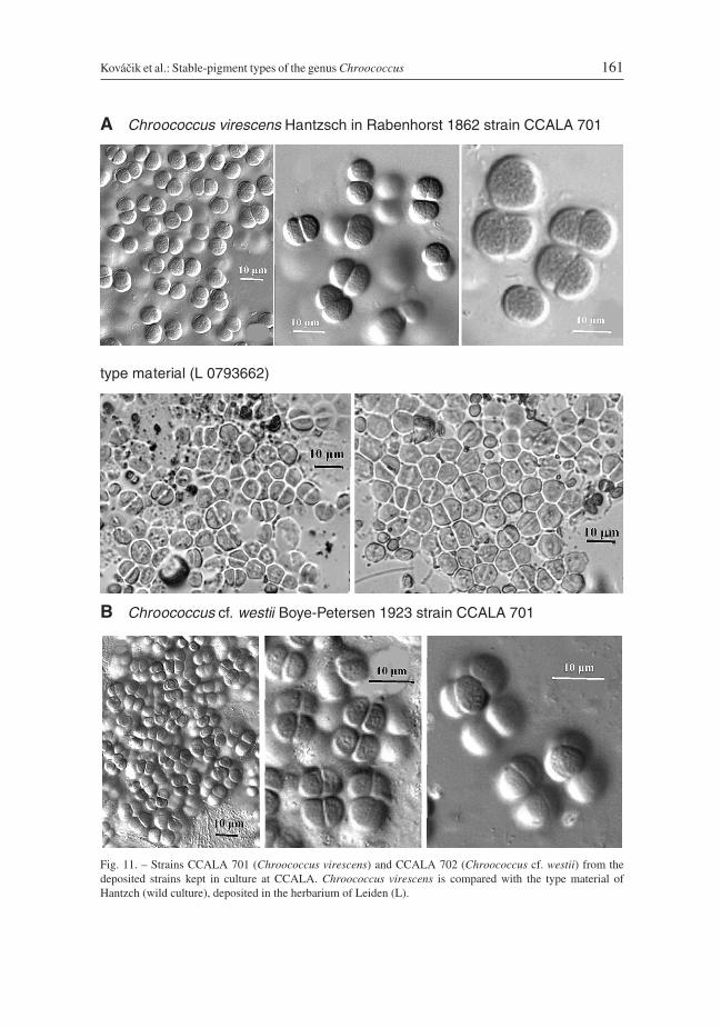
Chroococcus virescens Hantzsch in Rabenhorst 1862, Alg. Sachs. 133 & 134: 1332(Fig. 11A), strain CCALA 701
Colonies few-celled or in small packets, usually up to 4(8)-celled. The masses are dirtygreen. Cells blue-green or greenish with homogeneous or a slightly granular content,10.0–12.5 μm in diameter, or after division 6.2–10.0 (12.0) × 8.7–12.5 (17.5) μm. Muci-laginous envelopes are hyaline, colourless, diffluent and usually quite indistinct.
Reference strain: CCALA 701.Type material from Germany, Dresden (deposited in L); isotypes have the nos.
L0793662 and L0055202. The identification was controlled.Ecology: In unpolluted pools and backwaters with aquatic vegetation or in shallow
wetlands, metaphytic and periphytic on submerged plants and stones.
Kováčik et al.: Stable-pigment types of the genus Chroococcus 157

158 Preslia 83: 145–166, 2011
Fig. 9. – Phylogenetic tree with position of the genus Chroococcus and our strains CCALA 701 and CCALA 702,constructed with the Neighborjoining method (Saitou & Nei 1987). A bootstrap analysis involving 1000resamplings was performed and bootstrap values higher than 50% are given in from of the concerned nodes. 34sequences of chroococcal cyanobacteria deposited to GenBank were included. Dashed lines indicate positions ofthe Chroococcus strains from the GenBank that did not have sufficient sequence length.

Chroococcus cf. westii Boye-Petersen 1923, Bot. Icel. 2(2): 263 (Fig. 11B), strainCCALA 702:
Colonies in the form of three-dimensional, slightly irregular packets of cells, usually with upto 4 (–16)-cells, sometimes more or less in an agglomerate. The masses are dark violet-brownish to blackish. Cells reddish brown, when old brownish-yellowish, spherical, hemi-spherical or in form of a section of a sphere, sometimes slightly elongated (up to 2x longerthan wide), 5.2–11.0 μm in diameter, (3.8) 7.5–9.5 (10.0) × 5.0–12.5 (15.0) μm. Sometimesproduces smaller cells, ± 5.0 × 5.0–7.5 μm or 3–5 μm in diameter. Mucilaginous envelopeshomogeneous, colourless, delimited at the margin, sometimes layered, 2–3 μm thick.
Reference strain: CCALA 702.Type material is not available and the identification is based on descriptions in the liter-
ature. The specimens do not correspond exactly to the original diagnosis of material fromIceland by Boye-Petersen (1923), but agree with that of material collected more recentlyfrom populations in the northern parts of the European temperate zone. The ecology ofother populations of C. westii and the material studied is similar. These strains do not cor-respond to any other Chroococcus species.
Ecology: In clear pools, small ponds with aquatic plants, and in the littoral zone of clear(mountain) and mesotrophic streams, usually periphytic on stones. The information onthis species indicates it has a wider ecological range than C. virescens. This also accordswith the experimental results on the ecological plasticity of this species.
Discussion
Modifications and strategies of chromatic differences
The occurrence of differently pigmented specimens (with different PC/PE ratios but simi-lar morphology) was recorded as early as the beginning of the last century. Variously col-oured populations occur in the cyanobacterial genera Cyanobium, Synechocystis,Limnothrix, Phormidium, Lyngbya and others. Such variously pigmented, morphologi-cally very similar strains sometimes co-occur in nature in the same microhabitat and arestable in culture. The variation in colour in cyanobacteria is usually called “complemen-tary chromatic adaptation”, dependent on the environment, particularly the light condi-tions and considered as to be a more or less reversible. The first reviews of chromatic adap-tation were published by Schindler (1913) and Boresch (1921), who summarized interest-ing data on this phenomenon at that time. In the 1970s and 80s, the strain UTEX 481 of“Fremyella diplosiphon” was commonly used for studying chromatic adaptation (Bennett& Bogorad 1973, Haury 1980, Rosinski et al. 1981, Bogorad et al. 1983, Cobley &Miranda 1983). The model strain was incorrectly identified (both the generic and specificnames used to designate this strain are invalid according to nomenclatoric rules), but thedata on phycocyanin and phycoerythrin syntheses and phycobilisomes function are com-monly valid. Similar studies are reviewed by Bogorad (1975) and Tandeau de Marsac(1977). These later studies were focused particularly on chromatic adaptation in variousecological habitats, on the physiological conditions influencing the coloration of cells(Füglistaller et al. 1981, Tandeau de Marsac & Houmard 1983, 1993, Ohki & Fujita 1991,Latala & Misiewicz 2000, Pinevich et al. 2000) and on molecular mechanisms connected
Kováčik et al.: Stable-pigment types of the genus Chroococcus 159

with chromatic adaptations (Bryant 1994, MacColl 1998, Kehoe & Grossman 1999,Everroad & Wood 2006).
However, many other questions, concerning evaluation of the diversity and ecology ofvariously pigmented and stabilized cyanobacterial populations, remain to be resolved. Theproblem of variously pigmented cyanobaterial populations is not uniform or simple. Thereoccur various strategies of pigmentation in populations and strains (changes in ratios ofphotosynthetic pigments and carotenoids) and it is evident that all these features are inprinciple dependent on their ecology.
There has been great progress in studies on the genomic changes and lateral transfer ofphycobilisome rod genes and gene clusters in populations in which there is a wide variety ofcell pigmentation, e.g. in populations of the marine Synechococcus/Cyanobium (Ong &Glazer 1988, Palenik 2001, Six et al. 2007, Dufresne et al. 2008). In addition, there is a wideand variable mixture of numerous similar pigment types in natural population of planktic,freshwater “Oscillatoria” (= Limnothrix) redekei (Kohl & Nicklisch 1981).
Moreover, there are other adaptative changes in pigmentation and their stabilization innatural populations. Stable and differently coloured populations are known from nature,which differ sometimes only slightly morphologically but are clearly different in their eco-logical and other characteristics. The best known example is the complex Planktothrixagardhii/rubescens, in which both differently coloured types were considered as distinctand separate species already in the original description more than 100 years ago (cf.Geitler 1932). The differentiation and stabilization of these pigment types is interesting,because there are no distinct differences in the 16S rRNA gene sequences of the most com-mon types (P. agardhii and P. rubescens) and it is hypothesized that they are conspecific(Humbert & Le Berre 2001). However, the stability of both types and the ecological andecophysiological differences are so distinct that their separation in two taxa (species) isjustifiable (e.g. Suda et al. 2002, Davis et al. 2003).
160 Preslia 83: 145–166, 2011
Fig. 10. – Phylogenetic analysis of the traditional genus Chroococcus and the position of the strains CCALA 701and CCALA 702. The genetic (and specific) difference between the strains is clearly recognizable. The genusChroococcus is separated from morphologically similar, but genetically distant generic clusters (Limnococcus,Synechocystis and Eucapsis), which were evaluated using the modern polyphasic approach.
Kováčik et al.: Stable-pigment types of the genus Chroococcus 161
B Chroococcus cf. westii Boye-Petersen 1923 strain CCALA 701
Fig. 11. – Strains CCALA 701 (Chroococcus virescens) and CCALA 702 (Chroococcus cf. westii) from thedeposited strains kept in culture at CCALA. Chroococcus virescens is compared with the type material ofHantzch (wild culture), deposited in the herbarium of Leiden (L).
A Chroococcus virescens Hantzsch in Rabenhorst 1862 strain CCALA 701
type material (L 0793662)

The different pigmented Chroococcus-strains studied are not chromatic adaptations.The dependence of both strains on light conditions differed in spite of their occurring innature in the same habitat. This fact indicates the possibility of microzonation of both spe-cies within mats. This was not recorded during the collection of the original material, butsimilar zonation of differently coloured species in microcommunities is recorded for otherhabitats (see e.g. in Komárek & Komárek 2003). The molecular analyses confirmed thatthe two strains were genetically different.
Differently pigmented cyanobacteria include therefore a variety of modifications: (i)reversible chromatic adaptation (Kehoe & Gutu 2006), (ii) populations that are able to differ-entiate continually into variety of pigmented strains and rapidly adapt to a variety of lightniches (Six et al. 2007), (iii) morphologically similar and ecologically distinct morpho-typeswith different but stable pigment compositions, in which the process of diversification and sta-bilization occurred a long time ago. The Chroococcus strains studied belong to such alreadystabilized types (similar to e.g. Planktothrix rubescens/agardhii). They belong to the typicalChroococcus-cluster (Komárková et al. 2010) and are genetically different species (Fig. 9).
Ultrastructure of Chroococcus
The ultrastructure of both strains is almost identical and is probably characteristic of thewhole genus Chroococcus (cf. Potts et al. 1983). It is interesting that it was not easy to pre-pare Chroococcus cells (fixation) for ultrathin sectioning and the outline (periplast) of thecells in the sections was always wavy. This is commonly recorded for Chroococcus (cf.e.g. Figs 3a,b and 4a in Potts et al. 1983). Good sections were only obtained only afterusing a slightly different fixation procedure.
Taxonomic consequences
The modern classification of cyanobacteria is based on molecular methods, which eluci-date and explain the phylogenetic relations. A revision of the systematic classification atthe generic level has been carried out usually using 16S rRNA gene sequences (Castenholz2001, Hoffmann et al. 2005, Komárek 2006). However, comparison of various geneticunits from the GeneBank is sometimes difficult, because the similarity expressed in % isacceptable only as an indication and needs to be interpreted also with respect to a restrictednumber of sequenced bases. Moreover, according to the results of recent studies it is diffi-cult to use common uniform criteria for the delimitation of subgeneric units incyanobacteria (Johansen & Casamatta 2005, Komárek 2010).
Coccoid cyanobacteria are particularly heterogeneous and belong to various develop-mental lines, which differ in important phenotypic and ultrastructural markers. The largediversity of the genus Chroococcus (cf. Komárek & Anagnostidis 1998) was described innumerous morphological and ecological studies on strains and natural populations (Pottset al. 1983, Richert et al. 2006, Komárek & Komárková-Legnerová 2007 and others) andin the revisions based on molecular analyses. The strain “C. turgidus PCC 9340”, desig-nated by Castenholz (2001) as the reference strain for “Chroococcus cluster 2” (corre-sponding to the typical Chroococcus in the traditional sense) is probably close toChroococcus submarinus strain BM (Richert et al. 2006). The planktic types(“Chroococcus limneticus” = Limnococcus) and “small” Chroococcus morphospecies,which are more closely related to Aphanocapsa or Eucapsis, differ from the typicalmorphotypes (C. rufescens, C. turgidus, etc.; Komárková et al. 2010), etc.
162 Preslia 83: 145–166, 2011

The heterogeneity of Chroococcus is evident also from the database of sequences, inwhich the strains designated as “Chroococcus” occur in different clusters in phylogenetictrees. In this genus (in the revised, restricted form) there are types in which the differencein colour (pigment ratio) is stable, and which correspond to stable morphotypes at the spe-cies level (C. rufescens, C. turgidus, C. subnudus, C. virescens, C. westii and others). Thisis an example of taxonomic classification of a number of differently pigmented types,among which the strains studied also belong. The molecular, morphological andultrastructural analyses indicate that both of the strains studied are typical Chroococcus-taxa. The question if the studied strains belong into the category of “chromatic adapta-tions”, or if they represent genetically stable types (species) of the genus Chroococcus,was therefore the particular question in our study.
The studied strains were isolated from one mat and therefore it is likely they have simi-lar ecologies. Ecological differences are important arguments for deciding if two types canbe separated at the specific level (e.g. Planktothrix agardhii/rubescens). However, thesestrains only differ slightly in their ecophysiological characters, but are clearly distinguish-able phenotypically (stable pigment ratios) and have different positions in phylogenetictrees (96.4% genetic similarity). Their positions in the phylogenetic trees were particu-larly important in the assessment of their differences at the specific level. They cannot beconsidered as individuals that are differently chromatically adapted or a consequence ofa recent lateral transfer of phycobilisome rod genes or gene clusters, as described, e.g. bySix et al. (2007) and Dufresne et al. (2008) for oceanic planktic populations of small-celled Synechococcus/Cyanobium spp. The differences between the strains studied arestable, and therefore important in evaluating them as separate taxa. These two more or lessdelimited types do not differ morphologically from previously described C. virescens andonly slightly from C. westii. The preference is to use these taxonomic names, because theydo not differ morphologically from the original descriptions and type exsiccates (Drouet& Daily 1956). It is a pity it was not possible to sequence the 170 and 120 year old driedout types. However, the creation of new names in this case would be superfluous and com-plicating a nomenclatoric review of the genus. Both of the species studied can evidentlyoccur in one and the same habitat at the same locality, but may occupy different microniches.
There is an ongoing debate about the classification of stable, variously pigmented typesin nature. Chromatic adaptations or populations with continually occurring differently pig-mented strains (Kohl & Nicklisch 1981, Six et al. 2007, Dufresne et al. 2008) must be recog-nized, but it is difficult to evaluate them as taxonomic units. On the other hand, stable andisolated pigment mutants should be classified as different species, if they differ in ecology,as in the genus Planktothrix (P. agardhii/rubescens), or if the ecology is similar but they dif-fer in other specific criteria (molecular sequencing), as is the case of the strains studied.
Acknowledgement
This study was funded by the Slovak Grant Agency for Science VEGA, nos. 1/0868/11, 2/0073/11 (Ľ. Kováčik),and the grant agency of the Czech Republic nos. GA CR 206/09/0300, 208/05/0015 (J. Jezberová andJ. Komárková), GA 525/07/0338 (J. Kopecký) and GA AS CR IAA 600050704, AV0Z60050516 (J. Komárek).The authors are particularly thankful to J. Sulek for the electron microscope preparations, D. Švehlová andJ. Pergl for technical help, Š. Kubín, F. Partensky and R. Rippka for valuable discussions of the problems, andK. Edwards for the language correction. Tony Dixon kindly edited the language of final manuscript. We aredeeply indebted to National Herbarium Netherland in Leiden (L) and Naturhistorisches Museum Österreichs inVienna (W) for permission to study the type specimens of several species in their collections, which was impor-tant for the identification of the Chroococcus specimens studied.
Kováčik et al.: Stable-pigment types of the genus Chroococcus 163

Souhrn
Jedním z taxonomických problémů u cyanobakterií (sinic) je hodnocení populací s různým obsahem fykobilínů(poměr PC:PE). Dříve se takové případy vysvětlovaly pouze tzv. komplementární chromatickou adaptací, kde po-měr fotosyntetických barviv je závislý na vnějších podmínkách, především na světelném režimu a teplotě. Mo-derní, zejména molekulární metody však prokázaly, že morfologicky podobné ale různě zbarvené populace mo-hou být způsobeny různými mechanismy a strategiemi, a mohou být různě stabilní s různou taxonomickou hod-notou. Vedle reverzibilní, „klasické“ chromatické adaptace může v populacích dojít k vytvoření celé škály stálýchbarevných odchylek, které jsou v přírodních podmínkách vyvolány především horizontálním transferem přísluš-ných genů (stabilizují se jen po izolaci do monokultur), nebo se mohou dlouhodobě ustálit některé pigmentovémutace ve stálých odlišných ekologických podmínkách. Takové stabilizované přírodní typy se vyskytují zpravi-dla i v odlišných ekologických situacích, ale to neplatí absolutně. Pokud se mají podobné případy řešit, je nutnoprovést podrobné ekofyziologické, cytomorfologické i fylogenetické analýzy.
V našem případě šlo o vyřešení dvou důležitých, fenotypově podobných, ale pigmentově rozdílných typůz rodu Chroococcus, izolovaných ze stejného biotopu (mrtvé rameno Dunaje u Gabčíkova, Slovensko). Z našichvýsledků vyplynulo, že se jedná o geneticky i fenotypově rozdílné typy, které je nutno považovat za odlišné druhy.Kontrolou typových materiálů z Leidenu (L) a Vídně (W) vyplynulo, že lze dokonce tyto druhy ztotožnit již s dří-ve popsanými, ale dosud taxonomicky ne zcela jasnými druhy (Chroococcus virescens, C. westii). V našem pří-padě se jedná o výskyt ve stejném biotopu, což je u podobných případů určitá zvláštnost. V našem článku je vyře-šena taxonomická pozice obou studovaných druhů a jsou zde též poprvé stručně porovnány různé případypigmentových modifikací u sinic a diskutován způsob jejich taxonomického hodnocení.
References
Albertano P. & Kováčik Ľ. (1996): Light and temperature responses of terrestrial sciaphilous strains ofLeptolyngbya sp. in cross-gradient cultures. – Algol. Stud. 83: 17–28.
Albertano P., Kováčik Ľ. & Gardavský A. (1993): Cross-gradient cultures of filamentous cyanophytes. – Giorn.Bot. Ital. 123: 192–193.
Bennett A. & Bogorad L. (1973): Complementary chromatic adaptation in a filamentous blue-green alga. – J.Cell. Biol. 58: 419–435.
Bogorad L. (1975): Phycobiliproteins and complementary chromatic adaptation. – Ann. Rev. Plant Physiol. 26:369–401.
Bogorad L., Gendel S. M., Haury J. H. & Koller K.-P. (1983): Photomorphogenesis and complementary chro-matic adaptation in Fremyella diplosiphon. – In: Papageorgiou G. C. & Packer L. (eds), Photosyntheticprokaryotes: cell differentiation and function, p. 159–126, Elsevier.
Boresch K. (1921): Die komplementäre chromatische Adaptation. – Arch. Protistenk. 44: 1–69.Bourrelly P. (1970): Les algues d’eau douce III. – N. Boubée & Cie, Paris.Boye-Petersen J. (1923): The freshwater Cyanophyceae of Iceland. – Arb. Bot. Kobenhavn 101: 251–324.Bryant D. A. (1994): The moleclular biology of cyanobacteria. – Kluwer Acad. Publ., Dordrecht.Castenholz R. W. (2001): Oxygenic photosynthetic bacteria. – In: Boone D. R. & Castenholz R. W. (eds),
Bergey’s manual of systematic bacteriology, Ed. 2, p. 473–600, Springer-Verlag, New York.Cobley J. G. & Miranda R. D. (1983): Mutations, affecting chromatic adaptation in the cyanobacterium
Fremyella diplosiphon. – J. Bacteriol. 153: 1486–1492.Davis P. A., Beard S. J. & Walsby A. E. (2003): Variation in filament width and gas vesicles of red and green iso-
lates of Planktothrix spp. – Algol. Stud. 108: 15–29.Drouet F. & Daily W. A. (1956): Revision of the coccoid Myxophyceae. – Butler Univ. Bot. Stud. 12: 1–218.Dufresne A., Ostrowski M., Scanlan D. J., Garczarek L., Mazard S., Palenik B. P., Paulsen I. T., de Marsac N. T.,
Wincker P., Dossat C., Ferriera S., Johnson J., Post A. F., Hess W. R. & Partensky F. (2008): Unravelling thegenomic mosaic of a ubiquitous genus of marine cyanobacteria. – Genome Biology 9: R90.
Everroad R. C. & Wood A. M. (2006): Comparative molecular evolution of newly discovered picocyanobacterialstrains reveals a phylogenetically informative variable region of beta-phycoerythrin. – J. Phycol. 42:1300–1311.
Flechtner V. R., Boyer S. L., Johansen J. R. & DeNoble M. L. (2002): Spirirestis rafaelensis gen. et sp. nov.(Cyanophyceae), a new cyanobacterial genus from arid soils. – Nova Hedwigia 74: 1–24.
Füglistaller P., Widmer H., Sidler W., Frank G. & Zuber H. (1981): Isolation and characterization ofphycoerythrocyanin and chromatic adaptation of the thermophilic cyanobacterium Mastigocladuslaminosus. – Arch. Microbiol. 129: 268–274.
164 Preslia 83: 145–166, 2011

Geitler L. (1932): Cyanophyceae. – In: Rabenhorst’s Kryptogamenflora von Deutschland, Österreich und derSchweiz 14: 1–1196, Akad. Verlagsges., Leipzig.
Gilmore A. M. & Yamamoto H. Y. (1991): Resolution of lutein and zeaxanthin using a non-endcapped lightly car-bon-loaded C18 high-performance liquid chromatographic column. – J. Chromatogr. 543: 137–145.
Golubić S. (1967): Zwei wichtige Merkmale zur Abgrenzung der Blaualgengattungen. – Schweiz. Ztschr.Hydrol. 29: 176–184.
Guillard R. R. L. & Lorenzen C. J. (1972): Yellow-green algae with chlorophyllide. – J. Phycol. 8: 10–14.Hansgirg A. (1892): Prodromus der Algenflora von Böhmen. 2. – Arch. Naturwiss. Landesdurchforsch. Böhmen
8(4): 1–268.Haury J. F. (1980): Chromatic adaptation in Fremyella diplosiphon and morphogenetic changes. – In: Gantt E.
(ed.), Handbook of phycological methods, p. 219–229, CambridgeUniv. Press, Cambridge.Hoffmann L., Komárek J. & Kaštovský J. (2005): System of cyanoprokaryotes (cyanobacteria) – state in 2004. –
Algol. Stud. (Cyanobacterial Res. 6) 117: 95–115.Humbert J. F. & Le Berre B. (2001): Genetic diversity in two species of freshwater cyanobacteria Planktothrix
(Oscillatoria) rubescens and P. agardhii. – Arch. Hydrobiol. 150: 197–206.Jeffrey S. W. & Humphrey G. F. (1975): New spectrophotometric equations for determining chlorophylls a, b, c1
and c2 in a higher plants, algae and natural phytoplankton. – Biochem. Physiol. Pfl. 167: 191–194.Johansen J. R. & Casamatta D. A. (2005): Recognizing cyanobacterial diversity through adoption of a new spe-
cies paradigm. – Algol. Stud. 116:71–93.Kehoe D. M. & Grossman A. R. (1999): The molecular mechanisms controlling complementary chromatic adap-
tation. – In: Peschek G. A., Löffelhardt W. & Schmetterer G. (eds), The phototrophic prokaryotes, p. 61–69,Kluwer Academic/Plenum Publishers, New York.
Kehoe D. M. & Gutu A. (2006): Responding to color: the regulation of complementary chromatic adaptation. –Ann. Rev. Plant Biol. 57:127–150.
Kohl J.-G. & Nicklisch A. (1981): Chromatic adaptation of the planktic blue-green alga Oscillatoria redekei vanGoor and its ecological significance. – Int. Rev. Ges. Hydrobiol. 66: 83–94.
Komárek J. (2006): Cyanobacterial taxonomy: current problems and prospects for the integration of traditionaland molecular approach. – Algae 21: 349–375.
Komárek J. (2010): Recent changes (2008) in cyanobacteria taxonomy based on a combination of molecularbackground with phenotype and ecological consequences (genus and species concept). – Hydrobiologia 639:245–259.
Komárek J. & Anagnostidis K. (1998): Cyanoprokaryota 1.Teil: Chroococcales. – In: Ettl H., Gärtner G., HeynigH. & Mollenhauer D. (eds), Süsswasserflora von Mitteleuropa 19/1, Gustav Fischer, Jena, Stuttgart, Lübeck& Ulm.
Komárek J. & Komárek O. (2003): Diversity of cyanobacteria in seepages of King George Island, maritimeAntarctica. – In: Huiskes A. H. L., Gieskes W. W. C., Rozema J., Schorno R. M. L., van der Vies S. M. &Wolff W. J. (eds), Antarctic biology in a global context, p. 244–250, Backhuys Publishers, Leiden.
Komárek J. & Komárková-Legnerová J. (2007): Taxonomic evaluation of cyanobacterial microflora from alka-line marshes of northern Belize. 1. Phenotypic diversity of coccoid morphotypes. – Nova Hedwigia 84:65–111.
Komárková J., Jezberová J., Komárek O. & Zapomělová E. (2010): Variability of Chroococcus morphospecies(Cyanobacteria) in phylogenic relationships. – Hydrobiolgia 639: 69–83.
Kopecký J., Schoefs B., Loest K., Štys D. & Pulz O. (2000): Microalgae as a source for secondary carotenoid pro-duction: a sreening study. – Algol. Stud. 98: 153–168.
Latala A. & Misiewicz S. (2000): Effects of light, temperature and salinity on the growth and chlorophyll a con-tent of Baltic cyanobacterium Phormidium amphibium. – Algol. Stud. 100: 157–180.
Lichtenhaler H. K. & Wellburn A. R. (1983): Determination of total carotenoids and chlorophylls a and b of leafextracts in different solvents. – Biochem. Soc. Trans. 603: 591–592.
MacColl R. (1998): Cyanobacterial phycobilisomes. – J. Struct. Biol. 124: 311–334.Ohki K. & Fujita Y. (1991): Complementary chromatic adaptation in the cyanobacterium Tolypothrix tenuis:
location of phycoerythrin newly synthesized by green illumination. – Plant Cell. Physiol. 32: 483–488.Ong L. J. & Glazer A. N. (1988): Structural studies of phycobiliproteins in unicellular marine cyanobacteria. – In:
Stevens S. E. J. & Bryant D. A. (eds), Light-energy transduction in photosynthesis: higher plant and bacterialmodels, p. 102–121, American Society of Plant Physiologists, Rockville, M. D.
Palenik B. (2001): Chromatic adaptation in marine Synechococcus strains. – Appl. Environ. Microbiol. 67:991–994.
Kováčik et al.: Stable-pigment types of the genus Chroococcus 165

Pinevich A., Matthijs H., Garcia-Mendoza E. & Babanova A. (2000): The uncommon pigment composition andcomplementary chromatic adaptation in a marine Synechocystis sp. – Algol. Stud. 99: 67–77.
Potts M., Ocampo-Friedmann R., Bowman M. A. & Tözün B. (1983): Chroococcus-S24 and Chroococcus-N41(cyanobacteria): morphological, biochemical and genetic characterization and effects of water-stress onultrastructure. – Arch. Microbiol. 135: 81–90.
Richert L., Golubić S., Le Guédès R., Hervé A. & Payri C. (2006): Cyanobacterial populations build “Kopara”microbial mats, Rangiraa, Tuamotu Archipelago, F.P. – Eur. J. Phycol. 41: 259–279.
Rippka R., Deruelles J., Waterbury J. B., Herdman M. & Stanier R. Y. (1979): Generic assignments, strain histo-ries and properties of pure cultures of Cyanobacteria. – J. Gen. Microbiol. 111: 1–61.
Rosinski J., Hainfeld J. F., Rigbi M. & Siegelman H. W. (1981): Phycobilisome ultrastructure and chromaticadaptation in Fremyella diplosiphon. – Ann. Bot. 47: 1–12.
Saitou N. & Nei M. (1987): The neighbor-joining methods: a new method for reconstructed phylogenetic trees. –Mol. Biol. Evol. 4: 406–425.
Schindler B. (1913): Über den Farbenwechsel der Oscillarien. – Ztschr. Bot. 5: 497–575.Shibata K. (1958): Spectrophotometry of intact biological materials. – J. Biochem. (Tokyo) 45: 599–623.Six C., Thomas J.-C., Garczarek L., Ostrowski M., Dufresne A., Blot N., Scanlan P. J. & Partensky F. (2007):
Diversity and evolution of phycobilisomes in marine Synechococcus spp.: a comparative genomics study. –Genome Biology 8: R259.
Spurr A. R. (1969): A low-viscosity resin embedding medium for electron microscopy. – Clinical Microbiol. Rev.3: 197–218.
Stackebrandt E. & Goebel B. M. (1995): Taxonomic note: a place for DNA-DNA reassociation and 16S rRNAsequence analysis in the present species definition in bacteriology. – Int. J. Syst. Bacteriol. 44: 846–849.
Starmach K. (1966): Cyanophyta – sinice, Glaucophyta – glakofity. [Cyanophyta – cyanobacteria, Glaucophyta –glaucophytes]. – In: Starmach K. (ed.) Flora słodkowodna Polski [Freshwater flora of Poland] 2: 1–808, Pol.Wydaw. Nauk, Warszawa.
Suda S., Watanabe M. M., Otsuka S., Mahakahant A., Yongmanitchai W., Nopartnaraporn N., Liu Y. D. & Day J.G. (2002): Taxonomic revision of water-bloom-forming species of oscillatorioid cyanobacteria. – Int. J. Syst.Exper. Microbiol. 52: 1577–1595.
Tamura K., Dudley J., Nei M. & Kumar S. (2007): MEGA4: Molecular Evolutionary Genetics Analysis (MEGA)Software Version 4.0. – Mol. Biol. Evol. 24: 1596–1599.
Tandeau de Marsac N. (1977): Occurrence and nature of chromatic adaptation in cyanobacteria. – J. Bacteriol.130: 82–91.
Tandeau de Marsac N. & Houmard J. (1983): Complementary chromatic adaptation: physiological conditionsand action spectra. – In: Packer L. & Glazer A. N. (eds), Methods in enzymology 167 – Cyanobacteria, p.318–328, Academic Press.
Tandeau de Marsac N. & Houmard J. (1993): Adaptation of cyanobacteria to environmental stimuli: new stepstowards molecular mechanisms. – FEMS Microbiol. Rev. 104: 119–190.
Taton A., Grubisić S., Brambilla E., De Wit R. & Wilmotte A. (2003): Cyanobacterial diversity in natural and arti-ficial microbial mats of Lake Fryxell (McMurdo Dry Valleys, Antarctica): a morphological and molecularapproach. – Appl. Env. Microbiol. 69(9): 5157–5169.
Turicchia S., Ventura S., Schütte U., Soldati E., Zielke M. & Solheim B. (2005): Biodiversity of cyanobacteriaand changes on temporal and spiral scales of the cyanobacterial community in the foreland of Midtre Lovenglacier, Spitsbergen, Svalbard. – Algol. Stud. 117: 427–440.
Received 23 June 2010Revision received 25 October 2010
Accepted 27 October 2010
166 Preslia 83: 145–166, 2011


![[Taxonomic analysis and other taxometric methods]](https://img.pdfslide.net/doc/110x75/6360b29a4b9aa63a9e0068c0/taxonomic-analysis-and-other-taxometric-methods.jpg)


