Embed Size (px)
Citation preview
�����������������
Citation: Lara-Espinosa, J.V.;
Arce-Aceves, M.F.; López-Torres,
M.O.; Lozano-Ordaz, V.;
Mata-Espinosa, D.; Barrios-Payán, J.;
Silva-Islas, C.A.; Maldonado, P.D.;
Marquina-Castillo, B.;
Hernández-Pando, R. Effect of
Curcumin in Experimental
Pulmonary Tuberculosis:
Antimycobacterial Activity in the
Lungs and Anti-Inflammatory Effect
in the Brain. Int. J. Mol. Sci. 2022, 23,
1964. https://doi.org/10.3390/
ijms23041964
Academic Editors: Maria
Rosalia Pasca, Vadim Makarov,
Giulia Degiacomi and
Laurent Chiarelli
Received: 22 September 2021
Accepted: 1 February 2022
Published: 10 February 2022
Publisher’s Note: MDPI stays neutral
with regard to jurisdictional claims in
published maps and institutional affil-
iations.
Copyright: © 2022 by the authors.
Licensee MDPI, Basel, Switzerland.
This article is an open access article
distributed under the terms and
conditions of the Creative Commons
Attribution (CC BY) license (https://
creativecommons.org/licenses/by/
4.0/).
International Journal of
Molecular Sciences
Article
Effect of Curcumin in Experimental Pulmonary Tuberculosis:Antimycobacterial Activity in the Lungs and Anti-InflammatoryEffect in the BrainJacqueline V. Lara-Espinosa 1,* , María Fernanda Arce-Aceves 1, Manuel O. López-Torres 1 ,Vasti Lozano-Ordaz 1, Dulce Mata-Espinosa 1, Jorge Barrios-Payán 1, Carlos Alfredo Silva-Islas 2 ,Perla D. Maldonado 2 , Brenda Marquina-Castillo 1 and Rogelio Hernández-Pando 1,*
1 Sección de Patología Experimental, Instituto Nacional de Ciencias Médicas y Nutrición Salvador Zubirán,Mexico City 14080, Mexico; [email protected] (M.F.A.-A.);[email protected] (M.O.L.-T.); [email protected] (V.L.-O.);[email protected] (D.M.-E.); [email protected] (J.B.-P.); [email protected] (B.M.-C.)
2 Laboratorio de Patología Vascular Cerebral, Instituto Nacional de Neurología y Neurocirugía Manuel VelascoSuárez, Mexico City 14269, Mexico; [email protected] (C.A.S.-I.);[email protected] (P.D.M.)
* Correspondence: [email protected] (J.V.L.-E.); [email protected] (R.H.-P.)
Abstract: Tuberculosis (TB) is one of the ten leading causes of death worldwide. Patients with TB havebeen observed to suffer from depression and anxiety linked to social variables. Previous experimentsfound that the substantial pulmonary inflammation associated with TB causes neuroinflammation,neuronal death, and behavioral impairments in the absence of brain infection. Curcumin (CUR) is anatural product with antioxidant, anti-inflammatory and antibacterial activities. In this work, weevaluated the CUR effect on the growth control of mycobacteria in the lungs and the anti-inflammatoryeffect in the brain using a model of progressive pulmonary TB in BALB/c mice infected with drug-sensitive mycobacteria (strain H37Rv). The results have shown that CUR decreased lung bacilliload and pneumonia of infected animals. Finally, CUR significantly decreased neuroinflammation(expression of TNFα, IFNγ and IL12) and slightly increased the levels of nuclear factor erythroid2-related to factor 2 (Nrf2) and the brain-derived neurotrophic factor (BDNF) levels, improvingbehavioral status. These results suggest that CUR has a bactericidal effect and can control pulmonarymycobacterial infection and reduce neuroinflammation. It seems that CUR has a promising potentialas adjuvant therapy in TB treatment.
Keywords: tuberculosis; curcumin; neuroinflammation; antibacterial
1. Introduction
Tuberculosis (TB), the oldest human pandemic, generally caused by infection via thelung with Mycobacterium tuberculosis (Mtb), remains the foremost cause of death amongbacterial infectious diseases [1,2]. Bacillus Calmette–Guérin (BCG), a live attenuated strainof Mycobacterium bovis [3], the primary cause of bovine TB [4], is the only approved vaccineagainst TB and is the most widely used vaccine in history [4,5]. Unfortunately, thoughbillions of individuals were vaccinated in the past century, TB remains a severe threat toglobal health [5]. In 2019, 10 million persons developed TB, and approximately 1.4 millionpeople died of this infection (208,000 were HIV-infected). Due to the COVID-19 pandemic,the number of deaths attributable to TB is expected to increase to similar levels presentedin 2012, increasing to between 200,000 and 400,000 deaths (1.6–1.8 million deaths) [6]. TheCOVID-19 pandemic and related lockdown restrictions significantly impacted providingand monitoring TB surveillance strategies globally [7].
The typical treatment for pulmonary TB comprises two months of fourfold therapywith isoniazid (INH), rifampicin (RMP), ethambutol (EMB), and pyrazinamide (PZA)
Int. J. Mol. Sci. 2022, 23, 1964. https://doi.org/10.3390/ijms23041964 https://www.mdpi.com/journal/ijms
Int. J. Mol. Sci. 2022, 23, 1964 2 of 25
followed immediately by an additional four months of dual administration of RMP andINH [8]. This regimen is well known and has been generally adopted worldwide fordecades, and about 85% of patients will have a successful treatment outcome [9]. However,this treatment frequently produces side effects (gastric, neurologic and haematologicalalterations). Furthermore, the long duration affects patient adherence, resulting in treatmentabandonment, which results in the emergence of drug-resistant TB [10–12]. In addition,there has been a rapidly emerging problem of multidrug-resistant (MDR) TB, which isfrequently lethal, extremely expensive and complicated to treat [13]. A fluoroquinolone(moxifloxacin, levofloxacin) can be employed in patients with monoresistance to RMP orINH. The duration of treatment is then raised to a total of 6 to 9 months (INH resistance) or18 to 20 months (RMP resistance), depending on the individual course [14].
Although new antibiotics against TB have been developed in recent years, there is stilla need to discover new antituberculous agents that are effective in treating MDR TB casesand novel agents that can shorten the long conventional chemotherapy drug-sensitive TB.Within this context, new synthetic drugs and natural products from medicinal plants arepotential sources of new antimycobacterial products [15].
TB is a severe chronic systemic lung disease, although Mtb can spread to other organs,causing extrapulmonary disease [16]. Inflammation is a response to infection, antigen chal-lenge, or tissue injury designed to eradicate microbes or injury agents and potentiate tissuerepair [17]. However, excessive inflammation leads to tissue damage and can, if severe,cause physiological decompensation, organ dysfunction and death [17,18]. Perturbationsin host tissue homeostasis triggered by infectious microorganisms, such as Mtb, activateimmune surveillance mechanisms that promote inflammation. Infection with Mtb consistsof several phases, which begins with phagocytosis of bacteria by macrophages and progresstowards a TH1 lymphocyte response that fights the bacteria and causes tissue damage byexcessive inflammation [19,20]. Systemic inflammation occurs in pulmonary and extra-pulmonary TB diseases and is characterized by increased concentrations of inflammatorymolecules such as acute-phase proteins, lipid mediators (e.g., prostaglandin E2 [PGE2]),several pro-inflammatory cytokines, and chemokines [21].
The central nervous system (CNS) was long considered as a site of restricted immunesurveillance due to the absence of lymphatic vessels, the blood–brain barrier (BBB), andslow transplant rejection [18]. The CNS is a compartmentalized organ, including theparenchyma, the ventricles comprising the choroid plexus and cerebrospinal fluid (CSF),the meningeal layers that enclose the parenchyma, and various “absolute” (BBB) andsemi-permeable barriers (blood CSF, blood-leptomeningeal) [22]. The level of steady-stateimmune privilege varies considerably among these compartments [22]. During neuroin-flammation, the immune landscape of the CNS changes dramatically; resident immunecells become activated, and the inflammatory leukocytes can infiltrate the parenchyma [22].This process could be helpful by protecting the brain from pathogens and neurotoxic agentsand supporting tissue repair processes [23]. However, neuroinflammation is an essentialfeature of many neurodegenerative diseases such as multiple sclerosis (MS), Alzheimer’sdisease (AD), Parkinson’s disease (PD), narcolepsy, as well as psychiatric diseases andbehavioral disorders such as schizophrenia, autism, and depression [24,25].
Another problem related to neuroinflammation is psychiatric disorders, includingdepression and anxiety [26]. Lung diseases are among chronic medical conditions stronglyassociated with psychiatric disorders [27,28]. Evidence shows that anxiety, depression andemotional distress participate in the incapacity generated by TB, and they are related to theseverity of symptoms, the number of reported symptoms, higher rates of health servicesuse, short treatment compliance, more comprehensive course of treatment, reduced controlof the disease and death [29]. Epidemiological evidence establishes a relationship betweendepression, anxiety and TB [30]. For instance, in an Afghan study, 69.55% of the MDR-TBpatients with HIV-negative status presented with significant levels of depression [31]. ASouth African study determined that 81.1% of the TB patients presented with depressionand 31.9% with anxiety [32]. A recent study in India determined that 80.37% of TB patients
Int. J. Mol. Sci. 2022, 23, 1964 3 of 25
had depression and 74% anxiety [33]. A Brazilian study found a 60.2% increase in depres-sion in individuals with pulmonary TB [34]. Patients with a more prolonged disease have ahigher incidence of depression and anxiety [35], and depression is higher in patients withpulmonary than in extrapulmonary TB [33]. An important aspect is that TB patients stillsuffer from depression even under treatment, anxiety scores remain high [36,37], and adultpatients are more susceptible to depression [38]. Additionally, MDR-TB patients presenthigher levels of depression [31,39–41]. Furthermore, TB patients show low-to-moderaterates of suicidal ideation (9.0%) and a record of suicide attempts (3.1%) [42].
Even though the link between TB, depression and anxiety has not been clearly un-derstood [30], it seems that pro-inflammatory cytokines that are highly produced by thetuberculous lungs can reach the brain by specific carrier-mediated transport mechanisms.Furthermore, these cytokines are also overproduced in the brain during this peripheralinflammatory process by binding to their receptors in the endothelial cells and nerve cellsin the circumventricular organs and other brain areas lacking the BBB [43]. In addition,pro-inflammatory cytokines such as interferon-gamma (IFNγ), IFNα and tumor necrosisfactor (TNFα) contribute to the development of depressive disorder by regulating neuronalexcitability, reducing the levels of serotonin and causing changes in other mechanismsof neurotransmission and neuronal signaling in brain regions involved with depression,oxidative injury, and hippocampal neuronal damage [44–46]. Therefore, in TB patients, Mtbinfection’s peripheral inflammation generated in the lung could induce CNS inflammationand neuropsychiatric disorders, such as depression and anxiety.
In diseases with an inflammatory component, first-line medications have traditionallybeen agents that reduce inflammation [47]. Curcumin (CUR) or diferuloylmethane (1, 7-bis(4-hydroxy-3-methoxyphenol)-1, 6-heptadiene-3, 5-dione) is a polyphenolic compoundobtained from the rhizomes of Curcuma longa [48,49], a rhizomatous native plant fromSouth and Southeast Asia that belongs to the family Zingiberaceae [50]. Research hasrevealed that CUR has pleiotropic properties, including anti-inflammatory, antioxidant,chemopreventive, chemotherapeutic activity, neuroprotective properties, and antibacterialactivity [50–53]. The pleiotropic actions of CUR are derived from its complex chemistryand its ability to influence multiple signaling pathways [50]. CUR controls the inflamma-tory response by downregulating the activity of the enzymes cyclooxygenase-2 (COX-2),lipoxygenase, and inducible nitric oxide synthase (iNOS). In addition, CUR suppresses theactivation of nuclear factor kappa B (NF-κB) activation; inhibits the production of the inflam-matory cytokines TNF-α, interleukin (IL)-1, -2, -6, -8, and -12, monocyte chemoattractantprotein (MCP) and migration inhibitory protein; and down-regulates mitogen-activatedand Janus kinases [54]. In addition, CUR protects the brain from damage through theupregulated expression of the transcription factor, the nuclear factor erythroid 2-related tofactor 2 (Nrf2) expression [55], and the hippocampal levels of brain-derived neurotrophicfactor (BDNF) [56].
We have demonstrated neuroinflammation and distinct neuropsychiatric abnormali-ties in an experimental model of progressive pulmonary TB without brain infection [57].Therefore, we hypothesize that CUR administration could decrease the pulmonary bacilliburdens and neuroinflammation with its behavioral abnormalities in TB mice. Thus, thepresent study aimed to evaluate the efficacy of the administration of CUR on lung diseaseevolution, neuroinflammation, the Nrf2 and BDNF expression and behavioral alterationsin a murine model of pulmonary TB.
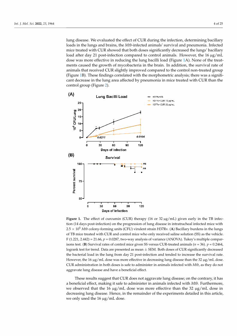
2. Results2.1. The Effect of Curcumin (CUR) Treatment on Survival, Bacilli Loads and Tissue Damage(Pneumonia) in Experimental Pulmonary Tuberculosis
Tuberculous animals were given CUR (16 or 32 µg/mL) via an intraperitoneal routestarting on day 14 after infection to see how these treatments affected the progression oflung disease in BALB/c mice after endotracheal infection with a high dose of the MtbH37Rv strain, trying to find a dose that reduced neuroinflammation without aggravating
Int. J. Mol. Sci. 2022, 23, 1964 4 of 25
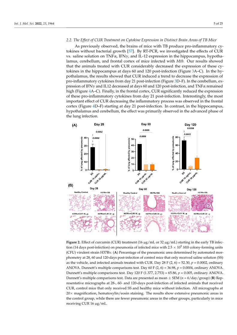
lung disease. We evaluated the effect of CUR during the infection, determining bacillaryloads in the lungs and brains, the Mtb-infected animals’ survival and pneumonia. Infectedmice treated with CUR showed that both doses significantly decreased the lungs’ bacillaryload after day 21 post-infection compared to control animals. However, the 16 µg/mLdose was more effective in reducing the lung bacilli load (Figure 1A). None of the treat-ments caused the growth of mycobacteria in the brain. In addition, the survival rate ofanimals that received CUR slightly improved compared to the control non-treated group(Figure 1B). These findings correlated with the morphometric analysis; there was a signifi-cant decrease in the lung area affected by pneumonia in mice treated with CUR than thecontrol group (Figure 2).
Int. J. Mol. Sci. 2021, 22, x FOR PEER REVIEW 4 of 25
2. Results 2.1. The Effect of Curcumin (CUR) Treatment on Survival, Bacilli Loads and Tissue Damage (Pneumonia) in Experimental Pulmonary Tuberculosis
Tuberculous animals were given CUR (16 or 32 μg/mL) via an intraperitoneal route starting on day 14 after infection to see how these treatments affected the progression of lung disease in BALB/c mice after endotracheal infection with a high dose of the Mtb H37Rv strain, trying to find a dose that reduced neuroinflammation without aggravating lung disease. We evaluated the effect of CUR during the infection, determining bacillary loads in the lungs and brains, the Mtb-infected animals’ survival and pneumonia. Infected mice treated with CUR showed that both doses significantly decreased the lungs’ bacillary load after day 21 post-infection compared to control animals. However, the 16 μg/mL dose was more effective in reducing the lung bacilli load (Figure 1A). None of the treatments caused the growth of mycobacteria in the brain. In addition, the survival rate of animals that received CUR slightly improved compared to the control non-treated group (Figure 1B). These findings correlated with the morphometric analysis; there was a significant de-crease in the lung area affected by pneumonia in mice treated with CUR than the control group (Figure 2).
These results suggest that CUR does not aggravate lung disease; on the contrary, it has a beneficial effect, making it safe to administer in animals infected with Mtb. Further-more, we observed that the 16 μg/mL dose was more effective than the 32 μg/mL dose in decreasing lung disease. Hence, in the remainder of the experiments detailed in this arti-cle, we only used the 16 μg/mL dose.
Figure 1. The effect of curcumin (CUR) therapy (16 or 32 μg/mL) given early in the TB infection (14 days post-infection) on the progression of lung disease in intratracheal infected mice with 2.5 × 10⁵ Mtb colony-forming units (CFU) virulent strain H37Rv. (A) Bacillary burdens in the lungs of TB mice treated with CUR and control mice who only received saline solution (SS) as the vehicle. F (1.221, 2.442) = 21.66, p = 0.0287, two-way analysis of variance (ANOVA). Tukey’s multiple compar-isons test. (B) Survival rates of control mice given SS versus CUR-treated animals (n = 36). p = 0.2464, logrank test for trend. Data are presented as mean ± SEM. Both doses of CUR significantly decreased the bacterial load in the lung from day 21 post-infection and tended to increase the survival rate. However, the 16 μg/mL dose was more effective in decreasing lung disease than the 32 μg/mL dose. CUR administration in both doses is safe to administer in animals infected with Mtb, as they do not aggravate lung disease and have a beneficial effect.
Figure 1. The effect of curcumin (CUR) therapy (16 or 32 µg/mL) given early in the TB infec-tion (14 days post-infection) on the progression of lung disease in intratracheal infected mice with2.5 × 105 Mtb colony-forming units (CFU) virulent strain H37Rv. (A) Bacillary burdens in the lungsof TB mice treated with CUR and control mice who only received saline solution (SS) as the vehicle.F (1.221, 2.442) = 21.66, p = 0.0287, two-way analysis of variance (ANOVA). Tukey’s multiple compar-isons test. (B) Survival rates of control mice given SS versus CUR-treated animals (n = 36). p = 0.2464,logrank test for trend. Data are presented as mean ± SEM. Both doses of CUR significantly decreasedthe bacterial load in the lung from day 21 post-infection and tended to increase the survival rate.However, the 16 µg/mL dose was more effective in decreasing lung disease than the 32 µg/mL dose.CUR administration in both doses is safe to administer in animals infected with Mtb, as they do notaggravate lung disease and have a beneficial effect.
These results suggest that CUR does not aggravate lung disease; on the contrary, it hasa beneficial effect, making it safe to administer in animals infected with Mtb. Furthermore,we observed that the 16 µg/mL dose was more effective than the 32 µg/mL dose indecreasing lung disease. Hence, in the remainder of the experiments detailed in this article,we only used the 16 µg/mL dose.
Int. J. Mol. Sci. 2022, 23, 1964 5 of 25
2.2. The Effect of CUR Treatment on Cytokine Expression in Distinct Brain Areas of TB Mice
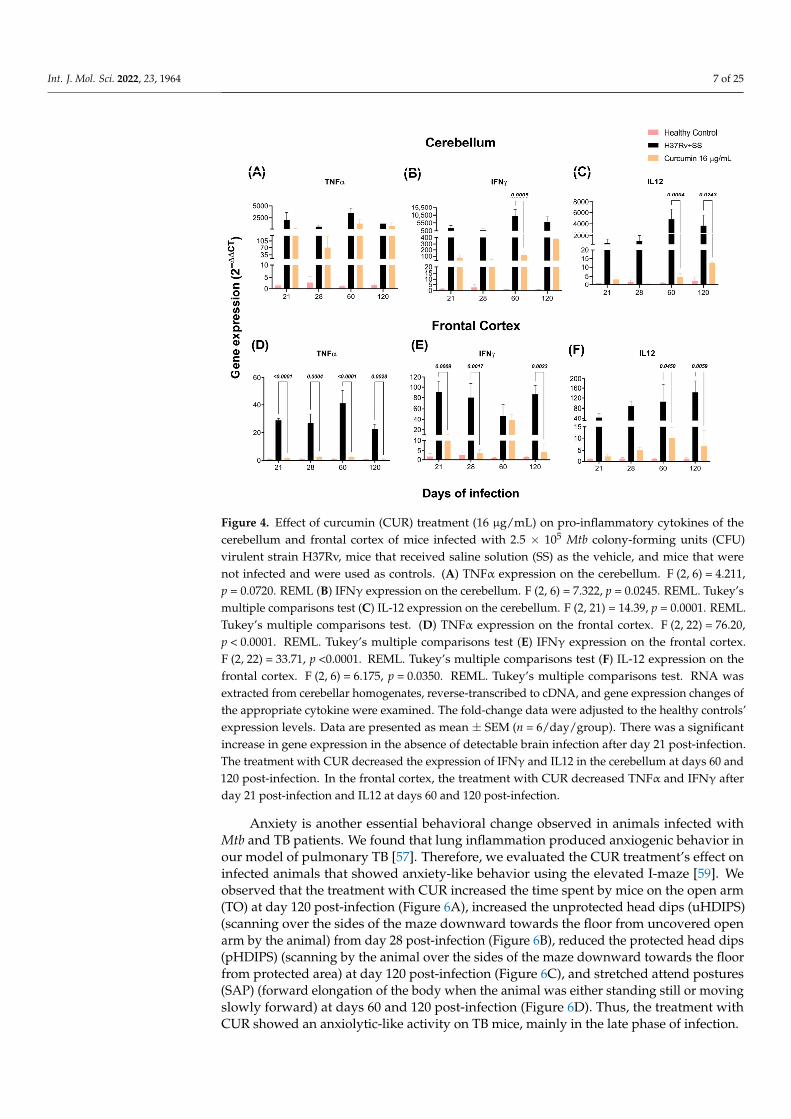
As previously observed, the brains of mice with TB produce pro-inflammatory cy-tokines without bacterial growth [57]. By RT-PCR, we investigated the effects of CURvs. saline solution on TNFα, IFNγ, and IL-12 expression in the hippocampus, hypotha-lamus, cerebellum, and frontal cortex of mice infected with Mtb. Our results showedthat the animals treated with CUR considerably decreased the expression of these cy-tokines in the hippocampus at days 60 and 120 post-infection (Figure 3A–C). In the hy-pothalamus, the results showed that CUR induced a trend to decrease the expression ofpro-inflammatory cytokines from day 21 post-infection (Figure 3D–F). In the cerebellum, ex-pression of IFNγ and IL12 decreased at days 60 and 120 post-infection, and TNFα remainedhigh (Figure 4A–C). Finally, in the frontal cortex, CUR significantly reduced the expressionof these pro-inflammatory cytokines from day 21 post-infection. Interestingly, the mostimportant effect of CUR decreasing the inflammatory process was observed in the frontalcortex (Figure 4D–F) starting at day 21 post-infection. In contrast, in the hippocampus,hypothalamus and cerebellum, the effect was primarily observed in the advanced phase ofthe lung infection.
Int. J. Mol. Sci. 2021, 22, x FOR PEER REVIEW 5 of 25
Figure 2. Effect of curcumin (CUR) treatment (16 μg/mL or 32 μg/mL) starting in the early TB infec-tion (14 days post-infection) on pneumonia of infected mice with 2.5 × 10⁵ Mtb colony-forming units (CFU) virulent strain H37Rv. (A) Percentage of the pneumonic area determined by automated mor-phometry at 28, 60 and 120-days post-infection of control mice that only received saline solution (SS) as the vehicle, and infected animals treated with CUR. Day 28 F (2, 6) = 52.30, p = 0.0002, ordinary ANOVA. Dunnett’s multiple comparisons test. Day 60 F (2, 6) = 36.98, p = 0.0004, ordinary ANOVA. Dunnett’s multiple comparisons test. Day 120 F (1.377, 2.753) = 65.86, p = 0.005, ordinary ANOVA. Dunnett’s multiple comparisons test. Data are presented as mean ± SEM (n = 6/day/group) (B) Rep-resentative micrographs at 28-, 60- and 120-days post-infection of infected animals that received CUR, control mice that only received SS and healthy mice without infection. All micrographs at 20× magnification, hematoxylin/eosin staining. The results show extensive pneumonic areas in the con-trol group, while there are fewer pneumonic areas in the other groups, particularly in mice receiving CUR 16 μg/mL.
2.2. The Effect of CUR Treatment on Cytokine Expression in Distinct Brain Areas of TB Mice As previously observed, the brains of mice with TB produce pro-inflammatory cyto-
kines without bacterial growth [57]. By RT-PCR, we investigated the effects of CUR vs. saline solution on TNFα, IFNγ, and IL-12 expression in the hippocampus, hypothalamus, cerebellum, and frontal cortex of mice infected with Mtb. Our results showed that the an-imals treated with CUR considerably decreased the expression of these cytokines in the hippocampus at days 60 and 120 post-infection (Figure 3A–C). In the hypothalamus, the results showed that CUR induced a trend to decrease the expression of pro-inflammatory cytokines from day 21 post-infection (Figure 3D–F). In the cerebellum, expression of IFNγ and IL12 decreased at days 60 and 120 post-infection, and TNFα remained high (Figure 4A–C). Finally, in the frontal cortex, CUR significantly reduced the expression of these pro-inflammatory cytokines from day 21 post-infection. Interestingly, the most important effect of CUR decreasing the inflammatory process was observed in the frontal cortex (Figure 4D–F) starting at day 21 post-infection. In contrast, in the hippocampus, hypothal-amus and cerebellum, the effect was primarily observed in the advanced phase of the lung infection.
Figure 2. Effect of curcumin (CUR) treatment (16 µg/mL or 32 µg/mL) starting in the early TB infec-tion (14 days post-infection) on pneumonia of infected mice with 2.5 × 10⁵ Mtb colony-forming units(CFU) virulent strain H37Rv. (A) Percentage of the pneumonic area determined by automated mor-phometry at 28, 60 and 120-days post-infection of control mice that only received saline solution (SS)as the vehicle, and infected animals treated with CUR. Day 28 F (2, 6) = 52.30, p = 0.0002, ordinaryANOVA. Dunnett’s multiple comparisons test. Day 60 F (2, 6) = 36.98, p = 0.0004, ordinary ANOVA.Dunnett’s multiple comparisons test. Day 120 F (1.377, 2.753) = 65.86, p = 0.005, ordinary ANOVA.Dunnett’s multiple comparisons test. Data are presented as mean ± SEM (n = 6/day/group) (B) Rep-resentative micrographs at 28-, 60- and 120-days post-infection of infected animals that receivedCUR, control mice that only received SS and healthy mice without infection. All micrographs at20× magnification, hematoxylin/eosin staining. The results show extensive pneumonic areas inthe control group, while there are fewer pneumonic areas in the other groups, particularly in micereceiving CUR 16 µg/mL.
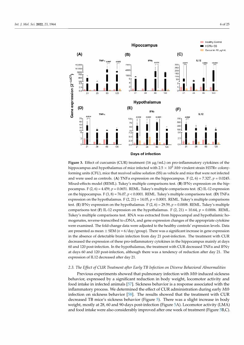
Int. J. Mol. Sci. 2022, 23, 1964 6 of 25Int. J. Mol. Sci. 2021, 22, x FOR PEER REVIEW 6 of 25
Figure 3. Effect of curcumin (CUR) treatment (16 μg/mL) on pro-inflammatory cytokines of the hip-pocampus and hypothalamus of mice infected with 2.5 × 10⁵ Mtb virulent strain H37Rv colony-forming units (CFU), mice that received saline solution (SS) as vehicle and mice that were not in-fected and were used as controls. (A) TNFα expression on the hippocampus. F (2, 6) = 7.327, p = 0.0245. Mixed-effects model (REML). Tukey’s multiple comparisons test. (B) IFNγ expression on the hippocampus. F (2, 6) = 4.459, p = 0.0651. REML. Tukey’s multiple comparisons test. (C) IL-12 ex-pression on the hippocampus. F (3, 8) = 76.07, p < 0.0001. REML. Tukey’s multiple comparisons test. (D) TNFα expression on the hypothalamus. F (2, 21) = 14.05, p = 0.0001. REML. Tukey’s multiple comparisons test. (E) IFNγ expression on the hypothalamus. F (2, 6) = 29.59, p = 0.0008. REML. Tukey’s multiple comparisons test (F) IL-12 expression on the hypothalamus. F (2, 21) = 10.64, p = 0.0006. REML. Tukey’s multiple comparisons test. RNA was extracted from hippocampal and hy-pothalamic homogenates, reverse-transcribed to cDNA, and gene expression changes of the appro-priate cytokine were examined. The fold-change data were adjusted to the healthy controls’ expres-sion levels. Data are presented as mean ± SEM (n = 6/day/group). There was a significant increase in gene expression in the absence of detectable brain infection from day 21 post-infection. The treat-ment with CUR decreased the expression of these pro-inflammatory cytokines in the hippocampus mainly at days 60 and 120 post-infection. In the hypothalamus, the treatment with CUR decreased TNFα and IFNγ at days 60 and 120 post-infection, although there was a tendency of reduction after day 21. The expression of IL12 decreased after day 21.
Figure 3. Effect of curcumin (CUR) treatment (16 µg/mL) on pro-inflammatory cytokines of thehippocampus and hypothalamus of mice infected with 2.5 × 10⁵ Mtb virulent strain H37Rv colony-forming units (CFU), mice that received saline solution (SS) as vehicle and mice that were not infectedand were used as controls. (A) TNFα expression on the hippocampus. F (2, 6) = 7.327, p = 0.0245.Mixed-effects model (REML). Tukey’s multiple comparisons test. (B) IFNγ expression on the hip-pocampus. F (2, 6) = 4.459, p = 0.0651. REML. Tukey’s multiple comparisons test. (C) IL-12 expressionon the hippocampus. F (3, 8) = 76.07, p < 0.0001. REML. Tukey’s multiple comparisons test. (D) TNFαexpression on the hypothalamus. F (2, 21) = 14.05, p = 0.0001. REML. Tukey’s multiple comparisonstest. (E) IFNγ expression on the hypothalamus. F (2, 6) = 29.59, p = 0.0008. REML. Tukey’s multiplecomparisons test (F) IL-12 expression on the hypothalamus. F (2, 21) = 10.64, p = 0.0006. REML.Tukey’s multiple comparisons test. RNA was extracted from hippocampal and hypothalamic ho-mogenates, reverse-transcribed to cDNA, and gene expression changes of the appropriate cytokinewere examined. The fold-change data were adjusted to the healthy controls’ expression levels. Dataare presented as mean ± SEM (n = 6/day/group). There was a significant increase in gene expressionin the absence of detectable brain infection from day 21 post-infection. The treatment with CURdecreased the expression of these pro-inflammatory cytokines in the hippocampus mainly at days60 and 120 post-infection. In the hypothalamus, the treatment with CUR decreased TNFα and IFNγ
at days 60 and 120 post-infection, although there was a tendency of reduction after day 21. Theexpression of IL12 decreased after day 21.
2.3. The Effect of CUR Treatment after Early TB Infection on Diverse Behavioral Abnormalities
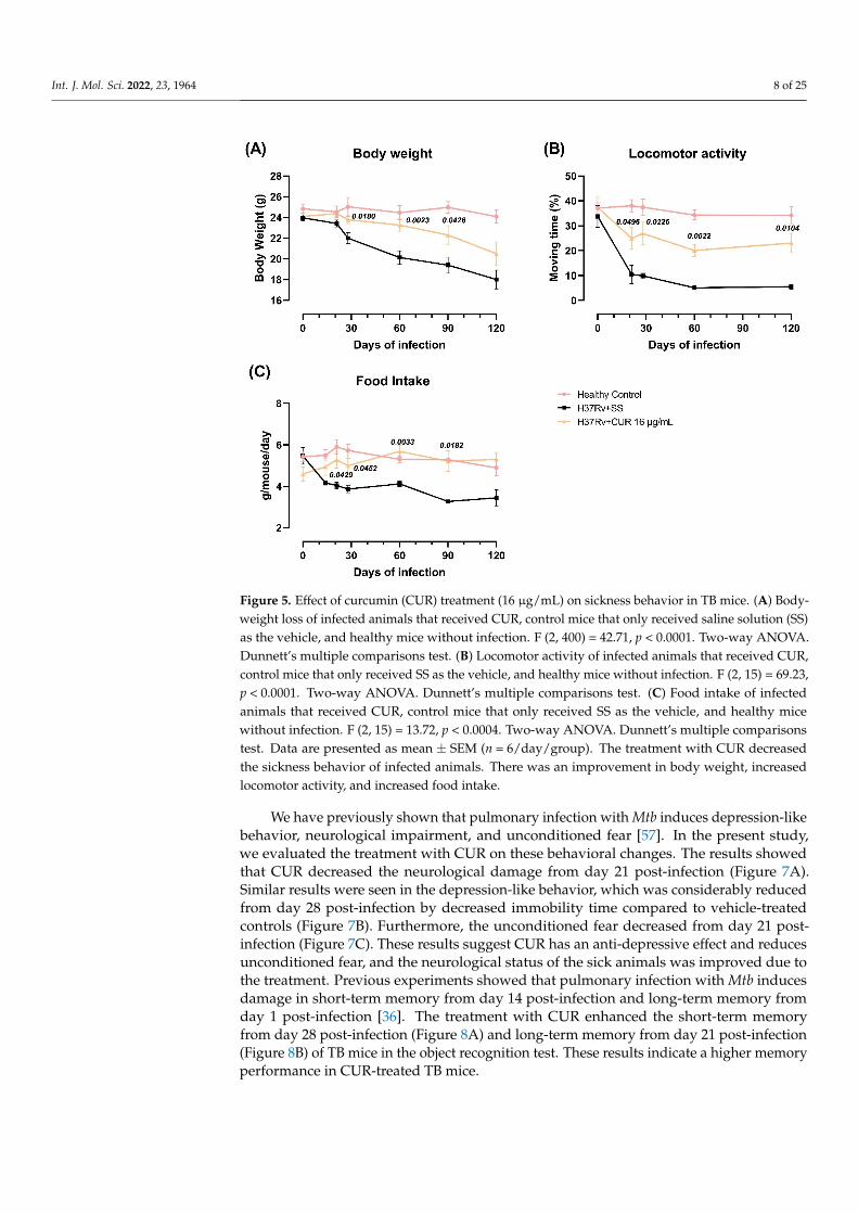
Previous experiments showed that pulmonary infection with Mtb induced sicknessbehavior, expressed by a significant reduction in body weight, locomotor activity andfood intake in infected animals [57]. Sickness behavior is a response associated with theinflammatory process. We determined the effect of CUR administration during early Mtbinfection on sickness behavior [58]. The results showed that the treatment with CURdecreased TB mice’s sickness behavior (Figure 5). There was a slight increase in bodyweight, mostly at 28, 60 and 90-days post-infection (Figure 5A). Locomotor activity (LMA)and food intake were also considerably improved after one week of treatment (Figure 5B,C).
Int. J. Mol. Sci. 2022, 23, 1964 7 of 25Int. J. Mol. Sci. 2021, 22, x FOR PEER REVIEW 7 of 25
Figure 4. Effect of curcumin (CUR) treatment (16 μg/mL) on pro-inflammatory cytokines of the cer-ebellum and frontal cortex of mice infected with 2.5 × 10⁵ Mtb colony-forming units (CFU) virulent strain H37Rv, mice that received saline solution (SS) as the vehicle, and mice that were not infected and were used as controls. (A) TNFα expression on the cerebellum. F (2, 6) = 4.211, p = 0.0720. REML (B) IFNγ expression on the cerebellum. F (2, 6) = 7.322, p = 0.0245. REML. Tukey’s multiple compar-isons test (C) IL-12 expression on the cerebellum. F (2, 21) = 14.39, p = 0.0001. REML. Tukey’s multiple comparisons test. (D) TNFα expression on the frontal cortex. F (2, 22) = 76.20, p <0.0001. REML. Tukey’s multiple comparisons test (E) IFNγ expression on the frontal cortex. F (2, 22) = 33.71, p <0.0001. REML. Tukey’s multiple comparisons test (F) IL-12 expression on the frontal cortex. F (2, 6) = 6.175, p = 0.0350. REML. Tukey’s multiple comparisons test. RNA was extracted from cerebellar homogenates, reverse-transcribed to cDNA, and gene expression changes of the appropriate cyto-kine were examined. The fold-change data were adjusted to the healthy controls’ expression levels. Data are presented as mean ± SEM (n = 6/day/group). There was a significant increase in gene ex-pression in the absence of detectable brain infection after day 21 post-infection. The treatment with CUR decreased the expression of IFNγ and IL12 in the cerebellum at days 60 and 120 post-infection. In the frontal cortex, the treatment with CUR decreased TNFα and IFNγ after day 21 post-infection and IL12 at days 60 and 120 post-infection.
2.3. The Effect of CUR Treatment after Early TB Infection on Diverse Behavioral Abnormalities Previous experiments showed that pulmonary infection with Mtb induced sickness
behavior, expressed by a significant reduction in body weight, locomotor activity and food intake in infected animals [57]. Sickness behavior is a response associated with the inflammatory process. We determined the effect of CUR administration during early Mtb infection on sickness behavior [58]. The results showed that the treatment with CUR de-creased TB mice’s sickness behavior (Figure 5). There was a slight increase in body weight, mostly at 28, 60 and 90-days post-infection (Figure 5A). Locomotor activity (LMA) and food intake were also considerably improved after one week of treatment (Figure 5B,C).
Figure 4. Effect of curcumin (CUR) treatment (16 µg/mL) on pro-inflammatory cytokines of thecerebellum and frontal cortex of mice infected with 2.5 × 10⁵ Mtb colony-forming units (CFU)virulent strain H37Rv, mice that received saline solution (SS) as the vehicle, and mice that werenot infected and were used as controls. (A) TNFα expression on the cerebellum. F (2, 6) = 4.211,p = 0.0720. REML (B) IFNγ expression on the cerebellum. F (2, 6) = 7.322, p = 0.0245. REML. Tukey’smultiple comparisons test (C) IL-12 expression on the cerebellum. F (2, 21) = 14.39, p = 0.0001. REML.Tukey’s multiple comparisons test. (D) TNFα expression on the frontal cortex. F (2, 22) = 76.20,p < 0.0001. REML. Tukey’s multiple comparisons test (E) IFNγ expression on the frontal cortex.F (2, 22) = 33.71, p <0.0001. REML. Tukey’s multiple comparisons test (F) IL-12 expression on thefrontal cortex. F (2, 6) = 6.175, p = 0.0350. REML. Tukey’s multiple comparisons test. RNA wasextracted from cerebellar homogenates, reverse-transcribed to cDNA, and gene expression changes ofthe appropriate cytokine were examined. The fold-change data were adjusted to the healthy controls’expression levels. Data are presented as mean ± SEM (n = 6/day/group). There was a significantincrease in gene expression in the absence of detectable brain infection after day 21 post-infection.The treatment with CUR decreased the expression of IFNγ and IL12 in the cerebellum at days 60 and120 post-infection. In the frontal cortex, the treatment with CUR decreased TNFα and IFNγ afterday 21 post-infection and IL12 at days 60 and 120 post-infection.
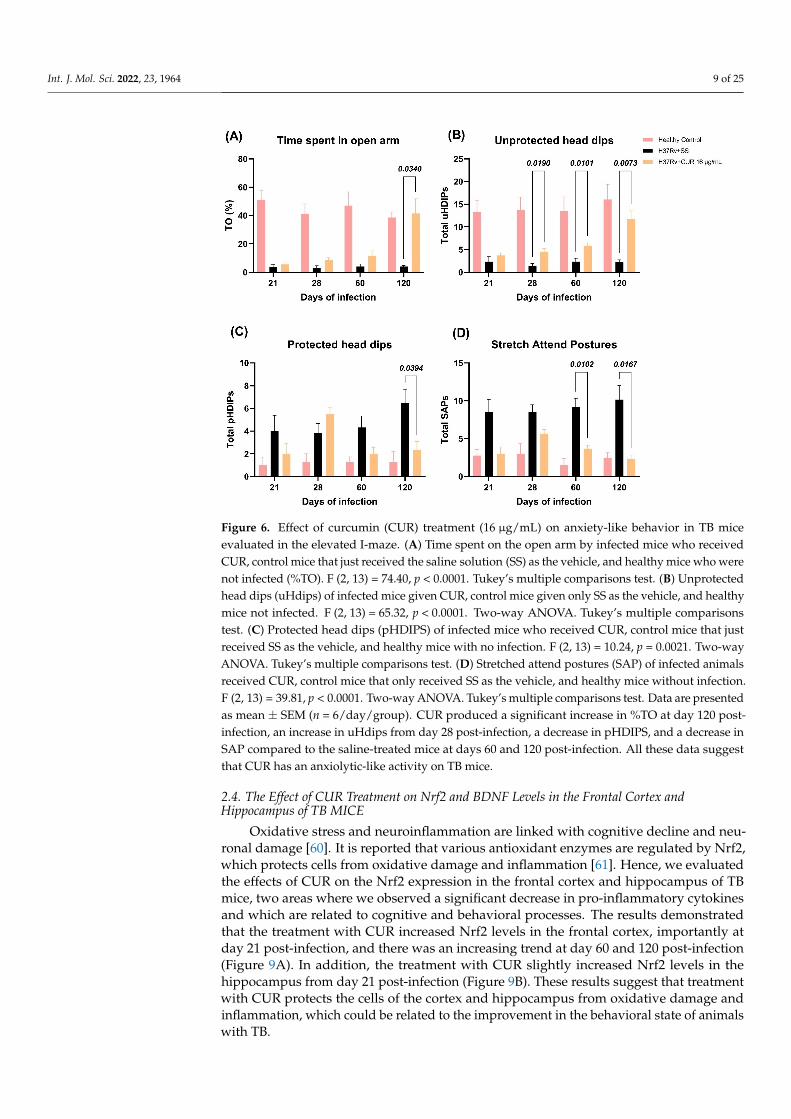
Anxiety is another essential behavioral change observed in animals infected withMtb and TB patients. We found that lung inflammation produced anxiogenic behavior inour model of pulmonary TB [57]. Therefore, we evaluated the CUR treatment’s effect oninfected animals that showed anxiety-like behavior using the elevated I-maze [59]. Weobserved that the treatment with CUR increased the time spent by mice on the open arm(TO) at day 120 post-infection (Figure 6A), increased the unprotected head dips (uHDIPS)(scanning over the sides of the maze downward towards the floor from uncovered openarm by the animal) from day 28 post-infection (Figure 6B), reduced the protected head dips(pHDIPS) (scanning by the animal over the sides of the maze downward towards the floorfrom protected area) at day 120 post-infection (Figure 6C), and stretched attend postures(SAP) (forward elongation of the body when the animal was either standing still or movingslowly forward) at days 60 and 120 post-infection (Figure 6D). Thus, the treatment withCUR showed an anxiolytic-like activity on TB mice, mainly in the late phase of infection.
Int. J. Mol. Sci. 2022, 23, 1964 8 of 25Int. J. Mol. Sci. 2021, 22, x FOR PEER REVIEW 8 of 25
Figure 5. Effect of curcumin (CUR) treatment (16 μg/mL) on sickness behavior in TB mice. (A) Bod-yweight loss of infected animals that received CUR, control mice that only received saline solution (SS) as the vehicle, and healthy mice without infection. F (2, 400) = 42.71, p < 0.0001. Two-way ANOVA. Dunnett’s multiple comparisons test. (B) Locomotor activity of infected animals that re-ceived CUR, control mice that only received SS as the vehicle, and healthy mice without infection. F (2, 15) = 69.23, p < 0.0001. Two-way ANOVA. Dunnett’s multiple comparisons test. (C) Food intake of infected animals that received CUR, control mice that only received SS as the vehicle, and healthy mice without infection. F (2, 15) = 13.72, p < 0.0004. Two-way ANOVA. Dunnett’s multiple compar-isons test. Data are presented as mean ± SEM (n = 6/day/group). The treatment with CUR decreased the sickness behavior of infected animals. There was an improvement in body weight, increased locomotor activity, and increased food intake.
Anxiety is another essential behavioral change observed in animals infected with Mtb and TB patients. We found that lung inflammation produced anxiogenic behavior in our model of pulmonary TB [57]. Therefore, we evaluated the CUR treatment’s effect on in-fected animals that showed anxiety-like behavior using the elevated I-maze [59]. We ob-served that the treatment with CUR increased the time spent by mice on the open arm (TO) at day 120 post-infection (Figure 6A), increased the unprotected head dips (uHDIPS) (scanning over the sides of the maze downward towards the floor from uncovered open arm by the animal) from day 28 post-infection (Figure 6B), reduced the protected head dips (pHDIPS) (scanning by the animal over the sides of the maze downward towards the floor from protected area) at day 120 post-infection (Figure 6C), and stretched attend pos-tures (SAP) (forward elongation of the body when the animal was either standing still or moving slowly forward) at days 60 and 120 post-infection (Figure 6D). Thus, the treatment with CUR showed an anxiolytic-like activity on TB mice, mainly in the late phase of infec-tion.
Figure 5. Effect of curcumin (CUR) treatment (16 µg/mL) on sickness behavior in TB mice. (A) Body-weight loss of infected animals that received CUR, control mice that only received saline solution (SS)as the vehicle, and healthy mice without infection. F (2, 400) = 42.71, p < 0.0001. Two-way ANOVA.Dunnett’s multiple comparisons test. (B) Locomotor activity of infected animals that received CUR,control mice that only received SS as the vehicle, and healthy mice without infection. F (2, 15) = 69.23,p < 0.0001. Two-way ANOVA. Dunnett’s multiple comparisons test. (C) Food intake of infectedanimals that received CUR, control mice that only received SS as the vehicle, and healthy micewithout infection. F (2, 15) = 13.72, p < 0.0004. Two-way ANOVA. Dunnett’s multiple comparisonstest. Data are presented as mean ± SEM (n = 6/day/group). The treatment with CUR decreasedthe sickness behavior of infected animals. There was an improvement in body weight, increasedlocomotor activity, and increased food intake.
We have previously shown that pulmonary infection with Mtb induces depression-likebehavior, neurological impairment, and unconditioned fear [57]. In the present study,we evaluated the treatment with CUR on these behavioral changes. The results showedthat CUR decreased the neurological damage from day 21 post-infection (Figure 7A).Similar results were seen in the depression-like behavior, which was considerably reducedfrom day 28 post-infection by decreased immobility time compared to vehicle-treatedcontrols (Figure 7B). Furthermore, the unconditioned fear decreased from day 21 post-infection (Figure 7C). These results suggest CUR has an anti-depressive effect and reducesunconditioned fear, and the neurological status of the sick animals was improved due tothe treatment. Previous experiments showed that pulmonary infection with Mtb inducesdamage in short-term memory from day 14 post-infection and long-term memory fromday 1 post-infection [36]. The treatment with CUR enhanced the short-term memoryfrom day 28 post-infection (Figure 8A) and long-term memory from day 21 post-infection(Figure 8B) of TB mice in the object recognition test. These results indicate a higher memoryperformance in CUR-treated TB mice.
Int. J. Mol. Sci. 2022, 23, 1964 9 of 25Int. J. Mol. Sci. 2021, 22, x FOR PEER REVIEW 9 of 25
Figure 6. Effect of curcumin (CUR) treatment (16 μg/mL) on anxiety-like behavior in TB mice eval-uated in the elevated I-maze. (A) Time spent on the open arm by infected mice who received CUR, control mice that just received the saline solution (SS) as the vehicle, and healthy mice who were not infected (%TO). F (2, 13) = 74.40, p < 0.0001. Tukey’s multiple comparisons test. (B) Unprotected head dips (uHdips) of infected mice given CUR, control mice given only SS as the vehicle, and healthy mice not infected. F (2, 13) = 65.32, p < 0.0001. Two-way ANOVA. Tukey’s multiple comparisons test. (C) Protected head dips (pHDIPS) of infected mice who received CUR, control mice that just received SS as the vehicle, and healthy mice with no infection. F (2, 13) = 10.24, p = 0.0021. Two-way ANOVA. Tukey’s multiple comparisons test. (D) Stretched attend postures (SAP) of infected ani-mals received CUR, control mice that only received SS as the vehicle, and healthy mice without infection. F (2, 13) = 39.81, p < 0.0001. Two-way ANOVA. Tukey’s multiple comparisons test. Data are presented as mean ± SEM (n = 6/day/group). CUR produced a significant increase in %TO at day 120 post-infection, an increase in uHdips from day 28 post-infection, a decrease in pHDIPS, and a decrease in SAP compared to the saline-treated mice at days 60 and 120 post-infection. All these data suggest that CUR has an anxiolytic-like activity on TB mice.
We have previously shown that pulmonary infection with Mtb induces depression-like behavior, neurological impairment, and unconditioned fear [57]. In the present study, we evaluated the treatment with CUR on these behavioral changes. The results showed that CUR decreased the neurological damage from day 21 post-infection (Figure 7A). Sim-ilar results were seen in the depression-like behavior, which was considerably reduced from day 28 post-infection by decreased immobility time compared to vehicle-treated con-trols (Figure 7B). Furthermore, the unconditioned fear decreased from day 21 post-infec-tion (Figure 7C). These results suggest CUR has an anti-depressive effect and reduces un-conditioned fear, and the neurological status of the sick animals was improved due to the treatment. Previous experiments showed that pulmonary infection with Mtb induces damage in short-term memory from day 14 post-infection and long-term memory from day 1 post-infection [36]. The treatment with CUR enhanced the short-term memory from day 28 post-infection (Figure 8A) and long-term memory from day 21 post-infection (Fig-ure 8B) of TB mice in the object recognition test. These results indicate a higher memory performance in CUR-treated TB mice.
Figure 6. Effect of curcumin (CUR) treatment (16 µg/mL) on anxiety-like behavior in TB miceevaluated in the elevated I-maze. (A) Time spent on the open arm by infected mice who receivedCUR, control mice that just received the saline solution (SS) as the vehicle, and healthy mice who werenot infected (%TO). F (2, 13) = 74.40, p < 0.0001. Tukey’s multiple comparisons test. (B) Unprotectedhead dips (uHdips) of infected mice given CUR, control mice given only SS as the vehicle, and healthymice not infected. F (2, 13) = 65.32, p < 0.0001. Two-way ANOVA. Tukey’s multiple comparisonstest. (C) Protected head dips (pHDIPS) of infected mice who received CUR, control mice that justreceived SS as the vehicle, and healthy mice with no infection. F (2, 13) = 10.24, p = 0.0021. Two-wayANOVA. Tukey’s multiple comparisons test. (D) Stretched attend postures (SAP) of infected animalsreceived CUR, control mice that only received SS as the vehicle, and healthy mice without infection.F (2, 13) = 39.81, p < 0.0001. Two-way ANOVA. Tukey’s multiple comparisons test. Data are presentedas mean ± SEM (n = 6/day/group). CUR produced a significant increase in %TO at day 120 post-infection, an increase in uHdips from day 28 post-infection, a decrease in pHDIPS, and a decrease inSAP compared to the saline-treated mice at days 60 and 120 post-infection. All these data suggestthat CUR has an anxiolytic-like activity on TB mice.
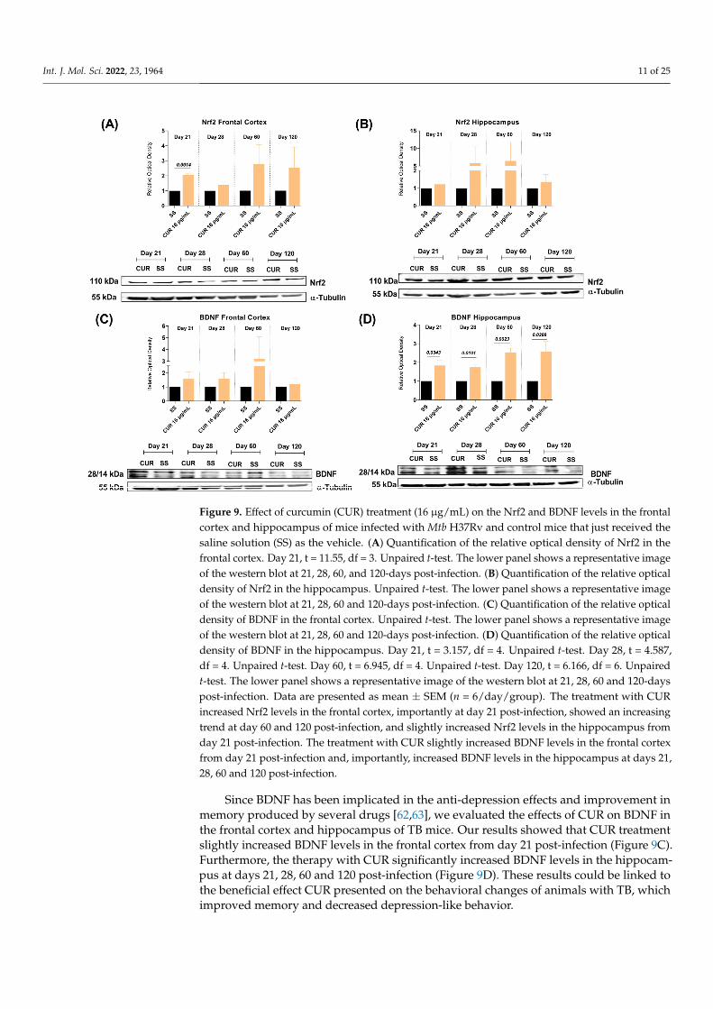
2.4. The Effect of CUR Treatment on Nrf2 and BDNF Levels in the Frontal Cortex andHippocampus of TB MICE
Oxidative stress and neuroinflammation are linked with cognitive decline and neu-ronal damage [60]. It is reported that various antioxidant enzymes are regulated by Nrf2,which protects cells from oxidative damage and inflammation [61]. Hence, we evaluatedthe effects of CUR on the Nrf2 expression in the frontal cortex and hippocampus of TBmice, two areas where we observed a significant decrease in pro-inflammatory cytokinesand which are related to cognitive and behavioral processes. The results demonstratedthat the treatment with CUR increased Nrf2 levels in the frontal cortex, importantly atday 21 post-infection, and there was an increasing trend at day 60 and 120 post-infection(Figure 9A). In addition, the treatment with CUR slightly increased Nrf2 levels in thehippocampus from day 21 post-infection (Figure 9B). These results suggest that treatmentwith CUR protects the cells of the cortex and hippocampus from oxidative damage andinflammation, which could be related to the improvement in the behavioral state of animalswith TB.

Int. J. Mol. Sci. 2022, 23, 1964 10 of 25Int. J. Mol. Sci. 2021, 22, x FOR PEER REVIEW 10 of 25
Figure 7. Effect of curcumin (CUR) treatment (16 μg/mL) on neurological damage, depression-like behavior and unconditioned fear of TB mice. (A) Neurological outcome of mice treated with CUR, control mice that only received saline solution (SS) as the vehicle, and healthy mice without infec-tion. F (2, 12) = 142.2, p < 0.0001. Two-way ANOVA. p < 0.0001. Dunnett’s multiple comparisons test. (B) Depression-like behavior of mice treated with CUR, control mice that only received SS as the vehicle, and healthy mice without infection. F (2, 12) = 154.6, p < 0.0001. Two-way ANOVA. Dun-nett’s multiple comparisons test. (C) Unconditioned fear of mice treated with CUR, control mice that only received SS as the vehicle, and healthy mice without infection. F (2, 15) = 45.82, p < 0.0001. Two-way ANOVA. Dunnett’s multiple comparisons test. Data are presented as mean ± SEM (n = 6/day/group). The treatment with CUR decreased neurological damage from day 21 post-infection and decreased the depression-like behavior of infected animals from day 28. The treatment de-creased the unconditioned fear from day 21.
Figure 8. Effect of curcumin (CUR) treatment (16 μg/mL) on TB mice’s memory damage. (A) Short-term memory of mice treated with CUR, control mice that only received saline solution (SS) as the vehicle, and healthy mice without infection. F (2, 15) = 77.03, p < 0.0001. Two-way ANOVA. Dun-nett’s multiple comparisons test. (B) Long-term memory of mice treated with CUR, control mice that only received SS as the vehicle, and healthy mice without infection. F (2, 12) = 86.60, p < 0.0001. Two-way ANOVA. Dunnett’s multiple comparisons test. Data are presented as mean ± SEM (n = 6/day/group). Animals with TB treated with CUR showed a significant improvement in short-term memory from day 28 post-infection and long-term memory from day 21.
Figure 7. Effect of curcumin (CUR) treatment (16 µg/mL) on neurological damage, depression-likebehavior and unconditioned fear of TB mice. (A) Neurological outcome of mice treated with CUR,control mice that only received saline solution (SS) as the vehicle, and healthy mice without infection.F (2, 12) = 142.2, p < 0.0001. Two-way ANOVA. p < 0.0001. Dunnett’s multiple comparisons test.(B) Depression-like behavior of mice treated with CUR, control mice that only received SS as thevehicle, and healthy mice without infection. F (2, 12) = 154.6, p < 0.0001. Two-way ANOVA. Dun-nett’s multiple comparisons test. (C) Unconditioned fear of mice treated with CUR, control mice thatonly received SS as the vehicle, and healthy mice without infection. F (2, 15) = 45.82, p < 0.0001.Two-way ANOVA. Dunnett’s multiple comparisons test. Data are presented as mean ± SEM(n = 6/day/group). The treatment with CUR decreased neurological damage from day 21 post-infection and decreased the depression-like behavior of infected animals from day 28. The treatmentdecreased the unconditioned fear from day 21.
Int. J. Mol. Sci. 2021, 22, x FOR PEER REVIEW 10 of 25
Figure 7. Effect of curcumin (CUR) treatment (16 μg/mL) on neurological damage, depression-like behavior and unconditioned fear of TB mice. (A) Neurological outcome of mice treated with CUR, control mice that only received saline solution (SS) as the vehicle, and healthy mice without infec-tion. F (2, 12) = 142.2, p < 0.0001. Two-way ANOVA. p < 0.0001. Dunnett’s multiple comparisons test. (B) Depression-like behavior of mice treated with CUR, control mice that only received SS as the vehicle, and healthy mice without infection. F (2, 12) = 154.6, p < 0.0001. Two-way ANOVA. Dun-nett’s multiple comparisons test. (C) Unconditioned fear of mice treated with CUR, control mice that only received SS as the vehicle, and healthy mice without infection. F (2, 15) = 45.82, p < 0.0001. Two-way ANOVA. Dunnett’s multiple comparisons test. Data are presented as mean ± SEM (n = 6/day/group). The treatment with CUR decreased neurological damage from day 21 post-infection and decreased the depression-like behavior of infected animals from day 28. The treatment de-creased the unconditioned fear from day 21.
Figure 8. Effect of curcumin (CUR) treatment (16 μg/mL) on TB mice’s memory damage. (A) Short-term memory of mice treated with CUR, control mice that only received saline solution (SS) as the vehicle, and healthy mice without infection. F (2, 15) = 77.03, p < 0.0001. Two-way ANOVA. Dun-nett’s multiple comparisons test. (B) Long-term memory of mice treated with CUR, control mice that only received SS as the vehicle, and healthy mice without infection. F (2, 12) = 86.60, p < 0.0001. Two-way ANOVA. Dunnett’s multiple comparisons test. Data are presented as mean ± SEM (n = 6/day/group). Animals with TB treated with CUR showed a significant improvement in short-term memory from day 28 post-infection and long-term memory from day 21.
Figure 8. Effect of curcumin (CUR) treatment (16 µg/mL) on TB mice’s memory damage. (A) Short-term memory of mice treated with CUR, control mice that only received saline solution (SS) as thevehicle, and healthy mice without infection. F (2, 15) = 77.03, p < 0.0001. Two-way ANOVA. Dunnett’smultiple comparisons test. (B) Long-term memory of mice treated with CUR, control mice thatonly received SS as the vehicle, and healthy mice without infection. F (2, 12) = 86.60, p < 0.0001.Two-way ANOVA. Dunnett’s multiple comparisons test. Data are presented as mean ± SEM(n = 6/day/group). Animals with TB treated with CUR showed a significant improvement in short-term memory from day 28 post-infection and long-term memory from day 21.
Int. J. Mol. Sci. 2022, 23, 1964 11 of 25
Int. J. Mol. Sci. 2021, 22, x FOR PEER REVIEW 11 of 25
2.4. The Effect of CUR Treatment on Nrf2 and BDNF Levels in the Frontal Cortex and Hippocampus of TB MICE
Oxidative stress and neuroinflammation are linked with cognitive decline and neu-ronal damage [60]. It is reported that various antioxidant enzymes are regulated by Nrf2, which protects cells from oxidative damage and inflammation [61]. Hence, we evaluated the effects of CUR on the Nrf2 expression in the frontal cortex and hippocampus of TB mice, two areas where we observed a significant decrease in pro-inflammatory cytokines and which are related to cognitive and behavioral processes. The results demonstrated that the treatment with CUR increased Nrf2 levels in the frontal cortex, importantly at day 21 post-infection, and there was an increasing trend at day 60 and 120 post-infection (Figure 9A). In addition, the treatment with CUR slightly increased Nrf2 levels in the hippocampus from day 21 post-infection (Figure 9B). These results suggest that treatment with CUR pro-tects the cells of the cortex and hippocampus from oxidative damage and inflammation, which could be related to the improvement in the behavioral state of animals with TB.
Figure 9. Effect of curcumin (CUR) treatment (16 μg/mL) on the Nrf2 and BDNF levels in the frontal cortex and hippocampus of mice infected with Mtb H37Rv and control mice that just received the saline solution (SS) as the vehicle. (A) Quantification of the relative optical density of Nrf2 in the frontal cortex. Day 21, t = 11.55, df = 3. Unpaired t-test. The lower panel shows a representative image of the western blot at 21, 28, 60, and 120-days post-infection. (B) Quantification of the relative optical density of Nrf2 in the hippocampus. Unpaired t-test. The lower panel shows a representative image of the western blot at 21, 28, 60 and 120-days post-infection. (C) Quantification of the relative optical density of BDNF in the frontal cortex. Unpaired t-test. The lower panel shows a representa-tive image of the western blot at 21, 28, 60 and 120-days post-infection. (D) Quantification of the relative optical density of BDNF in the hippocampus. Day 21, t = 3.157, df = 4. Unpaired t-test. Day 28, t = 4.587, df = 4. Unpaired t-test. Day 60, t = 6.945, df = 4. Unpaired t-test. Day 120, t = 6.166, df = 6. Unpaired t-test. The lower panel shows a representative image of the western blot at 21, 28, 60 and 120-days post-infection. Data are presented as mean ± SEM (n = 6/day/group). The treatment with CUR increased Nrf2 levels in the frontal cortex, importantly at day 21 post-infection, showed an increasing trend at day 60 and 120 post-infection, and slightly increased Nrf2 levels in the
Figure 9. Effect of curcumin (CUR) treatment (16 µg/mL) on the Nrf2 and BDNF levels in the frontalcortex and hippocampus of mice infected with Mtb H37Rv and control mice that just received thesaline solution (SS) as the vehicle. (A) Quantification of the relative optical density of Nrf2 in thefrontal cortex. Day 21, t = 11.55, df = 3. Unpaired t-test. The lower panel shows a representative imageof the western blot at 21, 28, 60, and 120-days post-infection. (B) Quantification of the relative opticaldensity of Nrf2 in the hippocampus. Unpaired t-test. The lower panel shows a representative imageof the western blot at 21, 28, 60 and 120-days post-infection. (C) Quantification of the relative opticaldensity of BDNF in the frontal cortex. Unpaired t-test. The lower panel shows a representative imageof the western blot at 21, 28, 60 and 120-days post-infection. (D) Quantification of the relative opticaldensity of BDNF in the hippocampus. Day 21, t = 3.157, df = 4. Unpaired t-test. Day 28, t = 4.587,df = 4. Unpaired t-test. Day 60, t = 6.945, df = 4. Unpaired t-test. Day 120, t = 6.166, df = 6. Unpairedt-test. The lower panel shows a representative image of the western blot at 21, 28, 60 and 120-dayspost-infection. Data are presented as mean ± SEM (n = 6/day/group). The treatment with CURincreased Nrf2 levels in the frontal cortex, importantly at day 21 post-infection, showed an increasingtrend at day 60 and 120 post-infection, and slightly increased Nrf2 levels in the hippocampus fromday 21 post-infection. The treatment with CUR slightly increased BDNF levels in the frontal cortexfrom day 21 post-infection and, importantly, increased BDNF levels in the hippocampus at days 21,28, 60 and 120 post-infection.
Since BDNF has been implicated in the anti-depression effects and improvement inmemory produced by several drugs [62,63], we evaluated the effects of CUR on BDNF inthe frontal cortex and hippocampus of TB mice. Our results showed that CUR treatmentslightly increased BDNF levels in the frontal cortex from day 21 post-infection (Figure 9C).Furthermore, the therapy with CUR significantly increased BDNF levels in the hippocam-pus at days 21, 28, 60 and 120 post-infection (Figure 9D). These results could be linked tothe beneficial effect CUR presented on the behavioral changes of animals with TB, whichimproved memory and decreased depression-like behavior.
Int. J. Mol. Sci. 2022, 23, 1964 12 of 25
These results suggest that the administration of CUR reduces the bacilli load in thelung, pneumonia, neuroinflammation, behavioral abnormalities, and slightly increasesNrf2 and BDNF in the murine model of experimental pulmonary TB.
3. Discussion
Previous work showed neuroinflammation and neuropsychiatric abnormalities in anexperimental model of progressive pulmonary TB without brain infection [57]. In thiswork, we evaluated the effect of CUR in lung disease evolution and the capability ofCUR to inhibit both the pro-inflammatory events induced by Mtb on the CNS and thebehavioral abnormalities.
TB is the leading cause of death amongst all bacterial diseases, accounting for morethan 1.4 million fatalities per year [64]. The emergence of multidrug-resistant (MDR) Mtbstrains is an increasing problem requiring novel treatment options [65]. Our results showedthat the treatment with 16 or 32 µg/mL of CUR reduced the bacilli lung load of mice infectedwith the drug-sensitive Mtb H37Rv. The decrease in bacilli lung load was related to reducedpneumonia and improved survival. These results agree with previous studies showing thatCUR is a potential anti-Mtb agent [65]. It has been demonstrated that CUR has a minimumconcentration inhibitory (MIC) activity of 16 µg/mL against drug-susceptible Mtb H37Rv,isoniazid-resistant Mtb H37Rv, rifampicin-resistant H37Rv, streptomycin-resistant H37Rvand ethambutol-resistant H37Rv [66]. The effect of CUR against Mtb could be direct,affecting the bacterial lipid metabolism since lipids and their metabolism is critical to allaspects of mycobacterial infection, pathogenicity, and persistence in the host [66]. CURcan also inhibit bacterial intracellular growth and promote sensible drug strain clearance,as demonstrated in differentiated THP-1 human monocytes, primary human alveolarmacrophages, and Raw 264.7 cells infected with Mtb H37Rv or MDR clinical isolates [67,68],by promoting caspase-3-dependent apoptosis and autophagy.
In antigen-presenting cells (APCs) infected with H37Rv, CUR nanoparticles improvedautophagy costimulatory activity promoting the generation of inflammatory cytokines andother mediators [69]. In H37Rv-challenged mice, nanocurcumin improves the effectivenessof the BCG vaccination by inducing long-lasting central memory T cells (TCM) of theTh1 and Th17 lineages [69]. CUR’s anti-inflammatory properties have also been used toincrease the efficacy of previously authorized anti-microbial drugs through synergisticeffects [70]. CUR also protected BALB/c mice against Klebsiella pneumonie-induced lunginflammation [71], which coincides with our results that showed a significant decrease in thepneumonia area of TB mice treated with CUR. It seems that CUR can directly damage Mtb,promote bacterial killing in the cytoplasm of macrophages, and has an immunomodulatoryeffect, which agrees with our results of efficient mycobacterial clearance in the lungs.
We have demonstrated that Mtb pulmonary infection caused neuroinflammation andbehavioral abnormalities in the absence of bacteria in the brain [57]. Neuroinflammationcan be caused by an increased peripheral inflammatory response and oxidative stress,activating microglia that contribute to brain pathology [72]. Therefore, the present workevaluated the effect of CUR in the inflammatory response in the hippocampus, hypothala-mus, cerebellum and frontal cortex. The results showed a significant effect of CUR on thepro-inflammatory cytokines TNFα, IL12, and IFNγ mRNA expression levels that were con-siderably lower in the treated group than the control non-treated TB group. Thus, CUR hasa substantial anti-inflammatory effect in the brain that constitutes a neuroprotective benefitclearly shown by our study. These results agree with various studies that have shownthe anti-inflammatory effect of CUR in the CNS, administrated either as a treatment or asadjuvant therapy in several illnesses [73], such as in the prevention of a cognitive deficit in-duced by ethanol through modulating oxidative-nitrosative stress and decreasing the levelsof pro-inflammatory cytokines (TNF, IL-1), NFκB, and caspase 3 in different brain regionsof ethanol-treated rat pups [74]. CUR decreased interleukin-23 (IL-23) and interleukin-17(IL-17) levels in the CNS in a model of retinal ischemia-reperfusion injury [75] and reducedthe infarct size and the levels of IL-1, TNFα, cyclooxygenase-2 (COX-2) and PGE-2 in
Int. J. Mol. Sci. 2022, 23, 1964 13 of 25
the cerebral ischemia model by activating the peroxisome proliferator-activated receptorgamma (PPARγ) [76]. CUR was also able to block Toll-like receptors 2 and 4 (TLR-2/4) andNF-κB in rats with persistent and localized cerebral ischemia [77]. In neurodegenerative dis-eases, CUR treatment reduced β-Amyloid Peptide (Aβ) deposits in the brain and improvedcognitive and synaptic dysfunction in Alzheimer’s disease (AD) patients [78]. CUR also re-duced the number of hypertrophic astrocytes in the hippocampus of Aβ(1-40)-treated mice,countered downregulated mRNA expression of the glial fibrillar acidic protein (GFAP) andimproved the spatial memory disorders (such disorders being symptomatic in AD) [79].Demethoxycurcumin, a CUR derivative, reduced the expression in the hippocampus ofpro-inflammatory IL-1 and GFAP in a rat model of AD [80]. CUR can also impact Aβ
metabolism and aggregation of the β-amyloid fibrils (fAβ) [81] and substantially inhibitIL-1, IL-6, and TNFα production in Aβ exposed microglia via the mitogen-activated proteinkinase (MEK1/MEK2) p38 pathways [82]. CUR also decreased pro-inflammatory cytokines(IL-6, IL-1, and TNFα) in the 1-methyl-4-phenyl-1,2,3,6-tetrahydropyridine (MPTP) modelof Parkinson’s disease (PD) and protected dopaminergic neurons from degeneration [83,84].Another study found that CUR specifically protects axons, but not neuronal cell bodies,from NO-mediated degeneration [85]. Thus, there is considerable evidence that CUR hasefficient anti-inflammatory effects that mediate neuroprotection. The present study extendsthe information about the therapeutical benefit of CUR by the demonstration for the firsttime of the prevention of neuroinflammation in experimental pulmonary TB.
Various cytokines and inflammatory factors that produce neuroinflammation alsoparticipate in the pathophysiology of neuropsychiatric disorders such as depression [86]by generating oxidative stress, affecting neurotransmitter production and even generatingneuronal death [86]. Thus, we also examined the effect of CUR treatment on a rangeof behavioral disorders as we have demonstrated that Mtb infection caused behavioralabnormalities in the absence of bacteria in the brain [57]. Our results show that CUR therapyreduced sickness, reduced anxiety-like, reduced depression-like behavior, improved theneurological outcome, and enhanced short- and long-term memory in tuberculous mice.Interestingly, similar results were found in patients with obesity, where supplementation of1 g/day of CUR had an antianxiety effect related to the antioxidant and anti-inflammatoryproperties of CUR [87]. Another study performed in rats subjected to immobilizationstress and pre-treated with CUR (200 mg/kg/day) for seven days showed a decrease inanxiety-like behavior, depression-like behavior and improved memory function, whichwas related to the activity of antioxidant enzymes [88]. Similar results have been observedin Cadmium (Cd) exposure Swiss-Webster mice that received CUR (300 mg/kg). In thiswork, the treatment improved body weight gain and locomotor activity, decreased anxietyin the plus-maze, and increased learning capability [89]. Moreover, CUR treatment had animportant suppressive effect on the cadmium-induced oxidative stress and increased thelevels of serotonin (5-HT) and dopamine (DA) in the forebrain area [89].
Indeed, there is evidence that CUR has a beneficial effect on humans suffering fromdepression and anxiety [90,91], linked to CUR anti-inflammatory effects, dopamine release,antioxidant activity, and neurotrophic factor regulation [91]. BDNF in the hippocampusis necessary for the cognition enhancement effect of chronic CUR in an AD model [63].Chronic CUR also resulted in a dose-dependent increase in hippocampal BDNF in a modelof depression [56]. These data coincide with our results, where we observed a significantincrease in BDNF levels in the hippocampus of animals with TB treated with CUR. BDNFplays a crucial role in regulating neuronal development, maintenance and survival, andcognition, formation, and storage of memories [92]. Therefore, the increase in BDNF in thehippocampus of TB animals could be related to the beneficial effect of CUR on memoryand the decrease in depression-like behavior in this model. Another factor that protects thebrain from injury is Nrf2, as oxidative damage plays a critical role in many central nervoussystem diseases [93]. CUR protected from injury in a model of an ischemic brain throughthe Akt/Nrf2 pathway [94]. CUR has a neuroprotective effect in a model of traumaticbrain injury (TBI) associated with activating the Nrf2 pathway [95]. These data coincide

Int. J. Mol. Sci. 2022, 23, 1964 14 of 25
with our results, as we observed a slight increase in the levels of Nrf2 in the TB animalstreated with CUR. These data suggest that treatment with CUR has a beneficial effect onvarious neuroinflammatory and neurodegenerative diseases, including those related topulmonary TB.
This investigation revealed the efficacy of CUR administration as a novel treatmentfor controlling neuroinflammation in chronic infectious diseases such as pulmonary TB. Inaddition, it is worth noting that CUR had a therapeutic effect on lung disease, indicatingthat CUR might be used as a coadjuvant treatment in TB chemotherapy.
4. Materials and Methods4.1. Reagents and Antibodies
The Middlebrook 7H9 and 7H10 media and the OADC (oleic acid, albumin, dextrose,and catalase) were obtained from Becton-Dickinson, (Detroit, MI, USA). The RNeasy® MiniKit for RNA extraction, the Omniscript® Reverse Transcription Kit for complementaryDNA acquisition, and the QuantiTectTM SYBR® for RT-PCR were obtained from Qiagen(Germantown, MD, USA). The primers for the cytokines studied were acquired from Invit-rogenTM Thermo Fisher Scientific (Waltham, MA, USA). CUR, DMSO (Dimethylsulfoxide)and ethanol was purchased from Sigma Aldrich (Zwijndrecht, The Netherlands). Primaryantibody against BDNF (ab72439) was obtained from Abcam Inc. (Cambridge, MA, USA).Primary antibody against Nrf2 (T-19; sc-30915) antibody was obtained from Santa CruzBiotechnology (Santa Cruz, CA, USA). Secondary antibodies against rabbit (711-035-152),goat (705-035-147) and mouse (715-035-150) were purchased from Jackson ImmunoResearchLaboratories Inc. (Jennersville, PA, USA). All other reagents were analytical grade andacquired from known commercial sources.
4.2. Animals
Three hundred pathogen-free adult male BALB/c mice, aged eight weeks, were ob-tained from Mexico’s Instituto Nacional de Ciencias Médicas y Nutrición Salvador Zubirán(INCMNSZ) animal house facility. Mice were housed in groups of five (n = 5) and dividedinto two sets: healthy controls (HC, n = 84) and infected mice (H37Rv, n = 216). The dis-tribution of animals within each experiment was made following the recommendationsfor the Replacement, Refinement and Reduction of Animals in Research (3R) [96] andadjusted to the total of animals provided by INCMNSZ. All efforts were made to minimizeanimal suffering and the number of animals used. All of the animals were housed in anapproved animal holding facility with a 12:12 h light-dark cycle (lights on at 07:00 h) and aregulated temperature (23 ± 1 ◦C), and humidity (50–20 percent). Food and drink werefreely available. All animal studies were carried out under the ARRIVE standards and Mex-ican Constitution statute NOM 062–Z00-1999, with consent from the INCMNSZ’s EthicalCommittee for Animal Experimentation, protocol number: PAT-1865-16/19-1 (approvedon 7 August 2016).
4.3. The Experimental Murine Model of Pulmonary TB
A mouse model of progressive pulmonary TB has previously been reported [47,97,98].The reference Mtb strain H37Rv was grown in a 7H9 medium enriched with OADC. For allof the experiments, mid-log-phase cultures were used. Mtb were counted and stored at atemperature of −80 ◦C until needed. Bacterial aliquots were thawed and pulse-sonicatedto eliminate clumping. After infecting mice, a portion of the bacterial inoculum was platedto validate the amount of CFU and viability of the CFU provided to the animals. Eight-week-old male BALB/c mice were anesthetized in a gas chamber with 0.1 mL sevofluraneper mouse. A blunt stainless-steel cannula was introduced through the mouth and guidedto the trachea. The cannula’s appropriate intratracheal (IT) placement was confirmed byrubbing the tracheal rings with the tiny ball from the cannula. Mice were infected with2.5 × 105 live bacilli by IT installation. Mice were kept in a vertical position until they
Int. J. Mol. Sci. 2022, 23, 1964 15 of 25
recovered spontaneously. Then, in a P-3 biosecurity level facility, a total of 184 infectedmice were kept in groups of five in cages equipped with micro-isolators.
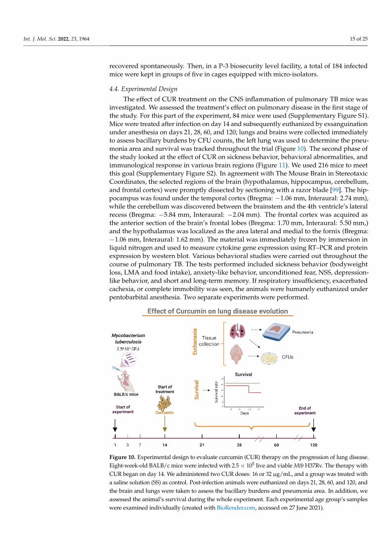
4.4. Experimental Design
The effect of CUR treatment on the CNS inflammation of pulmonary TB mice wasinvestigated. We assessed the treatment’s effect on pulmonary disease in the first stage ofthe study. For this part of the experiment, 84 mice were used (Supplementary Figure S1).Mice were treated after infection on day 14 and subsequently euthanized by exsanguinationunder anesthesia on days 21, 28, 60, and 120; lungs and brains were collected immediatelyto assess bacillary burdens by CFU counts, the left lung was used to determine the pneu-monia area and survival was tracked throughout the trial (Figure 10). The second phase ofthe study looked at the effect of CUR on sickness behavior, behavioral abnormalities, andimmunological response in various brain regions (Figure 11). We used 216 mice to meetthis goal (Supplementary Figure S2). In agreement with The Mouse Brain in StereotaxicCoordinates, the selected regions of the brain (hypothalamus, hippocampus, cerebellum,and frontal cortex) were promptly dissected by sectioning with a razor blade [99]. The hip-pocampus was found under the temporal cortex (Bregma: −1.06 mm, Interaural: 2.74 mm),while the cerebellum was discovered between the brainstem and the 4th ventricle’s lateralrecess (Bregma: −5.84 mm, Interaural: −2.04 mm). The frontal cortex was acquired asthe anterior section of the brain’s frontal lobes (Bregma: 1.70 mm, Interaural: 5.50 mm,)and the hypothalamus was localized as the area lateral and medial to the fornix (Bregma:−1.06 mm, Interaural: 1.62 mm). The material was immediately frozen by immersion inliquid nitrogen and used to measure cytokine gene expression using RT–PCR and proteinexpression by western blot. Various behavioral studies were carried out throughout thecourse of pulmonary TB. The tests performed included sickness behavior (bodyweightloss, LMA and food intake), anxiety-like behavior, unconditioned fear, NSS, depression-like behavior, and short and long-term memory. If respiratory insufficiency, exacerbatedcachexia, or complete immobility was seen, the animals were humanely euthanized underpentobarbital anesthesia. Two separate experiments were performed.
Int. J. Mol. Sci. 2021, 22, x FOR PEER REVIEW 16 of 25
Figure 10. Experimental design to evaluate curcumin (CUR) therapy on the progression of lung dis-ease. Eight-week-old BALB/c mice were infected with 2.5 × 10⁵ live and viable Mtb H37Rv. The ther-apy with CUR began on day 14. We administered two CUR doses: 16 or 32 μg/mL, and a group was treated with a saline solution (SS) as control. Post-infection animals were euthanized on days 21, 28, 60, and 120, and the brain and lungs were taken to assess the bacillary burdens and pneumonia area. In addition, we assessed the animal’s survival during the whole experiment. Each experimental age group’s samples were examined individually (created with BioRender.com, accessed on 27 June 2021).
Figure 11. Experimental design to evaluate curcumin (CUR) treatment on neuroinflammation and behavioral alterations of TB mice. The therapy with CUR began on day 14. We administered CUR (16 μg/mL), and a group received saline solution (SS) as control. Different behavioral assessments were performed on days 21, 28, 60, and 120 after infection. Animals were euthanized after the be-havioral tests, and the brain and lungs were taken to assess bacillary burdens. The cytokines gene expression was measured in the hypothalamus, hippocampus, cerebellum, and frontal cortex. Two separate experiments with n = 3 each were carried out for each of the measures. In addition, each experimental age group’s samples were examined individually. (Created with BioRender.com, ac-cessed on 20 January 2021).
Figure 10. Experimental design to evaluate curcumin (CUR) therapy on the progression of lung disease.Eight-week-old BALB/c mice were infected with 2.5× 10⁵ live and viable Mtb H37Rv. The therapy withCUR began on day 14. We administered two CUR doses: 16 or 32 µg/mL, and a group was treated witha saline solution (SS) as control. Post-infection animals were euthanized on days 21, 28, 60, and 120, andthe brain and lungs were taken to assess the bacillary burdens and pneumonia area. In addition, weassessed the animal’s survival during the whole experiment. Each experimental age group’s sampleswere examined individually (created with BioRender.com, accessed on 27 June 2021).

Int. J. Mol. Sci. 2022, 23, 1964 16 of 25
Int. J. Mol. Sci. 2021, 22, x FOR PEER REVIEW 16 of 25
Figure 10. Experimental design to evaluate curcumin (CUR) therapy on the progression of lung dis-ease. Eight-week-old BALB/c mice were infected with 2.5 × 10⁵ live and viable Mtb H37Rv. The ther-apy with CUR began on day 14. We administered two CUR doses: 16 or 32 μg/mL, and a group was treated with a saline solution (SS) as control. Post-infection animals were euthanized on days 21, 28, 60, and 120, and the brain and lungs were taken to assess the bacillary burdens and pneumonia area. In addition, we assessed the animal’s survival during the whole experiment. Each experimental age group’s samples were examined individually (created with BioRender.com, accessed on 27 June 2021).
Figure 11. Experimental design to evaluate curcumin (CUR) treatment on neuroinflammation and behavioral alterations of TB mice. The therapy with CUR began on day 14. We administered CUR (16 μg/mL), and a group received saline solution (SS) as control. Different behavioral assessments were performed on days 21, 28, 60, and 120 after infection. Animals were euthanized after the be-havioral tests, and the brain and lungs were taken to assess bacillary burdens. The cytokines gene expression was measured in the hypothalamus, hippocampus, cerebellum, and frontal cortex. Two separate experiments with n = 3 each were carried out for each of the measures. In addition, each experimental age group’s samples were examined individually. (Created with BioRender.com, ac-cessed on 20 January 2021).
Figure 11. Experimental design to evaluate curcumin (CUR) treatment on neuroinflammation andbehavioral alterations of TB mice. The therapy with CUR began on day 14. We administered CUR(16 µg/mL), and a group received saline solution (SS) as control. Different behavioral assessmentswere performed on days 21, 28, 60, and 120 after infection. Animals were euthanized after thebehavioral tests, and the brain and lungs were taken to assess bacillary burdens. The cytokinesgene expression was measured in the hypothalamus, hippocampus, cerebellum, and frontal cortex.Two separate experiments with n = 3 each were carried out for each of the measures. In addition,each experimental age group’s samples were examined individually. (Created with BioRender.com,accessed on 20 January 2021).
4.5. Curcumin Preparation and Administration
CUR powder was dissolved in DMSO to make a solution with a final concentration of0.05% of DMSO. After 14 days of infection, groups of three mice based on euthanasia timein two independent experiments were treated with 16 or 32 µg/mL of CUR administeredby intraperitoneal route (100 µL) three days per week (Monday, Wednesday, and Friday).Control mice received 100 µL of saline solution with 0.5% of DMSO.
4.6. Colony-Forming Units (CFU) in Lungs and Brain of TB Mice
Bacterial colonies were counted in the right lungs and right hemisphere of the brainsof six mice at each time point of two separate experiments. First, the lungs and brainswere homogenized in sterile tubes containing 1 mL of isotonic saline solution using aFastPrep homogenizer (MP Biomedicals). Four homogenate dilutions were spread ontoBacto Middlebrook 7H10 agar supplemented with OADC on triplicate plates. CFU countwas performed over 21 days of incubation at 37 ◦C and 5% CO2 [47,57].
4.7. Determination of Lung Affected Area by Pneumonia
For the histological/morphometric study, the left lung of six mice per group wasperfused (IT) with 100% ethanol. Dehydrated parasagittal portions were embedded inparaffin (Oxford Labware, St. Louis, MO, USA) sectioned at a width of 3 µm and stainedwith H&E. An automated image analyzer system was used to make a reconstruction ofthe lungs and to measure the complete lung surface area affected by pneumonia. (Q WinLeica, Milton Keynes, UK). The measurements were performed blind, and the results werepresented as mean values ± SEM from 3 individual mice in two separate experiments.
Int. J. Mol. Sci. 2022, 23, 1964 17 of 25
4.8. Expression of Cytokine Determined by RT-PCR
The RNeasy Mini Kit was used to isolate mRNA from the hippocampus, hypothalamus,cerebellum, and frontal cortex of six CUR-treated and control TB mice at each time point,following the manufacturer’s instructions. Spectrophotometry (260/280) and agarosegels were used to assess the quality and amount of RNA. One hundred ng of RNA, theoligo dT, and the Omniscript kit (Qiagen) were used to reverse-transcribe the mRNA. The7500 RT-PCR equipment (Applied Biosystems, San Francisco, CA, USA) and the QuantitecSYBR Green Mastermix kit were used for real-time PCR (Qiagen). In each PCR cycle,negative controls were added. Using the Primer Express software (Applied Biosystems),specific primers for genes encoding glyceraldehyde-3-phosphate dehydrogenase (GAPDH)as a housekeeping gene and TNFα, IFNγ, IL12 were designed (Table 1). Initial denaturationat 95 ◦C for 15 min was followed by 40 cycles at 95 ◦C for 20 s, 60 ◦C for 20 s, and 72 ◦C for34 s. Each sample was examined twice. The 2−(44Ct) technique calculates the fold changein gene expression [100].
Table 1. The primers’ sequences used to evaluate gene expression.
Gene Forward Reverse
GAPDH 5′-CATTGTGGAAGGGCTATGA-3′ 5′-GGAAGGCCATGCCAGTGAGC-3′
TNFα 5′-GCCGAGAAAGGCTGCTTG-3′ 5′-TGTGGCTTCGACCTCTACCTC-3′
IFNγ 5′-CCTCAACTTGGCAATCTCATGA-3′ 5′-GGTGACATGAAAATCCTGCAG-3′
IL12 5′-GGATGGAAGAGTCCCCCAAA-3′ 5′-GCTCTGCGGGCATTTAACAT-3′
4.9. Study of Nrf2 and BDNF by Western Blot Assay
To study if Nrf2 and BDNF are involved in protecting the brain mediated by CUR of TBmice, the expressions of these proteins were determined by western blot as described else-where [101]. The hippocampus and frontal cortex were dissected quickly and homogenizedin HB buffer (20 mM HEPES pH 7.4, 1 mM EDTA, 1 mM DTT, 1 mM PMSF, 1 µg/mL pep-statin A, 1 µg/mL aprotinin, 1 µg/mL leupeptin and 1X phosphatase inhibitor cocktail 3)plus 0.5% Nonidet P40 and incubated on ice for 15 min. The homogenates were centrifugedat 850× g for 10 min at 4 ◦C. The supernatants were collected, as they were part of thecytoplasmic fraction (F1), and the pellets were resuspended in HB buffer, incubated onice for 10 min, followed by the addition of 15 µL of 10% Nonidet P40 and incubated for5 min. The samples were centrifuged at 14,000× g for 2 min at 4 ◦C, the supernatantswere collected and added to the cytoplasmic fraction (F2) and the pellets were washedthree times with 300 µL of HB buffer resuspended in complete lysis buffer (20 mM HEPES,1.5 mM MgCl2, 0.2 mM EDTA, 20% Glycerol, 420 mM NaCl and 1 mM DTT) plus 1 mMPMSF, 1 µg/mL pepstatin A, 1 µg/mL aprotinin, 1 µg/mL leupeptin and 1X phosphataseinhibitor cocktail 3, vortexed on ice and incubated for 30 min at 150 rpm. The sampleswere vortexed for 30 s and centrifuged at 14,000× g for 10 min at 4 ◦C, and the supernatant(nuclear-enriched fraction) was collected. Protein quantification was determined by theLowry method in the cytoplasmic fractions and used in western blot analysis. Briefly,80 µg of protein of cytoplasmic fraction were loaded and separated in 10% SDS polyacry-lamide gel electrophoresis and transferred to polyvinylidene fluoride (PVDF) membranes(Millipore, Bedford, MA, USA). Membranes were blocked using 5% low-fat milk in TBSplus 0.1% Tween 20 (TBS-T) for 2 h at room temperature with slight agitation. Blots werethen incubated with anti-Nrf2 (1:500), anti-BDNF (1:500) or anti-β-tubulin (1:15,000) atroom temperature overnight. Membranes were washed three times (10 min) with TBS-T. Ahorseradish peroxidase-conjugated secondary polyclonal antibody anti-rabbit (1:10,000),anti-goat (1:10,000) and anti-mouse (1: 10,000) was then added for 2 h at room tempera-ture and after extensive washing with TBS-T. Bands were detected using the ImmobilonWestern kit (Millipore Co, Billerica, MA, USA), and the images were obtained with theimaging system Fusion Solo S (Eberhardzell, Biberach, Germany). To detect two or moreproteins, the membranes were washed with a stripping solution (containing 0.2 M glycine,
Int. J. Mol. Sci. 2022, 23, 1964 18 of 25
0.1% SDS and 1% Tween 20, pH 2.2). Area values were obtained from each band’s pixeldensities (PD) relationship. Area values of each group were standardized to the areavalue of the control group (value = 1). Data are expressed as relative optical density usingImageJ software [102].
4.10. Behavior Tests in Infected TB Mice
The approach for conducting behavior tests in the mouse model of progressive lungTB was previously reported [47,57]. Animals were exposed to the test environment 24 hbefore it was performed. To avoid any habituation, groups of mice were only examinedonce at the indicated time periods after treatment. All behavioral tests were conductedduring the first 4 h of the light cycle’s dark phase. From these recordings, a blind observerexamined and documented the results.
4.10.1. Sickness Behavior Study
We evaluated LMA, food consumption, and weight loss to determine sickness behavior.In an open environment, the effect of Mtb lung infection on LMA was assessed by measuringthe mice’s movement time for 10 min. The percentage of movement throughout the 10 minis shown in the corresponding graph. The amount of food fed to the mice was weighedtwice a week to assess food intake, and the total amount of food consumed by the micewas determined. The data are provided in grams per mouse each day. The weight loss ofMtb-infected mice was calculated from day one to day 120 after infection. The animals wereweighed every week, and their weight reduction was documented. The data are expressedin grams of body weight.
4.10.2. Depression-like Behavior Study
The tail suspension test [103] was used to assess depression-like behavior. Animalswere hung from the tail for 6 min in a tripod 30 cm high, and their activity was monitored,focusing on time spent in behavioral despair by mice. During those 6 min, the animal’sbehavioral despair was recorded.
4.10.3. Anxiety-like Behavior Study
The elevated I-maze, which is a modification of the elevated plus-maze model ofanxiety in mice [59], was used to assess anxiety-like behavior. The I-maze is made up ofthree sections: a straight wooden path that resembles the English letter “I,” two enclosedspaces (close arms) at both ends of the “maze,” and an open area in the middle of thetwo enclosed regions. Percent TO, pHDIPS, uHDIPS, and SAP were measured after theanimals were observed for 5 min.
4.10.4. Unconditioned Fear Study
The open-field test, which compares the intrinsic dread of being in the central openarea with the desire to explore different settings, was used to measure unconditioned fearand anxiety [104]. We used the same open field as in the LMA test, but we recorded a videofrom the top in a 5-min session and analyzed how much time was spent on the outside area.The data are provided as a percentage of the total time spent by the mice in the outside area.
4.10.5. Neurological Outcome Study
Infected mice’s motor function and reflexes were assessed using a modified neurologicseverity score (NSS) [105] and have been reported elsewhere [47,57]. These modifica-tions included assessments to test the animals’ senses. Test characteristics are shown inSupplementary Table S1, except for hypomobility, motor impairment, and balance, whichwere evaluated as weak (1), moderate (2), or strong (3), they were valued as absent (0) orpresent (1). The highest possible score was 31 (indicating neurological damage). The usualrate was between 3 and 6 percent (standard).
Int. J. Mol. Sci. 2022, 23, 1964 19 of 25
4.10.6. Memory Damage Study
After a lung infection with Mtb, the Object Recognition Test was used to measurememory and learning [106]. We used this test to assess both short- and long-term memory.We placed the animal in an open field for 10 min without any objects to familiarise it withthe surroundings during the initial habituation phase. Two identical objects (objects A)were put in different places after 24 h, and the animal was left in the box for 3 min. Short-term memory was tested 30 min later. For this, we placed an object A (familiar item) in onelocation and a new object in the other (object B), and we tallied the interactions with bothobjects (the animal sniffs or touches the object with its front legs) for 3 min. After 24 h, thelong-term memory was tested, item B was replaced with a new object (C), and the procedurewas repeated. The discrimination ratio, the fraction of the novel object interaction of thetotal interactions, displayed the results obtained as shown in the following equation [106].
Discrimination ratio =novel object interaction
total interaction with both objects(1)
4.11. Statistical Analysis
The mean and standard error of the mean (SEM) from 3 individual mice in two separateexperiments represents the data. All data collecting was performed in random order. TheShapiro–Wilk normality test was used to determine the normality of the data. The survivalcurve was analyzed using the logrank test. The statistical significance of the bacilli load,body weight, locomotor activity, and behavioral tests were determined using two-wayANOVA, followed by Dunnett’s multiple comparisons test or Tukey’s multiple compar-isons test (comparison of each group to the saline control), as described in the related text.Unpaired t-tests analyzed Western blot assays. The cytokine expression was analyzedwith the mixed-effects model. For all experiments, statistical significance was establishedat p < 0.05. GraphPad Prism was used to conduct the statistical analysis. (v 9.1.1.225)(GraphPad, San Diego, CA, USA).
5. Conclusions
CUR has a significant antibacterial and anti-inflammatory effect. In the present work,we showed that the administration of CUR in a murine model of pulmonary TB decreasedthe bacillary load of the lung, reduced the area of pneumonia, and tended to improve thesurvival of infected animals. Furthermore, CUR treatment decreased TNFα, IFNγ, andIL12 gene expression in the hippocampus, cerebellum, hypothalamus, and frontal cortex inthe brain of mice infected intratracheally with Mtb. There was a tendency to increase Nrf2levels in the frontal cortex and hippocampus. The results also showed a slight increase inBDNF levels in the frontal cortex. In the hippocampus, BDNF levels increase significantly.The treatment reduced the behavioral changes in tuberculous animals, such as sickness be-havior, depression, anxiety, neurological damage, unconditional fear and memory damage.Thereby CUR could be used as an adjuvant in the TB treatment (Figure 12).

Int. J. Mol. Sci. 2022, 23, 1964 20 of 25
Int. J. Mol. Sci. 2021, 22, x FOR PEER REVIEW 20 of 25
related text. Unpaired t-tests analyzed Western blot assays. The cytokine expression was analyzed with the mixed-effects model. For all experiments, statistical significance was established at p < 0.05. GraphPad Prism was used to conduct the statistical analysis. (v 9.1.1.225) (GraphPad, San Diego, CA, USA).
5. Conclusions CUR has a significant antibacterial and anti-inflammatory effect. In the present work,
we showed that the administration of CUR in a murine model of pulmonary TB decreased the bacillary load of the lung, reduced the area of pneumonia, and tended to improve the survival of infected animals. Furthermore, CUR treatment decreased TNFα, IFNγ, and IL12 gene expression in the hippocampus, cerebellum, hypothalamus, and frontal cortex in the brain of mice infected intratracheally with Mtb. There was a tendency to increase Nrf2 levels in the frontal cortex and hippocampus. The results also showed a slight in-crease in BDNF levels in the frontal cortex. In the hippocampus, BDNF levels increase significantly. The treatment reduced the behavioral changes in tuberculous animals, such as sickness behavior, depression, anxiety, neurological damage, unconditional fear and memory damage. Thereby CUR could be used as an adjuvant in the TB treatment (Figure 12).
Figure 12. In a mouse model of pulmonary TB, curcumin (CUR) treatment slightly increased Nrf2 and BDNF levels, reduced neuroinflammation, behavioral and neuropsychiatric symptoms as well as lung disease. (1) Intratracheal lung infection with Mtb (2) produced the development of active disease in the mice. (3) Intense inflammation in the lungs caused by an immunological response to mycobacteria promotes neuroinflammation via humoral and neuronal pathways, (4) characterized by increased production of various cytokines (5), and decreased Nrf2 and BDNF levels, which lead to behavioral changes and neuropsychiatric symptoms such as depression and anxiety. (6) Due to the anti-inflammatory, antioxidant and antibacterial actions of CUR, the treatment decreased lung disease and cytokine generation in the brain, slightly increased Nrf2 levels in the frontal cortex and hippocampus and increased BDNF levels in the hippocampus. All these changes reduced the be-havioral abnormalities in the TB animals. Therefore, CUR might be used as a coadjuvant treatment in TB chemotherapy. Created with BioRender.com (accessed on 20 January 2022).
Supplementary Materials: The following supporting information can be downloaded at: www.mdpi.com/xxx/s1.
Author Contributions: Conceptualization, J.V.L.-E. and R.H.-P.; formal analysis, J.V.L.-E., M.O.L.-T. and C.A.S.-I.; funding acquisition, B.M.-C. and R.H.-P.; investigation, J.V.L.-E.; methodology,
Figure 12. In a mouse model of pulmonary TB, curcumin (CUR) treatment slightly increased Nrf2and BDNF levels, reduced neuroinflammation, behavioral and neuropsychiatric symptoms as wellas lung disease. (1) Intratracheal lung infection with Mtb (2) produced the development of activedisease in the mice. (3) Intense inflammation in the lungs caused by an immunological response tomycobacteria promotes neuroinflammation via humoral and neuronal pathways, (4) characterizedby increased production of various cytokines (5), and decreased Nrf2 and BDNF levels, which leadto behavioral changes and neuropsychiatric symptoms such as depression and anxiety. (6) Due tothe anti-inflammatory, antioxidant and antibacterial actions of CUR, the treatment decreased lungdisease and cytokine generation in the brain, slightly increased Nrf2 levels in the frontal cortexand hippocampus and increased BDNF levels in the hippocampus. All these changes reduced thebehavioral abnormalities in the TB animals. Therefore, CUR might be used as a coadjuvant treatmentin TB chemotherapy. Created with BioRender.com (accessed on 20 January 2022).
Supplementary Materials: The following supporting information can be downloaded at: https://www.mdpi.com/article/10.3390/ijms23041964/s1.
Author Contributions: Conceptualization, J.V.L.-E. and R.H.-P.; formal analysis, J.V.L.-E., M.O.L.-T.and C.A.S.-I.; funding acquisition, B.M.-C. and R.H.-P.; investigation, J.V.L.-E.; methodology, J.V.L.-E.,M.F.A.-A., M.O.L.-T., V.L.-O., D.M.-E., J.B.-P., C.A.S.-I. and P.D.M.; project administration, B.M.-C.and R.H.-P.; supervision, R.H.-P.; writing—original draft, J.V.L.-E. and R.H.-P.; writing—review andediting, J.V.L.-E., C.A.S.-I., P.D.M., B.M.-C. and R.H.-P. All authors have read and agreed to thepublished version of the manuscript.
Funding: This research was funded by CONACYT, grant number 2015-257390-M to B.M.C and FonInst./58/2016. J.V.L.E. is a doctoral student from Programa de Doctorado en Ciencias Bioquimicas,Universidad Nacional Autónoma de México (UNAM) and receives a fellowship from CONACYT(CVU 630838).
Institutional Review Board Statement: The study was conducted according to the guidelines of theDeclaration of Helsinki and Mexican Constitution law NOM 062–Z00-1999 and approved by theEthical Committee for Experimentation in Animals of the INCMNSZ in Mexico, protocol number:PAT-1865-16/19-1 (approved on 7 August 2016).
Informed Consent Statement: Not applicable.
Int. J. Mol. Sci. 2022, 23, 1964 21 of 25
Data Availability Statement: The data presented in this study are available on request from thecorresponding author.
Conflicts of Interest: The authors of this manuscript declare that there are no actual or potentialconflicts of interest. The authors affirm that there are no financial, personal or other relationshipswith other people or organizations that have inappropriately influenced or biased their research.
References1. Smith, I. Mycobacterium tuberculosis Pathogenesis and Molecular Determinants of Virulence Mycobacterium tuberculosis
Pathogenesis and Molecular Determinants of Virulence. Society 2003, 16, 463–496. [CrossRef]2. Loddenkemper, R.; Lipman, M.; Zumla, A. Clinical aspects of adult tuberculosis. Cold Spring Harb. Perspect. Med. 2016, 6, a017848.
[CrossRef] [PubMed]3. Stockdale, L.; Fletcher, H. The Future of Vaccines for Tuberculosis. Clin. Chest Med. 2019, 40, 849–856. [CrossRef]4. Schrager, L.K.; Vekemens, J.; Drager, N.; Lewinsohn, D.M.; Olesen, O.F. The status of tuberculosis vaccine development. Lancet
Infect. Dis. 2020, 20, e28–e37. [CrossRef]5. Zatarain-Barrón, Z.L.; Ramos-Espinosa, O.; Marquina-Castillo, B.; Barrios-Payán, J.; Cornejo-Granados, F.; Maya-Lucas, O.;
López-Leal, G.; Molina-Romero, C.; Anthony, R.M.; Ochoa-Leyva, A.; et al. Evidence for the Effect of Vaccination on Host-Pathogen Interactions in a Murine Model of Pulmonary Tuberculosis by Mycobacterium tuberculosis. Front. Immunol. 2020,11, 930. [CrossRef] [PubMed]
6. World Health Organization. Global Tuberculosis Report 2020; World Health Organization: Geneva, Switzerland, 2020.7. Jain, V.K.; Iyengar, K.P.; Samy, D.A.; Vaishya, R. Tuberculosis in the era of COVID-19 in India. Diabetes Metab. Syndr. Clin. Res. Rev.
2020, 14, 1439–1443. [CrossRef]8. World Health Organization. Guidelines for Treatment of Drug-Susceptible Tuberculosis and Patient Care, 2017 Update; World Health
Organization: Geneva, Switzerland, 2017.9. World Health Organization. Treatment of Drug-Susceptible Tuberculosis: Rapid Communication; World Health Organization: Geneva,
Switzerland, 2021.10. Arbex, M.A.; Varella, M.d.C.L.; de Siqueira, H.R.; de Mello, F.A.F. Antituberculosis drugs: Drug interactions, adverse effects, and
use in special situations. Part 2: Second-line drugs. J. Bras. Pneumol. 2010, 36, 641–656. [CrossRef] [PubMed]11. Forget, E.J.; Menzies, D. Adverse reactions to first-time antituberculosis drugs. Expert Opin. Drug Saf. 2006, 5, 231–249. [CrossRef]
[PubMed]12. Nath, H.; Ryoo, S. First– and Second–Line Drugs and Drug Resistance. Tuberc. Curr. Issues Diagn. Manag. 2013, 20, 163–180.
[CrossRef]13. O’Donnell, M.R.; Daftary, A.; Frick, M.; Hirsch-Moverman, Y.; Amico, K.R.; Senthilingam, M.; Wolf, A.; Metcalfe, J.Z.; Isaakidis, P.;
Davis, J.L.; et al. Re-inventing adherence: Toward a patient-centered model of care for drug-resistant tuberculosis and HIV. Int. J.Tuberc. Lung Dis. 2016, 20, 430–434. [CrossRef]
14. World Health Organization. WHO Operational Handbook on Tuberculosis. Module 4: Treatment—Drug-Resistant Tuberculosis Treatment;World Health Organization: Geneva, Switzerland, 2020.
15. García, A.; Bocanegra-García, V.; Palma-Nicolás, J.P.; Rivera, G. Recent advances in antitubercular natural products. Eur. J. Med.Chem. 2012, 49, 1–23. [CrossRef]
16. Rodriguez-Takeuchi, S.Y.; Renjifo, M.E.; Medina, F.J. Extrapulmonary tuberculosis: Pathophysiology and imaging findings.RadioGraphics 2019, 39, 2023–2037. [CrossRef]
17. Sherwood, E.R.; Toliver-Kinsky, T. Mechanisms of the inflammatory response. Best Pract. Res. Clin. Anaesthesiol. 2004, 18, 385–405.[CrossRef]
18. Xu, W.; Larbi, A. Immunity and Inflammation: From Jekyll to Hyde. Exp. Gerontol. 2018, 107, 98–101. [CrossRef]19. Liu, C.H.; Liu, H.; Ge, B. Innate immunity in tuberculosis: Host defense vs pathogen evasion. Cell. Mol. Immunol. 2017,
14, 963–975. [CrossRef] [PubMed]20. Sia, J.K.; Rengarajan, J. Immunology of mycobacterium tuberculosis infections. Gram-Posit. Pathog. 2019, 7, 1056–1086. [CrossRef]21. Amaral, E.P.; Vinhaes, C.L.; Oliveira-De-Souza, D.; Nogueira, B.; Akrami, K.M.; Andrade, B.B. The Interplay between Systemic
Inflammation, Oxidative Stress, and Tissue Remodeling in Tuberculosis. Antioxid. Redox Signal. 2021, 34, 471–485. [CrossRef][PubMed]
22. Mrdjen, D.; Pavlovic, A.; Hartmann, F.J.; Schreiner, B.; Utz, S.G.; Leung, B.P.; Lelios, I.; Heppner, F.L.; Kipnis, J.; Merkler, D.; et al.High-Dimensional Single-Cell Mapping of Central Nervous System Immune Cells Reveals Distinct Myeloid Subsets in Health,Aging, and Disease. Immunity 2018, 48, 380–395.e6. [CrossRef]
23. Klein, R.S.; Garber, C.; Howard, N. Infectious immunity in the central nervous system and brain function. Nat. Immunol. 2017,18, 132–141. [CrossRef] [PubMed]
24. Shabab, T.; Khanabdali, R.; Moghadamtousi, S.Z.; Kadir, H.A.; Mohan, G. Neuroinflammation pathways: A general review. Int. J.Neurosci. 2017, 127, 624–633. [CrossRef] [PubMed]
25. Rhie, S.J.; Jung, E.Y.; Shim, I. The role of neuroinflammation on pathogenesis of affective disorders. J. Exerc. Rehabil. 2020, 16, 2–9.[CrossRef]
Int. J. Mol. Sci. 2022, 23, 1964 22 of 25
26. Rajeswari, R.; Muniyandi, M.; Balasubramanian, R.; Narayanan, P.R. Perceptions of tuberculosis patients about their physical,mental and social well-being: A field report from south India. Soc. Sci. Med. 2005, 60, 1845–1853. [CrossRef] [PubMed]
27. Van Den Heuvel, L.; Chishinga, N.; Kinyanda, E.; Weiss, H.; Patel, V.; Ayles, H.; Harvey, J.; Cloete, K.J.; Seedat, S. Frequency andcorrelates of anxiety and mood disorders among TB- and HIV-infected Zambians. AIDS Care Psychol. Socio-Med. Asp. AIDS/HIV2013, 25, 1527–1535. [CrossRef] [PubMed]
28. Lu, Y.; Feng, L.; Feng, L.; Nyunt, M.S.; Yap, K.B.; Ng, T.P. Systemic inflammation, depression and obstructive pulmonary function:A population-based study. Respir. Res. 2013, 14, 53. [CrossRef]
29. Wells, K.B.; Golding, J.M.; Burnam, M.A. Psychiatric disorder in a sample of the general population with and without chronicmedical conditions. Am. J. Psychiatry 1988, 145, 976–981. [CrossRef]
30. Lara-Espinosa, J.V.; Hernández-Pando, R. Psychiatric Problems in Pulmonary Tuberculosis: Depression and Anxiety. J. Tuberc.Res. 2021, 9, 31–50. [CrossRef]
31. Javaid, A.; Mehreen, S.; Khan, M.A.; Ashiq, N.; Ihtesham, M. Depression and its Associated Factors with Multidrug-ResistantTuberculosis at Baseline. J. Depress. Anxiety 2017, 6, 1–6. [CrossRef]
32. Peltzer, K.; Naidoo, P.; Matseke, G.; Louw, J.; Mchunu, G.; Tutshana, B. Prevalence of psychological distress and associated factorsin tuberculosis patients in public primary care clinics in South Africa. BMC Psychiatry 2012, 12, 89. [CrossRef] [PubMed]
33. Shyamala, K.K.; Sharadha Naveen, R.; Khatri, B. Depression: A neglected comorbidity in patients with tuberculosis. J. Assoc.Physicians India 2018, 66, 18–21.
34. De Castro-Silva, K.M.; Carvalho, A.C.; Cavalcanti, M.T.; Da Martins, P.S.; França, J.R.; Oquendo, M.; Kritski, A.L.; Sweetland, A.Prevalence of depression among patients with presumptive pulmonary tuberculosis in Rio de Janeiro, Brazil. Braz. J. Psychiatry2019, 41, 316–323. [CrossRef]
35. Singh, L.; Pardal, P.; Prakash, J. Psychiatric morbidity in patients of pulmonary tuberculosis-an observational study.Ind. Psychiatry J. 2015, 24, 168. [CrossRef] [PubMed]
36. Kruijshaar, M.E.; Lipman, M.; Essink-Bot, M.L.; Lozewicz, S.; Creer, D.; Dart, S.; Maguire, H.; Abubakar, I. Health status of UKpatients with active tuberculosis. Int. J. Tuberc. Lung Dis. 2010, 14, 296–302. [PubMed]
37. Rubeen, R.; Zareen, N.; Zameer, S.; Rasool, A.G.; Naqvi, S.S.N.; Iqbal, J. Anxiety and Depression in Tuberculosis Can CreateImpact on Quality of Life of Patient. Acta Med. Int. 2014, 1, 93–98. [CrossRef]
38. Kumar, K.; Kumar, A.; Chandra, P.; Kansal, H. A study of prevalence of depression and anxiety in patients suffering fromtuberculosis. J. Fam. Med. Prim. Care 2016, 5, 150. [CrossRef] [PubMed]
39. Aghanwa, H.S.; Erhabor, G.E. Demographic/socioeconomic factors in mental disorders associated with tuberculosis in southwestNigeria. J. Psychosom. Res. 1998, 45, 353–360. [CrossRef]
40. Vega, P.; Sweetland, A.; Acha, J.; Castillo, H.; Guerra, D.; Fawzi, M.C.S.; Shin, S. Psychiatric issues in the management of patientswith multidrug-resistant tuberculosis. Int. J. Tuberc. Lung Dis. 2004, 8, 749–759.
41. Walker, I.F.; Khan, A.M.; Khan, A.M.; Khan, N.M.; Ayub, R.M.; Ghias, K.N.; Walley, J.D. Depression among multidrug-resistanttuberculosis patients in Punjab, Pakistan: A large cross-sectional study. Int. J. Tuberc. Lung Dis. 2018, 22, 773–778. [CrossRef]
42. Yen, Y.F.; Chung, M.S.; Hu, H.Y.; Lai, Y.J.; Huang, L.Y.; Lin, Y.S.; Chou, P.; Deng, C.Y. Association of pulmonary tuberculosisand ethambutol with incident depressive disorder: A nationwide, population-based cohort study. J. Clin. Psychiatry 2015,76, e505–e511. [CrossRef]
43. Romeo, H.E.; Tio, D.L.; Rahman, S.U.; Chiappelli, F.; Taylor, A.N. The glossopharyngeal nerve as a novel pathway in immune-to-brain communication: Relevance to neuroimmune surveillance of the oral cavity. J. Neuroimmunol. 2001, 115, 91–100. [CrossRef]
44. Dantzer, R.; O’Connor, J.C.; Freund, G.G.; Johnson, R.W.; Kelley, K.W. From inflammation to sickness and depression: When theimmune system subjugates the brain. Nat. Rev. Neurosci. 2008, 9, 46–56. [CrossRef]
45. Maes, M.; Verkerk, R.; Bonaccorso, S.; Ombelet, W.; Bosmans, E.; Scharpé, S. Depressive and anxiety symptoms in the earlypuerperium are related to increased degradation of tryptophan into kynurenine, a phenomenon which is related to immuneactivation. Life Sci. 2002, 71, 1837–1848. [CrossRef]
46. Frank, M.G.; Weber, M.D.; Watkins, L.R.; Maier, S.F. Stress sounds the alarmin: The role of the danger-associated molecularpattern HMGB1 in stress-induced neuroinflammatory priming. Brain. Behav. Immun. 2015, 48, 1–7. [CrossRef]
47. Lara-Espinosa, J.V.; Arce-Aceves, M.F.; Mata-Espinosa, D.; Barrios-Payán, J.; Marquina-Castillo, B.; Hernández-Pando, R. Thetherapeutic effect of intranasal administration of dexamethasone in neuroinflammation induced by experimental pulmonarytuberculosis. Int. J. Mol. Sci. 2021, 22, 5997. [CrossRef]
48. Shakeri, F.; Boskabady, M.H. Anti-inflammatory, antioxidant, and immunomodulatory effects of curcumin in ovalbumin-sensitizedrat. BioFactors 2017, 43, 567–576. [CrossRef]
49. Avila-Rojas, S.H.; Tapia, E.; Briones-Herrera, A.; Aparicio-Trejo, O.E.; León-Contreras, J.C.; Hernández-Pando, R.; Pedraza-Chaverri, J. Curcumin prevents potassium dichromate (K2Cr2O7)-induced renal hypoxia. Food Chem. Toxicol. 2018, 121, 472–482.[CrossRef] [PubMed]
50. Hatcher, H.; Planalp, R.; Cho, J.; Torti, F.M.; Torti, S.V. Curcumin: From ancient medicine to current clinical trials. Cell. Mol. LifeSci. 2008, 65, 1631–1652. [CrossRef] [PubMed]
51. Bassani, T.B.; Turnes, J.M.; Moura, E.L.R.; Bonato, J.M.; Cóppola-Segovia, V.; Zanata, S.M.; Oliveira, R.M.M.W.; Vital, M.A.B.F.Effects of curcumin on short-term spatial and recognition memory, adult neurogenesis and neuroinflammation in a streptozotocin-induced rat model of dementia of Alzheimer’s type. Behav. Brain Res. 2017, 335, 41–54. [CrossRef]
Int. J. Mol. Sci. 2022, 23, 1964 23 of 25
52. Izui, S.; Sekine, S.; Maeda, K.; Kuboniwa, M.; Takada, A.; Amano, A.; Nagata, H. Antibacterial Activity of Curcumin AgainstPeriodontopathic Bacteria. J. Periodontol. 2016, 87, 83–90. [CrossRef] [PubMed]
53. Tyagi, P.; Singh, M.; Kumari, H.; Kumari, A.; Mukhopadhyay, K. Bactericidal activity of curcumin I is associated with damagingof bacterial membrane. PLoS ONE 2015, 10, e0121313. [CrossRef]
54. Jurenka, J.S. Anti-inflammatory Properties of Curcumin, a Major Constituent of. Altern. Med. Rev. 2009, 14, 141–154.55. Yang, C.; Zhang, X.; Fan, H.; Liu, Y. Curcumin upregulates transcription factor Nrf2, HO-1 expression and protects rat brains
against focal ischemia. Brain Res. 2009, 1282, 133–141. [CrossRef] [PubMed]56. Hurley, L.L.; Akinfiresoye, L.; Nwulia, E.; Kamiya, A.; Kulkarni, A.A.; Tizabi, Y. Antidepressant-like effects of curcumin in WKY
rat model of depression is associated with an increase in hippocampal BDNF. Behav. Brain Res. 2013, 239, 27–30. [CrossRef]57. Lara-Espinosa, J.V.; Santana-Martínez, R.A.; Maldonado, P.D.; Zetter, M.; Becerril-Villanueva, E.; Pérez-Sánchez, G.; Pavón, L.;
Mata-Espinosa, D.; Barrios-Payán, J.; López-Torres, M.O.; et al. Experimental pulmonary tuberculosis in the absence of detectablebrain infection induces neuroinflammation and behavioural abnormalities in male balb/c mice. Int. J. Mol. Sci. 2020, 21, 9483.[CrossRef] [PubMed]
58. Dantzer, R.; Kelley, K.W. Twenty years of research on cytokine-induced sickness behavior. Brain. Behav. Immun. 2007, 21, 153–160.[CrossRef] [PubMed]
59. Gilhotra, R.; Goel, S.; Gilhotra, N. Behavioral and biochemical characterization of elevated “I-maze” as animal model of anxiety.Beni-Suef Univ. J. Basic Appl. Sci. 2015, 4, 214–224. [CrossRef]
60. Solleiro-Villavicencio, H.; Rivas-Arancibia, S. Effect of chronic oxidative stress on neuroinflammatory response mediated byCD4+T cells in neurodegenerative diseases. Front. Cell. Neurosci. 2018, 12, 114. [CrossRef]
61. Singh, S.; Nagalakshmi, D.; Sharma, K.K.; Ravichandiran, V. Natural antioxidants for neuroinflammatory disorders and possibleinvolvement of Nrf2 pathway: A review. Heliyon 2021, 7, e06216. [CrossRef]
62. Monteggia, L.M.; Björkholm, C. BDNF—A key transducer of antidepressant effects. Neuropharmacology 2016, 102, 72–79. [CrossRef]63. Zhang, L.; Fang, Y.; Xu, Y.; Lian, Y.; Xie, N.; Wu, T.; Zhang, H.; Sun, L.; Zhang, R.; Wang, Z. Curcumin improves amyloid β-peptide
(1-42) induced spatial memory deficits through BDNF-ERK signaling pathway. PLoS ONE 2015, 10, e0131525. [CrossRef]64. Changtam, C.; Hongmanee, P.; Suksamrarn, A. Isoxazole analogs of curcuminoids with highly potent multidrug-resistant
antimycobacterial activity. Eur. J. Med. Chem. 2010, 45, 4446–4457. [CrossRef]65. Marathe, S.A.; Dasgupta, I.; Gnanadhas, D.P.; Marathe, S.A.; Dasgupta, I.; Gnanadhas, D.P. Multifaceted roles of curcumin: Two
sides of a coin! Expert Opin. Biol. Ther. 2011, 11, 1485–1499. [CrossRef]66. Singh, A.K.; Karaulia, P.; Yadav, P.; Narender, T.; Singh, S.P.; Sashidhara, K.V.; Pandey, A.K.; Chopra, S.; Dasgupta, A. Identification
of lipid metabolism-targeting compounds active against drug-resistant M. tuberculosis. J. Glob. Antimicrob. Resist. 2016, 7, 26–27.[CrossRef]
67. Gupta, P.K. Inhibition of Intracellular Survival of Multi Drug Resistant Clinical Isolates of Mycobacterium tuberculosis inMacrophages by Curcumin. Open Antimicrob. Agents J. 2013, 4, 1–5. [CrossRef]
68. Bai, X.; Oberley-Deegan, R.E.; Bai, A.; Ovrutsky, A.R.; Kinney, W.H.; Weaver, M.; Zhang, G.; Honda, J.R.; Chan, E.D. Curcuminenhances human macrophage control of Mycobacterium tuberculosis infection. Respirology 2016, 21, 951–957. [CrossRef] [PubMed]
69. Ahmad, S.; Bhattacharya, D.; Kar, S.; Ranganathan, A.; Van Kaer, L.; Das, G. Curcumin nanoparticles enhance mycobacteriumbovis BCG vaccine efficacy by modulating host immune responses. Infect. Immun. 2019, 87, e00291-19. [CrossRef] [PubMed]
70. Zorofchian Moghadamtousi, S.; Abdul Kadir, H.; Hassandarvish, P.; Tajik, H.; Abubakar, S.; Zandi, K. A review on antibacterial,antiviral, and antifungal activity of curcumin. Biomed. Res. Int. 2014, 2014, 186864. [CrossRef]
71. Bansal, S.; Chhibber, S. Curcumin alone and in combination with augmentin protects against pulmonary inflammation and acutelung injury generated during Klebsiella pneumoniae B5055-induced lung infection in BALB/c mice. J. Med. Microbiol. 2010,59, 429–437. [CrossRef]
72. Réus, G.Z.; Fries, G.R.; Stertz, L.; Badawy, M.; Passos, I.C.; Barichello, T.; Kapczinski, F.; Quevedo, J. The role of inflammation andmicroglial activation in the pathophysiology of psychiatric disorders. Neuroscience 2015, 300, 141–154. [CrossRef]
73. Kaufmann, F.N.; Gazal, M.; Bastos, C.R.; Kaster, M.P.; Ghisleni, G. Curcumin in depressive disorders: An overview of potentialmechanisms, preclinical and clinical findings. Eur. J. Pharmacol. 2016, 784, 192–198. [CrossRef]
74. Tiwari, V.; Chopra, K. Attenuation of oxidative stress, neuroinflammation, and apoptosis by curcumin prevents cognitive deficitsin rats postnatally exposed to ethanol. Psychopharmacology 2012, 224, 519–535. [CrossRef]
75. Zhang, H.J.; Xing, Y.Q.; Jin, W.; Li, D.; Wu, K.; Lu, Y. Effects of curcumin on interleukin-23 and interleukin-17 expression in ratretina after retinal ischemia-reperfusion injury. Int. J. Clin. Exp. Pathol. 2015, 8, 9223–9231.
76. Liu, Z.J.; Liu, W.; Liu, L.; Xiao, C.; Wang, Y.; Jiao, J.S. Curcumin protects neuron against cerebral ischemia-induced inflammationthrough improving PPAR-gamma function. Evid. Based Complement. Altern. Med. 2013, 2013, 470975. [CrossRef]
77. Tu, X.K.; Yang, W.Z.; Chen, J.P.; Chen, Y.; Ouyang, L.Q.; Xu, Y.C.; Shi, S.S. Curcumin Inhibits TLR2/4-NF-κB Signaling Pathwayand Attenuates Brain Damage in Permanent Focal Cerebral Ischemia in Rats. Inflammation 2014, 37, 1544–1551. [CrossRef]
78. Venigalla, M.; Gyengesi, E.; Münch, G. Curcumin and apigenin—Novel and promising therapeutics against chronic neuroinflam-mation in Alzheimer’s disease. Neural Regen. Res. 2015, 10, 1181–1185. [CrossRef]
79. Wang, Y.; Yin, H.; Wang, L.; Shuboy, A.; Lou, J.; Han, B.; Zhang, X.; Li, J. Curcumin as a potential treatment for Alzheimer’sdisease: A study of the effects of curcumin on hippocampal expression of glial fibrillary acidic protein. Am. J. Chin. Med. 2013,41, 59–70. [CrossRef] [PubMed]

Int. J. Mol. Sci. 2022, 23, 1964 24 of 25
80. Ahmed, T.; Gilani, A.H. A comparative study of curcuminoids to measure their effect on inflammatory and apoptotic geneexpression in an Aβ plus ibotenic acid-infused rat model of Alzheimer’s disease. Brain Res. 2011, 1400, 1–18. [CrossRef] [PubMed]
81. Ono, K.; Hasegawa, K.; Naiki, H.; Yamada, M. Curcumin Has Potent Anti-Amyloidogenic Effects for Alzheimer’s β-AmyloidFibrils In Vitro. J. Neurosci. Res. 2004, 75, 742–750. [CrossRef] [PubMed]
82. Shi, X.; Zheng, Z.; Li, J.; Xiao, Z.; Qi, W.; Zhang, A.; Wu, Q.; Fang, Y. Curcumin inhibits Aβ-induced microglial inflammatoryresponses in vitro: Involvement of ERK1/2 and p38 signaling pathways. Neurosci. Lett. 2015, 594, 105–110. [CrossRef] [PubMed]
83. Fu, W.; Zhuang, W.; Zhou, S.; Wang, X. Plant-derived neuroprotective agents in Parkinson’s disease. Am. J. Transl. Res. 2015,7, 1189–1202. [PubMed]
84. Ojha, R.P.; Rastogi, M.; Devi, B.P.; Agrawal, A.; Dubey, G.P. Neuroprotective effect of curcuminoids against inflammation-mediated dopaminergic neurodegeneration in the mptp model of parkinson’s Disease. J. Neuroimmune Pharmacol. 2012, 7, 609–618.[CrossRef]
85. Tegenge, M.A.; Rajbhandari, L.; Shrestha, S.; Mithal, A.; Hosmane, S.; Venkatesan, A. Curcumin protects axons from degenerationin the setting of local neuroinflammation. Exp. Neurol. 2014, 253, 102–110. [CrossRef]
86. Felger, J.C.; Lotrich, F.E. Inflammatory cytokines in depression: Neurobiological mechanisms and therapeutic implications.Neuroscience 2013, 246, 199–229. [CrossRef] [PubMed]
87. Esmaily, H.; Sahebkar, A.; Iranshahi, M.; Ganjali, S.; Mohammadi, A.; Ferns, G.; Ghayour-Mobarhan, M. An investigation of theeffects of curcumin on anxiety and depression in obese individuals: A randomized controlled trial. Chin. J. Integr. Med. 2015,21, 332–338. [CrossRef]
88. Haider, S.; Naqvi, F.; Batool, Z.; Tabassum, S.; Sadir, S.; Liaquat, L.; Naqvi, F.; Zuberi, N.A.; Shakeel, H.; Perveen, T. Pretreatmentwith curcumin attenuates anxiety while strengthens memory performance after one short stress experience in male rats. Brain Res.Bull. 2015, 115, 1–8. [CrossRef]
89. Abu-Taweel, G.M.; Ajarem, J.S.; Ahmad, M. Protective Effect of Curcumin on Anxiety, Learning Behavior, NeuromuscularActivities, Brain Neurotransmitters and Oxidative Stress Enzymes in Cadmium Intoxicated Mice. J. Behav. Brain Sci. 2013, 3, 74–84.[CrossRef]
90. Fusar-Poli, L.; Vozza, L.; Gabbiadini, A.; Vanella, A.; Concas, I.; Tinacci, S.; Petralia, A.; Signorelli, M.S.; Aguglia, E. Curcumin fordepression: A meta-analysis. Crit. Rev. Food Sci. Nutr. 2020, 60, 2643–2653. [CrossRef]
91. Wang, Z.; Zhang, Q.; Huang, H.; Liu, Z. The efficacy and acceptability of curcumin for the treatment of depression or depressivesymptoms: A systematic review and meta-analysis. J. Affect. Disord. 2021, 282, 242–251. [CrossRef] [PubMed]
92. Miranda, M.; Morici, J.F.; Zanoni, M.B.; Bekinschtein, P. Brain-Derived Neurotrophic Factor: A Key Molecule for Memory in theHealthy and the Pathological Brain. Front. Cell. Neurosci. 2019, 13, 363. [CrossRef] [PubMed]
93. Davies, D.A.; Adlimoghaddam, A.; Albensi, B.C. Role of Nrf2 in Synaptic Plasticity and Memory in Alzheimer’s Disease. Cells2021, 10, 1884. [CrossRef] [PubMed]
94. Wu, J.; Li, Q.; Wang, X.; Yu, S.; Li, L.; Wu, X.; Chen, Y.; Zhao, J.; Zhao, Y. Neuroprotection by Curcumin in Ischemic Brain InjuryInvolves the Akt/Nrf2 Pathway. PLoS ONE 2013, 8, e59843. [CrossRef] [PubMed]
95. Dong, W.; Yang, B.; Wang, L.; Li, B.; Guo, X.; Zhang, M.; Jiang, Z.; Fu, J.; Pi, J.; Guan, D.; et al. Curcumin plays neuroprotectiveroles against traumatic brain injury partly via Nrf2 signaling. Toxicol. Appl. Pharmacol. 2018, 346, 28–36. [CrossRef]
96. Aske, K.C.; Waugh, C.A. Expanding the 3R principles. EMBO Rep. 2017, 18, 1490–1492. [CrossRef] [PubMed]97. Hernández-Pando, R.; Orozcoe, H.; Sampieri, A.; Pavón, L.; Velasquillo, C.; Larriva-Sahd, J.; Alcocer, J.M.; Madrid, M. V
Correlation between the kinetics of Th1, Th2 cells and pathology in a murine model of experimental pulmonary tuberculosis.Immunology 1996, 89, 26–33.
98. Hernandez Pando, R.; Aguilar, D.; Cohen, I.; Guerrero, M.; Ribon, W.; Acosta, P.; Orozco, H.; Marquina, B.; Salinas, C.;Rembao, D.; et al. Specific bacterial genotypes of Mycobacterium tuberculosis cause extensive dissemination and brain infectionin an experimental model. Tuberculosis 2010, 90, 268–277. [CrossRef] [PubMed]
99. Franklin, K.B.J.; Paxinos, G. Paxinos and Franklin’s the Mouse Brain in Stereotaxic Coordinates; Academic Press: Cambridge, MA,USA; Elsevier: Amsterdam, The Netherlands, 2013; Volume 246.
100. Schmittgen, T.D.; Livak, K.J. Analysing real-time PCR data by the comparative CT method. Nat. Protoc. 2008, 3, 1101–1108.[CrossRef] [PubMed]
101. Silva-Islas, C.A.; Chánez-Cárdenas, M.E.; Barrera-Oviedo, D.; Ibarra-Rubio, M.E.; Maldonado, P.D. Acute expression of thetranscription factor Nrf2 after treatment with quinolinic acid is not induced by oxidative stress in the rat striatum. Neurotoxicology2019, 73, 120–131. [CrossRef] [PubMed]
102. Schneider, C.A.; Rasband, W.S.; Eliceiri, K.W. 10 NIH Image to ImageJ 25 years of image analysis. Nat. Methods 2012, 9, 671–676.[CrossRef]
103. Nestler, E.J.; Hyman, S.E. Animal models of neuropsychiatric disorders. Nat. Neurosci. 2010, 13, 1161–1169. [CrossRef]104. Multani, P.K.; Saini, N.; Kaur, R.; Sharma, P. Biomarkers for Drugs of Abuse and Neuropsychiatric Disorders: Models and Mechanisms;
Elsevier: Amsterdam, The Netherlands, 2014; ISBN 9780124046306.

Int. J. Mol. Sci. 2022, 23, 1964 25 of 25
105. Stahel, P.F.; Shohami, E.; Younis, F.M.; Kariya, K.; Otto, V.I.; Lenzlinger, P.M.; Grosjean, M.B.; Eugster, H.P.; Trentz, O.;Kossmann, T.; et al. Experimental closed head injury: Analysis of neurological outcome, blood-brain barrier dysfunction,in-tracranial neutrophil infiltration, and neuronal cell death in mice deficient in genes for pro-inflammatory cytokines. J. Cereb.Blood Flow Metab. 2000, 20, 369–380. [CrossRef]
106. Ennaceur, A.; Neave, N.; Aggleton, J.P. Spontaneous object recognition and object location memory in rats: The effects of lesionsin the cingulate cortices, the medial prefrontal cortex, the cingulum bundle and the fornix. Exp. Brain Res. 1997, 113, 509–519.[CrossRef]

![[Clinical, epidemiological and microbiological characteristics of a cohort of pulmonary tuberculosis patients in Cali, Colombia]](https://img.pdfslide.net/doc/110x75/6360ab9aa514f501cd0c6f52/clinical-epidemiological-and-microbiological-characteristics-of-a-cohort-of-pulmonary.jpg)


