Embed Size (px)
Citation preview
ARTICLE IN PRESS
0043-1354/$ - se
doi:10.1016/j.w
�Correspondfax: +351 253 6
E-mail addr
Water Research 39 (2005) 5142–5152
www.elsevier.com/locate/watres
Effect of mechanical stress on biofilms challengedby different chemicals
Manuel Simoes, Maria Olivia Pereira, Maria Joao Vieira�
Centro de Engenharia Biologica, Universidade do Minho, 4710-057 Braga, Portugal
Received 1 July 2005; received in revised form 19 September 2005; accepted 25 September 2005
Available online 9 November 2005
Abstract
In this study a methodology was applied in order to ascertain the mechanical stability of biofilms, by using a stainless-
steel (SS) rotating device immersed in a biological reactor where biofilms formed by Pseudomonas fluorescens were
allowed to grow for 7 days at a Reynolds number of agitation of 2400. The biofilms developed with this system were
characterised in terms of amount of total, extracellular and intracellular proteins and polysaccharides, amount of mass,
metabolic activity and mechanical stability, showing that the biofilms were active, had a high content of extracellular
constituents and an inherent mechanical stability. In order to assess the role of chemical agents on the mechanical
stability, the biofilms were exposed to chemical agents followed by mechanical treatments by submission to increase
Reynolds number of agitation. Seven different chemical agents were tested (two non-oxidising biocides, three
surfactants and two oxidising biocides) and their effects on the biofilm mechanical stability were evaluated. The increase
in the Reynolds number increased the biofilm removal, but total biofilm removal was not found for all the conditions
tested. For the experiment without chemical addition (only mechanical treatment), the biofilm remaining on the surface
was about 76%. The chemical treatment followed by the subsequent mechanical treatment did not remove all the
biofilms from the surface. The biofilm remaining on the SS cylinder ranged from 3% to 62%, depending on the
chemical treatment, showing that the chemical treatment is far from being a cause that induces massive biofilm
detachment and even the synergistic chemical and mechanical treatments did not promote biofilm removal. Some
chemical agents promoted an increase in the biofilm mechanical stability such as glutaraldehyde (GTA), benzalkonium
chloride (BC), except for the lower concentration tested, and sodium dodecyl sulphate (SDS), except for the higher
concentration tested. Treatments that promoted biofilm removal, to an extent similar to the control experiment
(without chemical treatment), were BC, for the lower and the higher concentration of SDS. Cetyltrimethyl ammonium
bromide (CTAB), ortho-phthalaldehyde (OPA), sodium hydroxide (NaOH) and sodium hypochlorite (SHC) promoted
the weakening of the biofilm mechanical stability.
r 2005 Published by Elsevier Ltd.
Keywords: Biofilm behaviour; Biofilm control; Chemical treatment; Mechanical stability; Mechanical stress
e front matter r 2005 Published by Elsevier Ltd.
atres.2005.09.028
ing author. Tel.: +351 253 604404;
78986.
ess: [email protected] (M.J. Vieira).
1. Introduction
Bacterial biofilms associated with surfaces are com-
plex three-dimensional structures where bacteria are
embedded in a matrix chiefly composed of extracellular
polymeric substances (EPS) (Campanac et al., 2002). A
better understanding of biofilm behaviour is particularly
ARTICLE IN PRESSM. Simoes et al. / Water Research 39 (2005) 5142–5152 5143
important due to the many serious problems associated
with their presence (Simoes et al., 2003b). The EPS
matrix provides biofilm mechanical stability by filling
and forming the space between the bacterial cells,
keeping them together (Korstgens et al., 2001). Once
developed, biofilms are harder to be removed completely
(Simoes et al., 2003b). Chemical agents and mechanical
forces are parameters often involved simultaneously in
the sanitation and removal of biofilms, since the
application of sole chemical agents tends to leave the
biofilm intact when no mechanical treatment is im-
plemented in the control process (Flemming, 1996).
Mechanical stability is an important factor in determin-
ing the structure and function of biofilm systems and
this parameter plays a key role in the removal and/or
control of biofilms in engineered systems (Poppele and
Hozalski, 2003). So far, very limited studies have been
conducted regarding the mechanical stability of biofilms
(Korstgens et al., 2001; Ohashi and Harada, 1994, 1996;
Ohashi et al., 1999; Poppele and Hozalski, 2003; Simoes
et al., 2003a, 2005b; Stoodley et al., 1999a). Moreover,
studies concerning the effect of chemical agents on this
biofilm parameter are even fewer. Physical forces acting
on the biofilm can also influence the biofilm structure
(Hall-Stoodley and Stoodley, 2002). One of the most
important factors affecting biofilm structure and beha-
viour is the velocity field of the fluid in contact with the
microbial layer (Pereira et al., 2002; Stoodley et al.,
1999b; Vieira et al., 1993). The hydrodynamic condi-
tions will determine the rate of transport of cells and
nutrients to the surface, as well as the magnitude of
shear forces acting on a developing biofilm.
In this paper, a reactor system that allows the
formation and subsequent exposure of biofilms to
different chemical and mechanical stresses is described.
With this system, it is possible to assess the synergistic
action of chemical and mechanical treatment on biofilm
removal and to characterise the intrinsic biofilm
mechanical stability.
2. Materials and methods
2.1. Microorganism and culture conditions
Pseudomonas fluorescens (ATCC 13525T) was the
microorganism used to produce biofilm. These bacteria
are good biofilm producers and are one of the several
microorganisms found in biofilms formed in industrial
environments (Pereira et al., 2002). Their growth
conditions were 2771 1C, pH 7, and glucose as the
carbon source (Oliveira et al., 1994). The bacterial
planktonic culture was grown in a chemostat, consisting
in a 0.5 l glass reactor, continuously fed with a sterile
concentrated nutrient solution—5g/l glucose, 2.5 g/l
peptone and 1.25 g/l yeast extract, in 0.02M phosphate
buffer (KH2PO4; Na2HPO4) at pH 7—at a flow rate of
10ml/h.
2.2. Biofilm formation
Biofilms were grown on ASI 316 stainless-steel (SS)
cylinders, with a surface area of 34.6 cm2 (diame-
ter ¼ 2.2 cm; length ¼ 5 cm), inserted in a 3.5 l reactor
and rotating at 300min�1. Three SS cylinders were used
in every experiment. This reactor was continuously fed
(1.7 l/h) with sterile diluted medium, containing 50mg/l
glucose, 25mg/l peptone, 12.5mg/l yeast extract in
phosphate buffer (pH 7, 0.02M), and P. fluorescens in
the exponential phase of growth supplied by the above
referred 0.5 l chemostat at a flow rate of 10ml/h. The
biofilm was allowed to grow for 7 days before the
assessment of the biofilm mechanical stability, in order
to obtain steady-state biofilms (Pereira et al., 2001).
2.3. Mechanical stability of the biofilm
The mechanical stability of the biofilms was assessed by
means of determining the biomass loss due to the
exposure of biofilms to increasing Reynolds number of
agitation in a rotating device described elsewhere
(Azeredo and Oliveira, 2000). This device was already
used to evaluate the mechanical stability of biofilms with
and without chemical treatment (Simoes et al., 2003b,
2005b). Biofilms were developed on three SS cylinders
rotating at 300min�1 and inserted in the above referred
3.5 l reactor (diameter ¼ 16.8 cm). After 7 days of biofilm
formation, the cylinders plus biofilm were carefully
removed from the 3.5 l reactor. One of the cylinders was
then immersed in a reactor with phosphate buffer (the
control cylinder), while the others were immersed in
reactors containing different chemical solutions (volume
of each reactor was 170ml). This chemical treatment was
carried out with the cylinders rotating at 300min�1 during
30min. Afterwards, the cylinders were removed from the
reactors containing the chemical solutions, accurately
weighed, introduced in other reactors with phosphate
buffer and consecutively subjected to serial velocities of
rotation, i.e., 500, 1000, 1500, and 2000min�1, for a
period of 30 s each. The wet weight of the cylinders plus
biofilm attached was determined before and after each
rotation. The experiments were repeated in three different
occasions for every chemical treatment tested.
For each experiment, the SS cylinders were identified
and weighed before being introduced in the reactor. The
same procedure was followed with the control assay, i.e.,
with the cylinder plus biofilm immersed in the buffer
solution.
The wet mass of the biofilm that was removed from
the surface area of each cylinder, after each rotation
speed, was expressed in percentage of biofilm removal,
and the amount of biofilm that remained adhered after
ARTICLE IN PRESS
Table 1
Reynolds number of agitation for each rotation speed used in
this study
min�1 N0ReA
300 2400
500 4000
1000 8100
1500 12,100
2000 16,100
M. Simoes et al. / Water Research 39 (2005) 5142–51525144
submission to the complete series of rotation speed was
expressed as percentage of biofilm remaining, according
to the following equations:
Biofilm remaining ð%Þ
¼ ðX 2000 � X cÞ=ðX after treat � X cÞ � 100, ð1Þ
Biofilm removal500 min�1 ð%Þ
¼ ðX after treat � X 500Þ=ðX after treat � X cÞ � 100, ð2Þ
Biofilm removal1000 min�1 ð%Þ
¼ ðX 500 � X 1000Þ=ðX after treat � X cÞ � 100, ð3Þ
Biofilm removal1500 min�1 ð%Þ
¼ ðX 1000 � X 1500Þ=ðX after treat � X cÞ � 100, ð4Þ
Biofilm removal2000 min�1 ð%Þ
¼ ðX 1500 � X 2000Þ=ðX after treat � X cÞ � 100, ð5Þ
where Xafter treat is the wet biofilm plus cylinder after the
treatment during 30min, Xc the wet masses of the
cylinder, and X500, X1000, X1500, X2000 are the wet masses
of the biofilm plus cylinder after submission to,
respectively, 500, 1000, 1500 and 2000min�1.
Assuming that the biological reactor had the beha-
viour of an agitated vessel, the Reynolds number of
agitation (N0ReA) as a consequence of each rotation
speed can be calculated (Table 1) according to the
following equation (Geankoplis, 1993):
N0ReA ¼Da2Nr
m, (6)
where Da (m) is the diameter of the cylinder—P40:5when comparing N0ReA with and without the biofilm
thickness (Pereira et al., 2002) associated with the
diameter, N (s�1) is the rotation speed, r (Kg/m3) is
the fluid density and m (Kg/m s) is the fluid viscosity.
2.4. Chemicals tested
In the present work, the following chemical agents
were used:
Two non-oxidising aldehyde-based biocides: Glutaral-
dehyde (GTA) that was purchased from Reidel-de-Haen
(Cat. No. 62621) and the concentrations tested were 100,
200, 500 and 1000mg/l.
Ortho-phthalaldehyde (OPA) that was purchased
from Sigma (Cat. No. P-1378) and the concentrations
tested were 50, 100, 200 and 300mg/l.
Three surfactants: Cetyltrimethyl ammonium bromide
(CTAB), a cationic surfactant, purchased from Merck
(Critical micellar concentration—1.00mM; Cat. No.
102342). The concentrations tested were 0.125, 0.250,
0.500 and 0.900mM.
Benzalkonium chloride (BC), a cationic surfactant,
purchased from Calbiochem (Critical micellar concen-
tration—5.00mM; Cat. No. 198901). The concentra-
tions tested were 0.125, 0.250, 0.500 and 0.900mM.
Sodium dodecyl sulphate (SDS), an anionic surfac-
tant, purchased from Riedel-de-Haen (Critical micellar
concentration—8.30mM; Cat. No. 62862). The concen-
trations tested were 0.5, 1, 3 and 7mM.
Two oxidising biocides: Sodium hydroxide (NaOH)
purchased from Merck (Cat. No. 106467). The concen-
trations tested were 50, 200, 300 and 500mM.
Sodium hypochlorite (SHC) purchased from Merck
(13% active chlorine; Cat. No. 105614). The concentra-
tions tested were 50, 200, 300 and 500mg/l.
The concentrations of each product tested were
obtained by preparation with sterile distilled water.
2.5. Biofilm characterisation
The biofilms that covered the SS slides were com-
pletely scraped from the metal slides, using a metal
scrapper, resuspended into 10ml phosphate buffer
(pH 7, 0.02M), homogenised in a vortex (Heidolph,
model Reax top) for 30 s with 100% power input and
used for further analysis. This biofilm suspension was
used to assess the cellular respiratory activity of
the biofilm through oxygen uptake rates and then
biofilm mass. Biofilm from another cylinder was
resuspended in extraction buffer for further quantifica-
tion of its extracellular and intracellular proteins and
polysaccharide content.
The experiments were repeated in three different
occasions by performing three independent biofilm
formation experiments.
2.6. Respiratory activity assessment
The respiratory activity of the biofilm was evaluated
by measuring oxygen uptake rates due to glucose
consumption in a biological oxygen monitor (BOM)
in short-term assays. The assays were performed in
Yellow Springs Instruments BOM (Model 53) and the
procedure used is described elsewhere (Simoes et al.,
2003b). The biofilm samples were placed in the
temperature-controlled vessel of the BOM
(T ¼ 27 1C� 1 1C). Each vessel contains a dissolved
ARTICLE IN PRESS
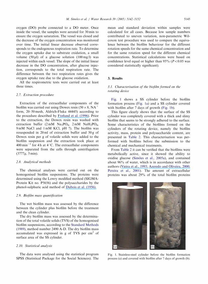
Fig. 1. Stainless-steel cylinder before the biofilm formation
process (a) and covered with biofilm after 7 days of growth (b).
M. Simoes et al. / Water Research 39 (2005) 5142–5152 5145
oxygen (DO) probe connected to a DO meter. Once
inside the vessel, the samples were aerated for 30min to
ensure the oxygen saturation. The vessel was closed and
the decrease of the oxygen concentration was monitored
over time. The initial linear decrease observed corre-
sponds to the endogenous respiration rate. To determine
the oxygen uptake due to substrate oxidation, a small
volume (50 ml) of a glucose solution (100mg/l) was
injected within each vessel. The slope of the initial linear
decrease in the DO concentration, after glucose injec-
tion, corresponds to the total respiration rate. The
difference between the two respiration rates gives the
oxygen uptake rate due to the glucose oxidation.
All the respirometric tests were carried out at least
three times.
2.7. Extraction procedure
Extraction of the extracellular components of the
biofilm was carried out using Dowex resin (50� 8, NA+
form, 20–50mesh, Aldrich-Fluka 44445) according to
the procedure described by Frølund et al. (1996). Prior
to the extraction, the Dowex resin was washed with
extraction buffer (2mM Na3PO4, 2mM NaH2PO4,
9mM NaCl and 1mM KCl, pH 7). The biofilm was
resuspended in 20ml of extraction buffer and 50 g of
Dowex resin per g of volatile solids were added to the
biofilm suspension and the extraction took place at
400min�1 for 4 h at 4 1C. The extracellular components
were separated from the cells through centrifugation
(3777g, 5min).
2.8. Analytical methods
The chemical analyses were carried out on the
homogenised biofilm suspensions. The proteins were
determined using the Lowry modified method (SIGMA-
Protein Kit no. P5656) and the polysaccharides by the
phenol-sulphuric acid method of Dubois et al. (1956).
2.9. Biofilm mass quantification
The wet biofilm mass was assessed by the difference
between the cylinder plus biofilm before the treatment
and the clean cylinder.
The dry biofilm mass was assessed by the determina-
tion of the total volatile solids (TVS) of the homogenised
biofilm suspensions, according to the Standard Methods
(1989), method number 2490 A-D. The dry biofilm mass
accumulated was expressed in g of TVS per cm2 of
surface area of the SS cylinder.
2.10. Statistical analysis
The data were analysed using the statistical program
SPSS (Statistical Package for the Social Sciences). The
mean and standard deviation within samples were
calculated for all cases. Because low sample numbers
contributed to uneven variation, non-parametric Wil-
coxon test procedure was used to compare the equiva-
lence between the biofilm behaviour for the different
rotation speeds for the same chemical concentration and
for the same rotation speed for the different chemical
concentrations. Statistical calculations were based on
confidence level equal or higher than 95% (Po0:05 was
considered statistically significant).
3. Results
3.1. Characterisation of the biofilm formed on the
rotating device
Fig. 1 shows a SS cylinder before the biofilm
formation process (Fig. 1a) and a SS cylinder covered
with biofilm after 7 days of growth (Fig. 1b).
This figure clearly shows that the surface of the SS
cylinder was completely covered with a thick and slimy
biofilm that seems to be strongly adhered to the surface.
Some characteristics of the biofilms formed on the
cylinders of the rotating device, namely the biofilm
activity, mass, protein and polysaccharide content, are
presented in Table 2. This characterisation was per-
formed with biofilms before the submission to the
chemical and mechanical treatments.
From Table 2 it can be verified that the biofilms were
metabolically active, since it showed the ability to
oxidise glucose (Simoes et al., 2005a), and contained
about 96% of water, which is in accordance with other
authors (Vieira et al., 1993; Azeredo and Oliveira, 2000;
Pereira et al., 2001). The amount of extracellular
proteins was about 29% of the total biofilm proteins
ARTICLE IN PRESSM. Simoes et al. / Water Research 39 (2005) 5142–51525146
and the amount of extracellular polysaccharides was
nearly 61.5% of the total biofilm polysaccharides. The
total protein content was similar to the total poly-
saccharide content. However, since the analytical
methods used to assess the amount of total proteins
and polysaccharides were different, the comparison
between quantitative amounts of proteins and poly-
saccharides cannot be accurately performed.
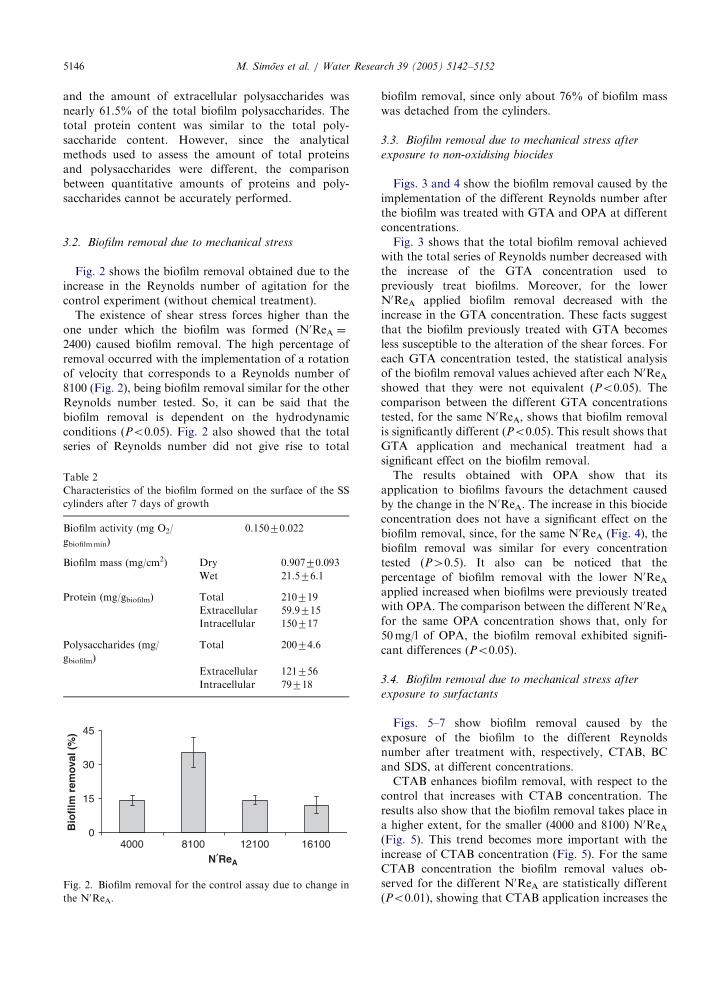
3.2. Biofilm removal due to mechanical stress
Fig. 2 shows the biofilm removal obtained due to the
increase in the Reynolds number of agitation for the
control experiment (without chemical treatment).
The existence of shear stress forces higher than the
one under which the biofilm was formed (N0ReA ¼
2400) caused biofilm removal. The high percentage of
removal occurred with the implementation of a rotation
of velocity that corresponds to a Reynolds number of
8100 (Fig. 2), being biofilm removal similar for the other
Reynolds number tested. So, it can be said that the
biofilm removal is dependent on the hydrodynamic
conditions (Po0:05). Fig. 2 also showed that the total
series of Reynolds number did not give rise to total
Table 2
Characteristics of the biofilm formed on the surface of the SS
cylinders after 7 days of growth
Biofilm activity (mg O2/
gbiofilmmin)
0.15070.022
Biofilm mass (mg/cm2) Dry 0.90770.093
Wet 21.576.1
Protein (mg/gbiofilm) Total 210719
Extracellular 59.9715
Intracellular 150717
Polysaccharides (mg/
gbiofilm)
Total 20074.6
Extracellular 121756
Intracellular 79718
0
15
30
45
4000 8100 12100 16100
Bio
film
rem
oval
(%
)
N′ReA
Fig. 2. Biofilm removal for the control assay due to change in
the N0ReA.
biofilm removal, since only about 76% of biofilm mass
was detached from the cylinders.
3.3. Biofilm removal due to mechanical stress after
exposure to non-oxidising biocides
Figs. 3 and 4 show the biofilm removal caused by the
implementation of the different Reynolds number after
the biofilm was treated with GTA and OPA at different
concentrations.
Fig. 3 shows that the total biofilm removal achieved
with the total series of Reynolds number decreased with
the increase of the GTA concentration used to
previously treat biofilms. Moreover, for the lower
N0ReA applied biofilm removal decreased with the
increase in the GTA concentration. These facts suggest
that the biofilm previously treated with GTA becomes
less susceptible to the alteration of the shear forces. For
each GTA concentration tested, the statistical analysis
of the biofilm removal values achieved after each N0ReAshowed that they were not equivalent (Po0:05). The
comparison between the different GTA concentrations
tested, for the same N0ReA, shows that biofilm removal
is significantly different (Po0:05). This result shows thatGTA application and mechanical treatment had a
significant effect on the biofilm removal.
The results obtained with OPA show that its
application to biofilms favours the detachment caused
by the change in the N0ReA. The increase in this biocide
concentration does not have a significant effect on the
biofilm removal, since, for the same N0ReA (Fig. 4), the
biofilm removal was similar for every concentration
tested (P40:5). It also can be noticed that the
percentage of biofilm removal with the lower N0ReAapplied increased when biofilms were previously treated
with OPA. The comparison between the different N0ReAfor the same OPA concentration shows that, only for
50mg/l of OPA, the biofilm removal exhibited signifi-
cant differences (Po0:05).
3.4. Biofilm removal due to mechanical stress after
exposure to surfactants
Figs. 5–7 show biofilm removal caused by the
exposure of the biofilm to the different Reynolds
number after treatment with, respectively, CTAB, BC
and SDS, at different concentrations.
CTAB enhances biofilm removal, with respect to the
control that increases with CTAB concentration. The
results also show that the biofilm removal takes place in
a higher extent, for the smaller (4000 and 8100) N0ReA(Fig. 5). This trend becomes more important with the
increase of CTAB concentration (Fig. 5). For the same
CTAB concentration the biofilm removal values ob-
served for the different N0ReA are statistically different
(Po0:01), showing that CTAB application increases the
ARTICLE IN PRESS
0
20
40
60
80
100
Control 200100 500 1000GTA concentration (mg/l)
Bio
film
rem
oval
(%
)
4000 8100 12100 16100
Fig. 3. Biofilm removal observed after the alteration of the N0ReA for the biofilm control and for the GTA treated biofilms.
0
20
40
60
80
100
Control 10050 200 300OPA concentration (mg/l)
Bio
film
rem
oval
(%
)
4000 8100 12100 16100
Fig. 4. Biofilm removal observed after the alteration of the N0ReA for the biofilm control and for the OPA treated biofilms.
0
20
40
60
80
100
Control 0.125 0.25 0.5 0.9CTAB concentration (mM)
Bio
film
rem
oval
(%
)
4000 8100 12100 16100
Fig. 5. Biofilm removal observed after the alteration of the N0ReA for the biofilm control and for the CTAB treated biofilms.
M. Simoes et al. / Water Research 39 (2005) 5142–5152 5147
biofilm susceptibility to detachment through the me-
chanical action. However, when comparing the biofilm
removal within concentrations and for the same N0ReA,
only for 4000 the biofilm removal was significantly
different (Po0:05).BC is a cationic surfactant as CTAB that caused
different biofilm removal results. The increase in BC
concentration used to treat biofilms increased the
difficulty of biofilm removal through the alteration of
the shear forces, especially when the lower N0ReA were
implemented. Biofilm removal is equivalent (P40:10)for the same N0ReA when comparing the different
concentrations tested, except for 0.900mM, where the
differences are statistically significant (Po0:05). In this
latter case, the higher amount of biofilm removal (30%)
was found for the highest N0ReA.
Concerning SDS, an anionic surface-active agent,
apart from 7mM, its application to the biofilm resulted
ARTICLE IN PRESS
0
20
40
60
80
100
BC Concentration (mM)
Bio
film
rem
ain
ing
(%
)
Control 0.125 0.25 0.5 0.9
4000 8100 12100 16100
Fig. 6. Biofilm removal observed after the alteration of the N0ReA for the biofilm control and for the BC treated biofilms.
0
20
0.5 1 3 7
40
60
80
100
ControlSDS concentration (mM)
Bio
film
rem
oval
(%
)
4000 8100 12100 16100
Fig. 7. Biofilm removal observed after the alteration of the N0ReA for the biofilm control and for the SDS treated biofilms.
M. Simoes et al. / Water Research 39 (2005) 5142–51525148
in the decrease of biofilm removal achieved with the
hydrodynamic change. Conversely, with the application
of 7mM of SDS biofilm removal takes place to a higher
extent, for N0ReA of 4000 and 8100, but similar to the
other N0ReA tested (P40:05). For 0.500mM the biofilm
removal is similar for every N0ReA tested (P40:1). Theapplication of 1 and 3mM of SDS promoted significant
differences in the posterior biofilm removal (Po0:05),when comparing the different N0ReA, being the high
amount of biofilm removal promoted with the exposure
to a N0ReA of 12,100. However, when comparing the
biofilm removal for the same N0ReA within different
concentrations, a significant difference (Po0:05) was
found only for a N0ReA of 4000, due to the high amount
of biofilm removal found after treatments with 0.5 and
7mM.
3.5. Biofilm removal due to mechanical stress after
exposure to oxidising biocides
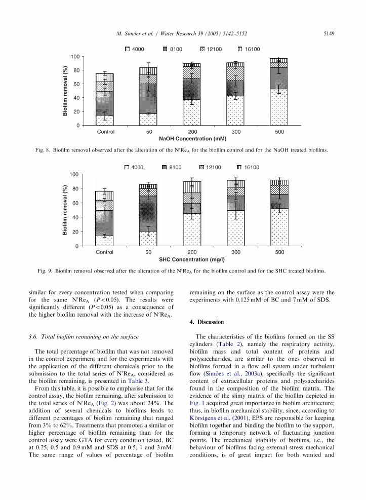
Figs. 8 and 9 show biofilm removal caused by the
exposure of the biofilm to the different N0ReA after
treatment with NaOH and SHC at different concentra-
tions.
Similar impacts on biofilm removal were found for
NaOH and SHC (Figs. 8 and 9). Both chemicals
similarly affected biofilm removal for every condition
tested. Concerning NaOH, with the exception for
50mM, the highest amount of biofilm removal is found
for a N0ReA of 4000 and with the trend to increase with
the increase in the concentration applied. For 50mM the
high amount of biofilm removal was found with an
exposure to a N0ReA of 8100. However, the biofilm
removal is statistically equivalent when compared with
the other N0ReA (Po0:05). Concerning the comparison
of the different N0ReA for the same NaOH concentra-
tion, the results are significantly different (Po0:05), withthe exception for the treatment with 200mM (P40:10),where the biofilm removal happened to a similar extent
with the submission to a N0ReA of 4000 and 8100.
The application of 50mg/l of SHC resulted in a
posterior biofilm removal that reached the highest
amount with the exposure to a N0ReA of 8100. For the
other concentrations tested, the biofilm removal was
high for a N0ReA of 4000. The biofilm removal was
ARTICLE IN PRESS
0
20
40
60
80
100
NaOH Concentration (mM)
Bio
film
rem
oval
(%
)
Control 50 200 300 500
4000 8100 12100 16100
Fig. 8. Biofilm removal observed after the alteration of the N0ReA for the biofilm control and for the NaOH treated biofilms.
0
20
40
60
80
100
SHC Concentration (mg/l)
Bio
film
rem
oval
(%
)
Control 50 200 300 500
4000 8100 12100 16100
Fig. 9. Biofilm removal observed after the alteration of the N0ReA for the biofilm control and for the SHC treated biofilms.
M. Simoes et al. / Water Research 39 (2005) 5142–5152 5149
similar for every concentration tested when comparing
for the same N0ReA (Po0:05). The results were
significantly different (Po0:05) as a consequence of
the higher biofilm removal with the increase of N0ReA.
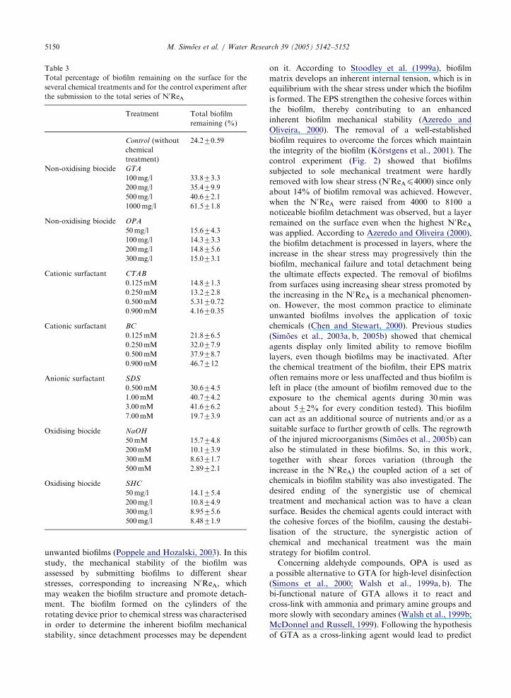
3.6. Total biofilm remaining on the surface
The total percentage of biofilm that was not removed
in the control experiment and for the experiments with
the application of the different chemicals prior to the
submission to the total series of N0ReA, considered as
the biofilm remaining, is presented in Table 3.
From this table, it is possible to emphasise that for the
control assay, the biofilm remaining, after submission to
the total series of N0ReA (Fig. 2) was about 24%. The
addition of several chemicals to biofilms leads to
different percentages of biofilm remaining that ranged
from 3% to 62%. Treatments that promoted a similar or
higher percentage of biofilm remaining than for the
control assay were GTA for every condition tested, BC
at 0.25, 0.5 and 0.9mM and SDS at 0.5, 1 and 3mM.
The same range of values of percentage of biofilm
remaining on the surface as the control assay were the
experiments with 0.125mM of BC and 7mM of SDS.
4. Discussion
The characteristics of the biofilms formed on the SS
cylinders (Table 2), namely the respiratory activity,
biofilm mass and total content of proteins and
polysaccharides, are similar to the ones observed in
biofilms formed in a flow cell system under turbulent
flow (Simoes et al., 2003a), specifically the significant
content of extracellular proteins and polysaccharides
found in the composition of the biofilm matrix. The
evidence of the slimy matrix of the biofilm depicted in
Fig. 1 acquired great importance in biofilm architecture;
thus, in biofilm mechanical stability, since, according to
Korstgens et al. (2001), EPS are responsible for keeping
biofilm together and binding the biofilm to the support,
forming a temporary network of fluctuating junction
points. The mechanical stability of biofilms, i.e., the
behaviour of biofilms facing external stress mechanical
conditions, is of great impact for both wanted and
ARTICLE IN PRESS
Table 3
Total percentage of biofilm remaining on the surface for the
several chemical treatments and for the control experiment after
the submission to the total series of N0ReA
Treatment Total biofilm
remaining (%)
Control (without
chemical
treatment)
24.270.59
Non-oxidising biocide GTA
100mg/l 33.873.3
200mg/l 35.479.9
500mg/l 40.672.1
1000mg/l 61.571.8
Non-oxidising biocide OPA
50mg/l 15.674.3
100mg/l 14.373.3
200mg/l 14.875.6
300mg/l 15.073.1
Cationic surfactant CTAB
0.125mM 14.871.3
0.250mM 13.272.8
0.500mM 5.3170.72
0.900mM 4.1670.35
Cationic surfactant BC
0.125mM 21.876.5
0.250mM 32.077.9
0.500mM 37.978.7
0.900mM 46.7712
Anionic surfactant SDS
0.500mM 30.674.5
1.00mM 40.774.2
3.00mM 41.676.2
7.00mM 19.773.9
Oxidising biocide NaOH
50mM 15.774.8
200mM 10.173.9
300mM 8.6371.7
500mM 2.8972.1
Oxidising biocide SHC
50mg/l 14.175.4
200mg/l 10.874.9
300mg/l 8.9575.6
500mg/l 8.4871.9
M. Simoes et al. / Water Research 39 (2005) 5142–51525150
unwanted biofilms (Poppele and Hozalski, 2003). In this
study, the mechanical stability of the biofilm was
assessed by submitting biofilms to different shear
stresses, corresponding to increasing N0ReA, which
may weaken the biofilm structure and promote detach-
ment. The biofilm formed on the cylinders of the
rotating device prior to chemical stress was characterised
in order to determine the inherent biofilm mechanical
stability, since detachment processes may be dependent
on it. According to Stoodley et al. (1999a), biofilm
matrix develops an inherent internal tension, which is in
equilibrium with the shear stress under which the biofilm
is formed. The EPS strengthen the cohesive forces within
the biofilm, thereby contributing to an enhanced
inherent biofilm mechanical stability (Azeredo and
Oliveira, 2000). The removal of a well-established
biofilm requires to overcome the forces which maintain
the integrity of the biofilm (Korstgens et al., 2001). The
control experiment (Fig. 2) showed that biofilms
subjected to sole mechanical treatment were hardly
removed with low shear stress (N0ReAp4000) since only
about 14% of biofilm removal was achieved. However,
when the N0ReA were raised from 4000 to 8100 a
noticeable biofilm detachment was observed, but a layer
remained on the surface even when the highest N0ReAwas applied. According to Azeredo and Oliveira (2000),
the biofilm detachment is processed in layers, where the
increase in the shear stress may progressively thin the
biofilm, mechanical failure and total detachment being
the ultimate effects expected. The removal of biofilms
from surfaces using increasing shear stress promoted by
the increasing in the N0ReA is a mechanical phenomen-
on. However, the most common practice to eliminate
unwanted biofilms involves the application of toxic
chemicals (Chen and Stewart, 2000). Previous studies
(Simoes et al., 2003a, b, 2005b) showed that chemical
agents display only limited ability to remove biofilm
layers, even though biofilms may be inactivated. After
the chemical treatment of the biofilm, their EPS matrix
often remains more or less unaffected and thus biofilm is
left in place (the amount of biofilm removed due to the
exposure to the chemical agents during 30min was
about 572% for every condition tested). This biofilm
can act as an additional source of nutrients and/or as a
suitable surface to further growth of cells. The regrowth
of the injured microorganisms (Simoes et al., 2005b) can
also be stimulated in these biofilms. So, in this work,
together with shear forces variation (through the
increase in the N0ReA) the coupled action of a set of
chemicals in biofilm stability was also investigated. The
desired ending of the synergistic use of chemical
treatment and mechanical action was to have a clean
surface. Besides the chemical agents could interact with
the cohesive forces of the biofilm, causing the destabi-
lisation of the structure, the synergistic action of
chemical and mechanical treatment was the main
strategy for biofilm control.
Concerning aldehyde compounds, OPA is used as
a possible alternative to GTA for high-level disinfection
(Simons et al., 2000; Walsh et al., 1999a, b). The
bi-functional nature of GTA allows it to react and
cross-link with ammonia and primary amine groups and
more slowly with secondary amines (Walsh et al., 1999b;
McDonnel and Russell, 1999). Following the hypothesis
of GTA as a cross-linking agent would lead to predict
ARTICLE IN PRESSM. Simoes et al. / Water Research 39 (2005) 5142–5152 5151
that biofilm treatment with GTA should actually
stabilise the biofilm, as found with this work. GTA
was not efficient in removing the biofilm from the SS
cylinders in spite of the fact that this biocide is
frequently used to chemically control the accumulation
of biofilms (Pereira and Vieira, 2001). On the contrary,
GTA contributed to the formation of a harder deposit,
since the percentage of biofilm remaining on the surface
was higher than for the control experiment.
Conversely, the results obtained with OPA are
consistent with its less effect of cross-linking when
compared with GTA. Probably this fact is related
with the aromatic ring presented in the molecular
structure of OPA, which confers a diminished flexibility
of the molecule, conversely to the aliphatic chain of
GTA (Simons et al., 2000; Walsh et al., 1999a, b).
Consequently, the biofilm remaining on the surface
decreases slightly after OPA application in relation to
the control.
The treatment with surfactants caused different biofilm
responses that may be related with their chemical nature.
Concerning the cationic surfactants, the behaviour of
CTAB differs significantly from the one observed with
BC. The action of those cationic surfactants is attributed
to their positive charge that forms an electrostatic bond
with negatively charged sites (Cloete et al., 1997). The
different biofilm behaviours may be related with the
chemical reaction of the surfactants with the biofilm
components used that can give rise to the strengthening
or the weakening of the biofilm structure. The electro-
static bonds created stress or cross-linking depending on
the chemical structure of the molecule, since CTAB is an
aliphatic compound while BC is an aromatic compound.
The increase in the CTAB concentration promoted the
subsequent higher biofilm removal due to the destabilisa-
tion of the biofilm cohesive forces, being biofilm removal
detected at a higher extent to the smaller shear stresses.
Conversely, the increase in the BC concentration
increased the biofilm mechanical stability face to mechan-
ical stress conditions.
The effect of SDS on the mechanical stability of the
biofilm may be due to the disruption of the hydrophobic
interactions involved in cross-linking the biofilm matrix
(Chen and Stewart, 2000). However, in this work, this
SDS effect was only felt for the higher concentration
(7mM) tested, proposing that low concentrations of
SDS can even promote the strength of the biofilm
structure.
The previous application of oxidising agents improved
biofilm removal by mechanical action, the effect being
more pronounced with the increase in their concentra-
tion. The oxidising biocides react strongly with the EPS
matrix, destroying the structure that becomes more
vulnerable to hydrodynamic stress. So, it is not
surprising to obtain more removal for the same N0ReAas the concentration increases.
5. Conclusions
The system presented in this work provided an
approach to investigate the influence of several para-
meters on the mechanical stability of biofilms, leading to
a better understanding of biofilms in different environ-
ments and the development of biofilm control strategies.
The characterisation of the biofilms showed that the
system tested allowed the formation of a great amount
of biofilm that covered the surface of the SS cylinder, the
biofilms being metabolically active, vastly comprising
EPS and having an inherent mechanical stability.
The effect of the chemical compounds on the biofilm
removal and consequent biofilm mechanical stability
varied with the chemical nature; even with the synergistic
chemical and mechanical treatment total biofilm eradica-
tion was not achieved in this work, for every condition
studied. The application of OPA to the biofilms favoured
the detachment caused by the increase in the mechanical
stress, being biofilm removal similar for every concentra-
tion tested. Also, OPA demonstrated to be an alternative
to GTA in the control of P. fluorescens biofilms, since the
biofilms treated with GTA showed posterior recalcitrance
properties when exposed to mechanical stress conditions,
increasing with the increase of GTA concentration. The
application of CTAB decreased the biofilm mechanical
stability, which was more pronounced with the increase
of the concentration and with the increase on the
mechanical stress conditions. Conversely, BC increased
the biofilm mechanical stability. This phenomenon was
more pronounced with the increase of concentration.
SDS caused biofilm removal due to increasing shear
forces only for the highest concentration tested, when
comparing with the control experiment. For the smaller
concentrations, a similar effect to the one found with
GTA and BC was observed. The previous application of
oxidising agents (NaOH and SHC) improved biofilm
removal by mechanical action, this effect being dependent
on the increase in their concentrations.
This chemical diversity of agents tested (non-oxidising
aldehyde-based biocides, surfactants and oxidising
biocides) emphasises that multiple interactive forces
contribute to biofilm mechanical stability.
Acknowledgements
The authors acknowledge the financial support provided
by IBQF, and the Portuguese Foundation for Science and
Technology (Post-Doc Grant—Manuel Simoes).
References
APHA, AWWA, WPCF, 1989. In: Clesceri, L.S., Greenberg,
A.E., Trussel, R.R. (Eds.), Standard Methods for the
ARTICLE IN PRESSM. Simoes et al. / Water Research 39 (2005) 5142–51525152
Examination of Water and Wastewater, 17th ed. American
Public Health Association, Washington, DC.
Azeredo, J., Oliveira, R., 2000. The role of exopolymers
produced by Sphingomonas paucimobilis in biofilm forma-
tion and composition. Biofouling 16, 17–27.
Campanac, C., Pineau, L., Payard, A., Baziard-Mouysset, G.,
Roques, C., 2002. Interactions between biocide cationic
agents and bacterial biofilms. Antimicrob. Agents Che-
mother. 46, 1469–1474.
Chen, X., Stewart, P.S., 2000. Biofilm removal caused by
chemical treatments. Water Res. 34, 4229–4233.
Cloete, T.E., Jacobs, L., Brozel, V.S., 1997. The chemical
control of biofouling in industrial water systems. Biode-
gradation 9, 23–37.
Dubois, M., Gilles, K.A., Hamilton, J.K., Rebers, A., Smith,
F., 1956. Colorimetric method for determination of sugars
and related substances. Anal. Chem. 28, 350–356.
Flemming, H.-C., 1996. The forces that keep biofilms together.
In: Biodeterioration and Biodegradation, DECHEMA
Monographien 133, pp. 311–317.
Frølund, B., Palmgren, R., Keiding, A., Nielsen, P.H., 1996.
Extraction of extracellular polymers from activated
sludge using a cation exchange resin. Water Res. 30,
1749–1758.
Geankoplis, C.J., 1993. Transport Processes and Unit Opera-
tions, third ed. Prentice-Hall International, Inc., New
Jersey, pp. 144–145.
Hall-Stoodley, L., Stoodley, P., 2002. Developmental regula-
tion of microbial biofilms. Curr. Opin. Biotechnol. 13,
228–233.
Korstgens, V., Flemming, H.-C., Wingender, J., Borchard, W.,
2001. Uniaxial compression measurement device for in-
vestigation of the mechanical stability of biofilms. J.
Microbiol. Methods 46, 9–17.
McDonnel, G., Russell, A.D., 1999. Antiseptics and disin-
fectants: activity, action and resistance. Clin. Microbiol.
Rev. 12, 147–179.
Ohashi, A., Harada, H., 1994. Adhesion strength of biofilm
developed in an attached-growth reactor. Water Sci.
Technol. 29, 281–288.
Ohashi, A., Harada, H., 1996. A novel concept for evaluation
of biofilm adhesion strength by applying tensile force and
shear force. Water Sci. Technol. 34, 201–211.
Ohashi, A., Koyama, T., Syutsubo, S., Harada, H., 1999. A
novel method for evaluation of biofilm tensile strength
resisting erosion. Water Sci. Technol. 39, 261–268.
Oliveira, R., Melo, L., Oliveira, A., Salgueiro, R., 1994.
Polysaccharide production and biofilm formation by
Pseudomonas fluorescens: effects of pH and surface material.
Colloids Surf. B: Biointerfaces 2, 41–46.
Pereira, M.O., Vieira, M.J., 2001. Effects of the interactions
between glutaraldehyde and the polymeric matrix on the
efficacy of the biocide against Pseudomonas fluorescens
biofilms. Biofouling 17, 93–101.
Pereira, M.O., Morin, P., Vieira, M.J., Melo, L.F., 2001.
A versatile reactor for continuous monitoring of biofilm
properties in laboratory and industrial conditions. Lett.
Appl. Microbiol. 34, 22–26.
Pereira, M.O., Kuehn, M., Wuertz, S., Neu, T., Melo, L., 2002.
Effect of flow regime on the architecture of a Pseudomonas
fluorescens biofilm. Biotechnol. Bioeng. 78, 164–171.
Poppele, E.H., Hozalski, R.M., 2003. Micro-cantilever method
for measuring the tensile strength of biofilms and microbial
flocs. J. Microbiol. Methods 55, 607–615.
Simons, C., Walsh, S.E., Maillard, J.-Y., Russell, A.D., 2000.
Ortho-phthalaldehyde: proposed mechanism of action of a
new antimicrobial agent. Lett. Appl. Microbiol. 31,
299–302.
Simoes, M., Pereira, M.O., Vieira, M.J., 2003a. Monitoring the
effects of biocide treatment of Pseudomonas fluorescens
formed under different flow regimes. Water Sci. Technol.
47, 217–223.
Simoes, M., Carvalho, H., Pereira, M.O., Vieira, M.J., 2003b.
Effect of different concentrations of ortho-phthalaldehyde
on biofilms formed by Pseudomonas fluorescens under
different flow conditions. Biofouling 19, 287–295.
Simoes, M., Pereira, M.O., Vieira, M.J., 2005a. Validation of
respirometry as a short-term method to assess the efficacy of
biocides. Biofouling 21, 9–17.
Simoes, M., Pereira, M.O., Vieira, M.J., 2005b. Action of a
cationic surfactant on the activity and removal of bacterial
biofilms formed under different flow regimes. Water Res.
39, 478–486.
Stoodley, P., Lewandowski, Z., Boyle, J.D., Lappin-Scott,
H.M., 1999a. Structural deformation of bacterial biofilms
caused by short-term fluctuations in fluid shear: an in situ
investigation of biofilm rheology. Biotechnol. Bioeng. 65,
83–92.
Stoodley, P., Boyle, J.D., DeBeer, D., Lappin-Scott, H.M.,
1999b. Evolving perspectives of biofilm structure. Biofoul-
ing 14, 75–90.
Vieira, M.J., Melo, L., Pinheiro, M.M., 1993. Biofilm forma-
tion: hydrodynamic effects on internal diffusion and
structure. Biofouling 7, 67–80.
Walsh, S.E., Maillard, J.-Y., Russel, A.D., 1999a. Ortho-
phthalaldehyde: a possible alternative to glutaraldehyde for
high level disinfection. J. Appl. Microbiol. 86, 1039–1046.
Walsh, S.E., Maillard, J.-Y., Simons, C., Russel, A.D., 1999b.
Studies on the mechanisms of the antimicrobial action of
ortho-phthaldehyde. J. Appl. Microbiol. 87, 702–710.


