Embed Size (px)
Citation preview
Journal of Electromyography and Kinesiology 21 (2011) 141–147
Contents lists available at ScienceDirect
Journal of Electromyography and Kinesiology
journal homepage: www.elsevier .com/locate / je lek in
Effect of unaccustomed eccentric exercise on proprioception of the kneein weight and non-weight bearing tasks
Carolina Vila-Chã a, Simone Riis a, Ditte Lund a, Anders Møller a, Dario Farina a,b, Deborah Falla a,b,⇑a Centre for Sensory-Motor Interaction (SMI), Department of Health Science and Technology, Aalborg University, Fredrik Bajers Vej 7, D-3, DK-9220, Aalborg, Denmarkb Department of Neurorehabilitation Engineering, Bernstein Center for Computational Neuroscience, Georg-August University of Göttingen, Göttingen, Germany
a r t i c l e i n f o
Article history:Received 2 July 2010Received in revised form 14 September2010Accepted 1 October 2010
Keywords:ProprioceptionQuadricepsEccentric exerciseDOMS
1050-6411/$ - see front matter � 2010 Elsevier Ltd. Adoi:10.1016/j.jelekin.2010.10.001
⇑ Corresponding author. Tel.: +45 99 40 74 59; fax:E-mail address: [email protected] (D. Falla).
a b s t r a c t
The study investigates the effects of eccentric exercise of the quadriceps on proprioception of the knee inweight and non-weight bearing tasks. Proprioception of the exercised leg was assessed at 120� and 150�of knee extension in 15 healthy adults (age 25.0 ± 3.6 yrs) before, immediately after, and 24 h followingeccentric exercise of the quadriceps. Three tests of proprioception were performed: 1. matching the posi-tion of the exercised leg (right leg) to the reference leg (left leg) in sitting (non-weight bearing matchingtask); 2. repositioning the exercised leg after active movement in sitting (non-weight bearing reposition-ing task); 3. repositioning the exercised leg after active movement in standing (weight bearing task).Maximum knee extension force was reduced by 77.0 ± 12.3 % immediately after the exercise, and by82.7 ± 16.2% 24 h post exercise, with respect to baseline (P < 0.001). The absolute error in the non-weightbearing matching task at 120� of knee extension was greater immediately following eccentric exercise(12.3 ± 5.6, P < 0.001) and 24 h after exercise (8.1 ± 4.5, P < 0.05) compared to baseline (5.8 ± 2.7). Simi-larly, the absolute error in the non-weight bearing repositioning task at 120� was greater both immedi-ately (5.9 ± 3.1�, P < 0.01) and 24 h post exercise (5.2 ± 3.0�, P < 0.05) compared to baseline (4.5 ± 2.6�).Therefore, in both non-weight bearing tasks, the subjects matched the position of their leg after eccentricexercise by adopting a more extended knee position of the exercised limb. Furthermore, the subjectsshowed higher variability in their performance immediately post exercise (P < 0.05, compared to base-line) but not 24 h after. In contrast, eccentric exercise did not affect the repositioning errors in the weightbearing task. In conclusion, eccentric exercise of the quadriceps impairs proprioception of the knee bothimmediately after and 24 h post exercise, but only in non-weight bearing tasks.
� 2010 Elsevier Ltd. All rights reserved.
1. Introduction
Unaccustomed eccentric exercise typically leads to myofibrillardamage, disturbance of the extracellular matrix and an inflamma-tory reaction (Howell et al., 1993; Jones et al., 1987; Yu and Thornell,2002). The sensation of pain and muscle stiffness normally beginsseveral hours after unaccustomed eccentric exercise, reaches a peak24–48 h after and may even persist for several days (Armstrong,1984; Jones et al., 1987). This phenomenon – delayed-onset muscu-lar soreness (DOMS) – is associated with prolonged muscle forceloss, reduction of joint range of motion, a sensation of unsteady limbsand clumsiness in precision movements (Brockett et al., 1997; Ho-well et al., 1993; Paschalis et al., 2007; Proske et al., 2003; Saxtonet al., 1995), and impaired proprioception (Proske and Allen, 2005).
Several studies have shown that immediately after eithereccentric or concentric exercise, the size of errors observed duringposition- and force-matching tasks increases significantly (Walsh
ll rights reserved.
+45 98 15 40 08.
et al., 2004; Allen and Proske, 2006; Givoni et al., 2007). Further-more, these studies show that the degree of matching errors isassociated with the degree of force reduction due to either musclefatigue or DOMS (Walsh et al., 2004; Givoni et al., 2007). Althoughproprioception can be impaired following both eccentric and con-centric exercise, it appears to be affected more following eccentricexercise (Walsh et al., 2004; Givoni et al., 2007). This is presumablydue to the greater reduction in force following eccentric comparedto concentric exercise (Walsh et al., 2004; Winter et al., 2005).After eccentric exercise, the impairment in force lasts for 24–48 h(Lephart and Fu, 2000; Proske and Morgan, 2001) and significantmatching errors are still observed after 24 h (Givoni et al., 2007).
These observations contributed to the effort hypothesis, whichsuggests that the sense of effort or heaviness generated by centralmotor commands play an important role in joint position sense(for review see Proske and Gandevia, 2009). Since maximal forceis reduced after exercise, the effort required to support the limb in-creases, altering the sense of effort (Walsh et al., 2006) resulting inreduced joint position accuracy. Nevertheless, the effort hypothesishas not been confirmed under all circumstances (Proske and
142 C. Vila-Chã et al. / Journal of Electromyography and Kinesiology 21 (2011) 141–147
Gandevia, 2009), indicating that other peripheral and/or centralmechanisms contribute to impaired proprioception.
Impairment of proprioception may influence joint stability andhas been associated with the occurrence of knee injuries in sportsand exercise (Granata et al., 1999; Givoni et al., 2007; Kelly, 2008;Sanna and O’Conner, 2008). However, the majority of studiesexamining the effects of exercise on proprioception have examinedthe upper extremities in non-weight bearing positions (Walshet al., 2004; Proske et al., 2004; Allen et al., 2007). Knowledge onthe effect of exercise on proprioception in weight bearing positionsis also needed since most knee injuries occur in weight bearingpositions. For example, anterior cruciate ligament injuries com-monly occur with change of direction in a weight bearing positionwith a fixed distal extremity (Kelly, 2008; Sanna and O’Conner,2008).
This study examines the immediate and delayed effects ofeccentric exercise on proprioception of the knee during weightand non-weight bearing tasks. It was hypothesized that proprio-ception would be less affected in weight bearing since propriocep-tive feedback from other sources may compensate for the loss ofproprioception induced by eccentric exercise.
2. Materials and methods
2.1. Subjects
Fifteen healthy volunteers participated in the experiment (9men; age, mean ± SD, 25.0 ± 3.6 yrs). To control for potential learn-ing effects, an additional 10 healthy subjects were recruited as acontrol group (5 men; age, 22.0 ± 0.8 yrs). The participants werefree of lower limb injuries. The study was conducted in accordancewith the Declaration of Helsinki and approved by the Local EthicsCommittee (N-20070019). Subjects provided informed writtenconsent prior to participation in the study.
2.2. Procedure
Maximum voluntary knee extension force and proprioception ofthe right knee were tested at baseline, immediately followingeccentric exercise, and 24 h after eccentric exercise. To confirmthe presence of DOMS 24 h post exercise, participants marked theirarea of pain on a body chart and verbally rated their perceived painon a scale from 0 (‘‘no soreness’’) to 10 (‘‘worst soreness imagin-able’’). The subjects were asked to rate the average pain intensityin the quadriceps during their regular activities of daily living(e.g. descending stairs) since their last visit to the laboratory (overthe past 24 h).
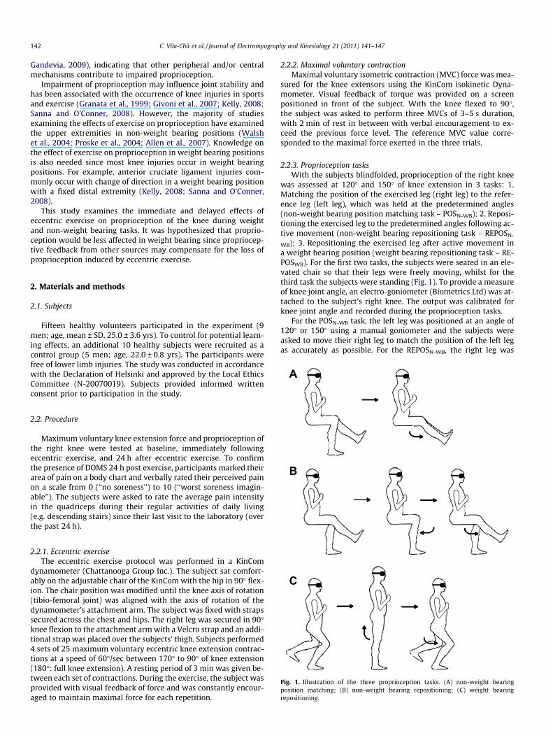
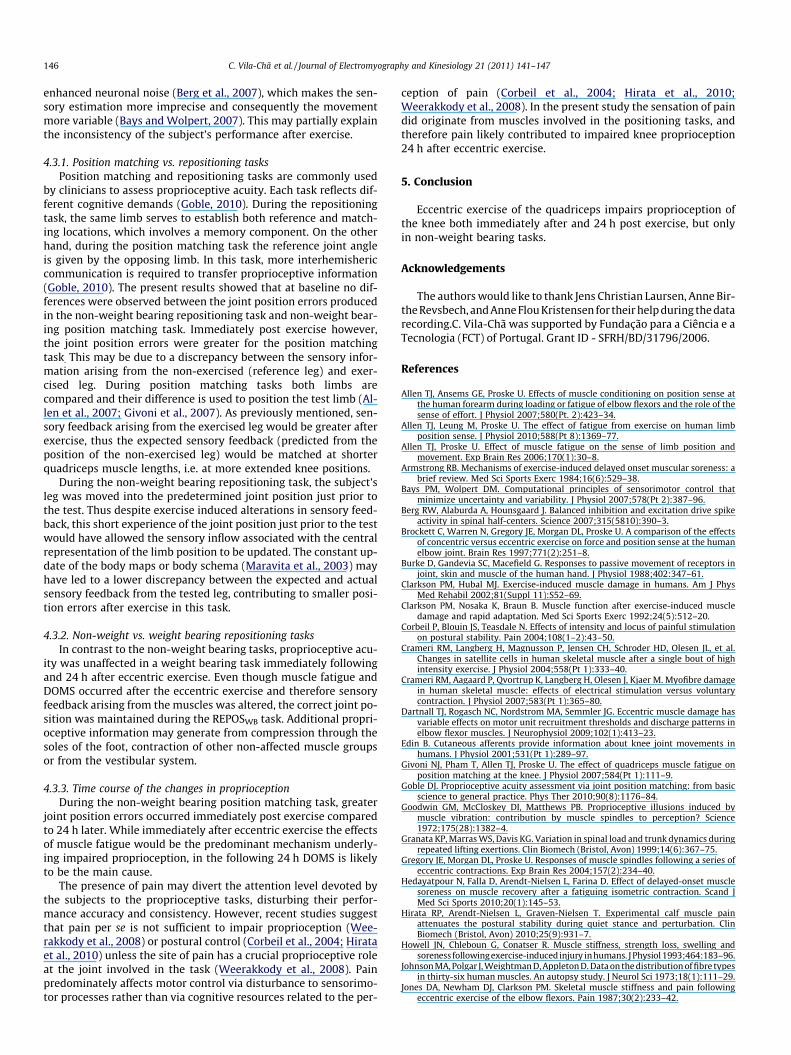
Fig. 1. Illustration of the three proprioception tasks. (A) non-weight bearingposition matching; (B) non-weight bearing repositioning; (C) weight bearingrepositioning.
2.2.1. Eccentric exerciseThe eccentric exercise protocol was performed in a KinCom
dynamometer (Chattanooga Group Inc.). The subject sat comfort-ably on the adjustable chair of the KinCom with the hip in 90� flex-ion. The chair position was modified until the knee axis of rotation(tibio-femoral joint) was aligned with the axis of rotation of thedynamometer’s attachment arm. The subject was fixed with strapssecured across the chest and hips. The right leg was secured in 90�knee flexion to the attachment arm with a Velcro strap and an addi-tional strap was placed over the subjects’ thigh. Subjects performed4 sets of 25 maximum voluntary eccentric knee extension contrac-tions at a speed of 60�/sec between 170� to 90� of knee extension(180�: full knee extension). A resting period of 3 min was given be-tween each set of contractions. During the exercise, the subject wasprovided with visual feedback of force and was constantly encour-aged to maintain maximal force for each repetition.
2.2.2. Maximal voluntary contractionMaximal voluntary isometric contraction (MVC) force was mea-
sured for the knee extensors using the KinCom isokinetic Dyna-mometer. Visual feedback of torque was provided on a screenpositioned in front of the subject. With the knee flexed to 90�,the subject was asked to perform three MVCs of 3–5 s duration,with 2 min of rest in between with verbal encouragement to ex-ceed the previous force level. The reference MVC value corre-sponded to the maximal force exerted in the three trials.
2.2.3. Proprioception tasksWith the subjects blindfolded, proprioception of the right knee
was assessed at 120� and 150� of knee extension in 3 tasks: 1.Matching the position of the exercised leg (right leg) to the refer-ence leg (left leg), which was held at the predetermined angles(non-weight bearing position matching task – POSN-WB); 2. Reposi-tioning the exercised leg to the predetermined angles following ac-tive movement (non-weight bearing repositioning task – REPOSN-
WB); 3. Repositioning the exercised leg after active movement ina weight bearing position (weight bearing repositioning task – RE-POSWB). For the first two tasks, the subjects were seated in an ele-vated chair so that their legs were freely moving, whilst for thethird task the subjects were standing (Fig. 1). To provide a measureof knee joint angle, an electro-goniometer (Biometrics Ltd) was at-tached to the subject’s right knee. The output was calibrated forknee joint angle and recorded during the proprioception tasks.
For the POSN-WB task, the left leg was positioned at an angle of120� or 150� using a manual goniometer and the subjects wereasked to move their right leg to match the position of the left legas accurately as possible. For the REPOSN-WB, the right leg was
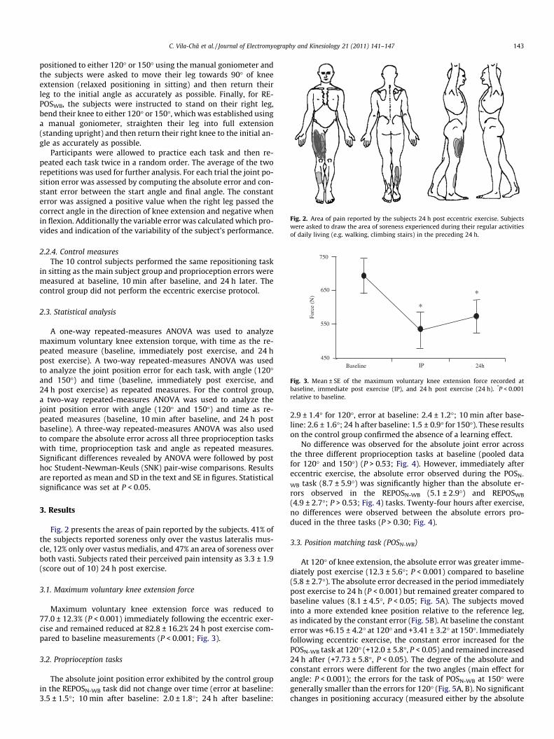
Fig. 2. Area of pain reported by the subjects 24 h post eccentric exercise. Subjectswere asked to draw the area of soreness experienced during their regular activitiesof daily living (e.g. walking, climbing stairs) in the preceding 24 h.
450
550
650
750
Baseline IP 24h
Forc
e (N
)*
*
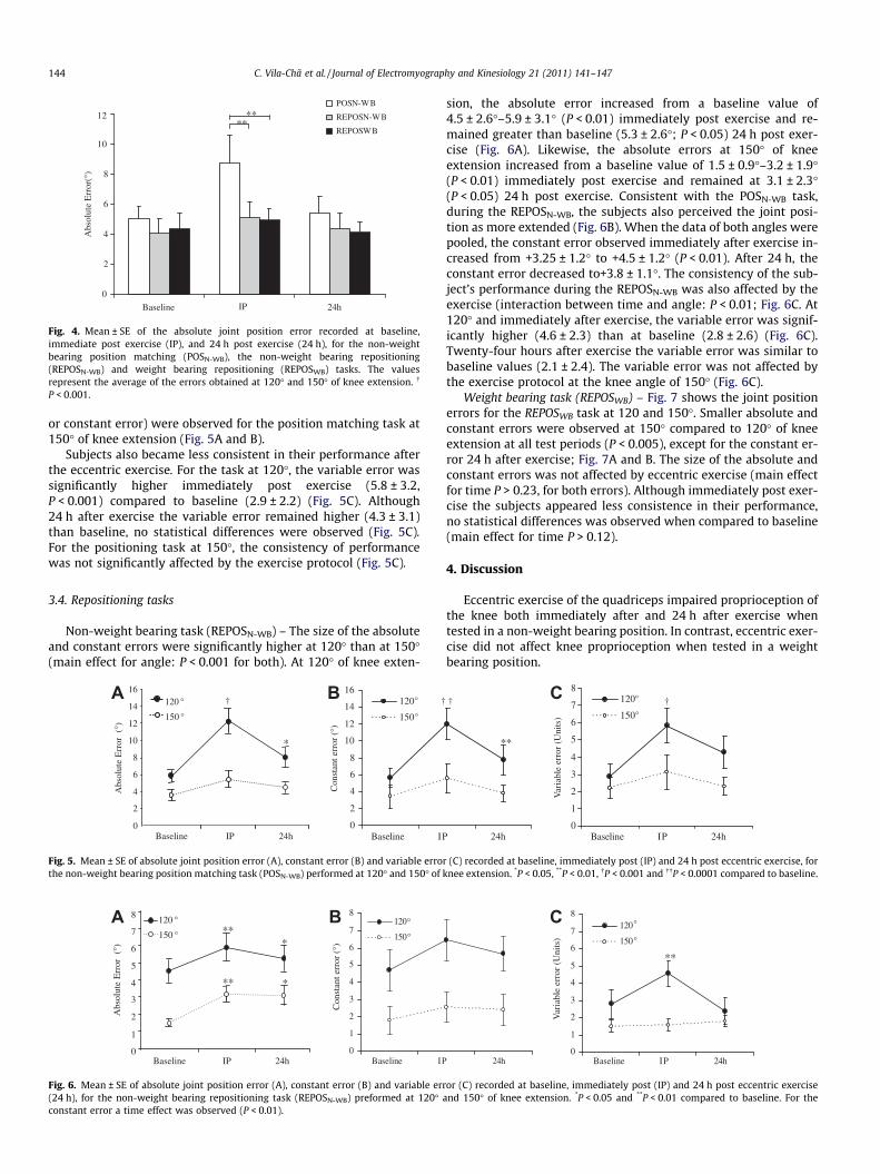
Fig. 3. Mean ± SE of the maximum voluntary knee extension force recorded atbaseline, immediate post exercise (IP), and 24 h post exercise (24 h). *P < 0.001relative to baseline.
C. Vila-Chã et al. / Journal of Electromyography and Kinesiology 21 (2011) 141–147 143
positioned to either 120� or 150� using the manual goniometer andthe subjects were asked to move their leg towards 90� of kneeextension (relaxed positioning in sitting) and then return theirleg to the initial angle as accurately as possible. Finally, for RE-POSWB, the subjects were instructed to stand on their right leg,bend their knee to either 120� or 150�, which was established usinga manual goniometer, straighten their leg into full extension(standing upright) and then return their right knee to the initial an-gle as accurately as possible.
Participants were allowed to practice each task and then re-peated each task twice in a random order. The average of the tworepetitions was used for further analysis. For each trial the joint po-sition error was assessed by computing the absolute error and con-stant error between the start angle and final angle. The constanterror was assigned a positive value when the right leg passed thecorrect angle in the direction of knee extension and negative whenin flexion. Additionally the variable error was calculated which pro-vides and indication of the variability of the subject’s performance.
2.2.4. Control measuresThe 10 control subjects performed the same repositioning task
in sitting as the main subject group and proprioception errors weremeasured at baseline, 10 min after baseline, and 24 h later. Thecontrol group did not perform the eccentric exercise protocol.
2.3. Statistical analysis
A one-way repeated-measures ANOVA was used to analyzemaximum voluntary knee extension torque, with time as the re-peated measure (baseline, immediately post exercise, and 24 hpost exercise). A two-way repeated-measures ANOVA was usedto analyze the joint position error for each task, with angle (120�and 150�) and time (baseline, immediately post exercise, and24 h post exercise) as repeated measures. For the control group,a two-way repeated-measures ANOVA was used to analyze thejoint position error with angle (120� and 150�) and time as re-peated measures (baseline, 10 min after baseline, and 24 h postbaseline). A three-way repeated-measures ANOVA was also usedto compare the absolute error across all three proprioception taskswith time, proprioception task and angle as repeated measures.Significant differences revealed by ANOVA were followed by posthoc Student-Newman-Keuls (SNK) pair-wise comparisons. Resultsare reported as mean and SD in the text and SE in figures. Statisticalsignificance was set at P < 0.05.
3. Results
Fig. 2 presents the areas of pain reported by the subjects. 41% ofthe subjects reported soreness only over the vastus lateralis mus-cle, 12% only over vastus medialis, and 47% an area of soreness overboth vasti. Subjects rated their perceived pain intensity as 3.3 ± 1.9(score out of 10) 24 h post exercise.
3.1. Maximum voluntary knee extension force
Maximum voluntary knee extension force was reduced to77.0 ± 12.3% (P < 0.001) immediately following the eccentric exer-cise and remained reduced at 82.8 ± 16.2% 24 h post exercise com-pared to baseline measurements (P < 0.001; Fig. 3).
3.2. Proprioception tasks
The absolute joint position error exhibited by the control groupin the REPOSN-WB task did not change over time (error at baseline:3.5 ± 1.5�; 10 min after baseline: 2.0 ± 1.8�; 24 h after baseline:
2.9 ± 1.4� for 120�, error at baseline: 2.4 ± 1.2�; 10 min after base-line: 2.6 ± 1.6�; 24 h after baseline: 1.5 ± 0.9� for 150�). These resultson the control group confirmed the absence of a learning effect.
No difference was observed for the absolute joint error acrossthe three different proprioception tasks at baseline (pooled datafor 120� and 150�) (P > 0.53; Fig. 4). However, immediately aftereccentric exercise, the absolute error observed during the POSN-
WB task (8.7 ± 5.9�) was significantly higher than the absolute er-rors observed in the REPOSN-WB (5.1 ± 2.9�) and REPOSWB
(4.9 ± 2.7�; P > 0.53; Fig. 4) tasks. Twenty-four hours after exercise,no differences were observed between the absolute errors pro-duced in the three tasks (P > 0.30; Fig. 4).
3.3. Position matching task (POSN-WB)
At 120� of knee extension, the absolute error was greater imme-diately post exercise (12.3 ± 5.6�; P < 0.001) compared to baseline(5.8 ± 2.7�). The absolute error decreased in the period immediatelypost exercise to 24 h (P < 0.001) but remained greater compared tobaseline values (8.1 ± 4.5�, P < 0.05; Fig. 5A). The subjects movedinto a more extended knee position relative to the reference leg,as indicated by the constant error (Fig. 5B). At baseline the constanterror was +6.15 ± 4.2� at 120� and +3.41 ± 3.2� at 150�. Immediatelyfollowing eccentric exercise, the constant error increased for thePOSN-WB task at 120� (+12.0 ± 5.8�, P < 0.05) and remained increased24 h after (+7.73 ± 5.8�, P < 0.05). The degree of the absolute andconstant errors were different for the two angles (main effect forangle: P < 0.001); the errors for the task of POSN-WB at 150� weregenerally smaller than the errors for 120� (Fig. 5A, B). No significantchanges in positioning accuracy (measured either by the absolute
Baseline IP 24h
0
2
4
6
8
10
12POSN-WB
REPOSN-WB
REPOSWB
Abs
olut
e E
rror
(°)
****
Fig. 4. Mean ± SE of the absolute joint position error recorded at baseline,immediate post exercise (IP), and 24 h post exercise (24 h), for the non-weightbearing position matching (POSN-WB), the non-weight bearing repositioning(REPOSN-WB) and weight bearing repositioning (REPOSWB) tasks. The valuesrepresent the average of the errors obtained at 120� and 150� of knee extension. �
P < 0.001.
144 C. Vila-Chã et al. / Journal of Electromyography and Kinesiology 21 (2011) 141–147
or constant error) were observed for the position matching task at150� of knee extension (Fig. 5A and B).
Subjects also became less consistent in their performance afterthe eccentric exercise. For the task at 120�, the variable error wassignificantly higher immediately post exercise (5.8 ± 3.2,P < 0.001) compared to baseline (2.9 ± 2.2) (Fig. 5C). Although24 h after exercise the variable error remained higher (4.3 ± 3.1)than baseline, no statistical differences were observed (Fig. 5C).For the positioning task at 150�, the consistency of performancewas not significantly affected by the exercise protocol (Fig. 5C).
3.4. Repositioning tasks
Non-weight bearing task (REPOSN-WB) – The size of the absoluteand constant errors were significantly higher at 120� than at 150�(main effect for angle: P < 0.001 for both). At 120� of knee exten-
0
2
4
6
8
10
12
14
16
Baseline IP
120
150
Con
stan
terr
or(°
)
*
0
2
4
6
8
10
12
14
16
Abs
olut
eE
rror
(°)
Baseline IP 24h
120 °
150 °
°
°
††A B
Fig. 5. Mean ± SE of absolute joint position error (A), constant error (B) and variable errothe non-weight bearing position matching task (POSN-WB) performed at 120� and 150� of k
0
1
2
3
4
5
6
7
8
Baseline IP
120
150
0
1
2
3
4
5
6
7
8
Baseline IP 24h
120 °
150 °°
°**
*
** *
Con
stan
t err
or (
°)
Abs
olut
eE
rror
(°)
A B
Fig. 6. Mean ± SE of absolute joint position error (A), constant error (B) and variable err(24 h), for the non-weight bearing repositioning task (REPOSN-WB) preformed at 120�constant error a time effect was observed (P < 0.01).
sion, the absolute error increased from a baseline value of4.5 ± 2.6�–5.9 ± 3.1� (P < 0.01) immediately post exercise and re-mained greater than baseline (5.3 ± 2.6�; P < 0.05) 24 h post exer-cise (Fig. 6A). Likewise, the absolute errors at 150� of kneeextension increased from a baseline value of 1.5 ± 0.9�–3.2 ± 1.9�(P < 0.01) immediately post exercise and remained at 3.1 ± 2.3�(P < 0.05) 24 h post exercise. Consistent with the POSN-WB task,during the REPOSN-WB, the subjects also perceived the joint posi-tion as more extended (Fig. 6B). When the data of both angles werepooled, the constant error observed immediately after exercise in-creased from +3.25 ± 1.2� to +4.5 ± 1.2� (P < 0.01). After 24 h, theconstant error decreased to+3.8 ± 1.1�. The consistency of the sub-ject’s performance during the REPOSN-WB was also affected by theexercise (interaction between time and angle: P < 0.01; Fig. 6C. At120� and immediately after exercise, the variable error was signif-icantly higher (4.6 ± 2.3) than at baseline (2.8 ± 2.6) (Fig. 6C).Twenty-four hours after exercise the variable error was similar tobaseline values (2.1 ± 2.4). The variable error was not affected bythe exercise protocol at the knee angle of 150� (Fig. 6C).
Weight bearing task (REPOSWB) – Fig. 7 shows the joint positionerrors for the REPOSWB task at 120 and 150�. Smaller absolute andconstant errors were observed at 150� compared to 120� of kneeextension at all test periods (P < 0.005), except for the constant er-ror 24 h after exercise; Fig. 7A and B. The size of the absolute andconstant errors was not affected by eccentric exercise (main effectfor time P > 0.23, for both errors). Although immediately post exer-cise the subjects appeared less consistence in their performance,no statistical differences was observed when compared to baseline(main effect for time P > 0.12).
4. Discussion
Eccentric exercise of the quadriceps impaired proprioception ofthe knee both immediately after and 24 h after exercise whentested in a non-weight bearing position. In contrast, eccentric exer-cise did not affect knee proprioception when tested in a weightbearing position.
0
1
2
3
4
5
6
7
8
Baseline IP 24h
120
150
24h
**
Var
iabl
e er
ror
(Uni
ts)
† †C °
°
r (C) recorded at baseline, immediately post (IP) and 24 h post eccentric exercise, fornee extension. *P < 0.05, **P < 0.01, �P < 0.001 and ��P < 0.0001 compared to baseline.
24h0
1
2
3
4
5
6
7
8
Baseline IP 24h
120
150
°
°
Var
iabl
e er
ror
(Uni
ts)
**
C
or (C) recorded at baseline, immediately post (IP) and 24 h post eccentric exerciseand 150� of knee extension. *P < 0.05 and **P < 0.01 compared to baseline. For the
0
1
2
3
4
5
6
7
8
120
150
0
1
2
3
4
5
6
7
8
120
150
1
2
3
4
5
6
7
8
Abs
olut
e E
rror
(o )
Baseline IP 24h Baseline IP 24h Baseline IP 24h
120°
150°°
°
0
Con
stan
t err
or (
°)
A B C
Var
iabl
e er
ror
(Uni
ts)
°
°
Fig. 7. Mean ± SE of absolute joint position error (A), constant error (B) and variable error (C) recorded at baseline, immediately post (IP) and 24 h post eccentric exercise, forthe weight bearing repositioning task (REPOSWB) performed at 120� and 150� of knee extension. No significant change in joint position error was observed either immediatelyor 24 h after exercise.
C. Vila-Chã et al. / Journal of Electromyography and Kinesiology 21 (2011) 141–147 145
4.1. Muscle performance
A reduction in maximal knee extension force was observed bothimmediately and 24 h after eccentric exercise which is in agree-ment with previous studies (Givoni et al., 2007; Hedayatpouret al., 2010). A prolonged force reduction following eccentric exer-cise is considered the most valid and reliable indirect measure ofmuscle damage (Clarkson and Hubal, 2002). The reduction in forcemay be due to disruption of sarcomeres and myofibrils especiallyin type II muscle fibers (Cheung et al., 2003). However, other alter-ations induced by unaccustomed eccentric exercise, such as musclesoreness (Jones et al., 1987), shift of the length-tension relationshipof the muscle (Howell et al., 1993; Proske and Allen, 2005) andalterations in muscle activation (Dartnall et al., 2009) may contrib-ute to the observed changes in maximal force.
4.2. Muscle soreness
The subjects reported soreness in the quadriceps muscle 24 hpost exercise, confirming the presence of DOMS. The average sore-ness level was 3.3 ± 1.9 (score out of 10), which is in accordancewith similar studies on the quadriceps (Newham et al., 1987).Although the mechanisms leading to DOMS are not clearly estab-lished, it has been proposed that the inflammatory process ob-served in the epimysium of the exercised muscle plays animportant role (Clarkson et al., 1992; Crameri et al., 2004). It is sug-gested that eccentric exercise induces tears within intramuscularconnective tissue, which leads to an increase in interstitial inflam-matory mediators (Crameri et al., 2007). Consequently, the inflam-matory substances activate muscle nociceptors and trigger a painresponse (Crameri et al., 2007; Proske and Allen, 2005).
Participants reported soreness more frequently in the area ofthe vastus lateralis compared to vastus medialis. This differencemay be due to greater loading of the vastus lateralis muscle duringthe eccentric exercise protocol, leading to a greater sensation ofDOMS. Furthermore, the vastus lateralis is composed of a higherproportion of type II muscle fibers compared to vastus medialis(Johnson et al., 1973) and type II muscle fibers are more suscepti-ble to exercise-induced disruption (Cheung et al., 2003).
4.3. Proprioception
Since the repetition of a task may be associated with learning,the results obtained after eccentric exercise were compared tothose obtained by a group of subjects who repeated a propriocep-tion task without the exercise intervention. The consistent reposi-tioning error in the control group confirmed that the observedchanges in the study group were not influenced by learning.
Impaired joint position sense was observed both immediatelyand 24 h after eccentric exercise particularly for the non-weightbearing tasks at 120� of knee extension. The subjects matched
the predetermined knee position by adopting a more extended po-sition of the tested limb. Similar results have been obtained previ-ously, suggesting that after eccentric exercise the subjectsperceived their exercised muscle to be longer than it is (Givoniet al., 2007; Paschalis et al., 2007). This disturbance of propriocep-tion was also accompanied by high variability in task performance.
During voluntary movement, the muscle spindles are consid-ered to be the principal peripheral receptor involved in the senseof limb position and moment (Goodwin et al., 1972). Peripheralsignals from stretch receptors in the skin may also contribute tothis sense (Edin, 2001). Conversely, the contribution of joint recep-tors seems to be small (Burke et al., 1988). Thus, one interpretationof the findings could be an abnormal function of the muscle recep-tors following eccentric exercise. However, animal studies haveshown that after intense eccentric exercise the responsiveness ofmuscle spindles is not disturbed despite extensive muscle damage(Gregory et al., 2004). Moreover, recent studies have shown thatimmediately after concentric exercise – where no disruption ofmuscle spindles is expected - the sense of joint position is also im-paired (e.g. Walsh et al. 2004). Thus the disturbance in propriocep-tion may be attributed to alterations in central commands ratherthan to abnormal function of the muscle receptors (Allen et al.,2010).
Disturbance of joint position sense may arise from the alteredsense of effort induced by exhaustive exercise. Nevertheless, as ex-plained by Givoni et al. (2007), if the sense of effort generated bysupporting the lower limb against the force of gravity contributesto joint position sense, the extra effort necessary to support the fa-tigued leg would have led the subjects to perceive that their kneewas more extended that it was (i.e., the subjects assumed a moreflexed knee position). Instead the subjects perceived their kneeas more flexed than it was and adopted a more extended knee po-sition (Givoni et al., 2007; Paschalis et al., 2007). The results of thepresent study also support these observations, which is in dis-agreement with the effort hypothesis.
Recently, it has been suggested that the effects of exercise onproprioception resides on the operation of an internal forwardmodel (Allen et al., 2007; Bays and Wolpert, 2007). This model esti-mates the sensory feedback expected for a particular limb positionbased on past memories and compares it with the actual sensoryfeedback arising from the fatigued limb. Based on previous experi-ence, the sensory feedback from the fatigued muscle might begreater than anticipated from the motor command. After eccentricexercise, increased muscle activity is required to support the sameforce level (e.g. lower limb) (Semmler et al., 2007; Turner et al.,2008). Subsequently additional motor unit recruitment can in-crease alpha-gamma motoneuron co-activation resulting in in-creased muscle spindle firing (Ploutz et al., 1994). This wouldlead to a discrepancy between the actual and expected feedback,increasing the size of the position errors (Allen et al., 2007; Givoniet al., 2007). Increased muscle activation is also accompanied by
146 C. Vila-Chã et al. / Journal of Electromyography and Kinesiology 21 (2011) 141–147
enhanced neuronal noise (Berg et al., 2007), which makes the sen-sory estimation more imprecise and consequently the movementmore variable (Bays and Wolpert, 2007). This may partially explainthe inconsistency of the subject’s performance after exercise.
4.3.1. Position matching vs. repositioning tasksPosition matching and repositioning tasks are commonly used
by clinicians to assess proprioceptive acuity. Each task reflects dif-ferent cognitive demands (Goble, 2010). During the repositioningtask, the same limb serves to establish both reference and match-ing locations, which involves a memory component. On the otherhand, during the position matching task the reference joint angleis given by the opposing limb. In this task, more interhemishericcommunication is required to transfer proprioceptive information(Goble, 2010). The present results showed that at baseline no dif-ferences were observed between the joint position errors producedin the non-weight bearing repositioning task and non-weight bear-ing position matching task. Immediately post exercise however,the joint position errors were greater for the position matchingtask. This may be due to a discrepancy between the sensory infor-mation arising from the non-exercised (reference leg) and exer-cised leg. During position matching tasks both limbs arecompared and their difference is used to position the test limb (Al-len et al., 2007; Givoni et al., 2007). As previously mentioned, sen-sory feedback arising from the exercised leg would be greater afterexercise, thus the expected sensory feedback (predicted from theposition of the non-exercised leg) would be matched at shorterquadriceps muscle lengths, i.e. at more extended knee positions.
During the non-weight bearing repositioning task, the subject’sleg was moved into the predetermined joint position just prior tothe test. Thus despite exercise induced alterations in sensory feed-back, this short experience of the joint position just prior to the testwould have allowed the sensory inflow associated with the centralrepresentation of the limb position to be updated. The constant up-date of the body maps or body schema (Maravita et al., 2003) mayhave led to a lower discrepancy between the expected and actualsensory feedback from the tested leg, contributing to smaller posi-tion errors after exercise in this task.
4.3.2. Non-weight vs. weight bearing repositioning tasksIn contrast to the non-weight bearing tasks, proprioceptive acu-
ity was unaffected in a weight bearing task immediately followingand 24 h after eccentric exercise. Even though muscle fatigue andDOMS occurred after the eccentric exercise and therefore sensoryfeedback arising from the muscles was altered, the correct joint po-sition was maintained during the REPOSWB task. Additional propri-oceptive information may generate from compression through thesoles of the foot, contraction of other non-affected muscle groupsor from the vestibular system.
4.3.3. Time course of the changes in proprioceptionDuring the non-weight bearing position matching task, greater
joint position errors occurred immediately post exercise comparedto 24 h later. While immediately after eccentric exercise the effectsof muscle fatigue would be the predominant mechanism underly-ing impaired proprioception, in the following 24 h DOMS is likelyto be the main cause.
The presence of pain may divert the attention level devoted bythe subjects to the proprioceptive tasks, disturbing their perfor-mance accuracy and consistency. However, recent studies suggestthat pain per se is not sufficient to impair proprioception (Wee-rakkody et al., 2008) or postural control (Corbeil et al., 2004; Hirataet al., 2010) unless the site of pain has a crucial proprioceptive roleat the joint involved in the task (Weerakkody et al., 2008). Painpredominately affects motor control via disturbance to sensorimo-tor processes rather than via cognitive resources related to the per-
ception of pain (Corbeil et al., 2004; Hirata et al., 2010;Weerakkody et al., 2008). In the present study the sensation of paindid originate from muscles involved in the positioning tasks, andtherefore pain likely contributed to impaired knee proprioception24 h after eccentric exercise.
5. Conclusion
Eccentric exercise of the quadriceps impairs proprioception ofthe knee both immediately after and 24 h post exercise, but onlyin non-weight bearing tasks.
Acknowledgements
The authors would like to thank Jens Christian Laursen, Anne Bir-the Revsbech, and Anne Flou Kristensen for their help during the datarecording.C. Vila-Chã was supported by Fundação para a Ciência e aTecnologia (FCT) of Portugal. Grant ID - SFRH/BD/31796/2006.
References
Allen TJ, Ansems GE, Proske U. Effects of muscle conditioning on position sense atthe human forearm during loading or fatigue of elbow flexors and the role of thesense of effort. J Physiol 2007;580(Pt. 2):423–34.
Allen TJ, Leung M, Proske U. The effect of fatigue from exercise on human limbposition sense. J Physiol 2010;588(Pt 8):1369–77.
Allen TJ, Proske U. Effect of muscle fatigue on the sense of limb position andmovement. Exp Brain Res 2006;170(1):30–8.
Armstrong RB. Mechanisms of exercise-induced delayed onset muscular soreness: abrief review. Med Sci Sports Exerc 1984;16(6):529–38.
Bays PM, Wolpert DM. Computational principles of sensorimotor control thatminimize uncertainty and variability. J Physiol 2007;578(Pt 2):387–96.
Berg RW, Alaburda A, Hounsgaard J. Balanced inhibition and excitation drive spikeactivity in spinal half-centers. Science 2007;315(5810):390–3.
Brockett C, Warren N, Gregory JE, Morgan DL, Proske U. A comparison of the effectsof concentric versus eccentric exercise on force and position sense at the humanelbow joint. Brain Res 1997;771(2):251–8.
Burke D, Gandevia SC, Macefield G. Responses to passive movement of receptors injoint, skin and muscle of the human hand. J Physiol 1988;402:347–61.
Clarkson PM, Hubal MJ. Exercise-induced muscle damage in humans. Am J PhysMed Rehabil 2002;81(Suppl 11):S52–69.
Clarkson PM, Nosaka K, Braun B. Muscle function after exercise-induced muscledamage and rapid adaptation. Med Sci Sports Exerc 1992;24(5):512–20.
Corbeil P, Blouin JS, Teasdale N. Effects of intensity and locus of painful stimulationon postural stability. Pain 2004;108(1–2):43–50.
Crameri RM, Langberg H, Magnusson P, Jensen CH, Schroder HD, Olesen JL, et al.Changes in satellite cells in human skeletal muscle after a single bout of highintensity exercise. J Physiol 2004;558(Pt 1):333–40.
Crameri RM, Aagaard P, Qvortrup K, Langberg H, Olesen J, Kjaer M. Myofibre damagein human skeletal muscle: effects of electrical stimulation versus voluntarycontraction. J Physiol 2007;583(Pt 1):365–80.
Dartnall TJ, Rogasch NC, Nordstrom MA, Semmler JG. Eccentric muscle damage hasvariable effects on motor unit recruitment thresholds and discharge patterns inelbow flexor muscles. J Neurophysiol 2009;102(1):413–23.
Edin B. Cutaneous afferents provide information about knee joint movements inhumans. J Physiol 2001;531(Pt 1):289–97.
Givoni NJ, Pham T, Allen TJ, Proske U. The effect of quadriceps muscle fatigue onposition matching at the knee. J Physiol 2007;584(Pt 1):111–9.
Goble DJ. Proprioceptive acuity assessment via joint position matching: from basicscience to general practice. Phys Ther 2010;90(8):1176–84.
Goodwin GM, McCloskey DI, Matthews PB. Proprioceptive illusions induced bymuscle vibration: contribution by muscle spindles to perception? Science1972;175(28):1382–4.
Granata KP, Marras WS, Davis KG. Variation in spinal load and trunk dynamics duringrepeated lifting exertions. Clin Biomech (Bristol, Avon) 1999;14(6):367–75.
Gregory JE, Morgan DL, Proske U. Responses of muscle spindles following a series ofeccentric contractions. Exp Brain Res 2004;157(2):234–40.
Hedayatpour N, Falla D, Arendt-Nielsen L, Farina D. Effect of delayed-onset musclesoreness on muscle recovery after a fatiguing isometric contraction. Scand JMed Sci Sports 2010;20(1):145–53.
Hirata RP, Arendt-Nielsen L, Graven-Nielsen T. Experimental calf muscle painattenuates the postural stability during quiet stance and perturbation. ClinBiomech (Bristol, Avon) 2010;25(9):931–7.
Howell JN, Chleboun G, Conatser R. Muscle stiffness, strength loss, swelling andsoreness following exercise-induced injury in humans. J Physiol 1993;464:183–96.
Johnson MA, Polgar J, Weightman D, Appleton D. Data on the distribution of fibre typesin thirty-six human muscles. An autopsy study. J Neurol Sci 1973;18(1):111–29.
Jones DA, Newham DJ, Clarkson PM. Skeletal muscle stiffness and pain followingeccentric exercise of the elbow flexors. Pain 1987;30(2):233–42.
C. Vila-Chã et al. / Journal of Electromyography and Kinesiology 21 (2011) 141–147 147
Kelly AK. Anterior cruciate ligament injury prevention. Curr Sports Med Rep2008;7(5):255–62.
Lephart SM, Fu FH. Proprioception and neuromuscular control in joint stability:Human Kinetics 2000.
Maravita A, Spence C, Driver J. Multisensory integration and the body schema: closeto hand and within reach. Curr Biol 2003;13(13):R531–9.
Newham DJ, Jones DA, Clarkson PM. Repeated high-force eccentric exercise: effectson muscle pain and damage. J Appl Physiol 1987;63(4):1381–6.
Paschalis V, Nikolaidis MG, Giakas G, Jamurtas AZ, Pappas A, Koutedakis Y. Theeffect of eccentric exercise on position sense and joint reaction angle of thelower limbs. Muscle Nerve 2007;35(4):496–503.
Ploutz LL, Tesch PA, Biro RL, Dudley GA. Effect of resistance training on muscle useduring exercise. J Appl Physiol 1994;76(4):1675–81.
Proske U, Allen TJ. Damage to skeletal muscle from eccentric exercise. Exerc SportSci Rev 2005;33(2):98–104.
Proske U, Gandevia SC. The kinaesthetic senses. J Physiol 2009;587(Pt 17):4139–46.Proske U, Gregory JE, Morgan DL, Percival P, Weerakkody NS, Canny BJ. Force
matching errors following eccentric exercise. Hum Mov Sci 2004;23(3–4):365–78.
Proske U, Morgan DL. Muscle damage from eccentric exercise: mechanism,mechanical signs, adaptation and clinical applications. J Physiol 2001;537(Pt2):333–45.
Proske U, Weerakkody NS, Percival P, Morgan DL, Gregory JE, Canny BJ. Force-matching errors after eccentric exercise attributed to muscle soreness. Clin ExpPharmacol Physiol 2003;30(8):576–9.
Sanna G, O’Connor KM. Fatigue-related changes in stance leg mechanics duringsidestep cutting maneuvers. Clin Biomech (Bristol, Avon) 2008;23(7):946–54.
Saxton JM, Clarkson PM, James R, Miles M, Westerfer M, Clark S, et al.Neuromuscular dysfunction following eccentric exercise. Med Sci Sports Exerc1995;27(8):1185–93.
Semmler JG, Tucker KJ, Allen TJ, Proske U. Eccentric exercise increases EMGamplitude and force fluctuations during submaximal contractions of elbowflexor muscles. J Appl Physiol 2007;103(3):979–89.
Turner TS, Tucker KJ, Rogasch NC, Semmler JG. Impaired neuromuscular functionduring isometric, shortening, and lengthening contractions after exercise-induced damage to elbow flexor muscles. J Appl Physiol 2008;105(2):502–9.
Walsh LD, Allen TJ, Gandevia SC, Proske U. Effect of eccentric exercise on positionsense at the human forearm in different postures. J Appl Physiol2006;100(4):1109–16.
Walsh LD, Hesse CW, Morgan DL, Proske U. Human forearm position sense afterfatigue of elbow flexor muscles. J Physiol 2004;558(Pt 2):705–15.
Weerakkody NS, Blouin JS, Taylor JL, Gandevia SC. Local subcutaneous and musclepain impairs detection of passive movements at the human thumb. J Physiol2008;586(13):3183–93.
Winter JA, Allen TJ, Proske U. Muscle spindle signals combine with the sense ofeffort to indicate limb position. J Physiol 2005;568(Pt 3):1035–46.
Yu JG, Thornell LE. Desmin and actin alterations in human muscles affected bydelayed onset muscle soreness: a high resolution immunocytochemical study.Histochem Cell Biol 2002;118(2):171–9
Carolina Vila-Chã received a B.Sc. and M.Sc. in SportsScience from the University of Porto, Portugal. Since2000, she has been assistant professor at theDepartment of Sports Science of the PolytechnicInstitute of Bragança, Portugal. In 2007 she received agrant from the Fundação para a Ciência e a Tecnologia(FCT) of Portugal and started her PhD in BiomedicalEngineering at the University of Porto; since then shehas been a guest researcher at the Center for Sensory-Motor Interaction, Aalborg University, Denmark. Herresearch is focused on the assessment of neuromus-cular adaptations to training by means of advancedEMG techniques. Her scientific interests also includeexploration of neuromuscular alterations induced by
muscle fatigue and delayed muscle onset soreness and their impact on motorcontrol.
Simone Riis is a Masters candidate in Medicine withIndustrial Specialization, Aalborg University, Den-mark.
Ditte Lund is a Masters candidate in Medicine withIndustrial Specialization, Aalborg University,Denmark.
Anders Møller is a Masters candidate in Medicine
with Industrial Specialization, Aalborg University,Denmark.Dario Farina obtained the MSc degree in Electronics
Engineering from Politecnico di Torino, Torino, Italy, in1998, and the PhD degree in Automatic Control andComputer Science and in Electronics and Communi-cations Engineering from the Ecole Centrale de Nantes,Nantes, France, and Politecnico di Torino, respectively,in 2002. In 2002–2004 he has been Research AssistantProfessor at Politecnico di Torino and in 2004–2008Associate Professor in Biomedical Engineering at Aal-borg University, Aalborg, Denmark. From 2008 to 2010he has been Full Professor in Motor Control and Bio-medical Signal Processing at Aalborg University and in2010 he has been appointed Full Professor andFounding Chair of the Department of Neurorehabilita-tion Engineering at the Georg-August University of Göttingen, Göttingen, Germany,within the Bernstein Center for Computational Neuroscience. Since 2010 he is the
Vice-President of the International Society of Electrophysiology and Kinesiology(ISEK). He is the recipient of the 2010 IEEE Engineering in Medicine and BiologySociety Early Career Achievement Award. He is an Associate Editor of IEEE Transac-tions on Biomedical Engineering and of Medical & Biological Engineering & Com-puting and member of the Editorial Boards of the Journal of Electromyography andKinesiology and of the Journal of Neuroscience Methods. His research focuses onbiomedical signal processing and modeling, neurorehabilitation, and neural controlof movement. Within these areas, he has (co)-authored approximately 200 papers inpeer-reviewed Journals and about 300 among conference papers/abstracts, bookchapters and encyclopedia contributions.Deborah Falla received her PhD from The University ofQueensland, Australia in 2003. In 2005 she was awar-ded Fellowships from the International Association forthe Study of Pain and the National Health and MedicalResearch Council of Australia to undertake postdoc-toral research at the Center for Sensory-Motor Inter-action, Aalborg University, Denmark. Since 2008 sheis Associate Professor in the Faculty in Medicine,Department of Health Science and Technology, AalborgUniversity, Denmark. Her research focus involves theintegration of neurophysiological and clinical researchto evaluate neuromuscular control of the spine inpeople with chronic pain. Her research interests alsoinclude motor skill learning and training for musculo-
skeletal pain disorders. In this field she has published over 50 papers in peer-reviewedJournals, more than 100 conference papers/abstracts and received the Delsys Prize for
Electromyography Innovation in 2004. She has given over 60 invited lectures and hasprovided professional continuing education courses on the management of neck painto health practitioners in 18 countries. She is co-author of the book entitled ‘‘Whip-lash, Headache and Neck Pain: Research Based Directions for Physical Therapies’’published by Elsevier and is Associate Editor of the journal Manual Therapy. Since2010 she is a Council member of the International Society of Electrophysiology andKinesiology (ISEK).