Embed Size (px)
Citation preview

Development, Growth and Differentiation, Vo1.17, No.4 (1975)
EFFECTS OF COLCEMID ON HYDRA VZRIDIS WITH MULTIPLE-PEDUNCLE GRAFTS
STANLEY SHOSTAK
Department of Biology, University of Pittsburgh, Pittsburgh, Pa. 15260, U. S. A .
The effect of Colcemid on multiple-graft Hydra viridis containing three peduncles (3p) grafted in tandem is reported. At grafting, the apical member of a 3p animal is a foot-less host, the middle member, an isolated peduncle, and the proximal member, a foot-bearing peduncle. In different experimental situations all three members are treated with Colcemid, the host member alone is treated, or the host member is not treated but the others are. Control animals have only non-treated members. Head and foot regeneration o n graft pieces and the formation of waists and separations at the borders between these pieces are monitored and the significance of differences between frequencies tested by Chi square. Head formation on the middle and proximal peduncles is promoted by Colcemid when all pieces are treated, but animals with treated host pieces have significantly higher frequencies of heads on their non- treated proximal pieces than other experimental and control animals. Animals with Colcemid-treated proximal peduncles do not have significantly more heads on the most proximal peduncle compared to controls. Colcemid does not alter the frequency of foot formation significantly on the host peduncles of animals in the different categories, but foot formation is increased on the middle pieces of animals with three treated pieces and on animals with middle and proximal treated pieces. The rate of waist formation and graft separation is retarded when all three or just the host piece is treated, but not when only the middle and proximal pieces are treated. The direct action of Colcemid on nerves and on dividing cells may be responsible for these effects.
Cut pieces of hydra can undergo regeneration or, under appropriate conditions, healing to each other in the process known as grafting. Regeneration does not depend on a cut end as ZWILLING (1939) first showed in the hydroid Tubularia, and healing between graft pieces is not incompatible with regeneration (TARDENT, 1954). But regeneration in graft hydras depends on elongating the animal, and unlike the permanently healed borders between pieces (graft pieces) in non-elongated animals, graft borders in elongated animals can break down and the pieces separate.
Elongated, or multiple-graft animals are lengthened with similar pieces from different hydras grafted in tandem. Each piece of such animals can regenerate (SHOSTAK, 1972, 1973). Gradients in the frequencies of head and foot regeneration occur and multiple budding regions make their appearance on the grafted pieces in a bipolar pattern (SHOSTAK, 1974a). Likewise, the frequencies of attentuation of
323

324 S. SHOSTAK
previously healed graft borders (waist formation) and of the ultimate rupture of the borders (separation) decrease from a peak at borders near the head end to a low at borders near the foot (SHOSTAK, 1974b). These gradients have been attributed to gradients of diffusible inhibitors and stimulators, and to differential competition of dominant regions for available cells (SHOSTAK, 1974~) .
Only one of the hypothetical diffusible substances, a promoter of head forma- tion, has been tentatively isolated (SCHALLER, 1973; SCHALLER and GIERER, 1973). It is a secretory product of nerve cells, but the bioassay employed to detect it may not be specific (BRAVERMAN, 1974). The other diffusible substances remain hypo- thetical and their investigation, like the investigation of competition for cells, de- pendent on indirect means. One such means involves the use of Colcemid.
Desacetylmethylcolchicine or Colcemid is known to have a variety of effects on cells many of which may be attributed to the disruption of microtubule assembly (SCHAR et al., 1954; BRINKELY et al., 1967). The drug inhibits mitosis, but may also effect interphase cells, for instance, causing the disappearance of cytoplasmic microtubules, interfering with DNA synthesis, RNA synthesis and protein synthesis, interfering with cell movement and polarity (KLEINFELD and SISKEN, 1966; ZIMMER- MAN and ZIMMERMAN, 1967; BISHOFF and HOLTZER, 1968; ISHIKAWA, 1968; HOLLY- FIELD, 1968).
The effects of Colcemid on Hydra are, not surprisingly, multiple. WEBSTER (1 967) reported that Colcemid causes a reversal of the polarity of regenerating hydras and the production of multiple distal structures. Hydra resembles planaria, in this respect, which forms bipolar heads when isolated pieces are treated, and forms increased numbers of heads during regeneration. (FLICKINGER, 1959 ; FLICKINCER and COWARD, 1962). In this respect Colcemid differs from colchicine which causes foot formation and the reversal of polarity (CORFF and BURNETT, 1969; CORFF, 1973). Colcemid also inhibits the development of a bud’s foot and bud separation when applied to parental animals with very early stage buds (SHOSTAK and TAMMARIELLO, 1969).
Recently we have begun to employ Colcemid treatment of multiplegraft hydras for the study of diffusible substances and cellular competition. Animals containing three gastric regions (3g animals or multiple-gastric region grafts) had all pieces treated with Colcemid, or only one of three pieces treated (SHOSTAK and ADAMS, 1975). The effects of this drug on wounded pieces of hydras were also produced on 3g animals. Colcemid promoted the regeneration of heads at the distal ends of the basal graft pieces and even caused the formation of more than one head at this site. Moreover, Colcemid treatment of the next more distal piece promoted head formation on the untreated proximal piece. Indeed, treated pieces did not regenerate significantly more heads than untreated control pieces unless the next more distal pieces were also treated with Colcemid. The drug did not profoundly

EFFECTS OF COLCEMID ON HYDRA 325
effect foot formation, but treatment of all three pieces of the graft animals signifi- cantly retarded the rate of breakdown at graft borders.
The present communication reports the results of similar experiments in which Colcemid is used to treat the parts of animals containing three peduncles (3p animals or multiple-peduncle grafts). The main reasons for employing multiple-peduncle grafts arise from a consideration of differences in the distribution of mitotic activity in Hydra of the 3g and 3p graft types. The burden of evidence now indicates that a greater rate of cell division occurs in the parts of a hydra distal to the lower peduncle and foot; indeed, some confusion in the literature seems to be a con- sequence of different investigators defining the foot as including mitotically inactive peduncle tissue (CORFF and BURNETT, 1969; DAVID and CAMPBELL, 1972; CORFF, 1973). Multiple-gastric region grafts and multiple-peduncle grafts, which both possess single heads, budding regions, and feet, originally, differ significantly in the distribution of mitotic activity. By comparing the results of Colcemid treatment on multiple-graft animals of the two types, one might gain insight into the signifi- cance of cell division for the control of morphogenesis, mechanisms of head and foot regeneration, and the rejection of grafts.
MATERIALS AND METHODS
Hydra viridis is cultured and grafts are made with the usual techniques as adapted to local conditions (SHOSTAK and KANKEL, 1967). All animals used in grafting have at least two buds present at the time of treatment. Multiple-graft animals with three peduncles (3p animals or multiple-peduncle grafts) are made by grafting a footless host animal containing a head, gastric region, budding region and the first peduncle (p-1) t o an isolated peduncle (p-2) and to a basal peduncle (p-3) bearing a terminal foot (Fig.1).
Colcemid (obtained from Ciba Pharmaceutical Company) in a 0.1% solution in hydra culture medium (see SHOSTAK and ADAMS, 1975) is administered by injection into the animals’ gastro- vascular cavities at the same time the animals are bathed in the solution. An aliquot of the Colcemid solution is injected via a glass needle placed through the mouth. The size of the aliquot is not measured, but depends on the amount of solution the animal accomodates before the positive pressure causes ejection from the needle. The glass needle is drawn from a capillary tube and is attached by a polyethylene tube to a syringe microburet. Animals are removed from the Colcemid solution after one hour and rinsed several times in culture medium before grafting. These conditions are known to completely block mitosis in Hydra viridk for 24 hr (TAM- MARIELLO, 1969).
Animals treated with the drug are cut into appropriate pieces and grafted together within three hours after Colcemid treatment. Likewise, pieces of treated and untreated animals are grafted together after the treated pieces have been rinsed in several changes of culture medium and have been cut into appropriate pieces. After grafting, animals are kept in 55 mm petri dishes containing 15 ml of culture medium. Beginning on the second day after grafting the animals are fed daily with freshly hatched Artemiu sp. nauplii, and the culture medium changed one hour after feeding.
The number of pieces regenerating structures and the number of waists and separations

326 S. SHOSTAK
accumulated are recorded daily. Frequencies are computed by dividing the number of times a morphogenic event occurs by the number of graft pieces prepared. The Chi square statistic is used to test the significance of differences between these frequencies. In computing Chi square the average for the frequencies in question is employed as the expected value. The null hypo- thesis is that the frequencies being compared are subsamples of a larger population to which the experimental and controls belong. The degrees of freedom are one less than the number of frequencies in any comparison.
RESULTS
Head and f o o t regeneration on graft pieces Heads and feet which regenerate in the vicinities of graft borders are designated
secondary (2") structures. The courses of their development are illustrated in Figs. 2a, b and c. Secondary feet usually develop by the first day after grafting, but 2" heads cannot be unambiguously identified until the second or third days after grafting. Incomplete heads in the form of isolated tentacles (tentacles, Table 1 ) and isolated hypostomes (spikes, Table 1) are also identifiable and distinguished from 2" heads by the second or third days. Since data are accumulated for five days, any ambiguity existing in the record of the second day after grafting is remedied by consulting the record for subsequent days. The second day record, as corrected, contains the results for animals which have been patent at least one day after grafting. The frequencies (as percentages) for the different morphogenic events on animals two days after grafting are presented in Table 1.
The untreated controls are 3p animals none of which pieces were exposed to Colcemid. The Colcemid treated animals are 3p animals with one or more pieces exposed to Colcemid prior to grafting. The animals listed in the column entitled
3 P
Host Donors Graft
Fig. 1 . Grafting protocol. A multiple-graft animal with three peduncles is constructed from pieces of three animals, a host and two donors, cut along lines indicated by dashes across the diagramatic hydras. The head of the animal is indicated by two solid bars extending from the pointed end, the budding region, by a solid triangle protruding to the left, and the foot by a solid bar at the squared end. A peduncle is defined as the region between the budding region and the foot. In the 3p animal, the host peice's peduncle, the distal peduncle, is called p l , the middle peduncle, p-2, and
the foot-bearing or proximal peduncle, p-3.

EFFECTS OF COWEMID ON HYDRA 327
Fig.2. The development of a Colcemid-treated 3p animal in the course of three days after grafting. 2a. One day after grafting the graft borders (arrows) are practically obscured. The differences in lengths among the three peduncles is attributable mainly to the degree of stretching in any peduncle at the time of photographing. The arrows surround the middle (p-2) peduncle. The host peduncle (p-1) is distal t o the upper arrow and proximal to the budding region, indicated by the presence of the bud. The proximal (p-3) peduncle is proximal t o the lower arrow and bears the terminal foot. 2b. Two days after grafting a constriction (waist) has formed at the distal graft border, between p-1 and p-2 (arrow). This graft border will ultimately break down and separation will occur at this point. A secondary (2”) foot is forming on the host peduncle distal t o the arrow. 2c. Three days after grafting. The buds present on the first day have detached from the host. A 2” head has appeared on p-3 (arrow), slightly proximal to the proximal graft border between the p-2 and p-3 pieces. This animal, bearing a 2” head on p-3, a 2” foot on p-1 and having a constriction at the distal graft border is statistically representative of a Colcemid-treated 3p animal a t this time.
“host, p-2, p-3” consist of three Colcemid treated pieces; those in the column entitled “host” consist sf a Colcemid treated host piece (see Fig.1) and untreated p-2 and p-3 pieces (i.e., proximal pieces); those in the column entitled “p-2, p-3” consist of Colcemid tr:ated p-2 and p-3 pieces and untreated host pieces. For convenience the animals with all three Colcemid treated pieces will be called 3p- Colcemid animals, and the others, host-Colcemid animals, and p-2, p-3-Colcemid animals.
The frequencies of 2” foot formation in control and Colcemid treated animals do not differ significantly on the second day after grafting. These frequencies did not differ on the first day after grafting either, although the frequency for p-2, p-3- Colcemid animals was only 83% of the total. The frequencies of waists, likewise did not differ statistically on the second day after grafting, but the frequencies of separations did. The frequency for the control and that for 3p-Colcemid animals

328 S. SHOSTAK
Table 1 . Percentages of different events two days after grafting.
Untreated Colcemid treated Control host, p-2, p-3 host p-2, p-3
No. Animals 86 59 50 40 host piece
2" feet 92 100 92 98 distal graft border
waists 24 36 20 30 separations 41 14 30 40
2" heads 9 41 66 38 tentacles 0 2 2 5 spikes 1 8 8 2 2" feet 28 58 26 70
waists 8 24 6 18 separations 5 8 0 32
2" heads 52 83 96 72 tentacles 8 12 8 0 spikes 10 3 2 2
p-2 graft piece
proximal graft border
p-3 graft piece
differed at the 0.001 probability level, but the frequency for the latter also differed significantly from that for the other Colcemid treated animals. The frequencies for the controls and the hostColcemid animals and p-2, p-3Colcemid animals did not differ significantly. Only treatment of all three parts of the 3p grafts, therefore, is sufficient to retard the frequency of separation at the distal graft border on the second day after grafting.
The frequencies of 2" head formation on the p-2 pieces of 3p animals is remarkably sensitive to Colcemid treatment. The frequencies are significantly, indeed, greatly increased by Colcemid treatment either of the p-2 piece directly or of the host piece alone. The frequency of 2" heads on hostColcemid animals is also significantly (0.025 > P > 0.010) greater than the frequencies for 3p-Colcemid and p-2, p-3Colcemid animals. Thus, treating a piece withcolcemid promotes head formation, and treating a more distal piece promotes h a d formation on a non- Colcemid treated piece. The formation of ectopic tentacles and spikes on the p-2 pieces also indicate an increase in head formation under the influence of Colcemid, but not a statistically significant one.
The frequencies of 2" feet on the p-2 pieces increase significantly on Colcemid treated pieces compared to non-Colcemid treated pieces in the control and host- Colcemid animals. This pattern is at variance with that on gistric regions (SHOSTAK and ADAMS, 1975) where Colcemid treatment had no effect on the frequency of 2" foot formation.
The frequencies of 2" heads, tentacles and spikes on the p-3 pieces suggest that the Colcemid treatment of a piece does not promote head formation direotly. The
EFFECTS OF COLCEMID ON HYDRA 329
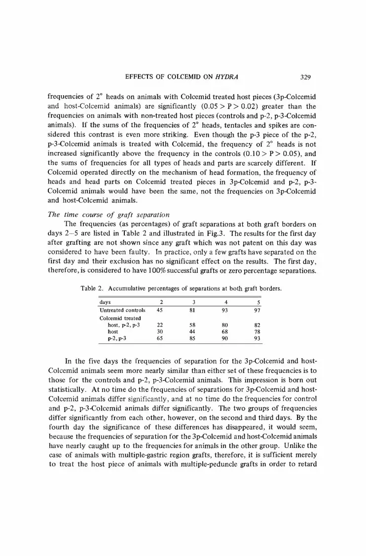
frequencies of 2" heads on animals with Colcemid treated host pieces (3p-Colcemid and host-Colcemid animals) are significantly (0.05 > P > 0.02) greater than the frequencies on animals with non-treated host pieces (controls and p-2, p-3-Colcemid animals). If the sums of the frequencies of 2" heads, tentacles and spikes are con- sidered this contrast is even more striking. Even though the p-3 piece of the p-2, p-3-Colcemid animals is treated with Colcemid, the frequency of 2" heads is not increased significantly above the frequency in the controls (0.10 > P > 0.05), and the sums of frequencies for all types of heads and parts are scarcely different. If Colcemid operated directly on the mechanism of head formation, the frequency of heads and head parts on Colcemid treated pieces in 3p-Colcemid and p-2, p-3- Colcemid animals would have been the same, not the frequencies on 3p-Colcemid and host-Colcemid animals.
The time course of graft separation The frequencies (as percentages) of graft separations at both graft borders on
days 2-5 are listed in Table 2 and illustrated in Fig.3. The results for the first day after grafting are not shown since any graft which was not patent on this day was considered to have been faulty. In practice, only a few grafts have separated on the first day and their exclusion has no significant effect on the results. The first day, therefore, is considered to have 100% successful grafts or zero percentage separations.
Table 2. Accumulative percentages of separations at both graft borders.
days 2 3 4 5 Untreated controls 45 81 93 97 Colcemid treated
host, p-2, p-3 22 58 80 82 host 30 44 68 78 P-2, P-3 65 85 90 93
In the five days the frequencies of separation for the 3p-Colcemid and host- Colcemid animals seem more nearly similar than either set of these frequencies is to those for the controls and p-2, p-3-Colcemid animals. This impression is born out statistically. A t no time do the frequencies of separations for 3p-Colcemid and host- Colcemid animals differ significantly, and at no time do the frequencies for control and p-2, p-3-Colcemid animals differ significantly. The two groups of frequencies differ significantly from each other, however, on the second and third days. By the fourth day the significance of these differences has disappeared, it would seem, because the frequencies of separation for the 3p-Colcemid and host-Colcemid animals have nearly caught up to the frequencies for animals in the other group. Unlike the case of animals with multiple-gastric region grafts, therefore, it is sufficient merely to treat the host piece of animals with multiple-peduncle grafts in order to retard

330 S. SHOSTAK
. - -0
1 2 3 4 5 D A Y S
Fig.3. Percentages of graft separations as a function of days after grafting for animals of different types. Controls: open circles and dotted line. Colcemid treated: closed symbols; 3pColcemid animals, circles and solid lines; host-Colcemid animals, squares
and long dashes; p-2, p-3-Colcemid animals, triangles and short dashes.
the rate of graft separation.
DISCUSSION
One of the main effects of Colcemid seems to be on the control system operating on head formation and originating in the distal, which is to say, the head- bearing piece. Since this piece is suspected of being the source of the hypothetical inhibitor of head regeneration (SHOSTAK, 1972, 1973, 1974a and c), Colcemid may reduce the rate of production or release of this head inhibitor, promoting head regeneration secondarily. The direct action of Colcemid on the mechanism of head formation would have to be in the opposite direction to account for the lower frequencies of 2” head formation on pieces treated with Colcemid compared to untreated pieces in hostColcemid animals. Other possibilities are not eliminated, however. Perhaps the inhibition of mitosis by Colcemid interferes with the accumu- lation of cell building blocks required for head formation, or possibly Colcemid alters nervous differentiation thereby disturbing head development.
The development of a set of tentacles and a hypostome in a hydra, and even the appellation “head” applied to these structures collectively, inevitably invites comparisons to head formation in other organisms. Although at one time it might have been thought that the induction of a head, for example, in amphibian embryos was clearly due to diffusible substances and had nothing to do with cell division, such a point of view is no longer acceptable. Several investigators have suggested that the decrease of DNA synthesis in late blastulas of developing amphibians

EFFECTS OF COLCEMID ON HYDRA 33 I
(GRAHAM and MORGAN, 1966; FLICKINGER et al., 1967) is related to the determination of the cells (GURDON, 1968; KLOSE and FLICKINGER, 1972; SUZUKI and KUWABARA, 19741, and FLICKINGER et al. (1 970) have suggested that neural or mesodermal differ- entiation can be determined by the same agents which control the length of S and G, phases of the cell cycle. A drug like Colcemid, with known anti-mitoticactivity in Hydra (TAMMARIELLO, 1969) thus could promote head formation by mimicking the anti-mitotic effects of an inductor, allowing the cells, thereby to reveal new potentials.
Recently much of the interest in hydra’s regeneration of its head was centered on the role of nerves and neurosecretory factors. Possibly, the action of Colcemid on head regeneration is mediated through nerve cells. The distribution of nerve cells (DAVID, 1973; BODE et al., 1973) and a neurosecretory product said to have head- inducing morphogenetic activity (SCHALLER, 1973 ; SCHALLER and GIERER, 1973) are parellel to a gradient in head-regenerating potential. MULLER and SPINDLER (1971), however, had earlier shown that the same sorts of activities reported to be induced by neurosecretory products are also elicited by substances released from discharging nematocysts, substances which hardly operate this way in the normal animal.
An important contribution to this controversy has recently been made by YASUGI (1 975) who obtained head regeneration from both ends of annuli cut out of the gastric regions of hydras and treated with lithium chloride. The most likely explanation for these bipolar regenerates would involve a direct effect of lithium ions on the control of head regeneration independent of induction by nerves. The accumulation of nerves at the sites of head regeneration or of bud formation (BODE et al., 1973) would then have to be seen as part of the mechanism of head formation, not part of the control system. The contention of GIERER et al. (1 972) that polarity in hydra regeneration is a function of the stable distribution of cells of particular types also has to be revaluated in light of YASUGI’S observations. It seems far more likely that lithium chloride worked on cells already present at the proximal end of the gastric region rather than on cells which had first to migrate to this end.
An explanation for the effect of Colcemid on the promotion of foot formation on treated middle peduncles is difficult to construct since Colcemid did not promote foot formation on multiple-graft animals containing three gastric regions ( SHOSTAK and ADAMS, 1975). The effect of Colcemid on peduncles would seem to be directly on the mechanism of foot formation inasmuch as only treated pieces are effected. Colchicine, however, promoted foot formation on gastric region pieces particularly in conjunction with the reversal of polarity of the pieces (CORFF, and BURNETT, 1969; CORFF, 1973; SHOSTAK and ADAMS, 1975), indicative of an indirect effect.
Colcemid’s ability to prolong the life of graft borders in 3g animals (SHOSTAK and ADAMS, 1975) suggests that graft rejection is under the control of dividing cells. If dividing cells produce a promoter of graft separation, the anti-mitotic activity of

332 S. SHOSTAK
Colcemid could effect a decrease in the amount of this promoter. Were this amount to fall below a threshold of tissue sensitivity, the rate of graft separation would be retarded. The present results on 3p animals are consistent with this interpretation, but in one situation, contrast sharply with the results on 3g animals: Colcemid retards the rate of breakdown at graft borders in 3g animals only when all three gastric regions are treated, but in 3p animals merely treating the host piece is sufficient to retard this rate.
This contrast may be attributable to differences in the distribution of dividing cells on animals of the two graft types. Mitotic activity is uniform throughout normal gastric regions (CAMPBELL, 1967; DAVID and CAMPBELL, 1972), but not throughout peduncles where it diminishes toward the foot (CORFF, 1973). Thus, 3g animals have a relatively uniform mitotic rate over the graft and host pieces, but 3p animals have the preponderance of their dividing cells on their host pieces. Dampen- ing mitotic activity about one third by Colcemid-treatment of one piece of a 3g animal might have been insufficient to diminish the amount of promoter of graft rejection below the tissue’s threshold of sensitivity. Eliminating mitotic activity by Colcemid-treatment of all pieces of 3g and 3p animals, or reducing the greatest part of mitotic activity by treating just the host piece of 3p animals, might have been sufficient to cause a large enough decline in the amount of this promoter to result in prolonging the life of the graft borders.
Some similarities in the mechanism and control of bud detachment and graft rejection suggest that the promoter of graft separation might also operate in bud detachment. Morphologically, the two processes are similar, depending on the formation of a waist or cleft between the separating pieces or parent and bud. Both processes are coupled to the formation of a foot, and while buds detach from the vicinity of the peduncle, graft separation occurs with a higher frequency between host and graft peduncles than between gastric regions (SHOSTAK, 1974b). Most importantly, the separation of a bud is inhibited (SHOSTAK and TAMMARIELLO, 1969), and the rejection of a graft retarded by treatment with Colcemid. A rapidly budding hydra utilizes budding as the chief method for removing excess cells produced following feeding (BRIEN, 1953; CAMPBELL, 1965, 1967; SHOSTAK and KANKEL, 1967; SHOSTAK, 1974a, c). When animals are experimentally elongated by the grafting of additional tissue, the rejection of this excess tissue may be controlled in the same way that the rejection of excess cells in buds is controlled. If this is, indeed, correct, multiple-graft animals will be an important new system for studying how a hydra regulates the rate at which its cells are normally rejected.
REFERENCES
BISHOFF, R. and H. HOLTZER, 1968. J . Cell Biol., 36, 111-127. BODE, H., S. BERKING, C. N . DAVID, A. GIERER, H. SCHALLER, and E. TRENKNER, 1973.

EFFECTS OF COLCEMID ON HYDRA 333
Roux’ Arch., 171, 269-285. BRAVERMAN, M., 1974. Oceanogr. Mar. Biol. Ann. Rev., 12, 129-221. BRIEN, P., 1953. Biol. Rev., 28, 308-349. BRINKLEY, B. R., E. STUBBLEFIELD, and T. C. HSU, 1967. J. Ultrastruct. Res. 19, 1-18. CAMPBELL, R. D., 1965. Science, 148, 1231-1232,
, 1967. Develop. Biol., 15, 487-502. CORFF, S. C., 1973. Organismal growth and the contribution of cell proliferation to net growth
and maintenance of form. In “Biology of hydra” (ed. A. L. Burnett), Academic Press. 346-393.
and A. L. BURNETT, 1969. J. Embryol. Exp. Morphol., 21, 417-443.
and R. D. CAMPBELL, 1972. J. Cell Sci., 11, 557-586.
and S. J. COWARD, 1962. Develop. Biol., 5, 179-204. , M. L. FREEMAN and P. J. STAMBROOK, 1967. Develop. Biol., 16, 457-473. , D. M. KOHL, M. R. LAUTH and P. J. STAMBROOK, 1970. Biochim. Biophys. Acta.,
DAVID, C. N., 1973. Roux’ Arch., 171, 259-268.
FLICKINGER, R. A., 1959. Growth, 23, 251-271.
209, 260-262.
E. TRENKNER, 1972. Nature, New Biol., 239, 98-101. GIERER, A. , S. BERKING, H. BODE, C. N. DAVID, K. FLICK, G. HANSMANN, H. SCHALLERand
GRAHAM, C. R. and R. W. MORGAN, 1966. Develop. Biol., 14, 439-460. GURDON, J., 1968. Essays Biochem., 4, 26-68. HOLLYFIELD, J . G., 1968. Develop. Biol., 18, 163-179. ISHIKAWA, H., 1968. J. Cell Biol., 38, 538-555. KLEINFELD, R. G. and J. E. SISKEN, 1966. J. Cell Biol., 31, 369-379. KLOSE, J. and R. A. FLICKINGER, 1972. Develop. Biol., 29, 214-219. MULLER, W. A. and K. D. SPINDLER, 1971. Roux’ Arch., 167, 325-335. SCHALLER, H. C., 1973. J . Embryol. Exp. Morph., 29, 27-38. ~- and A. GIERER, 1973. J. Embryol. Exp. Morph., 29, 39-52. ~ C H A R , B., P. LOUSTALOT and F. GROSS, 1954. Klin. Wschr., 32, 49-57. SHOSTAK, S., 1972. Develop. Biol., 28, 620-635. _ _ _ ~ , 1973. J. Embryol. Exp. Morph., 29, 311-330.
, 1974a. Am. Zoologist, 14, 619-632. , 1974b. Graft rejection and the regulation of length in Hydru viridis. In “Contemporary
, 1974c. Quart. Rev. Biol., 49, 287-310. and D. R. KANKEL, 1967. Develop. Biol., 15, 451-463.
___ and R. V. TAMMARIELLO, 1969. Nat. Cancer Instit. Monogr. (U.S.A.),31, 739-750. and J. A. ADAMS, 1975. J. Exp. Zool., 193, 43-56.
SUZUKI, A. and K. KUWABARA, 1974. Develop., Growth and Differ., 16, 29-40. TAMMARIELLO, R. V., 1969. The action of Colcemid in causing bud retention in Hydra viridis.
TARDENT, P. E., 1954. Roux’ Arch., 146,593-649. WEBSTER,G., 1967. J. Embryol. Exp. Morph., 18,181-197. YASUGI, S., 1975. Develop., Growth and Differ., 16, 171-180. ZIMMERMAN, A. M. and s. ZIMMERMAN, 1967. J. Cell Biol., 34, 483-488. ZWILLING, E., 1939. Biol. Bull., 76, 90-103.
topics in immunobiology,” Vol. 4 (ed. E. L. Cooper), Plenum Press. 127-139.
Ph. D. thesis in Biology. University of Pittsburgh, Pittsburgh, Pa.
(Received: April 4, 1975) (Revised version received: June 16, 1975)


