Embed Size (px)
Citation preview

Université de Grenoble __________________
MEMOIRE
pour l'obtention du Diplôme de Docteur en Médecine
__________________
Présenté et soutenu le 1er septembre 2011
par
BAILLET Athan
__________________
Intérêt de l’activité physique aérobie et du renfor cement musculaire dans la polyarthrite rhumatoïde : analyses systématiques de la littérature et méta-
analyses d’essais randomisés contrôlés
__________________
JURY
Pr. Robert JUVIN Pr. Dominique SARAGAGLIA
Pr. Philippe GAUDIN Dr. Michel GUINOT
Mémoire réalisé sous la direction du Pr Philippe GAUDIN
Clinique universitaire de Rhumatologie
Pôle Locomotion Rééducation et Physiologie
CHU Hôpital Sud Av. de Kimberley BP 338

Sommaire
1
SOMMAIRE
REMERCIEMENTS………………..………………………………………….….......................... 2
ABBREVIATIONS ………………………………………………………………………………… 4
INTRODUCTION………………..………………………………………….…............................... 5
PARTIE I : Intérêt de l’activité physique aérobie dans la polyarthrite rhumatoïde : Analyse systématique de la littérature et méta-analyse d’essais randomisés contrôlés....…. 6
SYNOPSIS………………………………………………………………….................................... 7
ARTICLE 1 : Baillet A, Zeboulon N, Gossec L et al. Efficacy of cardiorespiratory aerobic exercise in rheumatoid arthritis : meta-analysis of randomized controlled trials. Arthritis Care Res (Hoboken) 2010;62:984-92…..................................................................... 9
PARTIE II : Intérêt du renforcement musculaire dans la polyarthrite rhumatoïde : Analyse systématique de la littérature et méta-analyse d’essais randomisés contrôlés…… 18
SYNOPSIS………………………………………………………………….................................... 19
ARTICLE 2 : Baillet A, Vaillant M, Guinot M et al. Efficacy of resistance exercises in Rheumatoid Arthritis : Meta-analysis of randomized controlled trials. Rheumatology (Oxford) 2011; en révision…………………………………….………………… 21
DISCUSSION…………….……………………………………………………………………….… 42
CONCLUSION…………….………………………………………………………………………. 45
BIBLIOGRAPHIE…………….…………………………….………………………………….…… 46
ANNEXE 1 : Baillet A, Payraud E, Niderprim VA et al. A dynamic exercise programme to improve patients' disability in rheumatoid arthritis: a prospective randomized controlled trial. Rheumatology (Oxford) 2009;48:410-15……………………..……..………… 55
ANNEXE 2 : Résultats supplémentaires publiés en ligne pour l’article 1…………………... 61
ANNEXE 3 : Résultats supplémentaires publiés en ligne pour l’article 2………….……….. 67
SERMENT D’HIPPOCRATE………………………………………………….………….……….. 73

Remerciements
2
REMERCIEMENTS
Je remercie les membres du jury de thèse :
- Je tiens à exprimer ma très vive reconnaissance envers le Pr Robert Juvin pour
avoir accepté de présider ce jury. Peu de médecins m’ont inspiré un si grand
respect.
- Je désire remercier le Pr Philippe Gaudin pour son chaleureux soutien et pour la
qualité de son encadrement. « C'est le rôle essentiel du professeur d'éveiller la joie
de travailler et de connaître », Albert Einstein
- Merci au Pr Dominique Saragaglia et au Dr Michel Guinot d’avoir accepté de
juger mon travail.
Merci également au Pr Maxime Dougados et au Dr Laure Gossec pour leur
enseignement en épidémiologie clinique et pour m’avoir guidé dans les méandres de
l’analyse systématique de la littérature.
Bien entendu mes remerciements vont également à mes proches sans qui je n’aurais pas
pu réaliser ce travail :
Merci à mes fils, Antoine et Amaury , sources inépuisables de motivation et de
satisfaction. Un seul de vos sourires apaise toutes mes angoisses. Vous avez appris être
sages et à supporter les absences de votre père.
Merci à ma splendide épouse Cécile. Je ne mesure probablement pas suffisamment ton
apport dans l’accomplissement mon travail.
Grâce soit rendue à mes parents Alain et Christiane ainsi qu’à mon frère Axel ,
indéfectibles soutiens. Cette thèse est un peu la votre…
Je remercie également mes plus proches amis avec qui je partage plus de 30 ans d’éclats
de rire. Merci à vous Jérémy , Olivier et Joub .
Merci à ma tante Yolande

Abbreviations
3
ABBREVIATIONS
CLEAR-NPT = A Checklist to Evaluate a Non-Pharmacological Trial
DAS 28 = Disease Activity Score based on 28 joints
ES = Effect Size
EVA = Echelle Visuelle Analogique
HAQ = Health Assessment Questionnaire
IGF-1 = Insulin-like Growth Factor 1
IL-6 = Interleukine 6
OR = Odds Ratio
PR = Polyarthrite Rhumatoïde
RR = Risque Relatif
SDD = Smallest Dectectable Change
SMD = Standardized Mean Difference
WMD = Weighted Mean Difference
MCID = Minimally Clinically Important Difference

Introduction
4
INTRODUCTION
La polyarthrite rhumatoïde (PR) est une pathologie multifactorielle, avec un terrain
génétique [1] particulier impliquant des facteurs environnementaux tels que le tabac [2, 3]
et la flore microbienne [4], qui touche 0,4% de la population caucasienne [5]. Les
traitements immuno-modulateurs et les thérapeutiques ciblées ont largement contribué à
l’amélioration de la prise en charge des patients, permettant de diminuer l’activité de la
maladie [6], d’améliorer le handicap [7], la qualité de vie [6] et l’évolution structurale [6, 8-
10], mais au prix des effets indésirables [11] parfois sévères [12, 13] et avec un coût
important sur l’économie de santé [14, 15].
L'activité physique, définie comme un « mouvement du corps produit par un ou des
groupes musculaires à l’origine d’une dépense énergétique » [16] regroupe à la fois
l'exercice physique de la vie quotidienne (déplacements, travail, taches ménagères…),
l'activité physique de loisirs, et la pratique sportive qui est un "sous-ensemble spécialisé et
organisé de l'activité physique". On distingue généralement trois types d’activités
sportives : l’exercice aérobie, le renforcement musculaire et les assouplissements. Pendant
longtemps, l’exercice physique était suspecté d’augmenter l’activité des rhumatismes
inflammatoires [17] et de provoquer des lésions structurales en élevant la pression intra-
articulaire [18] responsable d’une baisse de la perfusion dans le pannus [19]. Il s’ensuivrait
des phénomènes d’ischémie-reperfusion localisés à l’origine d’une protéolyse matricielle
[20]. Cette destruction articulaire est, au moins en partie, secondaire à une production
accrue de dérivés radicalaire de l’oxygène [21-23]. Il faut attendre les années 90 pour que
des études néerlandaises et scandinaves suggèrent une relative innocuité de la pratique
de l’activité physique dans la PR stable [24-30].
Dans une étude récente nous avons montré qu’un programme d’exercices dynamiques
améliore la fonction des patients atteints de PR (Annexe 1 , [31]), allant à l’encontre des
résultats d’études publiées auparavant [30, 32]. Les articles de revue précédents [33, 34],

Introduction
5
n’ayant pas clairement permis de savoir si l’exercice physique était bénéfique pour les
patients atteints de PR, nous avons conduit une analyse systématique de la littérature afin
de savoir si l’exercice aérobie d’une part et le renforcement musculaire d’autre part
amélioraient les principaux critères d’évaluation de la PR. Nous avons effectué une méta-
analyse afin d’aboutir à des conclusions chiffrées, de dégager la pertinence clinique de
l’efficacité de l’intervention physique et de chercher un facteur d’hétérogénéité expliquant
les différences entre les études.
Dans une première partie, nous nous intéresserons à l’impact de l’exercice aérobie dans la
PR. Cette étude a été présentée en communication orale au congrès européen de
rhumatologie (European League Against Rheumatism) en 2009 à Copenhague et publiée
dans la revue Arthritis Care and Research en 2010 [35]. Dans une deuxième partie, nous
étudierons l’intérêt du renforcement musculaire dans la PR. Ce travail a été soumis en
mars 2011 pour publication au journal Rheumatology [36].

Partie I : entrainement aérobie et polyarthrite rhumatoïde
6
PARTIE I
Intérêt de l’activité physique aérobie dans la
polyarthrite rhumatoïde : Analyse systématique de l a
littérature et méta-analyse d’essais randomisés
contrôlés

Partie I : entrainement aérobie et polyarthrite rhumatoïde
7
PARTIE I : SYNOPSIS DU PREMIER ARTICLE
Introduction :
Il est déjà établi que la force de préhension et la capacité aérobie sont améliorées chez les
patients atteints de PR participant aux programmes de rééducation dynamique. L'objectif
de cette première étude était de déterminer l'effet de l’activité physique aérobie sur la
fonction, la qualité de vie, la douleur, l'activité de la maladie et la progression radiologique
des patients atteints de PR.
Patients et Méthodes :
Tous les essais randomisés contrôlés évaluant l'activité physique ont été
systématiquement recherchés dans les bases de données Medline, Cochrane et EMBase
en juillet 2009. Le groupe intervention devait bénéficier d'exercices aérobies alors que le
groupe contrôle était soumis à des exercices isométriques ou à une absence d'exercice. La
variation des données suivantes a été étudiée : la fonction sur le Health Assessment
Questionnaire (HAQ), la qualité de vie, l’échelle visuelle analogique douleur (EVA), l’activité
de la maladie sur le Disease Activity Score (DAS28), le compte articulaire et l’indice
radiologique de Larsen. La tolérance était évaluée par le nombre de patient qui achevaient
l’étude et le nombre d’effets indésirables. La qualité méthodologique de chaque étude était
analysée sur le score de Jadad (0-5) et CLEAR-NPT (A Checklist to Evaluate a Non-
Pharmacological Trial ; 0-14).
Une méta-analyse par le modèle de l’inverse de la variance a été effectuée. Les résultats
ont été exprimés au moyen de la différence des moyennes standardisées (Standardized
mean difference SMD) pour les variables quantitatives et en odds ratio (OR) pour les
variables qualitatives ainsi que leurs intervalles de confiance à 95% (IC95%). Les SMD et
les OR ont été agrégés en comparant le groupe actif au groupe contrôle. Une SMD>0.20
est considérée comme modeste et >0.40 comme modérée. Une analyse de sous-groupe a
été conduite pour identifier un facteur d’hétérogénéité entre les études. Un biais de
publication a été recherché par analyse graphique et par les tests statistiques de Begg et
Egger.

Partie I : entrainement aérobie et polyarthrite rhumatoïde
8
Résultats :
14 essais randomisés contrôlés ont été analysés (soit 1040 patients). Le score moyen de
Jadad était 2,4 ± 0,4 alors que seulement 2 articles validaient au moins 7 items du CLEAR-
NPT. L’exercice aérobie améliorait la fonction évaluée par le HAQ (SMD = 0,24, IC95%
[0,10, 0,38], p = 0,0009), la qualité de vie (SMD = 0,39, IC95% [0,23, 0,56], p < 0,0001),
l’EVA douleur (SMD = 0,31, IC95% [0,06, 0,55], p = 0,02). L'exercice physique n'avait pas
d'effet délétère sur l'activité de la maladie mesurée par le DAS 28 (SMD = -0,16 [-0,47,
0,15], p = 0,34), le compte articulaire (SMD = 0,14, IC95% [-0,05, 0,33], p = 0,14) et
semble même avoir un effet positif sur le score radiologique de Larsen (SMD = 0,36,
IC95% [0,16, 0,56], p = 0,0005). Le groupe qui bénéficiait d’exercices aérobies comptait le
même nombre de patients achevant l’étude que le groupe contrôle (respectivement 86,8%
et 87,3% ; OR = 0,80, IC95% [0,56, 1,16], p = 0,24). De même, il n’existait pas de
différence de tolérance entre le groupe intervention et le groupe contrôle (respectivement
47 (9,2%) et 34 (6,4%) évènements indésirables ; OR=1,67, IC95% [0,36, 7,69], p = 0,51).
La supervision des exercices améliorait l’effet positif sur la qualité de vie. On notait une
hétérogénéité des résultats en fonction de la qualité méthodologique des études. Un biais
de publication pour l’effet sur le HAQ était possible.
Discussion :
Nous avons pu démontrer un impact favorable de l'exercice aérobie sur le HAQ, la qualité
de vie, l'évolution structurale et la douleur sans effet péjoratif sur le compte articulaire ou
l'activité de la maladie. On notait une certaine hétérogénéité entre ces études dont la
qualité méthodologique était moyenne.
Conclusion :
Ces résultats plaident pour une augmentation de prescription des activités physiques
aérobies pour les patients atteints de PR.

Efficacy of Cardiorespiratory Aerobic Exercise inRheumatoid Arthritis: Meta-Analysis ofRandomized Controlled TrialsATHAN BAILLET,1 NADINE ZEBOULON,2 LAURE GOSSEC,2 CHRISTOPHE COMBESCURE,3
LOUIS-ANTOINE BODIN,4 ROBERT JUVIN,5 MAXIME DOUGADOS,2 AND PHILIPPE GAUDIN1
Objective. Several lines of evidence have emphasized an improvement in aerobic capacity and muscle strength afterphysical exercise programs in rheumatoid arthritis (RA) patients. Our objective was to evaluate the efficacy of aerobicexercises in RA on quality of life, function, and clinical and radiologic outcomes by a systematic literature review and ameta-analysis.Methods. A systematic literature search was performed in the Medline, EMBase, and Cochrane databases up to July 2009and in the abstracts presented at rheumatology scientific meetings during the last 5 years. Randomized controlled trials(RCTs) comparing aerobic exercises with non-aerobic interventions in RA patients were included. Outcomes studied werepostintervention quality of life, function assessed by the Health Assessment Questionnaire (HAQ), a pain visual analogscale (VAS), joint count, the Disease Activity Score in 28 joints (DAS28), and radiologic damage. Efficacy was assessed bystandardized mean differences (SMDs; difference between groups of mean outcome variation from baseline/SD atbaseline) of aerobic exercises versus non-aerobic rehabilitation. Heterogeneity was tested. SMDs were pooled by ameta-analysis using the inverse of variance model.Results. Fourteen RCTs, including 1,040 patients, met the inclusion criteria. Exercise improved the postinterventionquality of life (SMD 0.39, P < 0.0001), HAQ score (SMD 0.24, P ! 0.0009), and pain VAS (SMD 0.31, P ! 0.02). Exercisein this RA population appeared safe, since global compliance, DAS28, and joint count were similar in both groups.Conclusion. Cardiorespiratory aerobic conditioning in stable RA appears to be safe and improves some of the mostimportant outcome measures. However, the degree of the effect of aerobic exercise on the abovementioned parameters issmall.
INTRODUCTION
Impairment in range of motion, muscle strength, endur-ance, and aerobic fitness results in serious loss of function,
work disability, dependency, impaired social or familyfunction, and reduced quality of life in rheumatoid arthri-tis (RA) patients (1). Even if pharmacologic interventionshave largely improved RA management over the past de-cade, physical therapy remains an important part of treat-ment. Previously, the exercise therapy in RA aimed only atmaintaining joint mobility and muscle strength. Becauseexercises were thought to provoke joint damage by en-hancing disease activity (2), patients with inflammatoryarthritis, especially RA, were discouraged from performingweight-bearing exercises. In a previous randomized con-trolled trial (RCT), we showed that an exercise programhad a greater impact on both disability assessed by theHealth Assessment Questionnaire (HAQ) and quality oflife than conventional joint rehabilitation (3), contrastingwith most of the previous similar studies that failed todetect any statistical difference on the HAQ (4,5) or qualityof life (6). Several trials showed that exercises were safe inRA rehabilitation and effectively improved aerobic fitness(3,5,7,8). Previous systematic literature reviews of the ef-ficacy of physical exercises in RA patients confirmed theseresults (9,10). Given the limited number of studies, review-
Supported by an unrestricted educational grant from Ab-bott France.
1Athan Baillet, MS, Philippe Gaudin, MD, PhD: Univer-sity of Grenoble Medical School, Centre Hospitalier Univer-sitaire, Hopital Sud, and GREPI CNRS UMR 5525, Grenoble,France; 2Nadine Zeboulon, MD, Laure Gossec, MD, PhD,Maxime Dougados, MD: Paris Descartes University MedicalSchool, UPRES-EA 4058, Assistance Publique Hopitaux deParis, Cochin Hospital, Paris, France; 3Christophe Comb-escure, PhD: University of Geneva, Geneva, Switzerland;4Louis-Antoine Bodin, MD: Pierre and Marie Curie Univer-sity Medical School, Paris, France; 5Robert Juvin, MD, PhD:University of Grenoble Medical School, Centre HospitalierUniversitaire, Hopital Sud, Grenoble, France.
Address correspondence to Athan Baillet, MS, Clinic ofRheumatology, Hopital Sud, Grenoble Teaching Hospital,Avenue de Kimberley, BP 338, 38434 Echirolles Cedex,France. E-mail: [email protected].
Submitted for publication August 8, 2009; accepted inrevised form February 9, 2010.
Arthritis Care & ResearchVol. 62, No. 7, July 2010, pp 984–992DOI 10.1002/acr.20146© 2010, American College of Rheumatology
ORIGINAL ARTICLE
984

ers decided not to pool data and were therefore unable todraw numerical conclusions concerning the efficacy ofexercise programs on other important outcome measuresin RA. During the past few years, several trials have beencarried out to examine the effect of exercises in RA, butresults with respect to pain, disease activity, functionalability, quality of life, and structural damage are still un-clear. We therefore carried out this systematic literaturereview in order to determine whether aerobic exerciseseffectively improve the abovementioned parameters inRA.
MATERIALS AND METHODS
Type of intervention. A cardiorespiratory aerobic exer-cise is an exercise that improves VO2 and is usually per-formed at 50–80% of the maximal heart rate (220 ! age).The American College of Sports Medicine defined a dy-namic exercise program as aerobic exercises performed atbetween 60% and 80% of the maximal heart rate at leasttwice a week for 6 weeks (11). Strengthening exercises andaerobic exercises without training intensity (i.e., maximalheart rate) monitoring were not examined in this system-atic review.
Non-aerobic rehabilitation was defined by the followingterms: “static,” “range of motion,” “isometric,” “seatedimmersion,” “relaxation,” “stretching,” “no attention,” or“usual care.” As a consequence, trials with aerobic exer-cises in the control group were not considered.
Search strategy. An extensive search of PubMed, EM-Base, and the Cochrane Central Register of RCTs (until July28, 2009) was made by two reviewers (AB and L-AB). Thefollowing keywords were used for database screening:(“Arthritis, Rheumatoid” [medical subject headings(MeSH)]) AND (“Exercise Therapy” [MeSH] OR “Activitiesof Daily Living” [MeSH] OR “Physical Education andTraining” [MeSH]). The only limit of the search was “clin-ical trial.” A hand search of references from relevant arti-cles, from review articles, and from abstracts presented atthe Annual Scientific Meetings of the American College ofRheumatology (ACR), the Annual Congress of the Euro-pean League Against Rheumatism, and the Scientific Meet-ings of the French Society of Rheumatology published inthe past 5 years completed the search. A search on theClinicalTrials.gov Web site was also performed to identifyrandomized studies that were not yet published.
Selection. Inclusion criteria were an RCT evaluatingcardiorespiratory aerobic exercises performed at 50–90%of the maximal heart rate in comparison with non-aerobicrehabilitation in adult patients with RA as defined by theACR (formerly the American Rheumatism Association)criteria (12,13). Exclusion criteria consisted of postsurgeryrehabilitation and articles written in a language other thanEnglish, French, or German.
Quality assessment. A single author (AB) assessed themethodologic quality of each study included in the meta-
analysis on both the Jadad scale (14) and the Checklist toEvaluate A Report of a Non-Pharmacological Trial(CLEAR-NPT) (15), ranging from 0–5 and 0–14, respec-tively, where a high score indicates high quality.
Data extraction. One investigator (AB) selected the ar-ticles and collected the data using a predetermined form,including study design (randomization procedure, blind-ing, followup period, and intent-to-treat analysis), patientcharacteristics (number, age, sex, disease duration, func-tional status, treatment, rate of rheumatoid factor [RF]–positive patients, and proportion of completers), and in-tervention parameters (duration of an exercise session,overall duration of the intervention, exercise type, fre-quency, and intensity, i.e., maximal heart rate).
Outcomes. We used the Cochrane MusculoskeletalGroup recommendation to select outcome measures (16).We reported tender joint count, swollen joint count, pain,and disability. We also reported data concerning with-drawals (i.e., the number of completers) and adverseeffects in order to assess exercise safety. The followingoutcomes (the references of which are available in Supple-mentary Table 1, available in the online version of thisarticle at http://www3.interscience.wiley.com/journal/77005015/home) were extracted from the publications:1) quality of life, evaluated on the Nottingham HealthProfile (NHP), the Rheumatoid Arthritis Quality of Life(RAQol) questionnaire, the physical component of the 36-item Short Form health survey (SF-36), the Arthritis Im-pact Measurement Scales Health Status Questionnaire(AIMS), and the McMaster Toronto Arthritis Patient Pref-erence Disability Questionnaire (MACTAR); 2) function,assessed by the HAQ; 3) the Disease Activity Score in 28joints (DAS28); the DAS4 was converted into the DAS28 asfollow: DAS28 " (1.072 # DAS4) $ 0.938; 4) joint count(number of swollen joints, number of tender joints, andRitchie Articular Index); 5) pain on a visual analog scale(VAS); 6) radiologic evaluation by Larsen’s method; and7) exercise tolerance, evaluated both by exercise com-pliance (i.e., the number of completers) and by exercisesafety. For the latter parameter, we reported adverse effectssuch as steroid injection due to local disease flare, cardio-vascular events, and joint/muscle soreness.
Quality of life and joint count evaluations were ex-pressed as a percentage of the maximum possible score forthe method used. Standardized data abstraction concernedmeans or medians and measures of dispersion. Since themeasure of dispersion for change was not always available,a conservative estimate was taken from baseline data andconverted into an SD. When median values were giveninstead of mean values, the median was analyzed as amean. Data were collected at several end points: baseline,1 month % 2 weeks, 3 months % 4 weeks, 6 months % 2months, 1 year, and 2 years or more. When a trial wasreported in several publications, the more informativepublication was included in the meta-analysis. Whenstudies reported more than 2 subgroups, we included onlythe first intervention group described in this study and itscorresponding control group.
Meta-Analysis of Cardiorespiratory Aerobic Exercise in RA 985

Statistical analysis. Heterogeneity was tested usingCochran’s test. I2 values &50% were defined to indicatesignificant heterogeneity. The efficacy of intervention ver-sus non-aerobic rehabilitation was assessed in each studyby the calculation of the standardized mean difference(SMD; difference between both groups of mean outcomevariation from baseline/SD at baseline) and 95% confi-dence interval (95% CI). Individual SMDs were pooledusing the method of the inverse of variance. Interventionsafety was assessed by the odds ratio (OR) and 95% CI. Theresults of individual trials were pooled by meta-analysisusing the Mantel-Haenszel method. In case of heterogene-ity, a random-effects model was used (17). Otherwise, afixed-effects model was applied. SMDs between 0.2 and0.5 indicated a small effect, between 0.5 and 0.8 indicateda medium effect, and &0.8 indicated a large effect. Torelate the efficacy in SMD units to a more familiar out-come, we transformed the SMD into the difference in meanoutcome scores (experimental versus control) on that scale(18). Meta-analyses were done with Review Manager 5(Cochrane), and additional statistics were developed withStatsDirect software (StatsDirect).
Sensitivity analysis and heterogeneity assessment. Asensitivity analysis was conducted to evaluate the robust-ness of the meta-analysis by assessing the influence of anindividual study on the overall SMD. We therefore exam-ined the effect of removing each study individually fromthe meta-analysis. To explore heterogeneity, we combinedstudies into 2 or 3 subgroups according to the trial design(published before or after 2000, Jadad scale score '3 or!3), the disease characteristics (mean disease duration '5years or !5 years, functional status class I–II or includingclass III), and the intervention parameters (supervised orhome based; overall duration of the intervention '3, 3, or&3 months; exercise frequency '3 or !3 times/week; du-ration of an exercise session '30, 30–60, or &60 minutes).Heterogeneity between the subgroups was tested using achi-square test (19).
Publication bias was assessed using funnel plot analysis,Begg’s test, and Egger’s test.
RESULTS
Trial flow. A total of 513 abstracts were identified bydatabase searching and 6 articles by hand searching. Ofthese, 140 duplicates were removed and 298 abstractswere excluded because of no physical exercise (n " 95), noRA patients (n " 171), age '18 years (n " 13), postsurgeryrehabilitation (n " 9), no RCT (n " 9), and language (n "
1). Eighty-one full-text reports were analyzed and 67 arti-cles were excluded because of previous publication (n "
7), other joint disease (n " 18), no RCT or aerobic exercisein the control group (n " 14), no aerobic exercise in theintervention group (n " 11), training intensity not pro-vided (n " 5), and no extractable data (n " 12). Fourteenarticles were finally entered in the analysis.
Study characteristics. Study characteristics are shownin Table 1. The mean % SD Jadad scale score was 2.4 % 0.6.
Seven trials displayed a Jadad scale score '3 and only 2trials validated 7 CLEAR-NPT items. Among the 14 trialswith cardiorespiratory aerobic exercise conditioning, theintervention fulfilled the dynamic exercise program crite-ria in 5 studies (5,8,20–22). The control group underwentrange of motion exercises in 3 studies (5,22,23), non-aero-bic exercises in 1 study (24), education programs in 2studies (3,25), and usual care in 8 studies (8,20,21,26–30).
Patients’ characteristics. The meta-analysis included1,040 patients (510 patients in the intervention group and530 in the control group). Both groups were similar interms of age, disease duration, sex ratio, proportion ofcompleters, rate of RF-positive patients, and pharmaceuti-cal treatments (see Supplementary Table 2, available in theonline version of this article at http://www3.interscience.wiley.com/journal/77005015/home). Mean age and dis-ease duration in the studies ranged from 44–68 years and1–16 years, respectively (in the 13 trials reporting theseparameters). For the 11 studies for which sex was reported,46.7–100% were women. The rate of RF-positive patientsranged from 59–93.3% in the 5 studies assessing thisparameter.
Meta-analysis. Data on quality of life were available for5 studies, including 298 patients in the intervention groupand 288 in the control group. This outcome was measuredby the AIMS (26), the NHP (3), the RAQol (25), the SF-36(24), or the MACTAR (20) (see Supplementary Table 3,available in the online version of this article at http://www3.interscience.wiley.com/journal/77005015/home).A small beneficial effect of aerobic intervention on thequality of life of RA patients was shown (SMD 0.39 [95%CI 0.23, 0.56], P ' 0.0001) (Figure 1). Heterogeneity wasnot statistically significant in this subset of studies (I2 "
45%).Nine studies evaluated the impact of aerobic exercises
on the HAQ, comprising a total of 387 patients in theintervention group and 384 patients in the control group(Figure 2). Exercises provided a small positive effect on theHAQ (SMD 0.24 [95% CI 0.10, 0.38], P " 0.0009; I2 "
29%).Data on the pain VAS were available in 6 studies (138
patients in the intervention group and 123 in the controlgroup). The overall SMD for pain measured by the VASwas 0.31 (95% CI 0.06, 0.55; P " 0.02, I2 " 30%) (Figure 3).
Nine studies, including 228 patients in the interventiongroup and 209 patients in the control group, evaluatedjoint count. The Ritchie Articular Index was reported in 2trials (5,23), swollen joint count in 2 studies (21,27), ten-der joint count in 2 studies (26,28), and both swollen andtender joint counts in 3 studies (8,22,29) (see Supplemen-tary Table 3, available in the online version of this article athttp://www3.interscience.wiley.com/journal/77005015/home). Intervention tended to provide a positive effect onthis outcome, although the difference did not reachthe statistical significance (SMD 0.14 [95% CI !0.05, 0.33],P " 0.14; I2 " 0%) (Figure 4).
Only 3 studies evaluating 376 RA patients for radiologicdamage as assessed by radiologic findings were available
986 Baillet et al

Table
1.
Tri
al
ch
ara
cte
rist
ics*
Au
thor,
year
(ref.
)C
ou
ntr
y
Jad
ad
scale
score
Fu
ncti
on
statu
s
Inte
rven
tion
gro
up
Con
trol
gro
up
Pati
en
ts,
no.
Exerc
ise
typ
e
Max
HR
,%
Fre
qu
en
cy,
per
week
Du
rati
on
,
min
ute
s
Len
gth
,
week
s
Att
en
dan
ce,
%
Pati
en
ts,
no.
Com
para
tor
Bil
berg
et
al,
2005
(24)
Sw
ed
en
3S
tein
bro
cker
I–II
I
22
Su
perv
ised
card
iore
spir
ato
ry
aero
bic
con
dit
ion
ing
70
245
12
78
27
Non
-aero
bic
exerc
ises
Van
den
Berg
et
al,
2006
(25)
Th
e Neth
erl
an
ds
3n
r82
Hom
e-b
ase
d
card
iore
spir
ato
ry
aero
bic
con
dit
ion
ing
60–80
510–30
52
34
78
Ed
ucati
on
De
Jon
get
al,
2003
(20)
Th
e Neth
erl
an
ds
3A
CR
I–II
I150
Su
perv
ised
DE
P70–90
275
104
74†
150
Usu
al
care
West
by
et
al,
2000
(8)
Can
ad
a2
AC
RI–
II14
Hom
e-b
ase
dD
EP
60–75
345–60
52
71
16
Usu
al
care
Van
den
En
de
et
al,
2000
(22)
Th
e Neth
erl
an
ds
3n
r34
Su
perv
ised
DE
P60
315
(4
nr
30
RO
M exerc
ises
Meli
koglu
et
al,
2006
(23)
Tu
rkey
2A
CR
I–II
20
Su
perv
ised
card
iore
spir
ato
ry
aero
bic
con
dit
ion
ing
60
220
2n
r20
RO
M exerc
ises
Han
sen
et
al,
1993
(27)
Den
mark
2S
tein
bro
cker
I–II
60
Hom
e-b
ase
d
card
iore
spir
ato
ry
aero
bic
con
dit
ion
ing
70
!3
'90
104
&50
15
Usu
al
care
Hark
com
et
al,
1985
(21)
US
2S
tein
bro
cker
II
14
Su
perv
ised
DE
P70
315–35
12
nr
6U
sual
care
Van
den
En
de
et
al,
1996
(5)
Th
e Neth
erl
an
ds
3n
r75
Su
perv
ised
an
dh
om
e-
base
dD
EP
s
70–85
360
12
75
25
RO
M exerc
ises
Bail
let
et
al,
2009
(3)
Fra
nce
3A
CR
I–II
25
Su
perv
ised
card
iore
spir
ato
ry
aero
bic
con
dit
ion
ing
60–80
545
4100
23
Ed
ucati
on
Neu
berg
er
et
al,
2007
(29)
US
3n
r173
Su
perv
ised
an
dh
om
e-
base
d
card
iore
spir
ato
ry
aero
bic
con
dit
ion
ing
60–80
360
12
83
75
Usu
al
care
Nord
em
ar
et
al,
1981
(30)
Sw
ed
en
1A
CR
I–II
I23
Hom
e-b
ase
d
card
iore
spir
ato
ry
aero
bic
con
dit
ion
ing
70
‡60
52
nr
23
Usu
al
care
Nore
au
et
al,
1995
(26)
Can
ad
a2
AC
RI–
II19
Su
perv
ised
card
iore
spir
ato
ry
aero
bic
con
dit
ion
ing
50–70
215–30
12
83
10
Usu
al
care
Lyn
gberg
et
al,
1994
(28)
Den
mark
2S
tein
bro
cker
I–II
I
12
Su
perv
ised
card
iore
spir
ato
ry
aero
bic
con
dit
ion
ing
50–70
245
12
nr
12
Usu
al
care
*M
ax
"m
axim
al;
HR
"h
eart
rate
;n
r"
not
rep
ort
ed
/in
ap
pli
cable
;A
CR
"A
meri
can
Coll
ege
of
Rh
eu
mato
logy;
DE
P"
dyn
am
icex
erc
ise
pro
gra
m;
RO
M"
ran
ge
of
mo
tio
n.
†T
he
med
ian
perc
en
tage
of
sess
ion
satt
en
ded
was
74%
(in
terq
uart
ile
ran
ge
27%
),30%
of
pati
en
tsh
ad
a50
–75%
att
en
dan
ce
rate
,an
d4
9%
of
pati
en
tsh
ad
ah
igh
att
en
dan
ce
rate
(&7
5%
).‡
On
eh
ou
rd
ail
y(s
up
erv
ised
)every
secon
dw
eek
plu
s30
min
ute
sd
ail
y(h
om
ebase
d),
lead
ing
toa
mean
%S
Dtr
ain
ing
tim
eof
339
%1
79
min
ute
s/w
eek
.
Meta-Analysis of Cardiorespiratory Aerobic Exercise in RA 987

(188 patients in each group). Aerobic exercise training wassuspected to have a joint damage sparing effect because theSMD was 0.36 (95% CI 0.16, 0.56; P " 0.0005, I2 " 17%)(see Supplementary Figure 1, available in the online ver-sion of this article at http://www3.interscience.wiley.com/journal/77005015/home).
Disease activity, evaluated by the DAS28, was recordedin 4 studies (291 patients in the intervention group and281 patients in the control group). The exercise group didnot display a worse DAS28 score (SMD 0.08 [95% CI!0.08, 0.25], P " 0.34; I2 " 67%).
Both groups had similar numbers of completers: 460(86.8%) of 530 patients in the intervention group versus445 (87.3%) of 510 patients in the control group (OR 0.80[95% CI 0.56, 1.16], P " 0.24; I2 " 55%). Forty-sevenadverse events were reported in the intervention groupand 34 adverse events were reported in the control group.However, the difference was not statistically significant
(OR 1.67 [95% CI 0.36, 7.69], P " 0.51; I2 " 73%). Steroidinjection in both groups was similar (OR 0.85 [95% CI0.42, 1.29], P " 0.98; I2 " 0%). Three cardiovascularevents were reported in the 14 trials. One myocardialinfarction and one pulmonary embolism occurred in theintervention group. It was not possible to determine inwhich group the remaining cardiovascular event occurred.A single trial reported joint soreness and a compressionfracture in the exercise group.
Heterogeneity exploration and sensitivity analysis. Nostatistical heterogeneity was detected in the outcome mea-sures, except for disease activity on the DAS28.
Aerobic cardiorespiratory conditioning ameliorated theHAQ in patients with class I–II functional status, whereasit barely had any effect in patients with more severe func-tional status (Table 2). Disease duration influenced qualityof life and pain. In early RA (disease duration '5 years)
Figure 1. Efficacy of cardiorespiratory aerobic exercises on qual-ity of life. ¥ " percentage of rate reduction compared with thecontrol group; SMD " standardized mean difference; 95% CI "
95% confidence interval; IV " method of the inverse of variance.
Figure 2. Efficacy of cardiorespiratory aerobic exercises on func-tion, assessed by the Health Assessment Questionnaire (HAQ).¥ " percentage of rate reduction compared with the control group;SMD " standardized mean difference; 95% CI " 95% confidenceinterval; IV " method of the inverse of variance.
Figure 3. Efficacy of cardiorespiratory aerobic exercises on a painvisual analog scale (VAS). ¥ " percentage of rate reduction com-pared with the control group; SMD " standardized mean differ-ence; 95% CI " 95% confidence interval; IV " method of theinverse of variance.
Figure 4. Efficacy of cardiorespiratory aerobic exercises on jointcount. ¥ " percentage of rate reduction compared with the controlgroup; SMD " standardized mean difference; 95% CI " 95%confidence interval; IV " method of the inverse of variance.
988 Baillet et al

there was a positive impact of exercises on quality of life,but no impact in established RA (disease duration !5years). Pain responded in an opposite direction, with painreduced if the duration was &5 years, but no effect if it was'5 years. Cardiorespiratory aerobic exercise conditioninghad a positive impact on quality of life when performed'3 times per week, whereas it had no effect when per-formed !3 times per week. The duration of the individualsession and exercise supervision also had an impact onquality of life. If the duration of the exercise session was&60 minutes there was a positive impact, whereas exercisesessions lasting "60 minutes had no effect. If exercise wassupervised there was a positive impact, but there was noeffect if the exercise program was home based and unsu-pervised. The entire program duration impacted pain. Ifthe program lasted 3 months, less postintervention painwas improved, but it did not improve when the program
lasted more than 3 months. Methodologic quality had no
impact on any of the variables, whereas publication date
impacted the pain VAS results. Studies published after
2000 showed a positive effect on the VAS, whereas studies
published before 2000 did not. Sensitivity analyses
showed that the SMD and 95% CI were not substantially
altered by removing any of the trials (data not shown).
Publication bias. We did not identify any publication
bias for quality of life, pain, joint count, radiologic dam-
age, and DAS28 assessments. Concerning the HAQ meta-
analysis, both the Begg’s test result (P " 0.045) and the
Egger’s test result (P " 0.017) were significant, indicating a
potential bias. Inspection of funnel plots for the HAQ
assessment revealed that studies with an important stan-
dard error, i.e., with a few patients, and a negative SMD,
Table 2. Heterogeneity exploration*
Quality of life HAQ Joint count Pain VAS
SMD (95% CI) I2, % SMD (95% CI) I2, % SMD (95% CI) I2, % SMD (95% CI) I2, %
Patient parameters
Functional status
Class I–II 0.36 (!0.09, 0.82) 0 0.60 (0.27, 0.94) 28 0.21 (!0.14, 0.56) 0 0.34 (0.08, 0.59) 27
Including class III 0.55 (0.34, 0.77) 0 0.16 (!0.05, 0.37) 0 0.11 (!0.69, 0.91) 0 NA NA
Chi-square test, P 0.47 0.03 0.48 NA
Disease duration
'5 years 0.55 (0.34, 0.77) 0 0.22 (0.01, 0.42) 63 NA NA 0.03 (!0.34, 0.41) 0
!5 years 0.16 (!0.10, 0.42) 0 0.26 (0.07, !0.46) 15 0.15 (!0.05, 0.34) 0 0.56 (0.32, 0.79) 0
Chi-square test, P 0.02 0.75 NA 0.02
Exercise parameters
Frequency
'3 times/week 0.53 (0.32, 0.73) 0 0.16 (!0.07, 0.26) 0 0.01 (!0.54, 0.56) 0 0.31 (!0.25, 0.87) 0
!3 times/week 0.12 (!0.12, 0.43) 23 0.26 (0.11, 0.41) 36 0.35 (0.05, 0.66) 0 0.43 (0.21, 0.64) 47
Chi-square test, P 0.03 0.28 0.29 0.71
Duration of session
'30 minutes 0.07 (!0.38, 0.24) NA 0.19 (0.07, 0.46) 61 0.33 (!0.23, 0.88) 0 0.56 (0.04, 1.08) 0
30–60 minutes 0.28 (!0.18, 0.73) 0 0.41 (!0.04, 0.87) 0 0.01 (!0.54, 0.56) 0 NA NA
&60 minutes 0.57 (0.36, 0.78) 0 0.24 (!0.05, 0.42) 45 0.36 (!0.13, 0.86) 42 0.38 (0.17, 0.60) 73
Chi-square test, P 0.03 0.71 0.56 0.54
Program duration
'3 months 0.45 (!0.13, 1.02) 0 0.52 (0.09, 0.95) 62 0.27 (!0.35, 0.89) 0 0.67 (0.24, 1.09) 0
3 months 0.28 (!0.19, 0.74) NA 0.37 (!0.04, 0.77) 0 0.35 (!0.01, 0.70) 0 0.50 (0.23, 0.77) 0
&3 months 0.40 (0.22, 0.59) 86 0.18 (!0.02, 0.34) 34 0.09 (!0.46, 0.65) NA 0.04 (!0.46, 0.37) 11
Chi-square test, P 0.86 0.28 0.75 0.04
Supervision
Supervised 0.52 (0.32, 0.71) 0 0.25 (0.09, 0.42) 1 0.15 (!0.05, 0.34) 0 0.34 (0.08, 0.59) 27
Home based 0.07 (!0.24, 0.38) NA 0.20 (!0.08, 0.49) 73 NA NA NA NA
Chi-square test, P 0.02 0.28 NA NA
Trial design
Publication date
Before 2000 0.28 (!0.19, 0.74) 0 0.46 (0.12, 0.80) 26 0.14 (!0.18, 0.46) 0 0.03 (!0.34, 0.41) 0
After 2000 0.41 (0.23, 0.58) 72 0.19 (0.04, 0.35) 25 0.59 (0.09, 1.09) 0 0.56 (0.32, 0.79) 0
Chi-square test, P 0.60 0.16 0.14 0.02
Methodology
Jadad scale score '3 0.28 (!0.19, 0.74) 0 0.46 (0.08, 0.83) 36 0.17 (!0.22, 0.56) 0 0.46 (0.04, 0.88) 0
Jadad scale score !3 0.41 (0.23, 0.58) 68 0.20 (0.05, 0.36) 20 0.37 (!0.01, 0.74) 42 0.40 (0.17, 0.62) 22
Chi-square test, P 0.60 0.22 0.47 0.78
* The efficacy of aerobic exercises versus control was evaluated in the subgroups by standardized mean differences (SMDs) and 95% confidenceintervals (95% CIs). Heterogeneity between subgroups was tested using a chi-square test. HAQ " Health Assessment Questionnaire; VAS " visualanalog scale; NA " not applicable.
Meta-Analysis of Cardiorespiratory Aerobic Exercise in RA 989

i.e., showing better HAQ assessment in the control groupthan the intervention group, were lacking (see Supple-mentary Figure 2, available in the online version of thisarticle at http://www3.interscience.wiley.com/journal/77005015/home).
Clinical relevance. Quality of life, HAQ, and pain VASpostintervention changes were considered clinicallymeaningful when they were greater than 0.2 (31). It wasnoteworthy that several clinical trials achieved such clin-ical significance on the HAQ (3,8). However, the SMDswere smaller than 0.5, indicating a small effect of aerobicexercise on each outcome measure (Figures 1–4).
DISCUSSION
This systematic review and meta-analysis showed thatcardiorespiratory aerobic exercises improve some of themost important RA patient outcomes: function, quality oflife, and pain. Moreover, it appears that aerobic exercisedecreases radiologic damage and pain. DAS28, joint count,compliance, and adverse events were similar in bothgroups, indicating that exercises were safe in stable RA.Only RCTs focusing on the efficacy of aerobic exercise inRA patients were considered in this review, creating twogroups with similar demographic disease-related parame-ters and comparable treatments at baseline and providinga strong internal validity.
Although our review suggests that exercise, on its own,improves most disease outcomes, a few limitations shouldbe emphasized. First, data were extracted from the litera-ture by a single observer and although this extraction wassupervised every two months by two other reviewers, apotential bias may exist. Second, there was some method-ologic bias in most studies. Lack of blinding of outcomeassessors, for instance, may cause bias. A recent study (32)emphasized differences in the physical function level ofRA patients in different European countries with higherlevels of physically active patients in northern Europeancountries than in the South. We did not examine theinfluence of the baseline physical activity on either out-comes or heterogeneity, as this parameter was seldomreported. Since we focused on RCTs, this potential con-founder was probably equally distributed in control andintervention groups, but we could not determine whetherexercise programs were as effective with physically activepatients as with patients without regular physical activityor without exercise program experience.
This meta-analysis suggests that aerobic exercise ther-apy is safe in stable RA. Appraisal of adverse effects duringnonpharmacologic interventions is less stringent than dur-ing pharmacologic interventions, a fact explaining, at leastpartially, a lower detection of adverse events (33). There-fore, adverse events occurring during exercise programsmay be underestimated. Similarly, data on compliance aremore often described in pharmacologic treatment reportsthan in nonpharmacologic treatment reports. The rate ofcompleters is probably a less accurate measurement ofcompliance than the percentage of the maximum numberof sessions that could be attended. Unfortunately, only a
few studies reported this outcome measure, mostly inlong-term intervention trials. Fatigability with exertionand cardiovascular adverse events are important proxiesfor exercise tolerance, which have not yet been accuratelyevaluated. According to Metsios et al, these two outcomemeasures deserve more attention in future trials (34).
The efficacy of exercise on pain assessed by a VAS,quality of life, disability assessed by the HAQ, and jointcount was statistically significant, but the magnitude of theeffect was small (SMD '0.5). However, most of these out-come measures were designed for monitoring patients inpharmacologic trials and may not be appropriate for theevaluation of physical interventions. Therefore, a smallimprovement of the HAQ or quality of life may reflect aninability of these indices to detect the effects of an exerciseprogram rather than a failure of aerobic exercises to im-prove patients’ health status (9,35,36). The efficacy of aer-obic exercises on disability, quality of life, pain, and jointcount should be more rigorously compared with conven-tional pharmacologic treatment in further investigations inorder to determine their place in RA management.
Several lines of evidence suggested that any exercise isbetter than no exercise at all (32), but the exercise param-eters (intensity, duration, frequency, and type) that resultin better effects are not clearly defined. The ideal way oflooking at the effect of exercise intensity, duration, fre-quency, and type would be to have an RCT comparingthese parameters (head-to-head comparisons). Unfortu-nately, such data are sparse. In our meta-analysis, weperformed subgroup analysis–based heterogeneity explo-ration as an alternative approach to evaluating the influ-ence of patient characteristics or intervention parameterson the sizes of the treatment effect. Aerobic cardiorespira-tory conditioning improved the HAQ in patients with classI–II functional status but did not ameliorate patients withmore severe functional status. Both program duration anddisease duration impacted pain, with better results in caseof established RA and short-term programs, whereas dataconcerning quality of life suggested that exercise benefitsmore early RA patients than established RA patients. Su-pervised exercise and 60-minute exercise sessions per-formed biweekly or less had a greater effect on quality oflife than home-based exercises and 30-minute exercisesessions performed more than 3 times per week, respec-tively. Similarly, Neuberger et al showed that patientstaking part in a 12-week class exercise program experi-enced significant amelioration in disease-related parame-ters, whereas home-based exercise did not have that result,probably because patients in this group exercised at alower intensity (29). Indeed, most of the studies reportingpositive effects of exercises on quality of life were super-vised intervention trials (3,20,24), whereas most of thestudies in which this outcome was not modified werehome-based exercises (25). It is likely that supervised ex-ercises result in higher adherence to the exercise programand positive group enthusiasm, explaining the trend ofhigher SMDs in subsets of trials with supervised interven-tion. However, the impact of supervised dynamic exerciseprograms on work and on consumption of medical and ofparamedical resources is unclear. Van den Hout et al sug-gested that supervised class exercises in the Rheumatoid
990 Baillet et al

Arthritis Patients In Training cohort (37), which are moreexpensive than home-based interventions (3), providedinsufficient improvement to justify the additional costs asa medicoeconomic issue.
An exercise program could be of particular interest tothe elderly by reducing the risk of falls and fractures (38–40) and by improving the cardiovascular disease risk pro-file (41). Since a single RCT evaluated the efficacy of aer-obic exercise in RA patients age &65 years, we wereunable to look for heterogeneity of exercise efficacy orsafety according to this parameter. Therefore, our resultsshowing the benefit of aerobic exercises in middle-agedRA patients should not be generalized to the older RApopulation. However, Lyngberg et al (28) showed that anindividually adapted training session performed at 50–70% of the maximal heart rate can be performed in elderlyRA patients. Physiologic function capacity decreases lin-early with aging (42,43). As a consequence, a baselineevaluation of physiologic function capacity and cardiovas-cular risk factors is mandatory in order to adapt exerciseintensity to older RA patients. Moreover, stringent moni-toring of adverse events as well as a progressive increaseof exercise intensity appear appropriate in this population.A possible way to increase the compliance of older RApatients would be to perform “natural” aerobic exercisesuch as walking or cycling.
RA patients are dramatically physically inactive (32).This systematic review supports a more frequent recom-mendation of exercise to RA patients. Besides the positiveeffect of the intervention on patients’ psychological well-being, aerobic exercise should be considered as a safetherapy, the efficacy of which has been underestimated.The clinically meaningful and economic impact of suchtreatment must be investigated in further trials in order toclearly define the place of aerobic exercises in RA man-agement.
ACKNOWLEDGMENTSThe authors thank Drs. Michel Guinot, Catherine Bioteau,and Gaetan Gavazzi for their assistance in the manuscriptredaction.
AUTHOR CONTRIBUTIONS
All authors were involved in drafting the article or revising itcritically for important intellectual content, and all authors ap-proved the final version to be submitted for publication. Mr.Baillet had full access to all of the data in the study and takesresponsibility for the integrity of the data and the accuracy of thedata analysis.Study conception and design. Baillet, Zeboulon, Gossec, Juvin,Dougados, Gaudin.Acquisition of data. Baillet, Bodin.Analysis and interpretation of data. Baillet, Zeboulon, Gossec,Combescure, Juvin, Dougados.
ROLE OF THE STUDY SPONSOR
Abbott France organized a meta-analysis methods workshopbut played no further role in the project. Abbott France did notparticipate in drafting the article, did not see the manuscriptbefore submission; and in particular, Abbott France did notchoose the subject and did not participate in the study design,
the data collection, or the data analysis. This article was notsubmitted for approval to Abbott France.
REFERENCES
1. Mau W, Bornmann M, Weber H, Weidemann HF, Hecker H,Raspe HH. Prediction of permanent work disability in a fol-low-up study of early rheumatoid arthritis: results of a treestructured analysis using RECPAM. Br J Rheumatol 1996;35:652–9.
2. Semble EL, Loeser RF, Wise CM. Therapeutic exercise forrheumatoid arthritis and osteoarthritis. Semin ArthritisRheum 1990;20:32–40.
3. Baillet A, Payraud E, Niderprim VA, Nissen MJ, Allenet B,Francois P, et al. A dynamic exercise programme to improvepatients’ disability in rheumatoid arthritis: a prospective ran-domized controlled trial. Rheumatology (Oxford) 2009;48:410–5.
4. Brodin N, Eurenius E, Jensen I, Nisell R, Opava CH, and thePara Study Group. Coaching patients with early rheumatoidarthritis to healthy physical activity: a multicenter, random-ized, controlled study. Arthritis Rheum 2008;59:325–31.
5. Van den Ende CH, Hazes JM, le Cessie S, Mulder WJ, BelforDG, Breedveld FC, et al. Comparison of high and low intensitytraining in well controlled rheumatoid arthritis: results of arandomised clinical trial. Ann Rheum Dis 1996;55:798–805.
6. Stenstrom CH, Arge B, Sundbom A. Dynamic training versusrelaxation training as home exercise for patients with inflam-matory rheumatic diseases: a randomized controlled study.Scand J Rheumatol 1996;25:28–33.
7. Ekdahl C, Andersson SI, Moritz U, Svensson B. Dynamicversus static training in patients with rheumatoid arthritis.Scand J Rheumatol 1990;19:17–26.
8. Westby MD, Wade JP, Rangno KK, Berkowitz J. A randomizedcontrolled trial to evaluate the effectiveness of an exerciseprogram in women with rheumatoid arthritis taking low doseprednisone. J Rheumatol 2000;27:1674–80.
9. Stenstrom CH, Minor MA. Evidence for the benefit of aerobicand strengthening exercise in rheumatoid arthritis. ArthritisRheum 2003;49:428–34.
10. Van den Ende CH, Vliet Vlieland TP, Munneke M, Hazes JM.Dynamic exercise therapy in rheumatoid arthritis: a system-atic review. Br J Rheumatol 1998;37:677–87.
11. American College of Sports Medicine position stand: the rec-ommended quantity and quality of exercise for developingand maintaining cardiorespiratory and muscular fitness inhealthy adults. Med Sci Sports Exerc 1990;22:265–74.
12. Ropes MW, Bennett GA, Cobb S, Jacox R, Jessar RA. 1958revision of diagnostic criteria for rheumatoid arthritis. BullRheum Dis 1958;9:175–6.
13. Arnett FC, Edworthy SM, Bloch DA, McShane DJ, Fries JF,Cooper NS, et al. The American Rheumatism Association1987 revised criteria for the classification of rheumatoid ar-thritis. Arthritis Rheum 1988;31:315–24.
14. Jadad AR, Moore RA, Carroll D, Jenkinson C, Reynolds DJ,Gavaghan DJ, et al. Assessing the quality of reports of ran-domized clinical trials: is blinding necessary? Control ClinTrials 1996;17:1–12.
15. Boutron I, Moher D, Tugwell P, Giraudeau B, Poiraudeau S,Nizard R, et al. A checklist to evaluate a report of a nonphar-macological trial (CLEAR NPT) was developed using consen-sus. J Clin Epidemiol 2005;58:1233–40.
16. Maxwell L, Santesso N, Tugwell PS, Wells GA, Judd M, Buch-binder R. Method guidelines for Cochrane MusculoskeletalGroup systematic reviews. J Rheumatol 2006;33:2304–11.
17. Higgins JP, Thompson SG, Deeks JJ, Altman DG. Measuringinconsistency in meta-analyses. BMJ 2003;327:557–60.
18. Higgins JP, Green S, The Cochrane Collaboration. Cochranehandbook for systematic reviews of interventions, version5.0.2. 2008. URL: www.cochrane-handbook.org.
19. Deeks JJ. Systematic reviews in health care: systematic re-views of evaluations of diagnostic and screening tests. BMJ2001;323:157–62.
Meta-Analysis of Cardiorespiratory Aerobic Exercise in RA 991

20. De Jong Z, Munneke M, Zwinderman AH, Kroon HM, JansenA, Ronday KH, et al. Is a long-term high-intensity exerciseprogram effective and safe in patients with rheumatoid arthri-tis? Results of a randomized controlled trial. Arthritis Rheum2003;48:2415–24.
21. Harkcom TM, Lampman RM, Banwell BF, Castor CW. Ther-apeutic value of graded aerobic exercise training in rheuma-toid arthritis. Arthritis Rheum 1985;28:32–9.
22. Van den Ende CH, Breedveld FC, le Cessie S, Dijkmans BA, deMug AW, Hazes JM. Effect of intensive exercise on patientswith active rheumatoid arthritis: a randomised clinical trial.Ann Rheum Dis 2000;59:615–21.
23. Melikoglu MA, Karatay S, Senel K, Akcay F. Associationbetween dynamic exercise therapy and IGF-1 and IGFBP-3concentrations in the patients with rheumatoid arthritis.Rheumatol Int 2006;26:309–13.
24. Bilberg A, Ahlmen M, Mannerkorpi K. Moderately intensiveexercise in a temperate pool for patients with rheumatoidarthritis: a randomized controlled study. Rheumatology (Ox-ford) 2005;44:502–8.
25. Van den Berg MH, Ronday HK, Peeters AJ, le Cessie S, van derGiesen FJ, Breedveld FC, et al. Using Internet technology todeliver a home-based physical activity intervention for pa-tients with rheumatoid arthritis: a randomized controlledtrial. Arthritis Rheum 2006;55:935–45.
26. Noreau L, Martineau H, Roy L, Belzile M. Effects of a modifieddance-based exercise on cardiorespiratory fitness, psycholog-ical state and health status of persons with rheumatoid arthri-tis. Am J Phys Med Rehabil 1995;74:19–27.
27. Hansen TM, Hansen G, Langgaard AM, Rasmussen JO. Long-term physical training in rheumatoid arthritis: a randomizedtrial with different training programs and blinded observers.Scand J Rheumatol 1993;22:107–12.
28. Lyngberg KK, Harreby M, Bentzen H, Frost B, Danneskiold-Samsoe B. Elderly rheumatoid arthritis patients on steroidtreatment tolerate physical training without an increase indisease activity. Arch Phys Med Rehabil 1994;75:1189–95.
29. Neuberger GB, Aaronson LS, Gajewski B, Embretson SE, CaglePE, Loudon JK, et al. Predictors of exercise and effects ofexercise on symptoms, function, aerobic fitness, and diseaseoutcomes of rheumatoid arthritis. Arthritis Rheum 2007;57:943–52.
30. Nordemar R, Ekblom B, Zachrisson L, Lundqvist K. Physicaltraining in rheumatoid arthritis: a controlled long-term study.I. Scand J Rheumatol 1981;10:17–23.
31. Soubrier M, Dougados M. Selecting criteria for monitoringpatients with rheumatoid arthritis. Joint Bone Spine 2005;72:129–34.
32. Sokka T, Hakkinen A, Kautiainen H, Maillefert JF, Toloza S,Mork Hansen T, et al, for the QUEST-RA Group. Physical
inactivity in patients with rheumatoid arthritis: data fromtwenty-one countries in a cross-sectional, international study.Arthritis Rheum 2008;59:42–50.
33. Ethgen M, Boutron I, Baron G, Giraudeau B, Sibilia J, RavaudP. Reporting of harm in randomized, controlled trials of non-pharmacologic treatment for rheumatic disease. Ann InternMed 2005;143:20–5.
34. Metsios GS, Stavropoulos-Kalinoglou A, Veldhuijzen vanZanten JJ, Treharne GJ, Panoulas VF, Douglas KM, et al. Rheu-matoid arthritis, cardiovascular disease and physicalexercise: a systematic review. Rheumatology (Oxford) 2008;47:239–48.
35. Van den Ende CH, Breedveld FC, Dijkmans BA, Hazes JM.The limited value of the Health Assessment Questionnaire asan outcome measure in short term exercise trials. J Rheumatol1997;24:1972–7.
36. Gaudin P, Leguen-Guegan S, Allenet B, Baillet A, Grange L,Juvin R. Is dynamic exercise beneficial in patients with rheu-matoid arthritis? Joint Bone Spine 2008;75:11–7.
37. Van den Hout WB, de Jong Z, Munneke M, Hazes JM, Breed-veld FC, Vliet Vlieland TP. Cost-utility and cost-effectivenessanalyses of a long-term, high-intensity exercise program com-pared with conventional physical therapy in patients withrheumatoid arthritis. Arthritis Rheum 2005;53:39–47.
38. Heinonen A, Kannus P, Sievanen H, Oja P, Pasanen M, RinneM, et al. Randomised controlled trial of effect of high-impactexercise on selected risk factors for osteoporotic fractures.Lancet 1996;348:1343–7.
39. Hourigan SR, Nitz JC, Brauer SG, O’Neill S, Wong J, Richard-son CA. Positive effects of exercise on falls and fracture risk inosteopenic women. Osteoporos Int 2008;19:1077–86.
40. Korpelainen R, Keinanen-Kiukaanniemi S, Heikkinen J,Vaananen K, Korpelainen J. Effect of exercise on extraskeletalrisk factors for hip fractures in elderly women with low BMD:a population-based randomized controlled trial. J Bone MinerRes 2006;21:772–9.
41. Metsios GS, Stavropoulos-Kalinoglou A, Panoulas VF, WilsonM, Nevill AM, Koutedakis Y, et al. Association of physicalinactivity with increased cardiovascular risk in patients withrheumatoid arthritis. Eur J Cardiovasc Prev Rehabil 2009;16:188–94.
42. Fitzgerald MD, Tanaka H, Tran ZV, Seals DR. Age-relateddeclines in maximal aerobic capacity in regularly exercisingvs. sedentary women: a meta-analysis. J Appl Physiol 1997;83:160–5.
43. Tanaka H, Seals DR. Invited review. Dynamic exercise perfor-mance in masters athletes: insight into the effects of primaryhuman aging on physiological functional capacity. J ApplPhysiol 2003;95:2152–62.
992 Baillet et al

Partie II : renforcement musculaire et polyarthrite rhumatoïde
18
PARTIE II
Intérêt du renforcement musculaire dans la polyarth rite
rhumatoïde : Analyse systématique de la littérature et
méta-analyse d’essais randomisés contrôlés

Partie II : renforcement musculaire et polyarthrite rhumatoïde
19
PARTIE II : SYNOPSIS DU DEUXIEME ARTICLE
Introduction :
Nos précédentes études ont suggéré l’intérêt de d’entrainement aérobie chez les patients
atteints de PR. L'objectif de cette deuxième étude était de déterminer l'effet du
renforcement musculaire sur la force musculaire, la fonction, la marche, la douleur, et
l'activité de la maladie des patients atteints de PR.
Patients et Méthodes :
Nous avons conduit une analyse systématique de la littérature de tous les essais
randomisés contrôlés évaluant le renforcement musculaire dans les bases de données
Medline, Cochrane et EMBase (jusque novembre 2009) par 2 évaluateurs indépendants.
L’accord entre ces 2 lecteurs était apprécié au moyen de l’indice kappa. Le groupe
intervention devait bénéficier d’exercices de renforcement musculaire alors que le groupe
contrôle ne bénéficiait d’aucun exercice. La variation des données suivantes a été étudiée
avant et après l’intervention : la fonction sur le Health Assessment Questionnaire (HAQ), la
vitesse de marche, l’échelle visuelle analogique douleur (EVA), la force musculaire, le
compte articulaire, la vitesse de sédimentation, le nombre de patients sortis de l’étude et le
nombre d’effets indésirables. La qualité méthodologique de chaque étude était analysée
sur le score de Jadad (0-5).
Une méta-analyse par le modèle de l’inverse de la variance a été effectuée. Les résultats
ont été exprimés en différence de moyenne pondérée (weighted mean difference, WMD)
pour les variables quantitatives et en risque relatif (RR) pour les variables qualitatives ainsi
que leurs intervalles de confiance à 95% (IC95%). Les WMD et RR ont été agrégés en
comparant le groupe actif au groupe contrôle. Les WMD était comparées à la plus petite
variation cliniquement pertinente (smallest detectable change SDD ou minimally clinically
important difference MCID). Une analyse de sous-groupe a été conduite pour identifier un
facteur d’hétérogénéité entre les études. Un biais de publication a été recherché par
analyse graphique et par les tests statistiques de Begg et Egger.
Partie II : renforcement musculaire et polyarthrite rhumatoïde
20
Résultats :
10 essais randomisés contrôlés, incluant 547 patients, ont été analysés. L’accord entre les
2 évaluateurs pour la sélection des articles était de 98%, kappa=0,85, IC95% [0,78, 0,93].
Le score moyen de Jadad était 2,3 ± 0,6, avec un accord inter-observateur de 88% (kappa
= 0,60, IC95% [0,47, 0,73]). Le renforcement musculaire améliorait la force isocinétique
(WMD = 23,7%, IC95% [11,0, 36,4], p = 0,0002), isométrique (WMD = 35,8%, IC95% [24,4,
47,1], p < 0,00001) ainsi que la force de préhension (WMD = 26,4%, IC95% [12,3, 40,5], p
= 0,0003). L’amélioration de la fonction évaluée par le HAQ était à la fois statistiquement
significative et cliniquement pertinente dans le groupe intervention par rapport au groupe
contrôle (WMD = -0,22, IC95% [-0,35, -0,10], p = 0,0006). Le renforcement musculaire
avait un impact positif sur la vitesse de la marche (WMD = -1,90 s, IC95% [-2,95, -0,85], p
= 0,0004) et sur la vitesse de sédimentation à la première heure (WMD = -5,17 mm, IC95%
[-8,77, -1,58], p = 0,005). L'exercice physique était bien toléré ; aucune différence entre les
2 groupes n’a été constatée en ce qui concerne le nombre de patients sortis d’étude
(RR=0,95, IC95% [0,61, 1,48], p=0,82) ou sur la survenue d’évènements indésirables
(RR=1,08, IC95% [0,72, 1,63], p = 0,87). Les caractéristiques démographiques
n’influençaient pas les résultats. Les entrainements les plus intensifs avaient tendance à
être plus efficaces. Les résultats étaient robustes lors de l’analyse de sensibilité. Nous
n’avons pas mis en évidence de biais de publication.
Discussion :
Nous avons pu démontrer un effet positif du renforcement musculaire sur la force, le HAQ,
la vitesse de marche sans augmenter la survenue d’effets indésirables. Le faible nombre
d’études incluses et leur qualité méthodologique modérée nous incitent à conduire de
nouveaux essais ciblant notamment des populations plus fragiles et appréciant l’intérêt sur
d’autres critères d’évaluation tels que la qualité de vie, la survenue de chutes et de
fractures, les évènements cardiovasculaires et l’impact médico-économique.
Conclusion :
Le renforcement musculaire, efficace et bien toléré dans la PR, doit être proposé plus
fréquemment aux patients.

Partie II : renforcement musculaire et polyarthrite rhumatoïde
21
EFFICACY OF RESISTANCE EXERCISES IN RHEUMATOID ARTH RITIS: META-
ANALYSIS OF RANDOMISED CONTROLLED TRIALS
Athan Baillet 1&2, Mathieu Vaillant 2, Michel Guinot 3, Robert Juvin 1, Philippe Gaudin1&2
1 Rheumatology department, Grenoble teaching school.
² GREPI-FRE-3405 AGIM UJF- CNRS
3 Sport medicine department
Corresponding author: Athan Baillet
Address: Clinic of Rheumatology, Hôpital sud, Grenoble Teaching Hospital, Avenue de
Kimberley, BP 338, 38434 Echirolles Cedex, France.
Tel. +33 476 767 575x64842
E-mail address: [email protected]
Abstract: 247 words
Article: 3315 words
Funding : none
Disclosure: The authors have no conflict of interest
Key messages
1. Resistance exercise in rheumatoid arthritis is safe and improves muscle
strength, disability and walking performance.
2. The improvement on disability is clinically relevant.

Partie II : renforcement musculaire et polyarthrite rhumatoïde
22
ABSTRACT
Background: The objective of this meta-analysis was to evaluate the efficacy of resistance
exercises in Rheumatoid Arthritis (RA) patients.
Methods: A systematic literature search was performed using Medline, Embase and
Cochrane databases through November 2009 and in abstracts presented at rheumatology
scientific meetings over the last 3 years. Randomised controlled trials comparing resistance
exercise-based therapy to interventions without resistance exercise for the treatment of RA
patients were included. Outcomes studied were post-intervention, disability on the Health
Assessment Questionnaire (HAQ), functional capacity assessed by walking speed, pain on
the Visual Analogue Scale (VAS), joint count, isometric, isokinetic and grip strength. Efficacy
was assessed by weighted mean differences (WMD), and tolerance was assessed by relative
risk (RR). Data were pooled using the inverse of variance model, and heterogeneity was
tested.
Results: 10 randomised controlled trials, including 547 patients, met study inclusion criteria.
The mean Jadad score was 2.3±0.6. Resistance exercises significantly improved isokinetic
strength (WMD=23.7%, p<0.001), isometric strength (WMD=35.8%, p<0.001) and grip
strength (WMD=26.4%, p<0.001), and HAQ (WMD=-0.22, p<0.001). Exercise also had a
positive impact on the 50 feet walking test (WMD=-1.90s, p<0.001) and ESR (WMD=-5.17,
p=0.005). Withdrawals (RR=0.95, 95%CI [0.61, 1.48] and adverse events (RR=1.08, 95%CI
[0.72, 1.63]) were well-balanced in both groups. Patient and exercise characteristics did not
influence the results. Subgroup analysis revealed a trend toward higher efficacy associated
with high intensity programmes.
Conclusion: Resistance exercise in RA is safe; the improvement in most outcomes was
statistically significant and possibly clinically relevant for RA disability.
Keywords: Rheumatoid Arthritis, Strengthening exercise, Dynamic Exercise Program.

Partie II : renforcement musculaire et polyarthrite rhumatoïde
23
INTRODUCTION
Joint involvement in Rheumatoid Arthritis (RA) often leads to deformities and muscle atrophy,
which dramatically impact RA management and outcomes. Specifically, impairment in range
of motion and muscle strength increase RA-associated disability, widely impacting patient
quality of life and leading to increased health care costs, both for the patient and the health
care system. In addition, the decrease in joint mobility and muscle strength prevents RA
patients from performing regular physical activities. Therefore RA patients are often physically
inactive [1], leading to further muscle deconditioning and exercise intolerance.
Despite the significant impact of pharmaceutical interventions, physical therapy and exercise
training remain an important part of RA management [2]. Moreover, given that cardiovascular
events are an important issue in RA outcomes, improving cardiovascular risk [3] through
aerobic exercise seems to be the most relevant adjuvant therapy in RA management [4].
Indeed, aerobic exercises have been shown to improve cardiovascular fitness and patient
quality of life, while reducing RA associated disability and pain [5].
However, use of resistance exercise therapy for RA patients is still controversial, because its
effects on cardiovascular risk are still a concern [6]. Moreover, although some previous
studies have shown a statistically significant impact on RA disability [7-9], others have
suggested that this improvement is not statistically significant [10] or clinically relevant [11].
Similarly discrepancies were observed between studies reporting a positive impact from
exercise on functional capacity [11], versus others which did not find such a positive effect [8,
10]. These disparities are likely due to the sample size variations, and the fact that most of
the studies evaluating resistance exercises only addressed changes in muscle strength. In
fact few studies addressed the efficacy of resistance exercise based therapy for RA patients
with respect to pain, disease activity, functional capacity, quality of life and structural damage;
thus the effects of this therapy remain unclear. Therefore, we conducted a systematic review
of the literature, to determine whether resistance exercise effectively improves the above-
mentioned parameters in RA. Finally, we assessed whether this treatment addition is
clinically relevant, and evaluated its dependence on exercise modalities and/or patient
characteristics.

Partie II : renforcement musculaire et polyarthrite rhumatoïde
24
METHODS
Meta-analysis was conducted according to Cochrane Collaboration guidelines [12].
Type of intervention
Resistance exercises were (i) repetitive exercises (ii) specifically designed to improve muscle
strength (iii) through increased or adjusted resistance. Resistance was restricted to (iv) 30-
100% of the maximal load or adjusted by the therapist, (v) without substantial exercise in the
control group.
Non-aerobic rehabilitation was defined as: “static”, “range of motion”, “isometric”, “seated
immersion”, “relaxation”, “stretching”, “no attention” or “usual care”. Trials which included
aerobic exercise in the control group were not considered in this study.
Search strategy
An extensive search of PubMed, Embase and the Cochrane Central register of RCTs (until
November 18th, 2009) was conducted by 2 independent reviewers (AB and MV). The
following keywords were used to retrieve data from the Pubmed and Cochrane Arthritis
databases: Rheumatoid"[Mesh] AND ("strengthening"[tw] OR "strength"[tw]); and the limit
“clinical trial” was applied. Keywords for Embase database searches were: 'strength'/exp OR
strengthening OR 'exercise'/exp AND ('arthritis'/exp OR rheumatoid), with 'controlled clinical
trial'/lim as the limit. A hand search of references was also conducted from relevant articles,
review papers, and abstracts presented at the American College of Rheumatology’s (ACR)
annual scientific meetings, the European League Against Rheumatism’s (EULAR) annual
congress, and the French Society of Rheumatology’s (SFR) scientific meetings published
over the 5 past years. A search on the ClinicalTrials.gov website was also performed to
identify randomised studies not yet published.
Selection
Inclusion criteria were: (i) randomised controlled trials (RCT), which evaluated (ii) resistance
exercise (iii) in adult patients diagnosed with RA, (iv) as defined by the American Rheumatism
Association [13] or ACR criteria [14]. Exclusion criteria were: (i) post-surgery rehabilitation
studies, and (ii) articles written in a language other than English or French.

Partie II : renforcement musculaire et polyarthrite rhumatoïde
25
Study quality assessment
Two independent authors (AB and MV) assessed the methodological quality of each study
included in the present meta-analysis using the Jadad scale [15], ranging from 0 to 5 (a high
score indicating high quality). When disagreements remained after discussions between both
reviewers, a third reviewer (PG) was consulted regarding inclusion of the study.
Data extraction
Two independent investigators (AB and MV) selected the articles and collected the data using
a pre-determined form, which included: the study design (randomisation procedure, blinding,
follow-up period and intention-to-treat analysis), patient characteristics (number, age, gender,
disease duration, functional status, rate of rheumatoid factor positive patients and
withdrawals), and intervention parameters (duration of each exercise session, overall duration
of the intervention, exercise type, frequency, number of repetitions, and intensity or maximal
load). When disagreements remained after discussions between both reviewers, a third
reviewer (PG) was consulted.
Outcomes
In the present study, we report tender joint counts, swollen joint counts, pain and disability.
The safety of resistance exercises was assessed using withdrawals and adverse events. The
following outcomes were extracted from the publications:
Muscle strength: Isokinetic strength was measured as the peak torque of the knee extensor at
a constant speed of 60°/s; isometric and grip stren gth were determined as the maximal or
submaximal voluntary contraction, measured using a dynamometer.
Disability: assessed using the Health Assessment Questionnaire (HAQ) [16].
Functional capacity: assessed by walking speed, the 50 feet walking test (time required to
walk a distance of 50 feet), and the aggregate functional performance time [17]. When results
were expressed as walking speed, we calculated the time to walk a distance of 50 feet.
Disease activity score: DAS 28 [18].
Joint count: calculated from the number of swollen joints, tender joints and Ritchie index [19],
converted to a percent scale.

Partie II : renforcement musculaire et polyarthrite rhumatoïde
26
Pain on visual analogue scale (VAS).
Erythrocyte sedimentation rate: ESR measured at one hour.
Exercise tolerance: evaluated by the number of subjects completing the exercise, exercise
attendance and adverse events (whether or not related to RA and exercise).
Radiological damage: assessed according to the Larsen scoring system [20].
Data abstraction was performed using baseline and post-intervention means, as well as
measures of dispersion. We compared post-intervention means between both groups, with
the exception of HAQ and muscle strength, for which we also compared the change from
baseline values after completion of the intervention programme, because the randomisation
procedure generated comparable groups for these baseline outcome measures at baseline.
When median values were reported instead of mean values, the median was analysed as a
mean. Because measures of dispersion for change were not always available, a conservative
estimate was made from baseline data, and converted into a standard deviation. When a trial
was reported in several publications, the more informative publication was included in the
meta-analysis.
When disagreements remained after discussions between both reviewers, a third reviewer
(PG) was consulted regarding outcome extraction and Jadad scoring.
Statistical analysis
The efficacy of resistance exercise was assessed in each study, by calculating the weighted
mean difference (WMD) and 95% confidence interval (95% CI). Tolerance was expressed as
the relative risk for an adverse event or withdrawal (an RR> 1 indicates that the event is more
likely to occur in the resistance exercise group than in the control group). Results from
individual trials were pooled by meta-analysis, using the inverse of the variance method with
a random effect. When a statistically significant impact was found, WMDs were compared
with published values for minimum clinically important differences (MCID), in order to identify
clinically relevant improvements. The heterogeneity of outcomes between studies was
examined using an extension of the Q statistic, I² (and its 95% CI), which was considered to
be statistically significant at values >50% [21]. Inter-reviewer reproducibility for study
selection, data extraction and methodological quality assessment was calculated. Inter-
reviewer reproducibility was considered to be moderate for kappa coefficients ranging from
0.21 to 0.60; good for kappa coefficients >0.60; and excellent for kappa coefficients ≥0.80.

Partie II : renforcement musculaire et polyarthrite rhumatoïde
27
Meta-analyses were performed with Review Manager 5 (RevMan Version 5.0., Denmark),
while additional statistical analyses were conducted with StatsDirect (StatsDirect statistical
software, England).
Sensitivity analysis and heterogeneity assessment
Sensitivity analysis was conducted to evaluate the robustness of the meta-analysis, by
assessing the influence of an individual study on the pooled results; this was accomplished by
examining the effect of deleting each study individually from the meta-analysis. We also
assessed heterogeneity in HAQ changes, by combining studies into 2 subgroups according to
trial design (those published before or after 2000; and Jadad scale ≤2 or ≥3), disease
duration (mean disease duration <2 years or ≥2 years), and intervention parameters
(supervised or home-based; overall duration of the intervention ≤ 6 or >6 weeks; exercise
frequency ≤ 2 or >2 times/week; intensity of exercise ≤80% or >80% of the maximal load).
Heterogeneity between subgroups was tested using a Chi²-test [22].
Publication bias
Publication bias was assessed using funnel plot analysis, Begg’s test and Egger’s test.

Partie II : renforcement musculaire et polyarthrite rhumatoïde
28
RESULTS
Trial flow
The review process is summarised in Figure 5 additional material. In total, 693 abstracts
were identified by database searching, and an additional 3 articles by hand searching. Of
these, 61 full-text articles were analysed and 10 articles were finally included in this study [8-
11, 23-28]. Inter-reviewer reliability was good for abstract selection (kappa=0.85, 95%CI
[0.78, 0.93], 98% agreement).
Study characteristics
Intervention parameter characteristics are summarised in Table 1. The mean ± standard
deviation Jadad score was 2.3 ± 0.6, and 7 trials displayed a Jadad score ≤2. Of the 10
studies analysed: 3 studies focused on quadriceps resistance exercises [9, 23, 25]; 5 studies
evaluated the efficacy of upper and lower extremities training, with [8, 26] or without trunk
strengthening [10, 11, 27]; 1 study evaluated the impact of shoulder strengthening [28]; and 1
study evaluated hand [24] strengthening. Exercises were supervised in 5 studies [9-11, 23,
27]. The control group performed range of motion exercises in 3 studies [10, 26, 27]; non-
aerobic exercises in 1 study [28]; and usual care in 6 studies [8, 9, 11, 23-25].
Patient characteristics
The meta-analysis included 10 studies, comprising 547 patients (282 in the resistance
exercise group and 265 in the control group). Both groups were similar in terms of age,
disease duration, sex ratio, proportion of patients completing the program, rate of rheumatoid
factor positive patients, and DMARD treatments (Table 3 additional material ). The mean
age in the studies ranged from 41 to 62 years. Only 2 studies specifically included early RA
patients [25, 26]. For the 8 studies for which gender was reported, 47.6% to 100% were
female. The rate of rheumatoid factor positive patients was reported in 3 studies (range 54.3-
88.2%).
Meta-analysis
Inter-reviewer reliability was moderate for data extraction and Jadad scoring (kappa=0.60,
95%CI [0.47, 0.73], 88% agreement).
Efficacy

Partie II : renforcement musculaire et polyarthrite rhumatoïde
29
Data on isokinetic strength changes were available for 3 studies (76 patients in the
intervention group and 72 patients in the control group), with a positive effect from exercise
on this outcome (WMD=23.7%, 95% CI [11.0%, 36.4%], p<0.001; I²=0%, 95% CI [0%, 73%]).
Resistance exercise was also found to enhance isometric strength changes (WMD=35.8%,
95%CI [24.4%, 47.1%], p<0.001; I²=68%, 95% CI [0%, 86%]). In total, 5 studies, with 153
patients in the intervention group and 147 patients in the exercise group, evaluated isometric.
Data on grip strength changes was pooled for 4 studies, comprising 126 patients in the
resistance exercise group and 117 patients in the control group (Figure 1 ), and a positive
impact on this outcome was demonstrated (WMD=26.4%, 95% CI [12.3%, 40.5%], p<0.001;
I²=0%, 95% CI [0%, 68%]).
A post-intervention mean ± SD for HAQ was extractable from 7 studies [9-11, 23-27],
comprising 187 patients in the intervention group and 179 in the control group (WMD=-0.17,
95%CI [-0.38, 0.04], p=0.12; I²=47%, 95% CI [0%, 77%]) (Figure 2 ). Because baseline HAQ
scores were substantially higher in the intervention group than in the control group in 2
studies [9, 27], we also calculated a WMD for HAQ variation (-0.22, 95% CI [-0.35, -0.10],
p<0.001; I²=36%, 95% CI [0%, 69%]). for 9 studies, comprising 289 patients in the exercise
group and 324 patients in the control group.
Functional capacity was assessed for 5 studies, comprising 137 patients in the intervention
group and 138 patients in the control group. Three studies reported results for the 50 feet
walking test [8, 9, 27], while 1 study recorded either aggregate functional performance time,
or 30 meters walk speed [26]. Resistance exercise was associated with a positive effect on
functional capacity (WMD=-1.90 s, [-2.95 s, -0.85 s], p<0.001; I²=35%, 95% CI [0%, 75%]).
Data on joint counts were available in 6 studies, comprising 201 patients in the intervention
group and 189 patients in the control group. The Ritchie articular index was reported in 4
trials [7, 8, 24, 28], the swollen joint count in 1 study [10] and the tender joint count in 1 study
[23]. Resistance exercise was found to decrease joint count (WMD=-5.36%, 95% CI [-9.00%,
-1.72%] p=0.004; I²=73%, 95% CI [12%, 86%]). Only 2 studies reported a DAS28 [10, 27]. No
impact from resistance exercise on DAS28 was demonstrated in any of these studies.
In total, 5 studies evaluated the impact of resistance exercises on VAS pain, comprising 144
patients in the intervention group and 140 patients in the control group (Figure 3 ); a trend
toward a small positive effect on VAS pain was observed (WMD=-4.1mm, 95% CI [-11.0, 2.7],
p=0.24; I²=54%, 95% CI [0%, 82%])

Partie II : renforcement musculaire et polyarthrite rhumatoïde
30
ESR was recorded in 3 studies, comprising 133 patients in the resistance exercise group and
125 patients in the control group; and a significant decrease in ESR was found in the
intervention group (WMD= -5.17mm, 95%CI [-8.77, -1.58], p=0.005; I²=0%, 95% CI [0%,
73%]) (Table 5 additional material ).
Safety
Seven studies [8-11, 23, 25, 26], consisting of 199 patients in the resistance exercise group
and 172 patients in the control group, reported withdrawals: 37 (18.6%) patients in resistance
exercise group and 28 (16.3%) patients in the control group (RR=0.95, 95%CI [0.61, 1.48],
p=0.51 ; I²=0%, 95% CI [0%, 50%]) (Figure 4 ).
Seven studies [8-10, 23, 25, 26, 28], comprising 197 patients in the resistance exercise group
and 181 patients in the control group, reported adverse events; however, no statistical
difference was found in the number of adverse events (RR=1.08, 95%CI [0.72, 1.63], p=0.72;
I²=0%, 95% CI [0%, 59%]) between both groups for both RA-related and RA-unrelated
exercise adverse events. Notably, no difference was found for cardiovascular disease, flares
or psychological distress (Table 4 additional material ). Only 2 trials reported attendance [10,
11, 27], ranging from 73 to 82% (Table 1 ).
Heterogeneity and sensitivity analysis
Heterogeneity between the studies was moderate according to the outcome measures.
Disease duration, publication date, exercise supervision, exercise duration and exercise
frequency and had negligible impact on HAQ change (Table 2 ). However, methodological
quality and exercise intensity were found to slightly impact the results, with a trend toward
higher efficacy for studies with a lower Jadad score and higher exercise intensity.
Clinical Relevance
Although muscle strength improvement (ranging from 23.7% to 35.8%) appears to be
important, it is difficult to determine whether it is clinically relevant, because the MCID for this
outcome was found to vary widely across the studies [29]. The positive impact of resistance
exercise on HAQ (WMD=-0.25, 95% CI [-0.37, -0.13]) was higher than the MCID [30],
whereas the benefits of resistance exercise on pain ESR and VAS are unlikely to be clinically
relevant. The impact of functional capacity on the 50 feet walking test was greater than the
intra or inter-rater reliability [32].

Partie II : renforcement musculaire et polyarthrite rhumatoïde
31
Publication bias
We did not identify any publication bias (Figures 6 additional material).
DISCUSSION
In this systematic review and meta-analysis, we show that resistance exercise decreases
disability, functional capacity impairment and joint count. The positive impact on both
functional capacity and disability is likely to be clinically relevant. Assessment of
heterogeneity based on subgroup analysis did not identify any significant influence from
patient or intervention characteristics on the sizes of the treatment effect. However, a trend
toward higher efficacy was observed for studies with resistance exercise ≥80% maximum
load. Several studies suggested that supervised aerobic exercises were more efficient than
home-based exercises [5, 33]. However, our subgroup analysis results do not suggest that
supervised exercises are associated with better results than home-based exercises. Further
randomised controlled studies are required to determine whether supervised exercises are
clinically relevant, taking into consideration both patient improvement and the additional costs
of supervision.
Unfortunately, several important issues were beyond the scope of this meta-analysis. First,
improvements in neuromuscular function may decrease the risk of falls and fractures, which is
of particular interest given the significant risk of osteoporotic fractures for RA patients. In fact,
a reduction in the number of falls and fractures after exercise programmes has been
suggested in the elderly population in general [34-36], but this has not been confirmed in the
RA patient population. A single study concluded that the effect of resistance exercise on bone
mineral density is small [26]. Second, the medico-economic impact of resistance exercises for
the treatment of RA patients has not been evaluated to date. Therefore, further studies are
required to examine this issue, and to determine whether resistance exercise actually
improves patient quality of life and minimises structural damage. Third, the long-term
maintenance of the positive effects gained from resistance exercise has not been extensively
evaluated, and remains controversial. For example, Van den Ende et al. reported that post-
intervention muscle strength improvements only lasted for a few weeks; while, in contrast,
Hakkinen et al. suggested that the long-term benefits of strength exercises were maintained
according to outcome measures [37]. A possible explanation for this discrepancy may be
differences in physical activity levels after cessation of the intervention program. Therefore,
maintenance of a structured physical activity program after intervention seems to be critical
for long-term benefits, because the effects of detraining occur early [38]. Finally, the

Partie II : renforcement musculaire et polyarthrite rhumatoïde
32
molecular mechanisms behind the beneficial effects of resistance exercise were not explored
in this meta-analysis. Resistance training is associated with anabolic effects in muscle cells,
through activation of the Akt/mTor pathway [39]. In addition, local production of IGF-1 [40]
and growth factors in response to muscle contraction plays a key role, because it is a
powerful activator of this pathway. The anti-inflammatory effects of exercise may also
contribute to the decrease in ESR, and the beneficial effects observed on joints.
Nevertheless, the consequences are likely important in resistance exercises [27] and
cardiorespiratory fitness for both RA [41] and ankylosing spondylitis patients [42].
Although results from this meta-analysis are promising, this study is associated with some
potential limitations. First, only 2 studies evaluated the benefit of resistance exercises in early
RA, and most of the studies including middle-aged RA patients. Thus, our results should not
be generalised to the older RA population, who are more likely to suffer from sarcopenia, and
may benefit the most from resistance exercise. Second, information about previous exercise
programme experience or current physical activity levels were never reported. Although we
focused on RCT, a possible difference in physical activity levels between the intervention and
control group cannot be ruled out. Third, most of the studies included in this met-analysis did
not display high methodological quality. Potential sources of bias included inadequate
adverse reporting [43] and blinding procedures. However, in physical intervention studies,
complete blinding procedures are nearly impossible. Moreover, outcome measures
extrapolated from pharmacological evaluation studies are probably not appropriate for
physical intervention studies. For example, HAQ evaluates upper extremities, while
resistance exercises mainly improve limb function [38]. Furthermore, adverse events were
probably not thoroughly reported [44], because there is no consensus definition. Therefore,
the tolerance of resistance exercise was possibly overestimated, especially among the older
population. Adverse events should be clearly defined, in order to better report them in future
trials.
Despite the fact that physicians do not often recommend resistance exercise for RA patients,
this systematic review suggests that this type of physical intervention is safe and offers a
clinically significant amelioration of functional capacity and disability. Beside its anti-
inflammatory effects [45], an increasing body of evidence suggests that resistance exercise
decreases cardiovascular risk factors [45], possibly to a greater extent than aerobic exercise
[46]. However, the most effective proportion of aerobic and resistance exercise for the
treatment of RA patient requires further evaluation.

Partie II : renforcement musculaire et polyarthrite rhumatoïde
33
REFERENCES
1. Sokka T, Hakkinen A, Kautiainen H et al. Physical inactivity in patients with rheumatoid arthritis: data from twenty-one countries in a cross-sectional, international study. Arthritis Rheum 2008;59:42-50.
2. Gossec L, Pavy S, Pham T et al. Nonpharmacological treatments in early rheumatoid arthritis: clinical practice guidelines based on published evidence and expert opinion. Joint Bone Spine 2006;73:396-402.
3. Peters MJ, Symmons DP, McCarey D et al. EULAR evidence-based recommendations for cardiovascular risk management in patients with rheumatoid arthritis and other forms of inflammatory arthritis. Ann Rheum Dis 2009;69:325-31.
4. Metsios GS, Stavropoulos-Kalinoglou A, Veldhuijzen van Zanten JJ et al. Rheumatoid arthritis, cardiovascular disease and physical exercise: a systematic review. Rheumatology (Oxford) 2008;47:239-48.
5. Baillet A, Zeboulon N, Gossec L et al. Efficacy of cardiorespiratory aerobic exercise in rheumatoid arthritis: meta-analysis of randomized controlled trials. Arthritis Care Res (Hoboken) 2010;62:984-92.
6. Ibanez J, Izquierdo M, Martinez-Labari C et al. Resistance training improves cardiovascular risk factors in obese women despite a significative decrease in serum adiponectin levels. Obesity (Silver Spring) 2010;18:535-41.
7. Hakkinen A, Hakkinen K, Hannonen P. Effects of strength training on neuromuscular function and disease activity in patients with recent-onset inflammatory arthritis. Scand J Rheumatol 1994;23:237-42.
8. Komatireddy GR, Leitch RW, Cella K, Browning G, Minor M. Efficacy of low load resistive muscle training in patients with rheumatoid arthritis functional class II and III. J Rheumatol 1997;24:1531-9.
9. McMeeken J, Stillman B, Story I, Kent P, Smith J. The effects of knee extensor and flexor muscle training on the timed-up-and-go test in individuals with rheumatoid arthritis. Physiother Res Int 1999;4:55-67.
10. van den Ende CH, Breedveld FC, le Cessie S, Dijkmans BA, de Mug AW, Hazes JM. Effect of intensive exercise on patients with active rheumatoid arthritis: a randomised clinical trial. Ann Rheum Dis 2000;59:615-21.
11. Flint-Wagner HG, Lisse J, Lohman TG et al. Assessment of a sixteen-week training program on strength, pain, and function in rheumatoid arthritis patients. J Clin Rheumatol 2009;15:165-71.
12. Maxwell L, Santesso N, Tugwell PS, Wells GA, Judd M, Buchbinder R. Method guidelines for Cochrane Musculoskeletal Group systematic reviews. J Rheumatol 2006;33:2304-11.
13. Ropes MW, Bennett GA, Cobb S, Jacox R, Jessar RA. 1958 Revision of diagnostic criteria for rheumatoid arthritis. Bull Rheum Dis 1958;9:175-6.
14. Arnett FC, Edworthy SM, Bloch DA et al. The American Rheumatism Association 1987 revised criteria for the classification of rheumatoid arthritis. Arthritis Rheum 1988;31:315-24.

Partie II : renforcement musculaire et polyarthrite rhumatoïde
34
15. Jadad AR, Moore RA, Carroll D et al. Assessing the quality of reports of randomized clinical trials: is blinding necessary? Control Clin Trials 1996;17:1-12.
16. Fries JF, Spitz P, Kraines RG, Holman HR. Measurement of patient outcome in arthritis. Arthritis Rheum 1980;23:137-45.
17. Hurley MV, Scott DL, Rees J, Newham DJ. Sensorimotor changes and functional performance in patients with knee osteoarthritis. Ann Rheum Dis 1997;56:641-8.
18. Smolen JS, Breedveld FC, Eberl G et al. Validity and reliability of the twenty-eight-joint count for the assessment of rheumatoid arthritis activity. Arthritis Rheum 1995;38:38-43.
19. Ritchie DM, Boyle JA, McInnes JM et al. Clinical studies with an articular index for the assessment of joint tenderness in patients with rheumatoid arthritis. Q J Med 1968;37:393-406.
20. Larsen A, Dale K, Eek M. Radiographic evaluation of rheumatoid arthritis and related conditions by standard reference films. Acta Radiol Diagn (Stockh) 1977;18:481-91.
21. Higgins JP, Thompson SG, Deeks JJ, Altman DG. Measuring inconsistency in meta-analyses. Bmj 2003;327:557-60.
22. Deeks JJ. Systematic reviews in health care: Systematic reviews of evaluations of diagnostic and screening tests. Bmj 2001;323:157-62.
23. Bearne LM, Scott DL, Hurley MV. Exercise can reverse quadriceps sensorimotor dysfunction that is associated with rheumatoid arthritis without exacerbating disease activity. Rheumatology (Oxford) 2002;41:157-66.
24. Buljina AI, Taljanovic MS, Avdic DM, Hunter TB. Physical and exercise therapy for treatment of the rheumatoid hand. Arthritis Rheum 2001;45:392-7.
25. Hakkinen A, Malkia E, Hakkinen K, Jappinen I, Laitinen L, Hannonen P. Effects of detraining subsequent to strength training on neuromuscular function in patients with inflammatory arthritis. Br J Rheumatol 1997;36:1075-81.
26. Hakkinen A, Sokka T, Kotaniemi A, Hannonen P. A randomized two-year study of the effects of dynamic strength training on muscle strength, disease activity, functional capacity, and bone mineral density in early rheumatoid arthritis. Arthritis Rheum 2001;44:515-22.
27. Lemmey AB, Marcora SM, Chester K, Wilson S, Casanova F, Maddison PJ. Effects of high-intensity resistance training in patients with rheumatoid arthritis: a randomized controlled trial. Arthritis Rheum 2009;61:1726-34.
28. Bostrom C, Harms-Ringdahl K, Karreskog H, Nordemar R. Effects of static and dynamic shoulder rotator exercises in women with rheumatoid arthritis: a randomised comparison of impairment, disability, handicap, and health. Scand J Rheumatol 1998;27:281-90.
29. Bohannon RW. Minimal detectable change of measures of knee extension force obtained by handheld dynamometry from five patient groups: A systematic review. Isokinetics and Exercise Science 2010;18:133–135.
30. Kosinski M, Zhao SZ, Dedhiya S, Osterhaus JT, Ware JE, Jr. Determining minimally important changes in generic and disease-specific health-related quality of life questionnaires in clinical trials of rheumatoid arthritis. Arthritis Rheum 2000;43:1478-87.

Partie II : renforcement musculaire et polyarthrite rhumatoïde
35
31. Lee JS, Hobden E, Stiell IG, Wells GA. Clinically important change in the visual analog scale after adequate pain control. Acad Emerg Med 2003;10:1128-30.
32. Grace EM, Gerecz EM, Kassam YB, Buchanan HM, Buchanan WW, Tugwell PS. 50-foot walking time: a critical assessment of an outcome measure in clinical therapeutic trials of antirheumatic drugs. Br J Rheumatol 1988;27:372-4.
33. Hsieh LF, Chen SC, Chuang CC, Chai HM, Chen WS, He YC. Supervised aerobic exercise is more effective than home aerobic exercise in female chinese patients with rheumatoid arthritis. J Rehabil Med 2009;41:332-7.
34. Heinonen A, Kannus P, Sievanen H et al. Randomised controlled trial of effect of high-impact exercise on selected risk factors for osteoporotic fractures. Lancet 1996;348:1343-7.
35. Hourigan SR, Nitz JC, Brauer SG, O'Neill S, Wong J, Richardson CA. Positive effects of exercise on falls and fracture risk in osteopenic women. Osteoporos Int 2008;19:1077-86.
36. Korpelainen R, Keinanen-Kiukaanniemi S, Heikkinen J, Vaananen K, Korpelainen J. Effect of exercise on extraskeletal risk factors for hip fractures in elderly women with low BMD: a population-based randomized controlled trial. J Bone Miner Res 2006;21:772-9.
37. Hakkinen A, Sokka T, Hannonen P. A home-based two-year strength training period in early rheumatoid arthritis led to good long-term compliance: a five-year followup. Arthritis Rheum 2004;51:56-62.
38. Baillet A, Payraud E, Niderprim VA et al. A dynamic exercise programme to improve patients' disability in rheumatoid arthritis: a prospective randomized controlled trial. Rheumatology (Oxford) 2009;48:410-15.
39. Atherton PJ, Babraj J, Smith K, Singh J, Rennie MJ, Wackerhage H. Selective activation of AMPK-PGC-1alpha or PKB-TSC2-mTOR signaling can explain specific adaptive responses to endurance or resistance training-like electrical muscle stimulation. Faseb J 2005;19:786-8.
40. Adams GR. Invited Review: Autocrine/paracrine IGF-I and skeletal muscle adaptation. J Appl Physiol 2002;93:1159-67.
41. Melikoglu MA, Karatay S, Senel K, Akcay F. Association between dynamic exercise therapy and IGF-1 and IGFBP-3 concentrations in the patients with rheumatoid arthritis. Rheumatol Int 2006;26:309-13.
42. Karatay S, Yildirim K, Melikoglu MA, Akcay F, Senel K. Effects of dynamic exercise on circulating IGF-1 and IGFBP-3 levels in patients with rheumatoid arthritis or ankylosing spondylitis. Clin Rheumatol 2007;26:1635-9.
43. Ethgen M, Boutron I, Baron G, Giraudeau B, Sibilia J, Ravaud P. Reporting of harm in randomized, controlled trials of nonpharmacologic treatment for rheumatic disease. Ann Intern Med 2005;143:20-5.
44. Liu CJ, Latham N. Adverse events reported in progressive resistance strength training trials in older adults: 2 sides of a coin. Arch Phys Med Rehabil 2010;91:1471-3.
45. Donges CE, Duffield R, Drinkwater EJ. Effects of resistance or aerobic exercise training on interleukin-6, C-reactive protein, and body composition. Med Sci Sports Exerc 2010;42:304-13.
46. Stensvold D, Tjonna AE, Skaug EA et al. Strength training versus aerobic interval training to modify risk factors of metabolic syndrome. J Appl Physiol 2010;108:804-10

Partie II : renforcement musculaire et polyarthrite rhumatoïde
36
TABLE 1. STUDY CHARACTERISTICS
ITT= intention-to-treat analysis; ROM=range of motion; max=maximal; nr=not reported/inapplicable; ACR=American College of Rheumatology; wk=weeks. ## patients were instructed to exercise at 3 to 4 on a 1-10 scale.
Author Country Jadad ITT
Function Intervention group Control group
n= exercise type max load
repetitions frequency/wk duration
min length
wk attendance
% n= comparator
Bearne L, 2002
UK 3 yes
nr 47 Supervised quadriceps
strengthening 100 4×6 2 30-45 5 nr 46 Usual care
Boström A, 1998
Sweden 3 yes
ACR I-II 24 Non-supervised
shoulder Strengthening 30 1×30 3 40-60 10 nr 21
Non aerobic exercises
Buljina A, 2001 Bosnia 2 unclear
nr 50 Hand resistance
exercises 85 1×5 daily 20-30 3 nr 50 Usual care
Flint-Wagner HG, 2009
USA 1 no
ACR I-II 16 Supervised leg and
arm exercises 90 2×6-8 3 75 16 82 8 Usual care
Hakkinen A, 2001
Finland 3 no
nr 35 Non-supervised trunk, leg and arm exercises
50-70 2×8-12 2 30-45 104 nr 35 ROM
exercises
Hakkinen A, 1997
Finland 2 unclear
nr 21 Non-supervised
quadriceps strengthening
40-80 2×15-30 to
3×6-12 2 to 3 nr 24 nr 18 Usual care
Komatireddy GR, 1997
USA 2 no
ACR II-III 25 Non-supervised trunk, leg and arm exercises
30-40 ##
1×15-30 2 20-27 12 nr 24 Usual care
Lemmey A, 2009
UK 2 unclear
ACR I-II 13 Supervised leg and arm
exercise 80 3×8 2 nr 24 73 15
ROM exercises
Mc Meeken J, 1999
Australia 2 unclear
nr 17 Supervised quadriceps
strengthening 70 4×5 ≈2 45 6 nr 18 Usual care
Van den Ende CH, 2000
Netherlands 3 yes
nr 34 Supervised quadriceps
and shoulder strengthening
60 3×5 3 15 ~4 nr 30 ROM
exercises

Partie II : renforcement musculaire et polyarthrite rhumatoïde
37
TABLE 2. HETEROGENEITY EXPLORATION
The efficacy of resistance exercises versus control was evaluated in the subgroups by weighted
mean differences (WMD) and 95% confidence interval [95% CI]. Heterogeneity between subgroups
was tested using a Chi²-test.
WMD, [95%CI] I² WMD, [95%CI] I² P value
Disease duration
early vs. established RA
-0.12 [-0.32, 0.08]
26%
-0.28 [-0.45, -0.11]
74%
p=0.52
Publication date
before vs. after 2000 -0.27 [-0.47, -0.07] 57%
-0.18 [-0.37, 0.00] 4% p=0,99
Methodological quality
Jadad ≤2 vs. >2 -0.34 [-0.58, -0.10] 40%
-0.14 [-0.24, -0.05] 0% p=0,09
Exercise supervision
Home-based vs.supervised -0.20 [-0.43, 0.03] 64%
-0.30 [-0.46, -0.13] 0% p=0,12
Exercise duration
≤6 weeks vs. > 6 weeks -0.19 [-0.29, -0.09] 3%
-0.25 [-0.58, 0.08] 64% p=0,93
Exercise frequency
≤ twice vs. >twice a week -0.22 [-0.43, -0.02] 30%
-0.24 [-0.43, -0.05] 49% p=0,62
Exercise intensity
≤ 80% vs. > 80% of the max load -0.19 [-0.37, -0.01] 53%
-0.33 [-0.52, -0.14] 0% p=0,10

Partie II : renforcement musculaire et polyarthrite rhumatoïde
38
FIGURE 1. INFLUENCE OF RESISTANCE EXERCISES ON STRENGTH.
Data concerning isokinetic strength (A), isometric strength (B) and grip strength (C) were pooled using inverse variance weighting and random effect methods. 95% CI=95% confidence interval.

Partie II : renforcement musculaire et polyarthrite rhumatoïde
39
FIGURE 2. INFLUENCE OF RESISTANCE EXERCISES ON DISABILITY, FUNCTIONAL CAPACITY AND JOINT COUNT.
Data concerning disability assessed by the Health Assessment Questionnaire (HAQ) change (A), functional capacity assessed by the 50 feet walking test (B) and joint count (C) were pooled using inverse variance weighting and random effect methods. 95% CI=95% confidence interval.

Partie II : renforcement musculaire et polyarthrite rhumatoïde
40
FIGURE 3. INFLUENCE OF RESISTANCE EXERCISES ON PAIN AND ERYTHROCYTE
SEDIMENTATION RATE.
Data concerning pain assessed by the Visual analogue scale (A) and erythrocyte sedimentation
rate (ESR) at one hour (B) were pooled using inverse variance weighting and random effect
methods. 95% CI=95% confidence interval.

Partie II : renforcement musculaire et polyarthrite rhumatoïde
41
FIGURE 4. SAFETY OF RESISTANCE EXERCISES.
Data concerning withdrawals (A) and adverse events (B) were pooled using inverse variance
weighting and random effect methods. 95% CI=95% confidence interval.

Discussion
42
DISCUSSION
Les patients atteints de polyarthrite rhumatoïde ont une activité physique particulièrement
faible [75], à l’origine d’une amyotrophie et d’un déconditionnement. Notre étude suggère
que l’entrainement aérobie et le renforcement musculaire sont bien tolérés et permettent
d’améliorer la plupart des critères d’évaluation de la PR.
Au delà de l’effet positif de la pratique d’exercices physiques sur la thymie du patient [37-
39], il existe d’autres explications physiopathologiques aux bénéfices constatés dans notre
étude. Edmonds et al. [40] ont proposé que les phénomènes d’ischémie-reperfusion au
cours de l’exercice étaient comparables à une « auto-synovectomie » par diminution de
vascularisation du pannus. De plus, il est probable que la pratique sportive ait une activité
anti-inflammatoire intrinsèque par une modulation du réseau cytokinique avec une
augmentation de médiateurs anti-inflammatoires comme l’interleukine 10 (IL-10) et l’IL-1Ra
[41] contemporaine d’une production de cytokines pro-inflammatoires comme l’IL-6 [42]. En
outre, l’exercice physique a un effet anabolique sur le muscle, notamment par activation de
la voie Akt/mTOR [43] et par une production locale de facteurs de croissance tels que
l’Insuline Growth Factor IGF-1 [44]. Enfin l’effet de l’exercice physique est probablement
en partie attribuable à la diminution du volume du tissu adipeux, source majeure d’IL-6 [45,
46].
Certaines limitations doivent être prises en compte dans l’analyse de nos résultats. Tout
d’abord, nous avons pu confirmer la faiblesse méthodologique des études que nous le
suggérions dans une précédente revue [47]. Bien que la méta-analyse d’essais randomisés
constitue le plus haut niveau de preuve scientifique en recherche clinique [48, 49], la
mauvaise qualité méthodologique des études incluses dans nos méta-analyses limite la
portée des résultats. Contrairement aux études de traitements pharmacologiques, il est
difficile de respecter l’insu de l’intervention dans les travaux évaluant l’intérêt de l’activité
physique. De plus, il existe des limitations métrologiques inhérentes aux études
interventionnelles non pharmacologiques. En effet, la plupart des critères d’évaluation ont
été mis au point pour la recherche clinique médicamenteuse et ne s’appliquent pas
correctement aux interventions non pharmacologiques comme l’exercice physique [50, 51].
Le HAQ, par exemple, évalue surtout la fonction des membres supérieures des patients
Discussion
43
atteints de PR et ne semble pas très sensible au changement au cours de l’exercice
physique [52]. De plus, les évènements indésirables sont souvent mal décrits surestimant
probablement la tolérance des patients à l’exercice physique [51]. Une meilleure définition
a priori des effets adverses à analyser devrait permettre une amélioration substantielle de
leur description [53] dans les futures études. Enfin peu d’études intègrent dans leur
entrainement physique les recommandations de l’American College of Sport Medicine [54] :
20-60 min d’exercice aérobie 3-5 fois par semaine à 55-90% de la fréquence théorique
maximale ; 8-12 séries de musculation 2-3 fois par semaine. Notre étude [35] confirme que
l’entrainement pratiqué dans ces conditions est plus profitable qu’un entrainement moins
intensif [55]. Il semble donc important de proposer un entrainement physique supervisé
permettant d’améliorer la compliance et d'adapter l’intensité pour augmenter le bénéfice
des exercices. Il est également probable que les exercices supervisés en groupe
augmentent la motivation des patients, qui semble un facteur essentiel de la réussite d’un
programme d’exercice physique [56].
Plusieurs questions importantes, hors du cadre de notre étude, n’ont pas pu être abordées.
Tout d’abord nous n’avons pas clairement pu déterminer à quel type de patient
l’intervention était la plus profitable. Notre analyse de sous-groupes suggère que l’exercice
aérobie améliore d’autant plus la qualité vie que la maladie évolue depuis peu alors que les
propriétés antalgiques de l’exercice aérobie semblent plus importantes dans la PR établie.
En ce qui concerne le renforcement musculaire, nous n’avons pas détecté de critère
démographique modifiant l’effet de l’entrainement car les populations des essais
randomisés, sélectionnées sur des critères d’inclusions très semblables, étaient trop
homogènes. Nous manquons de données pour les populations exclues des essais
randomisés comme les patients âgés ou poly-pathologiques. En effet, une seule étude
incluait des patients de plus de 65 ans [57]. Il est donc difficile d’extrapoler nos résultats
aux patients de cette tranche d’âge, qui sont le plus à risque de fractures. L’amélioration de
la fonction neuromusculaire et du risque de chute ont été suggéré dans la population
générale [58-60], mais pas chez les patients atteints de PR pour qui l’exercice ne semble
pas modifier la densité minérale osseuse [61]. De plus, nous n’avons pas pu analyser les
conséquences de l’entrainement sur la survenue d’évènements cardio-vasculaires qui
constituent la première cause de mortalité de la PR [62, 63]. Certaines études suggèrent

Discussion
44
que l’inactivité corrèle avec la mortalité cardio-vasculaire [64, 65]. Dans la population
générale, l’entrainement aérobie comme le renforcement musculaire permettent de réduire
les facteurs de risque cardio-vasculaire [66, 67]. Des études devront être conduites pour
confirmer l’effet cardio-protecteur de l’exercice physique dans la PR [68]. Par ailleurs, nous
n’avons pas pu apprécier la rémanence du bénéfice du programme à l’issue de l’étude.
Van den Ende et al. rapportent dans leur étude [69] que le programme d’exercice
dynamique offre un effet d’une durée de quelques semaines alors que Hakkinen et al. [70]
suggèrent un bénéfice à plus long terme. Il est encore incertain que les patients inclus aient
poursuivi une activité physique régulière à l’issue de l’étude et que l’effet positif sur les
critères d’évaluation persiste. Enfin, nous n’avons pas pu déterminer l’intérêt médico-
économique de l’entrainement physique sur la prise en charge de patients atteints de PR.
Les nouveaux traitements ciblés de la polyarthrite entraînent une dépense de santé
conséquente de 16000 à 20000 € / an alors qu’un traitement de fond conventionnel est 5 à
7 fois moins important [71]. Ainsi le coût total de la maladie (coûts directs plus indirects)
moyen par patient varie de 3072 à 49786 € / an [72]. Il est possible que l’exercice physique
puisse réduire les dépenses liées aux biothérapies en abaissant l’indice de masse
corporelle [73]. Il est également probable que l’entrainement diminue le coût lié aux
complications cardio-vasculaires [68], aux chutes et aux fractures osseuses [74].
Les patients souffrant de PR sont volontaires pour participer à ces types d’intervention
mais regrettent la mauvaise maîtrise de la prescription d’exercices par le corps médical
[76], et souhaitent une meilleure information sur les risques et les bénéfices attendus ainsi
que sur les types d’exercices devant être pratiqués. Il est donc capital que les médecins
soient mieux informés sur l’efficacité et la tolérance de l’entrainement aérobie et du
renforcement musculaire dans cette pathologie. Enfin, il semble important de conduire de
nouvelles études afin de déterminer l’impact de l’exercice sur d’autres critères
d’évaluations pertinents comme la survenue de chutes, de fractures ou d’évènements
cardio-vasculaires et de déterminer à quels patients l’entrainement est le plus profitable.
Sur un plan fondamental, il serait intéressant de mieux cerner les voies
physiopathologiques par lesquelles l’entrainement exerce son effet bénéfique dans la PR
afin de le proposer comme une intervention synergique avec le traitement de fond.

Conclusion
45
Thèse soutenue par : Athan BAILLET
TITRE : Intérêt de l’activité physique aérobie et du renforcement musculaire dans la
polyarthrite rhumatoïde : analyses systématiques de la littérature et méta-analyses d’essais
randomisés contrôlés.
CONCLUSION : La méta-analyse des 14 essais randomisés contrôlés, soit 1040 patients
atteints de polyarthrite rhumatoïde, comparant une prise en charge classique à un
entrainement aérobie a suggéré que ce type d’exercice améliore significativement la qualité
de vie, la fonction, la douleur et l’évolution structurale avec une supériorité des
programmes où les exercices été supervisés. De même, la méta-analyse de 10 essais
randomisés contrôlés, soit 547 patients, montrait que le renforcement musculaire
augmente significativement la force musculaire isométrique, isocinétique et de préhension
ainsi que la fonction, le nombre d’articulations douloureuses ou gonflées et la vitesse de
marche. De manière surprenante la vitesse de sédimentation était diminuée chez les
patients ayant bénéficiés du renforcement musculaire.
La tolérance des deux types d’entrainement semblait bonne car on ne notait pas de
majoration du nombre d’effets indésirables ou de patients abandonnant l’étude dans le
groupe bénéficiant d’exercices par rapport au groupe contrôle.
Bien que notre étude ait montré l’intérêt de l’entrainement physique avec une bonne
tolérance pour les patients atteints de PR, la faible qualité méthodologique des essais
randomisés contrôlés inclus dans ces méta-analyses doit inciter la réalisation de nouvelles
études pour clairement définir chez quels patients souffrant de polyarthrite rhumatoïde ce
type d’intervention est le plus pertinent.

Bibliographie
46
BIBLIOGRAPHIE
1. Auger I, Sebbag M, Vincent C et al. Influence of HLA-DR genes on the production of
rheumatoid arthritis-specific autoantibodies to citrullinated fibrinogen. Arthritis Rheum
2005;52:3424-32.
2. Saag KG, Cerhan JR, Kolluri S, Ohashi K, Hunninghake GW, Schwartz DA.
Cigarette smoking and rheumatoid arthritis severity. Ann Rheum Dis 1997;56:463-9.
3. Silman AJ, Newman J, MacGregor AJ. Cigarette smoking increases the risk of
rheumatoid arthritis. Results from a nationwide study of disease-discordant twins. Arthritis
Rheum 1996;39:732-5.
4. Mikuls TR, Payne JB, Reinhardt RA et al. Antibody responses to Porphyromonas
gingivalis (P. gingivalis) in subjects with rheumatoid arthritis and periodontitis. Int
Immunopharmacol 2009;9:38-42.
5. Roux CH, Saraux A, Le Bihan E et al. Rheumatoid arthritis and
spondyloarthropathies: geographical variations in prevalence in France. J Rheumatol
2007;34:117-22.
6. Gartlehner G, Hansen RA, Jonas BL, Thieda P, Lohr KN. The comparative efficacy
and safety of biologics for the treatment of rheumatoid arthritis: a systematic review and
metaanalysis. J Rheumatol 2006;33:2398-408.
7. Aletaha D, Strand V, Smolen JS, Ward MM. Treatment-related improvement in
physical function varies with duration of rheumatoid arthritis: a pooled analysis of clinical
trial results. Ann Rheum Dis 2008;67:238-43.
8. Navarro-Sarabia F, Ariza-Ariza R, Hernandez-Cruz B, Villanueva I. Adalimumab for
treating rheumatoid arthritis. Cochrane Database Syst Rev 2005:CD005113.
9. Blumenauer B, Judd M, Cranney A et al. Etanercept for the treatment of rheumatoid
arthritis. Cochrane Database Syst Rev 2003:CD004525.
10. Jones G, Halbert J, Crotty M, Shanahan EM, Batterham M, Ahern M. The effect of
treatment on radiological progression in rheumatoid arthritis: a systematic review of
randomized placebo-controlled trials. Rheumatology (Oxford) 2003;42:6-13.
11. Singh JA, Christensen R, Wells GA et al. Biologics for rheumatoid arthritis: an
overview of Cochrane reviews. Cochrane Database Syst Rev 2009:CD007848.

Bibliographie
47
12. Maxwell L, Singh JA. Abatacept for rheumatoid arthritis. Cochrane Database Syst
Rev 2009:CD007277.
13. Mertens M, Singh JA. Anakinra for rheumatoid arthritis. Cochrane Database Syst
Rev 2009:CD005121.
14. Chen YF, Jobanputra P, Barton P et al. A systematic review of the effectiveness of
adalimumab, etanercept and infliximab for the treatment of rheumatoid arthritis in adults
and an economic evaluation of their cost-effectiveness. Health Technol Assess 2006;10:iii-
iv, xi-xiii, 1-229.
15. Merkesdal S, Ruof J, Huelsemann JL et al. Indirect cost assessment in patients with
rheumatoid arthritis (RA): comparison of data from the health economic patient
questionnaire HEQ-RA and insurance claims data. Arthritis Rheum 2005;53:234-40.
16. Caspersen CJ, Powell KE, Christenson GM. Physical activity, exercise, and physical
fitness: definitions and distinctions for health-related research. Public Health Rep
1985;100:126-31.
17. Semble EL, Loeser RF, Wise CM. Therapeutic exercise for rheumatoid arthritis and
osteoarthritis. Semin Arthritis Rheum 1990;20:32-40.
18. Merry P, Williams R, Cox N, King JB, Blake DR. Comparative study of intra-articular
pressure dynamics in joints with acute traumatic and chronic inflammatory effusions:
potential implications for hypoxic-reperfusion injury. Ann Rheum Dis 1991;50:917-20.
19. Gaffney K, Williams RB, Jolliffe VA, Blake DR. Intra-articular pressure changes in
rheumatoid and normal peripheral joints. Ann Rheum Dis 1995;54:670-3.
20. Kaur H, Edmonds SE, Blake DR, Halliwell B. Hydroxyl radical generation by
rheumatoid blood and knee joint synovial fluid. Ann Rheum Dis 1996;55:915-20.
21. Allen RE, Blake DR, Nazhat NB, Jones P. Superoxide radical generation by inflamed
human synovium after hypoxia. Lancet 1989;2:282-3.
22. Blake DR, Merry P, Unsworth J et al. Hypoxic-reperfusion injury in the inflamed
human joint. Lancet 1989;1:289-93.
23. Merry P, Grootveld M, Lunec J, Blake DR. Oxidative damage to lipids within the
inflamed human joint provides evidence of radical-mediated hypoxic-reperfusion injury. Am
J Clin Nutr 1991;53:362S-369S.
24. Bostrom C, Harms-Ringdahl K, Karreskog H, Nordemar R. Effects of static and
dynamic shoulder rotator exercises in women with rheumatoid arthritis: a randomised

Bibliographie
48
comparison of impairment, disability, handicap, and health. Scand J Rheumatol
1998;27:281-90.
25. Ekdahl C, Andersson SI, Moritz U, Svensson B. Dynamic versus static training in
patients with rheumatoid arthritis. Scand J Rheumatol 1990;19:17-26.
26. Hakkinen A, Hakkinen K, Hannonen P. Effects of strength training on neuromuscular
function and disease activity in patients with recent-onset inflammatory arthritis. Scand J
Rheumatol 1994;23:237-42.
27. Hakkinen A, Malkia E, Hakkinen K, Jappinen I, Laitinen L, Hannonen P. Effects of
detraining subsequent to strength training on neuromuscular function in patients with
inflammatory arthritis. Br J Rheumatol 1997;36:1075-81.
28. Hansen TM, Hansen G, Langgaard AM, Rasmussen JO. Longterm physical training
in rheumatoid arthritis. A randomized trial with different training programs and blinded
observers. Scand J Rheumatol 1993;22:107-12.
29. Nordstrom DC, Konttinen YT, Solovieva S, Friman C, Santavirta S. In- and out-
patient rehabilitation in rheumatoid arthritis. A controlled, open, longitudinal, cost-
effectiveness study. Scand J Rheumatol 1996;25:200-6.
30. van den Ende CH, Hazes JM, le Cessie S et al. Comparison of high and low
intensity training in well controlled rheumatoid arthritis. Results of a randomised clinical
trial. Ann Rheum Dis 1996;55:798-805.
31. Baillet A, Payraud E, Niderprim VA et al. A dynamic exercise programme to improve
patients' disability in rheumatoid arthritis: a prospective randomized controlled trial.
Rheumatology (Oxford) 2009;48:410-15.
32. Brodin N, Eurenius E, Jensen I, Nisell R, Opava CH. Coaching patients with early
rheumatoid arthritis to healthy physical activity: a multicenter, randomized, controlled study.
Arthritis Rheum 2008;59:325-31.
33. Stenstrom CH, Minor MA. Evidence for the benefit of aerobic and strengthening
exercise in rheumatoid arthritis. Arthritis Rheum 2003;49:428-34.
34. Van den Ende CH, Vliet Vlieland TP, Munneke M, Hazes JM. Dynamic exercise
therapy in rheumatoid arthritis: a systematic review. Br J Rheumatol 1998;37:677-87.
35. Baillet A, Zeboulon N, Gossec L et al. Efficacy of cardiorespiratory aerobic exercise
in rheumatoid arthritis: meta-analysis of randomized controlled trials. Arthritis Care Res
(Hoboken) 2010;62:984-92.

Bibliographie
49
36. Baillet A, Vaillant M, Guinot M, Juvin R, Gaudin P. Efficacy of resistance exercises in
Rheumatoid Arthritis: Meta-analysis of randomised controlled trials. Rheumatology (Oxford)
2011; en révision.
37. McCann IL, Holmes DS. Influence of aerobic exercise on depression. J Pers Soc
Psychol 1984;46:1142-7.
38. Noreau L, Martineau H, Roy L, Belzile M. Effects of a modified dance-based
exercise on cardiorespiratory fitness, psychological state and health status of persons with
rheumatoid arthritis. Am J Phys Med Rehabil 1995;74:19-27.
39. Scholten C, Brodowicz T, Graninger W et al. Persistent functional and social benefit
5 years after a multidisciplinary arthritis training program. Arch Phys Med Rehabil
1999;80:1282-7.
40. Edmonds S, Turnbull C, Blake D. Effect of intensive exercise on patients with active
RA. Ann Rheum Dis 2001;60:429.
41. Peake JM, Suzuki K, Hordern M, Wilson G, Nosaka K, Coombes JS. Plasma
cytokine changes in relation to exercise intensity and muscle damage. Eur J Appl Physiol
2005;95:514-21.
42. Peake JM, Suzuki K, Wilson G et al. Exercise-induced muscle damage, plasma
cytokines, and markers of neutrophil activation. Med Sci Sports Exerc 2005;37:737-45.
43. Atherton PJ, Babraj J, Smith K, Singh J, Rennie MJ, Wackerhage H. Selective
activation of AMPK-PGC-1alpha or PKB-TSC2-mTOR signaling can explain specific
adaptive responses to endurance or resistance training-like electrical muscle stimulation.
Faseb J 2005;19:786-8.
44. Adams GR. Invited Review: Autocrine/paracrine IGF-I and skeletal muscle
adaptation. J Appl Physiol 2002;93:1159-67.
45. Memoli B, Procino A, Calabro P et al. Inflammation may modulate IL-6 and C-
reactive protein gene expression in the adipose tissue: the role of IL-6 cell membrane
receptor. Am J Physiol Endocrinol Metab 2007;293:E1030-5.
46. Starr ME, Evers BM, Saito H. Age-associated increase in cytokine production during
systemic inflammation: adipose tissue as a major source of IL-6. J Gerontol A Biol Sci Med
Sci 2009;64:723-30.
47. Gaudin P, Leguen-Guegan S, Allenet B, Baillet A, Grange L, Juvin R. Is dynamic
exercise beneficial in patients with rheumatoid arthritis? Joint Bone Spine 2008;75:11-7.

Bibliographie
50
48. Rehabilitation BTSSoCSoP. Pulmonary rehabilitation. Thorax 2001;56:827-34.
49. ANAES. Guide d'analyse de la littérature et gradation des recommandations 2000.
http://www.anaes.fr/ANAES/publications.nsf/(ID)/70171430934C8F6CC125693E004C0903
/$file/analiterat.pdf.
50. Boutron I, Moher D, Tugwell P et al. A checklist to evaluate a report of a
nonpharmacological trial (CLEAR NPT) was developed using consensus. J Clin Epidemiol
2005;58:1233-40.
51. Ethgen M, Boutron I, Baron G, Giraudeau B, Sibilia J, Ravaud P. Reporting of harm
in randomized, controlled trials of nonpharmacologic treatment for rheumatic disease. Ann
Intern Med 2005;143:20-5.
52. van den Ende CH, Breedveld FC, Dijkmans BA, Hazes JM. The limited value of the
Health Assessment Questionnaire as an outcome measure in short term exercise trials. J
Rheumatol 1997;24:1972-7.
53. Liu CJ, Latham N. Adverse events reported in progressive resistance strength
training trials in older adults: 2 sides of a coin. Arch Phys Med Rehabil 2010;91:1471-3.
54. American College of Sports Medicine position stand. The recommended quantity
and quality of exercise for developing and maintaining cardiorespiratory and muscular
fitness in healthy adults. Med Sci Sports Exerc 1990;22:265-74.
55. Dagfinrud H, Halvorsen S, Vollestad NK, Niedermann K, Kvien TK, Hagen KB.
Exercise programs in trials for patients with ankylosing spondylitis: Do they really have the
potential for effectiveness? Arthritis Care Res (Hoboken) 2011;63:597-603.
56. Hurkmans EJ, van den Berg MH, Ronday KH, Peeters AJ, le Cessie S, Vlieland TP.
Maintenance of physical activity after Internet-based physical activity interventions in
patients with rheumatoid arthritis. Rheumatology (Oxford);49:167-72.
57. Lyngberg KK, Harreby M, Bentzen H, Frost B, Danneskiold-Samsoe B. Elderly
rheumatoid arthritis patients on steroid treatment tolerate physical training without an
increase in disease activity. Arch Phys Med Rehabil 1994;75:1189-95.
58. Heinonen A, Kannus P, Sievanen H et al. Randomised controlled trial of effect of
high-impact exercise on selected risk factors for osteoporotic fractures. Lancet
1996;348:1343-7.

Bibliographie
51
59. Hourigan SR, Nitz JC, Brauer SG, O'Neill S, Wong J, Richardson CA. Positive
effects of exercise on falls and fracture risk in osteopenic women. Osteoporos Int
2008;19:1077-86.
60. Korpelainen R, Keinanen-Kiukaanniemi S, Heikkinen J, Vaananen K, Korpelainen J.
Effect of exercise on extraskeletal risk factors for hip fractures in elderly women with low
BMD: a population-based randomized controlled trial. J Bone Miner Res 2006;21:772-9.
61. Hakkinen A, Sokka T, Kotaniemi A et al. Dynamic strength training in patients with
early rheumatoid arthritis increases muscle strength but not bone mineral density. J
Rheumatol 1999;26:1257-63.
62. Avina-Zubieta JA, Choi HK, Sadatsafavi M, Etminan M, Esdaile JM, Lacaille D. Risk
of cardiovascular mortality in patients with rheumatoid arthritis: a meta-analysis of
observational studies. Arthritis Rheum 2008;59:1690-7.
63. Meune C, Touze E, Trinquart L, Allanore Y. Trends in cardiovascular mortality in
patients with rheumatoid arthritis over 50 years: a systematic review and meta-analysis of
cohort studies. Rheumatology (Oxford) 2009;48:1309-13.
64. Metsios GS, Stavropoulos-Kalinoglou A, Panoulas VF et al. Association of physical
inactivity with increased cardiovascular risk in patients with rheumatoid arthritis. Eur J
Cardiovasc Prev Rehabil 2009;16:188-94.
65. Sokka T, Hakkinen A. Poor physical fitness and performance as predictors of
mortality in normal populations and patients with rheumatic and other diseases. Clin Exp
Rheumatol 2008;26:S14-20.
66. Donges CE, Duffield R, Drinkwater EJ. Effects of resistance or aerobic exercise
training on interleukin-6, C-reactive protein, and body composition. Med Sci Sports Exerc
2010;42:304-13.
67. Stensvold D, Tjonna AE, Skaug EA et al. Strength training versus aerobic interval
training to modify risk factors of metabolic syndrome. J Appl Physiol 2010;108:804-10.
68. Metsios GS, Stavropoulos-Kalinoglou A, Veldhuijzen van Zanten JJ et al.
Rheumatoid arthritis, cardiovascular disease and physical exercise: a systematic review.
Rheumatology (Oxford) 2008;47:239-48.
69. van den Ende CH, Breedveld FC, le Cessie S, Dijkmans BA, de Mug AW, Hazes JM.
Effect of intensive exercise on patients with active rheumatoid arthritis: a randomised
clinical trial. Ann Rheum Dis 2000;59:615-21.

Bibliographie
52
70. Hakkinen A, Sokka T, Hannonen P. A home-based two-year strength training period
in early rheumatoid arthritis led to good long-term compliance: a five-year followup. Arthritis
Rheum 2004;51:56-62.
71. Nurmohamed MT, Dijkmans BA. Efficacy, tolerability and cost effectiveness of
disease-modifying antirheumatic drugs and biologic agents in rheumatoid arthritis. Drugs
2005;65:661-94.
72. Rat AC, Boissier MC. Rheumatoid arthritis: direct and indirect costs. Joint Bone
Spine 2004;71:518-24.
73. Klaasen R, Wijbrandts CA, Gerlag DM, Tak PP. Body mass index and clinical
response to infliximab in rheumatoid arthritis. Arthritis Rheum 2011;63:359-64.
74. Carter ND, Khan KM, McKay HA et al. Community-based exercise program reduces
risk factors for falls in 65- to 75-year-old women with osteoporosis: randomized controlled
trial. Cmaj 2002;167:997-1004.
75. Sokka T, Hakkinen A, Kautiainen H et al. Physical inactivity in patients with
rheumatoid arthritis: data from twenty-one countries in a cross-sectional, international
study. Arthritis Rheum 2008;59:42-50.
76. Law RJ, Breslin A, Oliver EJ et al. Perceptions of the effects of exercise on joint
health in rheumatoid arthritis patients. Rheumatology (Oxford) 2011;49:2444-51.
77. Bilberg A, Ahlmen M, Mannerkorpi K. Moderately intensive exercise in a temperate
pool for patients with rheumatoid arthritis: a randomized controlled study. Rheumatology
(Oxford) 2005;44:502-8.
78. van den Berg MH, Ronday HK, Peeters AJ et al. Using internet technology to deliver
a home-based physical activity intervention for patients with rheumatoid arthritis: A
randomized controlled trial. Arthritis Rheum 2006;55:935-45.
79. de Jong Z, Munneke M, Zwinderman AH et al. Is a long-term high-intensity exercise
program effective and safe in patients with rheumatoid arthritis? Results of a randomized
controlled trial. Arthritis Rheum 2003;48:2415-24.
80. Westby MD, Wade JP, Rangno KK, Berkowitz J. A randomized controlled trial to
evaluate the effectiveness of an exercise program in women with rheumatoid arthritis taking
low dose prednisone. J Rheumatol 2000;27:1674-80.

Bibliographie
53
81. Melikoglu MA, Karatay S, Senel K, Akcay F. Association between dynamic exercise
therapy and IGF-1 and IGFBP-3 concentrations in the patients with rheumatoid arthritis.
Rheumatol Int 2006;26:309-13.
82. Harkcom TM, Lampman RM, Banwell BF, Castor CW. Therapeutic value of graded
aerobic exercise training in rheumatoid arthritis. Arthritis Rheum 1985;28:32-9.
83. Neuberger GB, Aaronson LS, Gajewski B et al. Predictors of exercise and effects of
exercise on symptoms, function, aerobic fitness, and disease outcomes of rheumatoid
arthritis. Arthritis Rheum 2007;57:943-52.
84. Nordemar R, Ekblom B, Zachrisson L, Lundqvist K. Physical training in rheumatoid
arthritis: a controlled long-term study. I. Scand J Rheumatol 1981;10:17-23.

Annexe 1
54
ANNEXE 1 :
A dynamic exercise programme to improve patients' d isability in rheumatoid
arthritis: a prospective randomized controlled tria l.
Baillet A, Payraud E, Niderprim VA, Nissen MJ, Alle net B, François P, Grange L,
Casez P, Juvin R, Gaudin P.
Rheumatology (Oxford) 2009;48:410-15
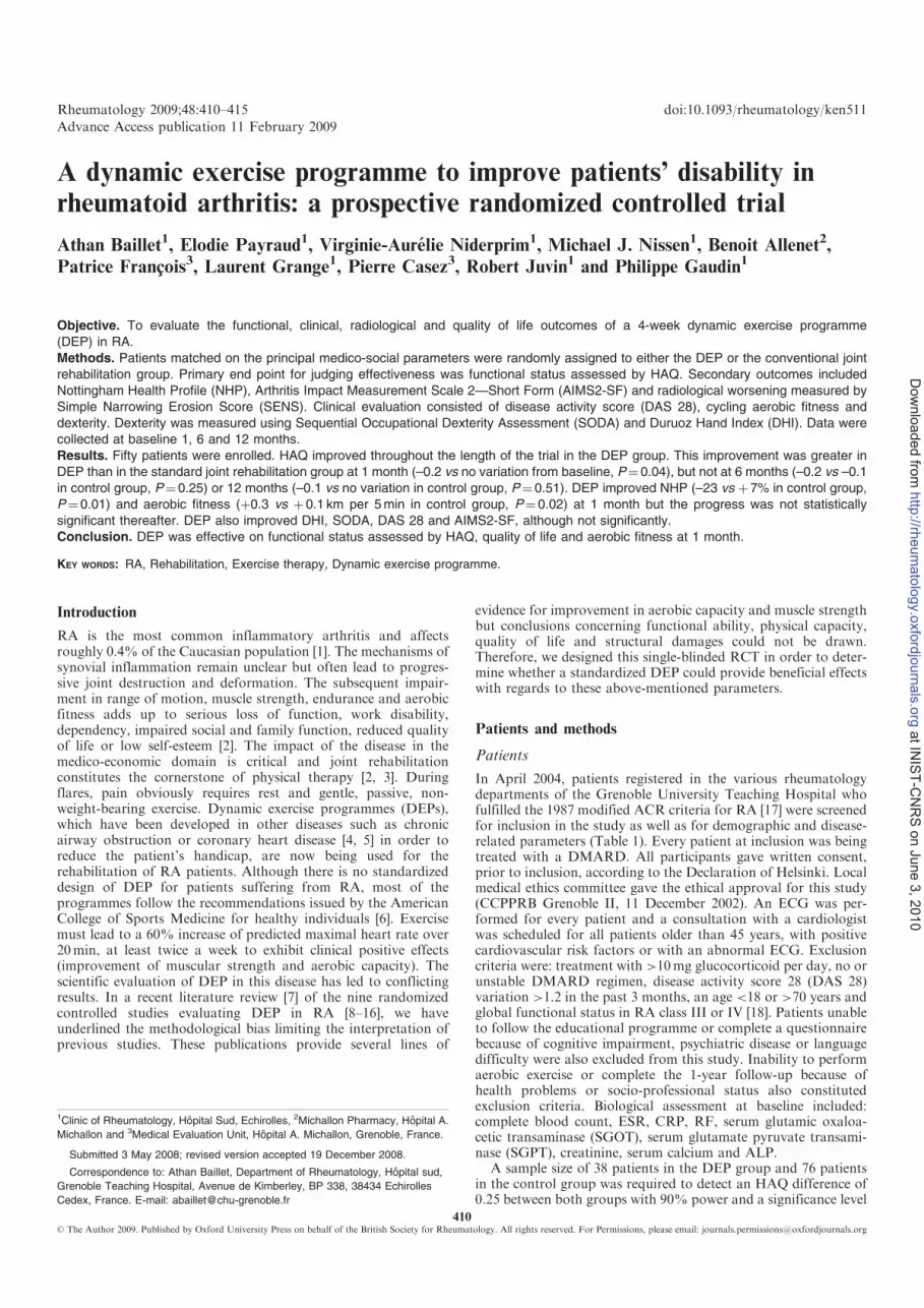
A dynamic exercise programme to improve patients’ disability inrheumatoid arthritis: a prospective randomized controlled trial
Athan Baillet1, Elodie Payraud1, Virginie-Aurelie Niderprim1, Michael J. Nissen1, Benoit Allenet2,
Patrice Francois3, Laurent Grange1, Pierre Casez3, Robert Juvin1 and Philippe Gaudin1
Objective. To evaluate the functional, clinical, radiological and quality of life outcomes of a 4-week dynamic exercise programme
(DEP) in RA.
Methods. Patients matched on the principal medico-social parameters were randomly assigned to either the DEP or the conventional joint
rehabilitation group. Primary end point for judging effectiveness was functional status assessed by HAQ. Secondary outcomes included
Nottingham Health Profile (NHP), Arthritis Impact Measurement Scale 2—Short Form (AIMS2-SF) and radiological worsening measured by
Simple Narrowing Erosion Score (SENS). Clinical evaluation consisted of disease activity score (DAS 28), cycling aerobic fitness and
dexterity. Dexterity was measured using Sequential Occupational Dexterity Assessment (SODA) and Duruoz Hand Index (DHI). Data were
collected at baseline 1, 6 and 12 months.
Results. Fifty patients were enrolled. HAQ improved throughout the length of the trial in the DEP group. This improvement was greater in
DEP than in the standard joint rehabilitation group at 1 month (–0.2 vs no variation from baseline, P¼ 0.04), but not at 6 months (–0.2 vs –0.1
in control group, P¼ 0.25) or 12 months (–0.1 vs no variation in control group, P¼ 0.51). DEP improved NHP (–23 vs þ 7% in control group,
P¼ 0.01) and aerobic fitness (þ0.3 vs þ 0.1 km per 5min in control group, P¼ 0.02) at 1 month but the progress was not statistically
significant thereafter. DEP also improved DHI, SODA, DAS 28 and AIMS2-SF, although not significantly.
Conclusion. DEP was effective on functional status assessed by HAQ, quality of life and aerobic fitness at 1 month.
KEY WORDS: RA, Rehabilitation, Exercise therapy, Dynamic exercise programme.
Introduction
RA is the most common inflammatory arthritis and affectsroughly 0.4% of the Caucasian population [1]. The mechanisms ofsynovial inflammation remain unclear but often lead to progres-sive joint destruction and deformation. The subsequent impair-ment in range of motion, muscle strength, endurance and aerobicfitness adds up to serious loss of function, work disability,dependency, impaired social and family function, reduced qualityof life or low self-esteem [2]. The impact of the disease in themedico-economic domain is critical and joint rehabilitationconstitutes the cornerstone of physical therapy [2, 3]. Duringflares, pain obviously requires rest and gentle, passive, non-weight-bearing exercise. Dynamic exercise programmes (DEPs),which have been developed in other diseases such as chronicairway obstruction or coronary heart disease [4, 5] in order toreduce the patient’s handicap, are now being used for therehabilitation of RA patients. Although there is no standardizeddesign of DEP for patients suffering from RA, most of theprogrammes follow the recommendations issued by the AmericanCollege of Sports Medicine for healthy individuals [6]. Exercisemust lead to a 60% increase of predicted maximal heart rate over20min, at least twice a week to exhibit clinical positive effects(improvement of muscular strength and aerobic capacity). Thescientific evaluation of DEP in this disease has led to conflictingresults. In a recent literature review [7] of the nine randomizedcontrolled studies evaluating DEP in RA [8–16], we haveunderlined the methodological bias limiting the interpretation ofprevious studies. These publications provide several lines of
evidence for improvement in aerobic capacity and muscle strengthbut conclusions concerning functional ability, physical capacity,quality of life and structural damages could not be drawn.Therefore, we designed this single-blinded RCT in order to deter-mine whether a standardized DEP could provide beneficial effectswith regards to these above-mentioned parameters.
Patients and methods
Patients
In April 2004, patients registered in the various rheumatologydepartments of the Grenoble University Teaching Hospital whofulfilled the 1987 modified ACR criteria for RA [17] were screenedfor inclusion in the study as well as for demographic and disease-related parameters (Table 1). Every patient at inclusion was beingtreated with a DMARD. All participants gave written consent,prior to inclusion, according to the Declaration of Helsinki. Localmedical ethics committee gave the ethical approval for this study(CCPPRB Grenoble II, 11 December 2002). An ECG was per-formed for every patient and a consultation with a cardiologistwas scheduled for all patients older than 45 years, with positivecardiovascular risk factors or with an abnormal ECG. Exclusioncriteria were: treatment with >10mg glucocorticoid per day, no orunstable DMARD regimen, disease activity score 28 (DAS 28)variation >1.2 in the past 3 months, an age <18 or >70 years andglobal functional status in RA class III or IV [18]. Patients unableto follow the educational programme or complete a questionnairebecause of cognitive impairment, psychiatric disease or languagedifficulty were also excluded from this study. Inability to performaerobic exercise or complete the 1-year follow-up because ofhealth problems or socio-professional status also constitutedexclusion criteria. Biological assessment at baseline included:complete blood count, ESR, CRP, RF, serum glutamic oxaloa-cetic transaminase (SGOT), serum glutamate pyruvate transami-nase (SGPT), creatinine, serum calcium and ALP.
A sample size of 38 patients in the DEP group and 76 patientsin the control group was required to detect an HAQ difference of0.25 between both groups with 90% power and a significance level
1Clinic of Rheumatology, Hopital Sud, Echirolles, 2Michallon Pharmacy, Hopital A.
Michallon and 3Medical Evaluation Unit, Hopital A. Michallon, Grenoble, France.
Submitted 3 May 2008; revised version accepted 19 December 2008.
Correspondence to: Athan Baillet, Department of Rheumatology, Hopital sud,
Grenoble Teaching Hospital, Avenue de Kimberley, BP 338, 38434 Echirolles
Cedex, France. E-mail: [email protected]
Rheumatology 2009;48:410–415 doi:10.1093/rheumatology/ken511
Advance Access publication 11 February 2009
410ß The Author 2009. Published by Oxford University Press on behalf of the British Society for Rheumatology. All rights reserved. For Permissions, please email: [email protected]
at IN
IST
-CN
RS
on
Jun
e 3
, 2010
h
ttp://rh
eum
ato
logy.o
xfo
rdjo
urn
als
.org
Do
wn
loa
de
d fro
m

of 0.05. To compensate for an expected 20% dropout, we plannedto include 48 patients in the DEP group and 96 patients in thecontrol group. Unfortunately, we were not able to enrol enoughpatients and the sample size was limited to 50 patients, based onprevious studies [9, 12–15].
Randomization procedure
The randomization process was carried out by an independentphysician who was not responsible for recruiting the patients, at acentral location. Patients were randomly assigned to either a DEPor a conventional joint rehabilitation by means of sealed andopaque envelopes. Study observers remained unaware of study-group assignments throughout the trial.
Interventions
The DEP was consistent with the 1990 recommendations of theAmerican College of Sports Medicine [6]. Three or four membersof the medical staff (rheumatologist, physiotherapist or occupa-tional therapist) took part in each session for 5 h a day during a4-week period (Table 2).
During the first week, following a multidisciplinary meeting,knowledge of the disease and physical capacity were evaluated foreach participant in order to design an individualized exercise pro-gramme. The second week of occupational therapy input focusedon the influence of RA on daily activities. The occupational ther-apy programme in the third week included skill exercises anddaily activities with increasing intensity (endurance and exercisesagainst resistance). During the fourth week the exercises focusedon office tasks.
The training programme was designed to improve musclestrength, flexibility, endurance and balance. Exercises for the
upper and lower limbs were performed five times a week in agymnasium (cycling, running or resisting pulley cord, 45min/day)and in a hydrotherapy pool (60min/day). During cycling, theheart rate was kept at 60–80% of the predicted maximal heart rate(220 – age). Resistance and intensity of exercise were designedafter individual evaluation of each patient and modified accordingto pain and fatigue. Regular breaks and relaxation sessions werescheduled in order to improve pain tolerance and self-esteem.Each session was preceded by a ‘warm-up’ and followed by a‘cool-down’. The patients were asked to keep a diary of their dailytraining.
Patients in the control group received a multidisciplinaryprogramme with a rheumatologist, a physiotherapist, an occupa-tional therapist, a social worker, a pharmacist and a psychologist.This 3-day intervention (�20 h) was designed to improve knowl-edge about disease pathogenesis, RA management and jointprotection. Each training group consisted of four or five patientsaccompanied by relatives or friends. The following fieldswere covered during lectures: mechanisms of RA, benefits andlimitations of drug therapy, psychological impact of the disease(first day); dietetic counselling (second day); and use of splints,surgical perspectives and counselling on activities of daily living orwork (third day). Patients benefited from a hydrotherapy session(for 45min at 358C) on the first day and relaxation exercises(45min) on the second day. Physical exercises (45min/day) aimedat preventing muscles from atrophy and tension. Each day endedwith an individual discussion where disease-related problems wereverbalized and possible solutions were offered. The training wascompleted by means of educational films or activities covering theaspects of the programme.
To standardize the delivery of interventions, written formsdetailed the coaching process based on the French RheumatologySociety recommendations, and patients received an educationalbooklet. Three physiotherapists were trained to carry out thephysical intervention.
Primary outcome
The primary outcome was the functional status evaluated byHAQ [19] at 1, 6 and 12 months. HAQ, whose total scoreranges from 0 (no functional limitation) to 3 (dramaticalfunctional impairment), was selected as the primary outcomemeasure because of its ability to measure the function of large andsmall joints by the patients’ multidimensional ability to performactivities of daily living. Its validity in assessing disability in RA iswell established.
TABLE 1. Demographic, clinical and radiological characteristic of the population
CharacteristicsDEP group(n¼25)
Control group(n¼ 23)
Age, mean � S.D., years 51.6�8.3 56.3� 12.8Female, n (%) 21 (84.0) 18 (78.3)Disease duration, mean � S.D. 10.5�8.0 11.7� 6.2Morning stiffness, mean� S.D. 59.8�75.1 55.7� 97.4Completers, n (%) 25 (100) 20 (80)Professional statusFull-time job, n (%) 6 (24.0) 5 (21.7)Part-time job, n (%) 6 (24.0) 3 (13.0)Unemployed, n (%) 0 (0.0) 1 (4.3)Disabled, n (%) 5 (20.0) 1 (4.3)Retired, n (%) 4 (16.0) 10 (43.5)
Education levelLast year of study, mean� S.D. 22.4�16.9 20.2� 7.3University training, n (%) 8 (32.0) 7 (30.4)
RA global functional ACR Indexa
ACR I, n (%) 7 (28.0) 11 (47.8)ACR II, n (%) 16 (64.0) 12 (52.2)
DAS 28 (0.14–9.3)a, mean� S.D. 4.9�1.4 4.0� 1.7<3.2 4 (16.0) 7 (30.4)3.2–5.1 11 (44.0) 8 (34.8)>5.1 10 (40.0) 6 (26.1)
VAS (0–100), mean� S.D. 34.4�23.0 31.4� 24.3Functional statusHAQ (0–3), mean� S.D. 0.9�0.6 0.7� 0.5
Quality of lifeNHP (0–600), mean� S.D. 220.7�122.4 201.6� 129.6AIMS2-SF (0–60), mean� S.D. 21.2�5.6 19.6� 4.9
Dexterity and aerobic fitnessDHI (0–90) mean� S.D. 28.8�21.8 16.0� 12.2SODA (0–108) mean� S.D. 60.0�12.6 63.6� 6.9Home bicycle, mean� S.D.b, km 1.1�0.4 1.2� 0.2
Treatmentc
Biotherapy (anti-TNF-� or IL-1�), n (%) 14 (56.0) 11 (48.0)Glucocorticoid, means (range), mg/day 6 (0–10) 5 (0–10)
SENS (0–86), median (IQR)d �3 (0–13) �5 (2–20)
aTwo missing values. bTwenty missing values. cAll patients were currently treated with aDMARD. dEight missing values.
TABLE 2. Content of the DEP: role of occupational therapists and physiotherapists
Occupational therapy Physiotherapy
First week Presentation of the multidisciplinary teamPatient’s individual expectationsEvaluation of the patient’s knowledgeof the disease and physical capacityin order to design specific programme
Explanation of the disease andits consequences
Occupational therapy advicesEvaluation of the patient’s splint andconstruction of rest splint
Dexterity exercises without resistance
HydrotherapyPain reliefImprovement inrange of motionMuscle strengthening
Fitness programme(gym, bicycle)
StretchingRelaxationEducational booklet
Second week Exercises concerning usual tasksSplint construction and evaluation
Third week Exercises concerning dexterity exercisesIncreasing resistance and range ofmotion in dexterity exercises
Fourth week Evaluation of disease-related difficultiesduring usual office tasks
Splint construction
A dynamic exercise programme for RA patients 411
at IN
IST
-CN
RS
on
Jun
e 3
, 2010
h
ttp://rh
eum
ato
logy.o
xfo
rdjo
urn
als
.org
Do
wn
loa
de
d fro
m

Secondary outcomes
Secondary outcomes included quality of life, functional, clinical,radiological, therapeutic and biological modifications in bothgroups. The French version of the Arthritis Impact Score 2—Short Form (AIMS2-SF) [20], is a specific questionnaire to assessquality of life in RA patients. AIMS2-SF was not converted toa score of 0–10, but was expressed on a scale from 0 to 60.Nottingham Health Profile (NHP) is a 38-item generic health-related quality of life measure (range 0–600, higher score forgreater health problems) [21]. Patient dexterity was evaluated byoccupational therapists using Sequential Occupational DexterityAssessment (SODA; [22]) or self-reported by patients usingDuruoz Hand Index (DHI) [23]. SODA consists of 12 standard-ized tasks evaluated by occupational therapists and ranges from 0(inability to perform tasks) to 108 (no difficulty in performingtasks). The purpose of DHI (range 0—excellent dexterity to 90—poor dexterity) was to measure hand ability while performingpersonal hygiene, office tasks and other general activities. SimpleErosions Narrowing Score (SENS: range 0–86; [24]) evaluateserosions in 32 joints in the hands, 12 in the feet and joint spacenarrowing in 30 and 12 joints, respectively. The total SENS scorewas assessed by a single radiologist unaware of the groupassignment. Disease activity was determined using disease activityscore (DAS 28: range 0.14–9.3; [25]). A 28-joint count for swellingand tenderness was assessed by a rheumatologist unaware of thetreatment allocation. Patients rated their general health on a100-mm visual analogue scale (VAS). DAS 28 was calculatedusing the three previously mentioned parameters and ESR.Aerobic fitness was measured on an exercise bike as the distancecovered in 5min [26]. Since the primary outcome (HAQ score) andsome of the secondary outcomes (DHI and SODA) are dependanton the upper limb function, we wanted to avoid a difference indexterity between DEP and control groups. Therefore, RoederManipulative Aptitude Test [27], five-handle position grip test[28], Grip and Pinch Strength test [29] were measured at baselineto avoid a potential confounder. The uses of oral and IA drugs aswell as other medical devices were noted.
Assessment of outcomes
Primary and secondary outcomes were evaluated at baseline, 1, 6and 12 months, except for SENS which was evaluated at the
baseline and 12 months. Questionnaires were sent by mail to eachpatient every 3 months. Every clinical evaluation was performedby a physician unaware of the patients’ group allocation.
Statistical analysis
Measures with a Gaussian distribution were expressed asmean� S.D. and measures with a non-Gaussian distribution wereexpressed as the median and interquartile range (IQR; expressedas the net result of 75th percentile – 25th percentile). We con-firmed with a Kolmogorov–Smirnov test that primary andsecondary outcomes, except SENS, were normally distributed.
Statistical significance of the variation of these values frombaseline between DEP and standard joint rehabilitation wasdetermined using analysis of covariance (ANCOVA) [30].P-values <0.05 were considered statistically significant. The anal-yses were based on the intent to treat as initially assigned. Allavailable data were used. Statistical analysis was performed usingStata 9.0 (Stata Corporation College Station, TX, USA).
Results
Baseline characteristics
Out of 827 patients eligible after screening, 440 were excludedbecause of exclusion criteria, 312 eligible participants refused theprogramme and 25 stopped the trial before the baseline visitbecause of personal reasons not included in the exclusion criteriaor unwillingness to participate (infection, travel, etc.). Twenty-fivepatients were randomly assigned to DEP and 25 to standard jointrehabilitation. Two individuals refused to participate immediatelyafter randomization (Fig. 1). The randomization procedurecreated two groups whose baseline demographic and disease-related parameters (clinical, functional, radiological and biologi-cal) were similar (Table 1) except for RA Global Functional ACRIndex which was better in the standard joint rehabilitation group(28% ACR I in DEP vs 47.8% in control). Functional status,measured by HAQ, did not differ significantly between the twogroups. Thirteen patients in the DEP group and 11 in the standardjoint rehabilitation group were treated by either anti-IL-1� oranti-TNF-�; five patients were taking glucocorticoids in the DEPgroup and nine in the standard joint rehabilitation group. Nopatients were lost to follow-up, but one subject was not assessed
Excluded n = 777
Refused to participate n = 312
Did not meet inclusion criteria n = 440
Withdrawal before baseline assessment
n = 2
Allocated to conventional management
n = 25
Allocated to DEP
n = 25
Randomization
n = 50
Assessed for eligibility
n = 827
Analysed n = 23 Analysed n = 25
Stopped the trial before the baseline visit
(infection, travel, etc.) n = 25
FIG. 1. Trial flowchart.
412 Athan Baillet et al.
at IN
IST
-CN
RS
on
Jun
e 3
, 2010
h
ttp://rh
eum
ato
logy.o
xfo
rdjo
urn
als
.org
Do
wn
loa
de
d fro
m

for NHP and AIMS2-SF at 1 month. Three patients were notassessed at 6 months and four patients were not assessed at 12months (Tables 3 and 4).
Primary end point
We observed an improvement in HAQ measurement throughoutthe length of the trial in DEP. This improvement was greater inDEP than in the standard joint rehabilitation group at 1 month(ÿ22% vs no variation from baseline, P¼ 0.04). The variation inHAQ scores from baseline between the two groups did not reachstatistical significance at 6 months (–22 vs –14%, P¼ 0.25) or12months (–11% vs no variation from baseline, P¼ 0.51)(Table 3). The change in HAQ score in the DEP group at 1month and 6 months (–0.2) almost reached clinical significance[31].
Secondary end points
In the DEP, all parameters globally improved during the trial.This change was most obvious after the first month comparedwith baseline (DAS 28: 3.8 vs 4.9; NHP: 170.2 vs 220.7; AIMS2-SF: 18.0 vs 21.2; DHI: 19.9 vs 28.8; SODA: 60.6 vs 60.0; andaerobic fitness: 1.4 vs 1.1 km, respectively). A variation of 0.6points in DAS 28 assessment is commonly considered to beclinically meaningful [31]. NHP and AIMS2-SF scores arepresented as a profile rather than overall score and the minimaldetectable change or the minimal important change still needs tobe assessed. Among the secondary end points, we demonstratedan increase in quality of life measured by NHP in DEP (ÿ23 vsþ 7% in control, P¼ 0.01) but not AIMS2-SF (–15 vs þ 2% incontrols, P¼ 0.09) at 1 month (Table 4). Improvement of aerobicfitness, measured on an exercise bike, was statistically better in theDEP group than in the standard joint rehabilitation group(þ0.2 km in 5min, P¼ 0.02), but no statistically functionalsuperiority of DEP could be detected on DHI (P¼ 0.35) orSODA (P¼ 0.30) at 1 month (Table 4).
Although quality of life and disease activity demonstrated apositive trend at 6 and 12 months in comparison with baseline(Tables 3 and 4), the changes of DAS 28, NHP, AIMS2-SF, DHIand SODA were not statistically significant between groups.Although not statistically different from the control group,improvement in DHI at 1 month (from 28.8 to 19.9), 6 months(from 28.8 to 15.4) and 12 months (from 28.8 to 19.2) is higherthan the minimal detectable change [32]. Aerobic fitness,measured on an exercise bike, improved with DEP but not withstandard joint rehabilitation at 6 months (þ18% vs no variation,P¼ 0.20) and at 12 months (þ18 vs –8%, P¼ 0.16). The radio-logical evaluation did not demonstrate any difference between thetwo groups at 12 months. We did not observe any significantworsening in SENS assessment in DEP in comparison withcontrol.
Safety and compliance
No adverse effects were seen in either group. Two patients in theDEP and four patients in the standard joint rehabilitation groupswitched to another anti-TNF-� treatment. There was no differ-ence in disease activity or inflammatory parameters (DAS 28,VAS, ESR and CRP) between the groups during the trial. Therewere no dropouts from either group.
Discussion
Previous studies supported evidence for improvement of aerobicfitness [9–11, 16, 33] and muscle strength [11, 33–37] after exerciseinterventions in RA. The specific effects of exercise on functionalstatus evaluated by HAQ or quality of life remain unclear [38].This randomized controlled single-blind study achieved itsprimary end point, i.e. the reduction of HAQ and demonstrated
that a DEP provided better effects on quality of life (NHP) thanconventional joint rehabilitation at 1 month.
Although our results are promising, some limitations must beconsidered. First, we did not record either patients’ current level ofphysical activity or their recent experience with rehabilitation,which could influence the baseline test parameters and the sub-sequent change at the different end points. Patients’ allocation wasrandomly performed but we cannot be sure that these potentialconfounders were equally distributed in DEP and control groups.Secondly, we evaluated compliance as the number of completersbut not as the percentage of the maximum possibly attendedsessions, which appears to be a more precise measurement ofcompliance. There was excellent compliance in our study with nowithdrawals from either group. This result is in concordance withprevious studies describing at least 70% completion in theintervention groups. As both groups in our study underwentdifferent types of interventions (intensive aerobic exercise vseducation and classical rehabilitation), a discrepancy in compli-ance is unlikely to explain the good result in the DEP group. Mostprevious studies reported compliance as the number of comple-ters. Only a few studies, mostly long-term intervention trialscomparing similar interventions, reported compliance as apercentage of the maximum possibly attended sessions. Amongthe 32 RCTs comparing an aerobic exercise programme to less
TABLE 4. Secondary outcomes (disease activity, disability, quality of life status,dexterity and aerobic fitness) during follow-up
DEP (N¼ 25) Control (N¼ 23)
Mean (S.D.)Change frombaseline (%) Mean (S.D.)
Change frombaseline (%) P-value�
1 monthDAS 28 3.8 (2.1) –22 4.0 (1.6) 0 0.19NHPa 170.2 (129.4) –23 215.7 (149.6) þ7 0.01AIMS2-SFa 18.0 (5.7) –15 19.9 (7.1) þ2 0.09DHI 19.9 (16.9) –31 14.6 (15.9) –9 0.35SODA 60.6 (11.4) þ1 60.8 (11.1) –4 0.30Exercise bike(km/5min)
1.4 (0.3) þ27 1.3 (0.2) þ8 0.02
6 monthsb
DAS 28 4.1 (1.9) –16 3.9 (2.1) –3 0.96NHP 192.5 (128.9) –13 187.5 (163.9) –7 0.46AIMS2-SF 20.4 (6.3) –4 20.1 (7.5) þ3 0.44DHI 15.4 (13.9) –47 11.8 (11.4) –26 0.38SODA 61.3 (10.8) þ2 60.8 (9.3) –4 0.18Exercise bike(km/5min)
1.3 (0.3) þ18 1.2 (0.3) 0 0.20
12 monthsc
DAS 28 4.5 (2.1) –8 3.8 (1.8) –5 0.39NHP 198.5 (134.1) –10 192.8 (130.2) –4 0.43AIMS2-SF 20.6 (7.5) –3 19.5 (6.6) –1 0.94DHI 19.2 (17.9) –33 13.3 (10.8) –17 0.74SODA 60.5 (9.4) þ1 60.3 (8.6) –5 0.18Exercise bike(km/5min)
1.3 (0.2) þ18 1.1 (0.3) –8 0.16
DAS 28: 0.14—no activity to 9.3—high activity; NHP: 0—good quality of life to 600—poorquality of life; AIMS2-SF: 0—good quality of life to 60—poor quality of life; DHI: 0—good dexterityto 90—poor dexterity; SODA: 0—poor dexterity to 108—good dexterity. aOne missing value.bThree patients not assessed. cFour patients not assessed. �
P-values are determined usingANCOVA.
TABLE 3. Primary outcome (HAQ) during follow-up
DEP (N¼25) Control (N¼23)
Mean (S.D.)Change frombaseline (%) Mean (S.D.)
Change frombaseline (%) P-value�
HAQ 1 month 0.7 (0.6) –22 0.7 (0.6) 0 0.04HAQ 6 monthsa 0.7 (0.6) –22 0.6 (0.6) –14 0.25HAQ 12 monthsb 0.8 (0.6) –11 0.7 (0.5) 0 0.51
HAQ: 0—no disability to 3—great disability. aThree patients not assessed. bFour patients notassessed. �
P-values are determined using ANCOVA.
A dynamic exercise programme for RA patients 413
at IN
IST
-CN
RS
on
Jun
e 3
, 2010
h
ttp://rh
eum
ato
logy.o
xfo
rdjo
urn
als
.org
Do
wn
loa
de
d fro
m

intensive rehabilitation in RA, only six trials (19%), whoseintervention lasted at least 12 weeks, evaluated this parameter. Ina home-based physical activity intervention, Van den Berg et al.[39] reported that 34% of patients were physically active in theintervention group. Thirdly, the small number of patients includedin this study is likely to be insufficient to detect a statisticaldifference in the primary and secondary outcomes between bothgroups at 6 and 12 months. Finally, missing data could yieldbiased results in our intent to treat statistical analysis. Contrary tothe numerous missing values in baseline home-bicycle sprintassessment that might limit the conclusion of a DEP effect onphysical capacity, only seven HAQ measurements (4%) weremissing, which is unlikely to constitute an attrition bias.
Our findings confirm the results of earlier studies [36, 40], whichreported a lower HAQ in an exercise intervention group, con-trasting with most of the previous studies that failed to detect anystatistical difference in HAQ variation between interventional andconventional joint rehabilitation [11, 14, 41, 42]. Two trials [8, 15]were able to show that DEP provides better functional results thancontrol. In these studies—the McMaster Toronto Arthritis PatientPreference Questionnaire (MACTAR) and SF-36–two quality oflife questionnaires containing functional ability items were used toassess functional impairment. To assess this last parameter weselected the widely used HAQ questionnaire that appears to bemore specific for the functional ability assessment in RA. Thisindex depends more on disease activity and pain than limbfunction and is more sensitive to detect change in function of theupper limbs than the lower limbs [43]. It is probably moreappropriate in pharmacological trials than in physical interventiontrials and might therefore be less sensitive to changes in groupswith a score below 1.00 [44]. Thus, by using HAQ we possiblymissed small but clinically relevant improvement of lower limbdisability, and the lack of significant improvement of HAQ after1 month may reflect an inability of this index to detect the effectsof DEP rather than a failure of DEP to improve patients’functional status [38, 43]. Similarly, as Stenstrom et al. [45]reported an improvement of NHP score after dynamic training inpatients with inflammatory rheumatic diseases, we have shownthat the intervention group displayed improved health-relatedquality of life with NHP being statistically higher in DEP than inthe control group as early as the first month. After the first month,the exercises were home-based and self-administrated. Thepatients could therefore no longer benefit from the groupdynamic, explaining the reduction in effect of DEP on NHP andother questionnaires concerning quality of life with time.Therefore, we cannot exclude that the enthusiasm of patientsand the health professionals involved in this short-term interven-tion trial influenced the outcome.
The cost effectiveness of DEPs compared with usual manage-ment could not be evaluated because of missing data on self-reported questionnaires. We were unable to quantify the impact ofa DEP on work and on the consumption of medical and para-medical resources, in order to determine whether the interventionprogramme, which is more expensive (10 000 euros for a DEP vs1500 euros for usual management), was economically relevant.
The radiological evaluation did not demonstrate any deleter-ious effects of the DEP, which is consistent with two previousstudies [8, 14]. Although our study was underpowered to proveany benefit in the DEP group in terms of joint damage, a previousstudy has reported less erosive change in patients treated with aDEP compared with usual care physical therapy [46]. Given thatboth study groups displayed differences of disease duration andbaseline joint damage, no solid conclusions could be drawn onthe protective effect of exercise on joints from this study [7].Molecular mechanisms are still unclear but some cytokines such asinsulin growth factor 1 (IGF1) are up-regulated in DEP [47].
Our study, based on stringent methodological procedure,provides evidence for quality of life improvement followingDEP. We have confirmed that this type of intervention has
positive consequences on aerobic fitness with excellentcompliance.
Acknowledgement
We thank Aurelie Petitprin for her help in writing this article.
Disclosure statement: The authors have declared no conflicts ofinterest.
References
1 Guillemin F, Saraux A, Guggenbuhl P et al. Prevalence of rheumatoid arthritis in
France: 2001. Ann Rheum Dis 2005;64:1427–30.
2 Mau W, Bornmann M, Weber H, Weidemann HF, Hecker H, Raspe HH. Prediction of
permanent work disability in a follow-up study of early rheumatoid arthritis: results of
a tree structured analysis using RECPAM. Br J Rheumatol 1996;35:652–9.
3 Wolfe F, Hawley DJ. The longterm outcomes of rheumatoid arthritis: work disability: a
prospective 18 year study of 823 patients. J Rheumatol 1998;25:2108–17.
4 Rossi G, Florini F, Romagnoli M et al. Length and clinical effectiveness of pulmonary
rehabilitation in outpatients with chronic airway obstruction. Chest 2005;127:105–9.
5 Carlson JJ, Johnson JA, Franklin BA, VanderLaan RL. Program participation,
exercise adherence, cardiovascular outcomes, and program cost of traditional
versus modified cardiac rehabilitation. Am J Cardiol 2000;86:17–23.
6 American College of Sports Medicine Position Stand. The recommended quantity
and quality of exercise for developing and maintaining cardiorespiratory and
muscular fitness in healthy adults. Med Sci Sports Exerc 1990;22:265–74.
7 Gaudin P, Leguen-Guegan S, Allenet B, Baillet A, Grange L, Juvin R. Is dynamic
exercise beneficial in patients with rheumatoid arthritis? Joint Bone Spine 2008;
75:11–17.
8 de Jong Z, Munneke M, Zwinderman AH et al. Is a long-term high-intensity exercise
program effective and safe in patients with rheumatoid arthritis? Results of a
randomized controlled trial. Arthritis Rheum 2003;48:2415–24.
9 Westby MD, Wade JP, Rangno KK, Berkowitz J. A randomized controlled trial to
evaluate the effectiveness of an exercise program in women with rheumatoid arthritis
taking low dose prednisone. J Rheumatol 2000;27:1674–80.
10 Minor MA, Hewett JE, Webel RR, Anderson SK, Kay DR. Efficacy of physical
conditioning exercise in patients with rheumatoid arthritis and osteoarthritis. Arthritis
Rheum 1989;32:1396–405.
11 van den Ende CH, Hazes JM, le Cessie S et al. Comparison of high and low intensity
training in well controlled rheumatoid arthritis. Results of a randomised clinical trial.
Ann Rheum Dis 1996;55:798–805.
12 Lyngberg KK, Harreby M, Bentzen H, Frost B. Danneskiold-Samsoe B. Elderly
rheumatoid arthritis patients on steroid treatment tolerate physical training without an
increase in disease activity. Arch Phys Med Rehabil 1994;75:1189–95.
13 Harkcom TM, Lampman RM, Banwell BF, Castor CW. Therapeutic value of graded
aerobic exercise training in rheumatoid arthritis. Arthritis Rheum 1985;28:32–9.
14 Hansen TM, Hansen G, Langgaard AM, Rasmussen JO. Longterm physical training
in rheumatoid arthritis. A randomized trial with different training programs and blinded
observers. Scand J Rheumatol 1993;22:107–12.
15 Bilberg A, Ahlmen M, Mannerkorpi K. Moderately intensive exercise in a temperate
pool for patients with rheumatoid arthritis: a randomized controlled study.
Rheumatology 2005;44:502–8.
16 Baslund B, Lyngberg K, Andersen V et al. Effect of 8 week of bicycle training
on the immune system of patients with rheumatoid arthritis. J Appl Physiol
1993;75:1691–5.
17 Arnett FC, Edworthy SM, Bloch DA et al. The American Rheumatism Association
1987 revised criteria for the classification of rheumatoid arthritis. Arthritis Rheum
1988;31:315–24.
18 Hochberg MC, Chang RW, Dwosh I, Lindsey S, Pincus T, Wolfe F. The American
College of Rheumatology 1991 revised criteria for the classification of global
functional status in rheumatoid arthritis. Arthritis Rheum 1992;35:498–502.
19 Guillemin F, Briancon S, Pourel J. Measurement of the functional capacity in
rheumatoid polyarthritis: a French adaptation of the Health Assessment
Questionnaire (HAQ). Rev Rhum Mal Osteoartic 1991;58:459–65.
20 Guillemin F, Coste J, Pouchot J, Ghezail M, Bregeon C, Sany J. The AIMS2-SF: a
short form of the Arthritis Impact Measurement Scales 2. French Quality of Life in
Rheumatology Group. Arthritis Rheum 1997;40:1267–74.
21 Bucquet D, Condon S, Ritchie K. The French version of the Nottingham Health
Profile. A comparison of items weights with those of the source version. Soc Sci Med
1990;30:829–35.
22 van Lankveld W, van’t Pad Bosch P, Bakker J, Terwindt S, Franssen M, van Riel P.
Sequential occupational dexterity assessment (SODA): a new test to measure hand
disability. J Hand Ther 1996;9:27–32.
Rheumatology key messages
� A DEP in RA improves HAQ at 1 month but not subsequently.� DEP provides beneficial effects on quality of life.
414 Athan Baillet et al.
at IN
IST
-CN
RS
on
Jun
e 3
, 2010
h
ttp://rh
eum
ato
logy.o
xfo
rdjo
urn
als
.org
Do
wn
loa
de
d fro
m

23 Duruoz MT, Poiraudeau S, Fermanian J et al. Development and validation of a
rheumatoid hand functional disability scale that assesses functional handicap.
J Rheumatol 1996;23:1167–72.
24 van der Heijde D, Dankert T, Nieman F, Rau R, Boers M. Reliability and sensitivity to
change of a simplification of the Sharp/van der Heijde radiological assessment in
rheumatoid arthritis. Rheumatology 1999;38:941–7.
25 Smolen JS, Breedveld FC, Eberl G et al. Validity and reliability of the twenty-eight-
joint count for the assessment of rheumatoid arthritis activity. Arthritis Rheum 1995;
38:38–43.
26 Astrand I. Aerobic work capacity in men and women with special reference to age.
Acta Physiol Scand. 1960;49:1–92.
27 Roeder, WS. Roeder Manipulative Aptitude Test. Indiana: Lafayette Instrument, 1967.
28 Goldman S, Cahalan TD, An KN. The injured upper extremity and the JAMAR five-
handle position grip test. Am J Phys Med Rehabil 1991;70:306–8.
29 Mathiowetz V, Kashman N, Volland G, Weber K, Dowe M, Rogers S. Grip and pinch
strength: normative data for adults. Arch Phys Med Rehabil 1985;66:69–74.
30 Frison L, Pocock SJ. Repeated measures in clinical trials: analysis using mean
summary statistics and its implications for design. Stat Med 1992;11:1685–704.
31 Soubrier M, Dougados M. Selecting criteria for monitoring patients with rheumatoid
arthritis. Joint Bone Spine 2005;72:129–34.
32 Sezer N, Yavuzer G, Sivrioglu K et al. Clinimetric properties of the Duruoz Hand
Index in patients with stroke. Arch Phys Med Rehabil. 2007;88:309–14.
33 Ekdahl C, Andersson SI, Moritz U, Svensson B. Dynamic versus static training in
patients with rheumatoid arthritis. Scand J Rheumatol 1990;19:17–26.
34 Hoenig H, Groff G, Pratt K, Goldberg E, Franck W. A randomized controlled trial of
home exercise on the rheumatoid hand. J Rheumatol 1993;20:785–9.
35 Hakkinen A, Sokka T, Kotaniemi A et al. Dynamic strength training in patients with
early rheumatoid arthritis increases muscle strength but not bone mineral density.
J Rheumatol 1999;26:1257–63.
36 McMeeken J, Stillman B, Story I, Kent P. The effects of knee extensor and flexor
muscle training on the timed-up-and-go test in individuals with rheumatoid arthritis.
Physiother Res Int 1999;4:55–67.
37 Van den Ende CHM, Breedveld FC, le Cessie S, Dijkmans BAC, de Mug AW,
Hazes JMW. Effect of intensive exercise on patients with active rheumatoid arthritis:
a randomized clinical trial. Ann Rheum Dis 2000;59:615–21.
38 Stenstrom CH, Minor MA. Evidence for the benefit of aerobic and strengthening
exercise in rheumatoid arthritis. Arthritis Rheum 2003;49:428–34.
39 van den Berg MH, Ronday HK, Peeters AJ et al. Using internet technology to deliver
a home-based physical activity intervention for patients with rheumatoid arthritis: a
randomized controlled trial. Arthritis Rheum. 2006;55:935–45.
40 Hakkinen A, Sokka T, Kotaniemi A, Hannonen P. A randomized two-year study of the
effects of dynamic strength training on muscle strength, disease activity, functional
capacity, and bone mineral density in early rheumatoid arthritis. Arthritis Rheum
2001;44:515–22.
41 Bostrom C, Harms-Ringdahl K, Nordemar R. Effects of static and dynamic shoulder
rotator exercises in women with rheumatoid arthritis. Scand J Rheumatol 1998;
27:281–90.
42 Brodin N, Eurenius E, Jensen I, Nisell R, Opava CH. Coaching patients with early
rheumatoid arthritis to healthy physical activity: a multicenter, randomized, controlled
study. Arthrtis Rheum 2008;59:325–31.
43 van den Ende CH, Breedveld FC, Dijkmans BA, Hazes JM. The limited value of the
Health Assessment Questionnaire as an outcome measure in short term exercise
trials. J Rheumatol 1997;24:1972–7.
44 Stenstrom CH. Home exercise in rheumatoid arthritis functional class II: goal setting
versus pain attention. J Rheumatol 1994;21:627–34.
45 Stenstrom CH, Arge B, Sundbom A. Dynamic training versus relaxation training as
home exercise for patients with inflammatory rheumatic diseases. A randomized
controlled study. Scand J Rheumatol. 1996;25:28–33.
46 de Jong Z, Munneke M, Zwinderman AH et al. Long term high intensity exercise
and damage of small joints in rheumatoid arthritis. Ann Rheum Dis 2004;63:
1399–405.
47 Karatay S, Yildirim K, Melikoglu MA, Akcay F, Senel K. Effects of dynamic exercise
on circulating IGF-1 and IGFBP-3 levels in patients with rheumatoid arthritis or
ankylosing spondylitis. Clin Rheumatol 2007;26:1635–9.
A dynamic exercise programme for RA patients 415
at IN
IST
-CN
RS
on
Jun
e 3
, 2010
h
ttp://rh
eum
ato
logy.o
xfo
rdjo
urn
als
.org
Do
wn
loa
de
d fro
m

Annexe 2
61
ANNEXE 2 :
Résultats supplémentaires publiés en ligne pour l’ article 1 :
Baillet A, Zeboulon N, Gossec L, Combescure C, Bodi n AL, Juvin R, Dougados M,
Gaudin P.
Efficacy of cardiorespiratory aerobic exercise in rheumatoid arthritis :
meta-analysis of randomized controlled trials
Arthritis Care Res (Hoboken) 2010;62:984-92

Annexe 2
62
SUPPLEMENTARY TABLE 1: REFERENCES OF OUTCOME MEASUR ES
1. Hunt SM, McKenna SP, McEwen J, Williams J, Papp E. The Nottingham Health Profile: subjective health status and medical consultations. Soc Sci Med [A] 1981;15:221-9.
2. de Jong Z, van der Heijde D, McKenna SP, Whalley D. The reliability and construct validity of the RAQoL: a rheumatoid arthritis-specific quality of life instrument. Br J Rheumatol 1997;36:878-83.
3. Ware JE, Jr., Sherbourne CD. The MOS 36-item short-form health survey (SF-36). I. Conceptual framework and item selection. Med Care 1992;30:473-83.
4. Meenan RF, Anderson JJ, Kazis LE, Egger MJ, Altz-Smith M, Samuelson CO, Jr., et al. Outcome assessment in clinical trials. Evidence for the sensitivity of a health status measure. Arthritis Rheum 1984; 27:1344-52.
5. Tugwell P, Bombardier C, Buchanan WW, Goldsmith CH, Grace E, Hanna B. The MACTAR Patient Preference Disability Questionnaire--an individualized functional priority approach for assessing improvement in physical disability in clinical trials in rheumatoid arthritis. J Rheumatol 1987;14:446-51.
6. Fries JF, Spitz P, Kraines RG, Holman HR. Measurement of patient outcome in arthritis. Arthritis Rheum 1980;23:137-45.
7. Smolen JS, Breedveld FC, Eberl G, Jones I, Leeming M, Wylie GL, et al. Validity and reliability of the twenty-eight-joint count for the assessment of rheumatoid arthritis activity. Arthritis Rheum 1995; 38:38-43.
8. Ritchie DM, Boyle JA, McInnes JM, Jasani MK, Dalakos TG, Grieveson P, et al. Clinical studies with an articular index for the assessment of joint tenderness in patients with rheumatoid arthritis. Q J Med 1968;37:393-406.
9. Larsen A, Dale K, Eek M. Radiographic evaluation of rheumatoid arthritis and related conditions by standard reference films. Acta Radiol Diagn (Stockh) 1977;18:481-91.
Health-related parameter Outcome measure Abbreviation Ref. Quality of life Nottingham Health Profile NHP (1) Rheumatoid Arthritis Quality of Life RAQol (2) Physical component of the 36-item
short-form health survey SF-36 Phys (3)
Original Arthritis Impact Measurement Scales Health Status Questionnaire
AIMS (4)
McMaster Toronto Arthritis MACTAR (5) Function Health Assessment Questionnaire HAQ (6) Disease activity Disease activity score DAS 28 (7) Joint count Ritchie index Ritchie (8) Radiological evaluation Larsen’s method Larsen (9)

Annexe 2
63
SUPPLEMENTARY TABLE 2: DEMOGRAPHIC CHARACTERISTICS IN 14 RANDOMIZED CONTROLLED TRIALS
Intervention group Control group
Age, yr
mean (SD) RA onset, yr mean (SD) n
Women %
Completers %
RF+ %
DMARD %
Age, yr mean (SD)
RA onset, yr mean (SD) n
Women %
Completers %
RF+ %
DMARD %
Bilberg [77] ¹ 49.0 (~10) 2.6 (1.3) 22 nr 91.7 nr 81.8 46.0 (~14.7) 2.9 (1.4) 27 nr 85.2 nr 80.0 Van den Berg [78] ¥² 49.5 (9.6) 7.6 (6.5) 82 75.6 93.9 64.6 90.2 49.8 (10.4) 5.5 (8.4) 78 76.9 96.2 59.0 89.7 de Jong [79] ¥ ² 54.0 (11.9) 5.0 (5.2) 150 79.3 90.7 71.3 74.0 53.5 (13.3) 7.5 (8.0) 150 78.7 96.7 70.7 78.0 Westby [80] 56.4 (10.1) 13.4 (10.4) 14 100.0 71.4 nr 28.6 56.4 (10.8) 12.6 (10.7) 16 100.0 62.5 nr 18.8 Van den Ende [69] 62.0 (13.0) 8.0 (8.0) 34 58.8 82.4 88.2 nr 58.0 (14.0) 7.0 (8.0) 30 66.7 66.7 63.3 60.0 Melikoglu [81] 46.2 (8.3) 6.7 (6.3) 20 100.0 95.0 nr nr 50.3 (9.7) 6.0 (5.7) 20 100.0 85.0 nr nr Hansen [28] ¥ ² 55.0 (10.4) 1.2 (3.6) 15 80.0 93.3 73.3 73.3 51.0 (8.2) 8.0 (4.4) 15 66.7 86.7 93.3 73.3 52.0 (8.9) 1.0 (3.6) 15 46.7 93.3 86.7 73.3 51.0 (10.4) 1.2 (3.7) 15 60.0 73.3 86.7 60.0 54.0 (8.9) 1.0 (5.9) 15 73.3 86.7 73.3 nr Harkcom [82] 51.5 (3.1) 12.2 (8.7) 5 100.0 80.0 nr nr 45.1 (19.3) 8.8 (10.1) 6 100.0 100.0 nr nr 47.3 (14.5) 10.6 (5.5) 4 100.0 75.0 nr nr 44.0 (18.3) 5.6 (3.7) 5 100.0 80.0 nr nr Van den Ende [30] 51.1 (9.5) 11.5 (8.4) 25 52.0 60.0 84.0 nr 56.1 (10.9) 11.2 (9.8) 25 72.0 nr 76.0 nr 47.7 (13.6) 8.4 (5.8) 25 64.0 80.0 80.0 nr 53.1 (12.1) 8.6 (7.1) 25 64.0 92.0 76.0 nr Baillet [31] 51.3 (8.3) 10.5 (8.3) 25 100.0 84.0 nr nr 56.3 (12.8) 11.7 (1.5) 23 78.3 87.0 nr nr Neuberger [83] nr nr nr nr 84 nr 81.0 nr nr nr nr nr nr 75 nr 97.3 nr nr nr nr nr nr 89 nr 88.8 nr nr Nordemar [84] 56.0 (9.0) 16.0 (7.0) 23 85.2 85.2 nr 56.5 58.0 (10.0) 14.0 (7.0) 23 82.6 92.0 nr 60.9 Noreau [38] 49.3 (13.0) 8.1 (8.2) 19 63.2 100.0 nr 68.4 49.4 (11.9) 11.2 (2.1) 10 80.0 100.0 nr 60.0 Lyngberg [57] 65.0 (10.8) 12.4 (11.1) 12 nr 83.3 nr 50.0 68.0 (7.2) 11.7 (10.1) 12 nr nr 16.7 Overall 52.8 7.8 76.9 86.8 77.5 70.6 54.5 9.3 80.2 87.2 75.9 66.2
Value dispersion is standard deviation (SD) as given in articles or inferred from range¹, interquartile range². Results are means except ¥, where results are medians. RA = rheumatoid arthritis; RF+ = rheumatoid factor positive; DMARD = disease-modifying antirheumatic drugs; yr = years.

Annexe 2
64
SUPPLEMENTARY TABLE 3: EFFICACY OF CARDIORESPIRATOR Y AEROBIC EXERCISES IN 14 RANDOMIZED CONTROLLED TRIALS ON QUALITY OF LIFE, FUNCTION, PAI N, JOINT COUNT, AND RADIOLOGIC DAMAGES
ACCORDING TO THE END POINT ASSESSMENTS
+ = Statistical difference in favor of exercise group; - = Statistical difference in favor of control group; ? = No statistical assessment of between-group difference; ns= no statistical difference between the two groups; * = the difference between groups was significant for both feet only and feet/hand Larsen scores, but not significant for large joint Larsen score; ** = the difference between groups was significant at 3 weeks but not at 6 weeks; *** = the difference between groups was significant for pain intensity assessed by McGill scale; § = the difference between groups was significant for swollen joint count but not for the Ritchie index. HAQ = Health Assessment Questionnaire; DAS 28 = disease activity score; VAS = Pain Visual Analogue Scale; NHP = Nottingham Health Profile; RAQol = Rheumatoid Arthritis Quality of Life; SF36-Phys = physical component of the 36-item short-form health survey; AIMS = Arthritis Impact Measurement Scales Health Status Questionnaire; MACTAR = McMaster Toronto Arthritis; SJC= swollen joint count; TJC= tender joint count; S+TJC= swollen and tender joint count; mo = months
Quality of life HAQ DAS 28 VAS Larsen score Joint count
outcome measure
≤3 mo
~6 mo
≥12 mo
≤3 mo
~6 mo
≥12 mo
≤3 mo
~6 mo
≥12 mo
≤3 mo
~6 mo
≥12 mo
≤3 mo
~6 mo
≥12 mo
outcome measure
≤3 mo
~6 mo
≥12 mo
Bilberg [77] SF-36 phys + ns ns ns Van den Berg [78] RAQol ns ns ns + + + ns ns De Jong [79] MACTAR + ns ns + ns* Westby [80] ns S+TJC ns Van den Ende [69] ns ns ns ns -/ns** ns S+TJC ns ns Melikoglu [81] ns + Ritchie + Hansen [28] ns ns ns SJC ns Harkcom [82] SJC + Van den Ende [30] ns ns ns ns Ritchie ns§ ns Baillet [31] NHP + ns ns + ns ns ns ns ns ns ns ns Neuberger [83] *** S+TJC + Nordemar [84] + Noreau [38] AIMS phys ? ? TJC ? Lyngberg [57] TJC ns

Annexe 2
65
SUPPLEMENTARY FIGURE 1. EFFICACY OF CARDIORESPIRATO RY AEROBIC EXERCISES ON DISEASE ACTIVITY SCORE (A) AND RADIOLO GICAL DAMAGE (B).
SMD = standardized mean difference (difference between both groups of mean outcome variation from baseline/SD at baseline); 95% CI = 95% confidence interval; IV = method of the inverse of the variance; DAS = Disease activity score; Larsen = Larsen’s radiologic score.
A. DAS
Baillet et al. (3)
de Jong et al. (20)
Van den Berg et al. (25)
Van den Ende et al. (22)
Total
Heterogeneity: I² = 67%, p = 0.03
Test for overall effect: p = 0.34
0.08 [-0.08, 0.25]
SMD [95% CI]IV, Fixed effect IV, Random effect
SMD [95% CI]
-1 -0.5 0 0.5 1
de Jong et al. (20)
Hansen et al. (27)
Nordemar et al. (30)
Total
Heterogeneity: I² = 17%, p = 0.30Test for overall effect: p = 0.0005
0.36 [0.16, 0.56]
SMD [95% CI]IV, Fixed effect IV, Fixed effect
SMD [95% CI]
-1 -0.5 0 0.5 1
B. Larsen
Study (ref)
Study (ref)
Favors control Favors exercise
Favors control Favors exercise
0% 30%% Rate reduction¥
% Rate reduction¥ 0% 3%

Annexe 2
66
SUPPLEMENTARY FIGURE 2. PUBLICATION BIAS ASSESSMENT .
Funnel plots for quality of life (A), function (B), visual analogue scale (VAS) pain (C), and joint count (D) meta-analysis. SMD = standardized mean difference; SE = standard error.
A. Quality of life
D. Joint countB. HAQ
C. VAS Pain
Begg’s test p=0.99; Egger’s test p=0.54
Begg’s test p=0.04; Egger’s test p=0.02
Begg’s test p=0.48 ; Egger’s test p=0.57
Begg’s test p=0.61; Egger’s test p=0.44
SE
SE
SMD
SMD1
0.5
0.4
-1 0
0.3
0.2
0.1
0.5
0.4
0.3
0.2
0.1
SE
SMD
0.5
0.4
0.3
0.2
0.1
SE
SMD
1-1 00.5
0.4
0.3
0.2
0.1
1-1 0
1-1 0

Annexe 3
67
ANNEXE 3
Résultats supplémentaires publiés en ligne pour l’ article 2 :
Baillet A, Vaillant M, Guinot M, Juvin R, Gau din P.
Efficacy of resistance exercises in Rheumatoid Arth ritis : Meta-analysis of
randomized controlled trials.
Rheumatology (Oxford) 2011; en révision
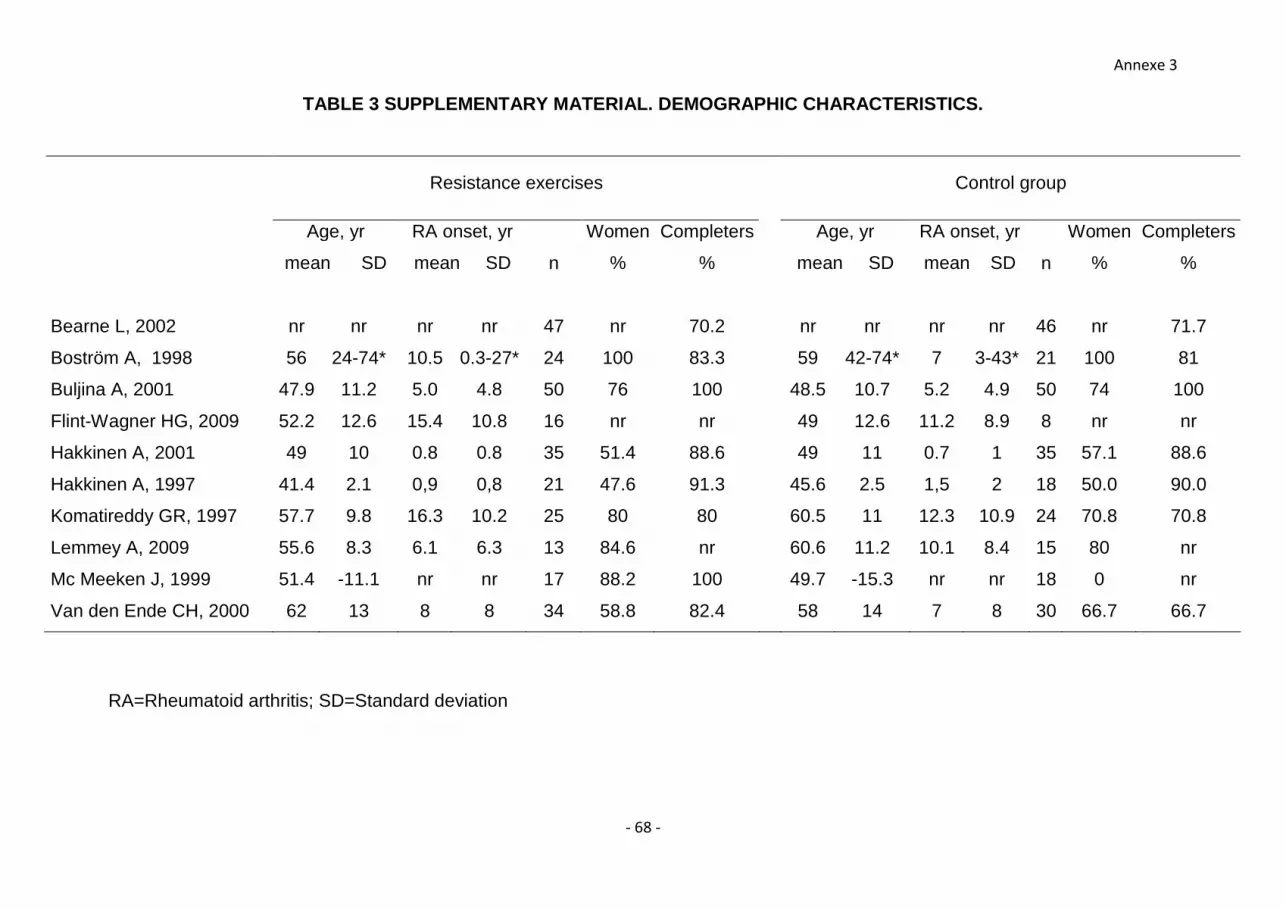
Annexe 3
- 68 -
TABLE 3 SUPPLEMENTARY MATERIAL. DEMOGRAPHIC CHARACT ERISTICS.
RA=Rheumatoid arthritis; SD=Standard deviation
Resistance exercises Control group
Age, yr
mean SD
RA onset, yr
mean SD n
Women
%
Completers
%
Age, yr
mean SD
RA onset, yr
mean SD n
Women
%
Completers
%
Bearne L, 2002 nr nr nr nr 47 nr 70.2 nr nr nr nr 46 nr 71.7
Boström A, 1998 56 24-74* 10.5 0.3-27* 24 100 83.3 59 42-74* 7 3-43* 21 100 81
Buljina A, 2001 47.9 11.2 5.0 4.8 50 76 100 48.5 10.7 5.2 4.9 50 74 100
Flint-Wagner HG, 2009 52.2 12.6 15.4 10.8 16 nr nr 49 12.6 11.2 8.9 8 nr nr
Hakkinen A, 2001 49 10 0.8 0.8 35 51.4 88.6 49 11 0.7 1 35 57.1 88.6
Hakkinen A, 1997 41.4 2.1 0,9 0,8 21 47.6 91.3 45.6 2.5 1,5 2 18 50.0 90.0
Komatireddy GR, 1997 57.7 9.8 16.3 10.2 25 80 80 60.5 11 12.3 10.9 24 70.8 70.8
Lemmey A, 2009 55.6 8.3 6.1 6.3 13 84.6 nr 60.6 11.2 10.1 8.4 15 80 nr
Mc Meeken J, 1999 51.4 -11.1 nr nr 17 88.2 100 49.7 -15.3 nr nr 18 0 nr
Van den Ende CH, 2000 62 13 8 8 34 58.8 82.4 58 14 7 8 30 66.7 66.7

Annexe 3
69
TABLE 4 SUPPLEMENTARY DATA. ADVERSE EVENTS (AE)
nr = not reported
Resistance exercises Control
Bearne L, 2002 4 flares, 9 unrelated to RA AEs 2 flares, 6 unrelated to RA AEs
Boström A, 1998 8 steroid injections 6 steroid injections
Buljina A, 2001 nr nr
Flint-Wagner HG, 2009 nr nr
Hakkinen A, 2001 1 unrelated to RA AE none
Hakkinen A, 1997 6 unrelated to RA AEs, 1 back
ache
5 unrelated to RA AEs, 2 back
aches
Komatireddy GR, 1997 5 unrelated to RA AEs, 1
coronary artery disease
2 unrelated to RA AEs, 1
coronary artery disease
Lemmey A, 2009 nr nr
Mc Meeken J, 1999 1 increased pain 1 diuretic prescription, 1 steroid
injection
Van den Ende CH, 2000 5 unrelated to RA AEs, 1
psychological problem
5 unrelated to RA AEs, 1
psychological problem, 4 RA
comorbidities
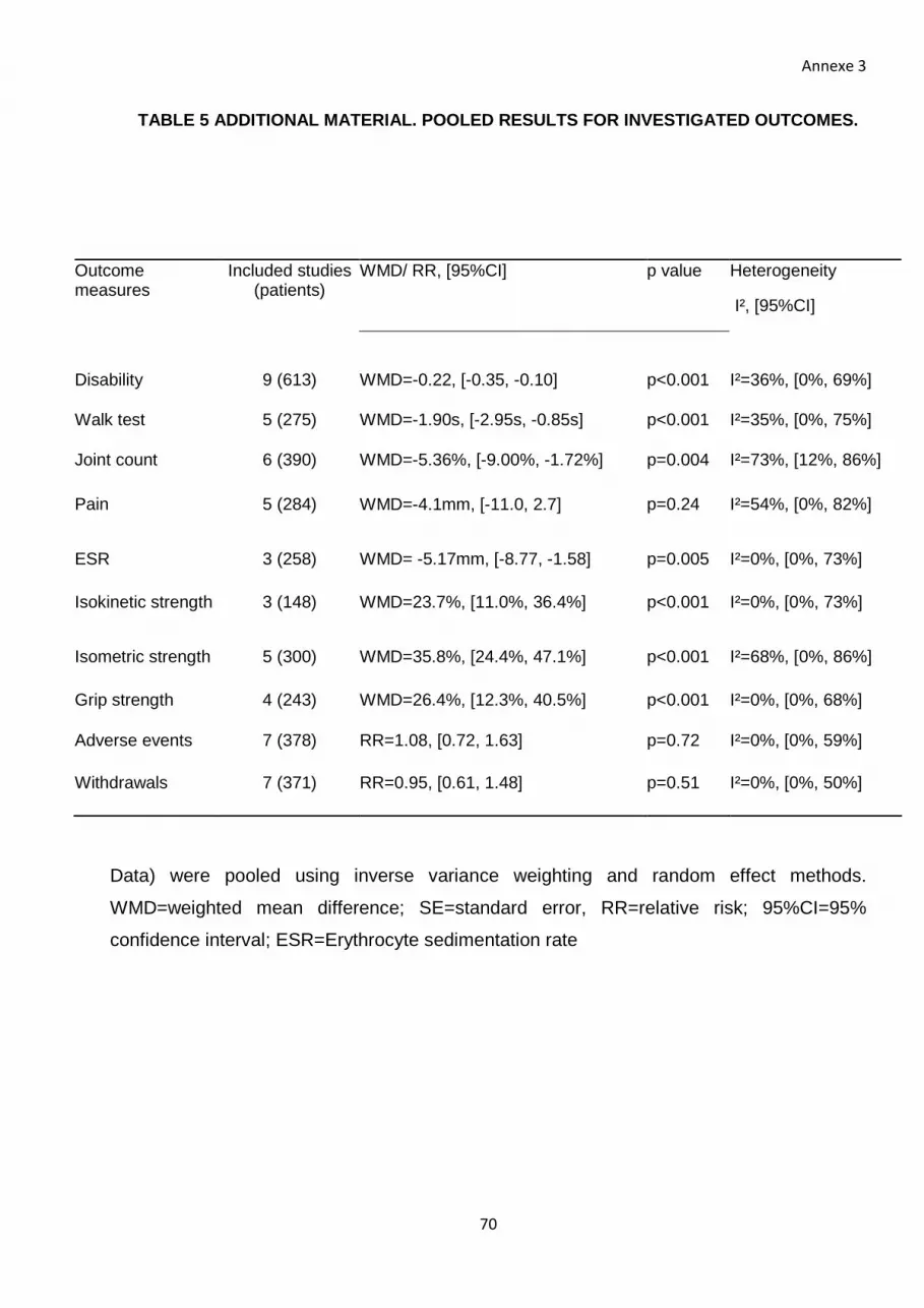
Annexe 3
70
TABLE 5 ADDITIONAL MATERIAL. POOLED RESULTS FOR INV ESTIGATED OUTCOMES.
Data) were pooled using inverse variance weighting and random effect methods.
WMD=weighted mean difference; SE=standard error, RR=relative risk; 95%CI=95%
confidence interval; ESR=Erythrocyte sedimentation rate
Outcome measures
Included studies (patients)
WMD/ RR, [95%CI] p value Heterogeneity
I², [95%CI]
Disability 9 (613) WMD=-0.22, [-0.35, -0.10] p<0.001 I²=36%, [0%, 69%]
Walk test 5 (275) WMD=-1.90s, [-2.95s, -0.85s] p<0.001 I²=35%, [0%, 75%]
Joint count 6 (390) WMD=-5.36%, [-9.00%, -1.72%] p=0.004 I²=73%, [12%, 86%]
Pain 5 (284) WMD=-4.1mm, [-11.0, 2.7] p=0.24 I²=54%, [0%, 82%]
ESR 3 (258) WMD= -5.17mm, [-8.77, -1.58] p=0.005 I²=0%, [0%, 73%]
Isokinetic strength 3 (148) WMD=23.7%, [11.0%, 36.4%] p<0.001 I²=0%, [0%, 73%]
Isometric strength 5 (300) WMD=35.8%, [24.4%, 47.1%] p<0.001 I²=68%, [0%, 86%]
Grip strength 4 (243) WMD=26.4%, [12.3%, 40.5%] p<0.001 I²=0%, [0%, 68%]
Adverse events 7 (378) RR=1.08, [0.72, 1.63] p=0.72 I²=0%, [0%, 59%]
Withdrawals 7 (371) RR=0.95, [0.61, 1.48] p=0.51 I²=0%, [0%, 50%]

Annexe 3
71
FIGURE 5 ADDITIONAL MATERIAL. LITERATURE SEARCH FLOW CHART .
632 abstract excluded- N o phys ical exercise n=498- Not RA, age<18 n=125- Post surgery n=2
693 ab stracts identified in databases- Medline n=342- Cochrane n=123- EMBASE n=228
55 articles excluded after full-text anal ysis
- Not only R A patients n=8
- Not specifically s trength exercise n=19
- Exerc ise in control group n=2
62 ar ticles potentially app ropr iate for inc lusion in the meta-analysis
Hand search n=0 - Sc ientific meet ings n=0- References n=3
10 studied includ ed in the meta-analysis (547 pati ents) -
- Not a RCT n=6
- Language n=1
- No RCT n=6
- No 30-80% resistance max load n=13
- Duplicate n=1
- No extractable data n=2

Annexe 3
72
FIGURE 6 ADDITIONAL MATERIAL. PUBLICATION BIAS ASSE SSMENT.
Funnel plots for resistance exercises vs. control concerning HAQ change (A), walking test (B), pain (C), joint count (D), Erythrocyte Sedimentation Rate at one hour (E), withdrawals (F), adverse events (G) . WMD=weighted mean difference; SE=standard error, RR=relative risk.

Annexe 3
73
Serment d’Hippocrate
En présence des Maîtres de cette Faculté, de mes chers condisciples et selon la tradition
d'Hippocrate, je promets et je jure d'être fidèle aux lois de l'honneur et de la probité dans l'exercice
de la Médecine.
Je donnerai mes soins gratuits à l'indigent, et n'exigerai jamais un salaire au-dessus de mon travail.
Admis dans l'intérieur des maisons mes yeux n'y verront pas ce qui s'y passe, ma langue taira les
secrets qui me seront confiés et mon état ne servira pas à corrompre les moeurs ni à favoriser le
crime.
Respectueux et reconnaissant envers mes Maîtres je rendrai à leurs enfants l'instruction que j'ai
reçue de leurs pères.
Que les hommes m'accordent leur estime si je suis fidèle à mes promesses. Que je sois couvert
d'opprobre et méprisé de mes confrères si j'y manque.


