Embed Size (px)
Citation preview
Published: April 20, 2011
r 2011 American Chemical Society 6426 dx.doi.org/10.1021/la2009144 | Langmuir 2011, 27, 6426–6432
ARTICLE
pubs.acs.org/Langmuir
Electrophoretic Characterization of Insulin Growth Factor (IGF-1)Functionalized Magnetic NanoparticlesJuli�an L. Viota,*,† Katarzyna Rudzka,§ �Angel Trueba,§ Ignacio Torres-Aleman,§ and �Angel V. Delgado‡
†Department of Physics, Campus Las Lagunillas, University of Ja�en, 23071, Ja�en, Spain‡Department of Applied Physics, School of Science, Campus Fuentenueva, University of Granada, 18071, Granada, Spain§Instituto Cajal, CSIC, and Ciberned, Avd. Doctor Arce 37, 28002, Madrid, Spain
’ INTRODUCTION
Nanomedicine is a rapidly growing field and expectations thatit will help in improving the treatment of different pathologies arerising high. Metal and semiconductor nanoparticles coupled tobiomolecules have recently attracted a great interest because theresulting hybrid materials call for new applications in biologicalsystems.1 Among such materials, those based on iron oxidenanoparticles are particularly attractive. Because of their mag-netic properties, they can be used as magnetic resonance imagingagents in diagnostic protocols, as heat mediators in hyperthermiatreatments, and additionally for magnetic guidance in drugdelivery applications.
In this scenario, surface functionalization of magnetic nano-particles is essential for biomedical applications, as that proce-dure allows targeting against diseased areas or interactingselectively with cells or biomolecules.1 The binding betweenmagnetite nanoparticles and biomolecules,2 achieved by self-assembly of molecules onto the particle surface3 or by methodslike the layer-by-layer technique4�6 for the deposition of highlycharged polyelectrolytes in an organized manner onto chargedsurfaces, opens new interesting paths to be studied.
In particular, the treatment of many neurodegenerative dis-orders or brain tumors requires the delivery of specified amountsof drugs to the brain. However, this is hindered by the blood�brain barrier (BBB): this is both a physical barrier and a trans-port mechanism associated with endothelial cells of the braincapillaries, and it controls the passage of substances from the
blood into the brain and the central nervous system. Therefore,a major challenge for the treatment of brain disorders is toovercome the impediment of therapeutic drugs for entering thebrain in sufficient amount.7
In the present study we describe the synthesis and character-ization of surface functionalized magnetite nanoparticles, withthe aim of developing a drug delivery system capable of crossingthe blood�brain barrier. We show how electrophoretic mobilitymeasurements allow the characterization of the functionalizednanoparticles serving as a template for the deposition of insulingrowth factor 1 (IGF-1). This is a 70-residue single-chain proteinthat mediates somatic growth and tissue remodeling. It consistsof a single polypeptide chain with four domains, which aredefined by homology within the insulin-related superfamily.8
Despite the similarities of their respective domain regions, insulinand IGF-1 appear to differ in how they bind to their cognatereceptors.9,10 Because of a functional interaction between theinsulin and IGF-1 signaling systems in target tissues, IGF-1 isunder investigation as a possible treatment for diverse endocrineand neurodegenerative diseases.11�13 An essential characteristicof IGF-1 that makes it an attractive candidate for the treatment ofbrain diseases is the existence of a physiological mechanism oftransfer of circulating IGF-1 into the brain.14,15 In addition to
Received: March 10, 2011Revised: April 8, 2011
ABSTRACT: The synthesis of composite nanoparticles con-sisting of a magnetite core coated with a layer of the hormoneinsulin growth factor 1 (IGF-1) is described. The adsorption ofthe hormone in the different formulations is first studied byelectrophoretic mobility measurements as a function of pH,ionic strength, and time. Because of the permeable characterexpected for both citrate and IGF-1 coatings surrounding themagnetite cores, an appropriate analysis of their electrophoreticmobilitymust be addressed. Recent developments of electrokinetic theories for particles covered by soft surface layers have renderedpossible the evaluation of the softness degree from raw electrophoretic mobility data. In the present contribution, the data arequantitatively analyzed based on the theoretical model of the electrokinetics of soft particles. As a result, information is obtained onboth the thickness and the charge density of the surrounding layer. It is shown that IGF-1 adsorbs onto the surface of citrate-coatedmagnetite nanoparticles, and adsorption is confirmed by dot-blot analysis. In addition, it is also demonstrated that the external layerof IGF-1 exerts a shielding effect on the surface charge of citrate�magnetite particles, as suggested by the mobility reduction uponcontacting the particles with the hormone. Aging effects are demonstrated, providing an electrokinetic fingerprint of changes inadsorbed protein configuration with time.

6427 dx.doi.org/10.1021/la2009144 |Langmuir 2011, 27, 6426–6432
Langmuir ARTICLE
fundamental roles of the IGF-1, in the early development of thenervous system, Sara et al.16 reported that IGF-1 contributes tothe regulation of neuronal survival and metabolism in theadult brain.
Because of the permeability of the surface layer, the use ofclassical interpretations (i.e., based on the assumption of rigid,impermeable interfaces) of electrokinetic properties applied tothese coated particles are of limited applicability in the electricalcharacterization of the coated particles. In spite of this, electro-kinetic techniques, particularly electrophoresis, have often beenemployed with that aim; for this it is required to consider thedegree of penetration of the flow inside the surface layer and itsdecay as the rigid core is approached, making the definition of theslip plane and in consequence of electrokinetic potential virtuallyimpossible. Instead, treatments based on the notion of softparticle, pioneered by Ohshima,17�20 have become really valu-able tools for the electrical characterization of soft interfaces. Themodel has progressively been improved and made more generalby Hill and Saville,21�23 Duval et al.,24,25 and L�opez-Garcíaet al.26 The case of concentrated suspensions was dealt with byAhualli et al.27 Microbial particles (bacteria, viruses) are arche-types of colloids with such soft interface architecture. Theusefulness of electrokinetic techniques and soft-particle theoriesin characterizing this kind of particles has been repeatedlydemonstrated: for example, Clements et al.28 investigated theelectrodynamics of Klsebsiella pnemoniae, relating the interactionof each bacterial strain with eukaryotic cells to the surface chargeexhibited by the polysccharide groups. Langlet et al.29 studied theelectrophoretic mobility of the bacteriophage MS2 viralparticles, demonstrating the role of the permeability of eventhe bulk RNA region of the virus. Further studies on the roleof physicochemical surface properties on bacterial adhesionand film-forming properties30,31 and even on the virulence of theinfection32 are also worth to be mentioned.
These treatments have also been applied to a variety ofsystems, including polymer-covered latex particles18 or red bloodcells.33 In previous works7,34�36 we have also applied such pro-cedures to the evaluation of themembrane and surface propertiesof montomorillonite clay/humic acid and magnetite/poly(acrylicacid) suspensions.
In this work we report on the coating of IGF-1 on magnetite�citrate particles and the characterization of the complexesproduced by means of electrophoresis. Consideration will begiven to the fact that the coating of both citrate and IGF-1 can bepartially permeable, and hence the mathematical description ofthe electrokientic response in these systemsmust be based on thesoft particle model.17�27
’MATERIALS AND METHODS
Materials. Water used for the preparation of all suspensions andsolutions was deionized in aMilli-Q Academic (Millipore, France) waterpurification system and filtered through 200 nm membranes; its exitconductivity was about 0.05 μS/cm. All chemicals were purchased fromSigma-Aldrich with analytical quality and were not further purified.Insulin growth factor was purchased from Genpharma S.A. (BuenosAires, Argentina).Methods. Preparation of Magnetite Particles. Magnetite nano-
particles were synthesized according to the coprecipitation methodreported by Massart.37 Briefly, it consists of adding at room temperature40 mL of an aqueous solution 1 mol/L FeCl3 and 10 mL of a 2 mol/LFeCl2 in 2 mol/L HCl solution to 500 mL of 0.7 mol/L aqueous
ammonia solution. After the synthesis, the magnetite nanoparticles weremagnetically decanted and redispersed in water at natural pH. Thisprocess was repeated several times until the conductivity of the super-natant was below 2 μS/cm. All the samples described below wereprepared with a 3% mother solution of magnetite. An average hydro-dynamic diameter of 14 ( 4 nm was determined by dynamic lightscattering (DLS, Nano ZS, Malvern Instruments, UK) with a polydis-persity index below 15%.
As a second step of this investigation, magnetite�citrate nanoparti-cles were prepared. It can be expected that citrate ions adsorbed onto thesurface ofmagnetite can improve the stability of themagnetic colloids. Inaddition, citrate coating may have a positive effect on the subsequentprotein adsorption as well as on the chances of the particle to cross theBBB, as discussed below.
The preparation of the citrate-coated magnetite particles consisted ofdropwise addition of 10 mL of the magnetite mother solution into 1 mLsodium citrate tribasic dihydrate solution at the desired molar concen-tration under vigorous stirring. It is worth to mention that the resultingdispersion of citrate-coated magnetite visually showed a higher stabilitythan the originally mother magnetite suspension. An average hydro-dynamic diameter of 21 ( 3 nm was determined by DLS. Thepolydispersity index was below 17%.
Finally, IGF-1�magnetite and IGF-1�(citrate-coated) magnetitenanoparticles were prepared by adding dropwise 1 mL of magnetite orcitrate (0.1 mol/L)-coated magnetite stock solution respectively to0.5 mL IGF-1 solutions at the indicated concentrations under perma-nent but moderate vortexing. The pH of both solutions was previouslyadjusted to 6 (the isoelectric point of IGF-1 is pHiep = 7.5
38). No changein the color of the suspension could be observed; however, it is worth tomention that the stability of the naked magnetite nanoparticles im-proved significantly when mixed with the IGF-1 hormone.
Electrical Surface Characterization. Electrophoretic mobility (ue)measurements were performed in a Malvern Zetasizer 2000 apparatus(Malvern Instruments, UK) at 25 �C. At least nine measurements wereperformed, changing the sample every three determinations; the mobi-lity result given is the average of all individual data, and the standarddeviation of each series is taken as the uncertainty of the determinations.All the samples were previously adjusted to the desired pH, and theywere left to equilibrate for 24 h before carrying out the measurements.
IGF-1 Determination by Dot-Blot Analysis. Binding of IGF-1 tonanoparticles was determined by dot-blot analysis. Samples of variouspreparations of nanoparticles coated with IGF-1 were adsorbed onto anitrocellulose membrane (10 μL), and after blockade of unspecificbinding with 5% defatted milk in Tris-saline buffer, membranes wereincubated with antihuman IGF-1 (1/1000 final concentration) for 2 h atroom temperature and washed three times with Tris-buffer, andimmunoreactivity developed with ECL chemiluminescence after incu-bation with a peroxidase-labeled secondary antibody. Positive controlincluded human recombinant IGF-1 while unspecific immunoreactivitywas ruled out for each sample by omission of the primary antibody.
’RESULTS AND DISCUSSION
Electrophoresis of IGF-1-Coated Magnetite. The charge ofthe magnetite particles is strongly dependent on environmentalconditions like pH or ionic strength. In order to optimize thepreparation conditions and the final surface charge best suited fordrug delivery and immune response, both the bare and coatedparticles were investigated by means of electrophoretic mobility.It is well-known that this electrokinetic technique is a very usefultool for qualitatively determining the coating efficiency.39
The graphs in Figure 1 depict the dependence of the electro-phoretic mobility on the pH of the medium for both magnetiteand IGF-1-coated magnetite particles. From an electrokinetic
6428 dx.doi.org/10.1021/la2009144 |Langmuir 2011, 27, 6426–6432
Langmuir ARTICLE
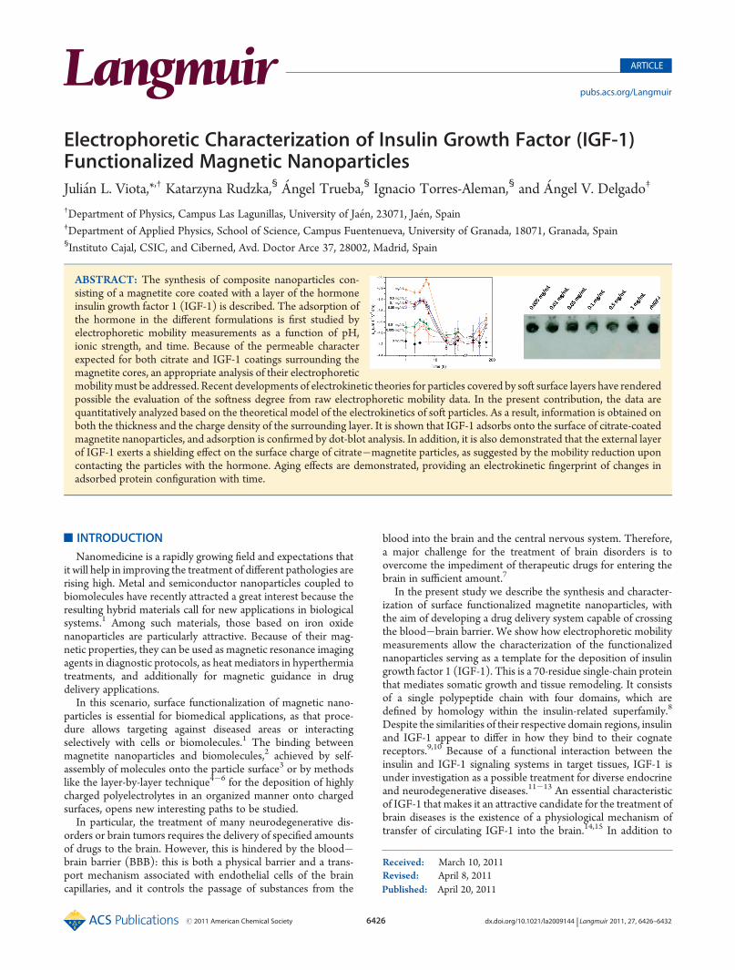
point of view, the difference between both kinds of particlescan be associated with an adsorption of IGF-1 on the magnetitesurface. Magnetite nanoparticles present an isoelectric point inthe vicinity of pH 6.5, and the IGF-1 coating produces a drop of|ue| for all pH values, and this effect is larger the higher theconcentration of IGF-1. It is interesting to note that there is aslight shift of the isoelectric point of the IGF-1-coated magnetiteparticles toward pH 7, whatever the hormone concentration.This shift can be explained by IGF-1 adsorption, considering itsabove-mentioned isoelectric point at pH 7.5. In addition, thecharge inhomogeneity of the protein coating may strongly affectthe isoelectric point in dependence of the flow penetration of theliquid in different regions of the protein layer. Such a shift of theisoelectric point is hence a manifestation of the changes in thesurface layer thickness when IGF-1 concentration is varied. Thiswas extensively discussed by Langlet et al.,29 both theoreticallyand by means of electrophoretic mobility measurements insuspensions of the MS2 bacteriophage (Figures 8 and 11 in ref29, respectively).We can get a further insight into the solid/liquid interface of
these composite systems if we estimate, among other relevant
quantities, the interfacial electric potential. The calculation ofthe electrokinetic potential (ζ-potential) from mobility data bymeans of the simple Helmholtz�Smoluchowski or more sophis-ticated methods, like those byO’Brien andWhite,40 may result ina misleading or even meaningless estimation of ζ for the protein-coated particles. This is because, as noted by Ohshima,17�20
electric potential, ionic concentrations, and liquid velocity dis-tribution must be considered, not only in the vicinity of the solidcharged surface but also in the soft (liquid penetrable) proteinlayer. For that reason, our discussion will, for the moment, limitto experimental mobilities. In the Soft-Particles Electrokineticssection we will come back to this issue.In Figure 2, the electrophoretic mobility of magnetite nano-
particles in the presence of different concentrations of IGF-1 isplotted as a function of NaNO3 concentration for two pHconditions far from magnetite isoelectric point, i.e., pH 3 andpH 9. As observed, whatever the pH, magnetite nanoparticlesshow an electrophoretic mobility that tends to zero as theNaNO3 concentration increases. This behavior, from an electro-phoretic point of view, is typical for hard spheres, in spite of thepossible presence of IGF-1 on the magnetite nanoparticles. Thisfact can be associated with a low adsorption of the hormone onthe surface of the magnetic carrier. For this reason, an alternativestrategy was applied.Sodium Citrate Coating. Although the interaction of IGF-1
with the magnetic carrier may be Coulombic, hydrophobic, and/or hydrogen bonding, Yamamoto et al.41 reported that a con-siderable high concentration of IGF-1 adsorption on acidicgelatin hydrogels was found, due to electrostatic interaction evenin solutions of high ionic strength. With this result in mind, andconsidering the large number of publications reporting highaffinity of biomolecules to citrate-gold nanoparticles,7 wedecided to try surface functionalization of the magnetite coreswith sodium citrate prior to adsorption of the IGF-1 hormone.This strategy is further supported by the fact that it was recentlyfound that large amounts of citrate-coated gold nanoparticles(15�50 nm) are able to cross the BBB.42 In fact, it has beenreported that the negative charge on these systems induces thebinding of plasma proteins, such as cationized albumin, whichtrigger the passage of the BBB and subsequent uptake of particles
Figure 1. Electrophoretic mobility of magnetite nanoparticles as afunction of pH, both bare (data labeled “0”) and after 24 h contact withIGF-1 solutions of the concentrations indicated.
Figure 2. Ionic strength effect on the electrophoretic mobility ofmagnetite nanoparticles at pH 3 and 9, after 24 h contact with IGF-1solutions of the concentrations indicated.
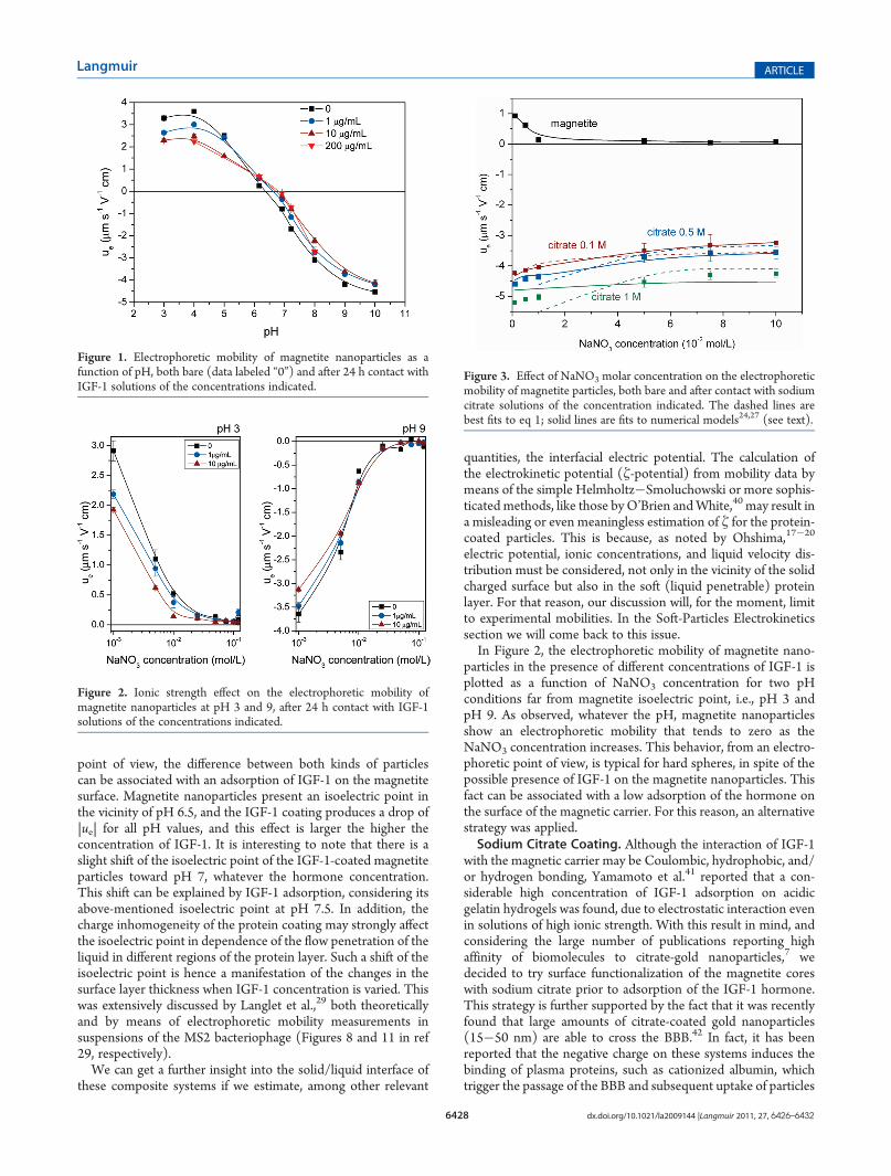
Figure 3. Effect of NaNO3 molar concentration on the electrophoreticmobility of magnetite particles, both bare and after contact with sodiumcitrate solutions of the concentration indicated. The dashed lines arebest fits to eq 1; solid lines are fits to numerical models24,27 (see text).

6429 dx.doi.org/10.1021/la2009144 |Langmuir 2011, 27, 6426–6432
Langmuir ARTICLE
by the brain parenchyma.43 Furthermore, the plasma residencetime of the particles can be increased by the protein coating, thusincreasing their chances to pass the barrier. It is worth mention-ing that the BBB, which is composed of a tightly sealed layer ofendothelial cells and numerous astrocytes (that regulate thepassage and diffusion of the substances), does not allow theaccess of (i) large molecules (if it is not via receptor-mediatedtranscytosis) as well as (ii) highly charged and (iii) hydrophilicmolecules.44,45 Therefore, it is necessary to study, first, if thecitrate groups are able to adsorb on themagnetic carriers, second,if the IGF-1 shows evidence of interacting with the citrate groups,and, third, which is the optimal concentration of IGF-1 that bestsuits the requirements concerning the final charge.Figure 3 shows the dependence of the electrophoretic mobility
with NaNO3 concentration for both naked and citrate-coatedmagnetite nanoparticles for three different concentrations (0.1,0.5, and 1 M) of sodium citrate. From the differences found, itis safe to conclude that citrate molecules adsorb onto the surfaceof the magnetite nanoparticles. In addition, the citrate-coatedmagnetite nanoparticles show a negative ue for all NaNO3 molarconcentration. In all cases the higher the sodium citrate con-centration the more negative the mobility.Dot-Blot Analysis. Figure 4 shows the results corresponding
to the dot-blot analysis. This confirmed the presence of immu-noreactive human IGF-1 on the nanoparticles. While no doseresponse was detected, probably due to the amounts of IGF-1present in the nanoparticle preparation saturating the antibody,specific IGF-1 immunoreactivity was found in all samples ofnanoparticles assayed.Interpretation Based on Soft Particle Electrokinetics. It is
worth to note that, contrary to the bare magnetite particles,citrate-coated ones show an electrophoretic mobility practicallyindependent of ionic strength, above 10�2 M concentration ofNaNO3 (Figure 3). This kind of behavior is typical of softparticles,17�20 as mentioned above. Hence, a study was under-taken of the surface electrical properties of the compositecitrate�magnetite and IGF-1�citrate�magnetite particles.Ohshima’s approach assumes that the electrical double layeraround a solid surface with adsorbed polymer or polyelectrolytecan be separated into two regions: a hydrogel layer of thicknessd which contains the adsorbed polyelectrolyte molecules and adiffuse double layer (like the one typically present in rigid particles)roughly starting at the limit of the polymer layer. It is assumedthat Donnan equilibrium is established between the two layers
because of the different salt concentrations inside the hydrogellayer and in the surrounding solution. Two electric potentials arethus involved in the overall description of the electrical doublelayer: the Donnan potential (ψDON) inside the hydrogel layerand the surface potential (ψ0) at the boundary between that layerand the bulk solution. The model assumes that the polyelec-trolyte segments can be regarded as resistance centers, exertingfrictional forces given by �γu (γ, friction coefficient; u, liquidvelocity inside the polyelectrolyte layer) on the liquid in thehydrogel layer. Considering this additional frictional force in theNavier�Stokes equation inside the hydrogel layer, an expressioncan be derived that relates the surface potential ψ0 and theelectrophoretic mobility ue. It is assumed that ionized groups ofcharge Ze of the polyelectrolyte molecule are uniformly distrib-uted over the polymer layer with a number density N (m�3) andthat the liquid contains a symmetrical electrolyte of valence zand bulk concentration n (m�3). More recently, Duval andOhshima24 analyzed, on the basis of the formalism alreadyproposed by Ohshima, the impact of the characteristic lengthand interfacial gradients associated with the polymer segmentdistribution on the electrophoretic behavior of the particle as awhole. Their numerical model applies to arbitrary values of theparticle charge, particle radius a, double-layer thickness, and for anycharge density profile of the membrane. These authors demon-strated that an approximate analytical expression for the mobilityfor sufficiently high ionic strengths and arbitrary λd, is given by
ue ¼ ZeN
ηλ2coshðλdÞ � 1coshðλdÞ þ ε
η
ψ0=km þψDON=λ
1=km þ 1=λð1Þ
where ε is the electric permittivity of the liquid medium, η isits viscosity, λ is a dimensionless frictional parameter given by(γ/η)1/2, and κm is the effective Debye�H€uckel parameter ofthe surface hydrogel layer, involving the contribution of the fixedcharge ZeN. The parameter 1/λ can be considered to character-ize the softness of the polyelectrolyte layer, and it is a measure ofthe flow penetration distance inside the polyelectrolyte mem-brane. The corresponding expressions forψDON,ψ0, and κm aregiven by17,20
ψDON ¼ kTze
sinh�1 ZN2zn
� �
ψ0 ¼ ψDON � kTze
tanhZeψDON
2kT
� �
km ¼ k coshzeψDON
kT
� �� �1=2ð2Þ
where k is Boltzmann’s constant, e is the electron charge, T is theabsolute temperature, and κ is the Debye�H€uckel parameter ofthe solution. It must be noted that eq 1 is valid if the electricdouble layer is thin in comparison with both the particle radiusand the membrane thickness.Briefly, the method followed involves fitting eq 1 to the
experimental data of the electrophoretic mobility of core/shellparticles as a function of electrolyte concentration. A least-squaresprocedure allows obtaining the best-fit parameters, d, ZN, and λ,for the model chosen, since the core size was independently ob-tained from TEM and DLS measurements.For the experiments, the electrophoretic mobility was mea-
sured immediately after the particles were mixed in solutionswith different salt concentrations. The results obtained using this
Figure 4. Results of the dot-blot analysis of IGF-1 coupled to citrate-coated ferromagnetic nanoparticles. Recombinant human IGF-1(rhIGF-1) was used as a positive control, and omission of primaryanti-IGF-1 was used for each sample to determine background inter-ference (upper row: results obtained with particles contacted with theIGF-1 concentrations indicated; lower row: control).

6430 dx.doi.org/10.1021/la2009144 |Langmuir 2011, 27, 6426–6432
Langmuir ARTICLE
procedure are displayed in Figure 3 (dashed lines), which showsthe expected electrophoretic nonzero mobility plateau for thehighest ionic strengths. This plateau is an evidence of the presenceof a soft, hydrodynamically permeable layer that surroundsthe particle.17 It must be recalled that, as depicted in Figure 2,the electrophoretic mobility of rigid particles at high ionicstrength should go to zero because of the so-called double-layer compression, i.e., the reduction of the potential at theelectrokinetic plane (the zeta potential) because of the screen-ing of the surface charge by the large amounts of counterionsin the double layer.The dashed lines in the Figure show how the model nicely fits
the experimental data in the high salt concentration range. Theiterative least-squares fitting procedure yielded the d, ZN, andλ values included in Table 1 for the three sodium citrate con-centrations. From the best-fit parameters, the effective Debye�H€uckel thickness (κm
�1), the Donnan potential (ψDON), andthe surface potential (ψ0) can be calculated using eq 2. Theresults from these calculations are also reported in Table 1. Thedeviations of the fits from the experimental data, found at the lowionic strength regime, are expected considering the validity ofeq 1, restricted to high ionic strengths. In addition, it is worth tomention that these deviations can be related to double-layerpolarization effects and/or to heterogeneity in surface layerdistribution following heterogeneous expansion of layer as aresult of swelling processes, as reported by Duval et al.25
For these reasons, we also used a more complete model, inwhich concentration polarization of the double layers is includedand where no limitations have to be applied to the parametervalues. This generalization of the theory of soft-particle electro-kinetics was elaborated, as above-mentioned, in refs 24 and 26 andextended to arbitrary volume fraction of solids in ref 27. For thiswork, the numerical treatment described by Ahualli et al.27 wasemployed. The fitting procedure involved again a least-squares
method, and the fitted parameters were d, λ, and the layer charge(Qcitrate, and from this the density of charged groups ZN). Asabove, the size and the charge (actually the electrokinetic charge)of the cores are known from independent determinations. Thebest-fit theoretical (solid) lines are included in Figure 3, andTable 2 displays the values of the fitted parameters. Note that theagreement with the experimental data is largely improved, asexpected, considering that the small particle radius of the magne-tite cores prevents the fulfilling of the condition κa. 1, except atvery high ionic strength. The full theory indicates that contact-ing the particles with increasing concentrations of sodiumcitrate gives raise to a thicker coating, but not necessarily amore highly charged one. It is suggested that the citrate ions willnot tend to form multilayers on the particles, due to electro-static repulsion, but rather we can picture the process as theformation of more extended coatings the larger the citrateconcentration, as citrate ions compete for adsorption sites onthe surface of the cores. Nevertheless, it is worth mentioningthat, in spite of the approximations involved in eq 1, the fittedparameters are very similar in both the approximate analyticalexpression and the numerical method. This reinforces thephysical significance of the Ohshima�Duval approach for theelectrokinetics of soft particles.
Table 1. Best-Fit Parameters of the Mobility Data in Figure 3 to the Duval�Ohshima Model24 a
Magnetite�Citratecitrate concn (M) d (nm) λa ZN (mmol/L) ψDON (mV) ψ0 (mV) κm
�1 (nm)
0.1 5 0.5 �70.6 �46.8 �28.2 2.89
0.5 11 0.7 �71.8 �48.6 �29.6 2.89
1.0 14 0.6 �77.6 �53.1 �33.2 3.93
Magnetite�0.1 M Citrate�IGF-1IGF-1 concn (mg/mL) d (nm) λa ZN (mmol/L) ψDON (mV) ψ0 (mV) κm
�1 (nm)
0.01 16 0.5 �56.6 �45.3 �27.2 2.89
0.5 23 0.5 �71.6 �43.6 �25.8 2.89a d = layer thickness, λa = softness parameter, ZN = layer charge density,ψDON = Donnan potential,ψ0 = surface potential, and κm
�1 = effective doublelayer thickness.
Table 2. Best-Fit Parameters of the Mobility Data in Figure 3to the Duval�Ohshima Numerical Model24,27 a
citrate concn (M) d (nm) λa Qcitrate (10�17 C) ZN (mmol/L)
0.1 2.5 0.3 �7.1 �88.5
0.5 3.3 0.5 �6.8 �61.3
1.0 6.0 0.5 �5.7 �28.7a d = layer thickness, λa = softness parameter,Qcitrate = total layer charge,and ZN = layer charge density.
Figure 5. Electrophoretic mobility of magnetite�citrate�IGF-1 nano-composites as a function of time, for different concentrations of IGF-1 insolution. Citrate concentration: 0.1 mol/L, pH 7.4.

6431 dx.doi.org/10.1021/la2009144 |Langmuir 2011, 27, 6426–6432
Langmuir ARTICLE
Plotted in Figure 5 are the experimentally determined electro-phoretic mobilities of IGF-1�citrate�magnetite complexes atpH 7.4 as a function of time, for six IGF-1 concentrations(0.005�1 mg/mL) and for a citrate concentration of 0.1 mol/L.As observed, ue is negative no matter the concentration of IGF-1present in the solution. At the initial time (when the citrate-coated magnetite nanoparticles are just mixed with the IGF-1solution), comparison between IGF-1-coated and uncoatedparticles suggests that the hormone has a screening effect onthe surface charge associated with the adsorbed citrate. In fact, forall samples it was found that the higher the concentration ofIGF-1, the higher is the screening of the charge, and thereforethe smaller is the electrophoretic mobility of the samples. Withthe aim of estimating the surface potential of the compositeIGF-1�citrate�magnetite nanoparticles, the analytical model(eq 1) was applied to two of the IGF-1-coated samples (IGF-1concentrations: 0.01 and 0.5 mg/mL). The results of the bestfits are presented in Table 1. As can be seen, our qualitativearguments are confirmed, and the higher the IGF-1 concentra-tion, the smaller the Donnan and surface potentials obtainedfor the composite particles. Note also that the layer expands(d increases) upon rising the hormone concentration, an addi-tional sign of protein coating.Aging Effects. Finally, with the aim of studying the process of
IGF-1 adsorption on the surface of citrate�magnetite particles,the electrophoretic mobility of the composite particles was studiedas a function of time (Figure 5). As observed, it was found thatfor all samples ue reaches a maximum between the third and thesixth hour after mixing the magnetite�citrate composites with theIGF-1 solutions. This maximum can be easily associated with anincreased deposition/adsorption of IGF-1 molecules on the sur-face of the citrate-coated nanoparticles. In addition, it is also worthto mention that a few hours after this maximum the mobilityincreases in absolute values and a sort of plateau is reached. Thisplateau in ue can be associatedwith a possible structural relaxation/desorption of the IGF-1molecule at the surface of the magnetite�citrate particles. According to Norde,46 once the protein moleculehas attached, it relaxes toward its equilibrium structure. Further-more, relaxation is retarded as the protein molecule has a stronginternal coherence, and it requires optimization of protein/surfaceinteractions, and a certain degree of “spreading” of the proteinmolecule over the sorbent surface, developing a larger numberof protein�surface contacts. Phenomenologically, a system isin equilibrium if no changes take place at constant surround-ings. It seems that this equilibrium is reached after ∼200 h.Note that after that time the value of ue for the IGF-1-coatedcitrate�magnetite nanoparticles is very similar whatever theinitial IGF-1 concentration within a certain range of values forue (3.7�3.8 μmV�1 s�1 cm). In all cases the presence of IGF-1on the surface can be confirmed from the difference betweenthe long-time mobility plateau and the ue value obtained in theabsence of hormone contact (ue ≈ �4.2 μmV�1 s�1 cm)(Figures 3 and 5).
’CONCLUSIONS
We have described the characterization of surface-functiona-lized magnetite prepared with the aim of developing magneticdrug delivery vehicles capable of surpassing the blood�brainbarrier and releasing their load of the insulin growth factor (IGF-1).To that aim, the magnetic nuclei were coated successively withsodium citrate and the IGF-1 hormone. All coating steps with
sodium citrate and IGF-1 were followed by electrophoreticmobilitymeasurements. The adsorption of the hormonemanifestsin a clear decrease of the absolute value of the electrophoreticmobility. In addition, the dot-blot analysis confirmed the presenceof immunoreactive human IGF-1 on the nanoparticles.
The presence of the hormone provides a soft coating onthe rigid core particles. This is demonstrated by the fact that theelectrophoretic mobility does not tend to zero at high ionicstrengths, so that a deformable layer can be identified which avoidsthe surface charge screening by ions in solution. A theoreticalmodel accounting for the existence of such charged layer is used forthe estimation of the friction parameter and Donnan potential ofthe adsorbed hormone coating. Interestingly, electrokinetic tech-niques such as electrophoresis can also be used to detect agingeffects associated with conformation changes in the adsorbed hor-mone. These results appear as a very promising connection be-tween electrokinetic characterization and design of nanoparticle-based drug vehicles.
’AUTHOR INFORMATION
Corresponding Author*E-mail: [email protected]; phoneþ34 953 212425; faxþ34 953212838.
’ACKNOWLEDGMENT
Financial support by Ministerio de Ciencia e Innovaci�on(Projects FIS2010-19493 and SAF 2009-06367-E), FEDERfunds, and Junta de Andalucía (Project PE-2008 FQM 3993)of Spain, is gratefully acknowledged. J.L.-V. was supported byMICINN, Spain, under the “Juan de la Cierva” programme. Thevaluable assistance of Dr. S. Ahualli in the numerical resolution ofsoft-particle electrokinetic equations is also acknowledged.
’REFERENCES
(1) Taton, T. A.; Mirkin, C. A.; Letsinger, R. L. Science 2000,289, 1757–1760.
(2) Viota, J. L.; Arroyo, F. J.; Delgado, A. V.; Horno, J. J. ColloidInterface Sci. 2010, 344, 144–149.
(3) Ulman, A. Chem. Rev. 1996, 96, 1533–1554.(4) Decher, G. Science 1997, 277, 1232–1237.(5) M€ohwald, H. Colloids Surf., A 2000, 171, 25–31.(6) Chanana, M.; Gliozzi, A.; Diaspro, A.; Chodnevskaja, I.; Huewel,
S.; Moskalenko, V.; Ulrichs, K.; Galla, H. J.; Krol, S. Nano Lett. 2005,5, 2605–2612.
(7) Viota, J. L.; Mandal, S.; Delgado, A. V.; Toca-Herrera, J. L.;M€oller, M.; Zanuttin, F.; Balestrino, M.; Krol, S. J. Colloid Interface Sci.2009, 332, 215–223.
(8) Blundell, T. L.; Humbel, R. E. Nature 1980, 287, 781–787.(9) de Meyts, P.; Whittaker, J. Nat. Rev. Drug Discovery 2002,
1, 769–783.(10) Whittaker, J.; Groth, A. V.; Mynarcik, D. C.; Pluzek, L.;
Gadsboll, V. L.; Whittaker, L. J.; Whittaker, J. J. Biol. Chem. 2001,276, 43980–43986.
(11) Pennisi, P.; Gavrilova, O.; Setser-Portas, J.; Jou, W.; Santopietro,S.; Clemmons, D.; Yakar, S.; LeRoith, D. Endocrinology 2006, 147, 2619–2630.
(12) Watanabe, T.; Miyazaki, A.; Katagiri, T.; Yamamoto, H.; Idei,T.; Iguchi, T. J. Am. Geriatr. Soc. 2005, 53, 1748–1753.
(13) Aleman, A.; Torres-Aleman, I. Prog. Neurobiol. 2009, 89, 256–265.
(14) Carro, E.; Nunez, A.; Busiguina, S.; Torres-Aleman, I. J. Neurosci.2000, 20, 2926–2933.

6432 dx.doi.org/10.1021/la2009144 |Langmuir 2011, 27, 6426–6432
Langmuir ARTICLE
(15) Nishijima, T.; Piriz, J.; Duflot, S.; Fernandez, A. M.; Gaitan, G.;Gomez-Pinedo, U.; Garcia Verdugo, J. M.; Leroy, F.; Soya, H.; Nu~nez,A.; Torres-Aleman, I. Neuron 2010, 67, 834–846.(16) Sara, V. R.; Clayton, K.; Cooke, P.; Craven, C. J.; Garcia-
Aragon, J.; Harmon, B.; Harvey, M.; Haase, H.; Plenderleith, M.;Richardson, N.; Sherrard, R.; Stahlbom, P.-A.; Walker, G.; Walsh,T. P. Dev. Brain Disfunct. 1996, 9, 85–92.(17) Ohshima, H. In Interfacial Electrokinetics and Electrophoresis;
Delgado, A. V., Ed.; Marcel Dekker: New York, 2002; pp 123�146.(18) Ohshima, H.; Makino, K.; Kato, T.; Fujimoto, K.; Kondo, T.;
Kawaguchi, H. J. Colloid Interface Sci. 1993, 159, 512–514.(19) Ohshima, H. Colloids Surf., A 1995, 103, 249–255.(20) Ohshima, H. In Electrical Phenomena at Interfaces: Fundamen-
tals, Measurements, and Applications; Ohshima, H., Furusawa, K., Eds.;Marcel Dekker: New York, 1998; pp 19�55.(21) Hill, R. J.; Saville, D. A. Colloids Surf., A 2005, 267, 31–49.(22) Hill, R. J.; Saville, D. A.; Russel, W. B. J. Colloid Interface Sci.
2003, 258, 56–74.(23) Hill, R. J. Phys. Rev. E 2004, 70, 051406.(24) Duval, J. F. L.; Ohshima, H. Langmuir 2006, 22, 3533–3546.(25) Duval, J. F. L.; Gaboriaud, F. Curr. Opin. Colloid Interface Sci.
2010, 15, 184–195.(26) Lopez-Garcia, J. J.; Grosse, C.; Horno, J. J. Colloid Interface Sci.
2003, 265, 327–340.(27) Ahualli, S.; Jim�enez, M. L.; Carrique, F.; Delgado, A. V.
Langmuir 2009, 25, 1986–1997.(28) Clement, A.; Gaboriaud, F.; Duval, F. J. L.; Farn, J. L.; Jenney,
A. W.; Lithgow, T.; Wijburg, O. L. C.; Hartland, E. L.; Strugnell, R. A.PLoS One 2008, 3, e3817.(29) Langlet, F.; Gaboriaud, F.; Gantzer, C.; Duval, J. F. L. Biophys. J.
2008, 94, 3293–3312.(30) Dague, E.; Duval, J. F. L.; Jorand, F.; Thomas, F.; Gaboriaud, F.
Biophys. J. 2006, 90, 2612–2621.(31) Duval, J. F. L.; Buscher, H. J.; van de Belt-Gritter, B.;
van der Mei, H. C.; Norde, W. Langmuir 2005, 21, 11268–11282.(32) Gosselin, F.; Duval, J. F. L.; Simonet, J.; Ginevra, C.; Gaboriaud,
F.; Jarraud, S.; Mathieu, L. Colloids Surf., B 2011, 82, 283–290.(33) Hyono, A.; Gaboriaud, F.; Mazda, T.; Takata, Y.; Ohshima, H.;
Duval, J. K. L. Langmuir 2009, 25, 10873–10885.(34) Ramos-Tejada,M.M.; Ontiveros, A.; Viota, J. L.; Duran, J. D. G.
J. Colloid Interface Sci. 2003, 268, 85–95.(35) Viota, J. L.; de Vicente, J.; Duran, J. D. G.; Delgado, A. V.
J. Colloid Interface Sci. 2005, 284, 527–541.(36) Viota, J. L.; de Vicente, J.; Ramos-Tejada, M.M.; Dur�an, J. D. G.
Rheol. Acta 2004, 43, 645–656.(37) Massart, R. IEEE Trans. Magn. 1981, 17, 1247–1248.(38) Holland, T. A.; Tabata, Y.; Mikos, A. G. J. Controlled Release
2005, 101, 111–125.(39) Goodman, C. M.; McCusker, C. D.; Yilmaz, T.; Rotello, V. M.
Bioconjugate Chem. 2004, 15, 897–900.(40) O’Brien, R. W.; White, L. R. J. Chem. Soc., Faraday Trans. 1978,
274, 1607–1626.(41) Yamamoto, M.; Ikada, Y.; Tabata, Y. J. Biomater. Sci. Polym.
2001, 12, 77–88.(42) Sonavane, G.; Tomoda, K.; Makino, K. Colloids Surf., B 2008,
66, 274–280.(43) Sousa, F.; Mandal, S.; Garrovo, C.; Astolfo, A.; Bonifacio, A.;
Latawiec, D.; Menk, R. H.; Arfelli, F.; Huewel, S.; Legname, G.;Galla, H.-J.; Krol, S. Nanoscale 2010, 2826–2834.(44) Rubin, L. L.; Staddon, J. M. Annu. Rev. Neurosci. 1999,
22, 11–28.(45) Spencer, B. J.; Verma, I. M. Proc. Natl. Acad. Sci. U.S.A. 2007,
104, 7594–7599.(46) Norde, W. In Physical Chemistry of Biological Interfaces; Baszkin,
A., Norde, W., Eds.; Marcel Dekker: New York, 2000; p 121.


