Embed Size (px)
Citation preview
Emotion Recognition and Psychosis-Proneness: Neural and Behavioral Perspectives
CitationGermine, Laura Thi. 2012. Emotion Recognition and Psychosis-Proneness: Neural and Behavioral Perspectives. Doctoral dissertation, Harvard University.
Permanent linkhttp://nrs.harvard.edu/urn-3:HUL.InstRepos:9556122
Terms of UseThis article was downloaded from Harvard University’s DASH repository, and is made available under the terms and conditions applicable to Other Posted Material, as set forth at http://nrs.harvard.edu/urn-3:HUL.InstRepos:dash.current.terms-of-use#LAA
Share Your StoryThe Harvard community has made this article openly available.Please share how this access benefits you. Submit a story .
Accessibility

© 2012 - Laura Thi Germine
All rights reserved
iii
Professor Christine Hooker Laura Germine
Emotion Recognition and Psychosis-Proneness:
Neural and Behavioral Perspectives
Abstract
Schizophrenia is associated with deficits in social cognition and emotion processing, but
it is not known how these deficits relate to other domains of neurocognition and whether they
might contribute to psychosis development. The current dissertation approaches this question by
looking at the relationship between psychosis proneness and face emotion recognition ability, a
core domain of social-emotional processing.
Psychosis proneness was inferred by the presence of psychosis-like characteristics in
otherwise healthy individuals, using self-report measures. Face emotion recognition ability was
found to be associated with psychosis-proneness across four large web-based samples and one
lab sample. These associations were relatively specific, and could not be explained by
differences in face processing or IQ. Using functional magnetic resonance imaging (fMRI),
psychosis-proneness was linked with reduced neural activity in brain regions that underlie
normal face emotion recognition, including regions that are implicated in self-representation.
Additional experiments were conducted to explore psychosis-proneness related differences in
self-representation, and a relationship was revealed between cognitive-perceptual (positive)
dimensions of psychosis-proneness and (1) flexibility in the body representation (as measured by
the rubber hand illusion), and (2) self-referential source memory (but not self-referential
iv
recognition memory). Neither of these relationships, however, explained the association
between psychosis-proneness and face emotion recognition ability.
These findings indicate that psychosis vulnerability is related to neural and behavioral
differences in face emotion processing, and that these differences are not a secondary
characteristic of psychotic illness. Moreover, poorer emotion recognition ability in psychosis-
prone individuals is not explained by generalized performance, IQ, or face processing deficits.
Although some dimensions of psychosis-proneness were related to differences in measures of
self-representation, no evidence was found that these abnormalities contribute to psychosis-
proneness related differences in emotion recognition ability.
v
Table of Contents
Background and Introduction….……………………………………………..……....1
Schizophrenia and Social-emotional Processing……………………………..1
Emotion Recognition in Schizophrenia………………………………………4
The High Risk Approach…………………………………………..…………7
Psychosis-proneness and Emotion Recognition: Previous Findings………..10
Emotion Recognition Deficits: Generalized or Specific Impairment? …….13
Research Questions…………………………………………………………………16
Experiment #1: Paper #1: Face emotion recognition is related to individual
differences in psychosis-proneness (pp. 1-11)….…………………………………..19
Experiment #2: Paper #2: Social anhedonia is associated with neural abnormalities
during face emotion processing (pp. 935- 945)…..……………..………………….40
Experiment #3a: Paper #3: Psychosis-proneness and the rubber hand illusion of body
ownership………………………………………….………………………………..74
Additional Findings…………………………………………………………....…...96
Experiment #3b: Psychosis-proneness, Flexibility in Body Representations,
and Face Emotion Recognition (FER)……………………………….…….96
Experiment #3c: Dimensions of Psychosis-proneness and FER……….…..98
Experiment #4: Psychosis-proneness and Self-referential Processing…….102
Experiment #4a: Self-referential source memory………………....103
Experiment #4b: Self-referential recognition memory…………....106
Experiment #5: Psychosis-proneness and Voice Emotion Recognition.......110
Discussion and Conclusion……………………………………………….……......113
Summary of Findings……………………………………………………....113
vi
Psychosis-proneness and FER: Interpretation of Behavioral Findings…......113
Psychosis-proneness and FER: Interpretation of Neural Findings………….115
Psychosis-proneness, Self-representation, and FER………………………...117
Psychosis-proneness and Voice Emotion Recognition………………….......120
The Relationship between Self-representation and Positive Symptoms….....120
Limitations……………………………………………………………….….122
Concluding Thoughts……………………………………………….………124
References………………………………………………………………………..…125
vii
Acknowledgements
I would like to thank my advisor, Dr. Christine Hooker, for her tremendous support and
mentorship over the course of my doctoral degree. In addition, I would like to thank the
members of my dissertation committee: Dr. Ken Nakayama, Dr. Jeremy Wilmer, and Dr. Felix
Warneken for helpful comments and assistance as I was preparing my dissertation experiments,
as well as Dr. Matthew Nock for serving as external examiner. I am also grateful to all the
members of the Harvard Social Neuroscience & Psychopathology and Harvard Vision Science
Labs for their support and feedback, particularly Laura Tully, Sarah Hope Lincoln, David
Dodell-Feder, Lucia Garrido, Taylor Benson, Roger Mercado, Francesca Cohen, Lori Bruce, and
Caitlin Carey as well as all the research volunteers that made these experiments possible.
Finally, I would like to thank my son, Raphael Strudwick, for taking naps that allowed this
dissertation to be written, and my husband, Tim Strudwick, for taking Raphael out of the house
during those times when naps were not possible (in addition to providing many helpful
comments). This work was generously funded by Harvard University research funds to Dr.
Christine Hooker and a National Science Foundation Graduate Research Fellowship.
1
Background and Introduction
Schizophrenia and Social-emotional Processing
Among the most pervasive and disabling deficits in schizophrenia are difficulties with
social interaction and emotional processing (APA, 2000; Bleuler, 1950; Kraepelin & Gosline,
1918). These deficits have been observed across schizophrenia spectrum disorders and include
poor social cognitive abilities (Pinkham, Hopfinger, Ruparel, & Penn, 2008) and poor social
functioning (Hooley, 2010; Hooker & Park, 2002). Social functioning has a tremendous impact
on an individual’s mental and physical health. The degree to which an individual seeks and
successfully obtains social support predicts risk for developing mental disorders (Dalgard, Bjork,
& Tambs, 1995), symptom severity (Norman et al., 2005), likelihood of remission (Corrigan &
Phelan, 2005), and chance of relapse after remission (Hooley, 2010). Number and quality of
social relationships predicts mortality and a wide range of health-related conditions including
vulnerability for infection, metabolic illness, and cancer (Miller, Chen, & Cole, 2009),
comparable to the relationship between smoking and risk of mortality from all causes (Cohen et
al., 1997; House, Landis, & Umberson, 1998). It is perhaps not surprising then that greater
deficits in social cognition in schizophrenia predict poorer outcomes and greater functional
disability (Green & Horan, 2010; Hooker & Park, 2002). Schizophrenia-related deficits in
social, cognitive, and emotional processing include difficulty recognizing and understanding
social stimuli, acquiring appropriate social and emotional responses, low-level mental state
inference, and context-sensitive mental state inference (Ochsner, 2008). The term “social-
emotional” will be used to refer to this range of processes, with the observation that social and
emotional functions are difficult to separate and often contribute to the same overall adaptive
capabilities (Keltner & Kring, 1998).
2
Symptoms of schizophrenia have traditionally been divided into positive and negative
symptom clusters thought to represent distinct domains of pathology (Crow, 1985). Positive
symptoms represent an excess of functions (Hughlings & Jackson, 1931) and comprise the so-
called psychotic symptoms: delusions, thought disorder, and hallucinations in visual, auditory,
somatosensory, olfactory, and gustatory domains (APA, 2000). Negative symptoms reflect a
deficit of functions (Andreasan, 1982) or social/cognitive withdrawal (Thiemann et al., 1987)
and include blunted expression of emotion and reduced feelings of pleasure (also known as
affective flattening and anhedonia; APA, 2000). Prominent negative symptoms tend to be
indicative of greater illness severity and poorer outcome (Addington, Addington, & Maticka-
Tyndale, 1991; Davidson & McGlashan, 1997; Ho et al., 1998) and respond less or not at all to
traditional antipsychotic treatment (Johnstone, Crow, & Ferrier, 1983; Kane & Mayerhoff,
1989). Social-emotional deficits are more closely linked with negative symptoms than positive
symptoms, in terms of classification (that is, social-emotional deficits are themselves negative
symptoms) but also in associations between specific social-emotional processes and symptom
severity (Lincoln, Mehl, Kesting, & Rief, 2011).
Deficits in social-emotional processing can be observed from the earliest stages of
psychotic disorders, many years before an individual’s first psychotic episode (Walker, Grimes,
Davis, & Smith, 1993). As mentioned, abnormalities in social-emotional functions are among
the negative symptoms of schizophrenia and include affective flattening and anhedonia.
Affective flattening is the reduced tendency among individuals with schizophrenia to display or
express emotion (APA, 2000). Importantly, affective flattening seems to be present even when
an individual’s experience of emotions and emotional intensity appears to be normal and thus
3
may reflect a failure to outwardly express felt emotions rather than a failure of emotion
generation (Keltner & Kring, 1998; Kring, Kerr, Smith, & Neale, 1993; Kring & Neale, 1996).
Although emotional experiences seem to be generally intact, schizophrenia spectrum
disorders are consistently linked to deficits in the experience of pleasure, or anhedonia.
Anhedonia has been noted particularly in the social domain, where patients report reduced
pleasure from social interactions and a reduced drive for social affiliation (Bleuler 1911/1950;
Kwapil, 1998). Early investigators, such as Rado (1953), described anhedonia as a chronic and
ubiquitous feature of psychotic disorders representing a primary abnormality in emotion
processing.
Positive symptoms can also be linked with social-emotional processing, as delusions and
hallucinations are frequently centered around social content. Some of the most common
delusions, for example, involve interpersonal threat, persecution, and/or social evaluation (APA,
2000). Auditory hallucinations of a human voice/voices are present in 50-70% of individuals
with schizophrenia (Hoffman et al., 2000). Delusions and cognitive distortions are also
typically grounded in socially or culturally salient themes, suggesting a relationship between
social conventions, social learning, and the content of delusional thought processes (Tateyama et
al., 1998). Social-emotional processing deficits, however, are less consistently linked with
positive symptoms (Sergi et al., 2007).
In the last two decades, translational research in cognitive neuroscience and psychology
has indicated that schizophrenia and related psychotic disorders are linked to measurable
differences in social-emotional aspects of neurocognition. These deficits have been measured
across a wide range of functions and include deficits in theory of mind (Frith & Corcoran, 1996),
source monitoring (Vinogradov et al., 1997), self-referential processing (Fisher et al., 2008), and
4
emotion perception (Kohler et al., 2010). Deficits in the ability to identify vocal and facial
expression of emotion, in particular, are a robust and highly replicated finding (Edwards,
Jackson, & Pattison, 2002; Edwards, Pattison, Jackson, & Wales, 2001; Habel et al., 2000;
Kohler, Bilker, Hagendoorn, Gur, & Gur, 2000; Kohler & Brennan, 2004; Mandal, Pandey, &
Prasad, 1998; Mueser, Penn, Blanchard, & Bellack, 1997; Penn et al., 2000).
Emotion Recognition in Schizophrenia
Emotion recognition is a widely studied neurocognitive ability that is a fundamental
component of social understanding and social functioning. Emotion recognition is a source of
rich moment-by-moment knowledge of another person’s changing mental states that shares
universal characteristics across cultures (Ekman, 1992), can occur automatically (Ohman, 2002),
and does not necessarily require conscious perception (Adolphs, 2002). Accurate and efficient
emotion recognition across modalities enables a person to understand and respond to the
changing beliefs, feelings, and intentions of another person. At the same time, it allows us to
judge whether our own beliefs, feelings and intentions are being understood and flexibly
navigate social interactions. Moreover, emotion recognition is a building block for other areas
of social and emotional processing (Adolphs, 2003), supporting the development of higher-level
social behavior by providing rich and instant feedback as social skills are refined through
development (Keltner & Kring, 1998). Specialized systems in the brain have been identified for
emotion recognition whose functions are dissociable from higher-order reasoning systems
(Adolphs, 2003) and from perceptual systems that process related types of information, such as
person identity (Haxby, Hoffmann, & Gobbini, 2000). Many studies of the neural substrates of
face emotion recognition (FER) have identified a network of regions involved in face emotion
5
processing, including the amygdala, superior temporal cortex, medial prefrontal cortex, and
somatosensory cortices (Adolphs et al., 2000; Adolphs, 2003; Allison, Puce, & McCarthy, 2000;
Haxby et al., 2000). Looking at variations in FER ability provides a specific and tractable
starting point for studying social-emotional processing in psychosis.
Evidence that FER is impaired in schizophrenia is robust and observed across a range of
measures (Edwards et al., 2001; Edwards et al., 2002; Habel et al., 2000; Kohler et al. 2000;
Kohler & Brennan, 2004; Mandal et al., 1998; Mueser et al. 1997; Penn, et al., 2000). FER
ability also predicts social functioning more so than other related neurocognitive and social
cognitive variables (Hooker & Park, 2002). Many studies have found schizophrenia-related
abnormalities in emotion recognition brain networks. Functional neuroimaging studies of
emotion recognition in schizophrenia have repeatedly identified abnormal responses in the
amygdala (Das et al., 2007; Gur et al., 2007a; Gur et al., 2002; Hall et al., 2004; Hempel et al.,
2003; Holt et al., 2006; Kosaka et al., 2002; Phillips et al., 1999; Pinkham et al., 2008; Schneider
et al., 1998; Taylor, Liberzon, Decker, & Koeppe, 2002; Williams et al., 2004) as well as
abnormalities in the medial prefrontal cortex (Brüne et al., 2008; Brunet, Sarfati, Hardy-Baylé, &
Decety, 2003; Das et al., 2007; Vinogradov, Luks, Schulman, & Simpson, 2008; Whitfield-
Gabrieli et al., 2009; Williams et al., 2004), superior temporal cortex (Brüne et al., 2008;
Leitman et al., 2008; Pinkham et al., 2008), and somatosensory-related cortices (Farrer et al.,
2004; Spence et al., 1997; Waberski et al., 2004). Structural measures suggest reduced volume
and/or gray matter volume in schizophrenia in temporal cortices (Davidson & Heinrichs, 2003;
Siever & Davis, 2004; Wright et al., 2000) and the amygdala-hippocampal complex (Exner et al.,
2004; Lawrie & Abukmeil, 1998; Nelson, Saykin, Flashman, & Riordan, 1998; Wright et al.,
2000).
6
Given the consistent associations between FER and schizophrenia, it has been suggested
that FER may serve as a useful neurocognitive endophenotype (Gur et al., 2007b). An
endophenotype is an observable characteristic that lies intermediate between a mental disorder
and the underlying genetic and/or neurobiological substrates of that disorder (Gottesman &
Gould, 2005). The concept was developed to provide a way for researchers to deal with
problems arising from diagnostic overlap, heterogeneity, and complexity in investigations of
high-level mental disorders (Insel & Cuthbert, 2009). Characteristics of a useful psychiatric
endophenotype include an association with illness, state-independence, heritability,
cosegregation with illness within families, and presence in unaffected relatives of a person with
the illness at higher rates than in the general population (Gottesman & Gould, 2005). FER
abnormalities are consistently associated with schizophrenia and appear to be state independent,
as they are measurable before psychosis-onset (Häfner et al., 2003; Walker et al., 1993; Yung &
McGorry, 1996), in first-episode psychosis (Edwards et al., 2001), and in individuals not
experiencing an active psychotic episode (Wolwer, Streit, Polzer, & Gaebel, 1996). FER ability
is also heritable (Greenwood et al., 2007), and impaired in individuals at familial risk of
developing schizophrenia but not themselves mentally ill, with FER performance intermediate
between patients and healthy control participants (Kee, Horan, Mintz, Green 2004; Bediou et al.,
2007). Based on the endophenotype approach, understanding FER deficits may provide a
means of linking high-level impairments with differences in underlying genetics and biological
pathways.
7
The High-risk Approach
The current literature has demonstrated that schizophrenia is associated with
abnormalities in FER behaviorally and neurally. Interpreting findings in schizophrenia research
can be challenging, however, due to the many secondary and/or confounding factors associated
with having a severe mental disorder. Schizophrenia and related psychotic disorders are
associated with significant psychosocial stress, long-term deterioration of function, chronic drug
treatment, and other secondary characteristics that significantly impact functioning and
neurocognition, but are not part of the core pathophysiology of the disease (Lenzenweger, 2006).
Social deficits could arise or be exacerbated, for example, by the widespread social stigma
associated with having a severe mental disorder in our society. Moreover, severe deficits in
sustained attention and information processing (Heinrichs, 2001) tend to impact performance
across a wide range of tasks, making it more difficult to distinguish specific impairments from
the background of generalized impairments in schizophrenia patients (Kerr & Neale, 1993).
These confounds make it difficult to interpret findings in schizophrenia, which may arise from
any of these factors.
One way of addressing the problem of secondary confounds is by investigating emotion
processing abnormalities in individuals who are at high-risk of developing schizophrenia or other
psychotic disorders, but are not psychotic. High-risk groups that have been used in this type of
investigation include individuals who show early clinical signs of psychosis (clinical high-risk
and/or schizophrenia prodromes), relatives of people with schizophrenia or related disorder
(genetic high-risk), individuals with schizotypal personality disorder, and nonpsychotic
individuals who are high in schizotypy – a latent dimension of psychosis vulnerability thought to
be expressed as subthreshold psychosis-like characteristics (Lenzenweger 2006; Meehl 1962,
8
1990). In the current set of studies, the focus will be on this latter form of psychometrically-
defined psychosis risk, as inferred by the presence of self-reported psychosis-like characteristics.
As the term schizotypy has come to be associated with the latent genetic predisposition to
developing schizophrenia (Meehl 1962/1990), the more etiologically-neutral term psychosis-
proneness will be used to refer to this form of psychosis vulnerability.
Individuals who are high in psychosis-proneness show similar symptoms and
characteristics as those with schizophrenia, but in a subthreshold or attenuated form. These
characteristics can include social isolation, social and physical anhedonia, magical thinking,
suspiciousness, and perceptual aberrations (Chapman & Chapman, 1980; Kwapil, 1998;
Lenzenweger et al., 2006; Rado, 1953; Raine, 1991). These can be assessed via interview, but
also by self-report / questionnaire measures. High scores on self-report measures are thought to
indicate an underlying genetic or biological predisposition to developing psychosis, as
individuals with high psychosis-proneness scores are more likely to develop a schizophrenia
spectrum disorder (Kendler, Thacker, & Walsh, 1996; Kwapil, 1998; Kwapil, Miller, Zinser,
Chapman, & Chapman, 1997). Along the same lines, psychosis-proneness scores are higher in
the relatives of schizophrenia patients than control participants (Faraone, Green, Seidman, &
Tsuang, 2001; Kendler & Walsh, 1995).
Many individuals high in psychosis-proneness qualify for a diagnosis of schizotypal
personality disorder, so that these two groups overlap (high psychometric psychosis-proneness
and schizotypal personality). Some measures of psychosis-proneness are specifically designed to
identify people with schizotypal personality traits, as schizotypal personality disorder is a
diagnosis defined by the presence of a certain number and type of schizophrenia-like symptoms
(Raine, 1991). Those with schizotypal personality disorder will almost invariably score high on
9
measures of psychosis-proneness. Schizotypal personality disorder is best conceptualized as a
particular phenotype associated with high levels of psychosis-proneness. Indeed, findings from
samples of individuals high in psychosis-proneness and schizotypal personality disorder often
converge (Raine, 1991; 2006).
As a means of identifying high-risk participants, self report measures of psychosis-
proneness have the advantage of yielding participant groups that are both phenotypically
homogeneous (unlike samples of genetic high-risk participants) and nontreatment-seeeking
(unlike clinically identified individuals with schizotypal personality disorder). Importantly,
research has indicated that scoring high in self-reported measures of psychosis-proneness is
related to a 10-fold or greater increased risk of developing psychosis relative to the baseline risk
in the normal population (Blanchard et al., 2011; Kwapil, 1998).
Studying dimensions of psychosis-proneness rather than diagnostic categories also maps
well onto recent theoretical shifts away from diagnostic categories and towards symptom
dimensions (Cuthbert & Insel, 2010). There is an increasing realization among mental health
researchers that mental disorders may not respect the diagnostic boundaries defined by the DSM
and that behavioral and neural dimensions may have more biological reality than specific
diagnoses (Cuthbert & Insel, 2010, Hyman, 2007; Smoller et al., 2008). Studying mental
disorders through a dimensional rather than categorical framework also increases phenotype
homogeneity, a major obstacle thusfar in mental health research, and facilitates a translational
approach (Hyman, 2010). Dimensional frameworks also provide clearer connections between
research on mental disorders and research on individual differences. This is especially relevant
as recent large-scale investigations in psychiatric genetics have revealed that a large proportion
of the genetic vulnerability to developing schizophrenia is based on variations in commonly
10
occurring alleles (Purcell et al., 2009). This means that genetic risk for schizophrenia is, in large
part, based on common alleles of small effect and thus may manifest itself in terms of individual
differences in neural and neurocognitive endophenotypes (Braff et al., 2007; Gottesman &
Gould, 2003; Ivleva et al., 2009; Meyer-Lindenberg & Weinberger, 2006).
Investigations of FER and psychosis vulnerability, from the perspective of individual
differences, are thus both timely and potentially informative given shifting approaches to
studying mental disorders and recent advances in our understanding of the way genetic
vulnerability is related to psychosis development.
Psychosis-proneness and Emotion Recognition: Previous Findings
FER deficits have been observed in individuals with schizotypal personality disorder
(Mikhailova et al., 1996; Waldeck & Miller, 2000), individuals at clinical high-risk of
developing schizophrenia (Addington et al., 2008), and relatives of individuals with
schizophrenia (Bediou et al., 2007; Loughland, Williams, & Harris, 2004; Toomey et al., 1999).
Previous studies have indicated that high levels of psychometric psychosis-proneness are related
to reduced social functioning (Henry, Bailey, & Rendell, 2008; Jahshan & Sergi, 2007) and
social cognitive abilities (Langdon & Coltheart, 2001; Langdon & Coltheart, 2004). Research on
the relationship between psychometric psychosis-proneness and FER has been less clear. Some
studies have reported reduced FER performance among psychosis-prone individuals (Brown &
Cohen, 2010; Poreh, Whitman, Weber, & Ross, 1994; van 't Wout et al., 2004; Williams, Henry,
& Green, 2007). Other studies have not found significant differences in FER related to
psychosis-proneness (Jahshan & Sergi, 2007; Toomey & Schuldberg,1995; Toomey et al.,
11
1999). Mixed findings may be due to lack of sensitivity in FER measures, symptom specific
relationships, or small sample sizes (Brown & Cohen, 2010).
Neuroimaging studies of social-emotional processing and psychosis-proneness (including
schizotypal personality disorder) are limited, and hence it is difficult to know how and to what
extent differences in social or emotional measures are related to differences in underlying neural
circuitry. Two studies that focus on emotion processing and psychosis-proneness both used tasks
engaging cognitive control systems. A study using an emotional stroop task found that
individuals high vs. low in psychosis-proneness (based on positive psychosis-like characteristics)
show different patterns of activity in right dorsolateral prefrontal cortex, hippocampus, and
amygdala when viewing negative emotion words (Mohanty et al., 2005). A study using an
emotion reappraisal task found reduced functional connectivity in frontal and limbic areas in
individuals high in psychosis-proneness when compared to a control sample (Modinos, Ormel, &
Aleman, 2010). These studies suggest abnormalities in fronto-limbic circuits during emotion
processing, which is consistent with findings in schizophrenia samples (Li, Chan, McAlonan, &
Gong, 2010).
Studies of structural brain abnormalities in regions implicated in FER have been more
plentiful, although these studies are mostly conducted with individuals diagnosed with
schizotypal personality disorder and it is not clear how structural differences might relate to FER
ability. One of the most consistent structural abnormalities in chronic schizophrenia is reduced
volume of the superior temporal gyrus (Davidson & Heinrichs, 2003; Downhill et al., 2001;
Siever & Davis, 2004; Wright, et al., 2000). Reduced superior temporal gyrus volume is also a
robust finding in schizotypal personality disorder (Dickey, McCarley, & Shenton, 2002; Dickey
et al., 2002; Dickey et al., 2003; Downhill et al., 2001; Siever & Davis, 2004). Reduced frontal
12
lobe volume is less widespread (Hazlett et al., 2008; Kawasaki et al., 2004; Siever et al., 2002;
Suzuki et al., 2005). Medial temporal lobe abnormalities (amygdala, hippocampus,
parahippocampal gyrus), although robustly identified in schizophrenia samples (Honea, Crow,
Passingham, & MacKay, 2005), are inconsistently identified in schizotypal personality disorder
(Siever & Davis, 2004). It has been suggested that reduced medial temporal lobe volume may be
more prevalent among familial or genetic high-risk individuals (Seidman et al., 2003; van Rijn,
Aleman, Swaab, & Kahn, 2005) than in nonfamilial high-risk phenotypes like schizotypal
personality disorder (Velakoulis et al., 2006). Altogether, these findings led Siever & Davis
(2004) to hypothesize that normal or compensatory frontal lobe function and/or subcortical
stability may protect psychosis-prone individuals from primary temporal lobe abnormalities that
could otherwise develop into full-blown psychosis. Along the same lines, Nakamura et al.
(2005) used diffusion tensor imaging to show that while schizophrenia patients have
abnormalities in both frontal-temporal and cortical-limbic connections, schizotypal personality
disorder was associated with reduced connectivity in frontal-temporal tracts only. Notably,
progressive gray matter loss in the medial temporal lobes and orbital prefrontal cortex has been
observed at the transition to psychosis (Pantelis et al., 2007; Velakoulis et al., 2006). Altogether,
structural brain measures suggest that temporal cortical abnormalities are related to psychosis-
proneness or vulnerability, with other (e.g. frontal and medial temporal) abnormalities emerging
and/or progressing with psychosis development.
Volumetric studies focusing on parietal cortex also reveal reduced volume in widespread
regions among schizophrenia patients. Reduced parietal lobe volume in schizotypal personality
disorder, however, appears to be limited to the postcentral gyrus / somatosensory cortex (Zhou et
al., 2007). This is consistent with behavioral evidence that basic aspects of somatosensory
13
processing are impaired in psychosis-prone participants (Chang & Lenzenweger, 2005;
Lenzenweger, 2000) and the relatives of schizophrenia patients (Chang & Lenzenweger, 2001,
2004, 2005; Hooley & Delgado, 2001; Lenzenweger, Nakayama, Chang, & Hooley, 2003).
Abnormalities in somatosensory representation may be related to the higher-level deficits in
emotion and self-other processing characteristic of psychosis, as would be predicted by action-
perception models of social cognition (Brunet-Gouet & Decety, 2006).
Emotion Recognition Deficits: Generalized or Specific Impairment?
Abnormalities in multiple systems could contribute to schizophrenia or psychosis-
proneness related deficits in FER. FER deficits could, for example, reflect generalized
performance deficits (Mohamed et al., 1999; Whittaker, Deakin, & Tomenson, 2001).
Schizophrenia is related to abnormalities in attention, motivation, and disorganized behavior
(Docherty, 2005) all of which could impact FER performance in the lab and in everyday life, but
do not reflect a specific social-emotional deficit. A generalized impairment explanation would
not reduce the importance of social-emotional processing deficits (e.g. deficits in attention that
drive FER deficits could still negatively impact social functioning), but it does suggest
mechanisms for impairment outside of specialized social-emotional processing networks.
Another possibility is that FER deficits reflect broader deficits in social perception or
face processing. Some studies have, for example, found similar deficits in emotional and
nonemotional aspects of face processing among patients with schizophrenia (Addington &
Addington, 1998) whereas other studies have found greater or selective impairments in
emotional face processing (Bediou et al., 2005). Behavioral, neuropsychological, and
neuroimaging studies indicate that FER relies on dissociable systems from face identity
14
recognition (Bruce & Young, 1986; Haxby et al., 2000), and that aspects of FER can be
selectively disrupted (Calder, 1996). Among schizophrenia patients, some studies have found
abnormal activity in inferior temporal face processing areas during recognition of non-emotional
face information (Gur et al., 2002a; Herrmann, Ellgring, & Fallgatter, 2004; Quintana et al.,
2003), whereas others have found that there are no differences between patients and control
participants in these regions (Foxe, Murray, & Javitt, 2005; Yoon, D'Esposito, & Carter, 2006).
Together with mixed behavioral findings, these studies suggest that psychosis may be related to
greater or more consistent impairment in FER than for other forms of face processing.
FER and social-emotional processing deficits may also be caused by impairments in self-
representation. Some researchers have suggested that abnormalities in self-processing are
fundamental to psychotic illness (Sass & Parnas, 2003). Early conceptualizations of
schizophrenia reflected this abnormality (e.g. the “orchestra without a conductor”: Kraepelin,
1896). Recent theories of social cognition and perception have proposed that understanding the
emotions of others involves the same mechanisms as expressing those emotions ourselves – that
is, we use our own somato-motor representations to simulate the observed actions (here, facial
expressions) of another individual and use the output of that simulation to understand what
another person is feeling (Adolphs, 2002; Gallese, Keysers, Rizzolatti, 2004). Evidence for
simulation theories of emotion understanding comes from literature showing contributions of
somatosensory, motor, and cognitive self-representation mechanisms to emotion recognition
(Adolphs et al., 2000; Adolphs, 2002; Heberlein & Atkinson, 2009; Oberman & Ramachandran,
2007). A large lesion-based study found that damage to brain areas involved in producing and
integrating somatosensory information about one’s own body impaired FER ability (primary and
secondary somatosensory cortices, insular cortex, and the anterior supramarginal gyrus: Adolphs
15
et al., 2000). A simulation-based framework of FER, where schizophrenia is related to
abnormalities in somato-motor systems, would explain the parallel deficits in emotion expression
and emotion recognition that have been found in patients with schizophrenia (Kring et al., 1993).
More broadly, abnormalities in the shared neural and neurocognitive mechanisms for
representing the self (including somatosensory and motor systems) may underlie the ubiquitous
deficits in self processing, emotional expression, and emotion understanding that are associated
with schizophrenia spectrum disorders (Brunet-Guoet & Decety, 2006). Abnormalities in this
action-perception coupling system have been identified in individuals with autistic spectrum
disorder, who exhibit consistent deficits in emotion recognition and social cognitive processing
(Dapretto et al., 2006; Oberman et al., 2005; Williams, Whiten, Suddendorf, & Perrett, 2001)
that share some features with deficits observed among patients with schizophrenia (Frith, 2004).
Differences in cognitive and somatic representations of the self may underlie deficits in FER and
social-emotional processing in schizophrenia.
The high-risk approach becomes particularly useful in understanding the contributions of
specific vs. generalized impairments in FER ability. Severe generalized impairments in attention
and/or perception make emotion specific deficits, if such exist, difficult or impossible to
measure. Although psychosis-proneness is also associated with deficits in attention and
perception that could affect performance across a range of tasks, these deficits tend to be less
marked than those of schizophrenia patients (Chen et al., 1998). Thus, investigations of
psychosis-proneness provide a potentially more sensitive approach for looking at specificity of
FER deficits and the contribution of deficits in other functions.
16
Research Questions
The current dissertation consists of a systematic investigation of the relationship between
emotion recognition and psychosis-proneness, at behavioral and neural levels, including
specificity and contributing processes. As FER deficits represent an important and highly
replicated finding in schizophrenia research, understanding the way these deficits relate to
psychosis-proneness provides a potentially informative approach for understanding the
mechanisms that underlie psychosis vulnerability and psychosis-related social-emotional deficits.
If FER differences are explained by deficits in more general functions (e.g. visual perception or
self-representation) this provides possible routes for understanding these deficits and targets of
interventions to ameliorate them, and ultimately address social functioning abnormalities that
contribute to disability and morbidity in schizophrenia patients.
In this dissertation, I provide data to address the following questions:
Experiment #1. Is psychosis-proneness related to reduced FER ability? Do FER impairments
reflect more generalized impairments in visual perception or face processing?
Paper #1: Germine, L., & Hooker, C.I. (2011). Face emotion recognition is related to individual
differences in psychosis-proneness. Psychological Medicine, 41(5), 937-948.
Experiment #2. Is psychosis-proneness related to neural response differences in FER-related
brain networks?
Paper #2: Germine, L., Garrido, L., Bruce, L., & Hooker, C.I. (2011). Social anhedonia is
associated with neural abnormalities during face emotion processing. Neuroimage, 58(3), 935-
945.
17
Experiment #3a. Is psychosis-proneness related to differences in bodily self-representation?
More specifically, is psychosis-proneness related to susceptibility of an individual’s body
representation to distortion?
Paper #3: Germine, L., Benson, T., Cohen, F., & Hooker, C.I. (under review). Psychosis-
proneness and the rubber hand illusion of body ownership.
Experiment #3b. Do differences in body representation flexibility explain the relationship
between psychosis-proneness and FER ability?
In Additional Findings, p.69
Experiment #3c. Which dimensions/measures of psychosis-proneness predict differences in
FER ability, after accounting for the relationship between psychosis-proneness and IQ?
In Additional Findings, p.71
Experiment #4. Is psychosis-proneness related to differences in self-referential processing?
Specifically, is psychosis-proneness related to biases in (4a) self-referential source memory
(remembering the source of information as arising from oneself) or (4b) self-referential
recognition memory (recognizing information that has been processed with respect to oneself)?
In Additional Findings, p.75
Experiment #5. Is psychosis-proneness related to emotion recognition ability in other
modalities, besides vision? Specifically, is psychosis-proneness related to voice emotion
recognition ability?
18
In Additional Findings, p.84
The goal of these experiments is to understand how FER relates to psychosis
vulnerability, and, ultimately, how social-emotional impairments contribute to the development
of severe mental disorders, like schizophrenia.
19
Paper #1: Face emotion recognition is related to individual differences in psychosis-
proneness
Published in Psychological Medicine, Vol. 41, Issue 5, pp. 937-948.
Authors
Laura T. Germine and Christine I. Hooker
Abstract
Deficits in face emotion recognition (FER) in schizophrenia are well-documented, and
have been proposed as a potential intermediate phenotype for schizophrenia liability. However,
research on the relationship between psychosis vulnerability and FER has mixed findings and
methodological limitations. Moreover, no study has yet characterized the relationship between
FER ability and level of psychosis-proneness. If FER ability varies continuously with psychosis-
proneness, this suggests a relationship between FER and polygenic risk factors. We tested two
large internet samples to see whether psychometric psychosis-proneness, as measured by the
Schizotypal Personality Questionnaire-Brief (SPQ-B) is related to differences in face emotion
identification and discrimination or other face processing abilities. Experiment 1 (n = 2332),
showed that psychosis-proneness predicts face emotion identification ability, but not face gender
identification ability. Experiment 2 (n = 1514) demonstrated that psychosis-proneness also
predicts performance on face emotion but not face identity discrimination. The tasks in
Experiment 2 used identical stimuli and task parameters, differing only in emotion/identity
20
judgment. . Notably, the relationships demonstrated in Experiments 1&2 persisted even when
individuals with the highest psychosis-proneness levels (the putative high-risk group) were
excluded from analysis. Our data suggest that FER ability is related to individual differences in
psychosis-like characteristics in the normal population, and that these differences cannot be
accounted for by differences in face processing and/or visual perception. Our results suggest that
FER may provide a useful candidate intermediate phenotype.
Introduction
Advances in the molecular genetics of schizophrenia increasingly support polygenic risk
models based on many genes of small effect (Gottesman & Shields, 1967, Purcell et al., 2009,
Shi et al., 2009, Stefansson et al., 2009). For example, in a recent large scale genome-wide
association study, Purcell and colleagues of the International Schizophrenia Consortium (2009)
reported that at least 1/3 of the variance in schizophrenia liability could be explained by a
polygenic model involving thousands of commonly occurring alleles. Polygenic models suggest
that the genetic liability may manifest as individual differences in specific neural circuits,
producing observable neurocognitive intermediate phenotypes (Braff et al., 2007, Gottesman &
Gould, 2003, Ivleva et al., 2009, Meyer-Lindenberg & Weinberger, 2006) .
Based on the criteria proposed by Gottesman and Gould (2003), deficits in face emotion
recognition (FER) provide a potential intermediate phenotype for schizophrenia and related
disorders (Gur et al., 2007a, 2007b). FER deficits are consistently related to schizophrenia
(Kohler and Brennan, 2004, Hooker and Park, 2002, Mandal et al., 1998, Mueser et al., 1997) are
observable in early (Edwards et al., 2001) and late psychosis (Mueser et al., 1997), remain after
21
treatment (Herbener et al., 2005), and are related to familial risk (Bediou et al., 2007, Kee et al.,
2004). Evidence suggests that FER ability is also highly heritable (Gur et al., 2007a, 2007b).
FER provides the advantage of implicating a well-studied neural network, including the
amygdala, superior temporal sulcus, and inferior parietal lobe (Adolphs, 2002), whose function
can be dissociated from the function of neural networks concerned with static face features
(Haxby et al., 2000) . Notably, people with schizophrenia spectrum disorders have structural and
functional abnormalities in neural regions that support FER processing (Aleman & Kahn, 2005,
Brunet-Gouet & Decety, 2006), but relatively normal function of neural regions such as the
fusiform gyrus that support face identity processing (Foxe et al., 2005, Yoon et al., 2006).
Recent evidence suggests that FER deficits are not limited to individuals with
schizophrenia, but are more broadly related to psychosis vulnerability (Phillips & Seidman,
2008) . FER deficits have been reported in the first-degree relatives of schizophrenia patients
(Bediou et al., 2007, Kee et al., 2004), even where other face processing abilities are unimpaired
(Bediou et al., 2007). If FER deficits contribute to the development of psychosis by influencing
the development of psychosis-like characteristics, they may also be observable in healthy, high-
risk individuals with psychosis-like or subthreshold characteristics (schizotypy or psychosis-
proneness). Individuals with high familial risk vary widely in how much they express
schizotypal or psychosis-like traits (Kremen et al., 1998, Tsuang et al., 1999, Vollema et al.,
2002), so studies of psychometric psychosis-proneness provide a critical means of addressing the
relationship between FER, phenotype, and psychosis vulnerability.
Results from studies looking at the relationship between psychometric psychosis-
proneness and FER have thus far been mixed or unclear. Some studies have shown FER deficits
in individuals high (versus low) in schizotypy or psychosis-proneness (Aguirre et al., 2008,
22
Mikhailova et al., 1996, Poreh et al., 1994, Waldeck & Miller, 2000, Williams et al., 2007)
whereas other studies have not (Jahshan & Sergi, 2007, Toomey & Schuldberg, 1995, van 't
Wout et al., 2004). Ceiling effects may have contributed to negative results however (e.g.
Jahshan & Sergi, 2007, Toomey & Schuldberg, 1995), by reducing the ability to detect between-
group differences. Sensitive FER tests are needed to detect individual differences in healthy
populations.
Furthermore, general cognitive impairment is associated with schizophrenia patients as
well as those at risk; therefore FER deficits could be part of more generalized deficits in face
processing or in visual perception rather than emotion processing (Addington & Addington,
1998). Of the studies that have employed face processing related control tasks, Poreh et al.
(2004) found evidence of general face processing impairment in psychosis-prone individuals,
whereas Williams et al. (2007) reported that high psychosis-proneness was related to face
emotion recognition impairments, but not face identity recognition impairments, based on the
Benton Face Recognition Test (although the Benton Face Recognition test may be a suboptimal
measure of face discrimination ability; see Duchaine & Nakayama, 2004). Moreover,
differences in procedure or face stimuli between tasks can contribute to misleading or artefactual
results. Hence, it is not clear from current research whether the relationship between psychosis-
proneness and FER, where observed, is related to more generic processes. Given the possible
role of FER as an intermediate phenotype, good behavioral assays in schizophrenia and
schizophrenia risk are an important tool, and more research is needed to determine how best to
test, characterize and quantify the extent and specificity of ER deficits in individuals with
schizophrenia or at risk for schizophrenia.
23
In addition, as evidence for polygenic models accumulates, it is increasingly important to
characterize the relationship between psychosis liability and neurocognition across the
continuum. FER differences may, for example, vary linearly with psychosis-proneness or only
be observable in individuals with the highest levels of psychosis-proneness. Clarifying the
nature of this relationship is needed for deciding whether a continuous individual differences
model (Claridge, 1997) or a discrete, discontinuous model (e.g. Meehl, 1962, 1990) is most
appropriate for characterizing FER as an intermediate phenotype. Thus far, no study has
examined the relationship between FER and psychosis liability at intermediate levels of
psychosis-proneness.
In two experiments using very large, psychometrically-defined samples, we tested the
hypothesis that variations across the continuum of psychosis-proneness are related to FER ability
but not to other face processing abilities. In Experiment 1, we administered tests of face emotion
and face gender identification to extend Bediou et al.’s (2007) finding of selective FER
impairments in familial high-risk participants to a sample of participants with varying levels of
psychometric risk. In Experiment 2, we replicated our results from Experiment 1 using a test of
face emotion and face identity discrimination. These discrimination tasks were designed to be
sensitive to individual differences in face processing, closely matched to minimize difficulty or
task-related artifacts, and have been shown to rely on specific and dissociable neural subsystems
(Garrido et al., 2009, Pitcher et al., 2008).
Experiment 1: Emotion Identification vs. Gender Identification
In order to determine whether individual differences in face emotion processing
24
performance is related to psychosis-proneness, we administered a face emotion and a face gender
identification task to individuals in the normal population with varying levels of psychosis-
proneness based on scores from the brief version of the Schizotypal Personality Questionnaire
(Raine & Benishay, 1995).
Methods.
Participants. Subjects were individuals who navigated to the website,
http://www.testmybrain.org and clicked on a link labeled “Recognizing Emotion and Gender
from Faces”. Data collected from face processing tests offered on testmybrain.org (different
from the ones described here) has been included in a previously published study (Wilmer et al.,
2010). There was no specific advertising conducted for the study or the website. Most users
arrived at the site through self-generated internet searches and by following links posted by other
volunteers on social networking websites and blogs. Subjects were given feedback on their
performance at the conclusion of the test as incentive for participating. There were no
limitations on who could participate in the experiment, but subjects in the reported sample had to
meet several criteria. After filling out an online consent form, participants completed a
questionnaire assessing demographics, psychiatric, neurological and medical history.
Participants were excluded if they endorsed any of the following: younger than 16 or older than
65 years old, neurological problems, psychological problems, vision problems, a physical
disability that might impact their performance, Asperger’s disorder or other autistic spectrum
disorder (ASD). At the end of the experiment, subjects who indicated they had had technical
problems were also excluded, as were those who may have participated in the experiment before
(as indicated by self-report and/or checking the individual’s web browser for a “cookie” that
indicated previous participation).

25
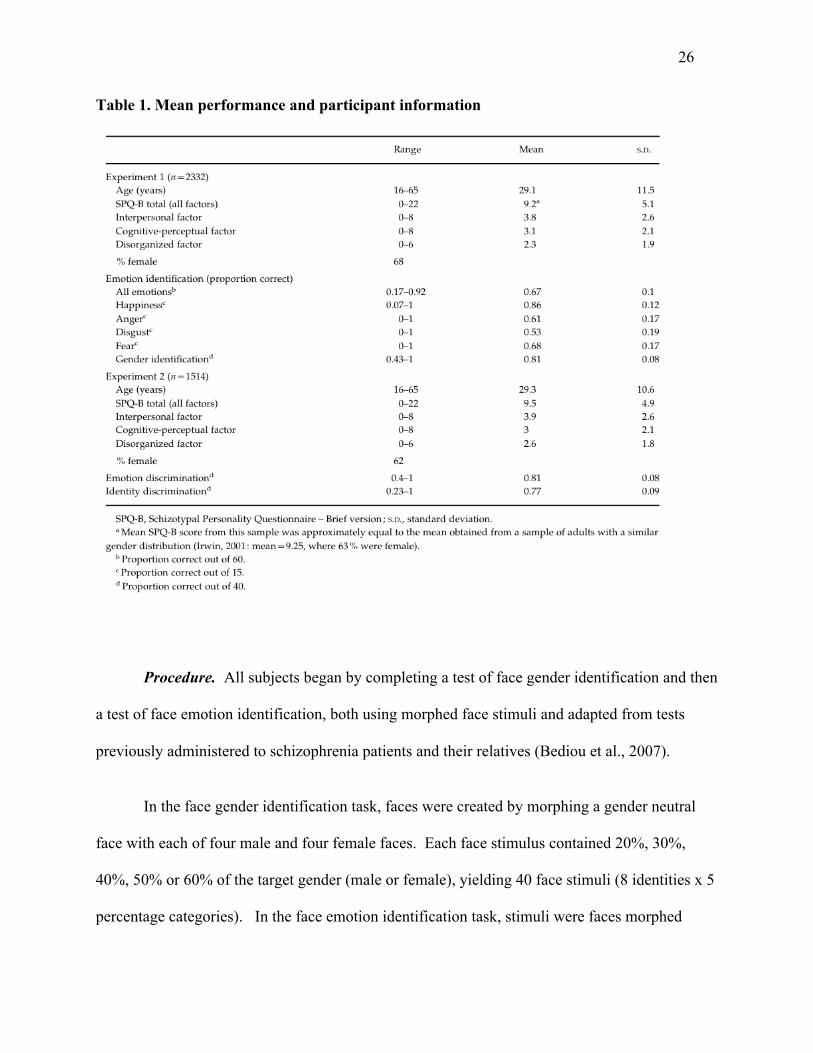
Our final group was comprised of 2332 subjects. Age, gender, and SPQ information for
this sample is shown in Table 1.
26
Table 1. Mean performance and participant information
Procedure. All subjects began by completing a test of face gender identification and then
a test of face emotion identification, both using morphed face stimuli and adapted from tests
previously administered to schizophrenia patients and their relatives (Bediou et al., 2007).
In the face gender identification task, faces were created by morphing a gender neutral
face with each of four male and four female faces. Each face stimulus contained 20%, 30%,
40%, 50% or 60% of the target gender (male or female), yielding 40 face stimuli (8 identities x 5
percentage categories). In the face emotion identification task, stimuli were faces morphed
27
between a neutral expression and an emotional expression. There were 4 different emotional
expressions: happy, disgusted, angry, and fearful. Faces were created from one male and two
female face identities. The faces contained 20%, 30%, 40%, 50%, and 60% of the emotional
expression for each identity and each type of facial expression. This yielded 60 face trials (4
emotion types x 3 identities x 5 percentage categories). The original tasks used by Bediou et al.
(2007) each contained 10 percentage categories, with trials containing 10% to 100% of the target
gender or expression. Based on the control data reported by Bediou et al. (2007), the range 20%
to 60% was chosen for the current experiment to maximize the range of difficulty levels in a
minimal number of trials. The different increments of emotion and gender intensities created
varying levels of difficulty, and therefore increased the sensitivity of the task to reveal individual
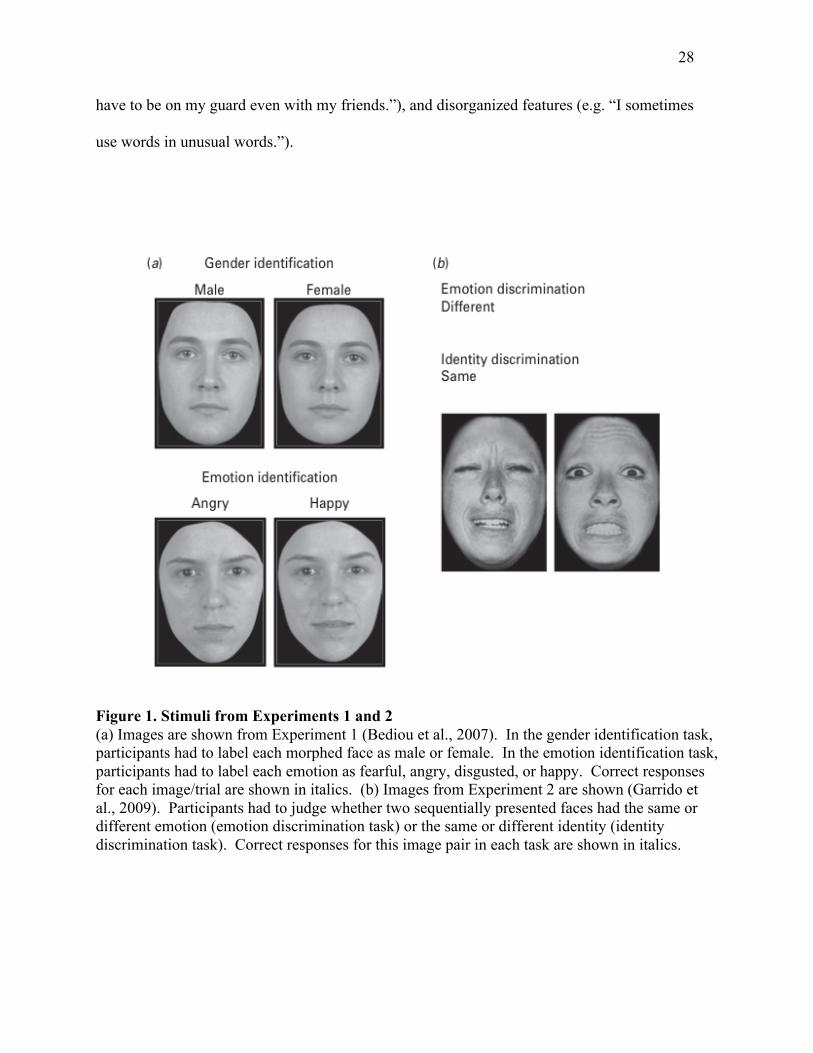
differences in performance. See Figure 1 for example stimuli.
In both tasks, each trial began with a fixation cross for 250ms, then the face was
presented on screen for 1000ms, followed by the list of answer choices. Participants made a
choice between “male or female” in the face gender test, and “angry, disgusted, fearful, or
happy” in the face emotion test. The answer choices remained on screen for seven seconds or
until the participant responded. Participants indicated their response by pressing a key (‘m’ or
‘f’; ‘a’,’d’,’f’, or ‘h’). For each task, participants who failed to respond within the time limit on
more than ten percent of trials were excluded from analysis.
After completing both tests, subjects responded to items from the brief version of the
Schizotypal Personality Questionnaire (SPQ-B) (Raine & Benishay, 1995), a measure of
psychosis-proneness. The SPQ-B is a 22 item self-report questionnaire that indexes the degree
to which an individual has schizophrenia-like cognitive-perceptual (e.g. “Have you ever noticed
a common event or object that seemed to be a special sign for you?”), interpersonal (e.g. “I feel I
28
have to be on my guard even with my friends.”), and disorganized features (e.g. “I sometimes
use words in unusual words.”).
Figure 1. Stimuli from Experiments 1 and 2 (a) Images are shown from Experiment 1 (Bediou et al., 2007). In the gender identification task, participants had to label each morphed face as male or female. In the emotion identification task, participants had to label each emotion as fearful, angry, disgusted, or happy. Correct responses for each image/trial are shown in italics. (b) Images from Experiment 2 are shown (Garrido et al., 2009). Participants had to judge whether two sequentially presented faces had the same or different emotion (emotion discrimination task) or the same or different identity (identity discrimination task). Correct responses for this image pair in each task are shown in italics.
29
Results. A summary of mean performance for this sample is given in Table 1. Paired
sample t-test results showed that participants were more accurate on gender identification as
compared with emotion identification (t(2331) = 59.4, p < 0.001).
Multiple regression was conducted (SPSS version 16.0, 2007) to test the hypothesis that
individual differences in psychosis-proneness were related to emotion identification, but not to
gender identification performance, by using age, participant sex, and SPQ-B scores as predictors
of face emotion identification. Previous research has indicated that face processing ability is
related to both participant sex and age (Bowles et al., 2010, McClure et al., 2000) so we
controlled for these effects in our analysis. Since both SPQ-B scores and age (r = -0.21) and
SPQ-B scores and sex (r = 0.06) were significantly related in this sample, controlling for age and
sex also allowed us to focus on variations in face processing with psychosis-proneness that were
not due to variations in age and sex. As expected, SPQ-B score significantly predicted emotion
identification performance (ß = -0.09, p < 0.001), controlling for the effects of sex (ß = -0.18, p <
0.001) and age (ß = -0.07, p < 0.01). The relationship between psychosis-proneness and emotion
identification did not change when gender identification performance was added as a predictor (ß
= -0.09, p < 0.001).
Two subgroups were defined by total SPQ-B score such that they roughly represented the
bottom and top 10% of the sample. The top 10% is traditionally defined as high-risk in studies
of psychometric schizotypy and individuals with schizophrenia spectrum disorders such as
schizotypal personality disorder are likely to be in the top 10% of scorers (Raine & Benishay,
1995) whereas the bottom 10% is unlikely to contain individual with schizophrenia spectrum
diagnoses (Raine, 1991). Individuals with the lowest SPQ-B scores (SPQ-B scores from 0 to 2,
bottom 10%) were significantly more accurate than those with the highest SPQ-B scores (SPQ-B
30
scores 17 and above, top 9%) [mean for low SPQ-B scorers: 0.66 (0.1); mean for high SPQ-B
scorers = 0.69 (0.1); independent samples t-test: t(430) = 2.7, p < 0.01] and corresponded to a
Cohen’s d effect size of 0.24. This relationship was not driven entirely by high SPQ-B scorers
(those with possible schizophrenia spectrum disorders): SPQ-B scores predicted emotion
identification performance even when individuals with high SPQ-B scores (scores of 16 / 22 or
higher) were excluded (2023 participants remaining; ß = -0.11, p < 0.001).
To see whether the observed relationship between psychosis-proneness and face
perception was specific to emotion processing, we conducted multiple regression of face gender
performance on age, sex, and SPQ-B score. Results indicated that although age and sex
significantly predicted gender identification performance (age: ß = 0.06, p < 0.01; sex: ß = -
0.002, p = 0.99), SPQ-B score did not (ß = -0.02, p = 0.43). Accordingly, high and low SPQ-B
scorers did not significantly differ in gender identification performance [mean for low SPQ-B
scorers = 0.80 (0.08); mean for high SPQ-B scorers = 0.81 (0.08); independent samples t-test:
t(430) = 1.0, p = 0.3].
Scores on the SPQ-B can be divided into three subscales: an interpersonal factor, a
cognitive-perceptual factor, and a disorganized factor. These three factors are analogous to the
three symptom clusters observed in schizophrenia (Arndt et al., 1991). After controlling for the
effects of age and sex, multiple regression analysis revealed that each of the factors predicted
emotion performance (interpersonal: ß = -0.09, p < 0.001; cognitive-perceptual: ß = -0.06, p <
0.01; disorganized: ß = -0.04, p < 0.05), but not gender performance (interpersonal: ß = -0.03, p
= 0.23; cognitive-perceptual; ß = 0.01, p = 0.66; disorganized: ß = -0.02, p = 0.27).
To identify whether the relationship between SPQ-B score and emotion identification
was significantly greater than the relationship between SPQ-B score and gender identification,
31
we used Steiger’s Z1* statistic for comparing two correlation coefficients from the same sample
(Steiger, 1980). This analysis showed that the partial correlation between SPQ-B score and
emotion identification and SPQ-B score was significantly greater than the partial correlation
between SPQ-B score and gender identification (Z = 2.8, p < 0.01).
Finally, to explore the relationship between SPQ-B scores and identification of specific
emotions, we conducted multiple regression with SPQ-B score, age, and participant sex as
predictors of proportion correct for happy, angry, disgusted, and fearful faces separately. Mean
performance for individual emotions is shown in Table 1. SPQ-B scores significantly predicted
identification of happy faces (ß = -0.07, p < 0.001), angry faces (ß = -0.07, p < 0.001), and
fearful faces (ß = -0.05, p < 0.05), but predicted disgusted faces only at the trend level (ß = -0.04,
p = 0.08). These results should be interpreted cautiously, however, as we did not have any a
priori predictions about the relationship between psychosis-proneness and specific emotions, and
the current task was not designed to reveal emotion-specific dissociations.
Figure 2 shows performance on face emotion and gender identification across the range
of SPQ-B scores, illustrating that differences in emotion identification begin to emerge at
moderate levels of psychosis-proneness.
32
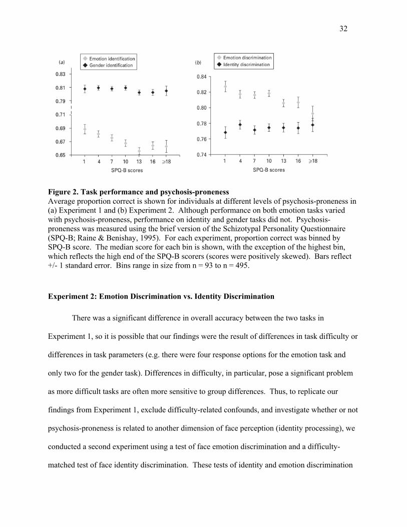
Figure 2. Task performance and psychosis-proneness Average proportion correct is shown for individuals at different levels of psychosis-proneness in (a) Experiment 1 and (b) Experiment 2. Although performance on both emotion tasks varied with psychosis-proneness, performance on identity and gender tasks did not. Psychosis-proneness was measured using the brief version of the Schizotypal Personality Questionnaire (SPQ-B; Raine & Benishay, 1995). For each experiment, proportion correct was binned by SPQ-B score. The median score for each bin is shown, with the exception of the highest bin, which reflects the high end of the SPQ-B scorers (scores were positively skewed). Bars reflect +/- 1 standard error. Bins range in size from n = 93 to n = 495.
Experiment 2: Emotion Discrimination vs. Identity Discrimination
There was a significant difference in overall accuracy between the two tasks in
Experiment 1, so it is possible that our findings were the result of differences in task difficulty or
differences in task parameters (e.g. there were four response options for the emotion task and
only two for the gender task). Differences in difficulty, in particular, pose a significant problem
as more difficult tasks are often more sensitive to group differences. Thus, to replicate our
findings from Experiment 1, exclude difficulty-related confounds, and investigate whether or not
psychosis-proneness is related to another dimension of face perception (identity processing), we
conducted a second experiment using a test of face emotion discrimination and a difficulty-
matched test of face identity discrimination. These tests of identity and emotion discrimination
33
have been used in two prior studies and were shown to tap into dissociable subsystems of face
perception, behaviorally and neurally (Garrido et al., 2009, Pitcher et al., 2008). Using a test of
emotion discrimination would also allow us to generalize our results from Experiment 1 to face
emotion processing more broadly. Whereas emotion discrimination is more purely perceptual,
emotion identification relies on other cognitive abilities, such as verbal labeling, that make
impairments difficult to interpret (Mandal et al., 1998).
Methods.
Participants. Subjects were individuals who navigated to the website,
http://www.testmybrain.org and clicked on a link labeled “Recognizing Emotion and Identity
from Faces”. Experiments 1 and 2 were never available on our website at the same time, so
participant overlap between the two experiments was unlikely to be significant. Exclusion
criteria were the same as for Experiment 1, except that we included two additional question
prompts to serve as validity checks. Participants were excluded if they responded ‘No’ to the
question “I am paying attention to my responses on this questionnaire.” or ‘Yes’ to the question
“I responded to most of the last 47 questions without reading them.” Our final group was
comprised of 1514 participants. Details of this sample are given in Table 1. All subjects first
completed a test of face identity discrimination followed by a test of face emotion
discrimination.
Procedure. Stimuli were the same for both emotion and identity discrimination tests, and
were comprised of six female models taken from the Ekman and Friesen’s (1976) facial affect
series expressing either happiness, sadness, surprise, fear, anger or disgust. Pictures were
grayscale and cropped using the same contour to hide the hair and neck. For both tasks, face
34
pairs were presented sequentially for 500 ms per face with 500ms fixation between images.
Participants then had up to seven seconds to indicate whether the two faces had the same or
different identity (identity discrimination test) or were expressing the same or different emotion
(emotion discrimination test). Half the trials on each test showed pairs with the same
identity/emotion, whereas half the trials showed pairs with different identities/emotions. In the
emotion test, identity always varied between the face pairs. In the identity test, emotion always
varied between the face pairs. Each test contained 40 trials. See Figure 1.
After finishing both tests, subjects again completed items from the brief version of the
Schizotypal Personality Questionnaire (SPQ-B) (Raine & Benishay, 1995), the same measure of
psychosis-proneness used in Experiment 1.
Results. Mean performance for this sample is given in Table 1. Participants were more
accurate on emotion discrimination as compared with identity discrimination (paired samples t-
test; t(1513) = 14.5, p < 0.001).
To test the hypothesis that psychosis-proneness was significantly related to emotion
discrimination performance, multiple regression was conducted in SPSS (version 16.0; 2007)
with age, participant sex, and total SPQ-B score as predictors of face emotion discrimination
performance. SPQ-B scores in this sample were significantly related to participant age (r = -
0.21), but not to sex. Participant sex significantly predicted emotion discrimination performance
(ß = -0.10, p < 0.001) whereas age did not (ß = -0.014, p=0.6). Psychosis-proneness, as
measured by the SPQ-B, significantly predicted emotion discrimination performance (ß = -0.11,
p < 0.001), even when controlling for identity discrimination performance (ß = -0.10, p < 0.001).
Performance was again significantly different between the participants lowest in psychosis-
35
proneness (SPQ-B scores from 0 to 2, bottom 8%) and those highest in psychosis-proneness
(SPQ-B scores 17 and above, top 9%) [mean for low SPQ-B scorers: 0.83 (0.8); mean for high
SPQ-B scorers = 0.79 (0.1); independent samples t-test: t(261) = 3.3, p < 0.001] corresponding to
a Cohen’s d effect size of 0.38. As in experiment 1, the relationship between SPQ-B score and
emotion recognition performance was not being driven entirely by individuals with the highest
levels of psychosis-proneness and possible schizophrenia spectrum diagnoses. When
individuals with scores of 16 (out of 22) or greater were excluded from analysis, multiple
regression again showed that SPQ-B score significantly predicted emotion discrimination (1322
participants remaining; ß = -0.07, p < 0.05).
To see whether psychosis-proneness related differences were limited to emotion
discrimination, we conducted multiple regression of face identity discrimination on age, sex, and
SPQ-B score. Age and sex predicted identity discrimination performance (age: ß = -0.17, p <
0.001; sex: ß = -0.14, p < 0.001) whereas psychosis-proneness did not (ß = -0.03, p = 0.22). This
was despite the fact that overall performance on the identity discrimination task was significantly
lower than on the emotion discrimination task, in contrast with Experiment 1 where the emotion
task was more difficult. Hence, the observed relationship between psychosis-proneness and
emotion processing cannot be explained by difficulty-related confounds.
Multiple regression of emotion discrimination performance on age, sex, and the three
factors of the SPQ-B again demonstrated a significant relationship between emotion performance
and all three factors (interpersonal: ß = -0.07, p < 0.05; cognitive-perceptual: ß = -0.10, p <
0.001; disorganized: ß = -0.08, p < 0.01). Only the interpersonal factor of psychosis-proneness
predicted identity discrimination performance (interpersonal: ß = -0.05, p < 0.05; cognitive-
perceptual: ß = 0.01, p = 0.82; disorganized: ß = -0.02, p = 0.54).
36
In addition, the correlations between SPQ-B score and emotion discrimination and SPQ-
B score and identity discrimination were significantly different, based on Steiger’s Z1* statistic
(1980) for comparing two correlation coefficients from the same sample (Z = 2.3, p < 0.01).
We did not conduct analyses looking at the relationship between psychosis-proneness and
specific emotions for this experiment, as the design (same/different; 6 emotion categories) was
not conducive to this type of analysis.
Figure 2 illustrates the relationship between psychosis-proneness based on SPQ-B scores
and discrimination performance. Consistent with our previous result in Experiment 1,
differences in emotion discrimination related to psychosis-proneness are visible at moderate
SPQ-B scores.
Discussion
We have demonstrated in two large samples that increasing psychosis-proneness, as
indicated by scores on the brief version of the Schizotypal Personality Questionnaire (Raine and
Benishay, 1995), is related to reductions in the ability to identify and discriminate facial
expressions of emotion. Further, this relationship cannot be accounted for by differences in face
processing, visual perception, or a general performance-related factor, as performance on a face
gender test (Experiment 1) and a face identity discrimination task (Experiment 2) did not show
reductions related to increasing psychosis-proneness. Finally, the relationship between face
emotion recognition and psychosis-proneness was significantly predicted by all three factors of
our psychosis-proneness measure (interpersonal, cognitive-perceptual, and disorganized). This
suggests that face emotion recognition (FER) ability is broadly related to psychosis-like
37
characteristics and not restricted to a single dimension of psychosis-proneness, such as positive
or negative symptoms.
Our data indicate that the phenotypic expression of subthreshold or psychosis-like
features is associated with small, but consistent differences in the ability to decode facial
expressions of emotion in the normal population. These differences are not likely to be clinically
significant, but indicate that FER ability varies with individual differences in psychosis-
proneness in the normal population. Schizotypal or psychosis-like features are related to genetic
vulnerability to schizophrenia (Kendler & Walsh, 1995, Vollema et al., 2002) and elevated
schizophrenia risk (Claridge, 1997, Kwapil, 1998, Kwapil et al., 1997, Vollema et al., 2002).
Our results suggest that FER deficits observed in schizophrenia and related disorders do not
solely emerge as a result disease-related confounds or secondary characteristics, but instead may
be a preexisting or even predisposing neurocognitive feature that vary broadly in the normal
population.
We have also shown that FER differences associated with psychosis vulnerability are not
associated with more general differences in visual or face processing. Our results are consistent
with the results of Bediou et al. (2007) who showed that schizophrenia patients and their
relatives have face emotion recognition impairments that are not related to deficits in another
type of face processing. This specificity suggests that differences in the neural systems
responsible for face emotion recognition may be related to psychosis vulnerability and the
expression of psychosis-like characteristics.
A polygenic model of vulnerability to schizophrenia (Gottesman & Shields, 1967)
suggests that vulnerability-related features may emerge in a continuous fashion across the
spectrum of psychosis-proneness (Chapman & Chapman, 1980, Eysenck, 1960, Raine, 2006) .
38
Differences in FER may, for example, reflect the expression of differing numbers of risk-
conferring genes and hence were present even at moderate levels of psychosis-proneness in our
samples (see Figure 2). Differences in performance at moderate levels of psychosis-proneness
also imply that reductions in FER ability are not solely attributable to early or subthreshold
pathology in at-risk participants.
Our study was conducted using a sample recruited entirely on the internet. An
increasingly large body of research demonstrates that results from populations tested over the
internet are reliable and empirically valid (Birnbaum, 2004, Gosling et al., 2004, Haworth et al.,
2007, Kraut et al., 2004, Mcgraw et al., 2000, Wilmer et al., 2010) and of broad theoretical
interest (Wilmer et al., 2010, Owen et al., 2010). A recent analysis of data collected from our
website (www.testmybrain.org) on a test of face recognition memory found that performance and
reliability from the internet-based sample was the same as a traditional lab-based sample
(Wilmer et al., 2010). Our average psychosis-proneness scores were also almost identical to
those reported in a community sample with a similar gender distribution (Irwin, 2001). However,
despite many precautions taken here to ensure valid data, it was not possible to monitor the
performance of each participant in real time, control for biases in self-selection, and verify the
accuracy of information provided by participants. These factors most likely added noise to the
data and may have interacted with our results in ways that cannot be ascertained based on
available data. Ultimately, testing over the internet allowed us to sample a large and diverse
population that would not have been practically feasible if this study were conducted in a
traditional lab setting. This large sample increased our ability to detect small but potentially
meaningful effects on both our FER and face processing control tasks.
Variations in face emotion processing have been documented for several psychiatric
39
disorders, including mood disorders (see Lappanen, 2006 for a review) and anxiety disorders
(e.g. McClure et al., 2003). Thus, it is possible that our results were partially driven by the
overlap between psychosis-like characteristics indexed by the interpersonal factor of the SPQ-B
and social anxiety. FER ability was related to multiple subscales of the SPQ-B, however,
including scores on the cognitive-perceptual factor, indicating that our results cannot be fully
explained by overlap between mood/anxiety symptoms and psychosis-proneness.
Our results recommend an individual differences approach to psychosis-proneness. An
individual differences approach has the advantage of complementing the increasing appreciation
that schizophrenia and other psychotic disorders are likely to arise from the influence of many
common genes of very small effect (Gottesman & Shields, 1967, Purcell et al., 2009, Shi et al.,
2009, Stefansson et al., 2009). The potential relationship between increasing vulnerability to
developing psychosis and FER ability suggests that differences in social-emotional processing
might contribute to the expression of psychosis like traits and, ultimately, to psychosis
development.
Acknowledgements
We would like to thank Benoit Bediou for providing us with the tasks and stimuli used in
Experiment 1 and Lucia Garrido for providing us with the tasks and stimuli used in Experiment
2. We would also like to thank Ken Nakayama for his financial support of
www.testmybrain.org, the website used to collect the current dataset. This research was
supported by a National Science Foundation (NSF) Graduate Research Fellowship to Laura
Germine. NSF had no further role in the research.
40
Paper #2: Social anhedonia is associated with neural abnormalities during face emotion
processing
Published in Neuroimage, Vol. 58, Issue 3, pp.935-945.
Authors
Laura T. Germine, Lucia Garrido, Lori Bruce, and Christine I. Hooker
Abstract
Human beings are social organisms with an intrinsic desire to seek and participate in
social interactions. Social anhedonia is a personality trait characterized by a reduced desire for
social affiliation and reduced pleasure derived from interpersonal interactions. Abnormally high
levels of social anhedonia prospectively predict the development of schizophrenia and contribute
to poorer outcomes for schizophrenia patients. Despite the strong association between social
anhedonia and schizophrenia, the neural mechanisms that underlie individual differences in
social anhedonia have not been studied and are thus poorly understood. Deficits in face emotion
recognition are related to poorer social outcomes in schizophrenia, and it has been suggested that
face emotion recognition deficits may be a behavioral marker for schizophrenia liability. In the
current study, we used functional magnetic resonance imaging (fMRI) to see whether there are
differences in the brain networks underlying basic face emotion processing in a community
sample of individuals low vs. high in social anhedonia. We isolated the neural mechanisms
related to face emotion processing by comparing face emotion discrimination with four other
41
baseline conditions (identity discrimination of emotional faces, identity discrimination of neutral
faces, object discrimination, and pattern discrimination). Results showed a group (high/low
social anhedonia) x condition (emotion discrimination/control condition) interaction in the
anterior portion of the rostral medial prefrontal cortex, right superior temporal gyrus, and left
somatosensory cortex. As predicted, high (relative to low) social anhedonia participants showed
less neural activity in face emotion processing regions during emotion discrimination as
compared to each control condition. The findings suggest that social anhedonia is associated with
abnormalities in networks responsible for basic processes associated with social cognition, and
provide a starting point for understanding the neural basis of social motivation and our drive to
seek social affiliation.
Introduction
As fundamentally social creatures, humans are driven by the desire for meaningful and
frequent social interaction (Baumeister & Leary, 1995). There are individual differences in the
strength of this desire, however, and some individuals exhibit a significantly reduced drive for
social affiliation known as social anhedonia (Brown et al., 2007; Kwapil, 1998; Kwapil et al.,
2009). Social anhedonia (SA) has been characterized as a deficiency in the need to belong to a
social group and is distinct from other constructs that might also predict abnormalities in social
interaction such as social anxiety (Brown et al., 2007; Kwapil et al., 2009). Individuals high in
SA exhibit a genuine preference for solitude, disengagement during social interactions (Brown et
al., 2007), and reduced negative affect when alone (Kwapil et al., 2009). Higher levels of SA are
related to lower levels of social support and social functioning (Blanchard et al., 2009). Reduced
42
social support and smaller social networks are associated with differences in immune functioning
and other clinically significant health outcomes (Miller et al., 2009). Furthermore, high SA has
been identified as one of the single most predictive traits for future onset of schizophrenia
spectrum disorders (Kwapil, 1998) and has long been recognized as a core attribute of psychosis
vulnerability (Bleuler, 1950; Horan et al., 2007; Kraepelin & Gosline, 1919; Meehl, 1962; Rado,
1953; Stone et al., 2005). Altogether, existing evidence indicates that SA is a deviation in a
psychologically and clinically important social and emotional process that has broad implications
for our understanding of normal and abnormal functioning.
Despite evidence for serious physical and mental health difficulties associated with
reduced desire for social affiliation, no research to our knowledge has been done exploring the
neural basis of SA in nonclinical populations. In schizophrenia, SA is considered a negative
symptom that is stable (Blanchard et al., 2001) and can be reliably assessed (Horan et al., 2006).
Studies of SA in schizophrenia have indicated that a number of neural systems may be involved
in reduced desire for social affiliation, including the amygdala (Becerril & Barch, 2010), caudate
nucleus (Dowd & Barch, 2010), dorsolateral prefrontal cortex (Becerril & Barch, 2010), and
somatosensory areas (Arnfred & Chen, 2004). However, disease-related confounds and
secondary characteristics of schizophrenia illness, such as psychosocial stress and
neurodegenerative processes, make it difficult to generalize these findings to SA among healthy
individuals or to identify whether neural abnormalities associated with SA are associated with
the vulnerability to psychosis (Lenzenweger, 2006).
Differences in the neural processing of face emotion provide a potential starting point for
identifying abnormalities associated with high SA. Accurate face emotion recognition is critical
for recognizing and responding to other’s mental states and is a building block to more complex
43
social behaviors (Adolphs, 2002). Importantly, face emotion recognition ability (but not face
identity processing ability) predicts social functioning in schizophrenia participants (Hooker &
Park, 2002) and varies with psychometric psychosis-proneness in nonclinical populations
(Germine & Hooker, 2011). Previous work has also shown that face emotion perception is
abnormal in individuals high in social anhedonia (Luh & Gooding, 1999). Thus, individual
differences in social anhedonia may be related to deficits in the neural mechanisms supporting
face emotion recognition.
The neural substrates of face emotion recognition are well characterized in healthy and
clinical populations. Previous work indicates that effective emotion recognition involves the
recruitment of a network of regions, including the amygdala (Adolphs, 2002; Adolphs et al.,
1994; Anderson & Phelps, 2001), superior temporal sulcus (Allison et al., 2000; Haxby et al.,
2000), medial prefrontal cortex (Amodio & Frith, 2006; Blair et al., 1999; Dolan et al., 1996;
Gorno-Tempini et al., 2001; Gur et al., 2002a; Phillips et al., 1998; Sprengelmeyer et al., 1998;
Wright et al., 2002), and somatosensory-related cortices (including insula, S1, S2, and anterior
supramarginal gyrus) (Adolphs, 2002; Adolphs et al., 2000). Using functional neuroimaging,
researchers have consistently found abnormalities in these regions during emotion recognition in
individuals with schizophrenia (Das et al., 2007; Farrer et al., 2004; Gur et al., 2007; Gur et al.,
2002b; Hall et al., 2004; Hempel et al., 2003; Holt et al., 2006; Kosaka et al., 2002; Phillips et
al., 1999; Pinkham et al., 2008; Schneider et al., 1998; Spence et al., 1997; Taylor et al., 2002;
Waberski et al., 2004; Williams et al., 2004).
Deficits in emotion recognition have been associated with lesions to the amygdala
(Adolphs et al., 1994), somatosensory and related cortices (Adolphs et al., 2000) and medial
prefrontal cortex (Heberlein et al., 2008). The medial prefrontal cortex, in particular, likely plays
44
a broad role in many social-cognitive processes and has been implicated in lower-level emotion
perception as well as higher-level processes including theory of mind attributions (Gallagher et
al., 2000), self-referential processing (Mitchell et al., 2005), and distinguishing between self and
other (Heatherton et al., 2006; Ochsner et al., 2004). In terms of functional divisions, the
anterior portion of rostral medial prefrontal cortex (arMFC) has been consistently identified in
measures of social cognition and emotion processing (Amodio & Frith, 2006) and in social
cognition in schizophrenia (Brunet-Gouet & Decety, 2006).
In the present study, we used functional magnetic resonance imaging (fMRI) to examine
differences in the neural circuitry underlying face emotion discrimination in otherwise normal
individuals who were high versus low in SA. As our face emotion recognition task, we used the
Queen Square Face Discrimination Test (QFDT; Garrido et al., 2009). Our primary hypothesis
was that high SA would have specific deficits in face emotion processing even when controlling
for broader, but equally complex aspects of face perception. The QFDT was chosen because it
can dissociate face emotion processing and face identity processing (Banissy et al., 2011;
Garrido et al., 2009; Germine & Hooker, 2011; Pitcher et al., 2008). In the QFDT, participants
view sequentially presented emotional faces; in one condition they judge whether the two faces
are expressing the same emotion, and in another condition they judge whether the two faces have
the same identity. Importantly, the two conditions have identical stimuli and are equally difficult
for healthy participants. As a result, any differences found between emotion discrimination and
identity discrimination can be attributed to differences in specific cognitive processes related to
emotion perception and cannot be attributed to differences in the stimuli, number of response
options, or difficulty level of the two conditions. This feature of the task is an improvement over
face processing studies where the experimental and control tasks differ along these dimensions
45
(e.g. labeling emotional faces using four options vs. same/different identity of paired neutral
faces). In the QFDT, the emotion recognition and identity recognition conditions use the same
task structure (both are a forced choice same/different judgment) and the same stimuli. Therefore
the comparison of emotion recognition and identity recognition of emotional faces isolates the
specific cognitive processes for attending to, processing, and judging face emotions. Using a
behavioral version of this task, we found that higher levels of psychosis risk (based on self-report
of cognitive-perceptual, interpersonal, and disorganized psychosis-prone characteristics) are
associated with reduced emotion discrimination performance, but normal identity discrimination
performance (Germine and Hooker, 2011). The QFDT was also used by Pitcher et al. (2008),
who found that applying transcranial magnetic stimulation (TMS) to the face area of
somatosensory cortex impaired performance in the emotion discrimination condition, but not the
identity discrimination condition. Thus, we have good reason to believe that the QFDT emotion
discrimination condition depends on one or more processes specific to emotion processing that
also vary with psychosis vulnerability. The current fMRI study included three additional control
conditions designed to reveal potential group differences in the broader face emotion processing
neural network. These conditions included identity discrimination of neutral faces, visual
discrimination of objects, and visual discrimination of patterns. Given the putative relationship
between SA and vulnerability for psychosis (Kwapil, 1998), we predicted that individuals high in
SA would exhibit reduced recruitment of cortical regions involved in face emotion recognition,
particularly superior temporal sulcus/gyrus, medial prefrontal cortex and somatosensory-related
parietal regions, as well as reduced responses in the amygdala. Between group differences in
one or more of these regions would indicate that higher levels of SA are associated with neural
46
abnormalities during emotion perception, and help us better understand the neural basis of
differences in the desire for social affiliation as well as psychosis vulnerability.
Material and Methods
Participants. We recruited a community-based sample comprised of thirty participants
who were high or low in social anhedonia based on their scores on the Revised Chapman Social
Anhedonia Scale (RSAS; Chapman & Chapman, 1980). Fifteen high social anhedonia
participants (high SA) were selected based on scoring in the top 10% on this measure (RSAS
score > 16 for females, > 19 for males). Fifteen low social anhedonia participants (low SA)
were selected based on having scores at or below the mean (RSAS score < 7 for females and < 9
for males). Participants were recruited from a combination of community advertisements and
the community-wide university study pool. Community advertisements were posted on sites
like Craigslist and targeted individuals with difficulties in interpersonal functioning associated
with social anhedonia and psychosis risk (e.g. “People sometimes find me aloof and distant.”).
In addition, items from the RSAS were used to pre-select individuals with high social anhedonia
from the community-wide university study pool. Anyone who had a score of 16 or greater on the
RSAS was invited to come into the lab for further screening. All participants took the full RSAS
after completing MR screening and demographic questionnaires. In total, 12/15 high SA
individuals were recruited through community advertisements and 3/15 high SA participants
came from prescreening of the community-wide university study pool using the RSAS.
Participants in the low SA groups were recruited from prescreening of the community-wide
university study pool for low-to-average levels of social anhedonia (based on the RSAS) and
47
demographic characteristics similar to our high SA group. Altogether, 4/15 participants in our
high SA group and 5/15 in our low SA group were university students. All participants were
administered the Structured Clinical Interview for DSM-IV (SCID; First et al., 2002) and were
excluded if they had any Axis I diagnosis, a history of alcohol or drug dependence, alcohol or
drug abuse within the last six months, a past major head injury involving a loss of consciousness
lasting more than 2-3 minutes, or did not speak English as a primary language. Socioeconomic
status was assessed using the Hollingshead Index (Hollingshead, 1957). One participant in the
high SA group and two participants in the low SA group were unable to give parental
information, and so the average parental education/ socioeconomic status from that individual’s
SA group was used to replace the missing values (e.g. the mean parental socioeconomic status
from the other 14 members of the high SA group was used in place of the final high SA
participant’s missing value). Groups did not differ significantly in terms of sex, age, education,
parental education, socioeconomic status, or parental socioeconomic status (see Table 2).
Informed written consent was obtained from all participants after the nature of the study
and procedures had been fully explained. The study was approved by the Institutional Review
Boards at Harvard University and Massachusetts General Hospital (MGH) (Partners health care
system).
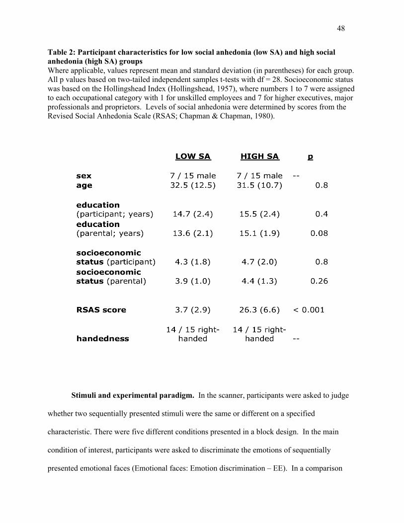
48
Table 2: Participant characteristics for low social anhedonia (low SA) and high social anhedonia (high SA) groups Where applicable, values represent mean and standard deviation (in parentheses) for each group. All p values based on two-tailed independent samples t-tests with df = 28. Socioeconomic status was based on the Hollingshead Index (Hollingshead, 1957), where numbers 1 to 7 were assigned to each occupational category with 1 for unskilled employees and 7 for higher executives, major professionals and proprietors. Levels of social anhedonia were determined by scores from the Revised Social Anhedonia Scale (RSAS; Chapman & Chapman, 1980).
Stimuli and experimental paradigm. In the scanner, participants were asked to judge
whether two sequentially presented stimuli were the same or different on a specified
characteristic. There were five different conditions presented in a block design. In the main
condition of interest, participants were asked to discriminate the emotions of sequentially
presented emotional faces (Emotional faces: Emotion discrimination – EE). In a comparison
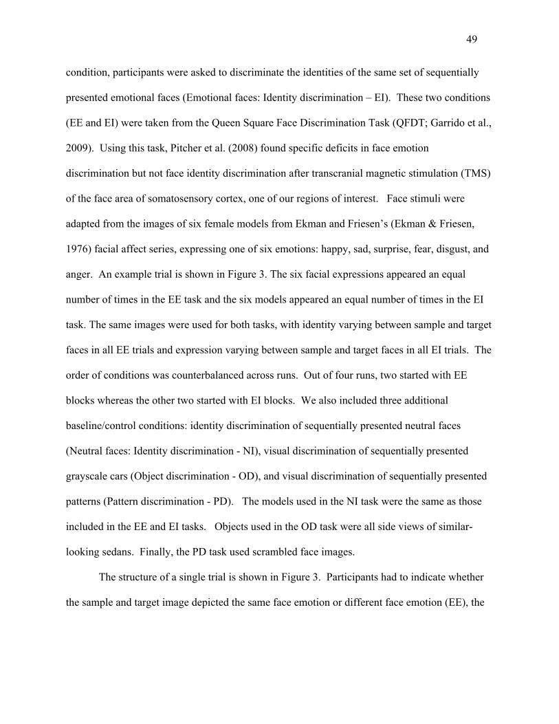
49
condition, participants were asked to discriminate the identities of the same set of sequentially
presented emotional faces (Emotional faces: Identity discrimination – EI). These two conditions
(EE and EI) were taken from the Queen Square Face Discrimination Task (QFDT; Garrido et al.,
2009). Using this task, Pitcher et al. (2008) found specific deficits in face emotion
discrimination but not face identity discrimination after transcranial magnetic stimulation (TMS)
of the face area of somatosensory cortex, one of our regions of interest. Face stimuli were
adapted from the images of six female models from Ekman and Friesen’s (Ekman & Friesen,
1976) facial affect series, expressing one of six emotions: happy, sad, surprise, fear, disgust, and
anger. An example trial is shown in Figure 3. The six facial expressions appeared an equal
number of times in the EE task and the six models appeared an equal number of times in the EI
task. The same images were used for both tasks, with identity varying between sample and target
faces in all EE trials and expression varying between sample and target faces in all EI trials. The
order of conditions was counterbalanced across runs. Out of four runs, two started with EE
blocks whereas the other two started with EI blocks. We also included three additional
baseline/control conditions: identity discrimination of sequentially presented neutral faces
(Neutral faces: Identity discrimination - NI), visual discrimination of sequentially presented
grayscale cars (Object discrimination - OD), and visual discrimination of sequentially presented
patterns (Pattern discrimination - PD). The models used in the NI task were the same as those
included in the EE and EI tasks. Objects used in the OD task were all side views of similar-
looking sedans. Finally, the PD task used scrambled face images.
The structure of a single trial is shown in Figure 3. Participants had to indicate whether
the sample and target image depicted the same face emotion or different face emotion (EE), the

50
same identity or a different identity (EI, NI tasks), or the same image or a different image (OD,
PD tasks). There were 72 trials of each task, with half requiring “same” judgments.
All participants were administered a brief practice including all conditions and
correct/incorrect feedback before placement in the scanner. The practice and scanning
experiments were administered using E-Prime software.
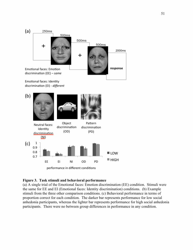
51
Figure 3. Task stimuli and behavioral performance (a) A single trial of the Emotional faces: Emotion discrimination (EE) condition. Stimuli were the same for EE and EI (Emotional faces: Identity discrimination) conditions. (b) Example stimuli from the three other comparison conditions. (c) Behavioral performance in terms of proportion correct for each condition. The darker bar represents performance for low social anhedonia participants, whereas the lighter bar represents performance for high social anhedonia participants. There were no between group differences in performance in any condition.
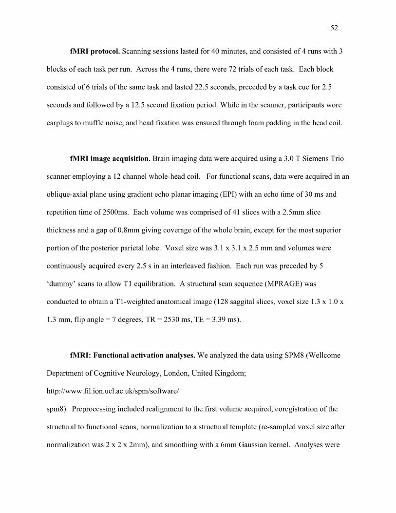
52
fMRI protocol. Scanning sessions lasted for 40 minutes, and consisted of 4 runs with 3
blocks of each task per run. Across the 4 runs, there were 72 trials of each task. Each block
consisted of 6 trials of the same task and lasted 22.5 seconds, preceded by a task cue for 2.5
seconds and followed by a 12.5 second fixation period. While in the scanner, participants wore
earplugs to muffle noise, and head fixation was ensured through foam padding in the head coil.
fMRI image acquisition. Brain imaging data were acquired using a 3.0 T Siemens Trio
scanner employing a 12 channel whole-head coil. For functional scans, data were acquired in an
oblique-axial plane using gradient echo planar imaging (EPI) with an echo time of 30 ms and
repetition time of 2500ms. Each volume was comprised of 41 slices with a 2.5mm slice
thickness and a gap of 0.8mm giving coverage of the whole brain, except for the most superior
portion of the posterior parietal lobe. Voxel size was 3.1 x 3.1 x 2.5 mm and volumes were
continuously acquired every 2.5 s in an interleaved fashion. Each run was preceded by 5
‘dummy’ scans to allow T1 equilibration. A structural scan sequence (MPRAGE) was
conducted to obtain a T1-weighted anatomical image (128 saggital slices, voxel size 1.3 x 1.0 x
1.3 mm, flip angle = 7 degrees, TR = 2530 ms, TE = 3.39 ms).
fMRI: Functional activation analyses. We analyzed the data using SPM8 (Wellcome
Department of Cognitive Neurology, London, United Kingdom;
http://www.fil.ion.ucl.ac.uk/spm/software/
spm8). Preprocessing included realignment to the first volume acquired, coregistration of the
structural to functional scans, normalization to a structural template (re-sampled voxel size after
normalization was 2 x 2 x 2mm), and smoothing with a 6mm Gaussian kernel. Analyses were
53
conducted with a general linear model framework. Vectors of onset times with durations of 22.5s
were defined for all five tasks: EE, EI, NI, OD, and PD. These onset vectors were convolved
with the SPM8 canonical hemodynamic response function (HRF) using a box-car function.
Additionally, regressors were created using an artifact detection tool (ART; Whitfield-Gabrieli,
2009) to exclude scans with gross motion (>0.6mm relative to previous time frame) or spiking
artifacts (global mean image intensity greater than 2.5 SD from mean of the entire time series
within a scan) from analysis. Where this procedure resulted in omission of more than 10% of
time frames, filters were adjusted to bring the number of excluded scans to approximately 10%.
There were no between group differences in number of outliers identified (high SA max = 83;
low SA max = 92) or filter parameters. A high-pass frequency filter (200s) was also applied to
the time series. For each subject, contrast images were calculated for each of the five tasks (EE,
EI, NI, OD, and PD) relative to baseline (blocks of fixation).
Second-level analysis. To verify that our task was activating our regions of interest, we
conducted a one sample t-test of EE vs. baseline (fixation) across all participants. To assess
whether our EE task was isolating regions for face emotion processing relative to face identity
processing, we also conducted a one sample t-test of the contrast of EE versus EI across
participants. To look at between group differences, we implemented a flexible factorial design in
SPM8. Our hypothesis was a group (high/low SA) x condition (EE/control) interaction, such that
high SA would show less activity for EE as compared to each control condition. Four group x
condition analyses were conducted: EE vs. EI, EE vs. NI, EE vs. OD, & EE vs. PD. All group
maps were thresholded at p < 0.001 uncorrected with an extent threshold of 5 voxels. For
regions of interest where suprathreshold clusters were identified in our flexible factorial analysis,
small volume corrections were performed using Family Wise Error correction (FWE, p < 0.05).
54
The WFU Pickatlas (Maldjian et al., 2003; Maldjian et al., 2004) was used to create anatomically
defined masks of the right superior temporal gyrus, bilateral postcentral gyri (somatosensory
cortices), and bilateral supramarginal gyri (somatosensory-related or somatosensory association
cortices). A mask of the anterior portion of rostral medial prefrontal cortex (arMFC) was defined
based on Amodio and Frith (2006). This mask was drawn to include all voxels in prefrontal
cortex with MNI coordinates of x < 20 and x > -20, y > 20, and z > 0 (4013 voxels total). Any
voxel in the left or right hemisphere that fell within these coordinate boundaries was included in
a single, bilateral arMFC mask. For regions of interest showing a significant interaction,
contrast estimates were extracted from the peak of primary clusters in order to conduct post hoc
comparisons.
Results
Behavioral. No significant between group differences were found in performance on any
condition (EE, EI, NI, OD, and PD) (all p > 0.2). Details of performance in each condition for
each group are shown in Figure 3.
Effects of task across all participants. We conducted one sample t-tests on EE (vs.
baseline) contrasts across all participants to verify that the EE task was inducing BOLD signal
changes in the expected face emotion and face processing regions. This analysis verified task-
related activity in a network of regions that included the right superior temporal sulcus, left
postcentral gyrus (left primary somatosensory cortex), bilateral supramarginal gyri
55
(somatosensory-related or somatosensory association cortices), bilateral fusiform gyri, and
bilateral medial prefrontal cortex, including the anterior rostral medial prefrontal cortex (arMFC)
(see Table 3). No suprathreshold voxels were detected in the amygdala.
To verify that the EE task was uniquely associated with activity in our emotion
perception regions of interest relative to other tasks, we also conducted a one sample t-test on the
EE vs. EI contrast across all participants. This analysis revealed a network of activation for the
EE task (relative to EI) that included the right superior temporal sulcus, bilateral postcentral gyri
(primary somatosensory cortices), left supramarginal gyrus (somatosensory-related cortex), and
bilateral medial prefrontal cortices, including anterior rostral medial prefrontal cortex (arMFC)
(see Table 3). As in our EE vs. baseline contrast, no suprathreshold voxels were detected in the
amygdala. With the exception of the amygdala, these results demonstrate that the EE task was
associated with activation across all of our regions of interest in the distributed emotion
perception network, above and beyond the most closely matched control condition (EI). Given
that this is the first time this task has been used in the scanner, our EE vs. EI contrast serves as
validation that the EE task is suited to tapping into neural networks associated with emotion
perception.
56
Table 3: Emotional faces: Emotion discrimination (EE) related fMRI BOLD responses across all participants Neural activity clusters are based on one sample t-tests across all participants with a significance threshold of p < 0.001 uncorrected and an extent threshold of 5 voxels. Neuroanatomical labels, MNI coordinates, and t-values are provided for the peak voxel of each cluster. Clusters that include a priori regions of interest are italicized. Superscripts denote clusters containing areas of activation that survive small volume correction based on regions of interest: (1) Includes voxels in anterior rostral medial frontal cortex (arMFC) that survive small volume correction over a bilateral region defined by voxels with MNI coordinates of |x| < 20, y > 20, and z > 0 (Amodio & Frith, 2006); (2) Includes voxels in postcentral gyrus / somatosensory cortex that survive small volume correction over anatomically defined bilateral postcentral gyri; (3) Includes voxels in right superior temporal gyrus that survive small volume correction over anatomically defined right superior temporal gyrus; (4) Includes voxels in supramarginal gyrus / somatosensory-related / somatosensory-association cortex that survive small volume correction over anatomically defined bilateral supramarginal gyri.
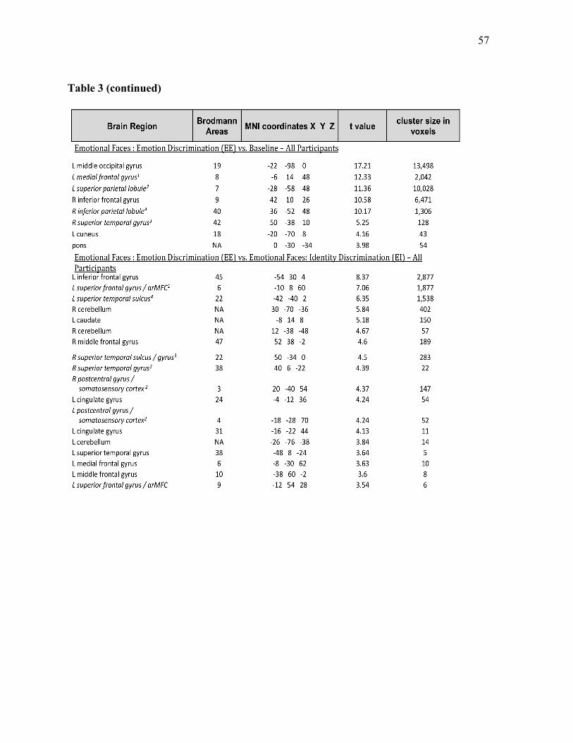
57
Table 3 (continued)
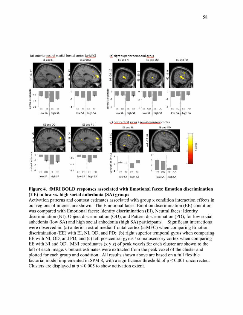
58
Figure 4. fMRI BOLD responses associated with Emotional faces: Emotion discrimination (EE) in low vs. high social anhedonia (SA) groups Activation patterns and contrast estimates associated with group x condition interaction effects in our regions of interest are shown. The Emotional faces: Emotion discrimination (EE) condition was compared with Emotional faces: Identity discrimination (EI), Neutral faces: Identity discrimination (NI), Object discrimination (OD), and Pattern discrimination (PD), for low social anhedonia (low SA) and high social anhedonia (high SA) participants. Significant interactions were observed in: (a) anterior rostral medial frontal cortex (arMFC) when comparing Emotion discrimination (EE) with EI, NI, OD, and PD; (b) right superior temporal gyrus when comparing EE with NI, OD, and PD; and (c) left postcentral gyrus / somatosensory cortex when comparing EE with NI and OD. MNI coordinates (x y z) of peak voxels for each cluster are shown to the left of each image. Contrast estimates were extracted from the peak voxel of the cluster and plotted for each group and condition. All results shown above are based on a full flexible factorial model implemented in SPM 8, with a significance threshold of p < 0.001 uncorrected. Clusters are displayed at p < 0.005 to show activation extent.
59
Group comparisons. The main hypothesis of the study was that participants with high
social anhedonia would show reduced activity in emotion processing regions during emotion
discrimination. Regions showing a significant interaction in the expected direction, i.e. where
low SA had greater activity than high SA participants for emotion discrimination (Emotional
faces – Emotion discrimination condition: EE) versus the control condition, are listed in Table 4.
Regions where there was a significant group x condition interaction but with the opposite pattern
(i.e. where high SA had more activity than low SA for EE > control) are shown in Table 5. None
of these regions (where high SA > low SA during EE) occurred in our regions of interest.
Clusters within a priori regions of interest were investigated by extracting contrast estimates
from the peak of the cluster. Primary clusters showing significant group x condition interactions
are shown together with contrast estimates in Figure 4, organized by region of interest. Finally,
Table 5 shows results of post hoc comparisons applied to the contrast estimates in primary
clusters located in our regions of interest.
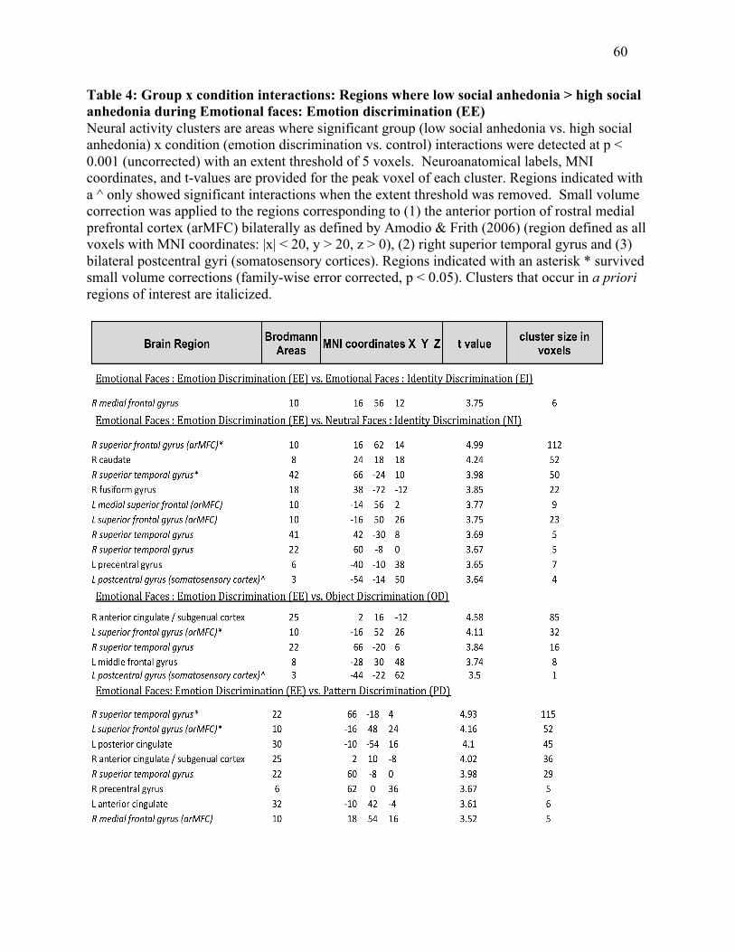
60
Table 4: Group x condition interactions: Regions where low social anhedonia > high social anhedonia during Emotional faces: Emotion discrimination (EE) Neural activity clusters are areas where significant group (low social anhedonia vs. high social anhedonia) x condition (emotion discrimination vs. control) interactions were detected at p < 0.001 (uncorrected) with an extent threshold of 5 voxels. Neuroanatomical labels, MNI coordinates, and t-values are provided for the peak voxel of each cluster. Regions indicated with a ^ only showed significant interactions when the extent threshold was removed. Small volume correction was applied to the regions corresponding to (1) the anterior portion of rostral medial prefrontal cortex (arMFC) bilaterally as defined by Amodio & Frith (2006) (region defined as all voxels with MNI coordinates: |x| < 20, y > 20, z > 0), (2) right superior temporal gyrus and (3) bilateral postcentral gyri (somatosensory cortices). Regions indicated with an asterisk * survived small volume corrections (family-wise error corrected, p < 0.05). Clusters that occur in a priori regions of interest are italicized.
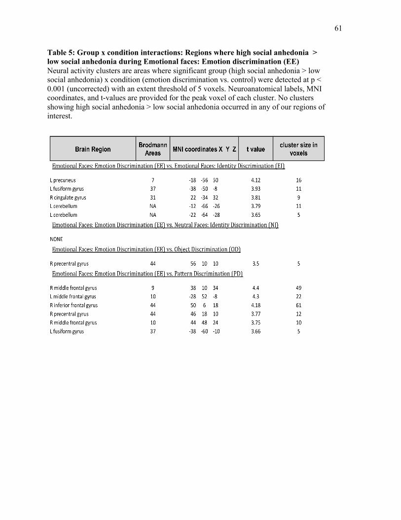
61
Table 5: Group x condition interactions: Regions where high social anhedonia > low social anhedonia during Emotional faces: Emotion discrimination (EE) Neural activity clusters are areas where significant group (high social anhedonia > low social anhedonia) x condition (emotion discrimination vs. control) were detected at p < 0.001 (uncorrected) with an extent threshold of 5 voxels. Neuroanatomical labels, MNI coordinates, and t-values are provided for the peak voxel of each cluster. No clusters showing high social anhedonia > low social anhedonia occurred in any of our regions of interest.
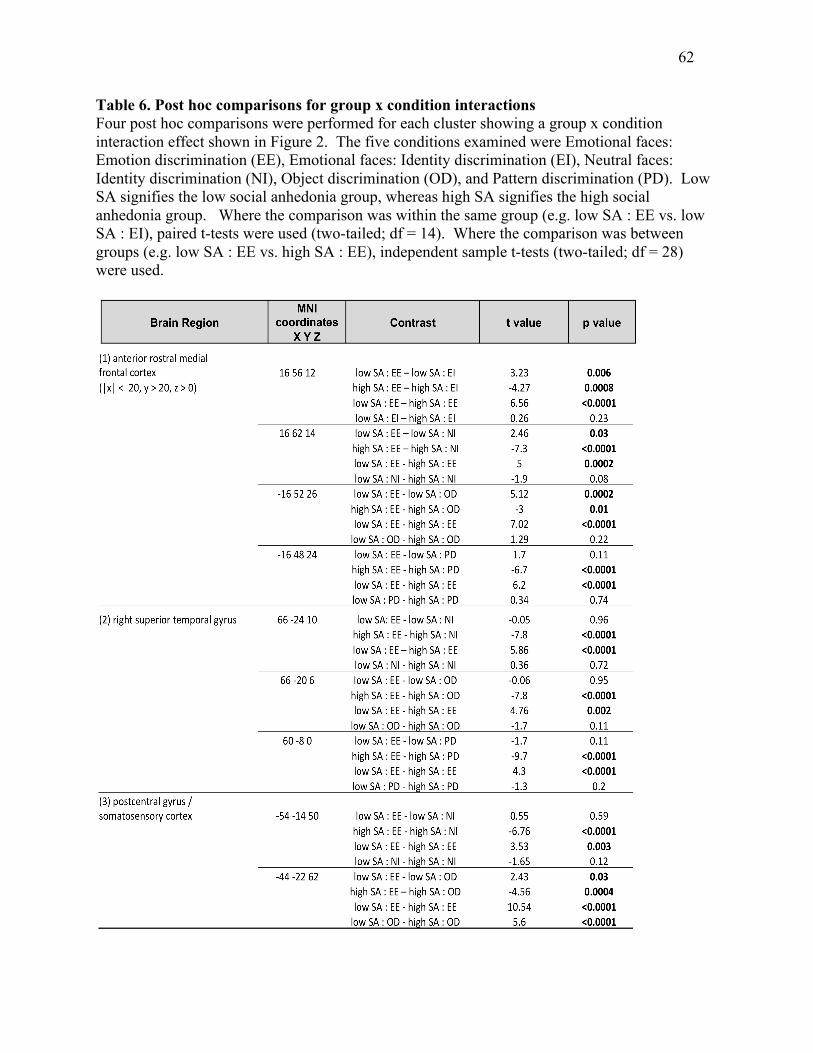
62
Table 6. Post hoc comparisons for group x condition interactions Four post hoc comparisons were performed for each cluster showing a group x condition interaction effect shown in Figure 2. The five conditions examined were Emotional faces: Emotion discrimination (EE), Emotional faces: Identity discrimination (EI), Neutral faces: Identity discrimination (NI), Object discrimination (OD), and Pattern discrimination (PD). Low SA signifies the low social anhedonia group, whereas high SA signifies the high social anhedonia group. Where the comparison was within the same group (e.g. low SA : EE vs. low SA : EI), paired t-tests were used (two-tailed; df = 14). Where the comparison was between groups (e.g. low SA : EE vs. high SA : EE), independent sample t-tests (two-tailed; df = 28) were used.
63
Emotional Faces: Emotion Discrimination (EE) vs. Emotional Faces: Identity
Discrimination (EI). For the comparison of EE and EI, there was a significant group x condition
interaction (where low SA > high SA) in arMFC (see Figure 4), but not other regions of interest.
Post hoc comparisons were conducted to investigate the interaction. Statistics for each
comparison are in Table 5. As predicted, the results showed that high SA participants had
reduced arMFC activity during emotion discrimination. In addition, low SA participants had
greater arMFC activity for emotion discrimination as compared to identity discrimination of
emotional faces (i.e. among low SA: EE > EI). However, high SA participants deactivated the
arMFC during EE, such that high SA had significantly less activity for emotion discrimination as
compared to identity discrimination of emotional faces (i.e. among high SA: EE < EI).
Moreover, the direct comparison of emotion discrimination between the groups showed that low
SA participants had significantly greater arMFC activity than high SA participants (i.e. low SA
EE > high SA EE). There was no significant difference between low SA and high SA
participants for arMFC activity during identity discrimination of emotional faces (EI condition)
(see Table 5).
Activation in arMFC did not survive small volume correction (over bilateral arMFC; see
Section 3.3.) in this contrast (p = 0.15).
Emotional Faces: Emotion Discrimination (EE) vs. Neutral Faces: Identity
Discrimination (NI). When comparing EE with NI, there was a significant group x condition
interaction (where low SA > high SA) in arMFC and right superior temporal gyrus (see Figure
4). Removing the extent threshold (p < 0.001 uncorrected, k = 0) revealed an additional
suprathreshold cluster in left somatosensory cortex (left postcentral gyrus). Post hoc
64
comparisons (see Table 5) revealed the same overall pattern in arMFC as was observed in the EE
vs. EI analysis (see Section 3.3.1.). In right superior temporal gyrus and left somatosensory
cortex, high SA participants deactivated regions of both right superior temporal gyrus and left
somatosensory cortex during EE relative to NI (i.e. among high SA: EE < NI) whereas low SA
participants did not show differences between these two conditions (i.e. among low SA: EE =
NI). As predicted, low SA was associated with greater activity in right superior temporal gyrus
and left somatosensory cortex during EE (i.e. low SA EE > high SA EE), but not differences
during NI (i.e. low SA NI = high SA NI).
Both arMFC and right superior temporal gyrus clusters survived small volume correction
in this comparison (p < 0.05).
Emotional Faces: Emotion Discrimination (EE) vs. Object Discrimination (OD).
When comparing EE with OD, there was again a significant group x condition interaction
(where low SA > high SA) in arMFC and right superior temporal gyrus (see Figure 4). As in the
comparison of EE and NI, removing the extent threshold (p < 0.001 uncorrected, k = 0) also
revealed an additional suprathreshold cluster in left somatosensory cortex (left postcentral
gyrus). Post hoc comparisons (see Table 5) revealed the same overall pattern in arMFC and right
superior temporal gyrus as was observed in the comparison of EE and NI (Section 3.3.2). In left
somatosensory cortex, significant differences were observed in all post hoc comparisons. High
SA participants deactivated this region during EE relative to OD (i.e. among high SA: EE < OD)
whereas low SA participants showed greater activation of this region during EE relative to OD
(i.e. among low SA: EE > OD). As predicted, low SA showed greater activity in somatosensory
cortex during EE than high SA (i.e. low SA EE > high SA EE), but also during OD (i.e. low SA
OD > high SA OD).
65
Differences in arMFC in the comparison of EE and OD survived small volume correction
at the trend level (p = 0.06), whereas differences in right superior temporal gyrus did not (p =
0.25).
Emotional Faces: Emotion Discrimination (EE) vs. Pattern Discrimination (PD).
When comparing EE with PD, there was again a significant group x condition interaction
(where low SA > high SA) in arMFC and right superior temporal gyrus (see Figure 4).
Removing the extent threshold did not reveal any additional suprathreshold clusters in other
regions of interest. Post hoc comparisons (see Table 5) revealed that, as predicted, low SA had
greater activation during EE than high SA in arMFC and right superior temporal gyrus (i.e. low
SA EE > high SA EE). High SA also showed deactivation during EE as compared with PD (i.e.
among high SA: EE < PD). Among low SA, there was no difference between EE and PD in
these regions (i.e. among low SA: EE = PD) and no differences in PD between low and high SA
(i.e. low SA PD = high SA PD).
Differences in right superior temporal gyrus survived small volume correction (p < 0.05)
in this comparison. Differences in arMFC survived small volume correction at the trend level (p
= 0.06).
Further Analyses.
Amygdala. Although the amygdala was one of our a priori regions of interest, we found
no within-subjects differences in this region when comparing EE with any of our control
conditions (EI, NI, OD and PD) across all participants. We also failed to detect any significant
group x condition interactions in the amygdala.
66
As the amygdala is considered a central part of the extended face emotion perception
network, we conducted further analyses to explore whether the combination of all conditions
using faces (EE, EI, and NI) would show suprathreshold amygdala activation when compared
with baseline. Using a within-subjects one sample t-test across all participants, we again found
no significant differences in the amygdala (p < 0.001). An analysis of signal-to-noise in the right
amygdala as compared with right superior temporal gyrus in our sample suggested significantly
lower signal-to-noise in the amygdala (paired samples t-test: t(29) = 11.8; p < 0.0001). Previous
research has indicated that the amygdala habituates rapidly to emotional information (Brieter et
al., 1996) and may show decreased activity during emotion labeling as compared with other
forms of encoding (Lieberman et al., 2007). Low signal-to-noise combined with our use of a
block design, continuous presentation of faces, and possible emotion labeling demands may have
interfered with our ability to detect amygdala differences.
Fusiform gyrus. Given its role in face processing more generally, we also looked at
differences in the degree to which emotion modulated BOLD responses in the fusiform gyrus
(Vuilleumier et al., 2001) in high vs. low SA participants. We observed a group x condition
interaction in both left and right fusiform gyri. In the right fusiform gyrus, the low SA group
showed greater BOLD response for EE > NI as compared to the high SA group. These results are
consistent with our hypothesis that high SA (vs. low SA) would be associated with reduced
neural responses during emotion discrimination. We also found, though, that high SA was
associated with greater BOLD response in the left fusiform gyrus for EE > NI and EE > PD as
compared to the low SA group. Since face processing is associated more strongly with right
fusiform responses in most individuals (Kanwisher et al., 1997; McCarthy et al., 1997), our
observation of fusiform gyrus response differences suggests variations in lateralization that may
67
relate to level of SA. These results are difficult to interpret, however, and warrant further
investigation.
Correlations with behavioral performance during emotion discrimination (EE). To
explore whether any of our task-related regions showed significant correlations with
performance, we extracted contrast estimates from clusters in our regions of interest that were
significantly associated with EE vs. EI across all participants (right superior temporal sulcus,
somatosensory cortices, and arMFC). None of these regions showed a significant or trend
relationship with EE performance across participants.
Discussion
Although social anhedonia has long been recognized as a key feature of schizophrenia
illness and liability, there is surprisingly little known about its underlying neural substrates. In
this study we investigated whether otherwise healthy individuals with high social anhedonia
(SA) had deficient neural activity during face emotion discrimination – a social cognitive process
associated with robust behavioral and neural deficits in schizophrenia. The results show that
people with high SA have reduced neural response in emotion perception regions during
discrimination of emotional faces. Compared to low SA, high SA was associated with reduced
neural activity in the anterior portion of the rostral medial prefrontal cortex (arMFC), right
superior temporal gyrus, and left somatosensory cortex during emotion discrimination relative to
control conditions. Deficient activity for emotion discrimination in the high SA group was most
consistent in arMFC. High SA participants showed reduced recruitment of this region when
emotion discrimination was compared to each control condition, including identity
68
discrimination of emotional faces – a condition that is comparable in difficulty and uses the exact
same emotional stimuli as the emotion discrimination condition.
Detailed examination of neural activity in the regions that showed a group x condition
interaction (arMFC, right superior temporal gyrus, and somatosensory cortex) revealed a few
consistent patterns. First, as predicted, there were significant between group differences during
emotion discrimination, such that low SA participants had more neural activity than high SA in
these regions. Second, the high SA group showed significantly less neural activity of these same
regions during emotion discrimination compared to the control conditions. In the arMFC, low
SA group showed greater neural activity for the emotion discrimination as compared to the
control conditions. There were no consistent differences between low SA and high SA in the
control conditions. Interestingly, these findings suggest that high SA participants were
deactivating these regions during emotion perception. It is unclear based on the current work
whether these differences were related to differences in strategy during emotion discrimination
(e.g. attending to low-level features to perform the task and thus down-regulating activity in
emotion processing regions) or other differences in emotional information processing. For
example, previous studies have shown that patients with schizophrenia exhibit abnormal visual
scanpaths of emotional faces (Loughland et al., 2002) and show greater interference from face
identity information during emotion matching compared to healthy control participants
(Baudouin et al., 2002).
These findings have important implications for our understanding of the mechanisms
underlying individual differences in SA, as well as the clinical and functional consequences of
these differences. The medial prefrontal cortex, superior temporal gyrus, and somatosensory
cortices are part of a network of brain regions that process face emotions. The medial prefrontal
69
cortex plays a role in emotion recognition (Heberlein et al., 2008), emotion experience
(Heberlein et al., 2008), mentalizing (Gallagher et al., 2000), and self-other processing
(Heatherton et al., 2006; Ochsner et al., 2004). The arMFC region, in particular, has been found
in previous studies to be consistently associated with mentalizing and other aspects of social
cognition (Amodio & Frith, 2006). This region has also been associated with abnormalities
during social-cognitive processing in schizophrenia samples (Brunet-Gouet & Decety, 2006).
Lesions to ventral (but not lateral or dorsal) regions of the medial prefrontal cortex are also
associated with impairments in emotion recognition (Heberlein et al., 2008). The superior
temporal gyrus and sulcus are involved in perceptual processing of dynamic social stimuli
including facial expressions of emotion and eye gaze (Haxby et al., 2000; Hooker et al., 2003;
Hooker et al., 2008; Hooker et al., 2010). Finally, somatosensory cortex and related areas are
thought to contribute to emotion processing by allowing facial expressions to be understood
using an internal representation of a facial expression maintained in one’s own somatosensory
cortex (Adolphs et al., 2000; Heberlein et al., 2008; Hooker et al., 2008). Disruption of activity
in somatosensory cortex leads to impairments in emotion discrimination of the same emotional
face stimuli used in the present study (the Queen Square Face Discrimination Task, QFDT;
Pitcher et al., 2008) and lesions to somatosensory and somatosensory-related areas are likewise
associated with emotion recognition deficits (Adolphs et al., 2000). As somatosensory cortex
and medial prefrontal cortex are involved in both emotion experience and emotion recognition,
researchers have suggested that these regions are involved in understanding other’s mental states
through simulation mechanisms (Adolphs, 2002; Adolphs et al., 2000; Heberlein et al., 2008;
Hooker et al., 2008). Given these previous findings, our results suggest that social anhedonia is
70
related to differences in the neural substrates responsible for self/other representation and social
perception, perhaps through their common relationship with simulation mechanisms.
As a personality trait, SA is specifically related with schizophrenia and not to other
disorders with anhedonic symptoms (Blanchard et al., 2001). Given this relationship, our
findings can also be interpreted in the context of schizophrenia vulnerability. Individuals with
schizophrenia have abnormalities in medial prefrontal cortex responses during emotion
perception (Hempel et al., 2003) and intention attribution (Brunet et al., 2003). Structural
abnormalities have also been consistently identified in superior temporal regions in individuals
with schizophrenia and schizophrenia spectrum disorders (Davidson & Heinrichs, 2003; Dickey
et al., 2002a; Dickey et al., 2002b; Dickey et al., 2003; Downhill et al., 2001; Siever & Davis,
2004; Wright et al., 2000). Superior temporal gyrus abnormalities may be related to deficits in
both emotion perception (Edwards et al., 2002; Hooker & Park, 2002; Mandal et al., 1998;
Mueser et al., 1996) and gaze perception (Hooker & Park, 2005; Hooker et al., 2003) observed in
individuals with schizophrenia. In addition, deficits in somatosensory processing (e.g.
differences in two point discrimination) are often associated with schizophrenia and
schizophrenia vulnerability (Chang & Lenzenweger, 2001, 2004, 2005; Hooley & Delgado,
2001; Lenzenweger et al., 2003).
Our neural findings from individuals with high levels of SA are consistent with a
relationship between SA and schizophrenia vulnerability (Kwapil, 1998; Stone et al., 2005).
High SA in young adults prospectively predicts schizophrenia diagnosis ten years later (Kwapil,
1998). In addition, first-degree relatives of schizophrenia patients have abnormally high
anhedonia levels (Stone et al., 2005). Understanding the neural basis of individual differences
in SA may thus contribute to our understanding of schizophrenia liability and development.
71
Although our results indicate a relationship between SA and differences in neural
networks related to basic emotion recognition, it is unclear whether these neural response
differences are a cause or a consequence of varying levels of SA. High SA is identified by self-
report of reduced approach motivation in social situations. If emotion recognition and social
approach motivation rely on shared neural substrates, lack of approach motivation may be
intrinsically related to reduced recruitment of social cognitive networks. For example, a reduced
tendency or ability to simulate the mental states of others might result in both reduced social
approach motivation as well as reduced emotion processing / recognition through the same basic
mechanisms. Alternatively, over the course of development, social isolation associated with
high SA may contribute to reduced engagement of social cognition systems during social
interaction. The result of this could then be a reduced tendency of these systems to respond to
even straightforward emotion recognition demands. Finally, it is also possible that
abnormalities in the neural networks responsible for processing social and emotional stimuli lead
to high-level trait differences in SA. That is, reduced responses in social perception networks
may create a predisposition to experience lower levels of pleasure from social interaction and
thus reduced drive for social affiliation. It is not possible to distinguish between these
possibilities based on the current study. Future work might address these questions by looking at
how differences in brain function predict differences in social pleasure over hours, days, or
years.
The present study has several limitations. First, our use of a block design did not permit
us to look at the relationship between brain activation and accuracy on individual trials. Using a
block design also meant that we were unable to investigate emotion-specific effects (e.g. positive
vs. negative valence) from emotion processing more generally. As social anhedonia is defined
72
by lack of pleasure from social interactions rather than increased negative affect during social
interactions, it is possible that our results were driven by abnormalities in neural responses to
positive emotional faces rather than emotional faces more generally. Due to the limited number
of trials per emotion and the use of same/different responses, we were not able to look at
emotion-specific brain responses and between-group differences in these responses. It is thus
unclear whether our results were driven by differences associated with a specific emotion.
Another limitation was that our task failed to produce suprathreshold activity in the
amygdala in either low or high SA groups. In addition to possible habituation effects from our
use of a block design and continuous face presentation, possible emotion labeling demands
(Lieberman et al., 2007) and reduced signal-to-noise in this region may have compromised our
ability to detect amygdala differences. Thus it is difficult to interpret our lack of between group
differences in medial temporal lobe areas.
Finally, although we assessed Axis I disorders in all participants, we did not conduct a
comprehensive assessment of Axis II personality disorders. One of the symptoms of Schizoid
Personality Disorder, in particular, is high levels of social anhedonia. It is possible that some of
our participants met criteria for this disorder. Although not a form of psychosis, Schizoid
Personality Disorder is considered a schizophrenia spectrum disorder and this diagnostic
information would have been useful for exploring differences between disordered and
nondisordered forms of SA in our sample.
Conclusion
73
The wide range of physical and mental health outcomes arising from differences in social
affiliation and social support argue that the experience of pleasure that accompanies social
interaction is a vital component of a functioning social cognitive system (Brown et al., 2007;
Kwapil et al., 2009) with broad and meaningful health consequences. Social impairments and
low levels of social affiliation are related to increased risk of mental illness (Hooley, 2010), as
well as differences in immune functioning and mortality (Miller et al., 2009). Understanding the
neural basis of differences in SA is thus both psychologically and clinically important. Our
results indicate that individual differences in SA are related to observable differences in neural
responses to social-emotional stimuli, especially in systems responsible for emotion perception
and higher-level social cognitive functions. Future work elucidating the neural mechanisms
underlying SA will have critical implications for our understanding of normal and abnormal
social functioning, and the basic processes that fuel our fundamental drive to be social beings.
Acknowledgements
We would like to thank Taylor Benson and Roger Mercado for help with data collection and
Tom Zeffiro for assistance with data analysis. We would also like to thank Yaoda Xu for
assistance with initial experimental design. This research was supported by Harvard University
research funds to Christine Hooker and a National Science Foundation Graduate Research
Fellowship to Laura Germine.
74
Paper #3: Psychosis-proneness and the rubber hand illusion of body ownership
Submitted for publication.
Authors
Laura T. Germine, Taylor L. Benson, Francesca Cohen, and Christine I. Hooker
Abstract
Psychosis and psychosis-proneness are associated with abnormalities in subjective
experience of the self, including distortions in bodily experience that are difficult to study
experimentally due to lack of structured methods. In 55 healthy adults, we assessed the
relationship between self-reported psychosis-like characteristics and susceptibility to the rubber
hand illusion of body ownership. In this illusion, a participant sees a rubber hand being stroked
by a brush at the same time that they feel a brush stroking their own hand. In some individuals,
this creates the bodily sense that the rubber hand is their own hand. Individual differences in
positive (but not negative) psychosis-like characteristics predicted differences in susceptibility to
experiencing the rubber hand illusion. This relationship was specific to the subjective experience
of rubber hand ownership, and not other unusual experiences or sensations, and absent when a
small delay was introduced between seeing and feeling the brush stroke. This indicates that
individual differences in susceptibility are related to visual-tactile integration and cannot be
explained by differences in the tendency to endorse unusual experiences. Our findings suggest
that susceptibility to body representation distortion by sensory information may be related or
75
contribute to the development of psychosis and positive psychosis-like characteristics
Introduction Before the rise of symptom-based classifications of mental illness, schizophrenia was
described as an abnormality in self-representation by both Kraepelin (the “orchestra without a
conductor”; 1913) and Bleuler (the loss of the “individual self”; 1916) (Parnas, 2011). Viewing
schizophrenia from a phenomenological perspective, Sass and Parnas more recently suggested
that a key factor in the pathogenesis of psychosis is a deficit in “ipseity” or the basic sense of
inhabiting the self (Sass & Parnas, 2003). Research in cognitive neuroscience supports the idea
that schizophrenia is related to basic deficits in self-processing, such as source monitoring (Frith,
1992; Ditman & Kuperberg, 2005) and self-referential processing (Vinogradov et al., 2008).
Deficits in self processing may underly the deficits in social cognitive processing characteristic
of schizophrenia (Fisher et al., 2008) and deficits in emotion perception in psychosis-prone
individuals (Germine & Hooker, 2011).
Individuals at high risk for developing psychosis report disruptions to the bodily self
(Lenzenweger, 2006), including abnormalities in the experience of inhabiting the body (Sass &
Parnas, 2003; Nelson et al., 2008) or the perception that the body has undergone some
morphological change (Nelson et al., 2008). The perception of one’s body is a basic dimension
of subjective experience, and is unique in its stability and consistency relative to external
percepts (James, 1890; Merleau-Ponty, 1962). Understanding how body representation stability
differs in individuals with varying levels of psychosis-proneness (i.e. with varying levels of
76
vulnerability to developing psychosis) may offer key insights into the disturbances of self-
identity and processing that may contribute to psychosis development (Nelson et al., 2008).
Despite the stability of the body in our perceptual experience, illusions of body
ownership are readily inducible in healthy, normal individuals under certain conditions
(Botvinick & Cohen, 1998; Tsakiris & Haggard, 2005). The rubber hand illusion, in particular,
has been used to investigate the structure of body representations (Tsakiris, 2010) and the
phenomenology of the bodily self (Longo et al., 2008). In this illusion, the participant feels the
touch of a brush on their own hand, hidden from view, at the same time that they see a brush
touching a rubber hand. After a brief period of simultaneous stimulation of the participant’s own
hand and the rubber hand, approximately 40% of healthy participants will experience the bodily
sense that the rubber hand is their own hand despite being fully aware that the rubber hand is not
their hand (Botvinick & Cohen, 1998). This distortion in the experience of the body is
associated with biases in judgment of the body’s location in space (proprioceptive drift;
Botvinick & Cohen, 1998), illusory sensations on the rubber hand (Durgin et al., 2007), and
cooling of the participant’s own hand (Moseley et al., 2008),
Susceptibility to the rubber hand illusion varies across individuals and experimental
conditions (Botvinick & Cohen, 1998; Tsakiris & Haggard, 2005). The tendency to experience
the illusion can be reduced or eliminated by disrupting perceptual cues that drive visual-tactile
integration through asynchronous stimulation (i.e. by stroking the rubber hand and the
participant’s hand asynchronously, such that the brush is seen to touch the rubber hand at a
different time than the touch is felt on the participant’s own hand; Tsakiris & Haggard, 2005) or
by violating constraints related to knowledge about the body (e.g. by stroking an object that is
77
structurally dissimilar from a hand, such as a wooden block; Tsakiris, 2010; Tsakiris et al.,
2010).
The relationship between the rubber hand illusion and individual differences in illusion
susceptibility has been less well-studied. To date, illusion susceptibility has been found to be
greater in individuals with low interoceptive sensitivity (Tsakiris et al., 2011) and those with a
malleable or distorted body image (Mussap & Salton, 2006; Eshkevari et al., 2011).
Two previous studies have attempted to link psychosis with susceptibility to the rubber
hand illusion. Peled et al. (2000) showed that participants with schizophrenia are more prone to
experiencing the rubber hand illusion than healthy control participants, and that this relationship
was related to degree of positive symptoms. Morgan et al. (2011) found that administration of
ketamine, a hallucinogenic drug that induces schizophrenia-like symptoms in healthy adults, is
also associated with greater susceptibility to the rubber hand illusion. Together, these studies
suggest a link between the positive symptoms of psychosis and illusion susceptibility.
Unfortunately, Peled et al. (2000) lacked a comparison condition in their study, and thus could
not control for the general tendency to endorse unusual experiences among schizophrenia
patients. Morgan et al. (2011) included an asynchronous stimulation control condition, but found
no difference in measures of the rubber hand illusion between the two conditions after ketamine
administration. Thus, it is unclear from these previous experiments whether rubber hand illusion
susceptibility differences could be explained by a tendency to endorse unusual experiences. It is
also unclear whether illusion susceptibility differences are related to psychosis vulnerability or
are secondary to the experience of positive symptoms.
The rubber hand illusion provides an experimentally tractable way of tapping into the
subjective experience of the body and investigating how individual differences in psychiatric
78
vulnerability relate to the bodily self. Psychosis and psychosis-proneness are associated with
deficits in somatosensory processing (Lenzenweger et al., 2003; Chang & Lenzenweger, 2005)
and abnormalities in the experience of the body are evident in the prodromal stages of psychosis,
representing a basic aspect of disturbed phenomenology (Sass & Parnas, 2003; Lenzenweger,
2006; Nelson et al., 2008). Given these previous findings, susceptibility to distortions of body
representations may be related to individual differences in psychosis-like characteristics
(psychosis-proneness) even in the absence of psychotic symptoms. If this is the case, body
representation abnormalities may be part of the fundamental vulnerability to developing
psychosis or psychosis-like experiences.
The goal of the current study was to identify whether there is a specific relationship
between experimentally-induced illusions of body ownership and psychosis-proneness. We
hypothesized that greater psychosis-proneness, as measured by self-reported psychosis-like
characteristics, would be related to a greater tendency to experience the rubber hand illusion after
synchronous stimulation (stroking the rubber hand and the participant’s own hand at the same
time). We predicted that the tendency to experience the rubber hand illusion, and its relationship
with psychosis-proneness, would be reduced or absent after asynchronous stimulation (stroking
the rubber hand and the participant’s own hand with a small temporal offset), as temporal
synchrony is needed for multisensory integration (Tsakiris & Haggard, 2005). In other words,
we predicted that the relationship between psychosis-proneness and the rubber hand illusion
would be driven by differences in the tendency to alter the body representation in response to
visual-tactile cues that lead to illusion formation in healthy adults. We further predicted that the
experience of the rubber hand illusion would be more closely associated with positive psychosis-
like characteristics (e.g. cognitive and perceptual distortions) than negative psychosis-like
79
characteristics (e.g. anhedonia), as positive symptoms often include abnormalities in bodily
experience. Finally, we predicted that psychosis-proneness would be specifically related to
subjective feelings of body ownership/agency and not a general tendency to have or endorse
unusual experiences. For example, the experimental procedure can induce feelings of
diminished or abnormal sensory perception in the participant’s own hand (which we refer to as
“reduced afference”, e.g. feelings of tingling or numbness; Longo et al., 2008). We expected
that variations in psychosis-proneness would not predict variations in feelings of reduced
afference. Confirmation of a link between individual differences in psychosis-like characteristics
and susceptibility to illusions of body ownership would provide an avenue for further exploration
into how the phenomenology of self, body, and psychosis are related.
Methods
Participants. Participants were 55 healthy volunteers (20/55 male) with a mean age of
28 (SD = 11) recruited through the community-wide Harvard University study pool. All
participants spoke English as a first or native language, were neurologically healthy, and had no
DSM-IV Axis I psychiatric disorders based on administration of the MINI clinical interview
before the experiment (Sheehan et al., 1998). All participants gave informed consent before
participating and the protocol was approved by the Committee for the Use of Human Subjects at
Harvard University.
Psychosis-proneness measures. We assessed psychosis-proneness with several widely
used self-report questionnaires that measure positive and negative psychosis-like characteristics.
These characteristics are analogous to the positive and negative symptoms of psychosis (Liddle,
80
1987). Our measure of positive psychosis-like characteristics (positive psychosis-proneness)
included 132 items taken from the cognitive-perceptual subscale of the Schizotypal Personality
Questionnaire (33 items; Raine, 1991), the Chapman Magical Ideation Scale (30 items; Eckblad
& Chapman, 1983), the Chapman Perceptual Aberration Scale (35 items; Chapman et al., 1978),
and the Referential Thinking Scale (34 items; Lenzenweger et al., 1997). Our measure of
negative psychosis-like characteristics (negative psychosis-proneness) included 73 items taken
from the interpersonal subscale of the Schizotypal Personality Questionnaire (33 items; Raine,
1991) and the Chapman Revised Social Anhedonia Scale (40 items; Eckblad et al., 1982;
Mishlove & Chapman, 1985). As previous work has indicated that illusions of body ownership
in schizophrenia are linked with positive symptoms (Peled et al., 2000), we hypothesized that
positive psychosis-proneness would be more strongly related to the rubber hand illusion than
negative psychosis-proneness.
Rubber Hand Illusion Procedure. After completing the questionnaires, participants sat
at a table and placed their nondominant hand inside of a large opaque box. An opening at the
top of the box allowed the participant to see a lifelike rubber hand, located at the participant’s
midline (Tsakiris & Haggard, 2005). The distance between the middle finger of the rubber hand
and of the participant’s hand was always 20cm (Lloyd, 2007). Participants wore a smock that
fastened to the front of the box, hiding both their arms. An opening on the other side of the box
allowed the experimenter to see the participant’s hand and the rubber hand. Two paintbrush
heads were attached to a rod that passed through the box lengthwise. The paintbrush heads were
20 cm apart, so that rotating the rod caused the paintbrushes to brush the participant’s hand and
the rubber hand in the same location. Figure 5 provides an illustration of the experimental set-
up.
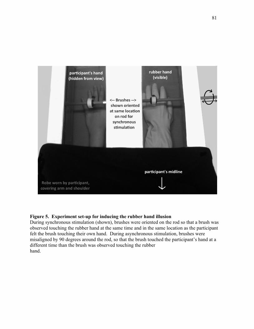
81
Figure 5. Experiment set-up for inducing the rubber hand illusion During synchronous stimulation (shown), brushes were oriented on the rod so that a brush was observed touching the rubber hand at the same time and in the same location as the participant felt the brush touching their own hand. During asynchronous stimulation, brushes were misaligned by 90 degrees around the rod, so that the brush touched the participant’s hand at a different time than the brush was observed touching the rubber hand.
82
There were two stimulation phases (Botvinick & Cohen, 1998). During the synchronous
phase, the paintbrush heads were aligned so that the participant saw the rubber hand being
touched by the paintbrush at the same time as the paintbrush was touching their own hand.
During the asynchronous phase, the paintbrush heads were misaligned by 90 degrees along the
rod, so that the brush touched the rubber hand a quarter of a second before or after touching the
participant’s hand (based on a 1 rotation/second frequency of brushing). Each stimulation phase
lasted 10 minutes.
Before and after each stimulation phase, the participant indicated the perceived location
of the middle finger of their hidden hand by reading off a meter stick that was held just above the
box and randomly translated left or right. Proprioceptive drift was estimated as the difference
between pre and post-stimulation hand location judgments (Botvinick & Cohen, 1998; Tsakiris
& Haggard, 2005). The participant was then asked a series of questions to assess their experience
of the illusion (see Table 7; taken from Longo et al., 2008 and Botvinick & Cohen, 1998) and
any nonspecific feelings of reduced afference (Longo et al., 2008).
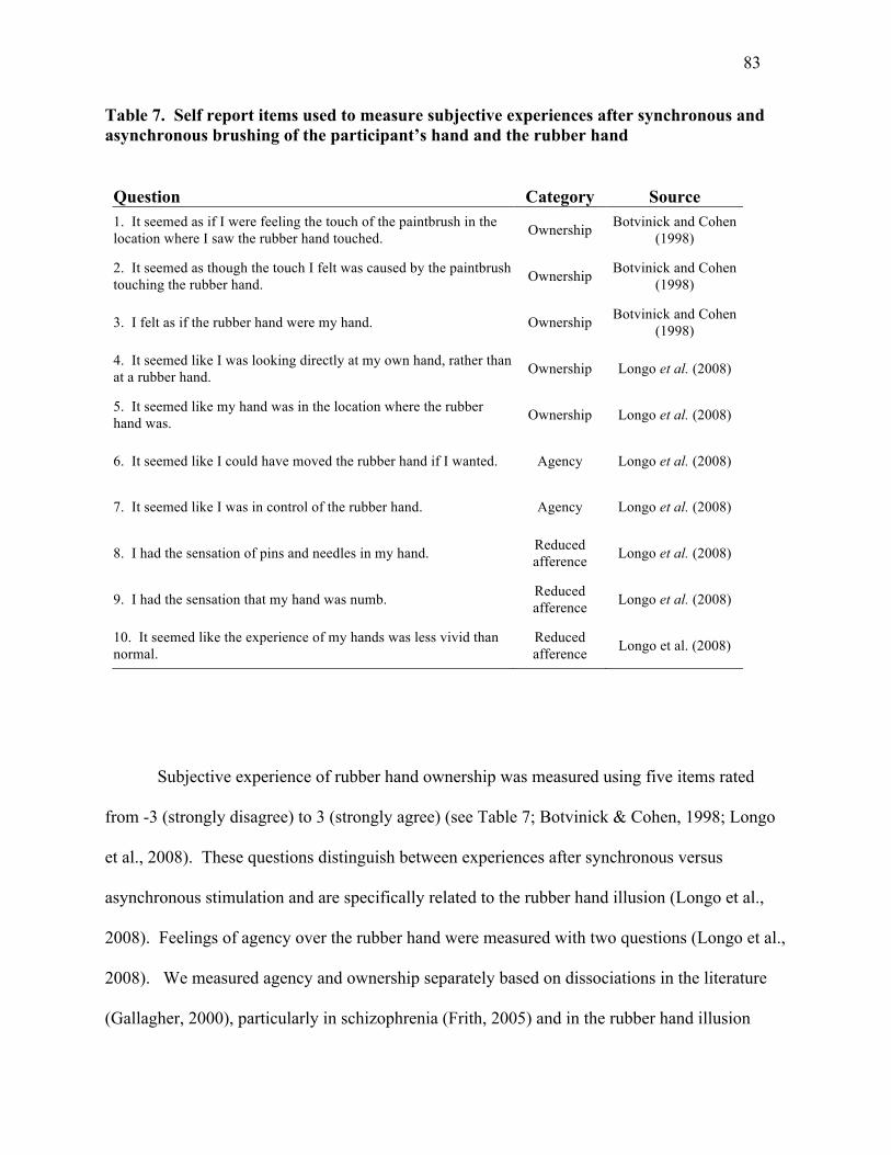
83
Table 7. Self report items used to measure subjective experiences after synchronous and asynchronous brushing of the participant’s hand and the rubber hand
Question Category Source 1. It seemed as if I were feeling the touch of the paintbrush in the location where I saw the rubber hand touched. Ownership Botvinick and Cohen
(1998)
2. It seemed as though the touch I felt was caused by the paintbrush touching the rubber hand. Ownership Botvinick and Cohen
(1998)
3. I felt as if the rubber hand were my hand. Ownership Botvinick and Cohen (1998)
4. It seemed like I was looking directly at my own hand, rather than at a rubber hand. Ownership Longo et al. (2008)
5. It seemed like my hand was in the location where the rubber hand was. Ownership Longo et al. (2008)
6. It seemed like I could have moved the rubber hand if I wanted. Agency Longo et al. (2008)
7. It seemed like I was in control of the rubber hand. Agency Longo et al. (2008)
8. I had the sensation of pins and needles in my hand. Reduced afference Longo et al. (2008)
9. I had the sensation that my hand was numb. Reduced afference Longo et al. (2008)
10. It seemed like the experience of my hands was less vivid than normal.
Reduced afference Longo et al. (2008)
Subjective experience of rubber hand ownership was measured using five items rated
from -3 (strongly disagree) to 3 (strongly agree) (see Table 7; Botvinick & Cohen, 1998; Longo
et al., 2008). These questions distinguish between experiences after synchronous versus
asynchronous stimulation and are specifically related to the rubber hand illusion (Longo et al.,
2008). Feelings of agency over the rubber hand were measured with two questions (Longo et al.,
2008). We measured agency and ownership separately based on dissociations in the literature
(Gallagher, 2000), particularly in schizophrenia (Frith, 2005) and in the rubber hand illusion
84
(Longo et al., 2008). Finally, three questions assessed feelings of reduced afference in the
participant’s own hand (e.g. numbness or tingling) (Longo et al., 2008). Feelings of reduced
afference are not directly related to the rubber hand illusion, but rather are more prominent
during asynchronous stimulation (Longo et al., 2008). Our prediction was that psychosis-
proneness would be associated with feelings of rubber hand agency and ownership, but not
reduced afference.
Due to our concern about the possible confound of individual differences in
suggestibility, the asynchronous stimulation condition always followed the synchronous
stimulation condition. Participants were naïve about the expected effect during synchronous
stimulation, but generally knew what to expect (either because of their experiences or post-
stimulation assessment) during the asynchronous stimulation condition.
Results
Average scores on measures of psychosis-proneness are shown in Table 8. Average
proprioceptive drift and question ratings after synchronous and asynchronous stimulation
conditions are shown in Table 8.
Due to positive skew, distributions of scores in our measures of psychosis-proneness and
subjective ratings of the rubber hand illusion were significantly nonnormal (Kolmogorov-
Smirnov test for normality; all P < 0.05). Thus, we report all effects in terms of both parametric
and nonparametric statistics.
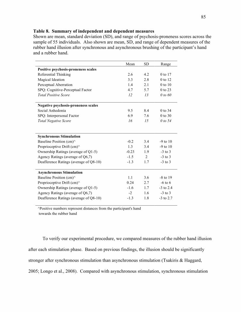
85
Table 8. Summary of independent and dependent measures Shown are mean, standard deviation (SD), and range of psychosis-proneness scores across the sample of 55 individuals. Also shown are mean, SD, and range of dependent measures of the rubber hand illusion after synchronous and asynchronous brushing of the participant’s hand and a rubber hand.
Mean SD Range Positive psychosis-proneness scales Referential Thinking 2.6 4.2 0 to 17 Magical Ideation 3.3 2.8 0 to 12 Perceptual Aberration 1.4 2.1 0 to 10 SPQ: Cognitive-Perceptual Factor 4.7 5.7 0 to 23 Total Positive Score 12 13 0 to 60 Negative psychosis-proneness scales Social Anhedonia 9.5 8.4 0 to 34 SPQ: Interpersonal Factor 6.9 7.6 0 to 30 Total Negative Score 16 15 0 to 54 Synchronous Stimulation Baseline Position (cm)^ -0.2 3.4 -9 to 10 Proprioceptive Drift (cm)^ 1.3 3.4 -9 to 10 Ownership Ratings (average of Q1-5) -0.23 1.9 -3 to 3 Agency Ratings (average of Q6,7) -1.5 2 -3 to 3 Deafference Ratings (average of Q8-10) -1.3 1.7 -3 to 3 Asynchronous Stimulation Baseline Position (cm)^ 1.1 3.6 -8 to 19 Proprioceptive Drift (cm)^ 0.24 2.7 -6 to 6 Ownership Ratings (average of Q1-5) -1.6 1.7 -3 to 2.4 Agency Ratings (average of Q6,7) -2 1.6 -3 to 3 Deafference Ratings (average of Q8-10) -1.3 1.8 -3 to 2.7 ^Positive numbers represent distances from the participant's hand towards the rubber hand
To verify our experimental procedure, we compared measures of the rubber hand illusion
after each stimulation phase. Based on previous findings, the illusion should be significantly
stronger after synchronous stimulation than asynchronous stimulation (Tsakiris & Haggard,
2005; Longo et al., 2008). Compared with asynchronous stimulation, synchronous stimulation
86
produced higher rubber hand ownership ratings (Wilcoxon Signed-Rank Test, one-tailed, z = -
5.0, P < 0.001; paired samples t-test, one-tailed, t(54) = 6.0, P < 0.001), higher agency ratings (z
= -2.4, P < 0.05; t(54) = 2.6, P < 0.01) and greater proprioceptive drift (z = -1.7, P = 0.07; t(54) =
1.9, P < 0.05). Although feelings of reduced afference were present in both conditions (z = -4.4
for both, P < 0.001; t(54) = -0.56, P < 0.001), there was no difference between conditions (z = -
0.21, P = 0.83; t(54) = 0.16, P = 0.43). Based on these data, we confirm that our manipulation
induced experiences associated with the rubber hand illusion in the synchronous condition
compared with the asynchronous (control) condition.
We predicted that positive psychosis-proneness would be associated with greater
susceptibility to the rubber hand illusion, as measured by feelings of rubber hand ownership and
agency after synchronous stimulation. We examined ownership and agency separately because
of ownership/agency dissociations in the schizophrenia literature (Frith, 2005).
Rubber hand ownership and psychosis-proneness. Ownership is the degree to which
the participant experiences the bodily sense that the rubber hand is their own hand. Consistent
with our hypothesis, positive psychosis-proneness was significantly associated with subjective
experiences of rubber hand ownership after synchronous stimulation (Spearman rank correlation,
ρ = 0.32, P < 0.05; Pearson correlation, r = 0.42, P < 0.01), even when controlling for rubber
hand ownership after asynchronous stimulation (Spearman rank partial correlation, ρ = 0.28, P <
0.05; Pearson partial correlation, r = 0.35, P < 0.05). There was no significant association
between rubber hand ownership and positive psychosis-proneness after asynchronous stimulation
(ρ = 0.17, P = 0.2; r = 0.26, P = 0.06). The absence of a relationship between positive
psychosis-proneness and experiences of rubber hand ownership after asynchronous stimulation
indicates that the relationship after synchronous stimulation cannot be explained by an increased
87
tendency to endorse unusual experiences among psychosis-prone participants.
Negative psychosis-proneness was not associated with the subjective experience of
rubber hand ownership (ρ = -0.08, P = 0.58; r = -0.1, P = 0.48), despite the strong relationship
between positive and negative psychosis-like characteristics in our sample (ρ = 0.59, P < 0.001; r
= 0.60, P < 0.001).
Figure 6 shows the relationship between ratings of rubber hand ownership after
synchronous stimulation, as related to positive and negative psychosis-proneness.
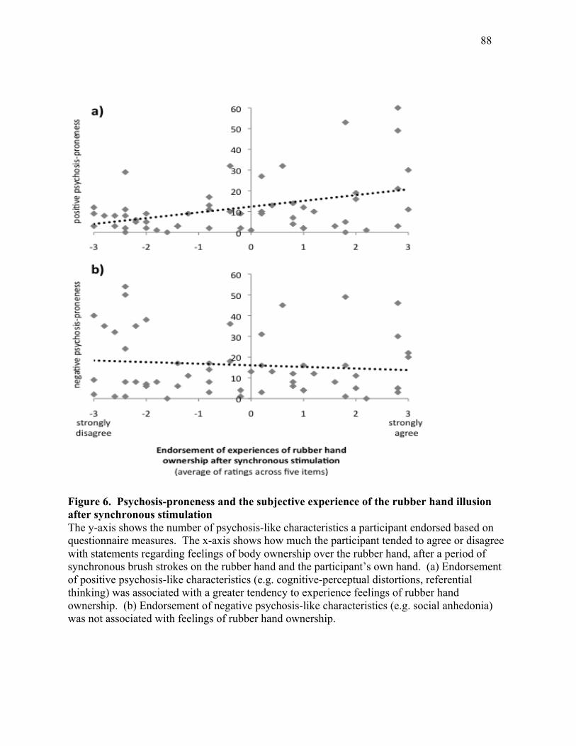
88
Figure 6. Psychosis-proneness and the subjective experience of the rubber hand illusion after synchronous stimulation The y-axis shows the number of psychosis-like characteristics a participant endorsed based on questionnaire measures. The x-axis shows how much the participant tended to agree or disagree with statements regarding feelings of body ownership over the rubber hand, after a period of synchronous brush strokes on the rubber hand and the participant’s own hand. (a) Endorsement of positive psychosis-like characteristics (e.g. cognitive-perceptual distortions, referential thinking) was associated with a greater tendency to experience feelings of rubber hand ownership. (b) Endorsement of negative psychosis-like characteristics (e.g. social anhedonia) was not associated with feelings of rubber hand ownership.
89
Agency and psychosis-proneness. Another component of the rubber hand illusion is the
subjective experience of agency, or the feeling that one can control the rubber hand. Positive
psychosis-proneness was significantly associated with the subjective experience of agency after
synchronous stimulation (ρ = 0.28, P < 0.05; r = 0.3, P < 0.05), but also after asynchronous
stimulation (ρ = 0.24, P = 0.08; r = 0.33, P < 0.05). Controlling for agency after asynchronous
stimulation abolished the relationship between positive psychosis-proneness and agency after
synchronous stimulation (ρ = 0.14, P = 0.3; r = 0.09, P = 0.52). It is possible that order effects
(asynchronous stimulation always followed synchronous stimulation) created a residual sense of
agency after asynchronous stimulation among psychosis-prone individuals. Alternatively,
differences in the experience of agency that vary with psychosis-proneness may be less closely
related to visual-tactile integration than differences in the experience of rubber hand ownership.
Negative psychosis-proneness was not associated with agency after either synchronous (ρ
= -0.05, P = 0.7; r = -0.04, P = 0.8) or asynchronous stimulation (ρ = -0.05, P = 0.7; r = 0.02, P
= 0.9).
Reduced afference and psychosis-proneness. To understand the specificity of the
relationship between subjective experiences and psychosis-proneness, we assessed whether
positive psychosis-proneness was associated with bodily experiences related to the manipulation,
but not to the rubber hand illusion. We measured experiences of reduced afference after illusion
induction (Longo et al., 2008) to explore this possibility.
Positive psychosis-proneness was not associated with feelings of reduced afference after
synchronous (ρ = 0.06, P = 0.67; r = 0.08, P = 0.58) or asynchronous stimulation (ρ = 0.11, P =
0.41; r = 0.13, P = 0.35). Differences in negative psychosis-proneness, similarly, showed no
association with feelings of reduced afference (synchronous: ρ = 0.09, P = 0.51; r = 0.13, P =
90
0.35; asynchronous: ρ = -0.04, P = 0.8; r = -0.1, P = 0.46).
Proprioceptive drift and psychosis-proneness. The rubber hand illusion is often
associated with biases in proprioception, where the participant judges their actual (hidden) hand
as being closer to the rubber hand after synchronous stimulation. Proprioceptive drift is often
used as an objective measure of the rubber hand illusion (Botvinick & Cohen, 1998; Tsakiris &
Haggard, 2005; Longo et al., 2008). We predicted that psychosis-proneness would be associated
with greater proprioceptive drift towards the rubber hand after synchronous stimulation.
Contrary to our hypothesis, proprioceptive drift after synchronous stimulation was not
associated with positive psychosis-proneness (ρ = 0.03, P = 0.85; r = 0.07, P = 0.62) or negative
psychosis-proneness (ρ = 0.01, P = 0.96; r = 0.06, P = 0.64), despite the relationship between
positive psychosis-proneness and subjective experiences of rubber hand ownership.
Dissociations between drift and subjective experience of the rubber hand illusion have been
documented in previous studies (Holmes et al., 2006), suggesting that these two measures may
map onto dissociable aspects of the illusion.
Discussion
We have shown that positive psychosis-like characteristics in otherwise healthy
individuals are associated with a greater susceptibility to the rubber hand illusion, an illusion of
body ownership. These positive psychosis-like characteristics include a tendency towards
referential thinking (Lenzenweger et al., 1997), magical ideation (Eckblad & Chapman, 1983),
cognitive-perceptual distortions (Raine, 1991), and perceptual aberrations (Chapman et al.,
1978). Our data suggest that susceptibility to body representation distortion may be a
91
vulnerability factor for developing psychosis, consistent with abnormalities in bodily experience
among individuals at high risk of developing psychosis (Sass & Parnas, 2003; Nelson et al.,
2008). A tendency to experience distortions in body representations may be linked to the
development of positive psychosis-like experiences and to broader deficits in self-processing
related to psychosis risk.
The relationship between positive psychosis-proneness and the experience of ownership
in the rubber hand illusion was only found when the participant saw a brush stroking a rubber
hand at the same time as feeling a brush stroking their own hand (synchronous stimulation).
When a small delay was introduced between these two events (asynchronous stimulation), there
was no longer a relationship between positive psychosis-proneness and illusion strength. Thus,
the relationship between positive psychosis-proneness and rubber hand illusion strength was not
being driven by a tendency to endorse unusual experiences, as these should impact both
synchronous and asynchronous stimulation conditions. Along similar lines, psychosis-proneness
was specifically related to feelings of rubber hand ownership, and not to feelings of reduced
afference that were also induced by the experimental procedure (Longo et al., 2008). The
specificity of the relationship between positive psychosis-proneness and the experience of rubber
hand ownership after synchronous stimulation rules out alternative explanations that previous
experiments looking at body ownership and psychosis have failed to exclude.
Rubber hand illusion susceptibility was specifically linked to positive (and not negative)
psychosis-like characteristics, suggesting possible mechanisms relating positive psychosis-like
characteristics to abnormalities in the subjective experience of the body. Perception involves
the integration of internal representations, acquired through previous experience, with incoming
sensory information. Perception is thus constrained by prior knowledge and context information
92
(Frith & Dolan, 1997; Gilbert & Sigman, 2007). Schizophrenia-related cognitive and perceptual
abnormalities may result from a failure to adequately couple sensory information with context
and previous experience (Fleminger, 1992; Fletcher & Frith, 2009; Gilbert & Sigman, 2007;
Hemsley, 1987; Hemsley, 2005)
For example, schizophrenia patients are less likely than control participants to experience
the hollow mask illusion, whereby a hollow mask viewed from behind is perceived as if being
viewed from the front (i.e. not depth-inverted). This has been interpreted as a reduced tendency
to incorporate previous experience in guiding perception towards the more familiar stimulus – a
normal face (Schneider et al., 2002).
A reliance on sensory information over previous experience may also increase
susceptibility to the rubber hand illusion. The rubber hand illusion is thought to be a
consequence of multisensory (visual-tactile) information overriding previous representations of
the body (Tsakiris, 2010). Greater susceptibility to the rubber hand could result from a reduced
tendency to use previous experience as a constraint on perception. Reduced constraints on
perception, across modalities, could give rise to the cognitive and perceptual distortions that we
think of as positive psychosis-like characteristics (Raine, 1991). The specific impact of this
tendency on the bodily sense, that is having body representations that are susceptible to
distortion, may further contribute to positive psychosis-like characteristics by disrupting the
stability of self experience (Sass & Parnas, 2003; Nelson et al., 2008; James, 1890).
To our knowledge, only one previous study has looked at the relationship between
psychosis-related personality variables and the rubber hand illusion (Asai et al., 2011). The
findings of this study are difficult to interpret, however, as a significant relationship was only
found between positive psychosis-like characteristics and self-report items not specifically
93
related to the experience of the rubber hand illusion (e.g. the sensation that the participant’s own
hand is moving; Asai et al., 2011, supplementary materials).
One limitation of our study is the absence of any relationship between psychosis-
proneness and proprioceptive drift after synchronous stimulation. Although self-reported
experiences of the rubber hand illusion and proprioceptive drift are generally highly associated
(Longo et al., 2008), dissociations between these two measures have been noted in several
studies (Holmes et al., 2006; Kammers et al., 2011; Kammers et al., 2009; Morgan et al., 2011;
Tsakiris et al., 2011). Positive psychosis-proneness may be related specifically to distortions in
the subjective experience of the body and not to differences in proprioceptive localization of the
body in space.
Another unexpected finding in our study was a relationship between psychosis-proneness
and the experience of rubber hand agency (but not ownership) after both synchronous and
asynchronous stimulation conditions. Previous evidence indicates that body ownership and
agency are dissociable aspects of subjective experience (Frith, 2005; Gallagher, 2000; Longo et
al., 2008). For example, a patient with schizophrenia-related delusions of control may recognize
that their hand is moving, but believe that some other agent is controlling that movement (Frith,
2005). Positive psychosis-proneness may be related to a tendency to feel a sense of agency over
a rubber hand based on visual similarity alone. Alternatively, the sense of agency induced by
synchronous stimulation (always occurring first) may have carried over to the asynchronous
stimulation condition, suggesting that the experience of agency might be more durable and/or
less dependent on visual-tactile integration than the experience of ownership. We cannot
distinguish between these possibilities based on the current study.
Our data suggest a provocative relationship between psychosis vulnerability and illusions
94
of body ownership. We do not mean to suggest, however, that individuals who are susceptible to
illusions of body ownership are therefore also vulnerable to developing psychosis. Those
individual differences that underly susceptibility to the rubber hand illusion are poorly
understood, and many other individually varying characteristics may increase susceptibility.
Previous literature has found, for example, that greater illusion susceptibility is related to low
interoceptive sensitivity (Tsakiris et al., 2011), a malleable body image (Mussap & Salton, 2006;
Eshkevari et al., 2011), and greater empathic concern (Asai et al., 2011).
In conclusion, our findings indicate that psychosis vulnerability and positive psychosis-
like characteristics are related to the tendency to experience illusions of body ownership, even in
otherwise healthy individuals. Our findings also demonstrate that susceptibility to body
representation distortion is a measurable phenomenon that varies with psychosis risk. Future
research might look at how rubber hand illusion susceptibility varies with other measures of self
processing that are disturbed in psychosis (e.g. self monitoring), increasing our understanding of
the way body representation relates to other forms of self-representation. One might also look at
experimental manipulations that interact with psychosis-proneness to alter illusion susceptibility,
to provide information about the mechanisms that underlie abnormalities of body experience
among psychosis-prone individuals. For example, if the relationship between psychosis-
proneness and the rubber hand illusion represents a failure of knowledge to constrain body
representation distortions, one might expect substitution of a rubber hand for another type of
object (e.g. a wooden block; Tsakiris, 2010) to disrupt illusion formation less for psychosis-
prone individuals. An increased understanding of the way body ownership illusions are related
to other forms of self-representation and psychosis-proneness can shed light on the distortions in
bodily experience that accompany positive symptoms and psychosis-like characteristics, and
95
ultimately the factors that might lead to psychosis development.
Acknowledgements
We would like to thank Daw-An Wu for advice about initial experimental set-up. We would also
like to thank Todd Wright, Juyoen Hur, Roger Mercado, and Lizz Langer for assistance with data
collection and analysis. This research was supported by Harvard University research funds to
Christine Hooker and a National Science Foundation Graduate Research Fellowship to Laura
Germine.
96
Additional Findings
Here, experimental findings are presented that were not included in Papers #1-3, but are
relevant to the current dissertation. These findings are organized by experiment number, with a
brief introduction, method, results, and discussion section for each experiment.
Experiment #3b: Psychosis-proneness, Flexibility in Body Representations, and Face
Emotion Recognition (FER)
Introduction. A significant motivation for Experiment #3 was to see whether differences
in flexibility of the body representation (as measured by susceptibility to the rubber hand
illusion) mediate the relationship between psychosis-proneness and FER, based on simulation
theories of FER that posit contributions from somatosensory and motor processing to emotion
understanding (Adolphs et al., 2000). The fact that there was no relationship between negative
psychosis-proneness and indices of the rubber hand illusion indicates that the latter could not
function as a mediator of the relationship between negative psychosis-proneness and FER ability.
Nevertheless, this hypothesis was tested for positive psychosis-proneness, which was
significantly related to rubber hand illusion susceptibility.
If differences in body representation flexibility account for differences in FER that vary
with positive psychosis-proneness, this would suggest that body representation abnormalities
may underly reduced FER ability in high psychosis-prone individuals (based on positive
psychosis-like characteristics). It was predicted that susceptibility to the rubber hand illusion
would be related to reduced FER ability, and mediate the relationship between positive
psychosis-proneness and FER performance.
97
Method. The rubber hand illusion and psychosis-proneness were measured as described
in Paper #3. Subjective experiences of ownership and subjective experiences of agency were
used as measures of the rubber hand illusion, as relationships between these measures and
positive psychosis-proneness were found in Experiment #3. No relationship was found between
the rubber hand illusion and negative psychosis-proneness, so negative psychosis-proneness was
not included in this analysis.
As a test of FER, the Reading the Mind in the Eyes test (hereafter referred to as the “Eyes
Test”) was administered. The Eyes Test is a test of complex emotion perception that has been
used to test individual differences in emotion perception and theory of mind (Baron-Cohen et al.,
2001). In this test, the participant sees an image of the eye region of a face and must choose
which of four complex emotion words (e.g. “arrogant”, “annoyed”, “cautious”) best fits the
emotion being expressed in that image.
Results. Positive psychosis-proneness predicted experiences of rubber hand ownership
(ρ = 0.32, p < 0.05, two-tailed) and rubber hand agency (ρ = 0.28, p < 0.05) (See Paper #3).
Positive psychosis-proneness also predicted Eyes Test performance (ρ = -0.28, p < 0.05).
Rubber hand ownership predicted Eyes Test performance, but in the opposite direction (ρ
= 0.35, p < 0.01). Agency predicted Eyes Test performance at trend level (ρ = 0.23, p = 0.1),
also in the opposite direction. This was contrary to prediction, and suggests that susceptibility to
the rubber hand illusion is related to better FER ability and thus cannot account for psychosis-
proneness related FER differences.
Discussion. The hypothesis was that variations in rubber hand illusion susceptibility
might mediate the relationship between psychosis-proneness and FER. This hypothesis was not
confirmed. If differences in somato-motor processing underlie differences in FER in psychosis-
98
prone individuals, these somato-motor differences do not appear to be measured by susceptibility
to the rubber hand illusion.
Another unexpected finding was the positive correlation between Eyes Test performance
and rubber hand illusion susceptibility. Previous literature has suggested that greater rubber
hand illusion susceptibility may arise from higher levels of empathy (Durgin et al., 2007).
Insofar as Eyes Test performance is related to empathy (Rodrigues et al., 2009), this could
explain why higher Eyes Test scores were associated with stronger illusions. On the other hand,
positive psychosis-proneness was related to rubber hand illusion susceptibility despite being
associated with reduced empathy (Henry, Bailey, & Rendell, 2008). Empathy and positive
psychosis-proneness may impact illusion susceptibility through different routes. Further
investigation would be needed to understand the complex relationship between psychosis-
proneness, FER, and illusions of body ownership.
Experiment #3c: Dimensions of Psychosis-proneness and FER
Introduction. Paper #1 (“Face emotion recognition is related to individual differences in
psychosis-proneness.”) reported findings from one self-report measure of psychosis-proneness,
the Schizotypal Personality Questionnaire (Brief; Raine & Benishay, 1995) and two different
tests of FER. Significant associations were found between positive psychosis-proneness and
FER as well negative psychosis-proneness and FER, even when controlling for variations in face
gender or face identity processing. Although not reported in Paper #3, Experiment #3 included a
measure of FER (complex emotion perception: Reading the Mind in the Eyes, Baron-Cohen et
al., 2001), a measure of verbal IQ, and a measure of performance IQ. These data thus allowed
an investigation of whether the relationship between FER and psychosis-proneness observed in

99
Experiment #1 replicates across measures of psychosis-proneness, with an FER paradigm that
relies on recognition of more complex emotional states, and while controlling for differences in
verbal and performance IQ.
Method. A set of widely used self-report questionnaires measuring positive and negative
psychosis-like characteristics was administered to 55 individuals, tested in the lab. These
included the Schizotypal Personality Questionnaire (cognitive-perceptual and interpersonal
subscales; Raine, 1991), the Chapman Magical Ideation Scale (Eckblad and Chapman, 1983), the
Chapman Perceptual Aberration Scale (Chapman et al., 1978), the Referential Thinking Scale
(Lenzenweger et al., 1997), and the Chapman Revised Social Anhedonia Scale (Eckblad et al.,
1982; Mishlove and Chapman, 1985). The brief version of the Schizotypal Personality
Questionnaire was also used in Experiment #1. The Revised Social Anhedonia Scale was also
used in Experiment #2. All measures were used in Experiment #3a. The sample of 55
individuals was the same as the sample described in Paper #3.
The test of FER was the Reading the Mind in the Eyes test (Eyes Test), described earlier.
The Vocabulary subtest of the Wechsler Abbreviated Scale of Intelligence was administered as a
measure of verbal IQ and the Matrix Reasoning subtest as a measure of performance IQ (WASI;
Wechsler, 1999).
Results are reported in terms of Spearman correlations between each measure of
psychosis-proneness (six measures in total) and scores on the Eyes Test. Spearman correlations
were chosen due to the nonnormal distributions in questionnaires scores (significant positive
skew).

100
Results. Table 9 shows zero-order correlations between Eyes Test scores and each
measure of psychosis-proneness, as well as partial correlations controlling for verbal IQ (based
on Vocabulary scores) and performance IQ (based on Matrix Reasoning scores).
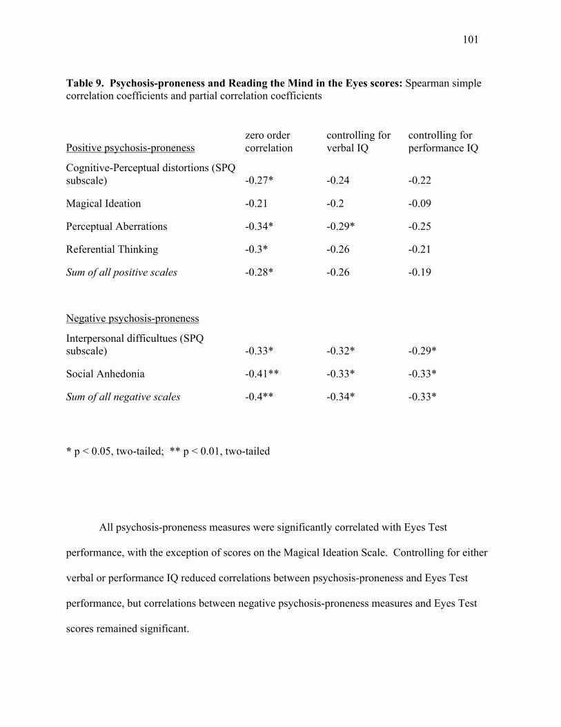
101
Table 9. Psychosis-proneness and Reading the Mind in the Eyes scores: Spearman simple correlation coefficients and partial correlation coefficients
Positive psychosis-proneness zero order correlation
controlling for verbal IQ
controlling for performance IQ
Cognitive-Perceptual distortions (SPQ subscale) -0.27* -0.24 -0.22
Magical Ideation -0.21 -0.2 -0.09
Perceptual Aberrations -0.34* -0.29* -0.25
Referential Thinking -0.3* -0.26 -0.21
Sum of all positive scales -0.28* -0.26 -0.19
Negative psychosis-proneness
Interpersonal difficultues (SPQ subscale) -0.33* -0.32* -0.29*
Social Anhedonia -0.41** -0.33* -0.33*
Sum of all negative scales -0.4** -0.34* -0.33*
* p < 0.05, two-tailed; ** p < 0.01, two-tailed
All psychosis-proneness measures were significantly correlated with Eyes Test
performance, with the exception of scores on the Magical Ideation Scale. Controlling for either
verbal or performance IQ reduced correlations between psychosis-proneness and Eyes Test
performance, but correlations between negative psychosis-proneness measures and Eyes Test
scores remained significant.
102
Discussion. The current findings suggest that the relationship between FER and
psychosis-proneness generalizes to measures of complex emotion perception, but is more robust
for negative than positive psychosis-like characteristics. In an analysis using multiple self-report
scales and controlling for IQ differences, FER was robustly related to measures of negative
psychosis-proneness. FER was also related to measures of positive psychosis-proneness, but this
relationship was not significant after controlling for differences in IQ.
This finding agrees with previous literature indicating that FER deficits are more closely
related to the presence of negative than positive symptoms (Sergi et al., 2007).
Experiment #4: Psychosis-proneness and self-referential processing
Introduction. Previous evidence has indicated that individuals with schizophrenia have
trouble judging the origin of information, or source monitoring (Vinogradov et al., 1997). In
particular, they have difficulties with remember the source of self-generated information
(Vinogradov et al., 1997; Fisher et al., 2008), showing a greater tendency to identify self-
generated information as arising from external sources. Harvey et al. (2011) similarly found that
schizophrenia patients had a smaller recognition memory advantage for self-referential
information than healthy control participants.
In a study of self-referential source memory and social perception, Fisher et al. (2008)
further found that source memory for self-generated information predicted face emotion
perception and identity recognition in healthy control and schizophrenia participants – but that
once the influence of other neurocognitive variables was removed, this relationship was only
significant in healthy control participants. In other words, self-referential source memory
specifically contributed to social perception ability in healthy control participants, but not in
103
patients with schizophrenia. This suggests that self-referential processing deficits contribute to
deficits in social cognition in schizophrenia.
Two paradigms were taken from the schizophrenia literature for measuring self-
referential source memory (Fisher et al., 2008; Vinogradov et al., 1997) and self-referential
recognition memory (Harvey et al., 2011) to see whether self-referential processing (1) is related
to positive and/or negative psychosis-proneness, (2) whether self-referential processing ability is
related to FER ability, and (3) whether the relationship between psychosis-proneness and FER
ability could be explained by differences in self-referential processing, as suggested by Fisher et
al. (2008).
Experiment 4a - Self-referential source memory.
Method. This study was conducted in the lab, using the same sample of 55 participants
that completed Experiment #3. The paradigm and stimuli were taken from Vinogradov et al.
(1997) and Fisher et al. (2008) to assess source memory for self-generated, experimenter-
generated, and new words. During the study phase, the participant was given a list of 20 short
sentences, where ten of the sentences had words already filled in and underlined and ten had
blank lines (e.g. “The boy threw the ball”, “The cat chased the ___”). The participant read each
sentence out loud, filling in the blank with a word where no word was given. The experimenter
recorded all words generated by the participant. Approximately two hours later, participants
were presented with a list of 30 word pairs (e.g. boy-ball, cat-mouse), with 10 word pairs from
sentences that the participant had previously read aloud (experimenter-generated), 10 word pairs
from sentences where the participant had filled in the blank with a word (self-generated), and 10
new word pairs. Participants were asked to identify whether the second word in the pair was
experimenter-generated, self-generated, or new.
104
Patterns of errors were calculated as dependent measures using the same procedure as
Vinogradov et al. (1997). For each category of word pair (self-generated, experimenter-
generated, and new) errors could be classified as misattribution errors or source discrimination
errors. For self-generated word pairs, for example, a judgment of “experimenter-generated” or
“new” would be a source discrimination error – a failure to remember the source of the word
pair. Where experimenter-generated or new word pairs were judged as self-generated, this was
classed as a self-generated misattribution error, or the incorrect judgment that oneself was the
source of a word pair. Thus, there were six classes of errors: self-generated source
discrimination errors, self-generated misattribution errors, experimenter-generated source
discrimination errors, experimenter-generated misattribution errors, new source discrimination
errors, and new misattribution errors.
People tend to be better at remembering the source of self-generated information than
experimenter-generated information (Vinogradov et al., 1997). Also, when new information is
falsely recognized, the source of that information is more likely to be attributed to the
experimenter than to oneself. The goal was to replicate these previous findings in this sample.
The main hypothesis with respect to psychosis-proneness was that higher psychosis-proneness
would be associated with a greater tendency to make self-generated source discrimination errors
(failure to remember that a self-generated word was self-generated), but that this could not be
explained by a greater tendency to make source discrimination errors overall. Finally, the role of
IQ in any observed relationships was examined.
Results. Due to significant positive skew in all variables, nonparametric tests were used.
Across all participants, significantly fewer self-generated source discrimination errors (9% error)
were made than experimenter-generated source discrimination errors (46%) (Wilcoxon signed
105
rank test: z = -5.9, p < 0.001). Similarly, participants made fewer self-generated misattribution
errors (mistakenly judging a word-pair as self-generated: 4%) than experimenter-generated
misattribution errors (14%) (p < 0.001). The most common errors were new misattribution
errors (49%), which correspond to a failure to recognize a word pair. For new word pairs that
were falsely recognized (new source discrimination errors), participants were much more likely
to judge the word pair as experimenter-generated (13%) than self-generated (0.2%) (z = -4.6, p <
0.001). Thus, these findings agree with previous literature on biases in source memory.
Greater positive psychosis-proneness was related to a significantly higher number of self-
generated source discrimination errors (Spearman rank correlation; ρ = 0.31, p < 0.05), but not
other types of source discrimination errors (ρ = 0.09, p = 0.5). Post hoc comparisons revealed no
other error types that were significantly related (even at trend level) to positive psychosis-
proneness. When controlling for other type of errors (total errors – self-generated source
discrimination errors), the correlation between positive psychosis-proneness and self-generated
source discrimination errors remained significant (ρ = 0.29, p < 0.05). Controlling for IQ did not
significantly impact this relationship either (performance IQ: ρ = 0.31, p < 0.05; verbal IQ: ρ =
0.29, p < 0.05). Negative psychosis-proneness was not related to any of the error types, at
significant or trend levels.
Finally, variations in self-generated source discrimination errors were looked at in
relation to FER performance based on the Eyes Test. There was no significant relationship (ρ = -
0.08, p = 0.5) indicating that differences in self-referential source memory do not explain FER
ability based on the current measures.
106
Experiment 4b - Self-referential recognition memory.
Method. This experiment was conducted on TestMyBrain.org, in two separate web-based
samples. In the first sample (n = 2006), the brief version of the Schizotypal Personality
Questionnaire was used as a measure of psychosis-proneness (Raine & Benishay, 1995). In the
second sample (n = 5071), the Revised Social Social Anhedonia Scale was used (Eckblad et al.,
1982). A new FER task was also included. In this task, participants were shown three faces
expressing either happiness, sadness, anger, fear, surprise, or disgust. Two of the faces
expressed the same emotion, and the third face expressed a different emotion. Participants were
asked to choose which of the three faces was the “odd one out”, that is the face that expressed a
different emotion from the other two (three alternative forced choice: 3AFC). Faces were shown
on screen all at once, for 4500ms. There were 50 trials in total.
As a measure of self-referential recognition memory, the paradigm and stimuli from
Harvey et al. (2011) were adapted for web-based administration. There were four conditions in
the encoding phase, each with 12 trials. Participants were presented with a prompt for 2000ms,
followed by a target adjective for another 2000ms. The prompt was one of four questions. (1) Is
this word uppercase? (structural condition), (2) Does this word describe you? (self condition), (3)
Is this word socially desirable? (social desirability condition), and a final question that was
created for this version of the task: (4) Does this word describe your computer? (object
condition). For each question the participant was to press 1 for yes, and 0 for no (response
options always on screen). The object condition was added to be certain that any memory biases
found were not due to deeper encoding arising from processing an adjective with reference to
some person/thing that is present in the local environment and could be used as a memory cue
107
(oneself or one’s computer). Within each condition, half the words were presented in uppercase
and half in lowercase. Half were positively valenced and half were negatively valenced.
After judging all 48 words, participants were taken to another test that lasted 10 minutes.
After this delay participants were tested in an unexpected recognition memory task. Participants
were asked to judge whether each of 96 adjectives were old or now. Half of the adjectives were
new, and these were matched to the other half in terms of number of letters and syllables. Both
lists were also matched in terms of how appropriate each adjective was for describing a
computer, using ratings from an in lab sample.
Harvey et al. (2011) used d’prime as an index of sensitivity for recognition memory in
each condition. Harvey et al. (2011) used the difference between d’ for self vs. social
desirability conditions as their index of self-referential memory biases. The d’ difference
between self and object conditions was also calculated as an additional index of self-referential
memory biases. As subtracting a control measure from a measure of interest can introduce
confounds owing to variations in the baseline (Wilmer, Garrido, & Herzmann, in prep), self-
referential memory bias was also computed by regressing d’ social or d’ object scores out of d’
self scores and using the residuals as the dependent measure.
Unfortunately, due to an error in transitioning tests to a new web server, demographic
information was lost for these participants. Thus, it is unknown whether any findings or lack of
findings might be explained by differences in participant age, sex, or education that would have
otherwise been collected.
Results. Revised Social Anhedonia Scale. Average d’ scores were 1.35 (SD = 0.88) for
the self condition, 1.04 (SD = 0.79) for the social desirability condition, 1.91 (SD = 1.43) for the
object condition, and 1.4 (SD = 1.1) for the structural condition. The magnitude of the self-
108
referential memory bias in the sample (self-social desirability) was 0.3 (SD = 0.73). This bias
was similar in magnitude to the bias found by Harvey et al. (2011) when comparing these two
(self and social desirability) conditions (M = 0.32, SD = 0.47 in Harvey et al., 2011). When
using the object condition as a comparison, however, there was no longer a self-referential
memory bias (M = -0.57, SD = 1.2).
Social anhedonia scores were related to d’ recognition sensitivity measures in the self (r =
-0.042, p < 0.01), social desirability (r = -0.038, p < 0.01), object (r = -0.055, p < 0.001), and
structural conditions (r = -0.04, p < 0.01). Based on the measure used by Harvey et al. (2011) (d’
self minus d’ social desirability for each participant), there was no relationship between social
anhedonia scores and self-referential memory biases (r = -0.009, p = 0.66). When self-referential
memory biases were calculated with reference to the object condition (d’ self minus d’ object),
there was a small, but significant association in the opposite direction as predicted (r = 0.035, p <
0.01). When regression based dependent measures were used, neither relationship was
significant (d’ self regressing out d’ social desirability: r = -0.006, p = 0.66; d’ self regressing
out d’ object: r = 0.01, p = 0.46).
In this sample, participants had an average 70% correct (SD = 11%) on the 3AFC FER
task. Social anhedonia scores were significantly related to FER scores (r = -0.15, p < 0.0001).
Since no robust relationship was found between measures of self-referential processing and
social anhedonia, however, no additional analyses were performed.
Schizotypal personality questionnaire (brief:SPQ-B). Averages across this sample in each
condition were very similar to the independent sample that completed the Revised Social
Anhedonia Scale. Average d’ scores were 1.33 (SD = 0.93) for the self condition, 1.02 (SD =
0.76) for the social desirability condition, 1.9 (SD = 1.46) for the object condition, and 1.36 (SD
109
= 1.0) for the structural condition. The magnitude of the self-referential memory bias was 0.31
(SD = 0.76) in this sample when comparing self and social desirability conditions, and -0.57 (SD
= 1.3) when comparing self and object conditions, similar to the social anhedonia sample.
The SPQ-B includes subscales for positive (cognitive-perceptual distortions), negative
(interpersonal difficulties), and disorganized symptoms. There was a slight relationship between
SPQ-B total scores and d’ self – d’ social (r = -0.047, p < 0.05), in the predicted direction. This
relationship was driven by a small but significant relationship between cognitive-
perceptual/positive psychosis-proneness characteristics and reduced self-referential memory
biases (r = -0.055, p < 0.01). These correlations were the same when using regression-based
measures. No significant relationships were found between the interpersonal or disorganized
subscales and self-referential memory biases with either subtraction or regression measures. No
significant relationships were found between self-referential memory biases and psychosis-
proneness when d’ object was used as the comparison measure.
In this sample, participants again had an average 69% correct (SD = 10%) on the 3AFC
FER task. FER scores were related to SPQ-B total scores (r = -0.13, p < 0.0001), as well as
scores on each subscale (cognitive-perceptual distortions: r = -0.12; interpersonal difficulties: r =
-0.09; disorganization: r = -0.10). FER scores were not related to self-referential memory biases
when either the social desirability (r = 0.03, p = 0.16) condition or the object condition (r = 0.01,
p = 0.54) were used as comparison measures, so no further analyses were conducted.
Discussion. The findings of Harvey et al. (2011) were replicated in terms of the direction
and magnitude of a self-referential memory bias (when comparing self judgments with social
desirability judgments) across participants. The overall findings of this experiment are difficult
to interpret, however, as there was no self-referential memory bias when comparing self
110
judgments with the other two conditions. In fact, accuracy was highest for adjectives where
participants judged whether or not the adjective described their computer.
Relationships with psychosis-proneness were weak and/or inconsistent. A weak
relationship was found between a measure of positive psychosis-proneness (SPQ-B cognitive-
perceptual subscale) and self-referential memory biases, but this relationship was only found
when comparing memory for words judged with respect to oneself with words judged for social
desirability. No relationship was found when self-referential memory biases were computed
with respect to memory for words judged for how well they described a computer. It is possible
that this was due to some problem or confound in this new condition (object) that has not been
identified, but these results do not support a relationship between self-referential memory biases
and psychosis-proneness.
Experiment #5: Psychosis-proneness and Voice Emotion Recognition
In Experiment #1, a relationship was observed between psychosis-proneness and FER,
but it is unclear whether this relationship extends to emotion recognition in other modalities. A
similar relationship between psychosis-proneness and voice emotion recognition would imply
that this relationship generalizes beyond visual perception of emotion.
Method. To answer this question, a voice emotion recognition task developed by Sauter
et al. (2010) was adapted for web-based administration on TestMyBrain.org. In this paradigm,
two male and two female British English speakers produced emotionally-inflected neutral
content speech (three-digit words) expressing one of ten positive or negative emotions:
amusement, anger, contentment, disgust, fear, relief, sadness, sensual pleasure, surprise, and
triumph. After hearing a brief audio clip, participants were asked to choose which of the ten
111
emotion words (all displayed on screen) best described the emotion being expressed. The
original experiment included 100 trials, with 10 trials of each emotion type, with an average
score of 72% (SD = 8%) based on 23 control participants. 5/10 trials of each emotion were
chosen to create a shorter version of this test more suitable for web administration.
As measures of psychosis-proneness, the Revised Social Anhedonia Scale (Eckblad et al.,
1982; Mishlove and Chapman, 1985; See Study #2 and #3) and the brief version of the
Schizotypal Personality Questionnaire (SPQ-B; Raine, 1991; Raine & Benishay, 1995; See
Study #1) were used. Due to technical difficulties, however, this experiment was only
conducted using the SPQ-B.
At the beginning of the experiment, an audio clip of a beeping sound was played
continuously in order to adjust volume for each participant. Participants were asked to adjust the
volume on their computers until the beeping sound was only just audible. This was followed by
three practice stimuli, with feedback, and 50 trials of the actual test. All measures were
administered over the web on TestMyBrain.org.
Results. Audio presentation over the web presented a wide range of problems evident by
very high attrition rates and numerous reports of technical issues. Also, cross browser
compatibility was an issue and no clear solution was found that allowed the test to be run in all
popular web browsers. When a major update of Firefox’s popular web browser created
compatibility problems for a significant proportion of users, the experiment was terminated early
and without administering the social anhedonia measure in a second sample. Thus, final sample
sizes were smaller than expected for this study and the voice emotion recognition task was only
administered in conjunction with one measures of psychosis-proneness, the brief version of the
Schizotypal Personality Questionnaire.
112
Based on a sample of 451 participants, mean performance was 63% (SD = 14) suggesting
this version was significantly more difficult than the original version --- although this
performance mean may have been reduced by technical problems with the test. Preliminary data
suggest a small but nonsignificant relationship between voice emotion recognition performance
and the SPQ-B interpersonal subscale (negative psychosis-proneness; r = -0.07, p = 0.14, two-
tailed) and the cognitive-perceptual subscale (positive psychosis-proneness; r = -0.07, p = 0.16),
but no relationship with the disorganized symptom subscale (r = 0.04, p = 0.35) or total SPQ-B
scores (r = -0.04, p = 0.34). Given the reduced mean and higher standard deviation of this
sample relative to the original control sample collected by Sauter et al. (2010), technical
difficulties likely interfered with the ability to accurately measure voice emotion recognition
ability in this experiment.
Discussion. No significant relationship was found between psychosis-proneness and
voice emotion recognition, but technical difficulties likely interacted with the results as
suggested by the relatively low performance means on this test. Previous comparisons between
web and lab using TestMyBrain.org for tests of social perception, including emotion recognition,
have revealed comparable means and standard deviations between web and lab samples
(Germine et al., submitted). Although this difference may arise from a difference in forms, it
also suggests problems for these web-based data that make results difficult to interpret.
113
Discussion and Conclusion
Summary of Findings
In this dissertation, it has been shown that both positive and negative psychosis-like
characteristics predict face emotion recognition (FER) abilities, even in otherwise healthy
individuals. This relationship was not explained by variations in IQ, visual perception, face
processing, body representation, or self-referential processing. Greater psychosis-proneness was
also related to reduced neural activity in brain regions thought to underlie normal FER.
Altogether, the current evidence indicates that behavioral and neural abnormalities in FER are
related to the expression of psychosis-like characteristics in otherwise healthy individuals. This
suggests that FER deficits are related to psychosis vulnerability, rather than secondary
characteristics of schizophrenia and other psychotic disorders.
Psychosis-proneness and FER: Interpretation of Behavioral Findings
Previous research has produced mixed findings regarding the relationship between FER
and psychosis-proneness (Brown & Cohen, 2010). This was addressed in the current set of
studies by using very large samples (Experiment #1, #4b, #5) and a range of psychosis-proneness
measures (Experiment #1, #3c, #4). A relationship between FER and psychosis-proneness was
identified and replicated using four different FER paradigms, five measures of psychometric
psychosis-proneness, and multiple control or comparison conditions. These findings indicate
that the relationship between FER and psychosis-proneness is highly replicable and generalizes
to different measures of FER when sample sizes are adequate and sensitive tests are used.
114
Another contribution of the current research is addressing whether the relationship
between FER and psychosis-proneness can be explained by differences in more general cognitive
factors that also vary with psychosis-proneness (Siever et al., 2002). It was found that
psychosis-proneness related variations in FER could not be explained by differences in face
processing (Experiment #1), IQ (Experiment #3c), flexibility of body representations
(Experiment #3b), or differences in self-referential processing (Experiment #4). Across
experiments, no single factor was identified that could explain the relationship between FER and
psychosis-proneness. Although there are many other possible neurocognitive factors that may
underlie FER deficits in psychosis-prone individuals, these findings indicate relative specificity
in psychosis-proneness related FER variations. This is in contrast with studies of schizophrenia
patients where specific deficits are difficult to isolate against a background of generalized
cognitive impairment (Kerr & Neale, 1993).
In the schizophrenia and psychosis-proneness literature, emotion recognition deficits are
typically linked with negative symptoms or characteristics (Lincoln et al., 2011). In the studies
reported here, both positive and negative psychosis-like characteristics were associated with
FER. There was some support, however, for a more robust link between negative psychosis-
proneness and FER. Although in experiments #1 and #4 the relationship between positive and
negative psychosis-like characteristics and FER was comparable, experiment #3c revealed a
stronger relationship between FER and negative psychosis-proneness measures. When
controlling for IQ in a smaller sample (n = 55), only negative psychosis-proneness still
significantly predicted FER ability. It should be noted though that in Experiment #3c,
composite measures of psychosis-proneness were used that may have masked symptom
heterogeneity. That is, some aspects or dimensions of positive psychosis-proneness (e.g.
115
perceptual distortions) may be better predictors of FER than others. Nevertheless, these data
suggest that FER may be broadly related to a range of psychosis-proneness characteristics and
not any single dimension, with the most consistent relationships between FER and negative
psychosis-like characteristics.
Psychosis-proneness and FER: Interpretation of Neural Findings
In Experiment #2, neural measures of FER were obtained using functional magnetic
resonance imaging (fMRI) in participants that differed in their expression of negative psychosis-
proneness, as indexed by social anhedonia. Social anhedonia is a lack of pleasure from social
interactions, and is one of the strongest predictors of psychosis development (Kwapil, 1998). In
participants with higher levels of social anhedonia / negative psychosis-proneness, reduced
responses were observed in a priori defined emotion perception brain regions including the
anterior medial frontal cortex, right superior temporal gyrus, and somatosensory cortices. These
regions have been implicated in previous studies of FER in healthy participants (Adolphs et al.,
2000; Adolphs, 2002; Haxby et al., 2000).
The right superior temporal sulcus (and neighboring regions of the right superior
temporal gyrus) is thought to be involved in integration of information across modalities,
contributing to social perception and processing dynamic social cues including eye gaze (Allison
et al., 2000), face emotion (Haxby et al., 2000), and voice emotion (Phillips et al., 1998). Single
cell recordings in monkeys have indicated that individual cells in superior temporal regions code
particular directions of head orientation and gaze (Allison et al., 2000). Thus, superior temporal
cortex seems to contain high-level representations of social stimuli and integrates semantic,
spatial, verbal, and perceptual information (Haxby et al., 2000). Psychosis-proneness related
116
differences in this region may reflect reduced recruitment of these computational processes for
FER, and may contribute to reduced FER ability among psychosis-prone individuals.
In Experiment #2, variations in neural activity in the anterior rostral subdivision of the
medial frontal cortex (arMFC; Amodio & Frith, 2006) were also observed. Numerous studies
have found activity in this region when judging emotional faces as compared to non-emotional
faces (Dolan et al., 1996; Kestler-West et al., 1991; Phillips et al., 1998; Wright et al., 2002) and
deficits in this region during emotion processing in schizophrenia (Hempel et al., 2003). Social
understanding, person perception, and mentalizing also engage arMFC across a range of tasks
(Frith & Frith, 2003; Gobbini et al., 2004; Iacoboni et al., 2004; Mitchell, Banaji, & Macrae,
2005; Saxe, Carey, & Kanwisher, 2004).
There is consistent evidence that arMFC is also involved in self-referential processing.
For example, Johnson et al. (2002) found that this region was more active when participants
were asked to reflect on their own personal characteristics than on information-based statements
(e.g. ‘I have a quick temper’ as opposed to ‘You need water to live’). Another study by Macrae
et al. (2004) found that not only was arMFC more active when judging self-related words, but
activity in arMFC also predicted subsequent memory for those words. Altogether, evidence
suggests that the arMFC is involved in processing self-related information, information about
other individuals, and information about the relationship between oneself and others.
In Experiment #2, regions of somatosensory and somatosensory-related cortex were also
found to be less active during emotion discrimination in psychosis-prone participants.
Somatosensory regions have been implicated in aspects of both self processing and social-
emotional processing (Adolphs, 2002). Somatosensory cortex is thought to contribute to
117
representation of the bodily self through touch and contributions to proprioception, as well as to
FER (Adolphs et al., 2000).
Neural findings reported in Paper #2 suggest that psychosis-proneness is related to
reduced neural activity in emotion processing regions, including regions that have been
implicated in processing self-related and social information. In addition to specifying potential
neural mechanisms that underlie psychosis-proneness related differences in FER, these findings
also suggest other areas of inquiry that may contribute to our understanding of the underlying
causes of FER deficits in psychosis-prone individuals.
Psychosis-proneness, Self-representation, and FER
Although differences in neural activity do not demonstrate differences in any particular
cognitive process or behavior, they do suggest further areas of investigation. Experiment #2
revealed that psychosis-proneness is related to variations in regions that putatively contain neural
substrates for representing self and others, suggesting that differences in self-representation may
contribute to differences in FER based on simulation or action-perception theories of social
cognition (Brunet-Gouet & Decety, 2006). Previous research supports this possibility, as
schizophrenia is associated with deficits in somatosensory processing (Arnfred & Chen, 2004)
and self-referential processing (Vinogradov et al., 1997). Moreover, Fisher et al. (2008) found
that differences in self-referential processing might contribute to differences in social cognition
among schizophrenia patients. Experiments #3 and #4 were designed to investigate the
relationship between self-representation and psychosis-proneness, and whether any observed
relationship contributes to psychosis-proneness related differences in FER. Experiment #3a-b
investigated the relationship between psychosis-proneness, FER, and flexibility of the body

118
representation. Experiment #4 investigated the relationship between psychosis-proneness, FER,
and self-referential processing.
Based on indications of somatosensory and body representation abnormalities in
schizophrenia and psychosis-prone individuals (Lenzenweger, Nakayama, Chang, & Hooley,
2003), the rubber hand illusion was used as a way of gaining traction on disturbances of the
bodily self that are related to psychosis-proneness (Experiment #3a-b). The rubber hand illusion
is an illusion of body ownership where stroking the participant’s hand with a paintbrush at the
same time as stroking a rubber hand induces the subjective experience of body ownership over
the rubber hand in approximately 40% of healthy participants (Botvinick & Cohen, 1998). In
this experiment, it was observed that greater psychosis-proneness was related to greater
susceptibility to the rubber hand illusion. The relationship was found only for positive
psychosis-proneness though, with no relationship between rubber hand illusion susceptibility and
negative psychosis-proneness. Contrary to hypothesis, rubber hand illusion susceptibility was
related to better FER, not poorer FER as predicted. Thus, although these findings suggest that
positive psychosis-like characteristics are linked with susceptibility of body representations to
distortion, this susceptibility does not appear to contribute to psychosis-proneness related
differences in FER ability.
The relationship between this finding and the finding of differences in somatosensory
regions in Experiment #2 cannot be interpreted based on the current data. Experiment #2 used a
measure of negative psychosis-proneness and Experiment #3a-b only identified a relationship
between bodily self-representation and positive psychosis-proneness. The lack of relationship
between negative psychosis-proneness (including social anhedonia) and the rubber hand illusion
119
suggests that the somatosensory differences observed in Experiment #2 are probably not related
to the differences in body representations observed in Experiment #3.
In Experiment #4, the relationship between psychosis-proneness and self-referential
processing was investigated. One previous study has reported that self-referential source
memory deficits may contribute to social cognition deficits (including FER) in schizophrenia
(Fisher et al., 2008). Experiment #4 focused on the relationship between self-referential
processing and FER in psychosis-proneness. Two forms of self-referential processing were
tested: (1) self-referential source memory, or memory for the source of a word pair as generated
by oneself, the experimenter, or a new word pair (Vinogradov et al., 1997; Fisher et al., 2008);
and (2) self-referential recognition memory, or memory for information that has been processed
with reference to oneself (Harvey et al., 2011). There was no consistent relationship between
self-referential recognition memory and psychosis-proneness. There was, however, a significant
relationship between self-referential source memory biases and positive psychosis-proneness.
Greater positive psychosis-proneness was associated with a greater tendency to judge a self-
generated word pair as being experimenter-generated or new. This replicates previous findings
in the literature with schizophrenia patient samples (Vinogradov et al., 1997). These findings did
not, however, replicate the finding of Fisher et al. (2008) showing a relationship between self-
referential source memory and FER, as FER was unrelated to self-referential source memory in
the current sample.
As before, the relationship between positive psychosis-proneness, self-referential
processing, and neural findings in Experiment #2 are difficult to interpret. Although neural
responses in medial prefrontal cortex are consistently linked with self-referential processing
(Amodio & Frith, 2006), no relationship was found between negative psychosis-proneness and
120
biases in self-referential source memory. Neural findings in Experiment #2 were based on
negative psychosis-proneness measures only.
Experiments #3 and #4 indicate that aspects of self-representation do vary with
psychosis-proneness, but are more closely linked with positive psychosis-like characteristics and
may not contribute to differences in FER.
Psychosis-proneness and Voice Emotion Recognition
Based on the finding that psychosis-proneness is related to differences in multimodal
cortical regions (arMFC and right superior temporal gyrus), the next question was whether
psychosis-proneness might be related to differences in emotion recognition in other modalities.
Deficits in voice emotion recognition have been observed in schizophrenia patients (Edwards,
Jackson, & Pattison, 2002). Thus, Experiment #5 looked at the relationship between voice
emotion recognition and psychosis-proneness.
Technical difficulties made results from Experiment 5 difficult to interpret. A weak,
nonsignificant relationship was observed between psychosis-proneness and voice emotion
recognition. Further experiments would be needed to clarify whether there is a relationship
between psychosis-proneness and voice emotion recognition, or whether psychosis-proneness
related deficits in emotion recognition are limited to FER.
The Relationship between Self-representation and Positive Symptoms
The current findings are consistent with a relationship between abnormalities in self-
representation and psychosis vulnerability, but only based on positive psychosis-like
characteristics.
121
Positive symptoms include distortions of self, such as attributing one’s own actions or
thoughts to an external agent (Frith, 1992) and the sense of not fully inhabiting one’s body,
experiences, thoughts, and actions (Sass & Parnas, 2003). Evidence from schizophrenia
research indicates that source monitoring deficits (e.g. self-referential source monitoring) are
more closely linked with positive symptoms (Sterling, Helliwell, Ndlovu, 2001) and perhaps
even inversely related with negative symptoms (Brebion et al., 2002). Positive symptoms have
been specifically linked with a failure to recognize one’s own actions as one’s own (Franck et al.,
2001) and discriminating self touch from other touch (Blakemore, Smith, Steel, Johnstone, &
Frith, 2000).
The specificity of the relationship between self-representation and positive symptoms
and/or positive psychosis-proneness calls into question the theoretical model that psychosis-
related deficits in self processing contribute to FER. Prominent theories of schizophrenia
development posit that self-representation and theory of mind (other representation) arise from
the same basic abnormality. For example, Frith (1992) suggested that both impairments arise
from failure of metarepresentation. If both theory of mind deficits and deficits in self-
representation are related to the same underlying abnormalities, they should show a similar
pattern of association with psychosis symptoms or dimensions of psychosis-proneness. As
noted above and suggested by the findings of Experiments #3 and #4, source monitoring and
self-representation deficits are more closely linked with positive symptoms whereas theory of
mind deficits (Pickup & Frith, 2001; Langdon et al., 2001; Garety & Freeman, 1999) and FER
deficits (Mandal, Jain, Haque-Nizamie, Weiss, & Schneider, 1999; Sergi et al., 2007) are more
closely linked with negative symptoms. Although self and other representations are thought to
arise from shared mechanisms in healthy individuals (Carruthers, 1996; Frith, 1994; Gopnik,
122
1993) deficits in representing one’s own mind and representing other minds may arise from
dissociable mechanisms in psychosis (Lysaker et al., 2005; Nichols & Stich, 2002). The current
findings support a relationship between FER and both positive and negative symptom
dimensions. However, the current findings also are consistent with dissociable mechanisms
underlying differences in self-representation and FER in psychosis-proneness. Further research
would be needed to resolve these discrepancies and advance our understanding of how different
dimensions of self-representation are related to one another and to social-emotional processing in
schizophrenia and in psychosis-prone individuals.
Limitations
The studies included in the current dissertation examined variations in psychosis-
proneness and attempted to link these variations with differences in social-emotional processing.
This approach was necessarily correlational, and relied on cross-sectional samples. Based on
this approach, it is not possible to ascertain whether these relationships are causal or the direction
of causality. That is, it is not clear whether FER deficits contribute to psychosis-like
characteristics, whether psychosis-like characteristics impact FER, or whether some third
variable influences FER and the expression of psychosis-like characteristics with no causal
relationship between the two. Studies that could begin to address questions of causality include
longitudinal studies examining the relationship between FER ability and the emergence of
psychosis-like characteristics over time and intervention studies targeting FER ability to see if
improvements in FER lead to improvements in symptoms or reduced psychometric psychosis-
proneness.
123
Another question is how specific FER deficits are to psychosis and psychosis-proneness,
or whether they represent a general neurocognitive impairment that is found in any disorder that
impacts social functioning. Some evidence suggests that social anhedonia, which has been
treated here as a psychosis vulnerability marker, also occurs in disorders such as major
depressive disorder (Blanchard, Horan, & Brown, 2001). One critical differences is that social
anhedonia in major depressive disorder is episodic as opposed to trait-like, whereas in
schizophrenia, anhedonia is thought to be a stable characteristic (Blanchard et al., 2001). Still,
investigations of social anhedonia tend to focus on psychotic disorders and psychosis
vulnerability so evidence about the relationship between social anhedonia and other disorders is
limited.
Even if we assume social anhedonia is a similar feature and risk factor for psychotic
disorders and mood disorders, this does not necessarily present a challenge to the importance of
social-emotional deficits (including social anhedonia) in psychosis development. There is an
increasing appreciation that many disorders are not as etiologically distinct as would be
suggested based on symptom-based classification. Evidence for boundaries between diagnostic
categories and boundaries with normality may be artifacts of a diagnostic system that emphasizes
reliability over validity, as well as research and funding practices that tend to reify these original
diagnoses (Hyman, 2010). Thus, differences between disorders may be more apparent than real,
and symptoms dimensions that cut across diverse categories may contain meaningful information
about underlying causes of mental disorders (Insel et al., 2010). Indeed, severe depression can
be accompanied by psychotic symptoms and schizophrenia spectrum disorders often include
significant mood symptoms (APA, 2000). The fact that these two phenomena are given
different classifications does not mean that they do not arise from many common environmental,
124
genetic, neural, or psychological risk factors (Bora, Yucel, & Pantelis, 2009) including a
tendency towards social-emotional withdrawal. In fact, there is significant inconsistency in these
diagnoses over time (Ruggero et al., 2011) with high rates of conversion from primary mood
disorders to primary psychotic disorders.
Concluding Thoughts
One can view the development of psychosis as a “perfect storm” where cognitive-
perceptual abnormalities, poor social-emotional functioning, and deficits in neurocognition
contribute to the break from reality that marks the transition to psychotic illness (Sergi et al.,
2007). In this view, social-emotional and cognitive/perceptual abnormalities (e.g. deficits in
source monitoring) can contribute to psychosis development in different ways. The primary
deficit that differentiate psychosis from other disorders is a fundamental disconnect between
beliefs and reality, but this is accompanied by an equally profound and important disconnect
between oneself and the social world. FER provides a crucial feedback mechanism for
developing social affiliations and correctly interpreting social signals and it may be for this
reason that FER significantly predicts social functioning in schizophrenia (Hooker & Park,
2002). Deficits in FER and other social-emotional processes may lead to impoverished social
feedback, allowing odd beliefs to develop and proliferate unchallenged in individuals with a
predisposition to abnormal associations (Meehl, 1962/1990), aberrant salience attributions
(Kapur, 2003), or source monitoring deficits (Frith, 1992). Over time, social-emotional
processing deficits may contribute to social-emotion withdrawal, social anhedonia, and affective
flattening as well as reducing stress resilience.
125
Given the complexity and diversity of plausible models linking emotion recognition to
the development of psychosis, it is difficult to say based on current evidence how FER is linked
with psychotic disorders. In this dissertation, I have reported a consistent relationship between
FER and psychosis-proneness as well as results from experiments linking positive psychosis-
proneness and aspects of self-representation. Unfortunately, self-representation is a broad
concept in the psychology literature that is often inconsistently operationalized. It may be that
certain dimensions of self-representation are related to negative psychosis-proneness and to FER
ability in psychosis-proneness, but simply not the dimensions studied here. Nevertheless, the
consistent link between FER and psychosis-proneness provides a challenge to theories of
schizophrenia and psychosis development that only account for positive symptom characteristics
at the exclusion of perhaps equally important social-emotional impairments.
References
Addington J., & Addington D. (1998). Facial affect recognition and information processing in
schizophrenia and bipolar disorder. Schizophrenia Research, 32, 171–181.
Addington, J., Addington, D., & Maticka-Tyndale, E. (1991). Cognitive functioning and positive
and negative symptoms in schizophrenia. Schizophrenia Research, 5(2), 123-134.
Addington, J., Penn, D., Woods, S. W., Addington, D., & Perkins, D. O. (2008). Facial affect
recognition in individuals at clinical high risk for psychosis. British Journal of
Psychiatry, 192(1), 67-68.

126
Adolphs, R. (2002). Neural systems for recognizing emotion. Current Opinion in Neurobiology,
12(2), 169-177.
Adolphs, R. (2003). Cognitive neuroscience of human social behaviour. Nature Reviews
Neuroscience, 4(3), 165-178.
Adolphs, R., Damasio, H., Tranel, D., Cooper, G., & Damasio, A. R. (2000). A role for
somatosensory cortices in the visual recognition of emotion as revealed by three-
dimensional lesion mapping. Journal of Neuroscience, 20(7), 2683-2690.
Adolphs, R., Tranel, D., Damasio, H., & Damasio, A. (1994). Impaired recognition of emotion in
facial expressions following bilateral damage to the human amygdala. Nature, 372, 669-
672.
Aguirre, F., Sergi, M. J. & Levy, C. A. (2008). Emotional intelligence and social functioning in
persons with schizotypy. Schizophrenia Research 104, 255-264.
Aleman, A. & Kahn, R. (2005). Strange feelings: do amygdala abnormalities dysregulate the
emotional brain in schizophrenia? Progress in Neurobiology 77, 283-98.
Allison, T., Puce, A., & McCarthy, G. (2000). Social perception from visual cues: Role of the
STS region. Trends in Cognitive Sciences, 4(7), 267-278.
Amodio, D.M., & Frith, C.D. (2006) Meeting of minds: the medial frontal cortex and social
cognition. Nature Reviews Neuroscience, 7, 268-277.
Anderson, A.K., & Phelps, E.A. (2001). Lesions of the human amygdala impair enhanced
perception of emotionally salient events. Nature, 411, 305-309.
Andreasan, N.C. (1982). Negative symptoms in schizophrenia: definition and validation.
Archives of General Psychiatry 39, 784-788.
127
APA (2000). Diagnostic and Statistical Manual of Mental Disorders (4th ed., text revision ed.).
Washington, DC: American Psychiatric Association.
Arndt, S., Alliger, R. & Andreasen, N. (1991). The distinction of positive and negative
symptoms. The failure of a two- dimensional model. The British Journal of Psychiatry
158, 317-322.
Arnfred, S.M., & Chen, A.C.N. (2004) Exploration of somatosensory P50 gating in
schizophrenia spectrum patients: reduced P50 amplitude correlates to social anhedonia.
Psychiatry Research, 125(2), 147-160.
Asai, T., Mao, Z., Sugimori, E., & Tanno, Y., (2011). Rubber hand illusion, empathy, and
schizotypal experiences in terms of self-other representations. Consciousness and
Cognition 20, 1744-1750.
Banissy, M.J., Garrido, L., Kusnir, L., Duchaine, B., Walsh, V., &Ward, J. (2011). Superior
facial expression, but not identity recognition, in mirror-touch synesthesia. Journal of
Neuroscience, 31, 1820-1824.
Baron-Cohen, S., Wheelwright, S., Hill, J., Raste, Y., & Plumb, I. (2003). The “Reading the
Mind in the Eyes” test revised version: A study with normal adults, and adults with
Asperger syndrome or high‐functioning autism. Journal of Child Psychology and
Psychiatry, 42(2), 241-251.
Baudouin, J.Y., Martin, G., Tiberghien, I.V., & Franck, N., (2002). Selective attention to facial
emotion and identity in schizophrenia. Neuropsychologia 40, 503-511.
Baumeister, R., & Leary, M. (1995). The need to belong: desire for interpersonal attachments as
a fundamental human motivation. Psychological Bulletin 117, 497-529.
128
Becerril, K., & Barch, D. (2010). Influence of Emotional Processing on Working Memory in
Schizophrenia. Schizophrenia Bulletin.
Bediou, B., Franck, N., Saoud, M., Baudouin, J., Tiberghien, G., Dalery, J., & D'Amato, T.
(2005). Effects of emotion and identity on facial affect processing in schizophrenia.
Psychiatry Research, 133(2-3), 149-157.
Bediou, B., Asri, F., Brunelin, J., Krolak-Salmon, P., D'Amato, T., Saoud, M., & Tazi, I. (2007).
Emotion recognition and genetic vulnerability to schizophrenia. British Journal of
Psychiatry, 191, 126-130.
Birnbaum, M. H. (2004). Human research and data collection via the internet. Annual Review of
Psychology 55, 803-832.
Blair, R.J.R., Morris, J.S., Frith, C.C., Perrett, D.I., & Dolan, R.J. (1999). Dissociable neural
responses to facial expressions of sadness and anger. Brain 122, 883-893.
Blakemore, S.J., Smith, J., Steel, R., Johnstone, C.E, & Frith, C.D. (2000). The perception of
self-produced sensory stimuli in patients with auditory hallucinations and passivity
experiences: Evidence for a breadown in self-monitoring. Psychological Medicine, 30,
1131-1139.
Blanchard, J.J., Collins, L.M., Aghevli, M., Leung, W., & Cohen, A.S. (2011). Social anhedonia
and schizotypy in a community sample: the Maryland Longitudinal Study of Schizotypy.
Schizophrenia Bulletin, 37(3), 587-602.
Blanchard, J.L., Horan, W.P., & Brown, S.A. (2001). Diagnostic differences in social anhedonia:
A longitudinal study of schizophrenia and major depressive disorder. Journal of
Abnormal Psychology, 110(3), 363-371.
Bleuler, E., (1916). Lehrbuch der Psychiatrie. Springer, Berlin.
129
Bleuler, E. (1950). Dementia praecox or the group of schizophrenias. Oxford , England:
International Universities Press.
Bora, E., Yucel, M., & Pantelis, C. (2009). Cognitive functioning in schizophrenia,
schizoaffective disorder and affective psychoses: A meta-analytic study. British Journal
of Psychiatry, 195(6), 475-482.
Botvinick, M., & Cohen, J . (1998). Rubber hands 'feel' touch that eyes see. Nature 391, 756.
Bowles, D.C., McKone, E., Dawel, A., Duchaine, B., Palermo, R., Schmalzl, L., Rivolta, D. &
Wilson, C.E,. (2009) Diagnosing prosopagnosia: Effects of aging, sex, and participant
stimulus ethnic match on the Cambridge Face Memory Test and Cambridge Face
Perception Test. Cognitive Neuropsychology 26, 423-455.
Braff, D., Freedman, R., Schork, N., & Gottesman, I. (2007). Deconstructing schizophrenia: an
overview of the use of endophenotypes in order to understand a complex disorder.
Schizophrenia Bulletin, 33, 21-32.
Brebion, G., Gorman, J.M., Amador, X., Malaspina, D., & Sharif, Z. (2002) Source monitoring
impairments in schizophrenia: Characterization and associations with positive and
negative symptomatology. Psychiatry Research, 112, 27–39.
Breiter, H.C., Etcoff, N.L., Whalen, P.J., Kennedy, W.A., Rauch, S.L., Buckner, R.L., Strauss,
M.M., Hyman, S.E., & Rosen, B.R., (1996). Response and habituation of the human
amygdala during visual processing of facial expression. Neuron, 17, 875-887.
Brown, L.A., & Cohen, A.S. (2010). Facial emotion recognition in schizotypy: The role of
accuracy and social cognitive bias. Journal of the International Neuropsychological
Society, 16, 474-483.
130
Brown, L., Silvia, P., Myin-Germeys, I., & Kwapil, T., (2007). When the need to belong goes
wrong: the expression of social anhedonia and social anxiety in daily life. Psychological
Science 18, 778-782.
Breiter, H.C., Etcoff, N.L., Whalen, P.J., Kennedy, W.A., Rauch, S.L., Buckner, R.L., Strauss,
M.M., Hyman, S.E., & Rosen, B.R. (1996). Response and habituation of the human
amygdala during visual processing of facial expression. Neuron 17, 875-887.
Bruce, V., & Young, A. (1986). Understanding face recognition. British Journal of Psychology,
77(3), 305-327.
Brüne, M., Lissek, S., Fuchs, N., Witthaus, H., Peters, S., Nicolas, V., Juckel, G., & Tegenthoff,
M. (2008). An fMRI study of theory of mind in schizophrenic patients with 'passivity'
symptoms. Neuropsychologia, 46(7), 1992-2001.
Brunet-Gouet, E., & Decety, J. (2006). Social brain dysfunctions in schizophrenia: A review of
neuroimaging studies. Psychiatry Research: Neuroimaging, 148(2), 75-92.
Brunet, E., Sarfati, Y., Hardy-Baylé, M.C., & Decety, J. (2003). Abnormalities of brain function
during a nonverbal theory of mind task in schizophrenia. Neuropsychologia, 41(12),
1574-1582.
Calder, A.J. (1996). Facial emotion recognition after bilateral amygdala damage: Differentially
severe impairment of fear. Cognitive Neuropsychology, 13(5), 699-745.
Carruthers, P. (1996). Autism as mind-blindness: an elaboration and partial defense. In P.
Carruthers, & P. Smith eds. Theories of theories of mind. Cambridge: Cambridge
University Press.
131
Chang, B. P., & Lenzenweger, M. F. (2001). Somatosensory processing in the biological
relatives of schizophrenia patients: A signal detection analysis of two-point
discrimination. Journal of Abnormal Psychology, 110(3), 433-442.
Chang, B. P., & Lenzenweger, M. F. (2004). Investigating Graphesthesia Task Performance in
the Biological Relatives of Schizophrenia Patients. Schizophrenia Bulletin, 30(2), 327-
334.
Chang, B. P., & Lenzenweger, M. F. (2005). Somatosensory Processing and Schizophrenia
Liability: Proprioception, Exteroceptive Sensitivity, and Graphesthesia Performance in
the Biological Relatives of Schizophrenia Patients. Journal of Abnormal Psychology,
114(1), 85-95.
Chapman, L.J., & Chapman, J.P., (1980). Scales for rating psychotic and psychotic-like
experiences as continua. Schizophrenia Bulletin, 6, 476-489.
Chapman, L.J., Chapman, J.P., & Raulin, M.L. (1978). Body-image aberration in Schizophrenia.
Journal of Abnormal Psychology, 87, 399-407.
Chen, W.J., Liu, S.K., Chang, C., Lien, Y., Chang, Y., & Hwu, H. (1998). Sustained attention
deficit and schizotypal personality features in nonpsychotic relatives of schizophrenic
patients. American Journal of Psychiatry, 155(9), 1214-1220.
Claridge, G. (1997). Theoretical background and issues. In Schizotypy: Implications for Illness
and Health (ed. G. Claridge). Oxford University Press: New York.
Cohen, S., Doyle, W.J., Skoner, D.P., Rabin, B.S., & Gwaltney, J.M. (1997). Social ties and
susceptibility to the common cold. JAMA, 277(24), 1940-1944.
Corrigan, P.W., & Phelan, S.M. (2005) Social support and recovery in people with serious
mental illnesses. Community Mental Health Journal, 40(6), 513-523.
132
Crow, T.J. (1985). The two syndrome concept: Origins and current status. Schizophrenia
Bulletin 11, 47-48.
Cuthbert, B.N., & Insel, T. (2010) Toward new approaches to psychotic disorders: the NIMH
Research Domain Criteria project. Schizophrenia Bulletin, 36(6), 1061-1062.
Dalgard, S., Bjork, S., & Tambs, K. (1995). Social support, negative life events, and mental
health. British Journal of Psychiatry 166(1), 29-34.
Dapretto, M., Davies, M. S., Pfeifer, J. H., Scott, A. A., Sigman, M., Bookheimer, S. Y., &
Iacoboni, I. (2006). Understanding emotions in others: Mirror neuron dysfunction in
children with autism spectrum disorders. Nature Neuroscience, 9(1), 28-30.
Das, P., Kemp, A. H., Flynn, G., Harris, A. W. F., Liddell, B. J., Whitford, T. J., Peduto, A.,
Gordon, E., & Williams, L.M. (2007). Functional disconnections in the direct and
indirect amygdala pathways for fear processing in schizophrenia. Schizophrenia
Research, 90(1), 284-294.
Davidson, L., & Heinrichs, R. (2003). Quantification of frontal and temporal lobe brain-imaging
findings in schizophrenia: a meta-analysis. Psychiatry Research, 122(2), 69-87.
Davidson, L., & McGlashan, T.H. (1997). The varied outcomes of schizophrenia. Canadian
Journal of Psychiatry 42(1), 34-43.
Dickey, C. C., McCarley, R. W., Voglmaier, M. M., Niznikiewicz, M. A., Seidman, L. J.,
Demeo, S., Frumin, M., & Shenton, M.E. (2003). An MRI study of superior temporal
gyrus volume in women with schizotypal personality disorder. American Journal of
Psychiatry, 160(12), 2198-2201.
133
Dickey, C. C., McCarley, R. W., & Shenton, M. E. (2002). The brain in schizotypal personality
disorder: A review of structural MRI and CT findings. Harvard Review of Psychiatry,
10(1), 1-15.
Dickey, C. C., McCarley, R. W., Voglmaier, M. M., Frumin, M., Niznikiewicz, M. A., Hirayasu,
Y., Fraone, S., Seidman, L.J., & Shenton, M.E. (2002). Smaller Left Heschl's gyrus
volume in patients with Schizotypal Personality Disorder. American Journal of
Psychiatry, 159(9), 1521-1527.
Ditman, T., & Kuperberg, G.R., (2005). A source-monitoring account of auditory verbal
hallucinations in patients with schizophrenia. Harvard Review of Psychiatry 13, 280-299.
Docherty, N.M. (2005) Cognitive impairments and disordered speech in schizophrenia: thought
disorder, disorganization, and communication failure perspectives. Journal of Abnormal
Psychology 114(2), 269-278.
Dolan, R.J., Fletcher, P., Morris, J., Kapur, N., Deakin, J.F., & Frith, C.D. (1996). Neural
activation during covert processing of positive emotional facial expressions. Neuroimage,
4, 194-200.
Dowd, E., & Barch, D. (2010). Anhedonia and emotional experience in schizophrenia: neural
and behavioral indicators. Biological Psychiatry 67, 902-911.
Downhill, J. E., Jr., Buchsbaum, M. S., Hazlett, E. A., Barth, S., Roitman, S. L., Nunn, M.,
Lekarev, O., Wei, T., Shihabuddin, L., Mitropoulou, V., Silverman, J., & Siever, L.J.
(2001). Temporal lobe volume determined by magnetic resonance imaging in schizotypal
personality disorder and schizophrenia. Schizophrenia Research, 48(2), 187-199.
Durgin, F.H., Evans, L., Dunphy, N., Klostermann, S., & Simmons, K., (2007). Rubber hands
feel the touch of light. Psychological Science, 18, 152-157.
134
Eckblad, M., & Chapman, L., (1983). Magical ideation as an indicator of schizotypy. Journal of
Consulting and Clinical Psychology, 51, 215-225.
Eckblad, M., Chapman, L., Chapman, J., & Mishlove, M. (1982). The revised social anhedonia
scale. Unpublished test.
Edwards, J., Jackson, H. J., & Pattison, P. E. (2002). Emotion recognition via facial expression
and affective prosody in schizophrenia: A methodological review. Clinical Psychology
Review, 22(6), 789-832.
Edwards, J., Pattison, P. E., Jackson, H. J., & Wales, R. J. (2001). Facial affect and affective
prosody recognition in first-episode schizophrenia. Schizophrenia Research, 48(2), 235-
253.
Ekman, P. (1992). An argument for basic emotions. Cognition and Emotion 6(3-4), 169-200.
Ekman, P., & Friesen, W.V. (1976). Measuring facial movement. Environmental Psychology
Nonverbal Behavior 1, 56-75.
Eshkevari, E., Rieger, E., Longo, M.R., Haggard, P., & Treasure, J., (2011). Increased plasticity
of the bodily self in eating disorders. Psychological Medicine.
Exner, C., Boucsein, K., Degner, D., Irle, E., & Weniger, G. (2004). Impaired emotional learning
and reduced amygdala size in schizophrenia: a 3-month follow-up. Schizophrenia
Research, 71(2-3), 493-503.
Eysenck, H. J. (1960). Classification and the problem of diagnosis. In Handbook of Abnormal
Psychology (ed. H. J. Eysenck). Pitman: London.
Faraone, S. V., Green, A. I., Seidman, L. J., & Tsuang, M. T. (2001). 'Schizotaxia': Clinical
implications and new directions for research. Schizophrenia Bulletin, 27(1), 1-18.
135
Farrer, C., Franck, N., Frith, C. D., Decety, J., Georgieff, N., d'Amato, T., & Jeannerod,
M.(2004). Neural correlates of action attribution in schizophrenia. Psychiatry Research:
Neuroimaging, 131(1), 31-44.
First, M.B., Spitzer, R.L., Gibbon, M., & Williams, J.B.W., (2002). Structure clinical interview
for DSM-IV-TR Axis I disorders, Research version, Non-patient Edition (SCID-I/NP).
New York: Biometrics Research, New York State Psychiatric Institute.
Fisher, M., McCoy, K., Poole, J., & Vinogradov, S. (2008). Self and other in schizophrenia: a
cognitive neuroscience perspective. American Journal of Psychiatry, 165(11), 1465-
1472.
Fleminger, S. (1992). Seeing is believing: the role of 'preconscious' perceptual processing in
delusional misidentification. British Journal of Psychiatry 160, 293-303.
Fletcher, P.C., & Frith, C.D., (2009). Perceiving is believing: a Bayesian approach to explaining
the positive symptoms of schizophrenia. Nature Reviews Neurosciene 10, 48-58.
Foxe, J., Murray, M., & Javitt, D. (2005). Filling-in in schizophrenia: a high-density electrical
mapping and source-analysis investigation of illusory contour processing. Cerebral
Cortex, 15(12), 1914-1927
Franck, N., Farrer, C., Georgieff, N., Marie-Cardine, M., Dalery, J., d'Amato, T., & Jeannerod,
M. (2001). Defective recognition of one's own actions in patients with schizophrenia.
American Journal of Psychiatry, 158(3), 454-459.
Frith, C.D. (1992). The cognitive neuropsychology of schizophrenia. Sussex: Lawrence Erlbaum
Associates.
Frith, C.D., (1994). Theory of mind in schizophrenia. In A. David, & J. Cutting eds. The
neuropsychology of schizophrenia. Hillsdale: Lawrence Erlbaum Associates Inc.
136
Frith, C. D. (2004). Schizophrenia and theory of mind. Psychological Medicine, 34(3), 385-389.
Frith, C.D., (2005). The neural basis of hallucinations and delusions. Comptes Rendus Biologies
328, 169-175.
Frith, C.M., & Corcoran, R. (1996). Exploring 'theory of mind' in people with schizophrenia.
Psychological Medicine, 26, 521-530.
Frith, C.D., & Dolan, R.J. (1997). Brain mechanisms associated with top-down processes in
perception. Proceedings of the Royal Society B: Biological Sciences 352, 1221-1230.
Frith, U., & Frith, C.D. (2003). Development and neurophysiology of mentalizing. Philosophical
Transactions of the Royal Society London B, 358, 459-473.
Gallagher, I., (2000). Philosophical conceptions of the self: implications for cognitive science.
Trends in Cognitive Sciences 4, 14-21.
Gallagher, H., Happé, F., Brunswick, N., Fletcher, P., Frith, U., & Frith, C., (2000). Reading the
mind in cartoons and stories: an fMRI study of 'theory of mind' in verbal and nonverbal
tasks. Neuropsychologia 38, 11-21.
Gallese, V., Keysers, C., & Rizzolatti, G. (2004). A unifying view of the basis of social
cognition. Trends in Cognitive Sciences, 8(9), 396-403.
Garety, P.A., & Freeman, D. (1999). Cognitive approaches to delusions: a critical review of
theories and evidence. British Journal of Clinical Psychology, 38, 47-62.
Garrido, L., Furl, N., Draganski, B., Weiskopf, N., Stevens, J., Tan, G., Driver, J., Dolan, R., &
Duchaine, B., (2009). Voxel-based morphometry reveals reduced grey matter volume in
the temporal cortex of developmental prosopagnosics. Brain 132, 3443-3455.
Germine, L.T., & Hooker, C.I., (2011). Face emotion recognition is related to individual
differences in psychosis-proneness. Psychological Medicine 41, 937-948.
137
Germine, L., Nakayama, K., Duchaine, B., Chabris, C., Chatterjee, G., & Wilmer, J. (submitted).
Is the web as good as the lab? Comparable performance from web and lab in
cognitive/perceptual experiments.
Gilbert, C.D., & Sigman, M., (2007). Brain states: Top-down influences in sensory processing.
Neuron 54, 677-696.
Gobbini, M.I., Leibenluft, E., Santiago, N., & Haxby, J.V. (2004). Social and emotional
attachment in the neural representation of faces. Neuroimage, 22, 1628-1635.
Gopnik, A. (1993). How we know our own minds: the illusion of first-person knowledge of
intentionality. Behavioral Brain Sciences, 16, 1-14.
Gorno-Tempini, M., Pradelli, S., Serafini, M., Pagnoni, G., Baraldi, P., Porro, C., Nicoletti, R.,
Umità, C., & Nichelli, P., (2001). Explicit and incidental facial expression processing: an
fMRI study. Neuroimage 14, 465-473.
Gosling, S. D., Vazire, S., Srivastava, S. & John, O. P. (2004). Should We Trust Web-Based
Studies? A Comparative Analysis of Six Preconceptions About Internet Questionnaires.
American Psychologist 59, 93-104.
Gottesman, I., & Gould, T. (2003). The endophenotype concept in psychiatry: etymology and
strategic intentions. American Journal of Psychiatry, 160, 636-645.
Gottesman, I. & Shields, J. (1967). A polygenic theory of schizophrenia. Proceedngs of the
National Academy of Sciences 58, 199-205.
Green, M., & Horan, W.P. (2010). Social cognition in schizophrenia. Current Directions in
Psychological Science, 19(4), 243-248.
Greenwood, T.A., Braff, D.L., Light, G.A., Cadenhead, K.S., Calkins, M.E., Dobie, D.J.,
Freedman, R., Green, M.F., Gur, R.E., Gur, R.C., Mintz, J., Nuechterlein, K.H., Olincy,
138
A., Radant, A.D., Seidman, L.J., Siever, L.J., Silverman, J.M., Stone, W.S., Swerdlow,
N.R., Tsuang, D.W., Tsuang, M.T., Turetsky, B.I., & Schork, N.J. (2007). Initial
heritability analyses of endophenotypic measures for schizophrenia: The consortium on
the genetics of schizophrenia. Archives of General Psychiatry, 64(11), 1242-1250.
Gur, R. E., McGrath, C., Chan, R. M., Schroeder, L., Turner, T., Turetsky, B. I., Kohler, C.,
Alsop, D., Maldjian, J., Ragland, J.D., & Gur, R.C. (2002a). An fMRI study of facial
emotion processing in patients with schizophrenia. American Journal of Psychiatry,
159(12), 1992-1999.
Gur, R., Schroeder, L., Turner, T., McGrath, C., Chan, R., Turetsky, B., Alsop, D., Maldjian, J.,
& Gur, R.E. (2002b). Brain activation during facial emotion processing. Neuroimage,
16(3 Pt 1), 651-662.
Gur, R. E., Loughead, J., Kohler, C. G., Elliott, M. A., Lesko, K., Ruparel, K., Wolf, D.H.,
Bilker, W.B., & Gur, R.C. (2007a). Limbic activation associated with misidentification of
fearful faces and flat affect in schizophrenia. Archives of General Psychiatry, 64(12),
1356-1366.
Gur, R.E., Calkins, M.E., Gur, R.C., Horan, W.P., Nuechterlein, K.H., Seidman, L.J., & Stone,
W.S. (2007b). The consortium on the genetics of schizophrenia: Neurocognitive
endophenotypes. Schizophrenia Bulletin, 33(1), 49-68.
Habel, U., Gur, R. C., Mandal, M. K., Salloum, J. B., Gur, R. E., & Schneider, F. (2000).
Emotional processing in schizophrenia across cultures: Standardized measures of
discrimination and experience. Schizophrenia Research, 42(1), 57-66
139
Häfner, H., Maurer, K., Löffler, W., an der Heiden, W., Hambrecht, M., & Schultze-Lutter, F.
(2003). Modeling the early course of schizophrenia. Schizophrenia Bulletin, 29(2), 325-
340.
Hall, J., Harris, J. M., Sprengelmeyer, R., Sprengelmeyer, A., Young, A. W., Santos, I. M.,
Johnstone, E.C., & Lawrie, S.M. (2004). Social cognition and face processing in
schizophrenia. British Journal of Psychiatry, 185(2), 169-170.
Harvey, P-O, Lee, J., Horan, W.P., Ochsner, K., & Green, M.F. (2011) Do patients with
schizophrenia benefit from a self-referential memory bias? Schizophrenia Research,
127(1-3), 171-177.
Haworth, C., Harlaar, N., Kovas, Y., Davis, O., Oliver, B., Hayiou-Thomas, M., Frances, J.,
Busfield, P., McMillan, A., Dale, P. & Plomin, R. (2007). Internet cognitive testing of
large samples needed in genetic research. Twin Research Human Genetics 10, 554-63.
Haxby, J. V., Hoffman, E. A., & Gobbini, M. I. (2000). The distributed human neural system for
face perception. Trends in Cognitive Sciences, 4(6), 223-233
Hazlett, E. A., Buchsbaum, M. S., Haznedar, M. M., Newmark, R., Goldstein, K. E., Zelmanova,
Y., Glanton, C.F., Torosjan, Y., New, A.S., Lo, J.N., Mitropoulou, V., & Siever, L.J.
(2008). Cortical gray and white matter volume in unmedicated schizotypal and
schizophrenia patients. Schizophrenia Research, 101(1), 111-123.
Heatherton, T., Wyland, C., Macrae, C., Demos, K., Denny, B., & Kelley, W., (2006). Medial
prefrontal activity differentiates self from close others. Social Cognitive Affective
Neuroscience 1, 18-25.
Heberlein, A.S., & Atkinson, A.P. (2009). Neuroscientific evidence for simulation and shared
substrates in emotion recognition: Beyond faces. Emotion Review 1(2), 162-177.
140
Heberlein, A., Padon, A., Gillihan, S., Farah, M., & Fellows, L., (2008). Ventromedial frontal
lobe plays a critical role in facial emotion recognition. Journal of Cognitive Neuroscience
20, 721-733.
Heinrichs, R. W. (2001). In Search of Madness: Schizophrenia and Neuroscience. New York:
Oxford University Press, Inc.
Hempel, A., Hempel, E., Schönknecht, P., Stippich, C., & Schöder, J. (2003). Impairment in
basal limbic function in schizophrenia during affect recognition. Psychiatry Research:
Neuroimaging, 122(2), 115-124.
Hemsley, D.R. (1987). An experimental psychological model for schizophrenia. In Search for
the Causes of Schizophrenia ., ed. H. Hafner, W.F. Gattaz, and W. Janzarik, pp 179-188.
Springer: New York.
Hemsley, D.R., (2005). The development of a cognitive model of schizophrenia: Placing it in
context. Neuroscience and Biobehavioral Reviews 29, 977-988.
Henry, J.D., Bailey, P.E., & Rendell, P.G. (2008). Empathy, social functioning and schizotypy.
Psychiatry Research, 160(1), 15-22.
Herbener, E.S., Hill, S.K., Marvin, R.W. & Sweeney, J.A. (2005). Effects of antipsychotic
treatment on emotion perception deficits in first-episode schizophrenia. American
Journal of Psychiatry 162, 1746-1748.
Herrmann, M., Ellgring, H., & Fallgatter, A. (2004). Early-stage face processing dysfunction in
patients with schizophrenia. American Journal of Psychiatry, 161(5), 915-917.
Ho, B., Nopoulos, P., Flaum, M., Arndt, S., & Andreasen, N.C. (1998). Two-year outcome in
first-episode schizophrenia: Predictive value of symptoms for quality of life. American
Journal of Psychiatry, 155(9), 1196-1201.
141
Hoffman, R.E., Boutros, N.N., Hu, S., Berman, R.M., Krystal, J.H., & Charney, D.S. (2000).
Transcranial magnetic stimulation and auditory hallucinations in schizophrenia. The
Lancet, 355(9209), 1073-1075.
Holt, D. J., Kunkel, L., Weiss, A. P., Goff, D. C., Wright, C. I., Shin, L. M., Rauch, S.L.,
Hootnick, J., & Heckers, S. (2006). Increased medial temporal lobe activation during the
passive viewing of emotional and neutral facial expressions in schizophrenia.
Schizophrenia Research, 82(2), 153-162.
Holmes, N.P., Snijders, H.J., & Spence, C., (2006). Reaching with alien limbs: visual exposure
to prosthetic hands in a mirror biases proprioception without accompanying illusions of
ownership. Perception and Psychophysics 68, 685-701.
Honea, R., Crow, T.J., Passingham, D., & Mackay, C.E. (2005). Regional deficits in brain
volume in schizophrenia: A meta-analyses of voxel-based morphometry studies.
American Journal of Psychiatry, 162(12), 2233-2245.
Hooker, C., Paller, K., Gitelman, D., Parrish, T., Mesulam, M., & Reber, P., (2003). Brain
networks for analyzing eye gaze. Brain Research 17, 406-418.
Hooker, C., & Park, S. (2002). Emotion processing and its relationship to social functioning in
schizophrenia patients. Psychiatry Research, 112(1), 41-50.
Hooker, C., & Park, S., (2005). You must be looking at me: The nature of gaze perception in
schizophrenia patients. Cognitive Neuropsychiatry 10, 327-245.
Hooker, C., Verosky, S., Germine, L., Knight, R., & D'Esposito, M., (2008). Mentalizing about
emotion and its relationship to empathy. Social Cognitive Affective Neuroscience 3, 204-
217.
142
Hooker, C., Verosky, S., Germine, L., Knight, R., & D'Esposito, M., (2010). Neural activity
during social signal perception correlates with self-reported empathy. Brain Research
1308, 100-113.
Hooley, J.M., (2010). Social Factors in Schizophrenia. Current Directions in Psychological
Science 19, 238-242.
Hooley, J., & Delgado, M. (2001). Pain insensitivity in the relatives of schizophrenia patients.
Schizophrenia Research, 47(2-3), 265-273.
Hooley, J.M. (2010). Social factors in schizophrenia. Current Directions in Psychological
Science 19, 238–242.
Horan, W.P., Brown, S.A., & Blanchard, J.J., (2007). Social anhedonia and schizotypy: The
contribution of individual differences in affective traits, stress, and coping. Psychiatry
Research 149, 147-156.
Horan, W., Kring, A., & Blanchard, J., (2006). Anhedonia in schizophrenia: a review of
assessment strategies. Schizophrenia Bulletin 32, 259-273.
House, J.S., Landis, K.R., & Umberson, D. (1988) Social relationships and health. Science,
241(4865), 540-545.
Hughlings, & Jackson, J. (1931) Selected Writings (ed. J. Taylor) London: Hodder & Stoughton.
Hyman, S.E. (2007). Can neuroscience be integrated into the DSM-V? Nature Reviews
Neuroscience, 8, 725-732.
Hyman, S.E. (2010). The diagnosis of mental disorders: the problem of reification. Annual
Review of Clinical Psychology, 6, 155-179.
Iacoboni, M., Lieberman, M.D., Knowlton, B.J., Molnar-Szakacs, I., Moritz, M., Throop, C.J., &
Fiske, A.P. (2004) Watching social interactions produces dorsomedial prefrontal and
143
medial parietal BOLD fMRI signal increases compared to a resting baseline.
Neuroimage, 21, 1167-1173.
Insel, T., Cuthbert, B., Garvey, M., Heinssen, R., Pine, D. S., Quinn, K., Sanislow, C. A., &
Wang, P. W. (2010). Research Domain Criteria (RDoC): Developing a valid diagnostic
framework for research on mental disorders. American Journal of Psychiatry, 167(7),
748-751.
Irwin, H. J. (2001). The relationship between dissociative tendencies and schizotypy: An artifact
of childhood trauma? Journal of Clinical Psychology 57, 331-342.
Ivleva, E., Morris, D., Moates, A., Suppes, T., Thaker, G., & Tamminga, C. (2010). Genetics and
intermediate phenotypes of the schizophrenia-bipolar disorder boundary. Neuroscience
and Biobehavioral Reviews, 34, 897-921.
Jahshan, C.S., & Sergi, M.J. (2007). Theory of mind, neurocognition, and functional status in
schizotypy. Schizophrenia Research 89, 278-286.
James, W. (1890). The principles of psychology. Dover, New York.
Johnson, S.C., Baxter, L.C., Wilder, L.S., Pipe, J.G., Heiserman, J.E., & Prigatano, G.P. (2002).
Neural correlates of self-reflection. Brain, 125, 1808-1814.
Johnstone, E.C., Crow, T.J., & Ferrier, N. (1983). Adverse effects of anticholinergic medication
on positive schizophrenia symptoms. Psychological Medicine 13, 513-552.
Kammers, M.P., Rose, K., & Haggard, P., (2011). Feeling numb: temperature, but not thermal
pain, modulates feeling of body ownership. Neuropsychologia 49, 1316-1321.
Kammers, M.P., Verhagen, L., Dijkerman, H.C., Hogendoorn, H., De Vignemont, F., &
Schutter, D.J., (2009). Is this hand for real? Attenuation of the rubber hand illusion by
144
transcranial magnetic stimulation over the inferior parietal lobule. Journal of Cognitive
Neuroscience 21, 1311-1320.
Kane, J.M., & Mayerhoff, D. (1989). Do negative symptoms respond to pharmacological
treatment? British Journal of Psychiatry, 155, 115-118.
Kanwisher, N., McDermott, J., & Chun, M.M., (1997). The fusiform face area: A module in
human extrastriate cortex specialized for face perception. Journal of Neuroscience 17,
4302-4311.
Kapur, S. (2003). Psychosis as a state of aberrant salience: a framework linking biology,
phenomenology, and pharmacology in schizophrenia. American Journal of Psychiatry,
160(1), 13-23.
Kawasaki, Y., Suzuki, M., Nohara, S., Hagino, H., Takahashi, T., Matsui, M., Yamashita, I.,
Chitnis, X.A., McGuire, P.K., Seto, H., & Kurachi, M. (2004). Structural brain
differences in patients with schizophrenia and schizotypal disorder demonstrated by
voxel-based morphometry. European Archives of Psychiatry and Clinical Neuroscience,
254(6), 406-414.
Kee, K., Horan, W., Mintz, J., & Green, M. (2004). Do the siblngs of schizophrenia patients
demonstrate affect perception deficits? Schizophrenia Research 67, 87-94.
Keltner, D., & Kring, A.M. (1998). Emotion, social function and psychopathology. Review of
General Psychology, 2(3), 320-342.
Kendler, K. S., & Walsh, D. (1995). Schizotypal personality disorder in parents and the risk for
schizophrenia in siblings. Schizophrenia Bulletin, 21(1), 47-52.
Kendler, K. S., Thacker, L., & Walsh, D. (1996). Self-report measures of schizotypy as indices
of familial vulnerability to schizophrenia. Schizophrenia Bulletin, 22(3), 511-520.
145
Kerr, S.L., & Neale, J.M. (1993). Emotion perception in schizophrenia: Specific deficit or further
evidence of generalized poor performance? Journal of Abnormal Psychology, 102(2),
312-318.
Kesler-West, M.L., Andersen, A.H., Smith, C.D., Avison, M.J., Davis, C.E., Kryscio, R.J., &
Blonder, L.X. (2001). Neural substrates of facial emotion processing using fMRI.
Cognitive Brain Research, 11, 213-226.
Kohler, C. G., & Brennan, A. R. (2004). Recognition of facial emotions in schizophrenia.
Current Opinion in Psychiatry, 17(2), 81-86.
Kohler, C. G., Bilker, W., Hagendoorn, M., Gur, R. E., & Gur, R. C. (2000). Emotion
recognition deficit in schizophrenia: Association with symptomatology and cognition.
Biological Psychiatry, 48(2), 127-136.
Kohler, C.G., Walker, J.B., Martin, E.A., Healey, K.M., & Moberg, P.J. (2010). Facial emotion
perception in schizophrenia: A meta-analytic review. Schizophrenia Bulletin, 36(5),
1009-1019.
Kosaka, H., Omori, M., Murata, T., Iidaka, T., Yamada, H., Okada, T., Takahashi, T., Sadato,
N., Itoh, H., Yonekura, Y., & Wada, Y. (2002). Differential amygdala response during
facial recognition in patients with schizophrenia: An fMRI study. Schizophrenia
Research, 57(1), 87-96.
Kraepelin, E. (1913). Ein Lehrbuch fur Studierende und Arzte. Barth: Leipzig.
Kraepelin, & Gosline, H. I. (1919). The signs of mental disorder. Alienist and Neurologist, 40,
85-108.
Kraepelin, E. (1896) Psychiatrie. In: Cutting, J., Shepherd, M., editors. The Clinical Roots of the
Schizophrenia Concept. New York, NY: Cambridge University Press: 1987.
146
Kraut, R., Olson, J., Banaji, M., Bruckman, A., Cohen, J. & Couper, M. (2004). Psychological
Research Online: Report of Board of Scientific Affairs' Advisory Group on the Conduct
of Research on the Internet. American Psychologist 59, 105-117.
Kremen, W. S., Faraone, S. V., Toomey, R., Seidman, L. J. & Tsuang, M. T. (1998). Sex
differences in self-reported schizotypal traits in relatives of schizophrenic probands.
Schizophrenia Research 34, 27-37.
Kring, A., & Moran, E. (2008). Emotional response deficits in schizophrenia: insights from
affective science. Schizophrenia Bulletin 34, 819-834.
Kring, A. M., & Neale, J. M. (1996). Do schizophrenic patients show a disjunctive relationship
among expressive, experiential, and psychophysiological components of emotion?
Journal of Abnormal Psychology, 105(2), 249-257.
Kring, A.M., Kerr, S.L., Smith, D.A., & Neale, J.M. (1993). Flat affect in schizophrenia does not
reflect diminished subjective experience of emotion. Journal of Abnormal Psychology,
102(4), 507-517.
Kwapil, T. R. (1998). Social anhedonia as a predictor of the development of schizophrenia-
spectrum disorders. Journal of Abnormal Psychology, 107(4), 558-565.
Kwapil, T. R., Miller, M. B., Zinser, M. C., Chapman, J., & Chapman, L. J. (1997). Magical
ideation and social anhedonia as predictors of psychosis proneness: A partial replication.
Journal of Abnormal Psychology, 106(3), 491-495.
Kwapil, T.R., Silvia, P.J., Myin-Germeys, I., Anderson, A.J., Coates, S.A., & Brown, L.H.,
(2009). The social world of the socially anhedonic: Exploring the daily ecology of
asociality. Journal of Research in Personality 43, 103-106.
147
Langdon, R., & Coltheart, M. (2001). Visual perspective-taking and schizotypy: Evidence for a
simulation-based account of mentalizing in normal adults. Cognition, 82(1), 1-26.
Langdon, R., & Coltheart, M. (2004). Recognition of metaphor and irony in young adults: The
impact of schizotypal personality traits. Psychiatry Research, 125(1), 9-20.
Langdon, R.,Coltheart, M., Ward, P.B., & Catts, S.V. (2001). Mentalizing, executive planning
and disengagement in schizophrenia. Cognitive Neuropsychiatry, 6, 81-108.
Lawrie, S., & Abukmeil, S. (1998). Brain abnormality in schizophrenia. A systematic and
quantitative review of volumetric magnetic resonance imaging studies. British Journal of
Psychiatry, 172, 110-120.
Leitman, D. I., Loughead, J., Wolf, D. H., Ruparel, K., Kohler, C. G., Elliott, M. A., Bilker,
W.B., Gur, R.E., & Gur, R.C. (2008). Abnormal superior temporal connectivity during
fear perception in schizophrenia. Schizophrenia Bulletin, 34(4), 673-678.
Lenzenweger, M. F. (2000). Two-point discrimination thresholds and schizotypy: Illuminating a
somatosensory dysfunction. Schizophrenia Research, 42(2), 111-124.
Lenzenweger, M. F. (2006). Schizotypy: An Organizing Framework for Schizophrenia Research.
Current Directions in Psychological Science, 15(4), 162-166.
Lenzenweger, M.F., Bennett, M.E., & Lilenfeld, L.R., (1997). The Referential Thinking Scale as
a measure of schizotypy : Scale development and initial construct validation.
Psychological Assessment, 9, 452-463.
Lenzenweger, M. F., Nakayama, K., Chang, B. P., & Hooley, J. M. (2003). Methodological
excursions in pursuit of a somatosensory dysfunction in schizotypy and schizophrenia. In
M. F. Lenzenweger & J. M. Hooley (Eds.), Principles of experimental psychopathology:
148
Essays in honor of Brendan A. Maher. (pp. 135-155). Washington, DC US: American
Psychological Association.
Leppanen, J.M. (2006). Emotional information processing in mood disorders: a review of
behavioral and neuroimaging findings. Current Opinion in Psychiatry 19, 34-39.
Li, H., Chan, R.C.K., McAlonan, G.M., & Gong, Q. (2010). Facial emotion processing in
schizophrenia: A meta-analysis of functional neuroimaging data. Schizophrenia Bulletin,
36(5), 1029-1039.
Liddle, P.F. (1987). The symptoms of chronic schizophrenia. A re-examination of the positive-
negative dichotomy. British Journal of Psychiatry 151, 145-151.
Lieberman, M.D., Eisenberger, N.I., Crockett, M.J., Tom, S.M., Pfeifer, J.H., & Way, B.M.
(2007). Putting feelings into words: Affect labeling disrupts amygdala activity in
response to affective stimuli. Psychological Science 18, 421-428.
Lincoln, T.M., Mehl, S., Kesting, M.L., & Rief, W. (2011). Negative symptoms and social
cognition: Identifying targets for psychological interventions. Schizophrenia Bulletin, 37,
23-32.
Lloyd, DM., (2007). Spatial limits on referred touch to an alien limb may reflect boundaries of
visuo-tactile peripersonal space surrounding the hand. Brain and Cognition 64, 104-109.
Longo, M.R., Schüür, F., Kammers, M.P., Tsakiris, M., & Haggard, P., (2008). What is
embodiment? A psychometric approach. Cognition 107, 978-998.
Loughland, C. M., Williams, L. M., & Harris, A. W. (2004). Visual scanpath dysfunction in first-
degree relatives of schizophrenia probands: Evidence for a vulnerability marker?
Schizophrenia Research, 67(1), 11-21.

149
Luh, K.E., & Gooding, D.C., (1999). Perceptual biases in psychosis-prone individuals. Journal
of Abnormal Psychology 108, 283-289.
Lysaker, P.H., Carcione, A., Dimaggio, G., Johannesen, J.K., Nicolo, G., Procacci, M., &
Semerari, A. (2005). Metacognition amidst narratives of self and illness in schizophrenia:
Associations with neurocognition, symptoms, insight and quality of life. Acta
Psychiatrica Scandinavica, 112(1), 64-71.
Macrae, C.N., Moran, J.M., Heatheron, J.F., Banfield, J.F., & Kelley, W.M. (2004). Medial
prefrontal activity predicts memory for self. Cerebral Cortex, 14, 647-654.
Maldjian, J.A., Laurienti, P.J., Burdette, J.B., & Kraft R.A., (2003). An Automated Method for
Neuroanatomic and Cytoarchitectonic Atlas-based Interrogation of fMRI Data Sets.
Neuroimage 19, 1233-1239.
Maldjian J.A., Laurienti P.J., & Burdette J.H., (2004). Precentral Gyrus Discrepancy in
Electronic Versions of the Talairach Atlas. Neuroimage 21, 450-455.
Mandal, M. K., Pandey, R., & Prasad, A. B. (1998). Facial expressions of emotions and
schizophrenia: A review. Schizophrenia Bulletin, 24(3), 399-412.
McCarthy, G., Puce, A., Gore, J.C., & Allison, T. (1997). Face-specific processing in the human
fusiform gyrus. Journal of Cognitive Neuroscience 9, 605-610.
McClure, E.B. (2000). A meta-analytic review of sex differences in facial expression processing
and their development in infants, children, and adolescents. Psychological Bulletin 126,
424-453.
McClure, E.B., Pope, K., Hoberman, A.J., Pine, D.S. & Leibenluft, E. (2003). Facial expression
recognition in adolescents with mood and anxiety disorders. American Journal of
Psychiatry 160, 1172-1174.
150
Mcgraw, K. O., Tew, M. D. & Williams, J. E. (2000). The integrity of Web-delivered
experiments: Can you trust the data? Psychological Science 11, 502-506.
Meehl, P. E. (1962). Schizotaxia, schizotypy, schizophrenia. American Psychologist,
17(12), 827-838.
Meehl, P. E. (1990). Toward an integrated theory of schizotaxia, schizotypy, and schizophrenia.
Journal of Personality Disorders, 4(1), 1-99
Merleau-Ponty, M. (1962). Phenomenology of perception ., trans. C. Smith. Routledge Kegan
Paul, London.
Meyer-Lindenberg, A., & Weinberger, D. (2006). Intermediate phenotypes and genetic
mechanisms of psychiatric disorders. Nature Reviews Neuroscience, 7(10), 818-827.
Mikhailova, E. S., Vladimirova, T. V., Iznak, A. F., & Tsusulkovskaya, E. J. (1996). Abnormal
recognition of facial expression of emotions in depressed patients with major depression
disorder and schizotypal personality disorder. Biological Psychiatry, 40(8), 697-705.
Miller, G., Chen, E., & Cole, S., (2009). Health psychology: developing biologically plausible
models linking the social world and physical health. Annual Review of Psychology, 60,
501–524.
Mishlove, M., & Chapman, L.J. (1985). Social anhedonia in the prediction of psychosis
proneness. Journal of Abnormal Psychology, 94, 384-396.
Mitchell, J.P., Banaji, M.R., & Macrae, C.N. (2005). General and specific contributions of the
medial prefrontal cortex to knowledge about mental states. Neuroimage, 28, 757-762.
Modinos, G., Ormel, J., & Aleman, A. (2010). Altered activation and functional connectivity of
neural systems supporting cognitive control of emotion in psychosis-proneness.
Schizophrenia Research, 118(1-3), 88-97.
151
Mohamed, S., Paulsen, J.S., O'Leary, D., Arndt, S., & Andreasen, N. (1999). Generalized
cognitive deficits in schizophrenia. Archives of General Psychiatry. 56(8), 749-754.
Mohanty, A., Herrington, J. D., Koven, N. S., Fisher, J. E., Wenzel, E. A., Webb, A. G., Heller,
W., Banich, M.T., & Miller, G.A. (2005). Neural Mechanisms of Affective Interference
in Schizotypy. Journal of Abnormal Psychology, 114(1), 16-27.
Morgan, H.L., Turner, D.C., Corlett, P.R., Absalom, A.R., Adapa, R., Arana, F.S., Pigott, J.,
Gardner, J., Everitt, J., Haggard, P., & Fletcher, P.C ., (2011). Exploring the impact of
ketamine on the experience of illusory body ownership. Biological Psychiatry 69, 35-41.
Moseley, G.L., Olthof, N., Venema, A., Don, S., Wijers, M., Gallace, A., & Spence, C., (2008).
Psychologically induced cooling of a specific body part caused by the illusory ownership
of an artificial counterpart. Proceedings of the National Academy of Sciences 105, 13169-
13173.
Mueser, K.T., Doonan, R., Penn, D.L., Blanchard, J.J., Bellack, A.S., Nishith, P., & DeLeon, J.,
(1996). Emotion recognition and social competence in chronic schizophrenia. Journal of
Abnormal Psychology 105, 271-275.
Mueser, K. T., Penn, D. L., Blanchard, J. J., & Bellack, A. S. (1997). Affect recognition in
schizophrenia: A synthesis of findings across three studies. Psychiatry: Interpersonal and
Biological Processes, 60(4), 301-308.
Mussap, A.J., Salton, N., (2006). A 'rubber-hand' illusion reveals a relationship between
perceptual body image and unhealthy body change. Journal of Health Psychology 11,
627-639.
Nakamura, M., McCarley, R. W., Kubicki, M., Dickey, C. C., Niznikiewicz, M. A., Voglmaier,
M. M., Seidman, L.J., Maier, S.E., Westin, C.F., Kikinis, R., & Shenton, M.E. (2005).
152
Fronto-Temporal Disconnectivity in Schizotypal Personality Disorder: A Diffusion
Tensor Imaging Study. Biological Psychiatry, 58(6), 468-478.
Nelson, B., Yung, A., Bechdolf, A., & McGorry, P. (2008). The phenomenological critique and
self-disturbance: implications for ultra-high risk ("prodrome") research. Schizophrenia
Bulletin, 34(2), 381-392.
Nelson, M., Saykin, A., Flashman, L., & Riordan, H. (1998). Hippocampal volume reduction in
schizophrenia as assessed by magnetic resonance imaging: a meta-analytic study.
Archives of General Psychiatry, 55(5), 433-440.
Nichols, S., & Stich, S. (2002). Reading one's own mind: a cognitive theory of self-awareness. In
Q. Smith, & A. Jokic eds. Aspects of Consciousness. Oxford: Oxford University Press.
Norman, R.M.G., Malla, A.K., Manchanda, R., Harricharan, R., Takhar, J., & Northcott, S.
(2005). Social support and three-year symptom and admission outcome for first episode
psychosis. Schizophrenia Research, 80(2-3), 227-234.
Oberman, L.M., & Ramachandran, V.S. (2007). The simulating social mind: The role of the
mirror neuron system and simulation in the social and communicative deficits of autism
spectrum disorders. Psychological Bulletin, 133(2), 310-327.
Oberman, L. M., Hubbard, E. M., McCleery, J. P., Altschuler, E. L., Ramachandran, V., &
Pineda, J. A. (2005). EEG evidence for mirror neuron dysfunction in autism spectrum
disorders. Cognitive Brain Research, 24(2), 190-198.
Ochsner, K. N. (2008). The social-emotional processing stream: five core constructs and their
translational potential for schizophrenia and beyond. Biological Psychiatry, 64(1), 48-61.
Ochsner, K., Knierim, K., Ludlow, D., Hanelin, J., Ramachandran, T., Glover, G., & Mackey, S.,
(2004). Reflecting upon feelings: an fMRI study of neural systems supporting the
153
attribution of emotion to self and other. Journal of Cognitive Neuroscience 16, 1746-
1772.
Ohman, A. (2002). Automaticity and the amygdala: Nonconscious responses to emotional faces.
Current Directions in Psychological Science, 11(2), 62-66.
Owen, A.M., Hampshire, A., Grahn, J.A., Stenton, R., Sajan, S., Burns, A.S., Howard, R.J. &
Ballard, C.G. (2010). Putting brain training to the test. Nature 464, 1111.
Pantelis, C., Velakoulis, D., Wood, S. J., Yücel, M., Yung, A. R., Phillips, L. J., Sun, D.Q., &
McGorry, P.D. (2007). Neuroimaging and emerging psychotic disorders: The Melbourne
ultra-high studies. International Review of Psychiatry, 19(4), 373-381
Parnas, J., (2011). A Disappearing Heritage: The Clinical Core of Schizophrenia. Schizophrenia
Bulletin 37, 1121-1130.
Peled, A., Ritsner, M., Hirschmann, S., Geva, A.B., & Modai, I., (2000). Touch feel illusion in
schizophrenic patients. Biological Psychiatry 48, 1105-1108.
Penn, D. L., Combs, D. R., Ritchie, M., Francis, J., Cassisi, J., Morris, S., & Townsend, M.
(2000). Emotion recognition in schizophrenia: Further investigation of generalized versus
specific deficit models. Journal of Abnormal Psychology, 109(3), 512-516.
Phillips, M.L., Bullmore, E.T., Howard, R., Woodruff, P.W.R., Wright, I.C., Williams, S.C.R.,
Simmons, A., Andrew, C., Brammer, M., & David, A.S., (1998). Investigation of facial
recognition memory and happy and sad facial expression perception: An fMRI study.
Psychiatry Research 83, 127-138.
Phillips, M. L., Williams, L., Senior, C., Bullmore, E. T., Brammer, M. J., Andrew, C., Williams,
S.C., & David, A.S. (1999). A differential neural response to threatening and non-
154
threatening negative facial expressions in paranoid and non-paranoid schizophrenics.
Psychiatry Research: Neuroimaging, 92(1), 11-31.
Phillips, M.L., Young, A.W., Scott, S.K., Calder, A.J., Andrew, C., Giampietry, V., Williams,
S.C., Bullmore, E.T., Brammer, M., & Gray, J.A. (1998). Neural responses to facial and
vocal expressions of fear and disgust. Proceedings of the Royal Society of London B,
265, 1809-1817.
Pickup, G.J., & Frith, C.D. (2001). Theory of mind impairments in schizophrenia:
Symptomatology, severity, and specificity. Psychological Medicine, 31, 207-220.
Pinkham, A. E., Hopfinger, J. B., Ruparel, K., & Penn, D. L. (2008). An investigation of the
relationship between activation of a social cognitive neural network and social
functioning. Schizophrenia Bulletin, 34(4), 688-697.
Pitcher, D., Garrido, L., Walsh, V., & Duchaine, B. (2008). Transcranial magnetic stimulation
disrupts the perception and embodiment of facial expressions. Journal of Neuroscience,
28(36), 8929-8933.
Poreh, A. M., Whitman, R. D., Weber, M., & Ross, T. (1994). Facial recognition in
hypothetically schizotypic college students: The role of generalized poor performance.
Journal of Nervous and Mental Disease, 182(9), 503-507.
Purcell, S., Wray, N., Stone, J., Visscher, P, O'Donovan, M., Sullivan, P., & Sklar, P. (2009).
Common polygenic variation contributes to risk of schizophrenia and bipolar disorder.
Nature, 460, 748-752.
Quintana, J., Wong, T., Ortiz-Portillo, E., Marder, S., & Mazziotta, J. (2003). Right lateral
fusiform gyrus dysfunction during facial information processing in schizophrenia.
Biological Psychiatry, 53(12), 1099-1112.
155
Rado, S. (1953). Dynamics and classification of disordered behavior. American Journal of
Psychiatry, 110, 406-426.
Raine, A. (1991). The SPQ: A Scale for the Assessment of Schizotypal Personality Based on
DSM-III-R Criteria. Schizophrenia Bulletin, 17(4), 555-564.
Raine, A. (2006). Schizotypal personality: Neurodevelopmental and psychosocial trajectories.
Annual Review of Clinical Psychology, 2, 291326-291326.
Raine, A., & Benishay, D. (1995). The SPQ-B: A brief screening instrument for Schizotypal
Personality Disorder. Journal of Personality Disorders, 9(4), 346-355.
Rodrigues, S.M., Saslow, L.R., Garcia, N., John, O.P., & Keltner, D. (2009). Oxytocin receptor
genetic variation relates to empathy and stress reactivity in humans. Proceedings of the
National Academy of Sciences, 106(50), 21437-21441.
Ruggero, C.J., Kotov, R., Carlson, G.A., Tanenberg-Karant, M., Gonzalez, D.A., & Bromet, E.J.
(2011). Diagnostic consistency of major depression with psychosis across 10 years.
Journal of Clinical Psychiatry, 72(9), 1207-1213.
Sass, L.A., & Parnas, J. (2003). Schizophrenia, consciousness, and the self. Schizophrenia
Bulletin, 29(3), 427-444.
Sauter, D.A., Eisner, F., Calder, A.J., & Scott, S.K. (2010). Perceptual cues in nonverbal vocal
expressions of emotion. Quarterly Journal of Experimental Psychology, 63(11), 2251-
2272.
Saxe, R., Carey, S., & Kanwisher, N. (2004). Understanding other minds: linking developmental
psychology and functional neuroimaging. Annual Review of Psychology, 55, 87-124.
156
Schneider, F., Weiss, U., Kessler, C., Salloum, J. B., Posse, S., Grodd, W., & Muller-Gartner,
H.W. (1998). Differential amygdala activation in schizophrenia during sadness.
Schizophrenia Research, 34(3), 133-142.
Schneider, U., Borsutzky, M., Seifert, J., Leweke, F.M., Huber, T.J., Rollnik, J.D., & Emrich,
H.M., (2002). Reduced binocular depth inversion in schizophrenic patients.
Schizophrenia Research 53, 101-108.
Seidman, L. J., Pantelis, C., Keshavan, M. S., Faraone, S. V., Goldstein, J. M., Horton, N. J.,
Makris, N., Falkai, P., Caviness, V.S., & Tsuang, M.T. (2003). A Review and New
Report of Medial Temporal Lobe Dysfunction as a Vulnerability Indicator for
Schizophrenia: A Magnetic Resonance Imaging Morphometric Family Study of the
Parahippocampal Gyrus. Schizophrenia Bulletin, 29(4), 803-830.
Sergi, M.J., Rassovsky, Y., Widmark, C., Reist, C., Erhart, S., Braff, D.L., Marder, S.R., &
Green, M.F. (2007). Social cognition in schizophrenia: Relationships with neurocognition
and negative symptoms. Schizophrenia Research, 90(1-3), 316-324.
Sheehan, D.V., Lecrubier, Y., Sheehan, K.H., Amorim, P., Janavs, J., Weiller, E., Hergueta, T.,
Baker, R., & Dunbar, G.C., (1998). The Mini-International Neuropsychiatric Interview .,
M.I.N.I.: the development and validation of a structured diagnostic psychiatric interview
for DSM-IV and ICD-10. Journal of Clinical Psychiatry 59, 22-33.
Shi, J., Levinson, D., Duan, J., Sanders, A., Zheng, Y., Pe'er, I., Dudbridge, F., Holmans, P.,
Whittemore, A., Mowry, B., Olincy, A., Amin, F., Cloninger, C., Silverman, J., Buccola,
N., Byerley, W., Black, D., Crowe, R., Oksenberg, J., Mirel, D., Kendler, K., Freedman,
R. & Gejman, P. (2009). Common variants on chromosome 6p22.1 are associated with
schizophrenia. Nature 460, 753-7.
157
Siever, L., & Davis, K. (2004). The pathophysiology of schizophrenia disorders: perspectives
from the spectrum. American Journal of Psychiatry, 161(3), 398-413.
Siever, L. J., Koenigsberg, H. W., Harvey, P., Mitropoulou, V., Laruelle, M., Abi-Dargham, A.,
Goodman, M., & Buchsbaum, M. (2002). Cognitive and brain function in schizotypal
personality disorder. Schizophrenia Research, 54(1), 157-167.
Smoller, J.W., Gardner-Schuster, E., & Misiaszek, M. (2008). Genetics of anxiety: Would the
genome recognize the DSM? Depression and Anxiety, 25(4), 368-377.
Spence, S. A., Brooks, D. J., Hirsch, S. R., Liddle, P. F., Meehan, J., & Grasby, P. M. (1997). A
PET study of voluntary movement in schizophrenic patients experiencing passivity
phenomena (delusions of alien control). Brain: A Journal of Neurology, 120(11), 1997-
2011.
Sprengelmeyer, R., Rausch, M., Eysel, U., & Przuntek, H., (1998). Neural structures associated
with recognition of facial expressions of basic emotions. Proceedings of the Royal
Society: Biological Sciences 265, 1927-1931.
Stefansson, H., Ophoff, R., Steinberg, S., Andreassen, O., Cichon, S., Rujescu, D., Werge, T.,
Pietiläinen, O., Mors, O., Mortensen, P., Sigurdsson, E., Gustafsson, O., Nyegaard, M.,
Tuulio-Henriksson, A., Ingason, A., Hansen, T., Suvisaari, J., Lonnqvist, J., Paunio, T.,
Børglum, A., Hartmann, A., Fink-Jensen, A., Nordentoft, M., Hougaard, D., Norgaard-
Pedersen, B., Böttcher, Y., Olesen, J., Breuer, R., Möller, H., Giegling, I., Rasmussen, H.,
Timm, S., Mattheisen, M., Bitter, I., Réthelyi, J., Magnusdottir, B., Sigmundsson, T.,
Olason, P., Masson, G., Gulcher, J., Haraldsson, M., Fossdal, R., Thorgeirsson, T.,
Thorsteinsdottir, U., Ruggeri, M., Tosato, S., Franke, B., Strengman, E., Kiemeney, L.,
Melle, I., Djurovic, S., Abramova, L., Kaleda, V., Sanjuan, J., de Frutos, R., Bramon, E.,
158
Vassos, E., Fraser, G., Ettinger, U., Picchioni, M., Walker, N., Toulopoulou, T., Need,
A., Ge, D., Yoon, J., Shianna, K., Freimer, N., Cantor, R., Murray, R., Kong, A.,
Golimbet, V., Carracedo, A., Arango, C., Costas, J., Jönsson, E., Terenius, L., Agartz, I.,
Petursson, H., Nöthen, M., Rietschel, M., Matthews, P., Muglia, P., Peltonen, L., St Clair,
D., Goldstein, D., Stefansson, K. & Collier, D. (2009). Common variants conferring risk
of schizophrenia. Nature 460, 744-7.
Steiger, J. H. (1980). Tests for comparing elements of a correlation matrix. Psychological
Bulletin 87, 245-251.
Stirling, J.D., Hellewell, J.S., & Ndlovu, D. (2001). Self-monitoring dysfunction and the positive
symptoms of schizophrenia. Psychopathology, 34, 198-202.
Stone, W.S., Faraone, S.V., Seidman, L.J., Olson, E.A., & Tsuang, M.T. (2005). Searching for
the liability to schizophrenia: concepts and methods underlying genetic high-risk studies
of adolescents. Journal of Child Adolescent Psychopharmacology 15, 403-417.
Suzuki, M., Zhou, S.-Y., Takahashi, T., Hagino, H., Kawasaki, Y., Niu, L., Matsui, M., Seto, H.,
& Kurachi, M. (2005). Differential contributions of prefrontal and temporolimbic
pathology to mechanisms of psychosis. Brain, 128(9), 2109-2122.
Tateyama, M., Asai, M., Hasimoto, M., Bartels, M., & Kasper, S. (1998). Transcultural study of
schizophrenic delusions. Psychopathology, 31(2), 59-68.
Taylor, S. F., Liberzon, I., Decker, L. R., & Koeppe, R. A. (2002). A functional anatomic study
of emotion in schizophrenia. Schizophrenia Research, 58(2), 159-172.
Thiemann, S., Csernansky, J., & Berger, P.A. (1987) Rating scales in research: the case of
negative symptoms. Psychiatry Research, 20, 47.
159
Toomey, R., & Schuldberg, D. (1995). Recognition and judgment of facial stimuli in schizotypal
subjects. Journal of Communication Disorders, 28, 193-203.
Toomey, R., Seidman, L. J., Lyons, M. J., Faraone, S. V., & Tsuang, M. T. (1999). Poor
perception of nonverbal social-emotional cues in relatives of schizophrenic patients.
Schizophrenia Research, 40(2), 121-130.
Tsakiris, M., (2010). My body in the brain: a neurocognitive model of body-ownership.
Neuropsychologia 48, 703-712.
Tsakiris, M., Carpenter, L., James, D., & Fotopoulou, A. (2010). Hands only illusion:
multisensory integration elicits sense of ownership for body parts but not for non-
corporeal objects. Experimental Brain Research 204, 343-352.
Tsakiris, M., & Haggard, P. (2005). The rubber hand illusion revisited: visuotactile integration
and self-attribution. Journal of Experimental Psychology: Human Perception and
Performance 31, 80-91.
Tsakiris, M., Jiménez, A.T., & Costantini, M. (2011). Just a heartbeat away from one's body:
interoceptive sensitivity predicts malleability of body-representations. Proceedings of the
Royal Society B: Biological Scienes 278, 2470-2476.
Tsuang, M. T., Stone, W. S. & Faraone, S. V. (1999). Schizophrenia: A Review of Genetic
Studies. Harvard Review of Psychiatry 7, 185.
van 't Wout, M., Aleman, A., Kessels, R. P. C., Laroi, F., & Kahn, R. S. (2004). Emotional
processing in a non-clinical psychosis-prone sample. Schizophrenia Research, 68(2),
271-281.
160
van Rijn, S., Aleman, A., Swaab, H., & Kahn, R. (2005). Neurobiology of emotion and high risk
for schizophrenia: role of the amygdala and the X-chromosome. Neuroscience and
Biobehavioral Reviews, 29(3), 385-397.
Velakoulis, D., Wood, S. J., Wong, M. T. H., McGorry, P. D., Yung, A., Phillips, L., Smith, D.,
Brewer, W., Proffitt, T., Desmond, P., & Pantelis, C. (2006). Hippocampal and amygdala
volumes according to psychosis stage and diagnosis: A magnetic resonance imaging
study of chronic schizophrenia, first-episode psychosis, and ultra-high-risk individuals.
Archives of General Psychiatry, 63(2), 139-149.
Vinogradov, S., Willis-shore, J., Poole, J.H., Marten, E., Ober, B.A., & Shenaut, G.K. (1997).
Clinical and neurocognitive aspects of source monitoring errors in schizophrenia.
American Journal of Psychiatry, 154(11), 1530-1537.
Vinogradov, S., Luks, T. L., Schulman, B. J., & Simpson, G. V. (2008). Deficit in a neural
correlate of reality monitoring in schizophrenia patients. Cerebral Cortex, 18(11), 2532-
2539.
Vollema, M. G., Sitskoorn, M. M., Appels, M. C. M. & Kahn, R. S. (2002). Does the
Schizotypal Personality Questionnaire reflect the biological-genetic vulnerability to
schizophrenia? Schizophrenia Research 54, 39-45.
Vuilleumier, P., Armony, J., Driver, J., & Dolan, R., (2001). Effects of attention and emotion on
face processing in the human brain: an event-related fMRI study. Neuron 30, 829-841.
Waberski, T., Norra, C., Kawohl, W., Thyerlei, D., Hock, D., Klostermann, F., Curio, G.,
Buchner, G., Hoff, P., & Gobbele, R. (2004). Electrophysiological evidence for altered
early cerebral somatosensory signal processing in schizophrenia. Psychophysiology,
41(3), 361-366.
161
Waldeck, T. L., & Miller, L. S. (2000). Social skills deficits in schizotypal personality disorder.
Psychiatry Research, 93(3), 237-246.
Walker, E. F., Grimes, K. E., Davis, D. M., & Smith, A. J. (1993). Childhood precursors of
schizophrenia: Facial expressions of emotion. American Journal of Psychiatry, 150(11),
1654-1660.
Wechsler, D. (1999). WASI: Wechsler Adult Scale - reduced. New York, NY: The Psychological
Corporation.
Whitfield-Gabrieli, (2009). Artifact detection tools.
Whitfield-Gabrieli, S., Thermenos, H. W., Milanovic, S., Tsuang, M. T., Faraone, S. V.,
McCarley, R. W., Shenton, M.E., Green, A.I., Nieto-Castanon, A., LaViolette, P.,
Wojcik, J., Gabrieli, J.D.E., & Seidman, L.J. (2009). Hyperactivity and hyperconnectivity
of the default network in schizophrenia and in first-degree relatives of persons with
schizophrenia. Proceedings of the National Academy of Sciences, 106(4), 1279-1284.
Whittaker, J.F., Deakin, J.F., & Tomenson, B. (2001). Face processing in schizophrenia:
Defining the deficit. Psychological Medicine, 31(3), 499-507.
Williams, L. M., Das, P., Harris, A. W. F., Liddell, B. B., Brammer, M. J., Olivieri, G., Skerrett,
D., Phillips, M.L., David, A.S., Peduto, A., & Gordon, E. (2004). Dysregulation of
Arousal and Amygdala-Prefrontal Systems in Paranoid Schizophrenia. American Journal
of Psychiatry, 161(3), 480-489.
Williams, B. T., Henry, J. D., & Green, M. J. (2007). Facial affect recognition and schizotypy.
Early Intervention in Psychiatry, 1(2), 177-182.
Williams, J. H. G., Whiten, A., Suddendorf, T., & Perrett, D. I. (2001). Imitation, mirror neurons
and autism. Neuroscience & Biobehavioral Reviews, 25(4), 287-295.

162
Wilmer, J.B., Garrido, L., & Herzmann, G. (in preparation). Use regression, not subtraction, in
correlational studies of behavior or brain.
Wilmer, J., Germine, L., Chabris, C., Chatterjee, G., Nakayama, K., Williams, M., Loken, E. &
Duchaine, B. (2010). Human face recognition ability is highly heritable. Proceedings of
the National Academy of Sciences. 107, 5238-5241.
Wolwer, W., Streit, M., Polzer, U., & Gaebel, W. (1996). Facial affect recognition in the course
of schizophrenia. European Archives of Psychiatry: Clinical Neuroscience, 246, 165-170.
Wright, C., Martis, B., Shin, L., Fischer, H., & Rauch, S., (2002). Enhanced amygdala responses
to emotional versus neutral schematic facial expressions. Neuroreport 13, 785-790.
Wright, I., Rabe-Hesketh, S., Woodruff, P., David, A., Murray, R., & Bullmore, E. (2000). Meta-
analysis of regional brain volumes in schizophrenia. American Journal of Psychiatry,
157(1), 16-25.
Yoon, J., D'Esposito, M., & Carter, C. (2006). Preserved function of the fusiform face area in
schizophrenia as revealed by fMRI. Psychiatry Research, 148(2-3), 205-216.
Yung, A. R., & McGorry, P. D. (1996). The prodromal phase of first-episode psychosis: Past and
current conceptualizations. Schizophrenia Bulletin, 22(2), 353-370.
Zhou, S.-Y., Suzuki, M., Takahashi, T., Hagino, H., Kawasaki, Y., Matsui, M., Seto, H., &
Kurachi, M. (2007). Parietal lobe volume deficits in schizophrenia spectrum disorders.
Schizophrenia Research, 89(1), 35-48.


