Embed Size (px)
Citation preview
Empathic responsiveness in amygdala and anteriorcingulate cortex in youths with psychopathic traits
Abigail A. Marsh,1 Elizabeth C. Finger,2 Katherine A. Fowler,3 Christopher J. Adalio,4 IlanaT. N. Jurkowitz,5 Julia C. Schechter,5 Daniel S. Pine,5 Jean Decety,6 and R. J. R. Blair51Department of Psychology, Georgetown University, Washington, DC, USA; 2Department of Clinical Neurological
Sciences, University of Western Ontario, London, ON, Canada; 3Division of Violence Prevention, Centers for DiseaseControl and Prevention, Atlanta, GA; 4Department of Psychology, University of California Berkeley, Berkeley, CA;5Mood & Anxiety Program, National Institute of Mental Health, Bethesda, MD; 6Department of Psychology and
Department of Psychiatry and Behavioral Neuroscience, The University of Chicago, Chicago, IL, USA
Background: Psychopathic traits are associated with increases in antisocial behaviors such as aggression and arecharacterized by reduced empathy for others’ distress. This suggests that psychopathic traits may also impairempathic pain sensitivity. However, whether psychopathic traits affect responses to the pain of others versus the selfhas not been previously assessed. Method: We used whole-brain functional magnetic resonance imaging to measureneural activation in 14 adolescents with oppositional defiant disorder or conduct disorder and psychopathic traits, aswell as 21 healthy controls matched on age, gender, and intelligence. Activation in structures associated withempathic pain perception was assessed as adolescents viewed photographs of pain-inducing injuries. Adolescentsimagined either that the body in each photograph was their own or that it belonged to another person. Behavioral andneuroimaging data were analyzed using random-effects analysis of variance. Results: Youths with psychopathictraits showed reduced activity within regions associated with empathic pain as the depicted pain increased. Theseregions included rostral anterior cingulate cortex, ventral striatum (putamen), and amygdala. Reductions inamygdala activity particularly occurred when the injury was perceived as occurring to another. Empathic painresponses within both amygdala and rostral anterior cingulate cortex were negatively correlated with the severity ofpsychopathic traits as indexed by PCL:YV scores. Conclusions: Youths with psychopathic traits show lessresponsiveness in regions implicated in the affective response to another’s pain as the perceived intensity of thispain increases. Moreover, this reduced responsiveness appears to predict symptom severity. Keywords:Psychopathy, adolescents, empathy, pain, amygdala, conduct disorder.
IntroductionPsychopathic traits, including reduced empathy andguilt, affect a subgroup of youths with the disruptivebehavior disorders Conduct Disorder and Opposi-tional Defiant Disorder (Christian, Frick, Hill, Tyler& Frazer, 1997). These traits are detectable early inchildhood, persist into adulthood, and heighten riskfor recurrent antisocial acts and criminal behaviors(Lynam, Loeber & Stouthamer-Loeber, 2008).Youths with psychopathic traits are characterizedclinically by reduced empathy, but whether psy-chopathy impairs youths’ empathic pain responsesis currently unknown.
Viewing or imagining pain typically elicits activa-tion in a distributed network of regions thatincludes the anterior cingulate cortex, anteriorinsula, amygdala, striatum, somatosensory cortex,supplementary motor cortex, and periaqueductalgray (see Lamm, Decety & Singer, 2011). Theseregions include those mediating both somatosen-sory (e.g. somatosensory cortex) and affectivedimensions of pain processing (e.g. anterior cingu-late cortex, insula, striatum, and amygdala) (Singeret al., 2004). In youths, the amygdala may play a
particularly strong role in empathy for pain (Decety& Michalska, 2010). If psychopathy impairs painresponding, youths with psychopathic traits shouldshow reduced activation within regions mediatingaffective pain responses. However, the opposite wasreported in a small sample (N = 8) of youths withConduct Disorder, who exhibited elevatedresponses to accidental pain in insula, anteriorcingulate cortex, amygdala and dorsal striatum(Decety, Michalska, Akitsuki & Lahey, 2009).Increased amygdala responding to aversive imageshas been previously reported in youths with Con-duct Disorder (Herpertz et al., 2009). However,these studies did not examine the moderatinginfluence of psychopathic traits, which affectapproximately 30% of youth with Conduct Disorder(Christian et al., 1997). Conduct Disorder withoutpsychopathic traits may be associated with height-ened emotional lability and responsiveness (Frick &Dickens, 2006). Thus, we wished to test whetheryouths with disruptive behavior disorders and psy-chopathic traits show increased responsiveness topain stimuli, consistent with the previous study ofConduct Disorder (Decety et al., 2009), or reducedresponsiveness, consistent with reports of individ-uals with psychopathic traits (Birbaumer et al.,2005; Jones, Laurens, Herba, Barker & Viding,Conflict of interest statement: No conflicts declared.
© 2013 The Authors Journal of Child Psychology and Psychiatry © 2013 Association for Child and Adolescent Mental Health.Published by Blackwell Publishing, 9600 Garsington Road, Oxford OX4 2DQ, UK and 350 Main St, Malden, MA 02148, USA
Journal of Child Psychology and Psychiatry **:* (2013), pp **–** doi:10.1111/jcpp.12063
2009; Kiehl et al., 2001; Marsh et al., 2008, 2011;White et al., 2012).
Given that reduced empathy is an essential clinicalcomponent of psychopathy, we also specificallyaimed to examine responses to the pain of others.Previous work has established that very similarpatterns of activation are observed in the pain matrixduring empathic pain and pain experienced by theself (Lamm et al., 2011). Therefore, following apreviously validated procedure, participants viewedpain stimuli while imagining the victim to be them-selves (own pain condition) or another person(other’s pain condition), the latter representingempathic pain perception (Jackson, Meltzoff & Dec-ety, 2005). This enabled us to assess whetheratypical pain responsiveness in youths with psycho-pathic traits is particularly marked for empathicpain.
MethodsParticipants
Thirty-seven participants recruited through newspa-per ads, fliers, and referrals underwent functionalmagnetic resonance imaging (fMRI) scanning,including 15 male and female adolescents (ages10–17) with Oppositional Defiant Disorder or Con-duct Disorder and psychopathic traits and 22 con-trol participants (Table 1). Data from twoparticipants, one from each group, were excludeddue to movement >4 mm. (These youths wereincluded in behavioral analyses; behavioralresponses were not used to create regressors, sothis does not affect imaging analyses.) Writteninformed assent and consent were obtained from
participating youths and parents. This study wasapproved by the Institutional Review Board at theNational Institute of Mental Health.
Youths and parents were administered the KiddieSchedule for Affective Disorders and Schizophrenia(K-SADS-PL; Kaufman et al., 1997) by an experi-enced clinician trained and supervised by an expertchild psychiatrist (D.S. Pine). This clinician wastrained to show good inter-rater reliability (kappa>0.75 for all diagnoses). A trained researcher admin-istered the Wechsler Abbreviated Scale of Intelli-gence (Wechsler, 1999). Exclusion criteria for allparticipants included pervasive developmental dis-order; substance dependence; Tourette’s syndrome;current or lifetime history of psychosis; depression;bipolar disorder; generalized, social, or separationanxiety disorder; posttraumatic stress disorder;neurologic disorder; history of head trauma; and IQless than 75. No youths in either group met criteriafor substance abuse or dependence. All parentscompleted the antisocial process screening device(APSD) (Frick, Bodin & Barry, 2000; Frick & Hare,2001), a 20-item parent-completed instrumentassessing psychopathic traits and conduct andimpulsivity problems in youths. Youths meetingK-SADS-PL criteria for Conduct Disorder or Opposi-tional Defiant Disorder returned to complete thePsychopathy Checklist-Youth Version (PCL:YV)(Forth, Kosson & Hare, 2003). This 20-item instru-ment assesses adolescent interpersonal/affectivedeficits and antisocial behavior, which correspondto the two-factor solution originally proposed for thisinstrument (Forth, 1995; Forth et al., 2003; Harpur,Hare & Hakstian, 1989). Researchers’ scoring fol-lowed separate semi-structured interviews of theparticipant and a parent or guardian and demon-strated good inter-rater reliability (R = 0.91). Youthsscoring �20/40 on both APSD and PCL:YV wereincluded in the psychopathic traits group consistentwith prior studies (Finger et al., 2008; Marsh et al.,2008, 2011). Healthy controls did not meet criteriafor any K-SADS-PL diagnosis and scored <20 on theAPSD.
Stimuli and task
The task is a validated paradigm in which partici-pants view 90 photographs of hands and feet, 30featuring severe pain (e.g. toes shut in a door), 30featuring moderate pain (e.g. toes being stubbedagainst a door frame), and 30 featuring neutralsituations (e.g. a foot on the floor next to a door)(Jackson, Brunet, Meltzoff & Decety, 2006). Partic-ipants completed three runs of the task after readingthe following instructions,
‘In this task you will be viewing pictures of handsand feet in different situations. Your job will be toevaluate how painful each situation is using thebuttons you are holding. Each picture you see will bepresented with a rating scale like this (a sample
Table 1 Characteristics of youths with psychopathic traits andhealthy comparison participants
VariableHealthy control
(N = 21)Psychopathictraits (N = 14) p-value
Age, years (SD) 14.3 (1.8) 15.4 (2.3) n.s.IQ, M (SD) 106.9 (12.9) 100.5 (10.9) n.s.Male gender, N(%)
15 (71) 8 (57) n.s.
DSM-IV diagnoses (current), N (%)Conductdisorder
0 7 –
Oppositionaldefiantdisorder
0 9 –
Attention-deficit
hyperactivity disorder08–Pediatric psychopathic trait ratingscale scores, M (SD)Antisocial process screening device(APSD)5.6 (3.8)28.4 (4.8)<.001Psychopathy Checklist:Youth Version (PCL:YV)–23.9 (3.5)– PCL:YV Factor111.8 (2.2)– PCL:YV Factor 29.9 (2.5)–Table includesyouths included in neuroimaging analyses (excluding twoyouths with psychopathic traits and one healthy youth withexcessive movement during scanning).
© 2013 The Authors Journal of Child Psychology and Psychiatry © 2013 Association for Child and Adolescent Mental Health.
2 Abigail A. Marsh et al.
rating scale was shown). You will use the buttons 1through 4 that you are holding to evaluate the level ofpain. Also, in between each small group of slides,you will see a slide that says either ‘YOURSELF’ or‘SOMEONE ELSE.’ For all the slides that follow,please imagine that the situation in the picture ishappening either to you or to somebody else. So forexample, if you saw this series of slides (sampleYOURSELF prompt and image slides were shown),you would rate what level of pain you would expe-rience in those situations. And then if you saw thisseries of slides next (sample SOMEONE ELSEprompt and image slides were presented), you wouldswitch to rating what level of pain someone else(meaning a specific but unfamiliar person) wouldexperience in those situations.’
The validity of this paradigm has been supportedby previously published work (Jackson et al., 2006;Lamm, Batson & Decety, 2007), which has estab-lished that this task results in pain network activa-tion comparable to that resulting from a variety ofpain tasks (Lamm et al., 2011; Singer et al., 2004).As noted, participants switched perspectives follow-
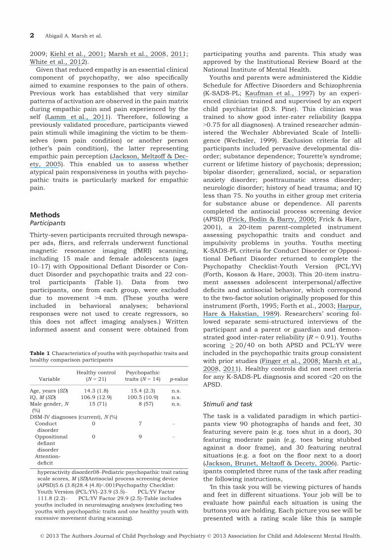
ing ‘Yourself’ and ‘Someone Else’ prompt slides. Aftereach prompt, participants viewed six images, duringwhich they rated how painful each image appearedusing a 4-point scale anchored by ‘No pain’ and‘Worst possible pain’ (Figure 1). Prompts comprisedthe blocked component of the design, and pain levelcomprised the event-related component. Responseswere collected using a 4-button box.
Participants viewed each stimulus twice: oncefollowing a Yourself prompt, and once following aSomeone Else prompt. Each prompt was 3 s andwas followed by a 3 s fixation, then six image trials.Each image trial was 3 s, consisting of an image(2.7 s) then a fixation (0.3 s). Response data werecollected during the entire trial. Each of three taskruns contained 10 prompts, 60 image trials, and 30‘jitter’ fixation trials (3.0 s) interspersed betweenimages. The inclusion of jitter trials increases powerin event-related fMRI paradigms (Dale, 1999). Eachrun of the task began and ended with a 15 s fixation,making each run of the task 5.5 min long. Fourpossible randomized sequences of stimulus presen-tations were created for the task. Each participant
Figure 1 Design of neuroimaging task and sample images
© 2013 The Authors Journal of Child Psychology and Psychiatry © 2013 Association for Child and Adolescent Mental Health.
Psychopathy and empathic pain responding 3
was randomly selected to view one of the foursequences.
Scanning acquisition
T2* weighted images were collected during fMRIscanning using a 1.5 Tesla GE Signa scanner (GEMedical Systems, Milwaukee, WI) (matrix 64 9 64;repetition time, 3000 ms; echo time, 30 ms; field ofview, 240 mm; voxels, 3.75 9 3.75 9 4). Functionalimages were acquired with a gradient echo-planarimaging (EPI) sequence (axial plane, 31 contiguousaxial slices). The task was yoked to the repetitiontime intervals. High-resolution T1-weighted anatom-ical images were also acquired (three-dimensionSpoiled GRASS with inversion recovery prep pulse;number of 1.5 mm axial slices, 128; field of view,240 mm; number of acquisitions, 1; repetition time,8.1 ms, echo time, 1.8 ms; matrix, 256 9 256).
Pre-processing of neuroimaging data
Imaging data were pre-processed and analyzedusing analysis of functional neuroimages (AFNI).The first four trials of each run were discarded, andthen functional images from the three time serieswere concatenated, despiked, motion corrected,spatially smoothed using a 6.0 mm full-width half-maximum Gaussian filter, and activation outsidethe brain was masked. The time series was thennormalized such that the resulting regression coef-ficients represented a percent signal change fromthe mean. Regressors were created for Own Pain andOther’s Pain trials and weighted according to theintensity of depicted pain (severe, moderate, ornone) according to a priori classifications of theimages into each category. A regressor of no interestwas created for trials in which participants did notprovide a response. Fixation trials were modeledimplicitly.
All regressors were created by convolving the trainof stimulus events with a gamma-variate hemody-namic response function. Linear regression model-ing used the full set of regressors to model baselinedrift and residual motion artifact. The baseline wasmodeled by a 1st-order function and motion artifactswere modeled using the six estimated rigid-bodymotion parameters. This produced a beta coefficientand associated t statistic for each voxel and regres-sor. Participants’ anatomical scans were individuallyregistered to the Talaraich and Tourneoux Atlas, asnormalization of adolescent brains does not appearto introduce major distortions during event-relatedfMRI (Burgund et al., 2002).
Data analysis
Behavioral data were analyzed using a 2 (group:psychopathic traits, healthy controls) 9 2 (condi-tion: Own Pain, Other’s Pain) 9 3 (pain intensity:
severe, moderate, none) repeated-measures analysisof variance (ANOVA) on both participants’ responsesto the images and on their response times. Maineffects and interactions are reported at p < 0.05,two-tailed.
Whole-brain group analyses on the event-relatedblood oxygen level dependent (BOLD) data wereconducted in AFNI. To identify the main effect ofpain, we conducted a whole-brain single samplecontrast comparing regressors weighted according tothe intensity of depicted pain to a baseline of zero.Thus, regions of activation would be identified to theextent that increasing activation corresponded toincreasing intensity of depicted pain. Again, consis-tent with prior work incorporating these stimuli,pain intensity in analyses of imaging data wasdetermined according to our a priori classifications,not according to participants’ own ratings (Jacksonet al., 2006), and regressors were weighted accord-ing to these classifications.
Next, to identify whether activation in the hypoth-esized brain regions is differentially associated withpain perception across groups, we used two analyticstrategies. First, to identify whether a truegroup 9 emotion interaction existed we conducteda whole-brain analysis of group differences acrossconditions using a 2 (group: psychopathic traits,healthy controls) 9 2 (condition: Own Pain, Other’sPain) ANOVA. Consistent with prior work featuringanalyses of variance (White et al., 2012), initialthresholding was set at a p-value of p < 0.005, withan extant threshold of 10 contiguous voxels, acombination that has been demonstrated to producea desirable balance between Type I and Type II errorrates, a critical consideration for omnibus interac-tions (Lieberman & Cunningham, 2009). Second, toinvestigate the nature of interaction effects, weperformed focused tests of our hypothesis, calcu-lated independently from the ANOVA, by conductingwhole-brain contrasts within AFNI. For these con-trast tests, we applied a threshold that yielded awhole-brain false discovery rate (FDR) <0.05 acrosscontrasts (p < 0.0001, uncorrected, with an extentthreshold of 10 voxels). Areas of differential activa-tion that survived both analytic strategies arereported. We also examined the main effect of painintensity in both groups combined to determineresponsiveness to pain stimuli across groups. Thiswas achieved using a single sample contrast test.
Additional planned analyses were performed onmean parameter estimates extracted from the func-tionally defined clusters identified by the ANOVA.Independent samples t tests were performed tospecify the nature of main effects and interactions.We also assessed the relationship between PCL:YVscores and patterns of atypical activation in youthswith psychopathic traits. These analyses were con-ducted within the clinical population only, so theyare independent from the original ANOVA conductedacross groups and do not introduce statistical bias.
© 2013 The Authors Journal of Child Psychology and Psychiatry © 2013 Association for Child and Adolescent Mental Health.
4 Abigail A. Marsh et al.
Two follow-up ANOVAs were also conducted thatexcluded, respectively, youths with diagnoses ofADHD or reported use of psychotropic medicationto rule these variables out as contributing to iden-tified group differences in neural activation.
ResultsBehavioral data
Results of the ANOVA revealed no main effects orinteractions involving the effects of condition orgroup (all p > .10). A significant main effect of painintensity was identified, F(2,70) = 202.94, p < .001,such that participants judged severe pain stimuli asthe most painful, M = 3.19, SD = 0.46, followed bymoderate pain, M = 2.51, SD = 0.43, followed byneutral, M = 1.57, SD = 0.44. Analysis of responsetimes revealed no significant main effects or inter-actions (all p > .10).
No significant group differences in average move-ment during scanning were observed in any plane(all p > .10).
Imaging data
As noted above, group differences in responses to theimages were analyzed using a 2 (group: psychopathictraits, healthy controls) 9 2 (condition: Own Pain,
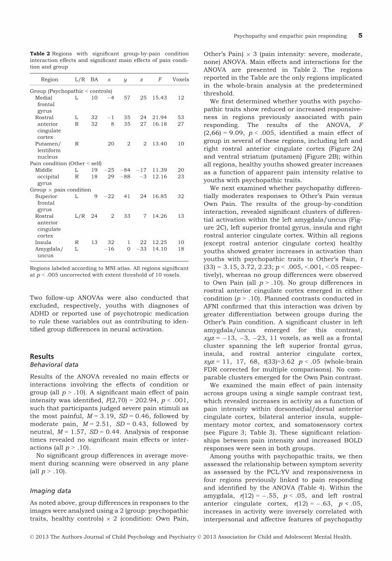
Other’s Pain) 9 3 (pain intensity: severe, moderate,none) ANOVA. Main effects and interactions for theANOVA are presented in Table 2. The regionsreported in the Table are the only regions implicatedin the whole-brain analysis at the predeterminedthreshold.
We first determined whether youths with psycho-pathic traits show reduced or increased responsive-ness in regions previously associated with painresponding. The results of the ANOVA, F
(2,66) = 9.09, p < .005, identified a main effect ofgroup in several of these regions, including left andright rostral anterior cingulate cortex (Figure 2A)and ventral striatum (putamen) (Figure 2B); withinall regions, healthy youths showed greater increasesas a function of apparent pain intensity relative toyouths with psychopathic traits.
We next examined whether psychopathy differen-tially moderates responses to Other’s Pain versusOwn Pain. The results of the group-by-conditioninteraction, revealed significant clusters of differen-tial activation within the left amygdala/uncus (Fig-ure 2C), left superior frontal gyrus, insula and rightrostral anterior cingulate cortex. Within all regions(except rostral anterior cingulate cortex) healthyyouths showed greater increases in activation thanyouths with psychopathic traits to Other’s Pain, t
(33) = 3.15, 3.72, 2.23; p < .005, <.001, <.05 respec-tively), whereas no group differences were observedto Own Pain (all p > .10). No group differences inrostral anterior cingulate cortex emerged in eithercondition (p > .10). Planned contrasts conducted inAFNI confirmed that this interaction was driven bygreater differentiation between groups during theOther’s Pain condition. A significant cluster in leftamygdala/uncus emerged for this contrast,xyz = �13, �3, �23, 11 voxels, as well as a frontalcluster spanning the left superior frontal gyrus,insula, and rostral anterior cingulate cortex,xyz = 11, 17, 68, t(33)=3.62 p < .05 (whole-brainFDR corrected for multiple comparisons). No com-parable clusters emerged for the Own Pain contrast.
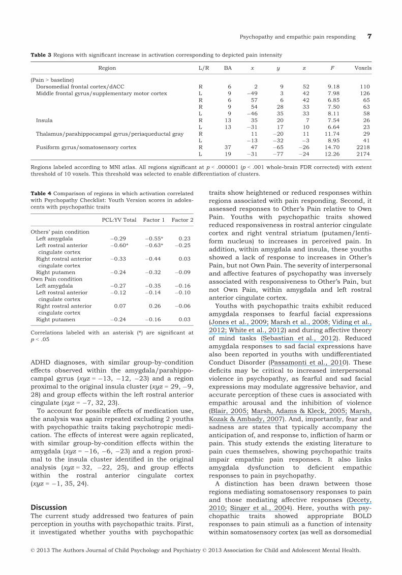
We examined the main effect of pain intensityacross groups using a single sample contrast test,which revealed increases in activity as a function ofpain intensity within dorsomedial/dorsal anteriorcingulate cortex, bilateral anterior insula, supple-mentary motor cortex, and somatosensory cortex(see Figure 3; Table 3). These significant relation-ships between pain intensity and increased BOLDresponses were seen in both groups.
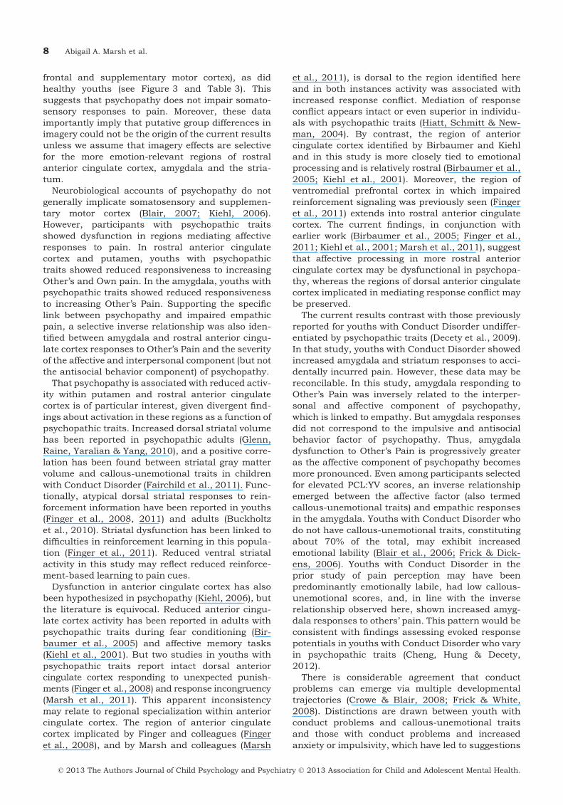
Among youths with psychopathic traits, we thenassessed the relationship between symptom severityas assessed by the PCL:YV and responsiveness infour regions previously linked to pain respondingand identified by the ANOVA (Table 4). Within theamygdala, r(12) = �.55, p < .05, and left rostralanterior cingulate cortex, r(12) = �.63, p < .05,increases in activity were inversely correlated withinterpersonal and affective features of psychopathy
Table 2 Regions with significant group-by-pain conditioninteraction effects and significant main effects of pain condi-tion and group
Region L/R BA x y z F Voxels
Group (Psychopathic < controls)Medialfrontalgyrus
L 10 �4 57 25 15.43 12
Rostralanteriorcingulatecortex
L 32 �1 35 24 21.94 53R 32 8 35 27 16.18 27
Putamen/lentiformnucleus
R 20 2 2 13.40 10
Pain condition (Other < self)Middleoccipitalgyrus
L 19 �25 �84 �17 11.39 20R 18 29 �88 �3 12.16 23
Group 9 pain conditionSuperiorfrontalgyrus
L 9 �22 41 24 16.85 32
Rostralanteriorcingulatecortex
L/R 24 2 33 7 14.26 13
Insula R 13 32 1 22 12.25 10Amygdala/uncus
L �16 0 �33 14.10 18
Regions labeled according to MNI atlas. All regions significantat p < .005 uncorrected with extent threshold of 10 voxels.
© 2013 The Authors Journal of Child Psychology and Psychiatry © 2013 Association for Child and Adolescent Mental Health.
Psychopathy and empathic pain responding 5
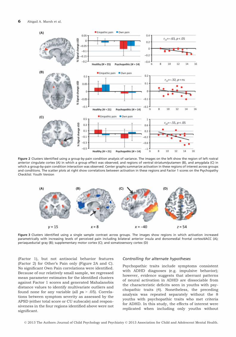
(Factor 1), but not antisocial behavior features(Factor 2) for Other’s Pain only (Figure 2A and C).No significant Own Pain correlations were identified.Because of our relatively small sample, we regressedmean parameter estimates for the identified clustersagainst Factor 1 scores and generated Mahalanobisdistance values to identify multivariate outliers andfound none for any variable (all ps > .05). Correla-tions between symptom severity as assessed by theAPSD (either total score or CU subscale) and respon-siveness in the four regions identified above were notsignificant.
Controlling for alternate hypotheses
Psychopathic traits include symptoms consistentwith ADHD diagnoses (e.g. impulsive behavior);however, evidence suggests that aberrant patternsof neural activation in ADHD are dissociable fromthe characteristic deficits seen in youths with psy-chopathic traits (4). Nonetheless, the precedinganalysis was repeated separately without the 8youths with psychopathic traits who met criteriafor ADHD. In this study, the effects of interest werereplicated when including only youths without
(A)
(B)
(C)
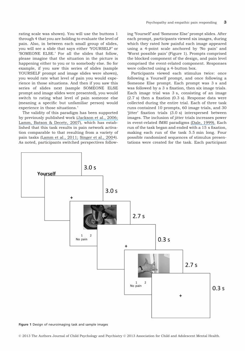
Figure 2 Clusters identified using a group-by-pain condition analysis of variance. The images on the left show the region of left rostralanterior cingulate cortex (A) in which a group effect was observed; and regions of ventral striatum/putamen (B), and amygdala (C) inwhich a group-by-pain condition interaction was observed. Center graphs summarize activation in these regions of interest across groupsand conditions. The scatter plots at right show correlations between activation in these regions and Factor 1 scores on the PsychopathyChecklist: Youth Version
(A) (B) (C) (D)
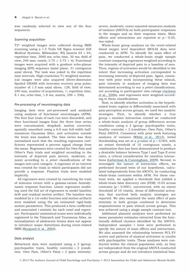
Figure 3 Clusters identified using a single sample contrast across groups. The images show regions in which activation increasedparametrically with increasing levels of perceived pain including bilateral anterior insula and dorsomedial frontal cortex/dACC (A);periaqueductal gray (B); supplementary motor cortex (C); and somatosensory cortex (D)
© 2013 The Authors Journal of Child Psychology and Psychiatry © 2013 Association for Child and Adolescent Mental Health.
6 Abigail A. Marsh et al.
ADHD diagnoses, with similar group-by-conditioneffects observed within the amygdala/parahippo-campal gyrus (xyz = �13, �12, �23) and a regionproximal to the original insula cluster (xyz = 29, �9,28) and group effects within the left rostral anteriorcingulate (xyz = �7, 32, 23).
To account for possible effects of medication use,the analysis was again repeated excluding 2 youthswith psychopathic traits taking psychotropic medi-cation. The effects of interest were again replicated,with similar group-by-condition effects within theamygdala (xyz = �16, �6, �23) and a region proxi-mal to the insula cluster identified in the originalanalysis (xyz = 32, �22, 25), and group effectswithin the rostral anterior cingulate cortex(xyz = �1, 35, 24).
DiscussionThe current study addressed two features of painperception in youths with psychopathic traits. First,it investigated whether youths with psychopathic
traits show heightened or reduced responses withinregions associated with pain responding. Second, itassessed responses to Other’s Pain relative to OwnPain. Youths with psychopathic traits showedreduced responsiveness in rostral anterior cingulatecortex and right ventral striatum (putamen/lenti-form nucleus) to increases in perceived pain. Inaddition, within amygdala and insula, these youthsshowed a lack of response to increases in Other’sPain, but not Own Pain. The severity of interpersonaland affective features of psychopathy was inverselyassociated with responsiveness to Other’s Pain, butnot Own Pain, within amygdala and left rostralanterior cingulate cortex.
Youths with psychopathic traits exhibit reducedamygdala responses to fearful facial expressions(Jones et al., 2009; Marsh et al., 2008; Viding et al.,2012; White et al., 2012) and during affective theoryof mind tasks (Sebastian et al., 2012). Reducedamygdala responses to sad facial expressions havealso been reported in youths with undifferentiatedConduct Disorder (Passamonti et al., 2010). Thesedeficits may be critical to increased interpersonalviolence in psychopathy, as fearful and sad facialexpressions may modulate aggressive behavior, andaccurate perception of these cues is associated withempathic arousal and the inhibition of violence(Blair, 2005; Marsh, Adams & Kleck, 2005; Marsh,Kozak & Ambady, 2007). And, importantly, fear andsadness are states that typically accompany theanticipation of, and response to, infliction of harm orpain. This study extends the existing literature topain cues themselves, showing psychopathic traitsimpair empathic pain responses. It also linksamygdala dysfunction to deficient empathicresponses to pain in psychopathy.
A distinction has been drawn between thoseregions mediating somatosensory responses to painand those mediating affective responses (Decety,2010; Singer et al., 2004). Here, youths with psy-chopathic traits showed appropriate BOLDresponses to pain stimuli as a function of intensitywithin somatosensory cortex (as well as dorsomedial
Table 3 Regions with significant increase in activation corresponding to depicted pain intensity
Region L/R BA x y z F Voxels
(Pain > baseline)Dorsomedial frontal cortex/dACC R 6 2 9 52 9.18 110Middle frontal gyrus/supplementary motor cortex L 9 �49 3 42 7.98 126
R 6 57 6 42 6.85 65R 9 54 28 33 7.50 63L 9 �46 35 33 8.11 58
Insula R 13 35 20 7 7.54 26L 13 �31 17 10 6.64 23
Thalamus/parahippocampal gyrus/periaqueductal gray R 11 �20 11 11.74 29L �13 �32 �3 8.95 41
Fusiform gyrus/somatosensory cortex R 37 47 �65 �26 14.70 2218L 19 �31 �77 �24 12.26 2174
Regions labeled according to MNI atlas. All regions significant at p < .000001 (p < .001 whole-brain FDR corrected) with extentthreshold of 10 voxels. This threshold was selected to enable differentiation of clusters.
Table 4 Comparison of regions in which activation correlatedwith Psychopathy Checklist: Youth Version scores in adoles-cents with psychopathic traits
PCL:YV Total Factor 1 Factor 2
Others’ pain conditionLeft amygdala �0.29 �0.55* 0.23Left rostral anteriorcingulate cortex
�0.60* �0.63* �0.25
Right rostral anteriorcingulate cortex
�0.33 �0.44 0.03
Right putamen �0.24 �0.32 �0.09Own Pain condition
Left amygdala �0.27 �0.35 �0.16Left rostral anteriorcingulate cortex
�0.12 �0.14 �0.10
Right rostral anteriorcingulate cortex
0.07 0.26 �0.06
Right putamen �0.24 �0.16 0.03
Correlations labeled with an asterisk (*) are significant atp < .05
© 2013 The Authors Journal of Child Psychology and Psychiatry © 2013 Association for Child and Adolescent Mental Health.
Psychopathy and empathic pain responding 7
frontal and supplementary motor cortex), as didhealthy youths (see Figure 3 and Table 3). Thissuggests that psychopathy does not impair somato-sensory responses to pain. Moreover, these dataimportantly imply that putative group differences inimagery could not be the origin of the current resultsunless we assume that imagery effects are selectivefor the more emotion-relevant regions of rostralanterior cingulate cortex, amygdala and the stria-tum.
Neurobiological accounts of psychopathy do notgenerally implicate somatosensory and supplemen-tary motor cortex (Blair, 2007; Kiehl, 2006).However, participants with psychopathic traitsshowed dysfunction in regions mediating affectiveresponses to pain. In rostral anterior cingulatecortex and putamen, youths with psychopathictraits showed reduced responsiveness to increasingOther’s and Own pain. In the amygdala, youths withpsychopathic traits showed reduced responsivenessto increasing Other’s Pain. Supporting the specificlink between psychopathy and impaired empathicpain, a selective inverse relationship was also iden-tified between amygdala and rostral anterior cingu-late cortex responses to Other’s Pain and the severityof the affective and interpersonal component (but notthe antisocial behavior component) of psychopathy.
That psychopathy is associated with reduced activ-ity within putamen and rostral anterior cingulatecortex is of particular interest, given divergent find-ings about activation in these regions as a function ofpsychopathic traits. Increased dorsal striatal volumehas been reported in psychopathic adults (Glenn,Raine, Yaralian & Yang, 2010), and a positive corre-lation has been found between striatal gray mattervolume and callous-unemotional traits in childrenwith Conduct Disorder (Fairchild et al., 2011). Func-tionally, atypical dorsal striatal responses to rein-forcement information have been reported in youths(Finger et al., 2008, 2011) and adults (Buckholtzet al., 2010). Striatal dysfunction has been linked todifficulties in reinforcement learning in this popula-tion (Finger et al., 2011). Reduced ventral striatalactivity in this study may reflect reduced reinforce-ment-based learning to pain cues.
Dysfunction in anterior cingulate cortex has alsobeen hypothesized in psychopathy (Kiehl, 2006), butthe literature is equivocal. Reduced anterior cingu-late cortex activity has been reported in adults withpsychopathic traits during fear conditioning (Bir-baumer et al., 2005) and affective memory tasks(Kiehl et al., 2001). But two studies in youths withpsychopathic traits report intact dorsal anteriorcingulate cortex responding to unexpected punish-ments (Finger et al., 2008) and response incongruency(Marsh et al., 2011). This apparent inconsistencymay relate to regional specialization within anteriorcingulate cortex. The region of anterior cingulatecortex implicated by Finger and colleagues (Fingeret al., 2008), and by Marsh and colleagues (Marsh
et al., 2011), is dorsal to the region identified hereand in both instances activity was associated withincreased response conflict. Mediation of responseconflict appears intact or even superior in individu-als with psychopathic traits (Hiatt, Schmitt & New-man, 2004). By contrast, the region of anteriorcingulate cortex identified by Birbaumer and Kiehland in this study is more closely tied to emotionalprocessing and is relatively rostral (Birbaumer et al.,2005; Kiehl et al., 2001). Moreover, the region ofventromedial prefrontal cortex in which impairedreinforcement signaling was previously seen (Fingeret al., 2011) extends into rostral anterior cingulatecortex. The current findings, in conjunction withearlier work (Birbaumer et al., 2005; Finger et al.,2011; Kiehl et al., 2001; Marsh et al., 2011), suggestthat affective processing in more rostral anteriorcingulate cortex may be dysfunctional in psychopa-thy, whereas the regions of dorsal anterior cingulatecortex implicated in mediating response conflict maybe preserved.
The current results contrast with those previouslyreported for youths with Conduct Disorder undiffer-entiated by psychopathic traits (Decety et al., 2009).In that study, youths with Conduct Disorder showedincreased amygdala and striatum responses to acci-dentally incurred pain. However, these data may bereconcilable. In this study, amygdala responding toOther’s Pain was inversely related to the interper-sonal and affective component of psychopathy,which is linked to empathy. But amygdala responsesdid not correspond to the impulsive and antisocialbehavior factor of psychopathy. Thus, amygdaladysfunction to Other’s Pain is progressively greateras the affective component of psychopathy becomesmore pronounced. Even among participants selectedfor elevated PCL:YV scores, an inverse relationshipemerged between the affective factor (also termedcallous-unemotional traits) and empathic responsesin the amygdala. Youths with Conduct Disorder whodo not have callous-unemotional traits, constitutingabout 70% of the total, may exhibit increasedemotional lability (Blair et al., 2006; Frick & Dick-ens, 2006). Youths with Conduct Disorder in theprior study of pain perception may have beenpredominantly emotionally labile, had low callous-unemotional scores, and, in line with the inverserelationship observed here, shown increased amyg-dala responses to others’ pain. This pattern would beconsistent with findings assessing evoked responsepotentials in youths with Conduct Disorder who varyin psychopathic traits (Cheng, Hung & Decety,2012).
There is considerable agreement that conductproblems can emerge via multiple developmentaltrajectories (Crowe & Blair, 2008; Frick & White,2008). Distinctions are drawn between youth withconduct problems and callous-unemotional traitsand those with conduct problems and increasedanxiety or impulsivity, which have led to suggestions
© 2013 The Authors Journal of Child Psychology and Psychiatry © 2013 Association for Child and Adolescent Mental Health.
8 Abigail A. Marsh et al.
of primary and secondary psychopathy (Kimonis,Frick, Cauffman, Goldweber & Skeem, 2012). Pri-mary psychopathy (cf. Kimonis) is associated withreduced amygdala responses to fearful expressionsand dysfunctional striatal and ventromedial frontalcortex activity during reinforcement-based decisionmaking (Finger et al., 2011; Jones et al., 2009;Marsh et al., 2008; Sebastian et al., 2012; Vidinget al., 2012; White et al., 2012). Conduct problemsassociated with secondary psychopathy are associ-ated with increased amygdala responses to threatand heightened sensitivity to threat (Herpertz et al.,2008; Kimonis et al., 2012; Passamonti et al., 2010;Viding et al., 2012); for a review of the putativeneurobiological differences between these forms ofconduct problems, see Crowe and Blair (2008). Thecurrent study focused primarily on youth withprimary psychopathy and observed, consistent withprevious research (Jones et al., 2009; Marsh et al.,2008; White et al., 2012), reduced amygdalaresponses to pain stimuli. This reduction in amyg-dala activity became more marked as psychopathictraits increased.
Limitations
Some limitations of this study should be considered.First, our behavioral measures revealed no maineffect of group or of target. This is consistent with theresults of prior studies that have employed simpleevaluation tasks during neuroimaging paradigms(Decety et al., 2009), and may reflect the use ofdifferent strategies by psychopathic and nonpsycho-pathic individuals to perform the task. Psychopathyis associated with increased reliance on areasinvolved in executive functioning and semanticknowledge during socio-affective tasks (Glenn,Raine, Schug, Young & Hauser, 2009; Marsh &Cardinale, 2012). Similar group differences mayhave facilitated task performance in psychopathicyouths in this task, albeit at subthreshold activationlevels. It is not known whether psychopathy resultsin any generalized effects on imagery, which we didnot assess in this paradigm using a questionnaire orother test. Imagery ability is clearly important in thepresent task, which required study participants toimagine that the limbs depicted in photographsbelonged to themselves or another person. However,the fact that we did find extensive activation in pain-relevant somatosensory regions across groups andacross conditions of the task is consistent withimagery being spared in psychopathy.
In addition, future research should precisely pin-point whether the identified group differences reflectdisruptive behavior diagnoses or psychopathic
traits. Mitigating this concern, however, are thepreviously discussed correlations between interper-sonal and affective psychopathic traits and activa-tion in amygdala and anterior cingulate cortex.These correlations strengthen our conclusion thatthe activation patterns we observed specificallyreflected psychopathic traits.
Potential alternate hypotheses should also beconsidered. A follow-up ANOVA excluded youthswith ADHD diagnoses, which are frequently comor-bid with psychopathic traits, and identified similarpatterns of activation. Two youths in the presentstudy were also taking psychotropic medications,but a second follow-up ANOVA excluding thesechildren again resulted in similar patterns ofresponding. The consistent results of these ANOVAs,which both resulted in a loss of statistical power,support the stability of the patterns we identified.Finally, our sample size was similar to sample sizesused in many previous studies of this population(Decety et al., 2009; Marsh et al., 2008, 2011; Whiteet al., 2012), but the present findings should bereplicated in a larger sample.
ConclusionAdolescents with disruptive behavior disorders andpsychopathic traits exhibited less responsiveness toincreasing perceived pain intensity within structurestypically implicated in affective responses to others’pain. Given suggestions that the pain of others maytrigger empathic distress in the observer, providing abasis for moral development (Hoffman, 1982), weconclude that dysfunction in response to others’pain may contribute to the behavioral deficitsobserved in this population.
AcknowledgmentsThis research was supported by the IntramuralResearch Program of the NIH:NIMH and the NICHD(R03HD 06 4906). The authors thank Ken Towbin,Andy Speer, and Steven Sinclair for their assistancewith this research. Data included in this manuscriptwere presented at the 2012 meeting of the Social &Affective Neuroscience Society.
CorrespondenceAbigailMarsh,DepartmentofPsychology,GeorgetownUniversity, 37th & O Streets NW, WGR 302-A,Washington, DC 20057, USA; Email: [email protected]
© 2013 The Authors Journal of Child Psychology and Psychiatry © 2013 Association for Child and Adolescent Mental Health.
Psychopathy and empathic pain responding 9
Key points
• Psychopathy is a developmental disorder associated with increases in disruptive behavior and with reducedempathy. However, no previous neuroimaging study has assessed empathic pain responding in psychopathy.
• The results of this neuroimaging study found that adolescents with psychopathic traits and disruptivebehavior disorders show reduced activation in areas associated with affective components of empathic pain,including striatum, anterior cingulate cortex, and amygdala.
• Reduced responsivity in amygdala and rostral anterior cingulate cortex in response to others’ pain (but notown pain) correlated with the severity of psychopathic traits as measured by PCL:YV Factor 1 scores.
• These findings add to accumulating evidence regarding the importance of assessing psychopathic traits todistinguish among adolescents with disruptive behavior disorders. Disruptive adolescents with and withoutpsychopathic traits exhibit contrasting patterns of neural activation to stimuli like distress cues and pain cues,suggesting that distinct neurobiological mechanisms underlie behavioral disturbances in these groups.
ReferencesBirbaumer, N., Veit, R., Lotze, M., Erb, M., Hermann, C.,
Grodd, W. & Flor, H. (2005). Deficient fear conditioning inpsychopathy: A functional magnetic resonance imagingstudy. Archives of General Psychiatry, 62, 799–805.
Blair, R.J., Peschardt, K.S., Bhudhani, S., Mitchell, D.G., &Pine, D.S. (2006). The development of psychopathy. Journalof Child Psychology and Psychiatry, 47, 262–276.
Blair, R.J. (2005). Applying a cognitive neuroscienceperspective to the disorder of psychopathy. Developmentand Psychopathology, 17, 865–891.
Blair, R.J. (2007). The amygdala and ventromedial prefrontalcortex in morality and psychopathy. Trends in CognitiveSciences, 11, 387–392.
Buckholtz, J.W., Treadway, M.T., Cowan, R.L., Woodward,N.D., Benning, S.D., Li, R.Ansari, M.S., . . . & Zald, D.H.(2010). Mesolimbic dopamine reward systemhypersensitivity in individuals with psychopathic traits.Nature Neuroscience, 13, 419–421.
Burgund, E.D., Kang, H.C., Kelly, J.E., Buckner, R.L., Snyder,A.Z., Petersen, S.E., . . . & Schlaggar, B.L. (2002). Thefeasibility of a common stereotactic space for children andadults in fMRI studies of development. Neuroimage, 17, 184–200.
Cheng, Y., Hung, A. Y., & Decety, J. (2012). Dissociationbetween affective sharing and emotion understanding injuvenile psychopaths.: Development and Psychopathology,Under review, 24, 623–636.
Christian, R.E., Frick, P.J., Hill, N.L., Tyler, L., & Frazer, D.R.(1997). Psychopathy and conduct problems in children: II.Implications for subtyping children with conduct problems.Journal of the American Academy of Child & AdolescentPsychiatry, 36, 233–241.
Crowe, S., & Blair, R.J.R. (2008). The development ofantisocial behavior: What can we learn from functionalneuroimaging studies? Development & Psychopathology,20, 1145–1159.
Dale, A.M. (1999). Optimal experimental design for event-related fMRI. Human Brain Mapping, 8, 109–114.
Decety, J. (2010). The neurodevelopment of empathy inhumans. Developmental Neuroscience, 32, 257–267.
Decety, J., & Michalska, K.J. (2010). Neurodevelopmentalchanges in the circuits underlying empathy and sympathyfrom childhood to adulthood. Developmental Science, 13,886–899.
Decety, J., Michalska, K.J., Akitsuki, Y., & Lahey, B.B. (2009).Atypical empathic responses in adolescents with aggressiveconduct disorder: A functional MRI investigation. BiologicalPsychology, 80, 203–211.
Fairchild, G., Passamonti, L., Hurford, G., Hagan, C.C., vondemHagen, E.A., van Goozen, S.H., . . . &Clader, A.J. (2011).
Brain structure abnormalities in early-onset andadolescent-onset conduct disorder. American Journal ofPsychiatry, 168, 624–633.
Finger, E.C., Marsh, A.A., Blair, K.S., Reid, M.E., Sims, C., Ng,P., . . . & Blair, R.J. (2011). Disrupted reinforcementsignaling in the orbitofrontal cortex and caudate in youthswith conduct disorder or oppositional defiant disorder and ahigh level of psychopathic traits. American Journal ofPsychiatry, 168, 152–162.
Finger, E.C., Marsh, A.A., Mitchell, D.G., Reid, M.E., Sims, C.,Budhani, S., . . . & Blair, J.R. (2008). Abnormal ventromedialprefrontal cortex function in children with psychopathictraits during reversal learning. Archives of GeneralPsychiatry, 65, 586–594.
Forth, A.E. (1995). Psychopathy and young offenders:Prevalence, family background, and violence. Ottawa, ON:Ministry of the Solicitor General of Canada.
Forth, A.E., Kosson, D.S., & Hare, R.D. (2003). Thepsychopathy checklist: Youth version. Toronto: Multi-Health Systems.
Frick, P., Bodin, S., & Barry, C. (2000). Psychopathic traits andconduct problems in community and clinic-referred samplesof children: Further development of the psychopathyscreening device. Psychological Assessment, 12, 382–393.
Frick, P.J., & Dickens, C. (2006). Current perspectives onconduct disorder. Current Psychiatry Reports, 8, 59–72.
Frick, P.J., & Hare, R.D. (2001). The antisocial processscreening device. Toronto: Multi-Health Systems.
Frick, P., & White, S. (2008). Research review: The importanceof callous-unemotional traits for developmental models ofaggressive and antisocial behavior. Journal of ChildPsychology & Psychiatry, 49, 359–375.
Glenn, A.L., Raine, A., Schug, R.A., Young, L., & Hauser, M.(2009). Increased DLPFC activity during moral decision-making in psychopathy. Molecular Psychiatry, 14, 909–911.
Glenn, A.L., Raine, A., Yaralian, P.S., & Yang, Y. (2010).Increased volume of the striatum in psychopathicindividuals. Biological Psychiatry, 67, 52–58.
Harpur, T.J., Hare, R.D., & Hakstian, A.R. (1989). Two-factorconceptualization of psychopathy: Construct validity andassessment implications. Psychological Assessment, 1, 6–17.
Herpertz, S.C., Huebner, T., Marx, I., Vloet, T.D., Fink, G.R.,Stoecker, T., … & Herpertz-Dahlmann, B. (2008). Emotionalprocessing in male adolescents with childhood-onsetconduct disorder. Journal of Child Psychology &Psychiatry, 49, 781–791.
Herpertz, S.C., Huebner, T., Marx, I., Vloet, T.D., Fink, G.R.,Stoecker, T., . . . & Herpertz-Dahlmann, B. (2009). Emotionalprocessing in male adolescents with childhood-onsetconduct disorder. Journal of Child Psychology &Psychiatry, 166, 95–102.
© 2013 The Authors Journal of Child Psychology and Psychiatry © 2013 Association for Child and Adolescent Mental Health.
10 Abigail A. Marsh et al.
Hiatt, K.D., Schmitt, W.A., & Newman, J.P. (2004). Strooptasks reveal abnormal selective attention amongpsychopathic offenders. Neuropsychology, 18, 50–59.
Hoffman, M.L. (1982). Affect and moral development. NewDirections for Child and Adolescent Development, 1982, 83–103.
Jackson, P.L., Brunet, E., Meltzoff, A.N., & Decety, J. (2006).Empathy examined through the neural mechanismsinvolved in imagining how I feel versus how you feel pain.Neuropsychologia, 44, 752–761.
Jackson, P.L., Meltzoff, A.N., & Decety, J. (2005). How do weperceive the pain of others? A window into the neuralprocesses involved in empathy. Neuroimage, 24, 771–779.
Jones, A.P., Laurens, K.R., Herba, C.M., Barker, G.J., &Viding, E. (2009). Amygdala hypoactivity to fearful faces inboys with conduct problems and callous-unemotional traits.American Journal of Psychiatry, 166, 95–102.
Kaufman, J., Birmaher, B., Brent, D., Rao, U., Flynn, C.,Moreci, P., . . . & Ryan, N. (1997). Schedule for AffectiveDisorders and Schizophrenia for School-Age Children –Present and lifetime version (K-SADS-PL): Initial reliabilityand validity data. Journal of the American Academy of Child& Adolescent Psychiatry, 36, 980–988.
Kiehl, K.A. (2006). A cognitive neuroscience perspective onpsychopathy: Evidence for paralimbic system dysfunction.Psychiatry Research, 142, 107–128.
Kiehl, K.A., Smith, A.M., Hare, R.D., Mendrek, A., Forster,B.B., Brink, J. & Liddle, P.F. (2001). Limbic abnormalities inaffective processing by criminal psychopaths as revealed byfunctional magnetic resonance imaging. BiologicalPsychiatry, 50, 677–684.
Kimonis, E., Frick, P., Cauffman, E., Goldweber, A., & Skeem,J. (2012). Primary and secondary variants of juvenilepsychopathy differ in emotional processing. Development &Psychopathology, 24, 1091–1103.
Lamm, C., Batson, C., & Decety, J. (2007). The neuralsubstrate of human empathy: Effects of perspective-takingand cognitive appraisal. Journal of Cognitive Neuroscience,19, 42–58.
Lamm, C., Decety, J., & Singer, T. (2011). Meta-analyticevidence for common and distinct neural networksassociated with directly experienced pain and empathy forpain. Neuroimage, 54, 2492–2502.
Lieberman, M.D., & Cunningham, W.A. (2009). Type I andType II error concerns in fMRI research: Re-balancingthe scale. Social Cognitive & Affective Neuroscience, 4,423–428.
Lynam, D.R., Loeber, R., & Stouthamer-Loeber, M. (2008). Thestability of psychopathy from adolescence into adulthood.Criminal Justice & Behavior, 35, 228.
Marsh, A.A., Adams, R.B. Jr, & Kleck, R.E. (2005). Why do fearand anger look the way they do? Form and social function infacial expressions. Personality & Social Psychology Bulletin,31, 73–86.
Marsh, A.A., & Cardinale, E.M. (2012). When psychopathyimpairs moral judgments: Neural responses duringjudgments about causing fear. Social Cognitive & AffectiveNeuroscience. in press.
Marsh, A.A., Finger, E.C., Fowler, K.A., Jurkowitz, I.T.,Schechter, J.C., Yu, H.H., . . . & Blair, R.J. (2011). Reducedamygdala-orbitofrontal connectivity during moraljudgments in youths with disruptive behavior disordersand psychopathic traits. Psychiatry Research, 194, 279–286.
Marsh, A.A., Finger, E., Mitchell, D.G.V., Reid, M.E., Sims, C.,Kosson, D.S., . . . & Blair, R.J.R. (2008). Reduced amygdalaresponse to fearful expressions in children and adolescentswith callous-unemotional traits and disruptive behaviordisorders. American Journal of Psychiatry, 165, 712–720.
Marsh, A.A., Kozak, M.N., & Ambady, N. (2007). Accurateidentification of fear facial expressions predicts prosocialbehavior. Emotion, 7, 239–251.
Passamonti, L.,Fairchild,G.,Goodyer, I.M.,Hurford,G.,Hagan,C.C., Rowe, J.B., & Calder A.J. (2010). Neural abnormalitiesin early-onset and adolescence-onset conduct disorder.Archives of General Psychiatry, 67, 729–738.
Sebastian, C., McCrory, E., Cecil, C., Lockwood, P.L., De Brito,S.A., Fontaine, N.M., & Viding, E. (2012). Neural responsesto affective and cognitive theory of mind in children withconduct problems and varying levels of callous-unemotionaltraits. Archives of General Psychiatry, 69, 814–822.
Singer, T., Seymour, B., O’Doherty, J., Kaube, H., Dolan, R.J.,& Frith, C.D. (2004). Empathy for pain involves the affectivebut not sensory components of pain. Science, 303, 1157–1162.
Viding, E., Sebastian, C.L., Dadds, M.R., Lockwood, P.L., Cecil,C.A., De Brito, S.A., & McCrory, E.J. (2012). Amygdalaresponse to preattentive masked fear in children withconduct problems: The role of callous-unemotional traits.American Journal of Psychiatry, 169, 1109–1116.
Wechsler, D. (1999). Wechsler Abbreviated Scaleof Intelligence.San Antonio, TX: The Psychological Corporation.
White, S.F., Marsh, A.A., Fowler, K.A., Schechter, J.C., Adalio,C., Pope, K., . . . & Blair, R.J. (2012). Reduced amygdalaresponse in youths with disruptive behavior disorders andpsychopathic traits: Decreased emotional response versusincreased top-down attention to nonemotional features.American Journal of Psychiatry, 169, 750–758.
Accepted for publication: 3 January 2013
© 2013 The Authors Journal of Child Psychology and Psychiatry © 2013 Association for Child and Adolescent Mental Health.
Psychopathy and empathic pain responding 11