Embed Size (px)
Citation preview

P1
Epidemiology of sepsis in pediatrics: fi rst Colombian multicenter pilot
survey
J Camilo Jaramillo-Bustamante1, A Marín-Agudelo1, M Fernández-Laverde1,
J Bareño-Silva2
1CES University, Pediatric Intensive Care Program, Medellin, Colombia; 2CES University, Epidemiology and Bio-statistic Research Group, Medellin,
Colombia
Critical Care 2010, 14(Suppl 2):P1 (doi: 10.1186/cc9104)
Introduction In 2002, the Surviving Sepsis Campaign defi ned a strategy that aimed to reduce the high mortality due to sepsis. One point of this strategy was a recommendation to recognize that sepsis is a frequent cause of death and high economic costs in the pediatric intensive care unit. Knowledge of the disease is the fi rst step to impact it. There are few studies on pediatric sepsis epidemiology in the world and none in Colombia.Hypothesis The epidemiological features of Colombian children are diff erent from other countries.Methods We constructed a website where 14 intensive care units across the country reported in a prospective way the epidemiological features of children with sepsis using an electronic process [1]. We asked for sociodemographics, microbiological data, sepsis classifi cation, complications, and outcome.Results We collected 253 patients from March to May 2009. Fifty-fi ve percent of the cases were male and 45% were female; 53% were less than 1 year old. A total of 67.2% came from urban areas and 33% came from rural villages. Eighty-fi ve percent were very poor (score 1 and 2 over 6 used in Colombia as socioeconomic classifi cation). Forty-fi ve percent have government-supported insurance. In total, 23.72% of the population presented with sepsis; 30.04% with severe sepsis; and 46.5% with septic shock. The infection origin was respiratory in 54.55%, followed by abdominal in 17.39%. In 50.2% no cause was identifi ed. A total of 75.1% required mechanical ventilation. The mortality rate was 20.4%.Conclusions Sepsis, severe sepsis, or septic shock is a common diagnosis in Colombian intensive care units. The majority of pediatric patients are 2 years or younger and from the poorest communities. It aff ected males more. In the majority, the process starts in the respiratory system. We had diffi culty identifying the cause. The disease causes high mortality and cost for a developing society. We need a complete survey to fi nd a correct approach to the problem.Reference
1. Sepsis en Columbia [www.sepsisencolombia.com]
P2
Randomized controlled trials are not designed to prove the safety of
third-generation hydroxyethyl starch for resuscitation: results from a
systematic review
CS Hartog, M Kohl, K Reinhart
Friedrich-Schiller-Universität Jena, Klinik für Anästhesiologie und Intensivtherapie,
Jena, Germany
Critical Care 2010, 14(Suppl 2):P2 (doi: 10.1186/cc9105)
Introduction Hydroxyethyl starch (HES) is widely used for volume therapy in intensive care. In critically ill and sepsis patients, HES use was
dose-dependently associated with increased renal failure, tissue storage with organ failure, and increased long-term mortality. There are other safety concerns with regard to coagulopathy, pruritus, and mortality. However, third-generation HES 130/0.4 is considered to have an improved risk profi le. Therefore, we wanted to assess whether published studies on HES 130/0.4 resuscitation are suffi ciently well designed to draw conclusions about the safety of this compound.Methods We derived clinically relevant outcome parameters to analyze safety outcomes from the literature and provided exemplary power calculations. Randomized controlled trials (RCT) on fl uid resuscitation with HES 130/0.4 were systematically searched and analyzed for clinical condition, sample size, study duration, cumulative dose, control fl uids, endpoints, and colloid–crystalloid volume ratios in studies with a goal-directed fl uid regimen. Due to the heterogeneity of included studies, all analyses were descriptive (SPSS 17.0).Results A total of 56 RCTs were included. Only two studies included severe sepsis patients, 80% were from the elective surgical setting and one study from the emergency surgical setting. In general, studies were underpowered (median sample size 25 patients in HES 130/0.4 groups, range 10 to 90 patients); of short duration (median study period 12 hours, range 0.5 to 144 hours) and with low cumulative HES doses (median 2,465 ml, range 328 to 6,229 ml). Sepsis studies (n = 2) included 18 patients (median, range 10 to 26 patients), study period was 96 hours (median, range 72 to 120 hours) and total fl uid volumes was 3,000 ml in one study. Sixty percent of control fl uids were synthetic colloids (other starches, gelatins, or dextran) that carry a similar risk profi le. Primary endpoints with power calculation (in 87% of studies) were mostly unspecifi c or clinically irrelevant. Only one sepsis study provided a primary endpoint, which was extravascular lung water. This did not diff er in comparison with the albumin 20% control group.Conclusions There is no reliable evidence from published clinical data that third-generation HES 130/0.4 is safe in septic patients or in the emergency or elective surgical setting.
P3
Eff ect of canine hyperimmune plasma on TNFα and infl ammatory
cell levels in a lipopolysaccharide-mediated rat air pouch model of
infl ammation
BE Essien, M Kotiw, H Butler, D Strunin
University of Southern Queensland, Centre for Systems Biology, Toowoomba,
Australia
Critical Care 2010, 14(Suppl 2):P3 (doi: 10.1186/cc9106)
Introduction Unregulated elevated levels of serum TNFα have been asso-ciated with proinfl ammatory cytokine cascades, which are characteristic in diseases such as septic shock. Endotoxic shock, which has a poorer prognosis than found with other forms of septic shock, is mediated by lipopolysaccharide (LPS), a molecule that is released from the outer membrane of Gram-negative bacteria. LPS is a potent stimulator of TNFα secretion by serum monocytes and tissue macrophages. While the use of monotherapeutic TNFα antagonists has been trailed, none has been registered for use in patients with sepsis.Objective The purpose of this study was to test the eff ect of canine hyperimmune frozen plasma (HFP), which is known to contain elevated levels of soluble TNFα receptor 1 (sTNFR1), on TNFα and infl ammatory cell levels in a LPS-mediated rat air pouch model of infl ammation.Methods A dorsal air pouch in 175 to 200 g Sprague–Dawley rats was formed by 20 ml subcutaneous infusions of sterile air. Prophylactic subcutaneous injections of canine HFP, canine fresh-frozen plasma (FFP), or carprofen © 2010 BioMed Central Ltd
Sepsis 2010Paris, France. 1–3 September 2010
Published: 1 September 2010
M E E T I N G A B S T R AC T S
Critical Care 2010, Volume 14 Suppl 2 http://ccforum.com/supplements/14/S2
© 2010 BioMed Central Ltd
were administered daily for 3 days into the lateral fl ank of the right foreleg at doses recommended by the manufacturers (n = 10 for each treatment group). Pouch fl uid was harvested by syringe at 1, 6, 12, 24, and 48 hours post LPS administration and subjected to histological and cytokine/cytokine receptor analysis. TNFα and sTNFR1 levels were determined by ELISA and an immunofl uorescent dot blot assay.Results Pouch fl uid analysis: maximal eff ects were detected at 6 hours post LPS administration. TNFα levels were signifi cantly depressed in animals dosed with HFP, but not in animals treated with FFP or carprofen (P <0.05). sTNFR1 levels were signifi cantly elevated in HFP, but not in FFP or carprofen-dosed animals (P <0.05). Neutrophil numbers were signifi cantly depressed in HFP-dosed, but not in FFP-treated or carprofen-treated animals (P <0.05).Conclusions There appears to be a correlation between elevated levels of sTNFR1 and depression of TNFα and neutrophil levels in the pouch fl uid of HFP-dosed rats (r = –0.73, P <0.0001). The data suggest that canine HFP, which has been demonstrated to contain elevated levels of sTNFR1 compared with FFP, has a direct eff ect on depressing TNFα levels and neutrophil sequestration in the rat air pouch model of infl ammation. These data suggest that HFP may be worthy of further investigation to determine whether such preparations have a therapeutic potential for treatment of acute infl ammatory diseases in which TNFα is implicated.
P4
Development of Klebsiella pneumoniae B5055-induced mouse model of
sepsis-associated brain infl ammation in BALB/c mice
V Kumar1, A Sharma2
1Dr Y. S. Parmar University, Microbiology, Himachal Pardesh, India; 2Saint-Justine
Hospital, University de Montreal, Medicine, Montreal, Canada
Critical Care 2010, 14(Suppl 2):P4 (doi: 10.1186/cc9107)
Introduction Incidence of sepsis is continuously increasing in the developing as well as the developed world. Severe sepsis is associated with the development of multiorgan dysfunction syndrome. In addition to other vital organs (that is, lungs, kidneys, heart, or liver), the brain is one of the severely aff ected organs in sepsis. Autopsy studies from septic patients reveal various cerebral lesions including ischemia, hemorrhage, microthrombi, microabscesses, multifocal necrotizing leukoencephalopathy, and bacterial invasion of the nervous system. Until now no animal model of sepsis has been developed that in a true sense represents the brain infl ammation associated with evolving sepsis originating from Gram-negative bacteria. This study comprises development of a mouse model of sepsis-induced brain infl ammation.Methods A mouse model of sepsis-associated brain infl ammation was developed by directly placing a selected dose (102 cfu) of Klebsiella pneumoniae B5055 entrapped in a fi brin–thrombin clot into the peritoneal cavity of mice, while in control group animals only a sterile fi brin clot was kept. Various infl ammatory cytokines (that is, IL-1α and TNFα) and other infl ammatory markers (that is, malondialdehyde, myeloperoxidase and nitric oxide) in serum and brain were estimated by ELISA, biochemical methods, and histopathology in both the experimental groups.Results Bacterial colonies were found to be established in brain on the very fi rst day of sepsis induction in experimental group but no bacteria were observed in sham-operated animals. Along with this, a signifi cant (P <0.05) increase in neutrophil infi ltration into the brain along with signifi cantly (P <0.05) increased levels of proinfl ammatory cytokines (TNFα and IL-1α) and other infl ammatory mediators like nitric oxide, malondialdehyde, and myeloperoxidase were observed in animals with sepsis. Also the septic animals survived until the 5th day of post-sepsis development, while 100% survival was observed in the sham-operated group on all experimental days without any infl ammatory change in the brain as observed by histopathologic examination and estimating the above-mentioned infl ammatory parameters.Conclusions This mouse model of sepsis-induced brain infl ammation may prove helpful to study immunopathogenesis of brain infl ammation observed during Gram-negative bacterial sepsis and may also prove helpful to study neuroimmunology of sepsis along with behavioral changes associated with sepsis.
P5
Abstract withdrawn
P6
Lipopolysaccharide is required for leukocyte adhesion to Toraymyxin®
fi lters used in the treatment of sepsis
EL Martin, B Assenzio, VM Ranieri
University of Turin, Anesthesia and Intensive Care, Turin, Italy
Critical Care 2010, 14(Suppl 2):P6 (doi: 10.1186/cc9109)
Introduction Extracorporeal hemoperfusion with polymyxin B is a novel septic treatment, shown to improve hemodynamics, organ dysfunction, and mortality through the removal of circulating lipopolysaccharide (LPS). This therapy can also remove activated leukocytes, which likely contributes to reduced infl ammation and improved patient outcome; however, the mechanistic role of LPS in the removal of leukocytes remains unclear.Objective To determine whether the presence of LPS and/or activation of leukocytes by LPS alters their ability to bind to polymyxin-bound fi lters used for extracorporeal hemoperfusion of septic patients.Methods Toraymyxin® fi lters were opened under sterile conditions and 2 cm2
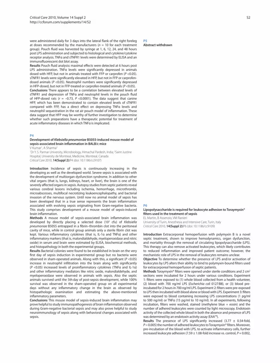
sections were incubated for 2 hours under various conditions. Experi ment 1: fi lters were exposed to (1) whole blood collected from a health volunteer, (2) blood with 700 ng/ml LPS (Escherichia coli 0127:B8), or (3) blood pre-incubated for 2 hours in 700 ng/ml LPS. Experiment 2: fi lters were pre-exposed to LPS then incubated with blood alone or blood with LPS. Experiment 3: fi lters were exposed to blood containing increasing LPS concentrations (1 pg/ml to 500 ng/ml) or TNFα (15 pg/ml to 10 ng/ml). In all experiments, following incubation, fi lters were washed, stained (methylene blue + eosin) and the number of adhered leukocytes were counted by light microscopy. Endotoxin activity of the collected whole blood in both the absence and presence of LPS was determined by an endotoxin activity assay (EAA™).Results The presence of LPS signifi cantly increased (3.77 ± 0.54-fold, P = 0.005) the number of adhered leukocytes to Toraymyxin® fi lters. Moreover, pre-incubation of the blood with LPS, to activate infl ammatory cells, further increased leukocyte adhesion (7.59 ± 1.08-fold increase vs. control, P = 0.002,
Critical Care 2010, Volume 14 Suppl 2 http://ccforum.com/supplements/14/S2
S2
or vs. non-incubated LPS, P = 0.03). Pre-exposure of Toraymyxin® fi lters to LPS versus vehicle control increased leukocyte adhesion, both for blood alone (7.60 ± 1.51-fold increase, P = 0.004) or blood incubated with LPS (24.43 ± 5.32-fols vs. 7.59 ± 1.08-fold increase, P = 0.019). Moreover, while the presence of TNFα or low levels of LPS did not induce leukocyte binding to Toraymyxin® fi lters, increasing LPS concentrations induced a dose-dependent increase in adhesion (Figure 1). Sterile blood was confi rmed by EAA to have low endotoxin activity (EAA™ <0.3), while blood containing 700 ng/ml LPS had high endotoxin activity (EAA™ = 0.8).Conclusions While leukocyte activation by LPS increases their adhesion to Toraymyxin® fi lters, the activation of leukocytes by TNF did not alter binding, indicating the essential need for the presence of LPS possibly as a bridging molecule in the mechanism responsible for the removal of leukocytes during extracorporeal hemoperfusion with Toraymyxin® fi lters.
P7
Risk of neonatal septicemia associated with neonatal–maternal–bacterial
determinants
M Douraghi1, MN Rostami2, H Goudarzi1, M-M Soltandallal3, M Radfar4, H Zeraati5
1Shahid Beheshti University of Medical Sciences, Department of Microbiology,
Faculty of Medicine, Tehran, Iran; 2Qom University of Medical Sciences,
Department of Public Health, Qom, Iran; 3School of Public Health, Tehran
University of Medical Sciences, Department of Bacteriology, Tehran, Iran; 4Shahid
Beheshti University of Medical Sciences, Neonatal Intensive Care Unit, Imam
Hossein Hospital, Tehran, Iran; 5School of Public Health, Tehran University of
Medical Sciences, Department of Epidemiology and Biostatistics, Tehran, Iran
Critical Care 2010, 14(Suppl 2):P7 (doi: 10.1186/cc9110)
Introduction The triad interaction of neonatal–maternal–bacterial deter-minants plays a crucial role in the increased incidence of bacterial sepsis during the neonatal period. This study was undertaken to determine whether neonatal–maternal predisposing factors and bacterial pathogens aff ect the risk of early or late onset sepsis.Methods Three hundred neonates in the NICU of two hospitals in Tehran were studied. Blood cultures from neonates with suspected sepsis were performed on BHI broth followed by identifi cation of isolates and testing for their susceptibility to antimicrobial agents. Collectively, neonatal and maternal risk factors such as birth weight, gestation age, PROM, Apgar score, and others were studied in the cultures of proven cases of neonatal sepsis. In univariate binary logistic regression models, the impact of neonatal and maternal factors on sepsis risk was estimated in terms of odds ratio (OR) with 95% confi dence interval (CI).Results The present study revealed the impact of bacterial pathogens and neonatal and maternal predisposing factors on sepsis as follows. Bacterial pathogens: 14/300 (4.7%) of neonates developed septicemia. Among infected neonates, 64.3% and 35.7% were considered with early-onset and late-onset sepsis, respectively. The most isolated Gram-negative organism was Stenotrophomonas maltophilia (42.8%) followed by Klebsiella pneumonia (28.6%), Escherichia coli (21.4%) and Serratia liquefaciens (7.2%). Neonatal factors: the mean age of neonates ± SD with early-onset sepsis (1.56 ± 0.88) was lower than that of those with late-onset sepsis (10.40 ± 5.50) and
this diff erence was statistically signifi cant (P <0.05). Low birth weight (LBW) <2,500 g increased the risk of sepsis to more than twofold (OR = 2.9, 95% CI = 1.17 to 9.86; P <0.01). Gestation age (GA) <29 weeks was signifi cantly associated with sepsis (P <0.01). The septicemia, in turn, increased the risk of death up to more than fi vefold (OR = 5.5; 95% CI = 1.98 to 15.3; P <0.01). More than one-half of septic neonates had positive result for CRP whereas only 1.9% of neonates with sepsis were CRP-negative, and this diff erence was statistically signifi cant (P <0.001). Maternal factors: PROM aff ected the sepsis risk to more than threefold (OR = 3.8; 95% CI = 1.37 to 10.56; P <0.05).Conclusions The present study reveals that specifi c neonatal and maternal factors are associated with increased risk of sepsis. Among the studied factors, prematurity of neonates explained as GA and LBW are the most important contributors to morbidity in neonate who suff ered from sepsis. Furthermore, PROM as a maternal risk factor predisposes a child to neonatal sepsis.
P8
Age-associated changes in the infl ammatory response to Gram-positive
challenge of the lung
HM Linge, K Ochani, K Lin, EJ Miller
Feinstein Institute for Medical Research, Cardiopulmonary Research, Manhasset, NY, USA
Critical Care 2010, 14(Suppl 2):P8 (doi: 10.1186/cc9111)
Introduction Mortality from sepsis is greater in the elderly than in the young although incidence only increases slightly. Pulmonary infections caused by Staphylococcus aureus that progress into sepsis are a major cause of death in elderly patients. Bacterial pneumonia is a common precipitating cause of sepsis. Gram-positive bacteria are increasing as causative agents of pneumonia in the elderly.Objective To investigate age-dependent changes in the intrapulmonary response to staphylococcal challenge.Methods Cell wall components lipoteichoic acid (LTA, 200 ng) and peptido-glycan (PGN, 660 ng) from S. aureus were instilled intratracheally to young (3 to 4 months) and old (>18 months) C57Bl6 mice (n >5/group). Controls received saline alone. After 6 hours, mice were euthanized by exsanguination, and blood saved for analysis. One-sided lavage was performed, the nonlavaged lung tissue collected and total RNA isolated.Results Using this relatively mild challenge of LTA and PGN, we observed signifi cant and age-dependent diff erences in the infl ammatory response. Macrophage migration inhibitory factor (MIF) protein was signifi cantly and age-dependently increased in BAL and plasma. A trend toward lower levels of total cells and neutrophils in the lung was noted in the old following stimulation, although the variation of the response was large. The dynamics of MIF at a transcriptional level in the lung was age-dependently altered, with a marked downregulation in the young mice after stimulation, whereas levels of MIF mRNA remained unchanged in the old mice. Transcriptional changes were also noted for the anti-infl ammatory cytokine IL-10 and other mediators involved in lymphocyte, macrophage and neutrophil recruitment. Interestingly, explanted lung cells from young and old mice showed a similar expressional pattern, with atypical expression levels in cells originating from old mice lungs.Conclusions The fi ndings support a hyperinfl ammatory response in the older individual at the measured time point. Interestingly, the diff erences were sustained in vitro in cells explanted from young and old mice. Together the data suggest an altered infl ammatory response to infectious challenge of the aged lung. Explanted cells from old animals may be a valuable tool in determining age-dependent diff erences in infl ammatory response and identifying novel targets for intervention.
P9
A novel molecular biomarker diagnostic for the early detection of sepsis
D Venter1, M Thomas2, J Lipman3, B Tang4, A McLean4, R Pascoe5, G Price1,
T Nguyen1, R Brandon2, A Sutherland2
1Mater Adult Hospital, Pathology, Brisbane, Australia; 2Athlomics Pty Ltd,
Immunobiology and Bioinformatics, Brisbane, Australia; 3Royal Brisbane & Women’s
Hospital, Intensive Care Medicine, Brisbane, Australia; 4Nepean Hospital, Intensive
Care Medicine, Sydney, Australia; 5Wesley Hospital, Intensive Care Medicine,
Brisbane, Australia
Critical Care 2010, 14(Suppl 2):P9 (doi: 10.1186/cc9112)
Introduction Sepsis is a complex immunological response to infection characterized by a sinusoidal pattern that represents early hyperinfl ammatory
Figure 1 (abstract P6). *, **, *** = versus all other groups, P <0.05.
Critical Care 2010, Volume 14 Suppl 2 http://ccforum.com/supplements/14/S2
S3

signals [1] followed by severe and protracted immunosuppression, suggesting that a multimarker approach has the greatest clinical utility in early detection within a clinical environment focused on SIRS diff erentiation. Preclinical research using an equine endotoxemia model identifi ed a panel of gene expression biomarkers that defi ne the aberrant immune activity during early sepsis. Thus, the primary objective was to apply these gene expression biomarkers to distinguish patients with sepsis from those who had undergone major open surgery and had clinical outcomes consistent with systemic infl ammation due to physical trauma and wound healing.Methods This was a multicenter, prospective clinical trial conducted across four tertiary critical care settings in Australia. Sepsis patients were recruited if they met the 1992 Consensus Statement [2] and had clinical evidence of systemic infection based on microbiology diagnoses (n = 27). Participants in the post-surgical (PS) group were recruited preoperatively and blood samples collected within 24 hours following surgery (n = 36). Healthy controls (HC) included hospital staff with no known concurrent illnesses (n = 19). Each participant had minimally 5 ml PAXgene blood collected for RNA isolation and gene expression analyses. Aff ymetrix Exon array and multiplex tandem (MT)-PCR studies were conducted to evaluate gene expression using a set of molecular markers that had been identifi ed a priori. A LogitBoost algorithm was used to create a machine-learning diagnostic rule in which to predict sepsis outcomes.Results Based on preliminary exon array analyses comparing HC and sepsis groups, a panel of 42 gene expression markers was identifi ed that linked to key innate immunity, cell cycle, endothelial, coagulation, and apoptotic pathways. When sepsis and PS groups were combined, the test had an ROC area >95%. Using subsets of these biomarkers in the MT-PCR assay, the ROC AUC for sepsis prediction was between 85 and 90%.Conclusions This novel molecular biomarker test has a clinically relevant sensitivity and specifi city profi le, and has the capacity for early detection of sepsis via the monitoring of critical care patients.
Acknowledgements DV is Director and Shareholder, Athlomics Pty Ltd. MT is Shareholder, Athlomics Pty Ltd. RB is Shareholder, Athlomics Pty Ltd. AS is Research Consultant, Athlomics Pty Ltd.References
1. Hotchkiss RS, Coopersmith CM, McDunn JE, Fergusan TA: Tilting toward immunosuppression. Nat Med 2009, 15:496-497.
2. American College of Chest Physicians/ Society of Critical Care Medicine
Consensus Conference: Defi nitions of sepsis and organ failure and guidelines for the use of innovative therapies in sepsis. Crit Care Med 1992,
20:864-874.
P10
In vivo and in vitro role of cholecystokinin in nitric oxide
R Simone Saia1, EC Cárnio2
1Universidade de São Paulo, Fisiologia, Ribeirão Preto, Brazil; 2Universidade de São
Paulo, Enfermagem Geral e Especializada, Ribeirão Preto, Brazil
Critical Care 2010, 14(Suppl 2):P10 (doi: 10.1186/cc9113)
Introduction Nitric oxide (NO) plays a key role in innate immune system controlling microbial infection; however, during septic shock its exacerbate formation is associated with several deleterious complications. Cholecystokinin (CCK) was fi rst described as a gastrointestinal hormone, but immune cells express their receptor, suggesting a possible involvement of this hormone in modulation of infl ammatory response. Our aim was to evaluate the role of CCK on NO production during endotoxemia in rats as well as lipopolysaccharide (LPS)-stimulated macrophages.Methods Male Wistar rats received an intravenous injection of CCK (0.4 and 40 μg/kg) 10 minutes before LPS (1.5 mg/kg) administration. The mean arterial pressure was monitored during 6 hours after endotoxin injection. Blood was collected for plasma nitrate level and vasopressin measurement at 2, 4 and 6 hours after LPS. Thioglicollate-elicited macrophages were obtained by peritoneal lavage and cultured in RPMI 1640 medium, supplemented with 10% fetal bovine serum and antibiotics. Macrophage culture was treated with CCK (10–14, 10–12, 10–10, 10–8, 10–6 M) 30 minutes before LPS stimulation (1 μg/ml) and supernatant nitrite concentration was determined at 6, 24 and 48 hours. The iNOS expression was evaluated by quantitative real-time PCR and the amount of gene transcription was measured using the delta–delta method. The presence of iNOS was analyzed by indirect immunofl uorescence at 12 and 24 hours after LPS incubation.
Results The LPS-induced hypotension was reverted by the pretreatment with CCK only at the lower dose. Moreover, CCK increased vasopressin levels at 2 and 4 hours after LPS administration and reduced nitrate levels during 2 and 6 hours. LPS-stimulated macrophages increased rapidly nitrite levels in supernatant and also iNOS expression. The pretreatment with CCK at all tested concentrations signifi cantly reduced nitrite levels at 6, 24 and 48 hours after LPS stimulation when compared with the LPS group (P <0.05). The iNOS/GAPDH expression ratio were also lower in CCK-treated cells at 6 and 24 hours (P <0.001). The qualitative analysis of iNOS protein was assessed at 12 and 24 hours after LPS stimulus by immunocytochemistry. In CCK-treated macrophages, a reduction of fl uorescence emission in comparison with the LPS group was observed. In control groups (without LPS), fl uorescence was not observed, suggesting the absence of iNOS protein in non-infl ammatory conditions.Conclusions These data suggest that CCK restores hypotension and reduces NO formation during endotoxemia in rats. Furthermore, CCK regulates negatively iNOS expression and also NO synthesis in LPS-activated peritoneal macrophage culture.
P11
Sepsis induces platelet mitochondrial uncoupling and a gradual increase
in respiratory capacity that is negatively associated with clinical outcome
F Sjövall1,2, S Morota1, MJ Hansson1,3, H Friberg4, E Gnaiger5, E Elmér1,6
1Lund University, Mitochondrial Pathophysiology Unit, Lund, Sweden; 2Copenhagen University Hospital, Rigshospitalet, Intensive Care Unit 4131,
Copenhagen, Denmark; 3Skåne University Hospital, Lund, Department of Clinical
Physiology, Lund, Sweden; 4Skåne University Hospital, Lund, Department of
Emergency Medicine, Lund, Sweden; 5Innsbruck Medical University, Department
of Transplant Surgery, Innsbruck, Austria; 6Skåne University Hospital, Lund,
Department of Clinical Neurophysiology, Lund, Sweden
Critical Care 2010, 14(Suppl 2):P11 (doi: 10.1186/cc9114)
Introduction Mitochondrial dysfunction has been suggested as a contributing factor in the pathogenesis of multiple organ dysfunction syndrome (MODS) and sepsis is the leading cause of MODS. Also, restoration of mitochondrial function, known as mitochondrial biogenesis, has been implicated as a key factor for the recovery of organ function in patients with sepsis. Here we investigated platelet mitochondrial respiratory function in patients with sepsis during the fi rst week after disease onset.Methods Platelets were isolated from blood samples taken from 18 patients with severe sepsis or septic shock within 48 hours of their admission to the intensive care unit. Subsequent samples were taken on days 3 to 4 and days 6 to 7. Eighteen healthy blood donors served as controls. Platelet mitochondrial function was determined by high-resolution respirometry. Endogenous respiration of intact platelets suspended in their own plasma or PBS glucose was determined and, in order to investigate the activity of individual complexes of the respiratory system, platelets were permeabilized with digitonin and stimulated with complex-specifi c substrates and inhibitors.Results There was a signifi cant increase in maximal respiratory capacity of platelets from days 1 to 2 to days 6 to 7 as well as compared with controls in both intact platelets and permeabilized platelets oxidizing complex I and/or II linked substrates. Platelets suspended in their own septic plasma exhibited increased leak respiration compared with platelets suspended in PBS glucose and to controls. No inhibition of respiration was detected in septic patients compared with controls. Mortality at 90 days was 33% (6/18). Nonsurvivors had a signifi cantly more elevated respiratory capacity at days 6 to 7 as compared with survivors. No correlation between respiratory capacity and severity of disease as measured by APACHE II, SAPS II, SOFA or noradrenaline dose were found. Platelet content of mitochondria-specifi c cytochrome c increased signifi cantly, but no change in mitochondrial DNA was detected over the time interval studied.Conclusions The results indicate the presence of a soluble plasma factor in the initial stage of sepsis inducing uncoupling of platelet mitochondria but not inhibition of oxidative phosphorylation. Further, the mitochondrial uncoupling was paralleled by a gradual and substantial increase in respiratory capacity that may refl ect mitochondrial biogenesis as a response to severe sepsis or septic shock. The enhanced respiratory capacity developing over the fi rst week seems to refl ect the severity of the condition and may be used as a prognostic marker of mortality.Acknowledgements EG is the founder of Oroboros instruments, Austria and has developed the oxygraph used in the present study.
Critical Care 2010, Volume 14 Suppl 2 http://ccforum.com/supplements/14/S2
S4

P12
New sepsis-related marker: endotoxin activity assay
T Ikeda, K Ikeda, H Taniuchi, M Hiramatsu, S Suda
Tokyo Medical University, Hachioji Medical Center, Division of Critical Care
Medicine, Hachioji, Tokyo, Japan
Critical Care 2010, 14(Suppl 2):P12 (doi: 10.1186/cc9115)
Introduction The endotoxin activity assay (EAA) is a rapid whole-blood chemiluminescent test for endotoxin that has proven clinical utility in the detection and risk stratifi cation of clinically ill patients with suspicion of sepsis.Methods The EAA was studied in a cohort of 153 septic patients admitted to the ICU. At the same time, IL-6 (chemiluminescent enzyme immunoassay), C-reactive protein (CRP), procalcitonin (PCT, chemiluminescent enzyme immunoassay) and plasminogen activator inhibitor-1 (PAI-1, latex photometric immunoassay) were measured within 24 hours after ICU admission. The patients were divided into the following three groups: L group: EAA <0.4, M group: 0.4 ≤EAA <0.6, H group: 0.6 ≤EAA. Nonrepeated-measures ANOVA was used to compare over three groups or conditions. Statistical signifi cance was assumed for values of P <0.05. Normally distributed data are presented as mean ± SD, and abnormally distributed data are presented as median values.Results Of the 153 patients, the L group contained 61 patients, M group 41 patients, and H group 51 patients, respectively. On the day of ICU admission, the rate of EAA ≥0.4 was 60.1% (MEDIC study: 57.2%). APACHE score in the L group was 21.0 ± 7.9, M group 24.8 ± 8.4, H group 26.4 ± 8.9, and SOFA score in the L group was 8.2 ± 4.3, M group 8.9 ± 4.1, H group 9.5 ± 4.3, respectively. There was no statistically signifi cant diff erence among the groups. The median value of PCT in the L group was 1.1 ng/ml, M group 5.9 ng/ml, H group 8.5 ng/ml, respectively. PCT values of the M and H groups were signifi cantly higher than those of the L group. Median IL-6 level of the H group was signifi cantly higher than that of the L group (H group: 2,635 pg/ml, L group: 177 pg/ml).Conclusions EAA has no signifi cant correlation with other sepsis-related markers, but may be associated with body insults (infl ammation or infection).
P13
A fast and accurate diagnostic test for severe sepsis using model-based
insulin sensitivity and clinical data
JD Parente1, D Lee2, J Lin3, JG Chase1, GM Shaw4
1University of Canterbury, Department of Mechanical Engineering, Centre for
Bioengineering, Christchurch, New Zealand; 2University of Canterbury, Department
of Mathematics and Statistics, Christchurch, New Zealand; 3University of Otago
Christchurch, Department of Medicine, Christchurch, New Zealand; 4Christchurch
Hospital, Department of Intensive Care Medicine, Christchurch, New Zealand
Critical Care 2010, 14(Suppl 2):P13 (doi: 10.1186/cc9116)
Introduction Severe sepsis occurs frequently in the ICU and is a leading cause of admission, mortality, and cost. Management guidelines defi ne treatment objectives within the fi rst 6 hours of clinical syndrome presentation. However, blood culture test confi rmation may return in up to 48 hours, with only 30 to 50% of presentations having positive blood cultures. Early treatment compliance has demonstrated a decrease in sepsis mortality. Thus, there remains a serious need for an early and accurate diagnostic test for severe sepsis. Insulin sensitivity (SI) is known to decrease with worsening condition and infl ammatory response, and could thus be used to aid clinical treatment decisions. Some glucose control protocols are able to accurately identify SI in real time, without high rates of hypoglycemia [1]. This research explores the diagnostic test properties of a real-time test for severe sepsis.Methods A diagnostic biomarker for severe sepsis was developed from retrospective SI and concurrent temperature, heart rate, respiratory rate, blood pressure, and SIRS score from 36 adult patients with sepsis. Patients were identifi ed as having severe sepsis based on a clinically validated sepsis score (ss). Kernel density estimates were used for the development of joint probability density profi les for ss ≥2 and ss <2 data hours (213 and 5,858, respectively, of 6,071 total hours) and for classifi cation. From the receiver operator characteristic (ROC) curve, the optimal probability cutoff values for classifi cation were determined, as well as AUC, positive and negative likelihood ratios (LHR), predictive values, and diagnostic odds ratios (DOR) for in-sample and out-of-sample estimates, respectively.
Results A biomarker including concurrent insulin sensitivity and clinical data for real-time diagnosis of severe sepsis (ss ≥2) achieves 69 to 94% sensitivity, 75 to 94% specifi city, 0.78 to 0.99 AUC, 3 to 17 LHR+, 0.06 to 0.4 LHR–, 9 to 38% PPV, 99 to 100% NPV, and 7 to 260 DOR for optimal probability cutoff values of 0.32 and 0.27 for in-sample and out-of-sample data, respectively. The overall result lies between these minimum and maximum error bounds. See Figure 1.Conclusions The clinical biomarker shows good to high accuracy and may provide useful information as an early real-time diagnostic test for severe sepsis.Reference
1. Wong XW, Chase JG, Shaw GM, Hann CE, Lotz T, Lin J, Singh-Levett I,
Hollingsworth LF, Wong OSW, Andreassen S: Model predictive glycaemic regulation in critical illness using insulin and nutrition input: a pilot study. Med Eng Phys 2006, 28:665-681.
P14
Serum procalcitonin as a diagnostic tool of bacteremia
C Gartzonika1, E Priavali1, N Zotos1, A Kallinteri1, I Katsoula1, H Sakkas1,
E Papapetrou1, E Kapsali1, G Vrioni2, A Mavridis1, G Nakos3, S Levidiotou1
1Medical School, University of Ioannina, Department of Microbiology, Ioannina,
Greece; 2Medical School, University of Athens, Department of Microbiology,
Athens, Greece; 3Medical School, University of Ioannina, Department of Intensive
Care Unit, Ioannina, Greece
Critical Care 2010, 14(Suppl 2):P14 (doi: 10.1186/cc9117)
Introduction Procalcitonin (PCT) is a highly specifi c marker of severe bacterial infections and organ failure due to sepsis. The aim of the present study was to determine the diagnostic value of serum PCT in ICU patients with bacteremia caused by either Gram-negative or Gram-positive bacteria.Methods During this prospective study, PCT levels were measured in 163 adult patients with proven systemic monobacterial infections. Bacteremia was defi ned as the recovery of any bacterial species and for coagulase-negative Staphylococci (CNS) the species that were included were those harboring the same antibiotic pattern grown from at least two consecutive samples. Blood for PCT levels and culture was drawn simultaneously at the onset of bacteremia. Eighty-eight episodes of bacteremia were caused by Gram-positive bacteria: Staphylococcus aureus 12, CNS 56, Enterococcus spp. 13, Streptococcus pneumoniae 3, Clostridium perfrigens 1 and Corynebacterium acnes 3. The remaining 75 episodes of bacteremia were caused by Gram-negative bacteria: Escherichia coli 16, Klebsiella pneumoniae 19, Pseudomonas aeruginosa 15, Acinetobacter baumannii 24, and Serratia marscensens 1. Serum PCT was estimated with an assay based on immunochemiluminescence (BRAHMS Diagnostica, Berlin, Germany).
Figure 1 (abstract P13).
Critical Care 2010, Volume 14 Suppl 2 http://ccforum.com/supplements/14/S2
S5

Results According to our results, PCT levels in all patients with bacteremia caused by Gram-negative bacteria (75/75) were >2 ng/ml. In more details in 41 patients with Gram-negative bacteremia (54.7%) the PCT levels were 2 to 10 ng/ml and in 34 patients (45.3%) were >10 ng/ml while in patients with CNS bacteremia the PCT levels were >2 ng/ml only in 14% (6/56). In addition, in all patients with bacteremia caused by S. aureus the PCT levels were >2 ng/ml and by Streptococcus spp., C. perfrigens, and C. acnes the PCT levels were 2 to 10 ng/ml.Conclusions PCT levels were markedly higher in patients with bacteremia associated with Gram-negative bacteria than in those with Gram-positive bacteremia, especially caused by CNS. Future research is needed to confi rm our results.
P15
Neonatal immune challenge impairs endotoxemic shock-induced
hypotension: potential role for vasopressin
EC Carnio, D Cola, LGS Branco
Universidade de São Paulo, Department of Physiology, Ribeirão Preto, Brazil
Critical Care 2010, 14(Suppl 2):P15 (doi: 10.1186/cc9118)
Administration of bacterial cell wall component lipopolysaccharide (LPS) stimulates the immune and endocrine systems, inducing acute phase of sickness and stress responses. Neonatal LPS exposure has been shown to alter many aspects of adult physiology, including neuroendocrine, neurochemical, and febrile responses. The aim of this study was to evaluate the eff ects of neonatal immune challenge on adults during septic shock-like condition assessing mean arterial pressure and heart rate, plasma vasopressin (AVP) concentration, body temperature (Tb), and macrophage nitric oxide (NO) synthesis. Male Wistar rats were exposed to LPS (100 μg/kg i.p.; nLPS) or saline administration (nSal) 14 days after birth (P14). On day 50 after birth, endotoxemic shock was induced by intraperitoneal injection of 10 mg/kg LPS, on rats previously implanted with polyethylene catheters in the femoral artery and loggers for Tb measurements. A diff erent set of animals was used to assess the eff ect of neonatal LPS exposure on NO synthesis by peritoneal macrophage in vitro, with (1 μg/ml) or without LPS, added to the culture. In nSal rats, LPS injection induced a transitory increase in AVP plasma concentration, a decrease in mean arterial pressure with a concomitant increase in heart rate, which were statistically signifi cant from 1 hour (P <0.01) up to 6 hours (P <0.001) after treatment. LPS-induced hypothermia (P <0.05) was observed for 2 hours after LPS administration, and was followed by an increased Tb (P <0.01). We also observed a signifi cant increase in nitrate plasma concentration as well as in macrophage culture medium after LPS stimulation. In nLPS rats we observed an attenuation to the development of hypotension, no signifi cant change in heart rate (P <0.05), an increased hypothermia, and a decreased febrile response, and further increased (P <0.01) AVP plasma levels were observed, in response to LPS administration. Interestingly, nitrite released in the culture medium was attenuated in nLPS animals. Neonatal exposure to LPS induces attenuation in hypotension during septic shock-like conditions and this response may involve an increased AVP release.
P16
Neonatal LPS exposure reduces stress fever in adult rats: modulation by
glucocorticoids and PGE2
LGS Branco, RN Soriano
University of São Paulo, Department of Physiology, Ribeirão Preto, Brazil
Critical Care 2010, 14(Suppl 2):P16 (doi: 10.1186/cc9119)
Immune challenges during the neonatal period may permanently program immune responses later in life, including endotoxin fever. We tested the hypo thesis that neonatal endotoxin exposure aff ects stress fever in adult rats. In control rats (treated with saline as neonates; nSal) body temperature peaked ~1.5°C during open-fi eld stress, whereas in rats exposed to endotoxin (lipopolysaccharide, LPS) as neonates (nLPS) stress fever was signifi cantly attenuated. Following stress, plasma corticosterone levels signifi cantly increased from 74.29 ± 7.05 ng/ml to 226.29 ± 9.87 ng/ml in nSal rats, and from 83.43 ± 10.31 ng/ml to 324.7 ± 36.87 ng/ml in nLPS rats. Animals treated with LPS as neonates and adrenalectomized 1 week before experimentation no longer displayed the attenuated febrile response to stress. This attenuated stress fever caused by an increased corticosterone secretion is likely to be
linked to an inhibitory eff ect of glucocorticoids on cyclooxygenase activity/PGE2 production in the preoptic/anteroventral third ventricular region (AV3V) since stress failed to cause a signifi cant increase in PGE2 in nLPS rats, and this eff ect was reverted by adrenalectomy. Altogether, the present results indicate that endogenous glucocorticoids are key modulators of the attenuated stress fever in adults rats treated with LPS as neonates, and they act downregulating PGE2 production. Moreover, our fi ndings also support the notion that neonatal immune stimulus aff ects programming of stress responses during adulthood, despite the fact that infl ammation and stress are two distinct processes mediated largely by diff erent neurobiological mechanisms.Acknowledgements Supported by Fundação de Amparo a Pesquisa do Estado de São Paulo (FAPESP), Conselho Nacional de Desenvolvimento Científi co e Tecnológico (CNPq) and Universidade de São Paulo (USP).
P17
Innate immunity and infl ammation in sepsis: mechanisms by which acute
ethanol exposure alters the course of sepsis and the eff ect to TLR4 signaling
B Jan, W Tan, X Deng, M Gadson, S Pruett
Mississippi State University, Department of Basic Sciences, College of Veterinary
Medicine, Mississippi State, MS, USA
Critical Care 2010, 14(Suppl 2):P17 (doi: 10.1186/cc9120)
Introduction Alcohol consumption is a signifi cant risk factor for mortality in patients with sepsis. Alcohol is the most widely abused substance worldwide and numerous studies have revealed that it has widespread eff ects on the immune system and leaves abusers at increased risk of a variety of infections. An increased predisposition to infection among patients with alcohol use problems may also mediate an association with sepsis.Objective The present study was carried out to investigate the mechanisms by which acute ethanol exposure alters the course of sepsis and the eff ect of TLR4 signaling.Methods Two diff erent strains of mice, C3H/HeJ (TLR4-mutants) and C3H/HeOuJ (wildtype), were treated with a dosage of 6 g/kg ethanol, which yields a blood-ethanol concentration of ~0.4%, similar to the blood-ethanol levels that occur in ethanol-dependent humans. Viable, indigenous Escherichia coli, log-phase, grown in LB broth was administered intraperitoneally. The dosage of E. coli was 2 × 10⁸ per mouse, which serves as a model for loss of intestinal integrity and release of bacteria in large numbers. Blood samples were obtained retro-orbitally while the animal was under halothane anesthesia. After euthanasia, peritoneal lavage was performed and samples of this fl uid were used to quantify bacteria by making serial dilutions in LB agar, and for cell-counting, for cytospin and cytokine and chemokine study. Spleen was also harvested from all the mice for carrying out bacterial quantifi cation, RNA analysis, and fl ow-cytometry analysis.Results Ethanol administration decreases resistance to E. coli and causes a decrease in the ability to clear bacteria both from the peritoneal cavity as well as the spleen. At early time points, ethanol also suppresses the production of proinfl ammatory cytokines (for example, IL-1, IL-17, IFNγ, TNFα, and so forth) and chemokines (for example, Eotaxin, RANTES, MIP-1, MIG, LIX, and so forth). Most (80 to 90%) of the cells in the peritoneal cavity were found to be macrophages (full of bacteria) and hardly any neutrophils could be found. See Figure 1.Conclusions Ethanol decreases clearance of bacteria in the peritoneal cavity and increases mortality. Ethanol also decreases production of most proinfl ammatory cytokines and chemokines. A large number of macrophages in the peritoneal fl uid indicates decreased attraction of neutrophils to the
Figure 1 (abstract P17). Survival of TLR4 mutant (HeJ) versus wildtype mice.
Critical Care 2010, Volume 14 Suppl 2 http://ccforum.com/supplements/14/S2
S6

peritoneal cavity, decreased clearance of bacteria by macrophages and neutrophils in the peritoneal cavity, and, hence, increased mortality. TLR4 is dispensable for survival in E. coli sepsis but it also contributes to lethality in wildtype mice. Although TLRs have been implicated as an important element of host defense against infections, evidence indicates that these receptors may also play a crucial role in the pathophysiology of sepsis.
P18
Lipopolysaccharide alters expression of incretin receptors in monocytic
and hepatocytic cell lines
H Daabo1, I Welters2, J Gallagher1, L Ranganath3
1University of Liverpool, Department of Human Anatomy & Cell Biology, Liverpool,
UK; 2University of Liverpool, Department of Critical Care Research Unit, Liverpool,
UK; 3University of Liverpool, Department of Clinical Chemistry, Liverpool, UK
Critical Care 2010, 14(Suppl 2):P18 (doi: 10.1186/cc9121)
Introduction Sepsis hyperglycemia is poorly understood. It is not known whether there is a role in sepsis hyperglycemia for glucagon-like peptide-1 (GLP-1) and glucose-dependent insulinotropic polypeptide (GIP), crucial for normal glucose metabolism. We developed an in vitro model of sepsis employing monocytes (crucial cells in mediating sepsis) and hepatocytes (crucial cells in carbohydrate homeostasis) to clarify the role of the incretin system in sepsis.Objective To establish an in vitro model of sepsis employing monocytic (U937) and hepatocytic (HUH7) cell lines by co-incubation with lipopolysaccharide (LPS) and to determine whether receptor expression for GIP, GLP-1, and insulin (INS) was altered.Methods U937 (monocyte cell line) and HUH7 (hepatocyte cell line) cells were cultured with diff erent concentrations of LPS for 24 hours. Real-time RT-PCR quantitation of gene expression was used to compare the rates for relative expression.Results U937 and HUH7 cells expressed mRNA GIPR (including GIPR protein expression in HUH7 cells), and INSR, but only HUH7 expressed GLP-1R. There was an inverse relationship between the LPS dose and mRNA expression for GIPR (P <0.05). For example at 5 μg/ml LPS, the expression of GIPR was reduced to 86% and INSR 72% of control in U937: while in HUH7 cells at 1 μg/ml LPS, the GIPR expression was decreased to 63%, GLP-1R 95% and INSR 89% compared with control (P <0.001). A direct signifi cant relationship between LPS and infl ammatory cytokines IL-1 (P <0.05) and IL-6 (P <0.05) in both cell lines validated our model.Conclusions We not only show for the fi rst time GIPR mRNA expression on U937 cells and expression of GIPR and GLP-1R on hepatocyte cell line, but also their downregulation with LPS. The LPS-mediated alteration in incretin receptor expression on these cell lines may be relevant to changes in cytokine secretion and carbohydrate metabolism in sepsis.
P19
The new sepsis marker, sCD14-ST, induction mechanism in the rabbit
sepsis models
K Shirakawa, K Naitou, J Hirose, M Nakamura, T Takeuchi, Y Hosaka, S Furusako
Mochida Pharmaceutical Co., Ltd, Pharmaceutical Research Center, Gotemba, Japan
Critical Care 2010, 14(Suppl 2):P19 (doi: 10.1186/cc9122)
Introduction Soluble CD14 subtype (sCD14-ST) is a fragment of CD14 and is markedly increased in sepsis patients. We developed a new immunoassay to detect sCD14-ST and evaluated the effi cacy of this marker for diagnosis of sepsis. For developing the strategies of sCD14-ST as a sepsis diagnostic marker, the induction mechanism must be known.Methods To determine the kinetics of sCD14-ST in the rabbit endotoxin shock model and the cecal ligation and puncture (CLP) model, we prepared the rabbit sCD14-ST immunoassay. Induction by infl ammatory inducers and inhibition of sCD14-ST production were assessed using rabbit abdominal cavity granulocytes. Fragmentation of CD14 by N-aspartic protease was analyzed by western blot analysis and immunoassay.Results sCD14-ST was induced in the CLP model. However, sCD14-ST was not induced in the endotoxin shock model. These results suggested that sCD14-ST was not induced after stimulation by physiologic activating agent but induced by bacterial infection. sCD14-ST was not induced after stimulation of rabbit granulocytes by LPS, IFNγ, FMLP, and PMA. In contrast, it was induced by adding Escherichia coli, indicating that sCD14-ST is produced
by phagocytosis rather than infl ammation. The phagocytosis inhibitors cytochalasin D and wortomanin inhibited the production of sCD14-ST in vitro. Additionally, N-asparagin protease inhibitor inhibited the production of sCD14-ST from granulocytes. Additionally sCD14-ST was detected from recombinant CD14 digested supernatant by cathepsin D enzyme.Conclusions These data suggested that induction mechanism of sCD14-ST is dependent on the phagocytosis and cathepsin D is one of the enzymes for fragmentation of CD14. This mechanism is strong evidence for explanation of the production of sCD14-ST in sepsis patients.
P20
Impact of delayed antimicrobial therapy in septic ITU patients
R Frost1, H Newsham1, S Parmar1, A Gonzalez-Ruiz2
1Dartford and Gravesham NHS Trust, Critical Care Directorate, Dartford, Kent, UK; 2Dartford and Gravesham NHS Trust, Microbiology, Dartford, Kent, UK
Critical Care 2010, 14(Suppl 2):P20 (doi: 10.1186/cc9123)
Introduction There is evidence that early delivery of antibiotics following the recognition of severe sepsis leads to decreased morbidity and indeed mortality. It is estimated that 36,800 people die annually in the UK as a result of severe sepsis, claiming more lives than bowel and breast cancer combined [1]. Patients admitted to ICUs with severe sepsis have a 39.8% risk of death [2], and for each hour delay in antibiotic administration, a 7.6% increase in mortality [3]. The Surviving Sepsis Campaign 2008 recommends that appropriate antimicrobial therapy be administered within 1 hour following recognition of severe sepsis [4].Methods We conducted a prospective audit of consecutive patients with severe sepsis admitted to an ITU between February and June 2010. The patients were identifi ed as those who fulfi lled two or more components of the systemic infl ammatory response syndrome (SIRS) criteria, and had evidence of organ dysfunction requiring critical care. Compliance to the Surviving Sepsis Campaign’s antibiotic care bundle was audited. The relationship between time of antibiotic administration and mortality was also determined.Results During the study period, 33 patients out of 187 admissions met the inclusion criteria. The population demographics are illustrated in Table 1. The mean time from fulfi lling SIRS criteria to delivery of antibiotics was 4.32 hours. Only eight (25%) of the patients received antibiotics within the
Table 1 (abstract P20). Demographic characteristics of 33 patients with
septic shock treated in an ICU
Variable Number (%)
Mean age (years) 62.1
Male gender 20 (65)
Deaths 11 (330
Source of sepsis
Chest 21 (75)
Urinary tract 5 (15)
Intraabdominal 3 (9)
Soft tissue 2 (6)
Other 2 (6)
SIRS criteria
Temperature >38 or <36°C 16 (49)
HR >90 bpm 33 (100)
RR >20/min or PCO2 <4.3 kPa 26 (78)
WBC >12 or <4 c/mm3 24 (72)
>2 SIRS criteria 33 (100)
Systolic BM <90 mmHg 17 (51)
Lactate >4 mol/l 5 (15) (unrecorded 36%)
Organ dysfunction 28 (64)
Critical Care 2010, Volume 14 Suppl 2 http://ccforum.com/supplements/14/S2
S7

hour, with the mortality rate for this group being 25%. Those patients who received antibiotics after 4 hours had a lower mortality rate than the group that received antibiotics after 12 hours (67% vs. 80%). See Figure 1.Conclusions Our results support published evidence that a delay in antibiotic delivery greater than 1 hour is associated with increased mortality in patients treated in the ITU. As a result of this study we have developed a standardized sepsis protocol to integrate into the AE triage pro forma, as well as a pathway to help instigate treatment earlier to those patients identifi ed as septic on the wards. Recruitment period has not concluded. More data analysis will be presented later.References
1. Daniels R: Incidence, mortality and economic burden of sepsis. NHS
Evidence 2009 [http://www.library.nhs.uk/emergency/ViewResource.
aspx?resID=269230]
2. Intensive Care National Audit and Research Centre [https://www.inarc.org/]
3. Kumar A, et al.: Duration of hypotension before initiation of eff ective antimicrobial therapy is the critical determinant of survival in human septic shock. Crit Care Med 2006, 34:1589-1596.
4. Dellinger RP, Levy MM, Carlet JM, et al.: International guidelines for management of severe sepsis and septic shock. Crit Care Med 2008,
36:296–327.
P21
Estimating coagulopathy in an ovine acute lung injury model of sepsis
using a disease progression model
BW Footer1,2, S Rehberg3, P Enkhbaatar3, DM Parish2, HM Linge4, LD Traber3,
EJ Miller4, DL Traber3, JJ Schentag1,2
1University at Buff alo, School of Pharmacy and Pharmaceutical Sciences, Buff alo,
NY, USA; 2CPL Associates LLC, Buff alo, NY, USA; 3The University of Texas Medical
Branch, Department of Anesthesiology, Galveston, TX, USA; 4The Feinstein Institute
for Medical Research, Center for Heart and Lung Research, Manhasset, NY, USA
Critical Care 2010, 14(Suppl 2):P21 (doi: 10.1186/cc9124)
Introduction Acute lung injury (ALI) caused by smoke inhalation with or without bacterial pneumonia remains a signifi cant cause of morbidity and mortality among burn patients. Bacterial pneumonia in an ALI patient is particularly worrisome because it often leads to sepsis. Although much of the literature surrounding ALI and pneumonia-induced sepsis has rightfully focused on pulmonary and endothelial changes, a major consequence of ALI and an area of continued research and drug development is coagulopathy. The objective, therefore, of our study was to determine whether coagulopathy diff ers between types of ALI and whether the dysregulation can be estimated using a disease progression model.Methods Nineteen sheep with acute lung injury were incorporated into this pneumonia-sepsis model. Pneumonia was induced by inoculating the airway with ~2.5 × 10¹¹ colony-forming units (CFUs) methicillin-resistant Staphylococcus aureus (MRSA), while smoke injury was created through inhalation of cotton smoke. The injury groups studied were as follows; MRSA and smoke inhalation (M+S), MRSA untreated (M), MRSA treated (M+T), and smoke inhalation only (S). Data were modeled over 24 hours. First, all
the sheep were modeled together to determine a rank-order of the injury groups. After rank-ordering the groups, the groups became model inputs and in conjunction with other clinical and laboratory variables were used to estimate the output parameter, prothrombin time (PT). In order to minimize overparameterization of a small patient population, the model was allowed to estimate PT using only two parameters.Results The number of sheep in each group was as follows; seven M+S, three M, three M+T, and six S. The rank-order of injury from least to greatest severity was M+T, S, M, M+S. The two highest-ranking parameters in estimating PT were calcium and injury. When using calcium and injury alone, the model estimate agreement with measured PT was r2 = 0.70 and r2 = 0.36, respectively. Allowing the model to combine the inputs did not improve the model estimate (r2 = 0.70) compared with when calcium was used alone.Conclusions The progression model allowed all individual sheep to be characterized as to the severity of resulting coagulopathy and identifi ed some important co-factors. Acute lung injury can lead to systemic coagulopathy even without MRSA infection, but the extent and severity is greater with infection.
P22
Overexpression of PD-1-related molecules is associated with lymphocyte
anergy, mortality, and development of nosocomial infections in septic
shock patients
C Guignant1, F Venet1, H Kherouf1, A Ayala2, A Lepape3, G Monneret1
1Hospices Civils de Lyon, Immunology Laboratory, Lyon, France; 2Brown University/
Rhode Island Hospital, Shock and Trauma Research Lab, Providence, RI, USA; 3Hospices Civils de Lyon, Intensive Care Units, Lyon, France
Critical Care 2010, 14(Suppl 2):P22 (doi: 10.1186/cc9125)
Introduction Septic syndromes are a culmination of multiple partially under stood dynamic processes. However, it is now established that, after a transient exacerbated proinfl ammatory response, a counter-regulatory phase develops, rapidly inducing immune alterations that are thought to play a major role in patients’ mortality and susceptibility to nosocomial infections. Programmed Death-1 (PD-1) receptor and its ligands PD-L1 and PD-L2 constitute a newly described pathway that negatively controls immune responses. Recently, improved bacterial clearance and decreased mortality were observed in PD-1 knockout mice [1]. The objective of the present study was to investigate PD-1-related molecule expressions in septic shock patients.Methods PD-1-related molecule expressions were measured by fl ow cytometry on circulating leukocytes from 64 septic shock patients and 49 healthy individuals. Severity scores (SAPS II, SOFA), clinical events (28-day mortality, occurrence of nosocomial infections) and the usual biomarkers of sepsis-induced immunosuppression (monocyte HLA-DR expression, lymphocyte phenotyping including Treg, plasmatic IL-10 concentration) were assessed. Ex vivo functional assays such as lymphocyte proliferation ([3H] thymidine incorporation) in response to phytohemagglutinin, and cytokine release (TNFα and IL-10 assessed by Bio-Plex technique) after overnight LPS incubation, were performed in the presence of blocking antibodies against PD-1-related molecules.Results Patients presented with typical features of sepsis-induced immuno-suppression (decreased mHLA-DR expression, increased Treg percentage, decreased LPS-induced TNFα release). At days 1 to 2 and days 3 to 5 after the onset of shock, patients displayed increased PD-1 and PD-L1 expressions on CD4+ T lymphocytes and enhanced PD-1, PD-L1 and PD-L2 expressions on monocytes. See Figure 1 overleaf. Nonsurvivors presented with increased mono cyte PD-L1 expression while enhanced monocyte PD-1 or PD-L2 expressions were associated with the occurrence of secondary nosocomial infections. In addition, decreased mitogen-induced lymphocyte proliferation was negatively correlated with increased lymphocyte PD-1 and PD-L1 expressions whereas monocyte PD-1-related molecule expressions were highly correlated with increased circulating IL-10 concentration. No benefi cial eff ects of anti-PD-1-related molecule antibodies were observed.Conclusions We describe here for the fi rst time the overexpression of PD-1-related molecules on circulating leukocytes in septic shock patients. Importantly, these increased expressions were signifi cantly associated with the occurrence of immune dysfunctions, secondary nosocomial infection, and death after septic shock. Taken together, our results suggest that PD-1-related molecules may constitute an additional regulatory system involved
Figure 1 (abstract P20). Time from diagnosis of severe sepsis to antibiotics.
Line represents percentage of mortality, and bars represent number of cases
in each outcome category for time periods.
Critical Care 2010, Volume 14 Suppl 2 http://ccforum.com/supplements/14/S2
S8

in sepsis-induced immune alterations. This may off er innovative therapeutic perspectives for the treatment of this hitherto deadly disease.Reference
1. Huang X, et al.: Proc Natl Acad Sci U S A 2009, 106:6303-6308.
P23
CCR2 drives neutrophil infi ltration and elicits tissue damage in remote
organs during sepsis
FO Souto1, JC Alves-Filho1,2, WM Turato3, DC Nascimento1,
M Auxiliadora-Martins4, A Basile-Filho4, F de Queiroz Cunha1
1School of Medicine of Ribeirão Preto, University of São Paulo, Department of
Pharmacology, Ribeirão Preto, Brazil; 2Glasgow Biomedical Research Centre,
University of Glasgow, Division of Immunology, Infection and Infl ammation,
Glasgow, UK; 3School of Medicine of Ribeirão Preto, University of São Paulo,
Department of Biochemistry and Immunology, Ribeirão Preto, Brazil; 4School of
Medicine of Ribeirão Preto, University of São Paulo, Department of Surgery and
Anatomy, Ribeirão Preto, Brazil
Critical Care 2010, 14(Suppl 2):P23 (doi: 10.1186/cc9126)
Introduction The severe form of sepsis is associated with multiple organ dysfunction syndrome (MODS), but the precise mechanisms by which MODS develops remain unclear. Neutrophils are essential cellular components of the innate immune system that have conserved roles in bacterial containment. Paradoxically, however, neutrophils also mediate tissue injury in varied human diseases, including sepsis.
Objective In the present study, we investigated the role of chemokine receptor CCR2 in driving neutrophil infi ltration and eliciting tissue damage in remote organs during sepsis.Methods and results We demonstrated that neutrophils, which are normally unresponsive to CCR2 chemokines, acquired substantial chemotaxis to CCL2 and CCL7 when exposed to LTA (4.33-fold and 3.02-fold increase, respectively) or LPS (4.67-fold and 3.29-fold increase, respectively). Moreover, consistent with the functional response, we found that TLR2 and TLR4 signaling through the MyD88/NF-κB pathway mediates the upregulation of CCR2 and chemotactic responsiveness to CCR2 ligands on neutrophils. In vivo, intravenous injection of TLR ligands or induction of cecal ligation and puncture (CLP)-induced sepsis triggered chemotaxis of circulating neutrophils to CCR2 chemokines, which was completely abolished in MyD88-defi cient mice. Notably, CCR2-defi cient (CCR2–/–) or WT mice treated with CCR2 antagonist (RS504393, 2 mg/kg) showed a signifi cant increased survival rate after CLP when compared with WT mice. Defi ciency or pharmacology blockade of CCR2 attenuated neutrophil infi ltration (by myeloperoxidase activity) into the lungs, heart, and kidneys, which was associated with reduction of serum biochemical markers of organ injury/dysfunction. Importantly, neutrophils from septic patients (n = 19, prospected in survivors (S) and nonsurvivors (NS)) showed an increase of median of fl uorescence intensity (MFI) of CCR2 by fl ow cytometry (S = 5.76 ± 2.30 vs. NS = 9.12 ± 1.72, MFI), which was related to the chemotactic response to CCL2 (S = 6.35 ± 0.68 vs. NS = 10.56 ± 2.38, neutrophils/fi eld). Furthermore, there was a positive correlation between SOFA scores with the neutrophil response to CCL2 (r2 = 0.62, P <0.01).
Figure 1 (abstract P22). PD-1, PD-L1 and PD-L2 measurements on circulating monocytes and CD4+ lymphocytes in septic shock patients and healthy volunteers.
PD-1-related molecule expressions were measured on (a) circulating monocytes and (b) CD4+ lymphocytes in whole blood from healthy volunteers (n =40) and
septic shock patients at D1 to D2 (n = 37) and at D3 to D5 (n =56) after the onset of shock. Results presented as percentages of positive cells among total population
of monocytes or CD4+ lymphocytes and as boxplots and individual values. *P <0.020, **P ≤0.002 (Mann–Whitney test). P <0.025 was considered statistically signifi cant
(with correction by the number of tests).
Critical Care 2010, Volume 14 Suppl 2 http://ccforum.com/supplements/14/S2
S9
Conclusions Collectively, our study identifi ed CCR2 as an important receptor that drives the inappropriate infi ltration of neutrophils into remote organs during sepsis. Therefore, CCR2 blockade could be an adjuvant therapeutic strategy for treatment of sepsis-induced MODS.Acknowledgements Financial support from FAPESP/CNPq/FAEPA.
P24
Prospective multicenter study of the eff ect of early fl uid resuscitation on
trends in IL-6 and TNFα levels in severe sepsis
R Arnold1, A Jones2, N Shapiro3, S Trzeciak1,4, RP Dellinger4
1Cooper University Hospital, UNDNJ-Robert Wood Johnson Medical School at
Camden, Department of Emergency Medicine, Camden, NJ, USA; 2Carolinas
Medical Center, Department of Emergency Medicine, Charlotte, NC, USA; 3Beth
Israel Deaconess Medical Center, Department of Emergency Medicine, Boston,
MA, USA; 4Cooper University Hospital, UNDNJ-Robert Wood Johnson Medical
School at Camden, Department of Critical Care Medicine, Camden, NJ, USA
Critical Care 2010, 14(Suppl 2):P24 (doi: 10.1186/cc9127)
Introduction The prognostic capability of TNFα and IL-6 is limited in septic shock. Previous studies were performed prior to publication of current therapeutic guidelines recommending aggressive early resuscitation. The objective of the present study was to evaluate the impact of early fl uid resuscitation on serial TNFα and IL-6 levels and its association with mortality in severe sepsis.Methods This is a substudy of a previously completed prospective, observational multicenter investigation of patients with severe sepsis. Inclusion criteria were age >17, infection with ≥2 SIRS, hypotension despite fl uid challenge, treatment with a standardized quantitative resuscitation protocol, and identifi cation within 3 hours of treatment initiation. Blood samples were obtained at enrollment, 6 hours, and 24 hours. Therapeutic amounts of intravenous crystalloid fl uid was defi ned by ≥5 l and <5 l over 24 hours (initial 2 l fl uid challenge over 4 hours followed by 150 ml/hour for 20 hours). Data analysis compared absolute levels of TNFα and IL-6 at each time point between survivors and nonsurvivors. The magnitude and direction of serial cytokine levels was quantifi ed by the percentage diff erence of each marker for each patient between 0 and 6 hours and 0 and 24 hours. Statistical
analysis was performed using the Wilcoxon-rank-sum test or the Student t test.Results Forty patients were enrolled; 11 died. Vasopressors were required in 60% of all patients. Absolute values of IL-6 (pg/ml) were higher in nonsurvivors than survivors at enrollment (5,479 vs. 710); 6 hours (4,180 vs. 405), and 24 hours (5,710 vs. 377) (P <0.05). There was no diff erence in TNFα values between the two groups (P = NS at 0, 6, 24 hours). Nonsurvivors had a larger percentage (diff erence) in both TNFα and IL-6 than survivors at 24 hours. See Figure 1. Treatment with ≥5 l intravenous fl uid over 24 hours was associated with a 32% decline in IL-6 compared with a 64% increase in IL-6 with <5 l fl uid therapy. See Figure 2.Conclusions In the context of a quantitative protocol for the treatment of severe sepsis, high-volume fl uid resuscitation is associated with a decline in the percentage diff erence of IL-6. Trends in the percentage diff erence of both TNFα and IL-6 diff erentiate survivors from nonsurvivors. Further investigation is needed into the impact fl uid resuscitation has on decreasing the infl ammatory insult and the use of serial cytokine measurements as a measure of therapeutic eff ectiveness.Acknowledgements Conducted within the Emergency Medicine Shock Research Network (EMShockNet). RA has no fi nancial disclosures relevant to this study but has received research funding from Hutchinson Technologies. The present study was supported in part by a grant from the Shock Society/Novo Nordisk research grant for Hemorrhagic Shock and Hemostasis to ST. AJ’s eff ort is supported by a grant from the National Institutes of Health/National Institutes of General Medical Sciences K23GM076652. NS is supported in part by grants from the National Institutes of Health L091757 and GM076659.
P25
Severity of illness scoring systems in community-acquired Legionella
pneumonia
Ø Simonsen, J Ringstad
Østfold Hospital Trust, Medical Department, Fredrikstad, Norway
Critical Care 2010, 14(Suppl 2):P25 (doi: 10.1186/cc9128)
Introduction Prognostic and severity-of-illness scoring systems are valuable tools for predicting mortality and choosing the site of care for patients with community-acquired pneumonia (CAP) [1]. Legionnaires’ disease (LD) is a pneumonia caused by Legionella spp. and carries a higher mortality rate (5 to 30%) than CAP of most other etiologies. The aim of our study was to evaluate fi ve scoring systems commonly used in CAP for predicting mortality in patients with Legionella pneumophila serogroup 1 infection admitted during a large LD outbreak [2,3].Methods Patients with microbiologically verifi ed LD (n = 103) and CAP patients with epidemiological association to the outbreak with no other bacteriological etiology identifi ed (n = 32) were included. A clinical protocol was initiated during an early phase of the outbreak, and clinical and biochemical data were collected from patients on admission to the regional hospital. The fi ve evaluated scoring systems were: pneumonia severity index (PSI), CURB-65 (confusion, uremia, respiratory rate ≥30, low blood pressure, age ≥65) and CRB-65 score, the modifi ed American Thoracic Society (ATS) score, and the IDSA/ATS guidelines. The endpoint was defi ned as 28-day mortality.
Figure 1 (abstract P24). %Δ IL-6 and TNF.
Figure 2 (abstract P24). %Δ at 24 hours.
Table 1 (abstract P25). Sensitivity, specifi city and predictive values for
mortality prediction of fi ve severity-of-illness scoring systems in 132
outbreak patients with confi rmed and presumptive Legionnaires’ disease
Sensitivity Specifi city
(n = 16) (%) (n = 119) (%) PPV (%) NPV (%)
PSI class IV and V 94 44 19 98
CURB-65 score ≥2 88 47 19 97
CRB-65 score ≥2 81 58 21 96
Modifi ed ATS 31 91 31 91
IDSA/ATS 63 77 26 94
PPV, positive predictive value; NPV, negative predictive value.
Critical Care 2010, Volume 14 Suppl 2 http://ccforum.com/supplements/14/S2
S10

Results The overall mortality rate was 12% (16/135), and 19% (25/135) were admitted to the ICU. The discriminatory power was highest for PSI, CURB-65 and CRB-65 with area under the receiver operator characteristic curve (AUC) of 0.79, 0.78, and 0.75, respectively. The AUC of the modifi ed ATS score and IDSA/ATS guidelines were 0.61 and 0.69, respectively. Table 1 shows that a PSI class IV or V, and a CURB-65 and CRB-65 score ≥2 yielded the highest sensitivity for prediction of mortality, but the specifi city and positive predictive value was low.Conclusions The PSI, the CURB-65 and CRB-65 scores proved sensitive in predicting mortality in patients with Legionella pneumonia admitted during an LD outbreak, but the low specifi cities and positive predictive values necessitate thorough clinical judgment in patients with a high severity score. The modifi ed ATS score and IDSA/ATS guidelines, which are decision recommendations for ICU admission, were not sensitive in predicting mortality from LD in this study.References
1. Mandell LA, Wunderink RG, Anzueto A, Bartlett JG, Campbell GD, Dean NC,
et al.; Infectious Diseases Society of America; American Thoracic Society:
Infectious Diseases Society of America/American Thoracic Society consensus guidelines on the management of community-acquired pneumonia in adults. [http://www.ncbi.nlm.nih.gov/pubmed/17278083].
Clin Infect Dis 2007, 44(Suppl 2):S27-S72.
2. Nygard K, Werner-Johansen O, Ronsen S, Caugant DA, Simonsen O,
Kanestrom A, et al.: An outbreak of legionnaires disease caused by long-distance spread from an industrial air scrubber in Sarpsborg, Norway. Clin
Infect Dis 2008, 46:61-69.
3. Simonsen Ø, Wedege E, Kanestrøm A, Bolstad K, Aaberge IS, Ringstad J:
A large outbreak of legionnaires disease: the impact of serological testing. Submitted 2010.
P26
Increased survival after a cecal ligation and puncture-induced sepsis in
mice consuming oleic acid
IM de Moraes, F Magno, C Campbell, P Estevam, C Araújo, P Bozza,
C Gonçalves-de-Albuquerque, A Silva, HCF Neto
Oswaldo Cruz Foundation (FIOCRUZ), Laboratory of Immunopharmacology,
Rio de Janeiro, Brazil
Critical Care 2010, 14(Suppl 2):P26 (doi: 10.1186/cc9129)
Introduction Sepsis accounts for a huge number of deaths in ICUs worldwide. Sepsis describes a complex clinical syndrome that results from an infection, setting off a cascade of systemic infl ammatory responses that can lead to multiple organ failure and death. Leite and colleagues have shown that mice fed for 6 weeks with an olive oil diet were resistant to endotoxic shock, with 60% survival at 168 hours [1]. Olive oil is composed of diff erent polyunsaturated fatty acids such as omega 3 and 6, but the monounsaturated fatty acid omega 9, also known as oleic acid (OA), that is the main component of olive oil, is highly consumed in the Mediterranean diet.Objective We aim to investigate the role of OA in an experimental model of sepsis.Methods Swiss mice were given daily doses (orally) of OA, at 282 μg/animal, for 15 days. Control animals received saline. On the 16th day, polymicrobial sepsis was induced by cecal ligation and puncture (CLP). Immediately after the procedure, all mice received volemic reposition and after 6 hours animals were given imipenem. Twenty-four hours after surgery, mice were euthanized and the peritoneal cavity was rinsed with sterile saline. Total leukocyte counts were performed in a Neubauer chamber and diff erential leukocyte were stained with May–Grunwald Giemsa. The supernatant and plasma were collected for cytokine quantifi cation. In another set of experiments, the survival rate was determined daily for 7 days in separate groups of 10 animals for each condition.Results Mice fed with OA were resistant to sepsis, with a 64% survival rate at 168 hours compared with saline-treated mice (33%). OA supplementation in CLP-subject animals led to a signifi cant decrease in the total leukocyte counts (10.69 × 10⁶ ± 1.71), mainly neutrophils, compared with mice that received saline (20.30 × 10⁶ ± 2.69). However, in mice that consumed OA the levels of TNFα, IL-10 and IL-6 were not signifi cantly diff erent from mice fed with saline submitted to CLP. Interestingly, preliminary data showed that mice fed with OA had a lower level of bacteria in the peritoneal lavage leukocyte compared with mice submitted to CLP. See Figure 1.
Conclusions Our data suggest that treatment with OA reduces mortality in an experimental model of sepsis and attenuates infl ammation. One mechanism involved may be due to an increased bacterial clearance in mice fed with OA. More data are required to clarify this mechanism of increased survival.Acknowledgements This presentation was made possible by partial support from CNPq, FIOCRUZ and FAPERJ.Reference
1. Leite MS, et al.: Mechanisms of increased survival after lipopolysaccharide-induced endotoxic shock in mice consuming olive oil-enriched diet. Shock
2005, 23:173-178.
P27
Anti-infl ammatory eff ect of procalcitonin on in vitro LPS-stimulated
human PBMC
A Foca, A Quirino, MC Pulicari, L Rametti, G Matera, MC Liberto
University Magna Graecia of Catanzaro, Department of Medical Sciences, Institute
of Microbiology, Catanzaro, Italy
Critical Care 2010, 14(Suppl 2):P27 (doi: 10.1186/cc9130)
Introduction Sepsis is a leading cause of death in critically ill patients and is characterized by a marked increase of the host proinfl ammatory cytokine release that is precipitated by infectious agents. The sepsis response to therapy has not appreciably improved. The procalcitonin (PCT) concentration is increased in the serum samples of septic patients and correlates with severity of the illness. LPS is a pivotal bacterial product involved in pathogenesis of sepsis and septic shock. Preventing the beginning of infl ammatory systemic cascade by means of LPS modulating agents might have a valuable eff ect in the control of such deadly illness. The aim of the present study was to
Figure 1 (abstract P26). Bacterial count in the peritoneal lavage leukocyte is
lower in oleic acid-treated mice submitted to CLP.
Critical Care 2010, Volume 14 Suppl 2 http://ccforum.com/supplements/14/S2
S11
evaluate the potential eff ect of procalcitonin on in vitro LPS-induced release of cytokines from human PBMC.Methods S. typhimurium LPS was preincubated with human PCT and then was added to freshly isolated human PBMC cultures in RPMI 1640. In such cultures the fi nal concentrations of LPS and PCT were 10 ng/ml and either 5,000 or 500 ng/ml respectively. A panel of cytokines was evaluated on culture supernatants by Biochip microarray (Randox) and PCT was tested by ELFA. Data analysis was carried out by a nonparametric method (Mann–Whitney U test, Graph Pad Prism version 4.03) to establish statistical diff erences between groups.Results Both of the PCT concentrations used signifi cantly (P <0.05 vs. LPS-stimulated PCT-free controls) reduced TNFα (after 4-hour incubation with LPS) and MCP-1 (24 hours following LPS challenge). The lower concentration of PCT was also able to signifi cantly (P<0.05 vs. LPS-stimulated PCT-free controls) decrease the TNFα and IL-2 levels in the 24-hour samples.Conclusions PCT, besides the well-known role as marker of sepsis, could be a potentially useful molecule to control systemic infl ammatory mediators in both sepsis and septic shock.
P28
Clinical eff ects of adsorption of lipopolysaccharide in the treatment of
Gram-negative severe sepsis
V Kulabukhov, A Chizhov, A Kleuzovich, A Kudryavtsev
National Medical Surgery Center named by N.I. Pirogov, Department of
Anesthesiology and Intensive Care, Purulent-Septic Surgery Clinic, Moscow,
Russian Federation
Critical Care 2010, 14(Suppl 2):P28 (doi: 10.1186/cc9131)
Introduction Sepsis is a major cause of morbidity and mortality in intensive care clinics and the incidence is continuously increasing. Estimated mortality rates are over 30% for patients with severe sepsis and over 50% for patients with septic shock. Endotoxins, or lipopolysaccharides (LPSs), are a major component of the cell membrane of Gram-negative bacteria. LPS is well known to induce a strong response from the immune system leading to release of infl ammatory mediators and occasionally sepsis, or even septic shock. The aim of our study is to investigate the clinical eff ects of adding treatment with the Alteco® LPS Adsorber (Alteco Medical AB, Lund, Sweden) to standard therapy for patients with septic shock.Methods Our study included 12 patients with septic shock and endotoxemia randomized 1:1 to standard therapy plus LPS adsorption (adsorber group (AdG), n = 6) or standard therapy alone (reference group (RefG), n = 6). Randomization of patients will be performed using sealed envelopes.
This included fi ve women and seven men; mean age 47.3 ± 24.8 years. All patients needed inotropic support and mechanical lung ventilation. The mean APACHE II score at start of treatment was 26.6 ± 2.3. Both groups in the study received standard therapy (Surviving Sepsis Campaign International Guidelines 2008) for patients in septic shock. For patients in the study group, treatment with the Alteco® LPS Adsorber was added to standard therapy. The adsorber treatment was initiated immediately after inclusion in the study; that is, as soon as possible after onset of septic shock. The duration of Alteco® LPS Adsorber treatment was 120 minutes repeated twice within 24 hours. Hemofi ltration/hemoperfusion in the study group was not carried out during the perfusion. Samples for endotoxin and PCT analysis shall be taken from the arterial line. Hemodynamic parameters are registered according to routines at the clinic (PICCO technology).Results Hemodynamic data, laboratory results, and blood gas analysis are summarized in Table 1. LPS was signifi cantly lower in all patients in the study group after treatment. No complications related to the use of the Alteco® LPS Adsorber were seen and all patients in this group were discharged from the ICU after 45 to 68 days, respectively.Conclusions We have received a statistically signifi cant improvement in hemodynamics, oxygenation, and reduced markers of endotoxemia in group therapy with Alteco® LPS Adsorber compared with traditional therapy. These eff ects were attributed with the removal of endotoxin from the systemic circulation. Only in one case using hemofi ltration for acute renal failure in the study group (in the reference group in all patients), 28-day mortality was 16.7% and 66.7% respectively. Negative eff ects were negligible.
P29
Abstract withdrawn
Table 1 (abstract P28). Results from analyses of samples
Stage
I II III IV
Parameter RefG AdG RefG Adg REfG Adg RefG AdG
LPS (EU/ml) 1.4 ± 0.0 1.4 ± 0.0 1.4 ± 0.2 0.8 ± 0.2 1.4 ± 0.6 0.4 ± 0.3 1.4 ± 0.2 0.2 ± 0.0
PCT (ng/ml) 15.6 ± 2.8 16.0 ± 2.5 16.4 ± 2.2 11.6 ± 3.7 14.3 ± 2.8 4.2 ± 2.2 13.6 ± 3.8 1.4 ± 0.9
CI (l/min/m2) 2.7 ± 0.8 2.9 ± 0.7 2.9 ± 0.8 3.9 ± 0.9 3.1 ± 0.6 3.0 ± 0.7 2.9 ± 0.5 4.7 ± 1.9
MAP (mmHg) 60.6 ± 4.3 59.1 ± 5.8 66.9 ± 5.5 79.7 ± 4.6 69.4 ± 1.4 81.1 ± 2.9 69.1 ± 3.2 83.8 ± 5.7
Pa2 (mmHg) 88.2 ± 2.1 84.5 ± 12.1 89.9 ± 14.4 108.7 ± 16.1 88.3 ± 9.4 129.4 ± 12.2 91.7 ± 7.8 130.6 ± 9.2
FlO2 (%) 60.0 ± 0.0 50.0 ± 4.1 50.0 ± 0.0 40.1 ± 4.3 50.0 ± 0.0 37.4 ± 3.5 50.0 ± 0.0 35.0 ± 0.0
ELWl (ml/kg) 12.9 ± 3.3 13.2 ± 1.2 13.1 ± 2.2 9.2 ± 3.1 11.0 ± 2.7 7.1 ± 1.1 9.7 ± 2.1 4.3 ± 2.1
PAO2/FiO
2 146.4 ± 12.1 168.4 ± 16.5 179.2 ± 10.1 270.1 ± 24.3 176.3 ± 9.5 345.0 ± 21.1 183.2 ± 12.1 373.5 ± 14.3
Dopamin (μg/kg/min) 12.0 ± 1.7 13.3 ± 3.9 14.2 ± 2.2 8.1 ± 2.1 13.5 ± 3.1 3.3 ± 1.4 11.7 ± 1.9 3.1 ± 0.3
SaO2 (%) 87.3 ± 2.2 86.7 ± 3.1 86.9 ± 1.9 94.9 ± 2.8 89.5 ± 3.5 98.2 ± 0.9 93.6 ± 5.4 98.5 ± 0.4
SVO2 venous (%) 79.8 ± 2.5 80.0 ± 1.5 82.2 ± 2.3 76.0 ± 3.4 85.3 ± 3.1 71.3 ± 2.4 79.8 ± 2.9 72.8 ± 3.7
Lactate (mol/l) 4.9 ± 2.1 4.8 ± 1.7 5.5 ± 1.3 3.2 ± 1.1 5.3 ± 1.3 2.4 ± 0.9 5.4 ± 0.9 1.7 ± 0.6
APTt (sec) 41.5 ± 5.9 42.1 ± 4.6 46.6 ± 7.7 58.4 ± 5.7 48.8 ± 12.1 66.3 ± 8.5 50.2 ± 11.8 62.3 ± 3.4
I, baseline, prior to fi rst adsorber treatment; II, prior to second adsorber treatment/day after fi rst treatment; III, 2 days after second adsorber treatment/3 days after fi rst adsorber treatment.
Critical Care 2010, Volume 14 Suppl 2 http://ccforum.com/supplements/14/S2
S12

P30
Clinical impact of a multiplex real-time PCR assay (SeptiFast®) for the
rapid detection of pathogens in patients with end-stage heart failure
bridged to heart transplantation with ventricular assist devices
A Chaidaroglou1, E Manoli2, A Gouziouta3, P Gourzi1, M Pantou1, G Saroglou4,
P Alivizatos4, P Sfyrakis4, D Degiannis1
1Onassis Cardiac Surgery Center, Immunology, Molecular Biology and
Histocompatibility Laboratory, Athens, Greece; 2Onassis Cardiac Surgery Center,
Microbiology Laboratory, Athens, Greece; 3Onassis Cardiac Surgery Center, 2nd
Cardiology Department, Athens, Greece; 4Onassis Cardiac Surgery Center, 1st
Cardiothoracic Surgery and Transplantation Department, Athens, Greece
Critical Care 2010, 14(Suppl 2):P30 (doi: 10.1186/cc9133)
Introduction Implantable ventricular assist devices (VADs) are widely used in patients with end-stage heart failure as a bridge to heart transplantation (HTx) or as destination therapy (DT); however, their use is associated with increased postoperative infection-related morbidity and mortality. Rapid identifi cation of responsible organisms is imperative for the initiation of appropriate treatment and for lowering mortality due to sepsis. Direct detection of pathogens in blood samples by nucleic acid amplifi cation is a sensitive and fast alternative to blood cultures. The multiplex real-time PCR system SeptiFast® (Roche Diagnostics) allows for rapid detection and identifi cation of the 25 most common pathogens (Gram-positive and Gram-negative bacteria and fungi) in blood, in less than 6 hours. The aim of this study was to evaluate the usefulness of SeptiFast® in patients with implanted VADs.Methods The study included 103 blood samples from 38 VAD patients analyzed over a period of 24 months (January 2008 to December 2009), using SeptiFast® in parallel with blood cultures. Blood samples were obtained only from patients suspected of harboring an infection, and in case of positive results follow-up samples were obtained on a weekly basis. PCR was performed according to manufacturers’ description (SeptiFast®) using MagnaLyser for extraction of DNA from 1.5 ml peripheral blood and LightCycler 2.0 (Roche Diagnostics) for amplifi cation and detection.Results SeptiFast® and blood cultures yielded concurrent negative results in 76% of the samples and positive results in 7.3% of them. There was a 75% concordance in species identifi cation. Diverging results were obtained in 10.3% of the samples where SeptiFast® only was positive and in 6.4% of the samples where blood cultures only were positive. In cases with concurrent positivity, acceptance of the SeptiFast® results could have led to an earlier targeted treatment.Conclusions The PCR-based SeptiFast® test in combination with traditional microbiological methods may facilitate fast and specifi c antibiotic treatment and may contribute to reduction of sepsis progressing infections in VAD patients.
P31
Extracellular metabolic alterations in critically ill septic patients studied
by adipose tissue microdialysis
M Theodorakopoulou1, N Nikitas2, S Orfanos1, D Vassiliadou3, A Diamantakis1,
A Kotanidou4, P Kopterides1, N Maniatis1, A Armaganidis1, I Dimopoulou1
1University Hospital of Athens ATTIKON, University of Athens School of Medicine
2nd Clinic of Intensive Care Medicine, Athens, Greece; 2Alexandra General Hospital,
University of Athens School of Medicine Department of Clinical Therapeutics,
Athens, Greece; 3University Hospital of Athens ATTIKON, University of Athens
School of Medicine Clinic of Endocrinology, Athens, Greece; 4Evangelismos
Hospital, University of Athens School of Medicine 1st Clinic of Intensive Care
Medicine, Athens, Greece
Critical Care 2010, 14(Suppl 2):P31 (doi: 10.1186/cc9134)
Introduction Tissue metabolic alterations during critical sepsis have not been well characterized.Objective To investigate the tissue metabolic alterations during sepsis.Methods A microdialysis (MD) catheter was inserted into the subcutaneous adipose tissue of the upper thigh in 65 (39 men) septic critically ill patients upon sepsis onset. Dialysate samples were analyzed for glucose, lactate, pyruvate, and glycerol. The lactate/pyruvate (L/P) ratio was calculated. Sampling was performed six times per day for a maximum of 6 days. The daily mean values of MD measurements were calculated for each patient. Eleven (fi ve men) critically ill nonseptic patients served as controls.Results Septic patients were older (66 ± 17 vs. 45 ± 20 years, P <0.001), and had a higher APACHE II score (21 ± 5 vs. 14 ± 6, P <0.001) along with a higher
SOFA score (8 ± 3 vs. 3 ± 3, P <0.001) compared with nonseptic patients. Septic patients had a high tissue glucose (>4.6 mmol/l), lactate (>2 mmol/l), pyruvate (>120 μmol/l), L/P ratio (>25), and glycerol (>200 μmol/l) during almost the entire observation period. Septic patients had higher tissue glucose (P = 0.02) and glycerol (P = 0.04) levels than nonseptic patients during the whole study period. They also tended to have higher lactate (P = 0.14) concentrations. In contrast, the two groups had similar tissue pyruvate (P = 0.35) and L/P ratios (P = 0.80).Conclusions Critical sepsis is characterized by an excessive release of extracellular glucose, lactate, and glycerol, with the latter refl ecting probably increased lipolysis. These mirror the well-known sepsis-related blood metabolic alterations. Thus, chemical monitoring with subcutaneous MD is accurate in severely ill septic patients.
P32
Two chromogranin A-derived peptides, chromofungin and catestatin, induce
neutrophil activation via a store-operated channel-dependent mechanism
MH Metz-Boutigue1,2, D Zhang1, T Lavaux2,3, F Schneider3, D Aunis1
1University of Strasbourg, Inserm Z575, Strasbourg, France; 2University of
Strasbourg, Inserm U977, Strasbourg, France; 3University of Strasbourg, University
Hospital, Strasbourg, France
Critical Care 2010, 14(Suppl 2):P32 (doi: 10.1186/cc9135)
Introduction New endogenous antimicrobial peptides derived from the natural processing of chromogranin A (CgA) are co-secreted with catecholamines upon stimulation of chromaffi n cells. Since PMNs play a central role in innate immunity, we examine responses by PMNs following stimulation by two antimicrobial CgA-derived peptides.Methods PMNs were treated with diff erent concentrations of CgA-derived peptides in the presence of several drugs. Calcium mobilization was observed using fl ow cytometry and calcium imaging experiments. Immunocytochemistry and confocal microscopy were performed to analyze the intracellular localization of the peptides. The calmodulin-binding and iPLA2-activating properties of the peptides were shown by surface plasmon resonance and iPLA2 activity assays. Finally, a proteomic analysis of the material released after PMN treatment with CgA-derived peptides was performed using HPLC and nano-LC MS–MS.Results Using fl ow cytometry we fi rst observed that after 15 seconds, in the presence of extracellular calcium, chromofungin (CHR) or catestatin (CAT) induce a concentration-dependent transient increase of intracellular calcium. In contrast, in the absence of extracellular calcium the peptides are unable to induce calcium depletion from the stores after 10 minutes of exposure. Treatment with 2-aminoethoxydiphenyl borate, a store-operated channel blocker, inhibits completely the calcium entry, as shown by calcium imaging. We also showed that they activate iPLA2 as the two CaM-binding
Figure 1 (abstract P32). CHR and CAT induce store-operated channel (SOC)
activation.
Critical Care 2010, Volume 14 Suppl 2 http://ccforum.com/supplements/14/S2
S13

factors (W7 and cmZ) and that the two sequences can be aligned with the two CaM-binding domains reported for iPLA2. We fi nally analyzed by HPLC and nano-LC MS–MS the material released by PMNs following stimulation by CHR and CAT. We characterized several factors important for infl ammation and innate immunity. See Figure 1.Conclusions For the fi rst time, we demonstrate that CHR and CAT penetrate into PMNs, inducing extracellular calcium entry by a CaM-regulated iPLA2 pathway [1]. Furthermore, new experiments show that CAT penetrates quickly into immune cells such as dendritic cells and macrophages. To conclude, this study highlights the role of two CgA-derived peptides in the active communication between neuroendocrine and immune systems.Reference
1. Zhang D, et al.: Two chromogranin a-derived peptides induce calcium entry in human neutrophils by calmodulin-regulated calcium independent phospholipase A2. PLoS One 2009, 4:e4501.
P33
Abstract withdrawn
P34
Cardiovascular hyporesponsiveness in sepsis is associated with G-protein
receptor kinase expression via a nitric oxide-dependent mechanism
D Dal Secco1, V Olivon2, T Corrêa1, MR Celes3, M Abreu3, M Rossi3, AM Oliveira2,
F Cunha2, J Assreuy1
1Federal University of Santa Catarina, Department of Pharmacology, Florianopolis,
Brazil; 2University of São Paulo, Department of Pharmacology, Ribeirão Preto, Brazil; 3University of São Paulo, Department of Pathology, Ribeirão Preto, Brazil
Critical Care 2010, 14(Suppl 2):P34 (doi: 10.1186/cc9137)
Introduction Septic shock is characterized by cardiac collapse and decreased peripheral resistance due to systemic resistance vessel dilatation, generally induced by large nitric oxide (NO). G-protein-coupled receptor (GPCR) kinases (GRKs), specifi c kinases interacting with GPCR proteins, induce receptor phosphorylation and thereby signal desensitization in the continuing agonist presence. Then, an increased expression of GRKs could augment adrenergic receptor desensitization and in turn reduce cardiovascular responses. Thus, we hypothesized that the hyporesponsiveness observed in sepsis could result from signal adrenergic receptor desensitization mediated by GRK2 via a NO-dependent mechanism.Methods C57Bl/6 mice were submitted to cecal ligation and puncture (CLP) surgery and sham-operated animals as controls. The cardiovascular responsive ness activity was evaluated in aorta rings or in cardiac ventricles. Aorta rings were contracted with phenylephrine (Phe; 1 μM), whereas ventricles were contracted with isoproterenol (Iso; 1 μM). The tissues responsiveness was evaluated 6, 12, and 24 hours after CLP surgery in the presence or absence of NO synthesis inhibitor (1400W; 100 μM; 30 min). GRK2 expression was analyzed on heart and aorta 6, 12, and 24 hours after CLP from sham, septic, and 1400W (1 mg/kg)-treated septic mice by immunofl uorescence analysis. The procedures have been approved by the Animal Use Ethics Committees of UFSC (PP003).Results The vascular responsiveness to vasoconstrictor Phe was signifi cantly reduced in aorta rings from septic mice evaluated 6 hours (55%), 12 hours (57%), and 24 hours (78%) after CLP. However, the 1400W incubation prevented this vascular hyporesponsiveness 6 and 12 hours after CLP. The cardiac responsive ness to Iso was signifi cantly reduced in ventricles from septic mice evaluated 12 hours (73%) and 24 hours (88%) after CLP. Conversely, the 1400W incubation prevented this cardiac hyporesponsiveness 12 hours after CLP. Moreover, high expression of GRK was detected in aorta 6 hours (65%), 12 hours (70%), and 24 hours (88%), and heart of septic mice 12 hours (52%) and 24 hours (63%) after CLP. The 1400W treatment reduced the GRK high expression on the aorta (75%) and heart (79%) of septic mice.Conclusions Our fi ndings identify that NO seems to activate GRK, which may induce adrenergic receptors’ desensitization to agonists, contributing to severe cardiovascular hyporesponsiveness observed during septic shock. Therefore, the results suggest that GRK could be a new potential target to sepsis pharmacotherapy.Acknowledgements The present study was supported by CNPq, CAPES, FAPESP, and FAPESC.
P35
Kinetics of TREM-1 expression on canine neutrophils after in vitro and in vivo stimulation with microbial products
J Li1, A Birkenheuer1, M Levy2, S Nordone3
1College of Veterinary Medicine, North Carolina State University, Department of
Clinical Sciences, Raleigh, NC, USA; 2College of Veterinary Medicine, North Carolina
State University, Department of Population Health and Pathobiology, Raleigh, NC,
USA; 3College of Veterinary Medicine, North Carolina State University, Department
of Molecular and Biomedical Sciences, Raleigh, NC, USA
Critical Care 2010, 14(Suppl 2):P35 (doi: 10.1186/cc9138)
The triggering receptor expressed on myeloid cells-1 (TREM-1) is a recently discovered cell surface molecule expressed on neutrophils, mature monocytes, and macrophages [1]. Activation of TREM-1 synergistically enhances proinfl am matory cytokine production induced by toll-like receptor (TLR) stimulation [2]. A soluble form of TREM-1 has shown promise as a sensitive and specifi c biomarker for sepsis in humans [3-5]. Expression and function of TREM-1 in the dog has yet to be characterized. We hypothesize that the expression of canine TREM-1 will be upregulated after stimulation with TLR agonists. We assessed TREM-1 expression on canine neutrophils after exposure to TLR agonists in vitro and in vivo after i.v. LPS administration. In vitro, expression of TREM-1 on neutrophils is signifi cantly upregulated by stimulation with microbe-derived agonists against TLR2/6 (Pam2CSK4), TLR1/2 (Pam3CSK4), and TLR4/MD2 (ultrapure LPS and wildtype LPS) (paired t test, P <0.05). The TLR5 agonist fl agellin did not signifi cantly upregulate TREM-1 expression at any time point. See Figure 1. In contrast, i.v. administration of LPS to dogs resulted in a signifi cant decrease in both TREM-1 expression and the percentage of TREM-1-positive neutrophils from 6 hours through 12 hours post LPS administration. See Figure 2. The disparity between in vitro and in vivo eff ects of LPS suggest other factors, such as systemic and local cytokine production and neutrophil turnover, may infl uence expression and shedding of TREM-1 on canine neutrophils. We suggest that naturally occurring sepsis in the dog represents the ideal model for defi ning diagnostic biomarkers and discovering effi cacious therapeutics for use in human sepsis.Acknowledgements This presentation was supported by Morris Animal Foundation and ICARE.
Figure 1 (abstract P35). Kinetics of TREM-1 expression after TLR agonist
stimulation in vitro.
Figure 2 (abstract P35). Kinetics of TREM-1 expression post LPS
administration in vivo.
Critical Care 2010, Volume 14 Suppl 2 http://ccforum.com/supplements/14/S2
S14

References
1. Bouchon A, Dietrich J, Colonna M: Cutting edge: infl ammatory responses can be triggered by TREM-1, a novel receptor expressed on neutrophils and monocytes. J Immunol 2000, 164:4991-4995.
2. Bouchon A, Facchetti F, Weigand MA, Colonna M: TREM-1 amplifi es infl ammation and is a crucial mediator of septic shock. Nature 2001,
410:1103-1107.
3. Gibot S, Kolopp-Sarda MN, Bene MC, et al.: Plasma level of a triggering receptor expressed on myeloid cells-1: its diagnostic accuracy in patients with suspected sepsis. Ann Intern Med 2004, 141:9-15.
4. Gibot S, Cravoisy A, Levy B, et al.: Soluble triggering receptor expressed on myeloid cells and the diagnosis of pneumonia. N Engl J Med 2004,
350:451-458.
5. Gibot S, Cravoisy A: Soluble form of the triggering receptor expressed on myeloid cells-1 as a marker of microbial infection. Clin Med Res 2004,
2:181-187.
P36
Severe sepsis and its impact on outcome in old and very old patients
admitted to the intensive care unit
D Juneja, O Singh, Y Javeri, N Garg
Max Super Specialty Hospital, Department of Critical Care Medicine, New Delhi,
India
Critical Care 2010, 14(Suppl 2):P36 (doi: 10.1186/cc9139)
Introduction Older patients comprise an increasing proportion of ICU admissions. Advanced age and multiple co-morbidities compromise their immunity and hence they may be more prone to succumbing to severe infection and have poorer outcome.Objective To assess the impact of severe sepsis on mortality in the old and very old subgroups of patients admitted to a medical ICU.Methods All patients admitted to a medical ICU of a tertiary care institute with severe sepsis or septic shock were prospectively included. Patients were divided into young (age below 60 years), old (age between 60 and 80 years), and very old (age above 80 years) groups. Data regarding baseline patient characteristics, admission APACHE II score, and ICU course including need for organ support and ICU length of stay were noted. Qualitative data were analyzed using the chi-squared test or Fisher exact test as appropriate and quantitative data were analyzed using Student’s t test. Inter-group and intra-group comparison for quantitative data was done by one-way ANOVA. The primary outcome measure was ICU mortality.Results Of 387 patients who were admitted with signs of SIRS or sepsis during the study period of 20 months, 132 patients who fulfi lled the criteria for severe sepsis/septic shock were included in the analysis. The most common suspected site of infection was the lungs (60 patients, 45.5%), followed by the urinary tract (28 patients, 21.2%) and the abdomen (22 patients, 16.7%). ICU mortality in younger patients was 45.6% as compared with 60.7% in old patients and 78.9% in very old patients (P = 0.035). The odds ratio (OR) and relative risk (RR) for dying in the old age group was 1.32 (95% CI = 0.655 to 2.659) and 1.125, respectively, and OR and RR for dying was 3.313 (95% CI = 1.035 to 10.6) and 1.487 in the very old age group. There was an increased need for organ support in the old and very old population as compared with the younger population. See Table 1.
Conclusions The risk of dying from severe sepsis is considerably higher in the old and very old subgroup of patients. Hence, early aggressive care to recognize and manage severe sepsis is required to improve outcome.
P37
Chromogranin A expression in plasma of critically ill patients
T Lavaux1,2, F Schneider3, C Bach2,4, J-E Herbrecht3, D Aunis4,
M-H Metz-Boutigue2,4
1University Hospital of Strasbourg, Laboratory of Biochemistry and Molecular
Biology, Strasbourg, France; 2INSERM U977, Bioactives Neuropeptides Group,
Strasbourg, France; 3University Hospital of Strasbourg, Medical Intensive Care
Unit, Strasbourg, France; 4INSERM Z575, Pathophysiology of the Nervous System,
Strasbourg, France
Critical Care 2010, 14(Suppl 2):P37 (doi: 10.1186/cc9140)
Introduction Risk assessments of patients should be based on objective variables, such as biological markers that can be measured routinely. Such a prediction remains a diffi cult challenge. The acute response to stress causes the release of catecholamines from the chromaffi n cells of the adrenal medulla accompanied by numerous proteins, peptides such as chromogranin A (CGA) and its natural fragments. To date, no study has evaluated the prognostic value of CGA in critically ill ICU patients.Methods We conducted a prospective study of ICU patients, admitted for a life-threatening condition with at least two organ failures, by measuring plasma procalcitonin (PCT), C-reactive protein (CRP), the Simplifi ed Acute Physiological Score II (SAPS II), and CGA on the 24th hour after admission. Continuous data are reported as the median (interquartile range), and group diff erences were evaluated with the Mann–Whitney U test or the Kruskal–Wallis test. A Cox proportional hazards regression model was used to evaluate the eff ect of the logarithmically transformed CGA concentration on the endpoint and to calculate hazard ratios (HRs) with 95% CIs. All statistical analyses were performed with the SPSS statistical package (SPSS for Windows version 11.5).Results In 120 consecutive patients, we found positive correlations between CGA and the following: CRP (r2 = 0.216; P = 0.02), PCT (r2 = 0.396; P <0.001), SAPS II (r2 = 0.438; P <0.001). Nonsurvivors had signifi cantly higher CGA concentrations than survivors (median (interquartile range): 293 μg/l (163 to 699 μg/l) vs. 86 μg/l (54 to 175 μg/l), respectively; P <0.001). Serum CGA concentrations were signifi cantly increased in SIRS patients with a median value of 115 mg/l (68 to 202), when compared with healthy controls (P <0.001). In cases where infection was associated with SIRS, patients had the highest increase in CGA with a median value of 138 mg/l (65 to 222; P <0.001). See Figure 1. In a multivariable linear regression analysis, creatinine (P <0.001), age (P <0.001), and SAPS II (P = 0.002) were the only signifi cant independent variables predicting CGA concentration (r2 = 0.352). A multivariate Cox regression analysis identifi ed three independent factors predicting death: log-normalized CGA concentration (hazard ratio (HR), 7.25;
Figure 1 (abstract P37).
Table 1 (abstract P36)
Young Old Very old
(<60 years) (60 to 80 years) (>80 years)
Number of patients (n = 132) 57 (43.2%) 56 (42.4%) 19 (14.4%)
Sex, males (%) 36 (63.2%) 38 (67.9%) 10 (52.6%)
Mean age, years (± SD) 44.4 ± 9.8 68 ± 5.4 86.7 ± 5
Mean APACHE II score (± SD) 10.6 ± 6.4 10.5 ± 7.3 13.7 ± 10.1
Inotropic support 51 (89.5%) 45 (80.5%) 16 (84.2%)
Renal support 18 (31.6%) 23 (41.1%) 5 (26.3%)
Mechanical ventilation 33 (57.9%) 41 (73.2%) 16 (84.2%)
Length of ICU stay, days (± SD) 4.56 ± 5.8 4.11 ± 5.4 3.47 ± 2.5
ICU mortality 26 (45.6%) 34 (60.7%) 15 (78.9%)
Critical Care 2010, Volume 14 Suppl 2 http://ccforum.com/supplements/14/S2
S15

95% confi dence interval (CI), 3.00 to 17.50), SAPS II (HR, 1.05; 95% CI, 1.03 to 1.07), and cardiogenic shock (HR, 3.92; 95% CI, 1.73 to 8.88).Conclusions The admission plasma CGA concentration is increased in the most severe critically ill patients; it correlates with the SAPS II measured after 24 hours and with infl ammatory/infectious markers (PCT and CRP). It may be useful in establishing an early stratifi cation for severity in nonselected critically ill patients with organ failures.
P38
Chlamydophila pneumoniae infection in macrophages and in lung
epithelial cells: IL-10 and the innate immunity response
JT Korhonen1,2, R Lahesmaa1, M Puolakkainen3
1University of Turku and Åbo Akademi University, Turku Centre for Biotechnology,
Turku, Finland; 2Turku University, Faculty of Medicine, Institute of Biomedicine,
Turku, Finland; 3University of Helsinki and HUSLAB, Department of Virology,
Haartman Institute, Helsinki, Finland
Critical Care 2010, 14(Suppl 2):P38 (doi: 10.1186/cc9141)
Introduction The genus of Chlamydiae comprises obligate intracellular pathogens that occasionally can disseminate and even cause septic infections. Chlamydophila pneumoniae causes acute and chronic respiratory tract infections from sinusitis to severe pneumonia, and phagocytes can transmit the bacteria from the lungs to the vasculature. We have previously shown in IL-10 knockout mice that IL-10 limits the severity of infl ammation but prolongs the clearance of the C. pneumoniae pneumonia [1].Objective Although IL-10 could contribute to the resolution of C. pneumonia infection by regulating the T-helper cell balance (Th1/Th2), we were interested in the direct eff ects of IL-10 and the IL-10-regulated genes in modulating C. pneumoniae growth in macrophages and in respiratory epithelial cells.Methods We investigated the eff ect of IL-10 and the expression of an IL-10-responsive anti-infl ammatory factor on mRNA and protein level in C. pneumoniae infected human monocyte/macrophage (MonoMac6 and Thp-1) and in lung epithelial adenocarcinoma (A549) and in HL (human lung) cell lines. We also applied a luciferase promoter assay to study the regulation of the anti-infl ammatory gene expression during the C. pneumoniae infection.Results In agreement with the previous studies, C. pneumoniae proliferated in epithelial cells, while in monocyte/macrophages the infection was often nonproductive and aberrant forms of bacteria were observed. The IL-10 responsive anti-infl ammatory factor was diff erentially regulated at transcriptional level in A549 and MonoMac6 cells in response to C. pneumoniae infection, which could potentially aff ect the outcome of infection. The luciferase promoter assay showed that the transcription was mediated via the E-box regulatory element of the gene.Conclusions Our results imply that the anti-infl ammatory response to intracellular C. pneumoniae infection varies in diff erent cell types and ongoing studies are needed to clarify the role of IL-10 response in limiting Chlamydia growth in these cells.Acknowledgements JTK was fi nancially supported by Drug Discovery Graduate School, Finland.Reference
1. Penttilä T, Haveri A, Tammiruusu A, Vuola JM, Lahesmaa R, Puolakkainen M:
Chlamydia pneumoniae infection in IL-10 knock out mice: accelerated clearance but severe pulmonary infl ammatory response. Microb Pathog
2008, 45:25-29.
P39
Infl uence of hydroxyethyl starch and gelatin versus crystalloids on renal
function, fl uid balance, and ICU length of stay in patients with severe
sepsis
O Bayer1, M Kohl1, B Kabisch1, N Riedemann1, U Settmacher2, K Hekmat3,
M Bauer1, K Reinhart1, C Hartog1
1University of Jena, Department of Anesthesiology and Intensive Care, Jena,
Germany; 2University of Jena, Department of General, Visceral and Vascular
Surgery, Jena, Germany; 3University of Jena, Department of Cardiac and Thoracic
Surgery, Jena, Germany
Critical Care 2010, 14(Suppl 2):P39 (doi: 10.1186/cc9142)
Introduction In a smaller sample of patients with severe sepsis, resuscitation with the synthetic colloids hydroxyethyl starch (HES) as well as gelatin (GEL)
increased the occurrence of acute kidney injury (AKI) in comparison with crystalloids (CRYS). We now performed the analysis in a large-scale sample with over 1,100 septic patients on our ICU.Methods A prospective controlled before-and-after study in patients with severe sepsis on a mixed ICU. AKI was defi ned by RIFLE criteria [1]. Statistical analysis was performed using SPSS 18.0.Results A total of 1,165 patients with severe sepsis were included. At baseline, the three groups had similar age, SAPS2 and SOFA scores and serum creatinine levels. Between January 2004 and January 2006, patients received fl uid resuscitation with HES (median cumulative dose 81 ml/kg (IQR 38 to 157), n = 391), mainly as 6% HES 130/0.4 (in 75% of patients) or 10% HES 200/0.5. Between February 2006 and March 2008 patients received 4% GEL (40 ml/kg (IQR 18 to 71), n = 396), and between April 2008 through April 2010 patients received only CRYS (n = 387). AKI by any criteria (risk, injury or failure) was 34% after CRYS, 55% after HES, and 47% after GEL (P <0.001 for HES or GEL vs. CRYS). Renal replacement therapy (RRT) was 28% after CRYS compared with 34% after HES (P = 0.04) or 39% after GEL (P = 0.002). Median cumulative fl uid input during ICU stay was 659 ml/kg (IQR 269 to 1,250) after HES, 526 ml/kg (IQR 174 to 817) after GEL and 360 ml/kg (IQR 174 to 817) after CRYS (P <0.001 HES vs. CRYS, P = 0.003 GEL vs. CRYS). Patients receiving synthetic colloids had a signifi cantly longer median length of stay in the ICU (HES: 17 (IQR 8 to 29) days; GEL: 13 (IQR 6 to 24) days vs. CRYS: 11 (IQR 5 to 20) days (HES vs. CRYS P <0.001, GEL vs. CRYS P = 0.001)). ICU mortality was 35% (HES), 32% (GEL), and 30% (CRYS, P = not signifi cant).Conclusions Patients with severe sepsis have a higher risk to develop AKI if they receive fl uid resuscitation with synthetic colloids (HES or gelatin). Interestingly, the need for RRT under fl uid therapy with mainly 6% HES 130/0.4 was higher than in the VISEP study under therapy with 10% HES 200/0.5 (RRT: 31.1%, n = 261) [2].Acknowledgements KR has in the past received honoraria from B Braun (Melsungen, Germany).References
1. Lopes et al.: Crit Care 2008, 12:R10.
2. Brunkhorst et al.: N Engl J Med 2008, 358:125-139.
P40
Study of the Helicobacter pylori infection in Iranian patients with multiple
sclerosis
N Sanadgol
Biotechnology Research Institute, Biology, Zabol, Iran
Critical Care 2010, 14(Suppl 2):P40 (doi: 10.1186/cc9143)
Introduction Multiple sclerosis (MS) is an infl ammatory demyelinating disease of the central nervous system (CNS), the etiology of which is believed to have both genetic and environmental components. During recent years, attention was paid to the role of bacterial infections in the process of MS development. Helicobacter pylori infections have been linked to peripheral neuropathies as they may trigger cellular and humoral immunity due to the sharing of similar epitopes present in the nervous tissue. We have investigated one of the candidate bacterials for the environmental component of MS, H. pylori.Objective To evaluate the roles of H. pylori in tendency toward MS in Iranian patients.Methods In a prospective case–control study, we studied 78 patients with MS and 123 healthy controls for viral DNA detection and antibody assay. DNA extracted from serum and real-time PCR was employed to detection of H. pylori genome. The levels of anti-H. pylori IgG were measured in samples by ELISA in Dr Ahmadi’s medical laboratory.Results We found H. pylori DNA in 84% and 71% of patients and healthy controls, respectively. Furthermore, higher levels of anti-H. pylori IgG were detected in patients in contrast with healthy controls. Moreover, the genome copy number of H. pylori was signifi cantly increased in patients. Results are expressed as the mean ± standard deviation and analyzed using Student’s t test for comparison between two groups. Results were considered signifi cant when P <0.05.Conclusions We did not observe signifi cant correlation between prevalence of H. pylori DNA and development of MS in selected patients, but active H. pylori infection was found in patients more than in controls. These results support the hypothesis that H. pylori may contribute to the MS disease thought to establish active infection process and induce immune response. The role of H. pylori in the modulation of MS requires further study.
Critical Care 2010, Volume 14 Suppl 2 http://ccforum.com/supplements/14/S2
S16

P41
Incidence of bacteremia at the time of ICU admission and its impact on
outcome
D Juneja, O Singh, G Singh, P Bajaj
Max Super Specialty Hospital, Critical Care Medicine, New Delhi, India
Critical Care 2010, 14(Suppl 2):P41 (doi: 10.1186/cc9144)
Introduction Blood culture is routinely taken at the time of admission to the ICU for all patients suspected to have any infection, even though it may be positive only in a few patients. Moreover, the impact of a positive blood culture in such a patient population is not clear.Objective To fi nd the incidence of bacteremia at the time of ICU admission, to assess its impact on the outcome, and to analyze which factors are related to poorer outcomes in these bacteremic patients.Methods A retrospective cohort study over a 2-year period. Data from all the admissions to a medical ICU, in a tertiary care hospital, with suspected infection in whom blood cultures were sent at the time of admission were analyzed. Data regarding patient demographics, probable source of infection, previous antibiotic use, and ICU course were recorded. Severity of illness on admission was assessed by APACHE II score. Qualitative data were analyzed using the chi-square test or Fisher exact test and quantitative data were analyzed using Student’s t test. Primary outcome measure was ICU mortality.Results A total of 567 patients were included in the analysis. A signifi cant proportion of these patients, 238/567 (42%), were already on antibiotics. Three hundred and sixty-three (64%) patients were direct ICU admissions from casualty, 61 (10.76%) were shifted from hospital wards, 35 (6.17%) from other ICUs in the hospital, and 108 (19.05%) were transfers from other hospitals. Blood cultures were positive in only 60/567 patients (10.6%). Mortality was signifi cantly higher in patients with positive blood cultures (27/60, 45% vs. 69/507, 13.6%; P = 0.000). Univariate analysis for assessing the risk factors for ICU mortality among bacteremic patients was done in which age (P = 0.061), sex (P = 0.253), type of admission (P = 0.203), type of organism, severity of illness (P = 0.234), and site of infection (P = 0.250) were analyzed, but only previous antibiotic use was statistically associated with higher mortality (P = 0.011). Bacteremic patients who were already on antibiotics had a signifi cantly higher mortality (54.2% vs. 8.3%) (OR 12.9, 95% CI: 1.6 to 100). Mortality was higher in patients with Pseudomonas bacteremia (72.7%) although it was not statistically signifi cant (P = 0.08). See Tables 1 and 2.Conclusions Blood cultures may be positive in only a minority of patients with suspected infection admitted to the ICU as most of these patients may already be taking antibiotics. Nevertheless, the prognosis of those patients
with positive blood culture is worse, especially if culture is positive in spite of the patient being on antibiotics.
P42
Diff erential kinetics of endothelial cell activation biomarkers E-selectin
and endocan during nonlethal endotoxemia in 129Sv mice: a role for
PMN-derived serine proteases in the transient decrease of circulating
endocan levels
J Pastré1, N De Freitas Caires2, M Delehedde2, A Scherpereel3, E Parmentier3,
D Mathieu3, P Lassalle1
1Institut Pasteur, Inserm U1019, Lille, France; 2Lunginnov, Institut Pasteur, Lille,
France; 3CHRU, Hopital Calmette, Lille, France
Critical Care 2010, 14(Suppl 2):P42 (doi: 10.1186/cc9145)
Introduction Severe septic syndrome remains one of the most frequent causes of death in ICUs. One of the main players in this pathology is the endothelium integrity. Our laboratory has demonstrated in preliminary clinical studies among the various biomarkers of endothelial dysfunction that blood levels of endocan (ESM-1), a pulmonary vascular endothelial cell-specifi c molecule participating in the control of endothelial–leukocyte interactions, are associated with the severity and evolution of septic states. On the other hand, we showed in vivo that antiprotease therapy is associated with a decrease of the leukocyte rolling and fi rm leukocyte adhesion to endothelium in sepsis. This decrease in the leukocyte–endothelial cell contacts was associated with an increase of blood endocan levels, suggesting a linkage between leukocyte proteases, endocan, and infl ammation during sepsis.Methods In order to characterize this linkage, we set up a mouse model of nonlethal shock induced by endotoxin (LPS), in mice genetically defi cient in cathepsin G (CG–/–) or double defi cient in cathepsin G and elastase (CGEL–/–). The neutrophil and endothelial activation biomarkers included myeloperoxidase, sE-selectin, and endocan ELISAs in mouse serum. In vitro tests of endocan proteolysis were also performed.Results During the nonlethal endotoxemia, clinical scores as well as E-selectin levels were maximal at 24 hours and progressively returned to the baseline. Circulating myeloperoxidase also increased early at 24 hours but remained elevated until 72 hours. By contrast, circulating endocan decreased early at day 1, remained undetectable at days 2 and 3, and then normalized at day 5. A strong inverse correlation was observed between endocan and myeloperoxidase levels. Similar fi ndings were observed CG–/–. However,
Table 1 (abstract P41)
Overall Blood culture positive Blood culture negative
(n = 567) (n = 60) (n = 507) P value
Sex, males (%) 332 (58.6%) 41 (68.3%) 291 (57.4%) 0.137
Mean age, years (± SD) 59.2 ± 8.5 59.6 ± 19.4 59.2 ± 18.4 0.859
Mean APACHE II score (± SD) 16.6 ± 8.5 18 ± 9.4 16.5 ± 18.4 0.184
Previous antibiotics 238 (46.9%) 48 (80%) 190 (37.5%) 0.000
Inotropic support 158 (27.9%) 35 (58.3%) 123 (24.3%) 0.000
Renal support 86 (15.2%) 19 (31.7%) 67 (13.2%) 0.000
Mechanical ventilation 164 (28.9%) 34 (56.7%) 130 (25.6%) 0.000
ICU length of stay, days (± SD) 5.4 ± 5.6 5.18 ± 7.4 5.4 ± 5.4 0.782
ICU mortality 96 (16.9%) 27 (45%) 68 (13.4%) 0.000
Table 2 (abstract P41)
Number of patients Need for Need for Need for
Organism (n = 60) inotropes renal support mechanical ventilation ICU mortality
E. coli 27 (45%) 15 (55.6%) 9 (33.3%) 13 (48.1%) 13 (48.1%)
P. aeruginosa 11 (18.3%) 9 (81.8%) 4 (36.4%) 9 (81.8%) 8 (72.7%)
K. pneumoniae 7 (11.7%) 2 (28.6%) 2 (28.6%) 4 (57.1%) 1 (14.3%)
S. aureus 5 (8.3%) 3 (60%) 2 (40%) 2 (40%) 2 (40%)
Others 8 (13.3%) 6 (75%) 2 (25%) 6 (75%) 3 (37.5%)
Critical Care 2010, Volume 14 Suppl 2 http://ccforum.com/supplements/14/S2
S17

CGEL–/– gained 1 day in health recovery, and showed less important reduction in endocan levels. Incubation of mouse endocan with PMN supernatants from WT, CG–/–, or CGEL–/– generated a major proteolytic fragment of 14 kDa. The proteolytic activity was inhibited by α1-antichemotrypsin.Conclusions In nonlethal endotoxemia, both endothelial cells and PMN are activated. The kinetics of PMN activation matched with the decrease of circulating endocan. In vitro, the PMN-derived serine proteases induce endocan cleavage, which may relate to the decrease of circulating level of endocan. Our results detail for the fi rst time the kinetics of endothelial cell and PMN activation markers in a mouse model of sepsis and revealed that a PMN-derived serine protease is involved in the degradation of endocan that diff ers from CG or EL.
P43
Soluble TLT-1 is a naturally occurring TREM-1 inhibitor and protects mice
from hyperresponsiveness and death during sepsis
M Derive1, Y Bouazza1, F Massin2, C Alauzet3, B Levy1,4, P-E Bollaert5, S Gibot1,5
1INSERM AVENIR Groupe CHOC, Nancy, France; 2Laboratoire d’Immunologie, CHU
Brabois, Nancy, France; 3EA 4369, Laboratoire de Bactériologie, UHP, Nancy, France; 4Service de Réanimation Médicale, TD6, CHU Brabois, Nancy, France; 5Service de
Réanimation Médicale, Hôpital Central, Nancy, France
Critical Care 2010, 14(Suppl 2):P43 (doi: 10.1186/cc9146)
Introduction Triggering receptor expressed on myeloid cells-1 (TREM-1) and TREM-like transcript 1 (TLT-1) belong to the TREM family. TREM-1 is expressed on neutrophils and monocytes/macrophages, and plays a crucial role during the onset of sepsis by cooperating with pattern recognition receptors in a synergistic way, thus amplifying the host immune response. TLT-1 is selectively expressed on activated platelets and is known to facilitate platelet aggregation through binding to fi brinogen. Interestingly, TLT-1 null mice displayed higher plasma cytokines concentrations and death rates than WT mice during experimental sepsis. We identifi ed a 17 amino acid peptide derived from the extracellular part of TLT-1, named LR17, which is responsible for TLT-1 anti-infl ammatory properties.Methods To quantify cellular activation, human neutrophils were isolated from whole blood by density gradient. After stimulation with LPS, αTREM-1
(a TREM-1 agonist) and/or LR17, p38-MAPK and ERK1/2 phosphorylation was quantifi ed by western blot; NF-κB activity and cytokine release by ELISA; mRNA levels of various gene of interest by quantitative RT-PCR; and ROS production by fl ow cytometry. The eff ect of siRNA-induced Trem-1 silencing was studied on purifi ed human monocytes. In vivo studies were performed on a CLP mouse model of sepsis.Results αTREM-1-induced or LPS-induced cytokine/chemokine production by human neutrophils or monocytes was dose-dependently reduced in the presence of recombinant TLT-1 or LR17, both at the gene (mRNA) and protein levels (ELISA). This decrease involves a broad set of cytokines and chemokines: TNFα, IL-1β, IL-6, IL-8, IL-16, GRO-α, MCP-1, MIP-1β, and RANTES. Exploration of intracellular signalling showed that LR17 also reduced αTREM-1-induced or LPS-induced p38-MAPK and ERK1/2 phosphorylation, NF-κB activation and then ROS production in neutrophils (Figure 1a to e). On Trem-1-silenced monocytes, TREM-1 agonist did not induce cytokine production and LR17 did not show any eff ect (Figure 1f ). As a result of this activity, both early and late LR17 administration to septic mice modulated the proinfl ammatory cascade triggered by infection with a decrease of plasma, bronchoalveolar lavage and peritoneal fl uid cytokine concentration, as well as of cytokine mRNA levels in the lung and liver. TLT-1 also prevented organ damage and coagulation abnormalities and fi nally improved survival by more than 60% versus controls (Figure 2 overleaf ).Conclusions TLT-1 plays a pivotal role during sepsis, linking haemostasis and infl ammation.
P44
Monoamino-oxidase-A function and potential benefi t of its inhibition in
sepsis
GP Otto, M Sossdorf, J Lemm, S Scholz, RA Claus, M Bauer
Jena University Hospital, Clinic for Anaesthesiology and Intensive Care, Jena,
Germany
Critical Care 2010, 14(Suppl 2):P44 (doi: 10.1186/cc9147)
Introduction The use of hydrocortisone (HC) for treatment of septic shock is controversially discussed. Microarray data from the CORTICUS trial showed the transcript encoding monoamino-oxidase-A (MAOA) as one of the
Figure 1 (abstract P43). LR17 decreases LPS-associated and αTREM-1-associated human neutrophil activation.
Critical Care 2010, Volume 14 Suppl 2 http://ccforum.com/supplements/14/S2
S18

strongest upregulated genes. Due to its involvement in generating reactive oxygen species (ROS) and apoptosis, MAOA might play an important role in infection and development of organ dysfunction in these patients. Blocking MAOA with the specifi c inhibitor clorgylin (CL) may provide a new therapeutic intervention for sepsis. The present study investigates the function of MAOA in sepsis and the potential benefi ts of its inhibition.Methods A total of 15 patients with severe sepsis or septic shock were enrolled and compared with 10 healthy controls. As a polymicrobial sepsis model, 16-week-old C57BL6 mice were injected intraperitoneally with 5 μg/g BW characterized human feces. Animals were treated once a day with 35 ml/kg BW saline and 4 mg/kg BW HC + 0.25 mg/kg BW CL. MAOA mRNA was quantifi ed by quantitative PCR, and protein levels were measured by fl ow cytometry. Phagotests determining phagocytotic activity were performed following the manufacturer’s instructions. For ex vivo stimulation, whole blood of healthy individuals was incubated with HC (50 μg/ml) and LPS (100 ng/ml). For labora tory and microbiological analyses, routine laboratory procedures were used. ROS were measured by difl uorodihydrofl uorescein diacetate and fl ow cytometry.Results Quantitative PCR showed a signifi cant increase of MAOA mRNA expression in patients with sepsis versus healthy controls (eightfold, P <0.05). The same is true for protein levels of MAOA (1.5-fold, P <0.05). Blocking of MAOA by CL enhanced phagocytosis ex vivo (140%, P <0.05). In the animal model after MAOA inhibition, the survival rate was signifi cantly higher (risk reduction 40%, P <0.05) and less bacterial burden was found in the blood, lung, and liver (1 log, P <0.05). Furthermore, less organ damage shown by LDH, ASAT and ALAT was observed (P <0.05). These results were associated with less ROS production in granulocytes (P <0.05).Conclusions MAOA is strongly upregulated during severe sepsis on RNA as well as on protein level. In septic mice, higher survival rates were observed by blocking MAOA. Inhibition of MAOA might have potential in sepsis and so provide a novel method for therapeutic intervention.Acknowledgements The project was supported in part by a grant from the State of Thuringia, Germany (ProExcellence Thurigia PE 114-1).
P45
Transcripts coding the VWF cleaving protease are decreased under
proinfl ammatory conditions, which is reversed by co-incubation with
activated protein C and selenate
F Conradi1, CL Bockmeyer2, M Sossdorf1, MW Sieber1, P Recknagel1, MF Blaess1,
T Schürholz1, W Lösche1, M Bauer1, RA Claus1
1Jena University Hospital, Clinic for Anaesthesiology and Intensive Care, Jena,
Germany; 2Hannover Medical School, Institute for Pathology, Hannover, Germany
Critical Care 2010, 14(Suppl 2):P45 (doi: 10.1186/cc9148)
Introduction In sepsis, the severity-dependent decrease of the VWF-cleaving protease ADAMTS13 is a common phenomenon, which may contribute to aggregation of platelets/platelet consumption and the development of sepsis-associated thrombotic microangiopathy (TMA) and organ failure. Up to now, hepatic stellate cells (HSC) are considered to function as the primary source of ADAMTS13 protein. The underlying mechanisms of the decrease in sepsis remain unclear.Methods We present data obtained in in vitro experiments using cultured human HSC (LX2-line) and microvascular endothelial cells (HMEC) stimulated under proinfl ammatory conditions. Monolayers were exposed to cytokines
known to be plasma abundant/relevant during systemic infl ammation (TNF, IL1β, IFNγ), to bacterial endotoxin (100 ng/ml), to a mixture of cytokines/endotoxin, or to freshly prepared serum obtained from patients (n = 12) with severe sepsis/septic shock.Results Both cell lines expressed ADAMTS13 mRNA as quantitated using quantitative PCR normalized to a set of unvaried genes. Overall, incubation with cytokines resulted in a decrease of ADAMTS13 mRNA to diff erent extents ranging between 40 and 80% of the basal transcription rate in between 24 hours. Furthermore, in endotoxin-treated cells, ADAMTS13 declined to 60% (HSC) or 65% of basal levels. This eff ect was more pronounced by the mixture of cytokines/endotoxin to levels of 55% (HSC) or 40% (HMEC). In monolayers treated with serum from patients with sepsis, only 10% (HSC) or 49% (HMEC) of the basal level was determined. Both the trace element selenium and activated protein C, which are used in the supportive therapy of patients with sepsis, ameliorates the decrease in serum-treated HSC and increased the level of ADAMTS13 transcript in endothelial cells. Continuous infusion adapted to body weight also abolished the decrease of ADAMTS13 expression in hepatic tissues during the course of polymicrobial sepsis in mice.Conclusions We found that mRNA coding ADAMTS13 protein is also present in endothelial cells. Also we observed a marked decrease in both cell lines undergoing proinfl ammatory stimulation. This mechanism may contribute to the decline of proteolytic activity of ADAMTS13 in patients with sepsis and sepsis-associated TMA. Furthermore, the amelioration of this eff ect by selenate and APC may function as mechanisms resulting in the more favorable outcome observed in a number of clinical studies.Acknowledgements Funding by the German Research Council (DFG CL 173/4-1) to RAC is gratefully acknowledged.
P46
Gastrin-releasing peptide receptor antagonist induces a protection from
lethal sepsis: involvement of toll-like receptor 4 signaling
F Petronilho1, F Felisberto1, L Constantino1, F Vuolo1, F Cardoso1, F Mina1,
CT de Souza2, FG Soriano3, EL Streck1, C Ritter1, JCF Moreira4, R Roesler5,6,7,
G Schwartsmann6,7,8, AV Schally9, F Dal-Pizzol1
1University of Southern Santa Catarina – UNESC, Experimental Physiopathology
Laboratory, Graduate Program in Health Sciences, Criciuma, Brazil; 2University
of Southern Santa Catarina – UNESC, Laboratory of Exercise Biochemistry and
Physiology, Graduate Program in Health Sciences, Criciuma, Brazil; 3São Paulo
University, Department of Internal Medicine, Academic Hospital, São Paulo, Brazil; 4Federal University of Rio Grande do Sul, Center for Oxidative Stress, Department
of Biochemistry, Institute for Basic Health Sciences, Porto Alegre, Brazil; 5Federal
University of Rio Grande do Sul, Department of Pharmacology, Institute for Basic
Health Sciences, Porto Alegre, Brazil; 6Federal University of Rio Grande do Sul,
Cancer Research Laboratory, University Hospital Research Center (CPE–HCPA),
Porto Alegre, Brazil; 7Federal University of Rio Grande do Sul, National Institute
for Translational Medicine, Porto Alegre, Brazil; 8Federal University of Rio Grande
do Sul, Department of Internal Medicine, Porto Alegre, Brazil; 9Divisions of
Endocrinology and Hematology-Oncology, University of Miami Miller School of
Medicine, Veterans Aff airs Medical Center, Miami, FL, USA
Critical Care 2010, 14(Suppl 2):P46 (doi: 10.1186/cc9149)
Introduction In lethal polymicrobial sepsis, toll-like receptor 4 (TLR-4) mediates a critical role in impeding the migration of neutrophils to infectious
Figure 2 (abstract P43). LR17 protects mice from caecal ligation and puncture (CLP)-induced mortality. (a) Dose–response curves. (b) Delayed LR17 administration.
Critical Care 2010, Volume 14 Suppl 2 http://ccforum.com/supplements/14/S2
S19

foci, thereby favoring increased bacteremia and ultimately leading to mortality. We have previously shown that the selective gastrin-releasing peptide receptor antagonist RC-3095 can reduce organ dysfunction in experimental sepsis. Thus the aim of the present study is to report a novel link between GRPR and TLR-4 signaling and its relationship with infl ammatory parameters in in vitro and in vivo experimental models as well as in sepsis patients.Methods For the in vitro experiment, RAW 264.7 macrophages were stimulated with LPS and treated with RC-3095 for RT-PCR analyses of TLR-4 mRNA, immunoblotting of pERK1/2, pJNK, pAkt, and EMSA of NF-κB and activator protein 1 (AP-1). In the in vivo studies, male Wistar rats were divided and submitted, into sham surgery, cecal ligation and puncture (CLP) surgery, and CLP plus RC-3095. Six hours after, all rats were anesthetized and sacrifi ced by cardiac puncture. Blood was collected for bacterial count and cytokine analyses; bronchoalveolar lavage fl uid for cell count, levels of TLR-4 and cytokines; peritoneal lavages for bacterial count; and lung tissue for levels of TLR-4 and RT-PCR analyses of TLR-4 mRNA. In a human study 12 patients, admitted to an adult medical ICU with a clinical diagnosis of septic shock, received a continuous infusion with RC-3095 over a period of 12 hours and concentrations of IL-6 and IL-10 in plasma were determined. Results are expressed as means ± SD. Diff erences between groups were determined by ANOVA, followed by Tukey’s post hoc test. Diff erences between two groups were determined by t test.Results RC-3095 inhibited expression of TLR-4 and reduced phosphorylation of extracellular signal-regulated kinase (ERK-1/2), c-Jun NH2-terminal kinase (JNK), and Akt, leading to decreased activation of NF-κB and AP-1 in macrophages. In a rat model of sepsis, RC-3095 treatment decreased lung TLR-4 content, reduced the migration of infl ammatory cells to the lung, reduced systemic cytokine levels, and attenuated bacterial dissemination. Continuous infusion with RC-3095 for 12 hours decreased IL-6 plasma levels in septic patients, but did not signifi cantly aff ect IL-10 plasma levels.Conclusions These fi ndings demonstrate the benefi cial action of GRPR antagonists in controlling the infl ammatory response in sepsis through a mechanism involving inhibition of TLR-4 signaling.Acknowledgements The present work was supported by the National Council for Scientifi c and Technological Development (CNPq), the South American Offi ce for Anticancer Drug Development (SOAD, Porto Alegre, Brazil), and the National Institute for Translational Medicine (INCT program).
P47
System biology of multiple organ dysfunction: formation of a ceramide-
enriched macro-domain in SIRS/sepsis
I Suckert, GP Otto, P Urban, B Acht, MJ Dorer, AC Bunck, DC Hupe, M Bauer,
RA Claus
Jena University Hospital, Clinic for Anaesthesiology and Intensive Care, Jena,
Germany
Critical Care 2010, 14(Suppl 2):P47 (doi: 10.1186/cc9150)
Generation of bioactive lipids such as ceramide (Cer) and the formation of Cer-enriched macrodomains are regarded as mediators of SIRS and the development of multiple organ failure. Therefore, we addressed the question of whether there is a diff erence in the plasma activity of the secreted isoform of the Cer-forming enzyme sphingomyelinase (SMPD1) in patients with various degrees of SIRS/sepsis of diff erent origin as well as in a murine loss of function model. We found plasma activity in critically ill patients (median 262.3 pmol/ml*hour) was signifi cantly higher than age-matched controls (123.6). In patients with fatal outcome, activity increased (+77.4) in comparison with survivors (–252.1). A severity-dependent increase was also analyzed in patients with multiple organ dysfunction syndrome (MODS) following elective cardiac surgery. Beyond immunological detection of increased pSMPD1 in septic patients, we found an increase in Cer-enriched macrodomains in endothelial cells after stimulation with patients’ plasma, endotoxin, or TNF.We also found formation of Cer-enriched macrodomains by immunostaining using specifi c antibodies directed against Cer, CD14, and Fas. In a loss of function model, we identifi ed 315 transcripts diff erentially regulated in circulating white blood cells, liver, and lung by use of microarray technology as well as in the cytokine pattern/organ function parameters following polymicrobial cavity infection. Furthermore, host responses in knockout mice were more pronounced with respect to bacterial load in lung, liver,
and blood, plasma cytokine levels, thrombocytopenia as well as delayed migration of neutrophils into hepatic tissue.In conclusion, the results provide demonstration of a biofunctional relevant activity of SMPD1 resulting in altered signal transduction in SIRS, which may contribute to the development of MODS.Acknowledgements The project was funded by German Research Council to RAC (DFG CL 173/3–1).
P48
Predicting organ failure at 24 hours from early clinical data in an ovine
pneumonia-sepsis model
BW Footer1,2, S Rehberg3, HM Linge4, DM Parish2, P Enkhbaatar3, LD Traber3,
EJ Miller4, DL Traber3, JJ Schentag1,2
1University at Buff alo, School of Pharmacy and Pharmaceutical Sciences, Buff alo,
NY, USA; 2CPL Associates LLC, Buff alo, NY, USA; 3The University of Texas Medical
Branch, Department of Anesthesiology, Galveston, TX, USA; 4The Feinstein Institute
for Medical Research, Center for Heart and Lung Research, Manhasset, NY, USA
Critical Care 2010, 14(Suppl 2):P48 (doi: 10.1186/cc9151)
Introduction Having the ability to predict organ failure could impact treatment decisions and potentially lessen the consequences of acute lung injury (ALI) and sepsis. While Acute Physiology and Chronic Health Evaluation (APACHE II) and the Pneumonia Severity Index are useful in predicting the risk of morbidity and mortality, they do not predict the risk of developing organ failure. Currently no accepted practice allows for the prediction of the development of organ failure. The objective, therefore, of our study was to predict the development of organ failure at 24 hours using only the data available from the fi rst 4 hours post inoculation.Methods This pneumonia-sepsis model included 19 sheep with ALI. Inoculation of ~2.5 × 10¹¹ colony-forming units methicillin-resistant Staphylococcus aureus (MRSA) induced pneumonia, while smoke injury was created through inhalation of cotton smoke. Four diff erent groups were studied and are as follows: MRSA and smoke inhalation (M+S, n = 7), MRSA untreated (M, n = 3), MRSA treated (M+T, n = 3), and smoke inhalation only (S, n = 6). In order to use the injury group as a model input, all the sheep were modeled independent of group and a rank order of severity was determined. Additional inputs included a number of clinical and laboratory parameters. Only the fi rst 4 hours of data were allowed to be used as an input. The model outputs were prothrombin time (PT) and mean arterial pressure (MAP) over the entire 24-hour time frame. To minimize overparameterization, only two inputs per output were used for prediction.Results The rank order of injury group from least to greatest severity was M+T, S, M, M+S. PT was best predicted by calcium and injury. The agreement between predicted and measured PT using only calcium as the input was r2 = 0.24. Adding the second input, in this case injury group, improved the model’s predictive ability (r2 = 0.48). MAP was best predicted by lactate with an agreement between predicted and measured of r2 = 0.64. Unlike PT, the model was not able to better predict MAP by adding a second input (r2 = 0.64).Conclusions Our model was able to provide an accurate prediction of MAP using only the fi rst 4 hours of data, while PT was less accurately predicted. However, this early study suggests that continued refi nement of the progression model could provide a viable tool to predict organ failure in sepsis.
P49
Modeling sepsis induced by methicillin-resistant Staphylococcus aureus
infection: a human/ovine approach
JJ Schentag1,2, S Rehberg3, BW Footer1,2, P Enkhbaatar3, HM Linge4, LD Traber3,
DM Parish2, EJ Miller4, DL Traber3
1University at Buff alo, School of Pharmacy and Pharmaceutical Sciences, Buff alo,
NY, USA; 2CPL Associates LLC, Buff alo, NY, USA; 3The University of Texas Medical
Branch, Department of Anesthesiology, Galveston, TX, USA; 4The Feinstein Institute
for Medical Research, Center for Heart and Lung Research, Manhasset, NY, USA
Critical Care 2010, 14(Suppl 2):P49 (doi: 10.1186/cc9152)
Introduction Methicillin-resistant Staphylococcus aureus (MRSA) is an invasive pathogen in critically ill patients, and is commonly the cause of nosocomial pneumonia. Infection with MRSA can lead to bacteremia, septic shock, and multisystem dysfunction. Previous work has demonstrated that a
Critical Care 2010, Volume 14 Suppl 2 http://ccforum.com/supplements/14/S2
S20

MRSA pneumonia-sepsis model in sheep mimics the vascular state in human sepsis. In the present study, we sought to combine sheep and clinical data from humans to determine whether common parameters existed across sepsis with regard to coagulopathy. Secondly, we wanted to model and provide estimates of MRSA bacterial load in both species.Methods Nineteen sheep with acute lung injury and 14 human patients were incorporated into this sepsis model. In sheep, pneumonia was induced by inoculating the airway with ~2.5 × 10¹¹ colony-forming units (CFU) MRSA. Thirteen of the sheep had smoke injury induced through inhalation of cotton smoke. All human patients were retrospectively studied and were bacteremic with MRSA from varying primary infection sites. Initial bacterial load in humans was modeled using clinical and microbiologic data available at the start of sepsis, while the initial load in sheep was the inoculating amount of bacteria. Load continues throughout the study period and is modifi ed by vital signs and antibiotic coverage. The bacterial load as well as clinical and laboratory parameters are inputs, with the output parameter being prothrombin time (PT). In order to minimize overparameterization of the population, the model was allowed to estimate PT using only three parameters. Data were modeled for 24 to 48 hours.Results Bacterial load was estimated to range from between 108 and 1011 CFU, with the high end of the range being similar to the inoculum used to induce pneumonia in the sheep. The highest-ranking parameters in estimating PT were calcium, potassium, and bacterial load. When using calcium alone, the model estimate agreement with measured PT was r2 = 0.25. Combining calcium and potassium improved agreement (r2 = 0.34), while using all three parameters further improved the estimate (r2 = 0.37).Conclusions Through progression modeling we were able to provide prediction of coagulopathy and bacterial load across two diff erent species of animals infected with the same organism.
P50
Identifi cation of immune modulators using a phage library displaying
Staphylococcus aureus secreted proteins
C Fevre, J van Strijp, P-J Haas
University Medical Center Utrecht, Medical Microbiology, Utrecht, The Netherlands
Critical Care 2010, 14(Suppl 2):P50 (doi: 10.1186/cc9153)
Understanding the mechanisms of bacterial immune modulation will teach us more about pathogenesis of infection and could lead to new strategies in anti-infl ammatory therapy during sepsis. Various approaches are already used to identify bacterial immune modulating proteins. However, these are ineffi cient and time consuming. Immune modulating proteins need to be secreted in order to act on their targets outside the bacterial cell. In the present study, phage display technology was used to specifi cally identify secreted immune modulating proteins with high effi cacy.Phage display technology is a technique to express a protein fused to a coat protein of a fi lamentous phage. The most widely used coat protein is pIII encoded in the phage genome by gIII. This gene contains a signal sequence that is essential for production of stable phage particles. When a bacterial genome is randomly fragmented and these fragments are inserted into a phage vector containing gIII lacking the signal sequence, intact phage particles are formed only when the inserted bacterial genomic fragment contains a signal sequence. This allows for selective expression of a bacterial secretome since secreted proteins also contain a signal sequence. The resulting secretome phage library can be used to select displayed proteins that specifi cally bind to various components of the immune system. This powerful technique has several advantages: it can be used for diff erent Gram-positive and Gram-negative bacterial genomes. There is no need for extensive culturing of bacteria so it can be used for diffi cult or slow-growing bacteria. Expressed proteins are not hampered by solubility problems.There is a direct relation between expressed protein and coding gene that allows for rapid identifi cation of selected proteins. As a proof of principle, a Staphylococcus aureus library was constructed. The goal is to evaluate the proportion of previously described secreted immune modulators that can be recovered and to identify new immune modulators. In order to express most of the 300 secreted proteins encoded by this microorganism, the library reached a diversity of 108 clones. The secretome phage library was screened for interaction with leukocytes and for modulation of the coagulation and complement pathways, which are highly activated in sepsis process.
P51
Building up an infection control strategy based on the e-health concept
C Palos1, A Pedroso1, J Mapril1, C Alves2
1Hospital da Luz, Infection Control Committee, Lisbon, Portugal; 2Siemens Medical
Solutions, Hospital da Luz, Lisbon, Portugal
Critical Care 2010, 14(Suppl 2):P51 (doi: 10.1186/cc9154)
Introduction Healthcare-associated infection (HAI) control is one of the most important challenges in quality and safety. Defi ned as the intensive use of the information and communication technologies, e-health can be a major part of this matter. Aiming for implementation of an infection control strategy based on the e-health concept in a 280-bed, paper-free, general hospital, the Infection Control Committee (ICC), along with the Quality and Antibiotics Committees and the IT team, has been working on the implementation of new tools for widespread use.Methods The authors used the electronic medical record, a special program for infection surveillance and data-mining, and some additional resources, such as messages and alerts sent by email or by SMS, as a global approach for improving infection control. On the electronic medical record (Soarian®; Siemens Medical Solutions), interventions are made at diff erent levels. On patient admission, through the fulfi llment by the physician of a questionnaire on detection of increased risk for infection/colonization, several protocols regarding isolation and screening testing are automatically activated and the ICC is informed via email, thus minimizing the spread of epidemiologically important microorganisms. During hospitalization, new templates for prescribing microbiological tests are in progress to improve the availability of clinical information to the laboratory and to the Vigiguard® (Biomérieux) program. New context-sensitive templates for antimicrobial prescription are being implemented to improve the quality of such therapeutics. Alerts to the pharmacy are sent when there is some inaccuracy in terms of the chosen antimicrobial or the duration of therapy, thus reducing the emergence of new drug resistances and minimizing costs. New fi elds were created to individualize infection control issues in the patient’s history, and to generate an automatic note on epidemiologically important issues at the transfer or discharge of the patient, thus complying with the recommendations for information transfer. Finally, surveillance of several infections/colonizations will be obtained in real time from Vigiguard®, a tool with a data-mining engine.Conclusions The authors hope that the application of the e-health concept to the infection control policy in a paper-free hospital will improve quality of and reduce the risk of HAI.
P52
Elimination of cytokine and soluble cytokine receptors by carbon
sorbents from blood
N Anisimova, M Kiselevsky, E Gromova, L Kuznetzova
N.N. Blokhin Russian Cancer Research Center, Laboratory of Cell Immunity,
Moscow, Russian Federation
Critical Care 2010, 14(Suppl 2):P52 (doi: 10.1186/cc9155)
Introduction As has been seen in several studies, many authors describe details of diff erent cytokines’ elimination from blood serum by carbon sorbents. Some data have been published about carbon immobilization of cytokines bounded and nonbounded in complexes with specifi c cytokine receptors. The aim of the present study was to research cytokine and soluble cytokine receptor elimination by carbon sorbents from blood.Methods The blood samples from 28 cancer patients with sepsis before and after extracorporeal detoxifi cation by Adsorba 300C (Gambro, Sweden) were analyzed. The sorbent washouts were also tested. We evaluated cytokine levels (IL-1β, IL-6, TNFβ) and their soluble receptors (sIL-1RII, sIL-6R, sTNFRI) in the samples.Results Our experiments showed that, after hemoperfusion, the cytokine levels in blood decreased or did not change compared with the initial cytokine level. At the same time the soluble cytokine receptor level increased considerably after the procedure (from 1.7 to 2.6 times). The cytokine level in the sorbent washouts was also very high. Therefore, the soluble receptor level was lower in the washouts than in the serum.Conclusions These results can partially be explained by the ability of carbon sorbent to eliminate cytokine molecules more actively than cytokine receptors. Therefore, it is tempting to suppose that carbon hemosorption leads to a considerable reduction of serum cytokines, bounded and soluble, but preserves the soluble receptors. These peptide molecules play an important role in the formation of adequate anti-infl ammatory response.
Critical Care 2010, Volume 14 Suppl 2 http://ccforum.com/supplements/14/S2
S21
P53
Regulation of neutrophil chemotaxis by toll-like receptor 9 is important
for sepsis survival
SC Trevelin1, JC Alves-Filho1, F Sônego1, W Turato2, D Nascimento2, FO Souto2,
TM Cunha1, R Gazzinelli3, F de Queiroz Cunha1
1School of Medicine of Ribeirão Preto, University of São Paulo, Department
of Pharmacology, Ribeirão Preto, Brazil; 2School of Medicine of Ribeirão Preto,
University of São Paulo, Department of Immunology and Biochemistry, Ribeirão
Preto, Brazil; 3Federal University of Minas Gerais, Belo Horizonte, Department of
Immunology and Biochemistry, Belo Horizonte, Brazil
Critical Care 2010, 14(Suppl 2):P53 (doi: 10.1186/cc9156)
Introduction Successful clearance of bacterial infection depends on effi cient neutrophil migration to infected tissues [1]. Chemotaxis is a crucial event for neutrophil migration to local infection and is controlled mainly through activation of G-protein-coupled receptors. Furthermore, the functionality of these receptors is regulated by G-protein-coupled receptor kinases (GRKs) [1]. Impaired chemotactic responses in sepsis was correlated with dysregulated neutrophil toll-like receptor (TLR) signaling, TLR2 and TLR4, while TLR9 inhibition in dendritic cells was associated with reduction of mortality in polymicrobial sepsis [2]. Despite the TLR9 expression, the role of this receptor in neutrophil chemotaxis has not been studied. Thus, the aim of the present study was to verify the importance of TLR9 activation on neutrophil migration during sepsis.Methods C57BL/6 wildtype (WT) and TLR9–/– mice were submitted to the cecal ligation and puncture (CLP) sepsis model and the survival rate was evaluated over 7 days. Also, neutrophil migration to the peritoneal cavity was measured 6 hours after CLP. Chemotaxis of blood neutrophils to CXCL2 in the Boyden camera, CXCR2 expression, and GRK2 induction on blood neutrophils, measured by fl ow cytometry and immunofl uorescence, respectively, were performed 2 hours after CLP. All experiments were developed in accordance with the ethical guidelines of the School of Medicine of Ribeirão Preto, University of São Paulo (protocol number 150/2009).Results TLR9–/– mice submitted to CLP had an enhanced survival rate when compared with WT mice (P = 0.096), and these knockout mice had increased the neutrophil migration to infectious focus (P = 0.0445). Investigating the mechanism by which the defi ciency of TLR9 could recover neutrophil migration, it was observed that neutrophil derived from TLR9–/– CLP-treated mice had restored the ability to migrate in vitro (chemotact assay) toward MIP-2, CXCR2 ligand (P = 0.0133). Moreover, the recovery in neutrophil chemotaxis was associated with an enhancement in CXCR2 expression on the neutrophil surface and a reduction in GRK2 induction.Conclusions In sepsis, TLR9 activation, similar to that previously observed with TLR2 and TLR4, can also be harmful to control bacterial growth, because it impairs neutrophils from reaching the infection focus.Acknowledgements This presentation was supported by grants from the Fundação de Amparo à Pesquisa do Estado de São Paulo (FAPESP), Coordenação de Aperfeiçoamento de Pessoal de Nível Superior (CAPES), and Conselho Nacional de Pesquisa e Desenvolvimento Tecnológico (CNPq).References
1. Alves-Filho JC, et al.: Regulation of chemokine receptor by toll-like receptor 2 is critical to neutrophil migration and resistance to polymicrobial sepsis. Proc Natl Acad Sci U S A 2009, 106:4018-4023.
2. Plitas G, et al.: Toll-like9 inhibition reduces mortality in polymicrobial sepsis. J Eexp Med 2008, 205:1277-1283.
P54
Disruption of sarcolemmal dystrophin and β-dystroglycan may be a
potential mechanism for myocardial dysfunction in severe sepsis
MR Celes1, D Torres-Duenas2, L Malvestio1, V Blefari1, E Campos1, C Prado1,
F Cunha1, M Rossi1
1University of São Paulo, Department of Pathology, Ribeirão Preto, Brazil; 2Universidad Autonoma de Bucaramanga, Department of Pharmacology,
Bucaramanga, Colombia
Critical Care 2010, 14(Suppl 2):P54 (doi: 10.1186/cc9157)
Introduction Evidence from our laboratory has shown alterations in myocardial structure in severe sepsis/septic shock. The morphological alterations are heralded by sarcolemmal damage, characterized by increased plasma membrane permeability caused by oxidative damage to lipids and proteins. The critical importance of the dystrophin–glycoprotein complex
(DGC) in maintaining sarcolemmal stability led us to hypothesize that loss of dystrophin and associated glycoproteins could be involved in early increased sarcolemmal permeability in experimentally induced septic cardiomyopathy.Methods and results Male C57Bl/6 mice were subjected to sham operation and moderate (MSI) or severe (SSI) septic injury induced by cecal ligation and puncture (CLP). Using western blot and immunofl uorescence, a downregulation of dystrophin and β-dystroglycan expression in both severe and moderate injury could be observed in septic hearts. The immunofl uorescent and protein amount expressions of laminin-α2 were similar in SSI and sham-operated hearts. Consonantly, the evaluation of plasma membrane permeability by intracellular albumin staining provided evidence of severe injury of the sarcolemma in SSI hearts, whereas antioxidant treatment signifi cantly attenuated the loss of sarcolemmal dystrophin expression and the increased membrane permeability.Conclusions The present study off ers novel and mechanistic data to clarify subcellular events in the pathogenesis of cardiac dysfunction in severe sepsis. The main fi nding was that severe sepsis leads to a marked reduction in membrane localization of dystrophin and β-dystroglycan in septic cardiomyocytes, a process that may constitute a structural basis of sepsis-induced cardiac depression. In addition, increased sarcolemmal permeability suggests functional impairment of the DGC complex in cardiac myofi bers. In vivo observation that antioxidant treatment signifi cantly abrogated the loss of dystrophin expression and plasma membrane increased permeability supports the hypothesis that oxidative damage may mediate the loss of dystrophin and β-dystroglycan in septic mice. These abnormal parameters emerge as therapeutic targets, and their modulation may provide benefi cial eff ects on future cardiovascular outcomes and mortality in sepsis.Acknowledgements This presentation was supported by a grant from FAPESP (n.07/58843–2) and CNPq (n.470536-2008).
P55
Endocan (endothelial cell-specifi c molecule-1) as a pertinent biomarker
of endothelial dysfunction in sepsis
E Parmentier1, N de Freitas Caires2, B Grigoriu2, J Pastré2, A Palud1, M Delehedde3,
D Mathieu4, A Scherpereel4, P Lassalle2
1CHRU de Lille, Pole de Réanimation, Lille, France; 2Institut Pasteur de Lille, Inserm
U1019, Lille, France; 3Lunginnov, Institut Pasteur de Lille, Lille, France; 4CHRU de
Lille, Pole Pneumologie, Lille, France
Critical Care 2010, 14(Suppl 2):P55 (doi: 10.1186/cc9158)
Introduction One of the main players in the severity of sepsis is the endothelium integrity. Endocan, also called endothelial cell-specifi c molecule-1 (ESM-1), was shown to be preferentially expressed in lung vasculature. Structurally, endocan/ESM-1 is a 50 kDa proteoglycan that can interact with ICAM-1 and LFA-1 integrins and consequently prevents infl ammatory events. In an experimental rat endotoxemic shock model, we previously showed that a decrease in the leukocyte–endothelial cell contacts (induced by drugs) is clearly linked to an increase of blood endocan levels. Blood levels of endocan/ESM-1 were also shown to be associated with the severity and evolution of septic states in preliminary studies.Methods We have designed a prospective observational larger clinical study with 125 septic patients recruited to assess endocan/ESM-1 blood levels concomitantly with a comparison with survival at D10, severity score (SAPS II) at D2 and D7, and other biomarkers such as procalcitonin, CRP, and interleukins. ICU patients were followed over a 28-day period. Time course kinetics of serum endocan/ESM-1 at D0, D2, and D7 were performed using an ELISA assay (EndoMark H1; Lunginnov).Results Our preliminary results for 39 patients showed that endocan/ESM-1 blood levels were increased at ICU admission in patients with poor prognosis (severe sepsis and septic shock). The monitoring of plasma endocan/ESM-1 at D2 and D7 revealed sustained elevated endocan levels in patients deceased within D10 (n = 12). By contrast, the endocan levels fall down as early as D2 in patients who survived at D10 (n = 27). Among the other molecules evaluated in this study, only anti-infl ammatory IL-10 presented similar variations to endocan/ESM-1. These results suggest that both endocan/ESM-1 and IL-10 may have potent predictive values for patient follow up.Conclusions We have demonstrated that a simple, accurate, and blood-based biomarker such as endocan/ESM-1 (EndoMark H1; Lunginnov) could assess the initial severity and closely follow the infl ammatory events and endothelial dysfunction in patients, and therefore would be hugely helpful for clinicians to predict outcome and to select more appropriate therapeutic strategies.
Critical Care 2010, Volume 14 Suppl 2 http://ccforum.com/supplements/14/S2
S22

P56
Epidemiological situation of Crimean-Congo hemorrhagic fever
S Chinikar, SM Ghiasi, M Moradi
Pasteur Institute of Iran, Arboviruses and Viral Hemorrhagic Fever Laboratory
(National Reference Laboratory), Tehran, Iran
Critical Care 2010, 14(Suppl 2):P56 (doi: 10.1186/cc9159)
Introduction Crimean-Congo hemorrhagic fever (CCHF) is a viral zoonotic disease with up to 50% mortality rate in humans and belongs to the Nairovirus genus and Bunyaviridae family. CCHF manifests with four distinct phases including incubation, prehemorrhagic, hemorrhagic, and convalescence. The virus is transmitted to humans by infected tick bite, handling of infected blood or tissues, or nosocomially. In the present study, serological and molecular epidemiology of CCHF infection was surveyed among the Iranian population during the past decade.Methods From 2000 to 2010 (30 May), probable sera of the human population throughout the country were collected. Then, the sera were analyzed through serological (IgM and IgG specifi c ELISA) and molecular (gel-based and real-time RT-PCR) testing.Results As the results show, among 1,377 human probable sera collected from diff erent parts of the country, 544 human cases were confi rmed for CCHF and 79 CCHF death cases were reported to date. Sistan and Baluchistan (383 confi rmed cases), Isfahan (44), Fars (26), Tehran (17), and Khorasan (12)
were the most infected provinces, respectively. Slaughterers, butchers, and farmers, with 21.6%, 17.64%, and 17.46%, ranked the highest among professions, respectively. Also, 52.2% of confi rmed cases were in an age range of 21 to 40 years and, interestingly, CCHF infection was shown in males (77.5%) more than females (22.5%).Conclusions Although CCHF has been confi rmed in 23 out of 30 provinces of Iran, the disease has occurred with the highest grade in Sistan and Baluchistan during the past decade, certainly because of its proximity to Pakistan and Afghanistan, two countries with endemic CCHF. In the present study, it was demonstrated that CCHF was seen much more in the active age range and is more common in high-risk professions related to livestock such as butchers, slaughterers, and farmers. Therefore, it seems, informing the groups of high-risk professions has been effi cient. Fortunately, with precise surveillance and laboratory detection, the mortality rate has been remarkably decreased recently.
Cite abstracts in this supplement using the relevant abstract number,
e.g.: Chinikar S, Ghiasi SM, Moradi M: Epidemiological situation of Crimean-Congo hemorrhagic fever. Critical Care 2010, 14(Suppl 2):P56.
Critical Care 2010, Volume 14 Suppl 2 http://ccforum.com/supplements/14/S2
S23


