Embed Size (px)
Citation preview

RESEARCH Open Access
Endothelial-like cells in chronic thromboembolicpulmonary hypertension: crosstalk withmyofibroblast-like cellsSeiichiro Sakao1*, Hiroyuki Hao2, Nobuhiro Tanabe1, Yasunori Kasahara1, Katsushi Kurosu1 and Koichiro Tatsumi1
Abstract
Background: Chronic thromboembolic pulmonary hypertension (CTEPH) is characterized by intravascular thrombusformation in the pulmonary arteries.Recently, it has been shown that a myofibroblast cell phenotype was predominant within endarterectomizedtissues from CTEPH patients. Indeed, our recent study demonstrated the existence of not only myofibroblast-likecells (MFLCs), but also endothelial-like cells (ELCs). Under in vitro conditions, a few transitional cells (co-expressingboth endothelial- and SM-cell markers) were observed in the ELC population. We hypothesized that MFLCs in themicroenvironment created by the unresolved clot may promote the endothelial-mesenchymal transition and/orinduce endothelial cell (EC) dysfunction.
Methods: We isolated cells from these tissues and identified them as MFLCs and ELCs. In order to test whetherthe MFLCs provide the microenvironment which causes EC alterations, ECs were incubated in serum-free mediumconditioned by MFLCs, or were grown in co-culture with the MFLCs.
Results: Our experiments demonstrated that MFLCs promoted the commercially available ECs to transit to othermesenchymal phenotypes and/or induced EC dysfunction through inactivation of autophagy, disruption of themitochondrial reticulum, alteration of the SOD-2 localization, and decreased ROS production. Indeed, ELCs includeda few transitional cells, lost the ability to form autophagosomes, and had defective mitochondrial structure/function. Moreover, rapamycin reversed the phenotypic alterations and the gene expression changes in ECs co-cultured with MFLCs, thus suggesting that this agent had beneficial therapeutic effects on ECs in CTEPH tissues.
Conclusions: It is possible that the microenvironment created by the stabilized clot stimulates MFLCs to induce ECalterations.
Keywords: neointima, myofibroblast, endothelial cells, CTEPH.
BackgroundIt is generally known that chronic thromboembolic pul-monary hypertension (CTEPH) is one of the leadingcauses of severe pulmonary hypertension. CTEPH ischaracterized by intravascular thrombus formation andfibrous stenosis or complete obliteration of the pulmon-ary arteries [1]. The consequence is increased pulmon-ary vascular resistance, resulting in pulmonaryhypertension and progressive right heart failure.
Pulmonary endarterectomy (PEA) is the current main-stream of therapy for CTEPH [2]. Moreover, recent stu-dies have provided evidence suggesting that, althoughCTEPH is believed to result from acute pulmonaryembolism [3,4], small-vessel disease appears and wor-sens later in the course of disease [5]. Histopathologicstudies of microvascular changes in CTEPH have shownindistinguishable vascular lesions from those seen inidiopathic pulmonary arterial hypertension (IPAH) andEisenmenger’s syndrome [6-8]. Especially in vitro and exvivo experiments, pulmonary artery endothelial cell (EC)in the group of pulmonary hypertensive diseases aresuggested to exhibit an unusual hyperproliferative
* Correspondence: [email protected] of Respirology (B2), Graduate School of Medicine, ChibaUniversity, 1-8-1 Inohana, Chuo-ku, Chiba 260-8670, JapanFull list of author information is available at the end of the article
Sakao et al. Respiratory Research 2011, 12:109http://respiratory-research.com/content/12/1/109
© 2011 Sakao et al; licensee BioMed Central Ltd. This is an Open Access article distributed under the terms of the Creative CommonsAttribution License (http://creativecommons.org/licenses/by/2.0), which permits unrestricted use, distribution, and reproduction inany medium, provided the original work is properly cited.

potential with decreased susceptibility to apoptosis[9,10], indicating that dysfunctional EC may contributeto the progression of the diseases.Recently, Firth et al showed that multipotent
mesenchymal progenitor cells are present in endarterec-tomized tissues from patients with CTEPH, and that amyofibroblast cell phenotype was predominant withinthese tissues, contributing extensively to the vascularlesion/clot [11]. Indeed, we have also demonstrated theexistence of not only myofibroblast-like cells (MFLCs),but also endothelial-like cells (ELCs) in these tissues[12]. Under in vitro conditions, morphological altera-tions were more easily detected in the ELCs. Smoothmuscle (SM)-like cells (defined by their expression of a-SM-actin (SMA)) and a few transitional cells (co-expres-sing both endothelial- (von Willebrand factor) and SM-(a-SMA) cell markers) were consistently observed byimmunohistochemical staining (preliminary data).In vitro experiments conducted to assess the contribu-
tion of ECs to the development of pulmonary arterialhypertension (PAH) have demonstrated that the shift toa transdifferentiated phenotype could be attributed toselection of distinct cell subpopulations (i.e., stem-likecells). These findings also suggest that the endothelial-mesenchymal transition (EnMT) might be an importantcontributor to pathophysiological vascular remodeling inthe complex vascular lesions of PAH [13], because,although bone marrow-derived cells could participate inarterial neointimal formation after mechanical injury,they did not contribute substantially to pulmonary arter-ial remodeling in an experimental PAH model [14].Autophagy is a catabolic process involving the degra-
dation of intracellular material that is evolutionarily con-served between all eukaryotes. During autophagy,cytoplasmic components are engulfed by double-mem-brane-bound structures (autophagosomes) and deliveredto lysosomes/vacuoles for degradation [15]. Recent stu-dies indicate that autophagy plays an important role inmany different pathological conditions. Indeed, bothactivation and inactivation of autophagy may impactcancer cell growth. If autophagy cannot be activated,protein synthesis predominates over protein degrada-tion, and tumor growth is stimulated. In contrast, autop-hagy may be activated in more advanced stages ofcancer to guarantee the survival of cells in minimally-vascularized tumors [16].The interactions between ECs and smooth muscle
cells (SMCs), which exist in close contact via a func-tional syncytium, are involved in the process of new ves-sel formation that occurs during development, as part ofwound repair, and during the reproductive cycle[17-19]. We hypothesized that MFLCs stimulated by themicroenvironment created by the unresolved clot maypromote ECs to transit to other mesenchymal
phenotypes and/or induce EC dysfunction, contributingto the vascular lesion, i.e., not only proximal vasculature,but also microvascular. In the experiments consideredhere, we isolated cells from endarterectomized tissuefrom patients with CTEPH and identified them asMFLCs and ELCs. In order to show the hypothesis,human pulmonary microvascular ECs were incubated ina serum-free medium conditioned by MFLCs, or ECswere co-cultured with MFLCs. The aim of this studywas to examine whether MFLCs in the microenviron-ment created by the unresolved clot can, in principle,affect EC disorder through the EnMT and autophagy.
MethodsCell lines and reagentsThe PEA tissues of patients with CTEPH were obtainedfollowing PEA performed by Dr. Masahisa Masuda atthe Chiba Medical Center, Japan. Control pulmonaryarteries were obtained following lung resection for per-ipheral cancer by Dr. Ichiro Yoshino at the Chiba Uni-versity Hospital, Japan. Written informed consent wasacquired before surgery from all patients from whomtissue samples were obtained. The study was approvedby the Research Ethics Committee of Chiba UniversitySchool of Medicine, and all subjects gave their informedconsent in writing. Although not clinically accurate, thePEA tissues were defined as mentioned below. PEAsamples obtained from the region directly surroundingthe fibrotic clot are referred to as “proximal” vasculartissue and those obtained from areas after the fibroticclot region are referred to as the “distal” vascular tissue[11]. The tissues were cultured and various explant out-growth cells were dissociated as described previously[12]. Myofibroblast-like cells (MFLCs) and endothelial-like cells (ELCs) were isolated and identified fromendarterectomized tissue from patients with CTEPHand pulmonary arterial fibroblast-like cells from controlpulmonary arteries. PEA samples obtained from a totalof six patients undergoing PEA were examined in thisstudy.Human pulmonary microvascular ECs were obtained
from Lonza Inc (Allendale, NJ, USA). The followingantibodies were used during our present studies: mouseanti-a-SMA (1:1000, Sigma, St. Louis, MO, USA),mouse anti-vimentin (1:200, DAKO, Carpinteria, CA,USA), mouse anti-human desmin (1:100, DAKO, Car-pinteria, CA, USA), anti-mouse IgG Ab conjugated withRhodamine dye (1:500, Molecular Probes, Eugene, OR,USA), rabbit anti-von Willebrand factor (Factor VIII)(1:1000, DAKO, Carpinteria, CA, USA), anti-rabbit IgGconjugated with Alexa-488 fluorescent dye (1:500, Mole-cular Probes, Eugene, OR, USA), and rabbit anti-CD31(1:1000, DAKO, Carpinteria, CA, USA). Rapamycin waspurchased from Merck (Frankfurter, Germany).
Sakao et al. Respiratory Research 2011, 12:109http://respiratory-research.com/content/12/1/109
Page 2 of 16

Immunofluorescence stainingThe cells were fixed in a 1:1 mixture of methanol andacetone for 2 minutes followed by blocking with normalgoat serum for 30 minutes as described previously [13].The cells were incubated with primary antibodies (anti-a-smooth muscle actin (SMA), anti-von Willebrand fac-tor, anti-vimentin and anti-desmin) for 1 hour at roomtemperature, and then with secondary antibodies (anti-mouse IgG conjugated with Alexa-594 fluorescent dyeand anti-rabbit IgG conjugated with Alexa-488 fluores-cent dye) for 1 hour at room temperature. Stained cellswere embedded in VectaShield mounting medium withDAPI (Vector Laboratories, Burlingame, CA, USA) andwere examined with a NIKON Eclipse 80 i microscope(Nikon, Tokyo, Japan) using the VB-7210 imaging sys-tem (Keyence, Tokyo, Japan). Positive cells were countedin 3 different fields at a magnification of × 200 using afluorescence microscope.
Double immunohistochemical stainingEndarterectomized samples were embedded in optimalcutting temperature (OCT) compound (Sakura TissueTek), frozen, and cut into 10- μm sections with a cryo-stat. For basic characterization, standard hematoxylinand eosin (H & E) staining was performed. The CD31antibody was used to stain endarterectomized tissue,together with aSMA to stain transitional cells. aSMAstaining (blue) was developed with alkaline phosphatase-conjugated secondary antibody, and then CD31 staining(brown) was developed with peroxidase-conjugated sec-ondary antibody. Transitional cells were confirmed byaSMA positively stained cytosol that also had concomi-tant positive cytoplasmic staining in CD31 positive cells.
ELISA (Enzyme-Linked ImmunoSorbent Assay)TGF-b1 were measured by sandwich ELISA techniquesby ELISA Tech (Aurora, CO, USA) utilizing reagentsfrom R&D systems (Minneapolis, MN, USA). The sam-ples were read in a spectrophotometer at 405 nm. Anti-bodies and tracer were bought from Cayman Chemicals(Ann Arbor, Mi, USA).
Human pulmonary microvascular ECs in the conditionedmediumAt passage 2 MFLCs or pulmonary arterial fibroblast-like cells were seeded at a density of 1.5 × 10⁴ cells/cm2
and were subcultured when they were to 90% con-fluences (4-8 days). They were washed 3 times usingphosphate-buffered saline (PBS) and were incubatedwith serum-free medium for 48 hours. HPMVEC wereseeded in 6 cm dishes at 1 × 10⁵ density and cultured inEGM supplemented with 5% fetal bovine serum. At 70to 80% confluence they were washed 3 times with PBS,incubated in the conditioned medium for 48 hours and
incubated in EGM again for 48 hours. After the incuba-tion periods, they were assessed microscopically, furthercharacterized by immunohistochemical staining and har-vested to extract RNA for quantitative RT-PCR and toextract protein for ELISA.
Co-culture of human pulmonary microvascular ECs andMFLCsCo-culture of human pulmonary microvascular ECs andMFLCs was done on a 6-well plate (BD Falcon) withCell Culture Inserts (Falcon, 353102, 1.0 microns poresize). Human pulmonary microvascular ECs or pulmon-ary arterial fibroblast-like cells (at 5 × 10⁴ density) andMFLCs (at 5 × 10⁴ density) were added into the loweror upper chamber with or without rapamycin (10 nM).After two weeks incubation periods, they were assessedmicroscopically and further characterized by immuno-histochemical staining, harvested to extract RNA forPCR array, and other assays.
Magnetic cell sorting (MACS)After trypsinization of ECLCs at passage 2, CD31 posi-tive cells were isolated by using CD31 MicroBeads(Direct CD31 progenitor cell isolation kit, Miltenyi Bio-tec Inc, Auburn, CA, USA) as described previously [13].After trypsinization of ECLCs at passage 2, 100 μl ofFcR Blocking Reagent (Direct CD31 progenitor cell iso-lation kit, Miltenyi Biotec Inc, Auburn, CA, USA) per108 total cells was added to the cell suspension to inhi-bit nonspecific or Fc-receptor mediated binding ofCD31 MicroBeads (Direct CD31 progenitor cell isolationkit, Miltenyi Biotec) to non-target cells. Cells werelabeled by adding 100 μl CD31 MicroBeads per 108 totalcells, and incubated for 30 min at 6-12°C. After washing,cells were resuspended in 500 μl buffer and applied tothe MS+/RS+ column with the column adapter in themagnetic field of the MACS separator. The column waswashed 3× with 500 μl buffer. The column was removedfrom the separator and the retained cells were flushedout with 1 ml buffer under pressure using the plungersupplied with the column. The cells were incubated inEGM and cultured until passage 5.
Total RNA isolation and Quantitative measurementTotal RNA was extracted from human pulmonarymicrovascular ECs with an RNeasy Mini Kit (Qiagen,CA, USA). RNA and cRNA yields were quantitated on aNano-Drop ND-1000 UV-Vis Spectrophotometer(NanoDrop Technologies, Wilmington, DE, USA) asdescribed previously [13].
PCR array analysisRT2 Profiler™ PCR Arrays (SABiosciences, Frederick,USA) are the reliable and sensitive tools for analyzing
Sakao et al. Respiratory Research 2011, 12:109http://respiratory-research.com/content/12/1/109
Page 3 of 16

the expression of a focused panel of genes in signaltransduction pathways, biological process or diseaserelated gene networks. The 96-well plate Human Autop-hagy PCR-array (PAHS-084) which profiles the expres-sion of 84 key genes involved in autophagy and HumanEndothelial Cell Biology PCR-array (PAHS-015) whichprofiles the expression of 84 genes related to endothelialcell biology were selected as the hypothesis.There is a better sensitivity of quantitative PCR in
comparison to microarray [20,21]. The PCR Arrays canbe used for research on various disease including cancer,immunology, and phenotypic analysis of cells.The mRNA of each co-cultured EC was converted
into cDNA using the RT2 First Strand Kit (SABios-ciences, Frederick, USA). This cDNA was then added tothe RT2 SYBR Green qPCR Master Mix (SABiosciences,Frederick, USA). Next, each sample was aliquotted onPCR-arrays. All steps were done according to the manu-facturer’s protocol for the ABI Prism 7000 SequenceDetection System. To analyze the PCR-array data, anMS-Excel sheet with macros was downloaded from themanufacturer’s website http://www.sabiosciences.com/pcrarraydataanalysis.php. The website also allowedonline analysis. For each PCR reaction, the excel sheetcalculated two normalized average Ct values, a paired ttest P value and a fold change. Data normalization wasbased on correcting all Ct values for the average Ct
values of several constantly expressed housekeepinggenes (HKGs) present on the array. PCR-array analysisresults were evaluated.
SMAD reporter assayThe SMAD reporter assay detects the activity of TGFbsignaling pathway through monitoring the SMAD tran-scriptional response in cultured cells. Cignal SMADReporter (GFP) Kit (SABiosciences, Frederick, USA) wasadapted to assess the activity of this signaling pathway.Co-cultured human pulmonary microvascular ECs
with pulmonary arterial fibroblast-like cells or MFLCswere trypsinized, suspended at 1 × 10⁴/well at density,and seeded into 96-well cell culture plates. Transfectioncomplexes including the signal reporters were aliquotedinto wells containing overnight cell cultures. After 40hours of transfection, expression of the GFP reporterwas monitored via the fluorometry (Infinite 200 PRO,Tecan Group Ltd., Männedorf, Switzerland). All stepswere done according to the manufacturer’s protocol.Reactive oxygen species (ROS) assayMeasuring ROS activity intracellularly, we adapted Oxi-Select ROS assay kit (Cell Biolabs, Inc., San Diego,USA).Co-cultured human pulmonary microvascular ECs
with pulmonary arterial fibroblast-like cells or MFLCswere trypsinized, suspended at 1 × 10⁴/well at density,
and seeded into 96-well cell culture plates. Media wasremoved from all wells and cells were washed withDPBS 3 times. 100 μL of 1 × 2,7-dichlorofluorescein dia-cetate (DCFH)-DA/media solution added to cells andthey were incubated at 37° for 60 minutes. Solution wasremoved and cells were washed with DPBS 3 times.DCFH-DA loaded cells were treated with hydrogen per-oxide (100 μM) in 100 μL medium. After 1 hour, thefluorescence was read via the fluorometry (Infinite 200PRO, Tecan Group Ltd., Männedorf, Switzerland). Allsteps were done according to the manufacturer’sprotocol.
Statistical analysisThree independent experiments were performed andsubjected to statistical analysis. The results wereexpressed as the means ± SEM. PCR array data wereanalyzed using a paired t test according to the manufac-turer’s protocol and other data were the Mann-WhitneyU test. A p < 0.05 was considered to be significant forall comparisons.
ResultsThe cellular composition of endarterectomized tissuefrom CTEPH patientsTwo different cell types were isolated from the “distal”vascular tissue in the patients with CTEPH. The celltypes were determined by morphology to be ELCs(rounded appearance and cell-cell contact in the mono-layer) and MFLCs (spindle-shaped with cytoplasmicextensions) (Figure 1). They were dissociated and pas-saged free from surrounding cells using cloning cylin-ders. MFLCs were prepared from each of the sixpatients and ELCs could be isolated from 4 of the sixpatients.
Figure 1 Cells from endarterectomized tissue. The MFCsL andELCs from endarterectomized tissue were microscopically assessed.The magnification was 100×. Scale bar = 100 μm; MFLCs =myofibroblast-like cells; ELCs = endothelial-like cells.
Sakao et al. Respiratory Research 2011, 12:109http://respiratory-research.com/content/12/1/109
Page 4 of 16
Moreover, another cell type was isolated from the ves-sel wall tissues of control pulmonary arteries, definedmorphologically as fibroblast-like cells (pulmonary arter-ial fibroblast-like cells) (data not shown). These cellswere used as control cells, and were prepared in thesame way as the CTEPH specimens.The cells outgrown from the organized thrombotic tis-
sue and control pulmonary arteries were further charac-terized by immunohistochemical staining for desmin,vimentin, von Willebrand factor (Factor VIII) anda-SMA. ELCs were positively stained for the endothelialcell (EC)-specific marker (Factor VIII) and the mesenchy-mal-specific marker (vimentin) and negative for the 2smooth muscle cell (SMC)-specific markers (desmin anda-SMA) [12]. MFLCs were Factor VIII and desmin nega-tive and vimentin and a-SMA positive [12]. Pulmonaryarterial fibroblast-like cells were Factor VIII, desmin anda-SMA negative, and vimentin positive (data not shown).
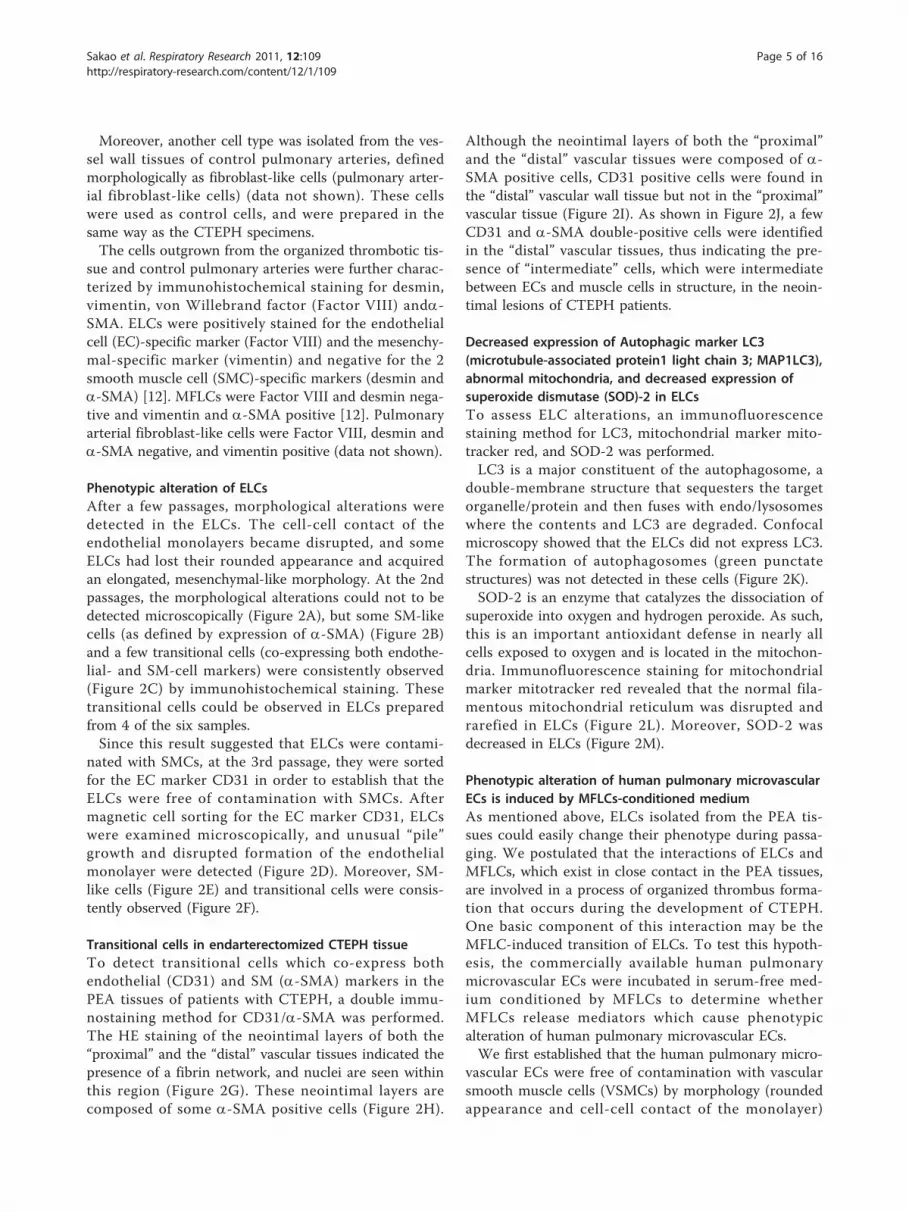
Phenotypic alteration of ELCsAfter a few passages, morphological alterations weredetected in the ELCs. The cell-cell contact of theendothelial monolayers became disrupted, and someELCs had lost their rounded appearance and acquiredan elongated, mesenchymal-like morphology. At the 2ndpassages, the morphological alterations could not to bedetected microscopically (Figure 2A), but some SM-likecells (as defined by expression of a-SMA) (Figure 2B)and a few transitional cells (co-expressing both endothe-lial- and SM-cell markers) were consistently observed(Figure 2C) by immunohistochemical staining. Thesetransitional cells could be observed in ELCs preparedfrom 4 of the six samples.Since this result suggested that ELCs were contami-
nated with SMCs, at the 3rd passage, they were sortedfor the EC marker CD31 in order to establish that theELCs were free of contamination with SMCs. Aftermagnetic cell sorting for the EC marker CD31, ELCswere examined microscopically, and unusual “pile”growth and disrupted formation of the endothelialmonolayer were detected (Figure 2D). Moreover, SM-like cells (Figure 2E) and transitional cells were consis-tently observed (Figure 2F).
Transitional cells in endarterectomized CTEPH tissueTo detect transitional cells which co-express bothendothelial (CD31) and SM (a-SMA) markers in thePEA tissues of patients with CTEPH, a double immu-nostaining method for CD31/a-SMA was performed.The HE staining of the neointimal layers of both the“proximal” and the “distal” vascular tissues indicated thepresence of a fibrin network, and nuclei are seen withinthis region (Figure 2G). These neointimal layers arecomposed of some a-SMA positive cells (Figure 2H).
Although the neointimal layers of both the “proximal”and the “distal” vascular tissues were composed of a-SMA positive cells, CD31 positive cells were found inthe “distal” vascular wall tissue but not in the “proximal”vascular tissue (Figure 2I). As shown in Figure 2J, a fewCD31 and a-SMA double-positive cells were identifiedin the “distal” vascular tissues, thus indicating the pre-sence of “intermediate” cells, which were intermediatebetween ECs and muscle cells in structure, in the neoin-timal lesions of CTEPH patients.
Decreased expression of Autophagic marker LC3(microtubule-associated protein1 light chain 3; MAP1LC3),abnormal mitochondria, and decreased expression ofsuperoxide dismutase (SOD)-2 in ELCsTo assess ELC alterations, an immunofluorescencestaining method for LC3, mitochondrial marker mito-tracker red, and SOD-2 was performed.LC3 is a major constituent of the autophagosome, a
double-membrane structure that sequesters the targetorganelle/protein and then fuses with endo/lysosomeswhere the contents and LC3 are degraded. Confocalmicroscopy showed that the ELCs did not express LC3.The formation of autophagosomes (green punctatestructures) was not detected in these cells (Figure 2K).SOD-2 is an enzyme that catalyzes the dissociation of
superoxide into oxygen and hydrogen peroxide. As such,this is an important antioxidant defense in nearly allcells exposed to oxygen and is located in the mitochon-dria. Immunofluorescence staining for mitochondrialmarker mitotracker red revealed that the normal fila-mentous mitochondrial reticulum was disrupted andrarefied in ELCs (Figure 2L). Moreover, SOD-2 wasdecreased in ELCs (Figure 2M).
Phenotypic alteration of human pulmonary microvascularECs is induced by MFLCs-conditioned mediumAs mentioned above, ELCs isolated from the PEA tis-sues could easily change their phenotype during passa-ging. We postulated that the interactions of ELCs andMFLCs, which exist in close contact in the PEA tissues,are involved in a process of organized thrombus forma-tion that occurs during the development of CTEPH.One basic component of this interaction may be theMFLC-induced transition of ELCs. To test this hypoth-esis, the commercially available human pulmonarymicrovascular ECs were incubated in serum-free med-ium conditioned by MFLCs to determine whetherMFLCs release mediators which cause phenotypicalteration of human pulmonary microvascular ECs.We first established that the human pulmonary micro-
vascular ECs were free of contamination with vascularsmooth muscle cells (VSMCs) by morphology (roundedappearance and cell-cell contact of the monolayer)
Sakao et al. Respiratory Research 2011, 12:109http://respiratory-research.com/content/12/1/109
Page 5 of 16
(Figure 3A) and by immunofluorescence staining usinganti-von Willebrand factor (Figure 3D), anti-a-SMA(Figure 3D), anti-vimentin (data not shown), and anti-human desmin (data not shown) antibodies. Theendothelial cell-specific marker and the mesenchymal-specific marker were positive, and the 2 smooth muscle-specific markers were negative, providing evidence thatthe human pulmonary microvascular ECs were not con-taminated with VSMCs.At the 2nd passage after incubation in serum-free
medium conditioned by pulmonary arterial fibroblast-like cells and MFLCs, the phenotypic alteration ofhuman pulmonary microvascular ECs was assessedmicroscopically and by immunofluorescence staining.The cell-cell contact of the endothelial monolayersbecame disrupted, and many ECs had lost their roundedappearance and acquired an elongated, mesenchymal-like morphology in the medium conditioned by MFLCs(Figure 3C) in comparison to the medium conditionedby pulmonary arterial fibroblast-like cells (Figure 3B).The number of ECs (as defined by expression of von
Willebrand factor) decreased, and SM-like cells (asdefined by expression of a-SMA) were consistentlyobserved in the medium conditioned by MFLCs (Figure3F, G), but not in the medium conditioned by pulmon-ary arterial fibroblast-like cells (Figure 3E, G).
Expression of TGF-b1 protein in the conditioned mediumBecause TGF-b1 is known to be involved in inducing theendothelial-mesenchymal transition [22] and is known topromote a-SMA expression in non-muscle cells (ECsand fibroblasts derived from various tissues) [23,24], theprotein levels in the conditioned medium were measuredby ELISA. Serum-free medium conditioned by MFLCscontained higher TGF-b1 levels than medium condi-tioned by pulmonary arterial fibroblast-like cells, but thedifference was not statistically significant (Figure 3H).
Phenotypic alteration of human pulmonary microvascularECs co-cultured with MFLCsAfter a 14 day incubation period, morphological altera-tions were detected in human pulmonary microvascular
Figure 2 ELCs from endarterectomized tissue. A-F), ELCs were assessed by immunofluorescence staining for anti-Factor VIII (green) and anti-a-SMA (red) to confirm the phenotypes of the cells. A), B) and C), ELCs before sorting; D), E) and F), ELCs after sorting; A) and D), themagnification was 100×. Scale bar = 100 μm; B) and E), the magnification was 200×. Scale bar = 50 μm; C) and F), the magnification was 400×.Scale bar = 25 μm. The blue staining was DAPI. G-J), Immunohistochemical staining of endarterectomized tissue. The neointimal layer of distalvascular wall tissues was assessed by immunohistochemical staining. G), Hematoxylin and Eosin (HE) staining; H), Single staining for a-SMA; I),Single staining for CD31; J), Double staining for CD31 and a-SMA; the magnification was 200×. Scale bar = 50 μm. K, L, M), Immunofluorescencestaining of ELCs for the autophagic marker, LC3 (K), mitochondrial marker mitotracker red (L), and SOD-2 (M). K), The formation ofautophagosomes (green punctate structures) was not detected. L), The normal filamentous mitochondrial reticulum (red punctate structures) wasnot detected. M), SOD-2 expression (green punctate structures) was not detected. The blue staining was DAPI. The magnification was 400×.Scale bar = 25 μm. ELCs = endothelial-like cells.
Sakao et al. Respiratory Research 2011, 12:109http://respiratory-research.com/content/12/1/109
Page 6 of 16

Figure 3 Human pulmonary microvascular ECs (HPMVECs) in serum-free medium conditioned by pulmonary arterial fibroblast-likecells (PAFLCs) or myofibroblast-like cells (MFLCs). The phenotypic alteration of HPMVECs was assessed microscopically and byimmunofluorescence staining. A) and D), Before incubation in serum-free medium conditioned by PAFLCs and MFLCs; B) and E), At the 2ndpassage after incubation in serum-free medium conditioned by PAFLCs; C) and F), At the 2nd passage after incubation in serum-free mediumconditioned by MFLCs; A), B) and C), microscopic findings; the magnification was 100×. Scale bar = 100 μm; D), E) and F), Immunofluorescencestaining for anti-Factor VIII (green) and anti-a-SMA (red). The blue staining was DAPI. The magnification was 200×. Scale bar = 50 μm. F), Somecells were positive for smooth muscle actin fibers (see inset); HPMVECs = human pulmonary microvascular endothelial cells; MFLCs =myofibroblast like cells; PAFLCs = fibroblast-like cells from control pulmonary arteries. G) Positive cells for anti-von Willebrand factor and anti-a-SM-actin were counted in 3 different fields at a magnification of × 200 in a fluorescence microscope. *P < 0.05 VS. PAFLCs, n ≥ 3. H) The TGF-b1protein levels in the conditioned medium were measured by ELISA. There were no significant differences between the serum-free mediumconditioned by PAFLCs and MFLCs.
Sakao et al. Respiratory Research 2011, 12:109http://respiratory-research.com/content/12/1/109
Page 7 of 16

ECs co-cultured with MFLCs (Figure 4B, D), but notthose cultured with pulmonary arterial fibroblast-likecells (Figure 4A, C). The cell-cell contact of theendothelial monolayers (Figure 4A) became disrupted,and hill and valley formation appeared. Moreover, someECs had lost their rounded appearance and acquired anelongated, mesenchymal-like morphology (Figure 4B).Some SM-like cells (as defined by their expression of a-SMA) and a few transitional cells (co-expressing bothendothelial- and SM- cell markers) were consistently
observed (Figure 4D, E) by immunohistochemicalstaining.
Autophagy PCR array analysis of human pulmonarymicrovascular ECs co-cultured with MFLCsThere were decreases in the expression of 17 autop-hagy-related genes in ECs co-cultured with MFLCs incomparison to the expression in ECs co-cultured withpulmonary arterial fibroblast-like cells (Figure 5A)(Table 1). Four of these genes; AMBRA1, ATG4D,MAP1LC3B, and RGS19, are involved in autophagicvacuole formation. In particular, ATG4D is responsi-ble for protein targeting to the membrane/vacuole,and is responsible for protein transport and proteaseactivity. Ten of the 17 genes; BCL2, BID, CDKN2A,CTSB, HSP90AA1, HTT, IFNG, IGF1, INS, andPRKAA1 are co-regulators of autophagy and apopto-sis. Three genes; RPS6KB1, TMEM77, and UVRAGare related to autophagy in response to other intracel-lular signals.
Autophagic marker LC3 expression in human pulmonarymicrovascular ECs co-cultured with MFLCsConfocal microscopy showed that the ECs co-culturedwith pulmonary arterial fibroblast-like cells expressedLC3. The formation of autophagosomes (green punctatestructures) was detected in these cells (Figure 6A), butnot in ECs co-cultured with MFLCs (Figure 6B) nor inELCs (Figure 2K).
Abnormal mitochondria and decreased expression ofsuperoxide dismutase (SOD)-2 in human pulmonarymicrovascular ECs co-cultured with MFLCsImmunofluorescence staining for mitochondrial markermitotracker red revealed that the normal filamentousmitochondrial reticulum observed in ECs co-culturedwith pulmonary arterial fibroblast-like cells (Figure 6D)was disrupted and rarefied in both ECs co-cultured withMFLCs (Figure 6E) and ELCs (Figure 2L). Moreover,SOD-2 was decreased in ECs co-cultured with MFLCs(Figure 6H) and ELCs (Figure 2M) compared to thoseco-cultured with pulmonary arterial fibroblast-like cells(Figure 6G). The decrease in SOD-2 expression in ECsco-cultured with MFLCs and ELCs might be associatedwith a reduction in SOD-2 activity.
Endothelial cell biology PCR array of human pulmonarymicrovascular ECs co-cultured with MFLCsThese results, including the phenotypic alterations, inac-tivation of autophagy, and mitochondrial dysfunction,suggested that the endothelial cell biology is altered inpatients with CTEPH. Therefore, an endothelial cellbiology PCR array was done to further explore theeffects of MFLCs on endothelial cell biology.
Figure 4 Human pulmonary microvascular ECs (HPMVECs) co-cultured with pulmonary arterial fibroblast-like cells (PAFLCs)or myofibroblast-like cells (MFLCs). The phenotypical alteration ofHPMVECs was assessed microscopically and by immunofluorescencestaining after a 14 day incubation period. A) and C), HPMVECs co-cultured with PAFLCs; B) and D), HPMVECs co-cultured with MFLCs;A) and B), Microscopic findings; the magnification was 100×. Scalebar = 100 μm; C) and D), Immunofluorescence staining for anti-Factor VIII (green) and anti-a-SMA (red). The blue staining was DAPI.The magnification was 400×. Scale bar = 25 μm. D), Some cellscoexpressed both anti-Factor VIII and anti-a-SMA (see inset);HPMVECs = human pulmonary microvascular endothelial cells;MFLCs = myofibroblast-like cells; PAFLCs = fibroblast-like cells fromcontrol pulmonary arteries. E) Positive cells for anti-von Willebrandfactor and anti-a-SM-actin were counted in 3 different fields at amagnification of × 200 in a fluorescence microscope. *P < 0.05 VS.
PAFLCs, n ≥ 3.
Sakao et al. Respiratory Research 2011, 12:109http://respiratory-research.com/content/12/1/109
Page 8 of 16

There were decreases in the expression of 15 andincreases in the expression of 3 genes in ECs co-cul-tured with MFLCs in comparison to the expression inthose co-cultured with pulmonary arterial fibroblast-like
cells (Figure 5B). The 15 decreased genes were ANXA5,BCL2, CDH5, COL18A1, CX3CL1, ITGA5, ITGAV,ITGB1, MMP1, NPPB, PGF, PLA2G4C, PLAU, RHOB,and SOD1 (Table 2). CDH5, COL18A1, CX3CL1,ITGA5, ITGAV, ITGB1 and RHOB are related toendothelial cell activation as adhesion molecules.MMP1, NPPB, PLAU and RHOB are related to endothe-lial cell activation, and are part of the extracellularmatrix (ECM) molecules. ANXA5 and PLAU are relatedto endothelial cell activation with regard to thrombinactivity. PGF is related to angiogenesis. PLA2G4C andSOD-1 are both related to the endothelial cell responseto stress.The 3 genes with increased expression were AGTR1,
CASP1, and TIMP1 (Table 3). AGTR1 is related to thepermissibility and vessel tone of the angiotensin system.CASP1 is related to endothelial cell injury and resultingapoptosis. TIMP1 is related to endothelial cell activationand cell growth.
SMAD reporter signal in human pulmonary microvascularECs co-cultured with MFLCsThe SMAD2 and SMAD3 proteins are phosphorylatedand activated by TGF-b signaling. These activatedSMAD 2 and SMAD 3 then form complexes with theSMAD4. These SMAD complexes then migrate to thenucleus, where they activate the expression of TGF-b-responsive genes.Besides simple concentration measurements of TGF-
b1 in the conditioned medium (Figure 3H), the activa-tion of the TGF-b signaling in human pulmonary micro-vascular ECs co-cultured with MFLCs were measured bythe SMAD reporter assay. There was no statistical dif-ference in the expression of SMAD reporter signal inECs co-cultured with MFLCs in comparison to theexpression in those co-cultured with pulmonary arterialfibroblast-like cells (Figure 5C).
Accumulation of ROS in human pulmonary microvascularECs co-cultured with MFLCsAccumulation of ROS coupled with an increase in oxi-dative stress has been implicated in the pathogenesis ofnumerous disease states. As SOD1 and SOD2 downre-gulation have been shown by the PCR-Arrays (Figure5B) and immunofluorescence (Figure 6H), the missingproduction of ROS might be involved in ECs co-cul-tured with MFLCs [25]. The decreased production ofROS has been detected in ECs co-cultured with MFLCsin comparison to the expression in those co-culturedwith pulmonary arterial fibroblast-like cells (Figure 5D).
Rapamycin treatmentProlonged rapamycin treatment of ECs co-cultured withMFLCs reversed the decrease in the 17 autophagy-
Figure 5 Human pulmonary microvascular ECs (HPMVECs) co-cultured with pulmonary arterial fibroblast-like cells (PAFLCs)or myofibroblast-like cells (MFLCs). Autophagy and Endothelialcell biology. A) Autophagy PCR array analysis of HPMVECs co-cultured with PAFLCs, MFLCs or MFLCs+Rapamycin. There weredecreases in the expression of 17 autophagy-related genes in theECs co-cultured with MFLCs in comparison those co-cultured withPAFLCs (P < 0.05; n = 3). This result is related to 3 different patientsout of six of co-culture or conditioned medium. See table 1 fordefinitions of the abbreviations. B) Endothelial cell biology PCR arrayanalysis of HPMVECs co-cultured with PAFLCs, MFLCs or MFLCs+Rapamycin. There were decreases in 15 and increases of 3 genesin ECs co-cultured with MFLCs in comparison to the expression inECs co-cultured with PAFLCs (P < 0.05; n = 3). This result is relatedto 3 different patients out of six of co-culture or conditionedmedium. See table 2 and 3 for the definitions. C) SMAD reportersignal in HPMVECs co-cultured with MFLCs. There was no statisticaldifference in the expression of SMAD reporter signal in ECs co-cultured with MFLCs in comparison to the expression in those co-cultured with PAFLCs treated with or without rapamycin. D)Accumulation of ROS in HPMVECs co-cultured with MFLCs. Thedecreased production of ROS has been detected in ECs co-culturedwith MFLCs in comparison to the expression in those co-culturedwith PAFLCs (P < 0.05; n = 3). Although there was a tendency thatrapamycin treatment of ECs co-cultured with MFLCs reversed thedecreased production of ROS, there was no statistical differencebetween them.
Sakao et al. Respiratory Research 2011, 12:109http://respiratory-research.com/content/12/1/109
Page 9 of 16

related genes (Figure 5A) (Table 1) and prevented thechanges in expression in 11 of the 15 decreased and allthree of the increased genes related to endothelial cellbiology (Figure 5B) (Table 2, 3). There was no statisticaldifference in the expression of SMAD reporter signal inECs co-cultured with MFLCs with rapamycin (Figure5C). Although rapamycin treatment of ECs co-culturedwith MFLCs seemed to reverse the decreased produc-tion of ROS (Figure 5D), there was no statistical differ-ence between them.Confocal microscopy showed that the ECs co-cultured
with MFLCs that were treated with rapamycin expressedLC3. Although the formation of autophagosomes (greenpunctate structures) was not detected in ECs co-
cultured with MFLCs (Figure 6B), it was detected inthese cells when they were treated with rapamycin (Fig-ure 6C). In the ECs co-cultured with MFLCs, the co-localization of Mitotracker red and SOD-2 was lost,indicating that the mitochondrial reticulum is disrupted(Figure 6E, 2M). However, the mitochondria in the ECsco-cultured with MFLCs that were treated with rapamy-cin form an intricate, filamentous network, in whichSOD-2 and Mitotracker red are tightly co-localized (Fig-ure 6F, I).
DiscussionEnMT is a term which has been used to describe theprocess through which ECs lose their endothelial
Table 1 Autophagy PCR array
Biological process description Gene name Genesymbol
Public ID P-value
Autophagy Machinary Components: Genes Involved inAutophagic Vacuole Formation
Autophagy/beclin-1 regulator 1 AMBRA1 NM_017749 0.00308
Autophagy Machinary Components: Genes Involved inAutophagic Vacuole Formation
Genes Responsible for Protein Targeting to Membrane/Vacuole
Genes Responsible for Protein TransportGenes with Protease Activity
ATG4 autophagy related 4 homolog D (S.cerevisiae)
ATG4D NM_032885NM_017749
0.01167
Regulation of Autophagy:Co-Regulators of Autophagy and Apoptosis
B-cell CLL/lymphoma 2 BCL2 NM_000633 0.000727
Regulation of Autophagy:Co-Regulators of Autophagy and Apoptosis
BH3 interacting domain death agonist BID NM_001196 0.047933
Regulation of Autophagy:Co-Regulators of Autophagy and Apoptosis
Cyclin-dependent kinase inhibitor 2A(melanoma, p16, inhibits CDK4)
CDKN2A NM_000077 0.044888
Regulation of Autophagy:Co-Regulators of Autophagy and Apoptosis
Cathepsin B CTSB NM_001908 0.010802
Regulation of Autophagy:Chaperone-Mediated Autophagy
Heat shock protein 90 kDa alpha (cytosolic),class A member 1
HSP90AA1 NM_001017963 0.037151
Regulation of Autophagy:Co-Regulators of Autophagy and Apoptosis
Huntingtin HTT NM_002111 0.033212
Regulation of Autophagy:Co-Regulators of Autophagy and Apoptosis
Co-Regulators of Autophagy and the Cell Cycle
Interferon, gamma IFNG NM_000619 0.017749
Regulation of Autophagy:Co-Regulators of Autophagy and Apoptosis
Insulin-like growth factor 1 (somatomedin C) IGF1 NM_000618 0.017282
Regulation of Autophagy:Co-Regulators of Autophagy and Apoptosis
Insulin INS NM_000207 0.045037
Autophagy Machinary Components: Genes Involved inAutophagic Vacuole Formation
Microtubule-associated protein 1 light chain 3beta
MAP1LC3B NM_022818 0.011251
Regulation of Autophagy:Co-Regulators of Autophagy and Apoptosis
Autophagy in Response to Other Intracellular Signals
Protein kinase, AMP-activated, alpha 1 catalyticsubunit
PRKAA1 NM_006251 0.005633
Autophagy Machinary Components: Genes Involved inAutophagic Vacuole Formation
Regulator of G-protein signaling 19 RGS19 NM_005873 0.021592
Regulation of Autophagy:Autophagy in Response to Other Intracellular Signals
Ribosomal protein S6 kinase, 70 kDa,polypeptide 1
RPS6KB1 NM_003161 0.024072
Regulation of Autophagy:Autophagy in Response to Other Intracellular Signals
Transmembrane protein 77 TMEM77 NM_178454 0.019285
Regulation of Autophagy:Autophagy in Response to Other Intracellular Signals
UV radiation resistance associated gene UVRAG NM_003369 0.016479
Functional classification of low expressed genes in co-cultured HPMVECs with MFLCs in comparison to PAFLCs
Sakao et al. Respiratory Research 2011, 12:109http://respiratory-research.com/content/12/1/109
Page 10 of 16

characteristics and gain expression of mesenchymal,myofibroblast-like characteristics [26]. In the presentstudy, a few transitional cells (co-expressing bothendothelial- and SM- cell markers) were shown in theprimary culture of endarterectomized tissue specimen(Figure 2C, F). The microenvironment created by thestabilized clot is suggested to induce EnMT (Figure 3F,G, 4D, E). Moreover, CD31 and a-SMA double-positivecells were identified in the neointimal layer of vascularwall tissue, thus indicating the presence of transitionalcells in the neointimal lesions of CTEPH (Figure 2J). Insupport of our finding, Yao et al showed the presence ofCD34 (endothelial marker) positive cells co-expressinga-SMA (SM-cell marker) in endarterectomized tissuesfrom patients with CTEPH [27]. Moreover, they sug-gested that the microenvironment provided by throm-boemboli might promote the putative progenitor cells todifferentiate and enhance intimal remodeling [27]. Inthis study, our data suggest that MFLC-related EnMTmay enhance intimal remodeling. However, we fully rea-lize the limitations of our data interpretation, which was
based on in vitro studies of cultured cells, and acknowl-edge that data provided in this study were not strong tosupport EnMT hypothesis because this study failed toshow mechanisms responsible for this process. More-over, it may be possible that transitional cells are morelikely progenitor cells rather than they are transdifferen-tiated by EnMT.There was no significant difference in TGF-b1 levels
between serum-free medium conditioned by MFLCs andby pulmonary arterial fibroblast-like cells (Figure 3H).Moreover, there was no statistical difference in theexpression of SMAD reporter signal in ECs co-culturedwith MFLCs in comparison to the expression in thoseco-cultured with pulmonary arterial fibroblast-like cells(Figure 5C). A recent study provides evidence that Ras/MAPK, via TGF-b1 signaling, mediates completion ofEnMT in a bleomycin model of pulmonary fibrosis [28].However, an endothelial cell biology PCR array in thisstudy demonstrated the decreased expression of RHOB(Ras homolog gene family, member B) in co-culturedhuman pulmonary microvascular ECs with MFLCs in
Figure 6 Immunofluorescence staining of human pulmonary microvascular ECs (HPMVECs) and endothelial-like cells (ELCs) for theautophagic marker, LC3 (A-C), mitochondrial marker mitotracker red (D-F), and SOD-2 (G-I). A), HPMVECs co-cultured with PAFLCs; B),with MFLCs; C), with MFLCs + Rapamycin; A) and C), The formation of autophagosomes (green punctate structures) was detected (see inset). D),HPMVECs co-cultured with PAFLCs; E), MFLCs; F), with MFLCs + Rapamycin; D) and F), The normal filamentous mitochondrial reticulum (redpunctate structures) was detected (see inset). G), HPMVECs co-cultured with PAFLCs; H), with MFLCs; I), with MFLCs + Rapamycin; G) and I), SOD-2 expression (green punctate structures) was detected (see inset). The blue staining was DAPI. The magnification was 400×. Scale bar = 25 μm.ELCs = endothelial-like cells.
Sakao et al. Respiratory Research 2011, 12:109http://respiratory-research.com/content/12/1/109
Page 11 of 16

comparison to pulmonary arterial fibroblast-like cells.These results suggest that not only TGF-b1 nor Ras, butalso additional factors, may be essential for this transi-tional pathway. Indeed, TGF-b1 is currently thought tobe insufficient to induce the late stage of SM differentia-tion in non-SMC lineage cells [24]. Moreover, neither
TGF-b1 nor activated Ras alone were capable of indu-cing a-SMA expression [28].The effects of conditioned media may be particularly
remarkable if chemically defined culture media withoutserum additions is employed. Therefore, serum freemedia was adapted for the conditioned media
Table 2 Endothelial Cell Biology PCR Array
Biological processdescription
Gene name Genesymbol
Public ID P-value
Endothelial Cell Activation:Thrombin Activity
Annexin A5 ANXA5 NM_001154 0.023464
Endothelial Cell Injury:Response to StressAnti-Apoptosis
B-cell CLL/lymphoma 2 BCL2 NM_000633 0.000247
Endothelial Cell Activation:Adhesion Molecules
Cadherin 5, type 2 (vascular endothelium) CDH5 NM_001795 0.003968
Endothelial Cell Activation:Adhesion Molecules
Collagen, type XVIII, alpha 1 COL18A1 NM_030582 0.004024
Endothelial Cell Activation:Adhesion Molecules
Chemokine (C-X3-C motif) ligand 1 CX3CL1 NM_002996 0.000779
Endothelial Cell Activation:Adhesion Molecules
Integrin, alpha 5 (fibronectin receptor, alpha polypeptide) ITGA5 NM_002205 0.000147
Endothelial Cell Activation:Adhesion Molecules
Integrin, alpha V (vitronectin receptor, alpha polypeptide, antigen CD51) ITGAV NM_002210 0.02125
Endothelial Cell Activation:Adhesion Molecules
Integrin, beta 1 (fibronectin receptor, beta polypeptide, antigen CD29includes MDF2, MSK12)
ITGB1 NM_002211 0.018399
Endothelial Cell Activation:Extracellular Matrix (ECM)
Molecules
Matrix metallopeptidase 1 (interstitial collagenase) MMP1 NM_002421 0.021783
Permissibility and Vessel Tone:Regulation of Blood Pressure
Regulation of VascularPermeabilityAngiogenesis:
Negative Regulation ofAngiogenesis
Endothelial Cell Activation:Extracellular Matrix (ECM)
Molecules
Natriuretic peptide precursor B NPPB NM_002521 0.041345
Angiogenesis:Other Genes Involved in
Angiogenesis
Placental growth factor PGF NM_002632 0.029734
Endothelial Cell Injury:Response to Stress
Phospholipase A2, group IVC (cytosolic, calcium-independent) PLA2G4C NM_003706 0.014626
Endothelial Cell Activation:Extracellular Matrix (ECM)
MoleculesThrombin Activity
Plasminogen activator, urokinase PLAU NM_002658 0.039877
Endothelial Cell ActivationAdhesion Molecules
Angiogenesis:Positive Regulation of
AngiogenesisEndothelial Cell Activation:
Adhesion MoleculesEndothelial Cell Injury:Other Genes Related to
Apoptosis
Ras homolog gene family, member B RHOB NM_004040 0.035874
Endothelial Cell Injury:Response to Stress
Superoxide dismutase 1, soluble SOD1 NM_000454 0.001855
Functional classification of low expressed genes in co-cultured HPMVECs with MFLCs in comparison to PAFLCs
Sakao et al. Respiratory Research 2011, 12:109http://respiratory-research.com/content/12/1/109
Page 12 of 16

experiments. However, this leads to serum starvation onthe cells, which commonly leads to cell cycle arrest andinduces changes in protein synthesis. Accordingly, co-culture experiments were conducted in media withserum, which allows different cell types to grow oneither side of the membrane and may be able to detectthe mutual effects of cell types on one another.An inactivation of autophagy was found in both ELCs
(Figure 2K) and human pulmonary microvascular ECsco-cultured with MFLCs (Figure 6B) compared to theexpression in human pulmonary microvascular ECs co-cultured with pulmonary arterial fibroblast-like cells(Figure 6A), thus suggesting that in these cells, proteinsynthesis predominates over protein degradation. More-over, the decreased expression of cell death-relatedgenes indicated that cell growth may be stimulated (Fig-ure 5A). This inactivation could benefit cancer cells.Recently several genetic links between autophagy defectsand cancers have been shown, providing increasing sup-port for the concept that autophagy is a genuine tumorsuppressor pathway [29]. Signaling pathways that regu-late autophagy overlaps with those that regulate tumori-genesis [16].This study has shown that human pulmonary micro-
vascular ECs co-cultured with MFLCs and ELCs havefewer mitochondria with an organized reticulum (Figure6E, 2H) and SOD-2, which is an enzyme found only inthe mitochondria, is decreased in these cells (Figure 6H,2L). Endothelial cell biology PCR array demonstratedthe decreased expression of SOD1 (Table 2), which islocated in the cytoplasm. Both SOD1 and 2 are animportant antioxidant defense in almost all cells exposedto oxygen. Moreover, the decreased production of ROShas been detected in ECs co-cultured with MFLCs incomparison to the expression in those co-cultured withpulmonary arterial fibroblast-like cells (Figure 5D).These results including fewer mitochondria, thedecreased expression of SOD, and normoxic decreasesin ROS are compatible with the characteristics of mito-chondrial abnormalities in PAH, demonstrated byArcher et al [25]. The metabolic shift from oxidativemitochondrial metabolism to the glycolytic metabolism
inhibits acetyl-CoA to enter the Krebs’ Cycle, resultingin reduced production of ROS. However, gene and pro-tein expression of SOD are not directly translated intoactivity and the decreased production of ROS is not suf-ficient to determine SOD activity. It has been shownthat pulmonary artery SMCs in PAH are associated withmitochondrial disorders [30-32]. Xu and colleagues usedan in vitro experiment with pulmonary artery ECs fromidiopathic PAH (IPAH ECs) and control lungs (controlECs) to show that glucose metabolism plays the primaryrole in the energy requirements of IPAH ECs, based onthe 3-fold greater glycolytic rate of IPAH ECs comparedwith control ECs. This indicates that there is mitochon-drial dysfunction in ECs in patients with idiopathicPAH, similar to the SMCs in PAH [33]. The existenceof mitochondrial disorder/dysfunction in commerciallyavailable pulmonary microvascular ECs co-cultured withMFLCs in CTEPH and ECs in PAH, may support thesimilarities in the microvascular remodeling in the twodisease.Although several protein kinases regulate autophagy,
the mammalian target of rapamycin (mTOR), whichnegatively regulates the pathway in organisms fromyeast to man, is the best characterized [15]. Rapamycinis an inhibitor of mTOR and an anti-proliferative immu-nosuppressor that arrests cells in the G1 phase of thecell cycle [34]. Rapamycin is used clinically in cardiovas-cular medicine as an anti-proliferative agent applied tocoronary stents to reduce local restenosis [35]. Rapamy-cin inhibits hypoxia-induced activation of S6 kinase inpulmonary arterial adventitial fibroblasts [36], suggestingthe possibility that there may be a therapeutic benefit inPAH. Moreover, rapamycin has an anti-proliferativeeffect on pulmonary arterial SMCs derived from endar-terectomized tissues of CTEPH patients [37]. In thisstudy, we demonstrated that rapamycin reversed thedecrease in autophagy in the ECs co-cultured withMFLCs (Figure 5A, 6C). Moreover, rapamycin alsoreversed the disruption of the mitochondrial reticulumand restored the localization of SOD-2 (Figure 6F, I). Itis acknowledged that mTOR activity antagonize induc-tion of the general stress response genes including
Table 3 Endothelial Cell Biology PCR Array
Biological process description Gene name Genesymbol
Public ID P-value
Permissibility and Vessel Tone:Angiotensin System
Angiotensin II receptor, type 1 AGTR1 NM_031850 0.030612
Endothelial Cell Injury:Caspase Activation
Caspase 1, apoptosis-related cysteine peptidase (interleukin 1, beta,convertase)
CASP1 NM_033292 0.005519
Endothelial Cell Activation:Other Genes Involved in Cell
Growth
TIMP metallopeptidase inhibitor 1 TIMP1 NM_003254 0.049858
Functional classification of highly expressed genes in co-cultured HPMVECs with MFLCs in comparison to PAFLCs
Sakao et al. Respiratory Research 2011, 12:109http://respiratory-research.com/content/12/1/109
Page 13 of 16

SOD-2 gene [38-40]. This may explain the mechanismsby which rapamycin exerts its beneficial changes on cel-lular mitochondria and SOD2 expression. Indeed, SOD2is located within the mitochondrial matrix and wasstrongly induced in response to rapamycin in normaland neoplastic mammalian cells [41]. It also reversedthe change in expression of 11 of the 15 genesdecreased by co-culture and the 3 genes increased byco-culture that were related to endothelial cell biology(Figure 5B) (Table 2, 3), thus suggesting that rapamycin(as an anti-proliferative agent) has beneficial therapeuticeffects, not only on pulmonary arterial SMCs, but alsoon pulmonary arterial ECs which exist in the close con-tact with MFLCs, in the patients with CTEPH. However,because rapamycin may act on the proliferation rate ofMFLCs more than pulmonary arterial fibroblast-likecells [37], it is possible that this action may be an alter-native explanation for the observed differences. More-over, a few transitional cells were observed in the ECsco-culture with MFLCs that were treated with rapamy-cin (data not shown), indicating that rapamycin mightexert no beneficial effect on EnMT.
ConclusionsOur experiments with ECs and MFLCs demonstratedthat factors associated with MFLCs in the microenviron-ment created by the unresolved clot might induce ECdysfunction through EnMT (3F, 3G, 4D, 4E),
inactivation of autophagy (Figure 5A, 6B), disruption ofthe mitochondrial reticulum, and improper localizationof SOD-2 (Figure 6E, H). Indeed, ELCs, which were iso-lated from the PEA tissues of CTEPH patients, includeda few transitional cells (coexpressing both endothelial-and SM- cell markers) (Figure 2J), lost their ability toform autophagosomes (Figure 2K) and had defectivemitochondrial structure/function (Figure 2L). Althoughit is uncertain whether MFLCs induce EC dysfunctionin vivo and whether EC dysfunction contribute to thevascular lesions in the patients with CTEPH, it is possi-ble that there exist dysfunctional ECs in the microenvir-onment created by the unresolved clot (Figure 7).However, non-resolving pulmonary thromboemboli inCTEPH mainly consist of fibrotic tissue representing theend-stage of a thrombus organization process. There-fore, it remains uncertain whether any of the cellular ormolecular findings at this stage of disease are causallyinvolved in disease pathogenesis. We should acknowl-edge the purely descriptive nature of this study thatdoes not confer any pathophysiological evidence inCTEPH.
AcknowledgementsAll authors read and approved the final manuscript. We thank Dr. J. T.Reeves, who enriched our research for many years.All sources of supportThis study was supported by Research Grants for the Respiratory FailureResearch Group, the Cardiovascular Diseases (19-9), and Research onIntractable Diseases (22-33) from the Ministry of Health, Labor and Welfare,Japan, and a Grant-in-Aid for Scientific Research (Category C 22590851) fromthe Japanese Ministry of Education and Science.
Author details1Department of Respirology (B2), Graduate School of Medicine, ChibaUniversity, 1-8-1 Inohana, Chuo-ku, Chiba 260-8670, Japan. 2Department ofSurgical Pathology, Hyogo College of Medicine, 1-1 Mukogawa-cho,Nishinomiya, Hyogo, 663-8501, Japan.
Authors’ contributionsSS conceived of the report, contributed to its design and conception,drafted the manuscript and carried out the all studies. HH carried out thepathological studies. NT drafted the manuscript and contributed to itsdesign and conception. YK contributed to its design. KK carried out thepathological studies. KT contributed to its design and drafted themanuscript. All authors read and approved the final manuscript.
Competing interestsDr. Tatsumi has received honoraria for lectures from Glaxo Smith Kline,Actelion Pharmaceutical Ltd. Dr. Tanabe has received honoraria for lecturesfrom Actelion, Glaxo Smith Kline, Astellas and Pfizer and research grantsupport from Actelion Pharmaceutical Ltd. The other authors report noconflicts.
Received: 23 May 2011 Accepted: 22 August 2011Published: 22 August 2011
References1. Klepetko W, Mayer E, Sandoval J, Trulock EP, Vachiery JL, Dartevelle P,
Pepke-Zaba J, Jamieson SW, Lang I, Corris P: Interventional and surgicalmodalities of treatment for pulmonary arterial hypertension. J Am CollCardiol 2004, 43(suppl S):73S-80S.
Figure 7 EC dysfunction in CTEPH (a proposed mechanism).Our experiments with ECs and MFLCs demonstrated that themicroenvironment provided by the thrombus cells in CTEPHpatients might induce EC dysfunction through EnMT, inactivation ofautophagy, disruption of mitochondrial reticulum, and the improperSOD-2 localization, although it remains unknown whether bothEnMT and other cell function alterations are taking placesimultaneously in the same ECs. Although it is uncertain whetherMFLCs induce EC dysfunction in vivo and whether EC dysfunctioncontribute to the vascular lesions in the patients with CTEPH, it ispossible that there exist dysfunctional ECs in the microenvironmentcreated by the unresolved clot.
Sakao et al. Respiratory Research 2011, 12:109http://respiratory-research.com/content/12/1/109
Page 14 of 16

2. Jamieson SW, Kapelanski DP, Sakakibara N, Manecke GR, Thistlethwaite PA,Kerr KM, Channick RN, Fedullo PF, Auger WR: Pulmonary endarterectomy:experience and lessons learned in 1,500 cases. Ann Thorac Surg 2003,76:1457-1462; discussion 1462-1454.
3. Fanikos J, Piazza G, Zayaruzny M, Goldhaber SZ: Long-term complicationsof medical patients with hospital-acquired venous thromboembolism.Thromb Haemost 2009, 102:688-693.
4. Cohen AT, Agnelli G, Anderson FA, Arcelus JI, Bergqvist D, Brecht JG,Greer IA, Heit JA, Hutchinson JL, Kakkar AK, Mottier D, Oger E, Samama MM,Spannagl M: VTE Impact Assessment Group in Europe (VITAE). Venousthromboembolism (VTE) in Europe. The number of VTE events andassociated morbidity and mortality. Thromb Haemost 2007, 98:756-764.
5. Hoeper MM, Mayer E, Simonneau G, Rubin L: Chronic thromboembolicpulmonary hypertension. Circulation 2006, 113:2011-2020.
6. Moser KM, Bloor CM: Pulmonary vascular lesions occurring in patientswith chronic major vessel thromboembolic pulmonary hypertension.Chest 1993, 103:685-692.
7. Azarian R, Wartski M, Collignon MA, Parent F, Herve P, Sors H, Simonneau G:Lung perfusion scans and hemodynamics in acute and chronicpulmonary embolism. J Nucl Med 1997, 38:980-983.
8. Yi ES, Kim H, Ahn H, Strother J, Morris T, Masliah E, Hansen LA, Park K,Friedman PJ: Distribution of obstructive intimal lesions and their cellularphenotypes in chronic pulmonary hypertension: a morphometric andimmunohistochemical study. Am J Respir Crit Care Med 2000,162:1577-1586.
9. Sakao S, Taraseviciene-Stewart L, Lee JD, Wood K, Cool CD, Voelkel NF:Initial apoptosis is followed by increased proliferation of apoptosis-resistant endothelial cells. FASEB J 2005, 19:1178-1180.
10. Masri FA, Xu W, Comhair SA, Asosingh K, Koo M, Vasanji A, Drazba J,Anand-Apte B, Erzurum SC: Hyperproliferative apoptosis-resistantendothelial cells in idiopathic pulmonary arterial hypertension. Am JPhysiol Lung Cell Mol Physiol 2007, 293:L548-L554.
11. Firth AL, Yao W, Ogawa A, Madani MM, Lin GY, Yuan JX: Multipotentmesenchymal progenitor cells are present in endarterectomized tissuesfrom patients with chronic thromboembolic pulmonary hypertension.Am J Physiol Cell Physiol 2010, 298:C1217-C1225.
12. Maruoka M, Sakao S, Kantake M, Tanabe N, Kasahara Y, Kurosu K,Takiguchi Y, Masuda M, Yoshino I, Voelkel NF, Tatsumi K: Characterizationof myofibroblasts in chronic thromboembolic pulmonary hypertension.Int J Cardiol 2011.
13. Sakao S, Taraseviciene-Stewart L, Cool CD, Tada Y, Kasahara Y, Kurosu K,Tanabe N, Takiguchi Y, Tatsumi K, Kuriyama T, Voelkel NF: VEGF-R blockadecauses endothelial cell apoptosis, expansion of surviving CD34+precursor cells and transdifferentiation to smooth muscle-like andneuronal-like cells. FASEB J 2007, 21:3640-3652.
14. Sahara M, Sata M, Morita T, Nakamura K, Hirata Y, Nagai R: Diversecontribution of bone marrow-derived cells to vascular remodelingassociated with pulmonary arterial hypertension and arterial neointimalformation. Circulation 2007, 115:509-517.
15. Klionsky DJ, Emr SD: Autophagy as a regulated pathway of cellulardegradation. Science 2000, 290:1717-1721.
16. Levine B, Kroemer G: Autophagy in the pathogenesis of disease. Cell2008, 132:27-42.
17. Lindahl P, Johansson BR, Levéen P, Betsholtz C: Pericyte loss andmicroaneurysm formation in PDGF-B-deficient mice. Science 1997,277:242-245.
18. Hirschi K, Rohovsky SA, D’Amore PA: PDGF, TGF-β and heterotypic cell-cellinteractions mediate the recruitment and differentiation of 10T1/2 cellsto a smooth muscle cell fate. J Cell Biol 1998, 141:805-814.
19. Hellström M, Kalén M, Lindahl P, Abramsson A, Betsholtz C: Role of PDGF-Band PDGFR-β in recruitment of vascular smooth muscle cells andpericytes during embryonic blood vessel formation in the mouse.Development 1999, 126:3047-3055.
20. Allanach K, Mengel M, Einecke G, Sis B, Hidalgo LG, Mueller T, Halloran PF:Comparing microarray versus RT-PCR assessment of renal allograftbiopsies: similar performance despite different dynamic ranges. Am JTransplant 2008, 8:1006-1015.
21. Wang Y, Barbacioru C, Hyland F, Xiao W, Hunkapiller KL, Blake J, Chan F,Gonzalez C, Zhang L, Samaha RR: Large scale real-time PCR validation ongene expression measurements from two commercial long-oligonucleotide microarrays. BMC Genomics 2006, 7:59.
22. Frid MG, Kale VA, Stenmark KR: Mature vascular endothelium can give riseto smooth muscle cells via endothelial-mesenchymaltransdifferentiation: in vitro analysis. Circ Res 2002, 14:1189-1196.
23. Arciniegas E, Sutton AB, Allen TD, Schor AM: Transforming growth factorbeta 1 promotes the differentiation of endothelial cells into smoothmuscle-like cells in vitro. J Cell Sci 1992, 103:521-529.
24. Hautmann MB, Adam PJ, Owens GK: Similarities and differences insmooth muscle α-actin induction by TGF-s in smooth muscle versusnon-smooth muscle cells. Arterioscler Thromb Vasc Biol 1999, 19:2049-2058.
25. Archer SL, Gomberg-Maitland M, Maitland ML, Rich S, Garcia JG, Weir EK:Mitochondrial metabolism, redox signaling, and fusion: a mitochondria-ROS-HIF-1alpha-Kv1.5 O2-sensing pathway at the intersection ofpulmonary hypertension and cancer. Am J Physiol Heart Circ Physiol 2008,294:570-578.
26. Arciniegas E, Frid MG, Douglas IS, Stenmark KR: Perspectives onendothelial-to-mesenchymal transition: potential contribution tovascular remodeling in chronic pulmonary hypertension. Am J PhysiolLung Cell Mol Physiol 2007, 293:L1-8.
27. Yao W, Firth AL, Sacks RS, Ogawa A, Auger WR, Fedullo PF, Madani MM,Lin GY, Sakakibara N, Thistlethwaite PA, Jamieson SW, Rubin LJ, Yuan JX:Identification of putative endothelial progenitor cells (CD34+CD133+Flk-1+) in endarterectomized tissue of patients with chronicthromboembolic pulmonary hypertension. Am J Physiol Lung Cell MolPhysiol 2009, 296:L870-878.
28. Hashimoto N, Phan SH, Imaizumi K, Matsuo M, Nakashima H, Kawabe T,Shimokata K, Hasegawa Y: Endothelial-mesenchymal transition inbleomycin-induced pulmonary fibrosis. Am J Respir Cell Mol Biol 2010,43:161-172.
29. Levine B: Cell biology: autophagy and cancer. Nature 2007, 446:745-747.30. Bonnet S, Archer SL, Allaluis-Turner J, Haromy A, Beaulieu C, Thompson R,
Lee CT, Lopaschuk GD, Puttagunta L, Bonnet S, Harry G, Hashimoto K,Porter CJ, Andrade MA, Thebaud B, Michelakis ED: A mitochondria-Kchannel axis is suppressed in cancer and its normalization promotesapoptosis and inhibits cancer growth. Cancer Cell 2003, 11:37-51.
31. Bonnet S, Michelakis ED, Porter CJ, Andrade-Navarro MA, Thébaud B,Bonnet S, Haromy A, Harry G, Moudgil R, McMurtry MS, Weir EK, Archer SL:An abnormal mitochondrial-HIF-1-Kv channel pathway disrupts oxygen-sensing and triggers pulmonary arterial hypertension (PAH) in fawn-hooded rats: similarities to human PAH. Circulation 2006, 113:2630-2641.
32. Bonnet S, Rochefort G, Sutendra G, Archer SL, Haromy A, Webster L,Hashimoto K, Bonnet SN, Michelakis ED: The nuclear factor of activated Tcells in pulmonary arterial hypertension can be therapeutically targeted.Proc Natl Acad Sci USA 2007, 104:11418-11423.
33. Xu W, Koeck T, Lara AR, Neumann D, DiFilippo FP, Koo M, Janocha AJ,Masri FA, Arroliga AC, Jennings C, Dweik RA, Tuder RM, Stuehr DJ,Erzurum SC: Alterations of cellular bioenergetics in pulmonary arteryendothelial cells. Proc Natl Acad Sci USA 2007, 104:1342-1347.
34. Brown EJ, Albers MW, Shin TB, Ichikawa K, Keith CT, Lane WS, Schreiber SL:A mammalian protein targeted by G1-arresting rapamycinreceptorcomplex. Nature 1994, 369:756-758.
35. Morice MC, Serruys PW, Sousa JE, Fajadet J, Ban Hayashi E, Perin M,Colombo A, Schuler G, Barragan P, Guagliumi G, Molnàr F, Falotico R, RAVELStudy Group. RAVEL Study Group: A randomized comparison of asirolimus-eluting stent with a standard stent for coronaryrevascularization. N Engl J Med 2002, 346:1773-1780.
36. Gerasimovskaya EV, Tucker DA, Stenmark KR: Activation ofphosphatidylinositol 3-kinase, Akt, and mammalian target of rapamycinis necessary for hypoxia-induced pulmonary artery adventitial fibroblastproliferation. J Appl Physiol 2005, 98:722-731.
37. Ogawa A, Firth AL, Yao W, Madani MM, Kerr KM, Auger WR, Jamieson SW,Thistlethwaite PA, Yuan JX: Inhibition of mTOR attenuates store-operatedCa2+ entry in cells from endarterectomized tissues of patients withchronic thromboembolic pulmonary hypertension. Am J Physiol Lung CellMol Physiol 2009, 297:L666-676.
38. Beck T, Hall MN: The TOR signalling pathway controls nuclear localizationof nutrient-regulated transcription factors. Nature 1999, 402:689-692.
39. Görner W, Durchschlag E, Martinez-Pastor MT, Estruch F, Ammerer G,Hamilton B, Ruis H, Schüller C: Nuclear localization of the C2H2 zincfinger protein Msn2p is regulated by stress and protein kinase Aactivity. Genes Dev 1998, 12:586-597.
Sakao et al. Respiratory Research 2011, 12:109http://respiratory-research.com/content/12/1/109
Page 15 of 16

40. Görner W, Durchschlag E, Wolf J, Brown EL, Ammerer G, Ruis H, Schüller C:Acute glucose starvation activates the nuclear localization signal of astress-specific yeast transcription factor. EMBO J 2002, 21:135-144.
41. Zurita-Martinez SA, Cardenas ME: Tor and cyclic AMP-protein kinase A:two parallel pathways regulating expression of genes required for cellgrowth. Eukaryot Cell 2005, 4:63-71.
doi:10.1186/1465-9921-12-109Cite this article as: Sakao et al.: Endothelial-like cells in chronicthromboembolic pulmonary hypertension: crosstalk with myofibroblast-like cells. Respiratory Research 2011 12:109.
Submit your next manuscript to BioMed Centraland take full advantage of:
• Convenient online submission
• Thorough peer review
• No space constraints or color figure charges
• Immediate publication on acceptance
• Inclusion in PubMed, CAS, Scopus and Google Scholar
• Research which is freely available for redistribution
Submit your manuscript at www.biomedcentral.com/submit
Sakao et al. Respiratory Research 2011, 12:109http://respiratory-research.com/content/12/1/109
Page 16 of 16


