Embed Size (px)
Citation preview
Enhancement of gene delivery by an analogue of K-MSH in
a receptor-independent fashion
Johanna Chluba *, Debora Lima de Souza, Beno|ªt Frisch, Francis Schuber 1
Laboratoire de Chimie Bioorganique, UMR 7514 CNRS-ULP, Faculte de Pharmacie, 74, route du Rhin, 67400 Illkirch, France
Received 23 March 2000; received in revised form 26 September 2000; accepted 3 October 2000
Abstract
In order to transfect melanoma specifically by receptor-mediated endocytosis we prepared dioctadecyl aminoglycyl-
spermine (lipospermine)^DNA complexes with [Nle4,D-Phe7]-K-MSH(4^10), a pseudo-peptide analogue of K-melanocyte
stimulating hormone (K-MSH) linked to a thiol-reactive phospholipid. With these complexes we obtained an up to 70-fold
increase of transfection with B16-F1 melanoma cells. However when B16-G4F, an K-MSH receptor negative melanoma cell
line was transfected, an up to 700-fold increased transfection efficiency was observed. The peptide hormone analogue was
equally efficient when it was only mixed with lipospermine^DNA complexes without covalent coupling. In addition to
melanoma cells we also obtained up to 30-fold increased transfection with BN cells (embryonic liver cells). Our data show
that an K-MSH analogue increased transfection independently of the MSH receptor expression but reaches efficiencies
approaching those obtained with peptides derived from viral fusion proteins. The absence of targeting of constructs
containing [Nle4,D-Phe7]-K-MSH(4^10) can probably be attributed due to the relatively modest number of MSH receptors at
the surface of melanoma. We suggest, however, that the peptide hormone analogue used in this study has membrane-active
properties and could be of interest as helper agent to enhance non-viral gene delivery presumably by endosomal-destabilizing
properties. ß 2001 Elsevier Science B.V. All rights reserved.
Keywords: Melanoma; Cationic lipid-based lipofection; Melanocyte stimulating hormone; Receptor-mediated endocytosis;
Membrane-active agent
1. Introduction
One of the critical steps for gene therapy is the
transfer of genes into target cells. With viral vectors
high transfection e¤ciencies may be achieved, but
these systems carry limited lengths of DNA, are po-
tentially hazardous for patients and do not allow a
convenient targeting of speci¢c cell types. In compar-
ison to viral vectors, synthetic transfection constructs
are less e¤cient but they are still attractive because
of the possibility to overcome most of these prob-
lems. For these reasons many e¡orts are concen-
0005-2736 / 01 / $ ^ see front matter ß 2001 Elsevier Science B.V. All rights reserved.
PII: S 0 0 0 5 - 2 7 3 6 ( 0 0 ) 0 0 3 4 8 - 5
Abbreviations: K-MSH, K-melanocyte stimulating hormone;
[NDP]-K-MSH, [Nle4, D-Phe7]-K-MSH; [NDP]-K-MSH(4-10),
[Nle4, D-Phe7]-K-MSH fragment 4^10; compound 1, HS-CH2-
CH2-CONH-Gly-[Nle4, D-Phe7]-K-MSH(4-10); compound
1-DPPE, product obtained after reaction of 1 with DPPE-
(PEG)3-Mal; FCS, fetal calf serum; MEM, modi¢ed Eagle's me-
dium; HBS, HEPES-bu¡ered saline; lipospermine, dioctadecyl
aminoglycylspermine; HPLC, high-performance liquid chroma-
tography; RLU, relative light unit; TCEP, tris-(2-carboxyethyl)-
phosphine; MC1, melanocortin receptor 1
* Corresponding author. Fax: +33-388-24-33-99;
E-mail : [email protected] Also corresponding author. Fax: +33-388-67-88-91; E-mail :
Biochimica et Biophysica Acta 1510 (2001) 198^208
www.elsevier.com/locate/bba
trated on increasing transfection e¤ciencies and tis-
sue speci¢city of synthetic vectors. (Poly)cationic
molecules, able to complex and condense DNA [1^
4] seemed to be very attractive but no real break-
through has occurred. E¡orts on improving lipid-
based gene transfer have been done by di¡erent
ways: direct modi¢cation of the cationic lipid [5,6],
optimization of the formulation of lipid^DNA com-
plexes [7,8] and addition of compounds, such as am-
phipathic peptides [9^11] or helper lipids [5,7,8], that
facilitate transfer across the endosomal membranes.
On the other hand, concerning tissue speci¢c target-
ing, a variety of ligands have been examined, for
review see [12,13]. Recently, a small peptide ligand
has been successfully used to transfect pancreatic
cells [14]. In an other study, ¢broblasts and HeLa
cells have been targeted by an integrin binding pep-
tide coupled to PEI [15]. Such small peptides have
the advantage that they can be readily synthesized
and may be less antigenic in vivo.
The incidence of melanoma in the Caucasian pop-
ulation is still increasing [16] and because of the dif-
¢culties to treat its disseminating forms, there is a
considerable interest in speci¢c therapies for this
type of tumors. In gene therapy, several approaches
seem to be promising, as for example the use of genes
encoding HLA or cytokines [17,18]. Localized gene
transfer will improve the treatments, as the destruc-
tion of healthy tissue could be considerably limited.
In order to target speci¢cally melanoma cells we have
chosen to use a superpotent analogue of K-melano-
cyte stimulating hormone (K-MSH), derived from
[Nle4,D-Phe7]-K-MSH(4^10) (compound 1, Fig. 1)
[19]. Compared to the natural hormone, this stable
pseudo-peptide analogue of the fragment 4^10 of
K-MSH, abbreviated here [NDP]-K-MSH(4^10),
binds with a slightly lower a¤nity to the MSH re-
ceptor melanocortin receptor 1 (MC1) [19], a G-pro-
tein-coupled receptor which is speci¢cally expressed
on pigmented cells and in higher density on melano-
ma cells [20,21]. Importantly, it was shown that
[NDP]-K-MSH(4^10) has a very low non-speci¢c up-
take in vivo and could be modi¢ed at both N- or C-
terminal ends, for example by coupling to macromo-
lecules or other molecular moieties, without a¡ecting
its a¤nity for the receptor [19,22^24]. For e¤cient
gene delivery by ligand^receptor interaction, endocy-
tosis of the complex is necessary. In this respect, it
has been shown for B16 melanoma cells, that [NDP]-
K-MSH is rapidly internalized, accompanied by a
loss or down-regulation of surface MSH receptors,
suggesting an internalization of the [NDP]-K-MSH^
receptor complex [25].
In this study we have investigated the e¤ciency of
gene transfer to mouse melanoma cells using [NDP]-
K-MSH(4^10) coupled to an amphiphilic anchor
mixed to a dioctadecyl aminoglycylspermine (lipo-
spermine)^DNA complex. We have found that this
ligand originally used for targeting purposes en-
hanced transfection but independently of the MSH-
receptor expression. Our results suggest that [NDP]-
K-MSH(4^10) might be endowed with hitherto un-
recognized membrane-active properties that favor
transfection.
2. Materials and methods
2.1. Materials
Compound 1, HS-CH2-CH2-CONH-Gly-[Nle4,
D-Phe7]-K-MSH(4^10) (see Fig. 1) was obtained
high-performance liquid chromatography (HPLC)-
puri¢ed from Neosystem (Strasbourg, France).
DPPE-(polyethylene glycol (PEG))3-Mal was pre-
pared in our laboratory as described [26,27].
2.2. Cell cultures
B16-F1 and B16-G4F mouse melanoma cell lines
were maintained at 37³C in a humidi¢ed air^CO2
(5%) atmosphere using modi¢ed Eagle's medium
(MEM) with Earle's salts (Gibco, Paisley, UK) sup-
plemented with 10% heat-inactivated fetal calf serum
(FCS, Gibco), 2 mM L-glutamine, 1% MEM non-
essential amino acid solution, 1% MEM vitamin
solution, 50 U/ml of penicillin and 50 Wg/ml strepto-
mycin. 3T3 cells were cultured in DMEM supple-
mented with 2 mM L-glutamine, 100 U/ml penicillin,
100 Wg/ml streptomycin and 10% FCS. BN cells were
cultured in DMEM with 0.4% glucose, supplemented
with 10% FCS, 100 U/ml streptomycin, 100 IU/ml
penicillin, 0.1 mg/ml kanamycin and 286 mg/ml
L-glutamine.
J. Chluba et al. / Biochimica et Biophysica Acta 1510 (2001) 198^208 199

2.3. Transfection protocols
Transfections were performed with 2 Wg of the
plasmid pCMV-Luc (Dr. M.A. Zanta, Strasbourg,
France) per well in 24 well plates. For this amount
of DNA, 6 nmol of lipospermine (Transfectam,
Promega, gift of J.-P. Behr) are required to neutralize
all the negative charges carried by the phosphates of
the plasmid (i.e., molar ratio of lipospermine nitro-
gens to DNA phosphate N/P= 1).
Preparation of the transfection particles:
Method A (Fig. 1): Ligand 1 was ¢rst conjugated
to DPPE-(PEG)n-Mal, a thiol-reactive amphiphilic
anchor and then added to lipospermine and plasmid
to yield the transfection particles at given N/P ratios.
A solution of DPPE-(PEG)n-Mal (0.12^0.6 nmol) in
chloroform was evaporated under vacuum. The re-
sulting ¢lm was dispersed by bath-sonication in a
mixture of 40 Wl ethanol and 40 Wl of HEPES-bu¡-
ered saline (HBS) (pH 6.7) containing 15 mM NaCl
and reacted for 2 h, at room temperature, with com-
pound 1 previously treated with tris-(2-carboxy-
ethyl)phosphine (TCEP) to reduce any disul¢de
bonds; ¢nally, unreacted DPPE-(PEG)n-Mal was
treated with an excess (¢ve equivalents) of 2-mercap-
toethanol during 20 min. The same was done with
controls which lacked 1. After evaporation under
vacuum, the resulting ¢lm was bath sonicated in
the presence of 20 Wl H2O and 70 Wl HBS. For three
wells, lipospermine (in 10% ethanol) was then added
Fig. 1. Structure of the [Nle4,D-Phe7]-K-MSH(4^10) analogue compound 1 and of the thiol-reactive DPPE derivatives. Schematic rep-
resentation of the preparation of lipoplexes for the targeted transfection of melanoma cells. Method A: compound 1 was reacted ¢rst
with DPPE-(PEG)n-Mal and the conjugation product was mixed with lipospermine and plasmid, at given N/P ratios, to yield the tar-
geted transfection construct. Method B: alternatively, compound 1 was conjugated to preformed and freshly prepared transfection
complexes containing DPPE-(PEG)n-Mal.
J. Chluba et al. / Biochimica et Biophysica Acta 1510 (2001) 198^208200

to the coupling reaction, the solution was vortex-
mixed and 6 Wg DNA (in double-distilled water)
added.
Method B (Fig. 1): Alternatively, the coupling re-
action of compound 1 was performed with pre-
formed lipospermine^DNA particles of given N/P
ratios containing the thiol reactive derivative
DPPE-(PEG)n-Mal. In this case lipospermine and
DPPE-(PEG)n-Mal (0.12^0.6 nmol) in chloroform
were evaporated under vacuum. The lipid ¢lm was
sonicated in 90 Wl HBS, 6 Wg DNA added and vor-
texed. TCEP-treated compound 1 was reacted for 2 h
with the lipospermine^DNA complexes and un-
reacted DPPE-(PEG)n-Mal was treated with an ex-
cess (¢ve equivalents) of 2-mercaptoethanol during
20 min.
After the preparation of compound 1-DPPE^lipo-
spermine^DNA complexes 200 Wl of MEM without
serum was added to them and the transfection mix-
ture was added to cells (5U104 cells/well) which were
seeded in 24 well plates (Costar) 18 h before trans-
fection to reach 60^70% con£uence during transfec-
tion. Culture medium was replaced with 1 ml serum-
free medium before transfection and 10% FCS was
added 2 h after transfection. Transfections were also
done in 1 ml fresh complete medium containing 10%
FCS. In this case relative values for transfections
with compound 1 were only slightly lower compared
to transfections in serum-free medium (results not
shown). The cells were cultured for an additional
24 h period and tested for reporter gene expression.
Luciferase expression was quanti¢ed on cell lysates
by measuring the light emission with a luminometer
(Biolumat LB 9500; Berthold, Paris, France) using a
commercial kit (Promega). Each transfection experi-
ment was done in triplicate and is expressed as rela-
tive light units (RLU) integrated over 10 s, per mg of
cell protein (BCA assay, Pierce, Paris, France). If not
indicated otherwise experiments were performed
twice or three times. For all studies representative
experiments are shown because of variations of ab-
solute values between individual experiments, de-
pending on plasmid batches and state of the cells.
3. Results
3.1. Optimization of lipospermine^DNA lipoplexes for
targeted transfection of mouse melanoma cells
To observe targeted transfection it is, in many
cases, necessary to transfect cells with complexes
that otherwise, in the absence of speci¢c ligands,
lead to only low transfection e¤ciencies. This can
be observed for example with constructs that have
a global charge near neutrality (N/P= 1) [8] or which
are negatively charged as reported by Lee and Huang
[28] for folate targeted constructs. In this latter work,
it was shown that under optimal transfection condi-
tions, when using positively charged complexes, no
Fig. 2. E¡ect of N/P and bu¡er conditions on lipospermine^
DNA complexes-mediated transfection of B16-F1 melanoma
cells. Complexes were prepared with increasing charge equiva-
lents of lipospermine in 150 mM NaCl (A) or HBS containing
15 mM NaCl (B). Cells were transfected with pCMVLuc plas-
mid (2 Wg per well). Luciferase activities were measured 24 h
after transfection and are represented as the mean values
( þ S.D.) of triplicates.
J. Chluba et al. / Biochimica et Biophysica Acta 1510 (2001) 198^208 201

further enhancement of transfection could be ob-
served in the presence of the speci¢c ligand; more-
over this transfection was no more cell speci¢c sug-
gesting that the non-speci¢c electrostatic interactions
between cells and the positively charged complexes
were superseding the speci¢c ligand^receptor interac-
tions. Similar considerations were discussed by
Scha¡er et al. [29] favoring the existence of a limited
window where ligand-mediated transfection can be
observed.
In comparison to the many other cell types trans-
fected with the lipospermine^DNA complexes encod-
ing luciferase reporter gene [30], B16-F1 mouse mel-
anoma cells were the most easily transfected, even
with constructs prepared at low N/P in the presence
of 150 mM NaCl (Fig. 2A). In contrast, when the
complexes were formed in HBS containing only 15
mM NaCl, transfection reached lowest e¤ciencies at
N/P= 1 and increased again when the particles were
globally negatively charged (analyzed down to N/P=
0.3 in Fig. 2B). Similar e¡ects have been observed by
Lee and Huang [28], which can be explained by the
occurrence of small particles size for positively and
negatively charged particles and large particle size for
`isoelectric' (i.e. neutral) particles.
As low N/P are su¤cient to transfect e¤ciently
B16-F1 cells, we wondered whether the transfection
of these cells was still dependent on proteoglycans as
reported for other cell types [31,32]. Proteoglycans
are the most negative-charged components on the
cell surface and they were shown to interact with
lipoplexes/polyplexes carrying net positive charges.
The involvement of proteoglycans in transfection
could be demonstrated e.g. by inhibiting the glycos-
amine sulfation with sodium chlorate or by competi-
tion with free glycosaminoglycans [31,32]. To test the
role of sulfated proteoglycans in the transfection of
B16-F1 melanoma cells, the cells were grown in the
presence of 35 mM sodium chlorate for 4 and 48 h
and transfected with lipospermine^DNA complexes
prepared at di¡erent N/P ratios. At 48 h, luciferase
expression was less than 30% of the control even in
the case of negatively charged transfection complexes
(Fig. 3). To test if free exogenous glycosaminogly-
cans could competitively inhibit gene delivery, B16-
F1 cells were transfected in the presence of heparin
(40 Wg/ml). Con¢rming the results of other groups
[31,32], this resulted in a dramatic decrease of trans-
fection e¤ciency to levels less than 1% of controls
under all charge conditions tested (not shown). Thus,
even in the case of negatively charged particles it
seems that there is disruption of the complexes and
liberation of DNA as predicted for positively
charged particles by Szoka et al. [33,34].
3.2. Preparation of targeted constructs
The preparation of the targeted constructs was
achieved according to a strategy we have developed
before for the coupling of e.g. triantennary galactosyl
ligands to transfection constructs [5] or peptides to
the surface of liposomes [27]. Thus compound 1,
which corresponds to the pseudo-peptide [Nle4,D-
Phe7]-K-MSH(4^10) that was extended at its N-ter-
minus with a thiol containing spacer-arm, can be
easily reacted with a thiol-reactive derivative of phos-
phatidylethanolamine such as DPPE-(PEG)3-Mal
that serves as an amphipilic anchor (Fig. 1) in lipo-
plexes. The conjugation of 1 to the transfection con-
structs was realized according to two alternative
methods (Fig. 1): (i) method A: 1 was ¢rst coupled
to DPPE-(PEG)3-Mal (2^10 mol% of lipospermine)
and the reaction product was mixed with liposper-
mine before making the complexes with the plasmid;
(ii) method B: 1 was coupled to preformed transfec-
Fig. 3. E¡ect of sodium chlorate on transfection of B16-F1 cells
by lipospermine^DNA complexes. Cells were grown for 4 or 48
h in the presence of sodium chlorate (35 mM) and transfected
with complexes of di¡erent N/P. Luciferase activity was mea-
sured after 48 h in both cases. Results are presented as a per-
centage ( þ S.D.) of luciferase expression relative to control
transfections obtained with untreated cells.
J. Chluba et al. / Biochimica et Biophysica Acta 1510 (2001) 198^208202

tion complexes made by mixing lipospermine and
DPPE-(PEG)3-Mal (2^10 mol% of lipospermine) to
the plasmid. As seen in Fig. 4B, coupling with di¡er-
ent amounts of 1 after formation of the lipid^DNA
complexes (method B) resulted in strongly decreased
transfection e¤ciencies; however when 1 was ¢rst
coupled to DPPE-(PEG)3-Mal and the complexes
formed after (method A), there was a 40-fold in-
crease in e¤ciency starting at 5 mol% of 1 (Fig.
4A). When 10 mol% of compound 1 was used we
did not observe any further increase in transfection
e¤ciency (results not shown).
We also tested the in£uence on the transfection
e¤ciencies of the spacer-arm length between the li-
gand and the transfection particle. We tried two dif-
ferent PEG-based spacers, i.e. a short spacer with
three oxyethylene units and a longer spacer with 45
(average) units. We observed more than a 1000-fold
decrease of transfection e¤ciencies when liposper-
mine is mixed with DPPE-(PEG)45-Mal-compound
1 (with long PEG-spacers), independently of the cou-
pling method, suggesting an inhibitory e¡ect of the
longer spacer (Fig. 4A,B). In fact, it has been shown
that PEG alone on liposomes inhibits adhesion to
cells [35]. As the long spacers did not improve trans-
fections, all further experiments were than done with
short spacers. For all following studies, complexes
were prepared in HBS (pH 6.7) containing 15 mM
NaCl.
3.3. Transfection of K-MSH receptor positive or
negative mouse melanoma cells
When B16-F1 cells, which have been shown to
express about 10 000 MSH receptors on the cell sur-
face [21], were transfected with targeted constructs,
prepared according to method A, we observed in-
creased e¤ciencies with complexes containing
5 mol% of compound 1 (Figs. 4 and 5) and a up
to 70-fold increase over controls (Fig. 5) when com-
pound 1 (TCEP- and 2-mercaptoethanol-treated)
was simply mixed with lipospermine, in the absence
of DPPE-(PEG)3-Mal, before formation of the
complexes with DNA. However when an excess of
free [NDP]-K-MSH was added to the cell culture
medium we never could observe any competition;
Fig. 4. E¡ect of the spacer-arm length and coupling method on
the transfection e¤ciencies of B16-F1 cells by lipospermine^
DNA complexes (N/P= 1). Compound 1 was conjugated before
(method A, A) or after (method B, B) mixing the thiol-reactive
derivative DPPE-(PEG)n-Mal with lipospermine and pCMVLuc
plasmid as described in Section 2. Luciferase activities were
measured 24 h after transfection (2 Wg plasmid per well) and
are represented as the mean values ( þ S.D.) of triplicates.
Spacers: S= (PEG)3 and L= (PEG)45. The proportions of the li-
gand are given as mol% of lipospermine used to prepare the
lipoplexes.
Fig. 5. In£uence of compound 1 on the transfection of B16-F1
cells (MC1 receptor positive cells) by lipospermine^DNA com-
plexes. The cells were transfected with complexes (N/P= 1) that
contained compound 1 coupled to DPPE-(PEG)3-Mal (method
A) or complexes, lacking the thiol-reactive derivative, in the
presence of free compound 1. In competition experiments an
excess of [NDP]-K-MSH (1 Wg/ml) was added per well before
transfection. Luciferase activity was monitored 24 h after trans-
fection. The activities are means ( þ S.D.) of triplicates. The
proportions of thiol-reactive DPPE derivative and of compound
1 are given as mol% of lipospermine used to prepare the lipo-
plexes. This experiment was performed six times with very simi-
lar results.
J. Chluba et al. / Biochimica et Biophysica Acta 1510 (2001) 198^208 203
on the contrary, in most cases we even observed a
further increase in transfection yield (Fig. 5).
As a control, the same experiments were per-
formed with B16-G4F, i.e., a mutant line derived
of B16 melanoma cells which is lacking the K-MSH
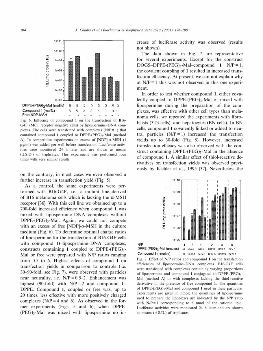
receptor [36]. With this cell line we obtained up to a
700-fold increased e¤ciency when compound 1 was
mixed with lipospermine^DNA complexes without
DPPE-(PEG)3-Mal. Again, we could not compete
with an excess of free [NDP]-K-MSH in the culture
medium (Fig. 6). To determine optimal charge ratios
of lipospermine for the transfection of B16-G4F cells
with compound 1/^lipospermine^DNA complexes,
constructs containing 1 coupled to DPPE-(PEG)3-
Mal or free were prepared with N/P ratios ranging
from 0.5 to 6. Highest e¡ects of compound 1 on
transfection yields in comparison to controls (i.e.
30^90-fold, see Fig. 7), were observed with particles
near neutrality, i.e. N/P= 0.5^2. Enhancement was
highest (90-fold) with N/P= 2 and compound 1^
DPPE. Compound 1, coupled or free was, up to
20 times, less e¡ective with more positively charged
complexes (N/P= 4 and 6). As observed in the for-
mer experiments (Figs. 5 and 6), when DPPE-
(PEG)3-Mal was mixed with lipospermine no in-
crease of luciferase activity was observed (results
not shown).
The data shown in Fig. 7 are representative
for several experiments. Except for the construct
DOGS^DPPE-(PEG)3-Mal^compound 1 N/P=1,
the covalent coupling of 1 resulted in increased trans-
fection e¤ciency. At present, we can not explain why
at N/P= 1 this was not observed in this one experi-
ment.
In order to test whether compound 1, either cova-
lently coupled to DPPE-(PEG)3-Mal or mixed with
lipospermine during the preparation of the com-
plexes, was e¡ective with other cell types than mela-
noma cells, we repeated the experiments with ¢bro-
blasts (3T3 cells), and hepatocytes (BN cells). In BN
cells, compound 1 covalently linked or added to neu-
tral particles (N/P= 1) increased the transfection
yields up to 30-fold (Fig. 8). However, increased
transfection e¤cacy was also observed with the con-
struct containing DPPE-(PEG)3-Mal in the absence
of compound 1. A similar e¡ect of thiol-reactive de-
rivatives on transfection yields was observed previ-
ously by Kichler et al., 1995 [37]. Nevertheless the
Fig. 6. In£uence of compound 1 on the transfection of B16-
G4F (MC1 receptor negative cells) by lipospermine^DNA com-
plexes. The cells were transfected with complexes (N/P= 1) that
contained compound 1 coupled to DPPE-(PEG)3-Mal (method
A). In competition experiments an excess of [NDP]-K-MSH (1
Wg/ml) was added per well before transfection. Luciferase activ-
ities were monitored 24 h later and are shown as means
( þ S.D.) of triplicates. This experiment was performed four
times with very similar results.
Fig. 7. E¡ect of N/P ratios and compound 1 on the transfection
e¤ciencies of lipospermine^DNA complexes. B16-G4F cells
were transfected with complexes containing varying proportions
of lipospermine and compound 1 conjugated to DPPE-(PEG)3-
Mal (method A) or with complexes lacking the thiol-reactive
derivative in the presence of free compound 1. The quantities
of DPPE-(PEG)3-Mal and compound 1 used in these particular
experiments are given in nmol; the quantities of lipospermine
used to prepare the lipoplexes are indicated by the N/P ratio
with N/P= 1 corresponding to 6 nmol of the cationic lipid.
Luciferase activities were monitored 24 h later and are shown
as means ( þ S.D.) of triplicates.
J. Chluba et al. / Biochimica et Biophysica Acta 1510 (2001) 198^208204

fact that constructs that did not contain the malei-
mide group were also potent in the presence of com-
pound 1 (Fig. 8, last bar) indicates that this peptide
is active per se in the transfection process.
In contrast, with 3T3 cells we could not observe
any signi¢cant increase in transfection triggered by
compound 1 (Fig. 8).
4. Discussion
It is now well established that mouse and human
melanoma cells possess speci¢c high-a¤nity recep-
tors for K-MSH [20,21,38,39]. For that reason this
peptide was utilized for the development of diagnos-
tic tools of melanoma but also for therapeutic pur-
poses; thus, K-MSH derivatives have been success-
fully used to visualize melanoma metastases in
patients and malignant melanomas have been specif-
ically targeted with MSH^toxin fusion proteins [40].
For targeting purposes K-MSH has many potential
advantages: it presents a high a¤nity for its recep-
tors, analogues have been synthesized which are
highly stable in vivo and which have also excellent
a¤nities for example for the MC1 receptor [19,21],
because if their small size these peptides should be
poorly immunogenic. Finally these analogues can be
easily modi¢ed, e.g. at their N-terminus, without
losing a¤nity for the receptor. For these reasons
K-MSH, and in particular its superpotent analogue
[NDP]-K-MSH(4^10), seemed to us an attractive tool
for targeting melanoma cells with the aim of gene
delivery.
In many cases, a condition to observe targeted
gene transfer is to associate speci¢c ligands to trans-
fection particles that present by themselves a limited
interaction with the target cells and thus generally
achieve only low transfection levels. In our prelimi-
nary assays to determine optimal conditions for tar-
geting gene transfer to melanoma cells with com-
pound 1 we have observed that B16-F1 cells are
easily transfectable with lipoplexes. In general with
other cell lines tested with cationic lipid-based gene
transfer complexes, e.g. with lipospermine[30], opti-
mal transfection is observed with highly positive par-
ticles (i.e. N/Pv 6). This positive charge is thought to
promote the binding of the complexes to the nega-
tively charged surface of cells and their uptake via
adsorptive endocytosis. In contrast, B16-F1 melano-
ma cells are optimally transfected with lesser posi-
tively particles (N/P ratio between 2 and 4) and are
transfectable, albeit in lower yield, with neutral or
even net negatively charged complexes obtained
with lipospermine under standard conditions. This
could be explained tentatively by the fact that mela-
noma cells overexpress negatively charged proteogly-
cans [41]. In agreement with this proposal we have
shown that inhibition of the sulfation of its surface
proteoglycans decreased drastically the transfection
e¤ciency. This result, however, does not explain
why cells are still transfected with complexes pre-
Fig. 8. E¡ect of compound 1 on the transfection e¤ciency of
BN and 3T3 cells by lipospermine^DNA complexes. The cells
were transfected with complexes (N/P= 1) containing increasing
proportions (0^5 mol%) of compound 1 conjugated to DPPE-
(PEG)3-Mal (method A). In competition experiments an excess
of [NDP]-K-MSH (1 Wg/ml) was added per well before transfec-
tion. Luciferase activities were monitored 24 h later and are
shown as means ( þ S.D.) of triplicates.
J. Chluba et al. / Biochimica et Biophysica Acta 1510 (2001) 198^208 205

pared with a global negative charge. Related to these
results it should be noted that relatively e¤cient
transfection with particles that carry a net negative
charge have also been observed by others [42,43]. In
line with our aim, we could however decrease back-
ground transfection by preparing the complexes in
HBS and the lowest levels were reached with neutral
particles.
In our transfection assays we have shown that a
superactive analogue of K-MSH used with liposper-
mine^DNA complexes increased strongly the trans-
fection of melanoma cells. Enhancement was only
observed when coupling was performed using meth-
od A. As the coupling methods described by Remy et
al. [5], even decreased transfection e¤ciencies of B16-
F1 cells (not shown) we changed the protocol and
included a drying step after coupling and 2-mercap-
toethanol treatment which eliminated ethanol and
2-mercaptoethanol (see Section 2.3). We suppose
that particles prepared according method B inhibit
transfection of melanoma cells because 2-mercapto-
ethanol remains in the transfection solution.
With these complexes, we could however not dem-
onstrate any cell speci¢c targeting, as we could not
compete targeting with an excess of free [NDP]-K-
MSH. In addition B16-G4F cells which lack MC1
receptors on their surface are even better transfected
than B16-F1 cells. With these cells transfection is
improved up to 700-fold by the presence of the li-
gand 1.
Thus, even if there is some cell speci¢c targeting,
the unspeci¢c enhancement due to compound 1 is so
high that it would mask it. This seems likely, consid-
ering the relatively low number of MSH receptors
expressed on the cell surface in comparison to other
receptors which have been used successfully for tar-
geting gene delivery constructs, such as for example
folate receptors (V2.5U105 sites/cell) [24]. As com-
pound 1 was also found to have an e¡ect on trans-
fection yield when it is just mixed with lipospermine
when making the complexes with the plasmid, we
suppose that the free ligand could interact with the
constructs most probably via hydrophobic interac-
tions (see below).
Using the same lipospermine^DNA lipoplexes, the
e¡ect of 1 on the transfection yield was also tested of
other cell types such as murine ¢broblasts and liver-
derived cells. We could observe an enhanced trans-
fection only with the liver cells. When compound 1 is
just mixed with lipospermine, transfection increases
as observed with coupled 1. We conclude from these
results that the e¡ect of compound 1 is not restricted
to cells that express the MSH receptor and therefore
does not increase the transfection yields of the lipo-
spermine-based lipoplexes by targeting this recep-
tor.
The e¡ect of the peptide [NDP]-K-MSH on the
transfection e¤ciency of lipospermine^DNA com-
plexes is therefore more reminiscent to the one de-
scribed for viral fusogenic peptides when associated
to polyplexes/lipoplexes [44]. These membrane-active
peptides, which act by destabilizing the endosomal
membranes, also lack cell speci¢city and for example
INF6, a peptide deriving from in£uenza virus, which
is one the most e¡ective of these agents, was found
to increase transfection up to 1000-fold with 1.5 and
2 charge eq. particles [11]. In contrast to compound
1, no enhancement was observed with neutral par-
ticles. Under optimal transfection conditions, i.e.
with the most positively charged particles, only slight
enhancement could be obtained with compound 1
and this feature was also observed with viral fuso-
genic peptides [11]. But in contrast to fusogenic pep-
tides compound 1 strongly increases transfection
with particles which are nearly neutral, N/P between
0.5 and 2.
The mechanism for the membrane-destabilizing ac-
tion of compound 1 remains to be fully elucidated.
But it seems that it might be di¡erent from the
known fusogenic peptides: among the peptides that
Kichler et al. have tested [11], only the acidic (neg-
atively charged) ones gave good results with liposper-
mine. In contrast to these viral peptides, compound 1
has a net positive charge at neutral and acidic pH.
There are, however, some hints on a possible mem-
brane activity of K-MSH and [NDP]-K-MSH. The
group of Schwyzer has shown some time ago that
K-MSH and other peptide hormones that bind to
G-protein-coupled receptors interact directly with
lipid bilayers [45,46]. More recent biophysical studies
by Biaggi et al. [47,48] and Macedo et al. [49] have
demonstrated that indeed K-MSH penetrates lipid
bilayers. Our own studies in this area demonstrate
that the analogues of K-MSH are endowed with hith-
erto unrecognized, and unexpected, membrane-active
properties such as the promotion of membrane fu-
J. Chluba et al. / Biochimica et Biophysica Acta 1510 (2001) 198^208206

sion (D. Lima de Souza et al., to be published) which
could well explain our present data.
Taken together, compound 1 does not seem to be
a useful peptide for the targeting of cells such as
melanomas. This is probably due to the relatively
low expression of MC1 receptors on the surface of
these cells. However, 1 seems to be a new kind of
membrane-active peptide that could be used under
neutral transfection conditions, in particular for mel-
anoma cells. The other advantages, as low immuno-
genicity, resistance to degradation are good prereq-
uisites for in vivo gene transfer.
Acknowledgements
This work was supported by Grants from the
Association pour la Recherche Contre le Cancer,
the Ligue Contre le Cancer and the European Com-
munity (BIO4-97-2191). J.C. was also a recipient of a
research fellowship from the Deutsche Forschungsge-
meinschaft. We would like to thank A.M. Zanta and
O. Boussif for helpful discussions, J.-P. Behr for the
kind gift of lipospermine and A.N. Eberle for mela-
noma cells.
References
[1] J.-P. Behr, B. Demeneix, J.P. Loe¥er, J. Perez-Mutul, E¤-
cient gene transfer into mammalian primary endocrine cells
with lipopolyamine-coated DNA, Proc. Natl. Acad. Sci.
USA 86 (1989) 6982^6986.
[2] P.L. Felgner, T.R. Gadek, M. Holm, R. Roman, H.W.
Chan, M. Wenz, J.P. Northrop, G.M. Ringold, M. Daniel-
sen, Lipofection: a highly e¤cient, lipid-mediated DNA-
transfection procedure, Proc. Natl. Acad. Sci. USA 84
(1987) 7413^7417.
[3] R. Leventis, J.R. Silvius, Interaction of mammalian cells
with lipid dispersion containing novel metabolizable cationic
amphophiles, Biochim. Biophys. Acta 1023 (1990) 124^132.
[4] X. Gao, L. Huang, A novel cationic liposome reagent for
e¤cient transfection of mammalian cells, Biochem. Biophys.
Res. Commun. 179 (1991) 280^285.
[5] J.S. Remy, A. Kichler, V. Mordinov, F. Schuber, J.-P. Behr,
Targeted gene transfer into hepatoma cells with lipopoly-
amine-condensed DNA particles bearing multiantennary gal-
actosyl ligands: a stage towards arti¢cial viruses, Proc. Natl.
Acad. Sci. USA 92 (1995) 1744^1748.
[6] J.H. Felgner, R. Kumar, C.N. Sridhar, C.J. Wheeler, Y.J.
Tsai, R. Border, P. Ramsay, M. Martin, P.L. Femgner, En-
hanced gene delivery and mechanism studies with a novel
series of cationic lipid formulations, J. Biol. Chem. 269
(1994) 2550^2561.
[7] A.E. Thierry, P. Rabinovich, B. Peng, L.C. Mahan, J.L.
Bryant, R.C. Gallo, Characterization of liposome-mediated
gene delivery expression, stability and pharmacokinetics of
plasmid DNA, Gene Ther. 4 (1997) 226^237.
[8] A. Kichler, W. Zauner, M. Ogris, E. Wagner, In£uence of
the DNA complexation medium on the transfection e¤-
ciency of lipospermine^DNA particles, Gene Ther. 5 (1998)
855^860.
[9] C. Plank, B. Oberhauser, K. Mechtler, C. Koch, E.J. Wag-
ner, The in£uence of endosome-disruptive peptides on gene
transfer using synthetic virus-like gene transfer systems,
J. Biol. Chem. 269 (1994) 12918^12924.
[10] K. Mechtler, E. Wagner, Gene transfer mediated by in£uen-
za virus peptides: the role of the peptide sequences, New J.
Chem. 21 (1997) 105^111.
[11] A. Kichler, K. Mechtler, J.-P. Behr, E. Wagner, In£uence
of membrane-active peptides on lipospermine^DNA com-
plex-mediated gene transfer, Bioconj. Chem. 8 (1997) 213^
221.
[12] F. Schuber, A. Kichler, D. Lima de Souza, B. Frisch, Li-
gand-mediated gene delivery, in: G. Gregoriadis and B. Mc-
Cormack, Targeting of Drugs: Strategies for Gene Con-
structs and Delivery, NATO ASI Series, Life Sciences 323
(2000) 210^225.
[13] M.P. Deonarain, Ligand-targeted receptor-mediated vectors
for gene delivery, Exp. Opin. Ther. Patents 8 (1998) 53^69.
[14] D.S. Carpenter, R.F. Minchin, Targeting of a cholecystoki-
nin^DNA complex to pancreatic cells in vitro and in vivo,
Gene Ther. 5 (1998) 848^854.
[15] P. Erbacher, J.-S. Remy, J.-P. Behr, Gene transfer with syn-
thetic virus-like particles via the integrin-mediated endocyto-
sis pathway, Gene Ther. 6 (1999) 138^145.
[16] J.M. Elwood, H.K. Koh, Etiology, epidemiology, risk fac-
tors, and public health issues of melanoma, Curr. Opin. On-
col. 6 (1994) 179^187.
[17] G.J. Nabel, D. Gordon, D.K. Bishop, B.J. Nickolo¡, Z.-Y.
Yang, A. Aruga, M. Cameron, E. Nabel, A.E. Chang, Proc.
Natl. Acad. Sci. USA 93 (1996) 15388^15393.
[18] P. Jantsche¡, R. Herrmann, C. Rochlitz, Cancer gene and
immunotherapy: recent developments, Med. Oncol. 16
(1999) 78^85.
[19] C. Bagutti, B. Stolz, R. Albert, C. Bruns, J. Pless, A.N.
Eberle, 111In-DTPA-labelled analogues of K-melanocyte-
stimulating hormone for melanoma targeting receptor bind-
ing in vitro and in vivo, Int. J. Cancer 58 (1994) 749^755.
[20] A.N. Eberle, in: The Melanotropins: Chemistry, Physiology
and Mechanisms of Action, S. Karger, Basel, 1988.
[21] W. Siegrist, F. Solca, S. Stutz, L. Giu¡re, S. Carrel, J. Gir-
ard, A.N. Eberle, Characterization of receptors for K-mela-
nocyte-stimulating hormone on human melanoma cells, Can-
cer Res. 49 (1989) 6352^6358.
[22] D.R. Bard, C.G. Knight, D.P. Page-Thomas, Targeting of a
chelating derivative of a short-chain analogue of K-melano-
J. Chluba et al. / Biochimica et Biophysica Acta 1510 (2001) 198^208 207

cyte-stimulating hormone to Cloudman S91 melanomas,
Biochem. Soc. Trans. 18 (1990) 882^883.
[23] D.R. Bard, C.G. Knight, D.P. Page-Thomas, E.P. Wraight,
T.S. Maugham, The use of derivatives of K-MSH for target-
ing of melanoma in vivo, in: G. Gregordiadis, G. Poste,
A.T. Florenz (Eds.), Targeting of Drugs: The Challenge of
Peptides and Proteins, Plenum, New York, 1992.
[24] S. Sharma, M. Granberry, J. Jiang, S. Leong, M. Hadley, V.
Hruby, Multivalent melanotropic peptide and £uorescent
macromolecule conjugates: new reagents for characteriza-
tion of melanotropin receptors, Bioconj. Chem. 5 (1994)
591^601.
[25] W. Wong, R.F. Minchin, Binding and internalization of
melanocyte stimulating hormone receptor ligand [Nle4,
D-Phe7] K-MSH in B16 melanoma cells, Int. J. Biochem.
Cell. Biol. 28 (1996) 1223^1232.
[26] H.C. Loughrey, L.S. Choi, P.R. Cullis, M.B. Bally, Opti-
mized procedures for the coupling of proteins to liposomes,
J. Immunol. Methods 132 (1990) 25^35.
[27] B. Frisch, C. Boeckler, F. Schuber, Synthesis of short poly-
oxyethylene-based heterobifunctional cross-linking reagents.
Application to the coupling of peptides to liposomes, Bio-
conj. Chem. 7 (1996) 180^186.
[28] R.J. Lee, L.J. Huang, Folate-targeted, anionic liposome-en-
trapped poly-lysine-condensed DNA for tumor cell-speci¢c
gene transfer, J. Biol. Chem. 271 (1996) 8481^8487.
[29] D.V. Scha¡er, D.A. Lau¡enburger, Optimization of cell sur-
face binding enhances e¤ciency and speci¢city of molecular
conjugate gene delivery, J. Biol. Chem. 273 (1998) 28004^
28009.
[30] O. Boussif, M.A. Zanta, J.-P. Behr, Optimized galenics im-
prove in vitro gene transfer with cationic molecules up to
1000-fold, Gene Ther. 3 (1996) 1074^1080.
[31] K.A. Mislik, J. Baldeschwieler, Evidence for the role of pro-
teoglycans in cation-mediated gene transfer, Proc. Natl.
Acad. Sci. USA 93 (1996) 12349^12354.
[32] L.C. Mounkes, W. Zhong, G. Cipres-Palacin, T. Heath, R.J.
Debs, Proteoglycans mediate cationic liposome^DNA com-
plex-based gene delivery in vitro and in vivo, J. Biol. Chem.
273 (1998) 26164^26170.
[33] Y. Xu, F.C. Szoka, Mecanism of DNA release from cationic
liposome^DNA complexes used in cell transfection, Bio-
chemistry 35 (1996) 5616^5623.
[34] O. Zelphati, F.C. Szoka, Mechanism of oligonucleotide re-
lease from cationic liposomes, Proc. Natl. Acad. Sci. USA 93
(1996) 11493^11498.
[35] H. Du, P. Chandaroy, S.W. Hui, Grafted poly-(ethylene
glycol) on lipid surfaces inhibits protein adsorption and
cell adhesion, Biochim. Biophys. Acta 1326 (1997) 236^248.
[36] F.F. Solca, J. Chluba-de Tapia, K. Iwata, A.N Eberle, B16-
G4F mouse melanoma cells : an MSH receptor de¢cient cell
clone, FEBS Lett. 322 (1993) 177^180.
[37] A. Kichler, J.S. Remy, O. Boussif, B. Frisch, C. Boeckler,
J.P. Behr, F. Schuber, E¤cient gene delivery with neutral
complexes of lipospermine and thiol-reactive phospholipids,
Biochem. Biophys. Res. Commun. 209 (1995) 444^450.
[38] G.E. Ghanem, G. Comunale, A. Liebert, G.A. Verkcammen,
F.J. Lejeune, Evidence for a-melanocyte stimulating hor-
mone (K-MSH) receptors on human malignant melanoma
cells, Int. J. Cancer 41 (1988) 248^255.
[39] J.B. Tatro, M. Atkins, J.W. Mier, S. Hardason, H. Wolfe, T.
Smith, M.L. Entwistle, S.J. Reichlin, Melanotropin receptors
demonstrated in situ in human melanoma, Clin. Invest. 85
(1990) 1825^1832.
[40] J.B. Tatro, Z. Wen, M.L. Entwistle, M.B. Atkins, T.J.
Smith, S. Reichlin, J.R. Murphy, Interaction of an K-mela-
nocyte stimulating hormone^diphtheria toxin fusion protein
with melanotropin receptors, Cancer Res. 52 (1992) 2545^
2548.
[41] J. Timar, A. Ladanyi, K. Lapis, M. Moczar, Di¡erential
expression of proteoglycans on the surface of human mela-
noma cells characterized by altered experimental metastatic
potential, Am. J. Pathol. 141 (1992) 467^474.
[42] E.R. Lee, J. Marshall, C.S. Siegel, C. Jiang, N.S. Yew, M.R.
Nichols, J.B. Nietupski, R.J. Ziegler, M.B. Lane, K.X.
Wang, N.C. Wan, R.K. Scheule, D.J. Harris, A.E. Smith,
S.H. Cheng, Detailed analysis of structures and formulations
of cationic lipids for e¤cient gene transfer to the lung, Hum.
Gene Ther. 14 (1996) 1701^1717.
[43] A.J. Fasbender, J. Zabner, M.J. Welsh, Optimization of cat-
ionic lipid-mediated gene transfer to airway epithelia, Am. J.
Physiol. 269 (1995) L45^L51.
[44] E. Wagner, E¡ects of membrane-active agents in gene deliv-
ery, J. Control. Release 53 (1998) 155^158.
[45] H.U. Gremlich, U.P. Fringeli, R. Schwyzer, Conformational
changes of adrenocorticotropin peptides upon interaction
with lipid membranes revealed by infrared attenuated total
re£ection spectroscopy, Biochemistry 22 (1983) 4257^4264.
[46] D.F. Sargent, R. Schwyzer, Membrane lipid phase as cata-
lyst for peptide receptor interactions, Proc. Natl. Acad. Sci.
USA 83 (1986) 5774^5778.
[47] M.H. Biaggi, T.J.T. Pinheiro, A. Watts, M.T. Lamy-Freund,
Spin label and 2H-NMR studies on the interaction of mela-
notropic peptides with lipid bilayers, Eur. Biophys. J. 24
(1996) 251^259.
[48] M.H. Biaggi, K.A. Riske, M.T. Lamy-Freund, Melanotropic
peptides lipid bilayer interaction. Comparison of the hor-
mone K-MSH to a biologically more potent analog, Biophys.
Chem. 67 (1997) 139^149.
[49] Z.S. Macedo, T.A. Furquim, A.S. Ito, Estimation of average
depth of penetration of melanotropins in dimyristoylphos-
phatidylglycerol vesicles, Biophys. Chem. 59 (1996) 193^202.
J. Chluba et al. / Biochimica et Biophysica Acta 1510 (2001) 198^208208


