Embed Size (px)
Citation preview

Enteric Glia Cells Attenuate Cytomix-Induced IntestinalEpithelial Barrier BreakdownGerald A. Cheadle, Todd W. Costantini, Nicole Lopez, Vishal Bansal, Brian P. Eliceiri, Raul Coimbra*
Division of Trauma, Surgical Critical Care, and Burns, Department of Surgery, University of California San Diego Health Sciences, San Diego, California, UnitedStates of America
Abstract
Background: Intestinal barrier failure may lead to systemic inflammation and distant organ injury in patients followingsevere injury. Enteric glia cells (EGCs) have been shown to play an important role in maintaining gut barrier integritythrough secretion of S-Nitrosoglutathione (GSNO). We have recently shown than Vagal Nerve Stimulation (VNS)increases EGC activation, which was associated with improved gut barrier integrity. Thus, we sought to further studythe mechanism by which EGCs prevent intestinal barrier breakdown utilizing an in vitro model. We postulated thatEGCs, through the secretion of GSNO, would improve intestinal barrier function through improved expression andlocalization of intestinal tight junction proteins.Methods: Epithelial cells were co-cultured with EGCs or incubated with GSNO and exposed to Cytomix (TNF-α, INF-γ, IL-1β) for 24 hours. Barrier function was assessed by permeability to 4kDa FITC-Dextran. Changes in tight junctionproteins ZO-1, occludin, and phospho-MLC (P-MLC) were assessed by immunohistochemistry and immunoblot.Key Results: Co-culture of Cytomix-stimulated epithelial monolayers with EGCs prevented increases in permeabilityand improved expression and localization of occludin, ZO-1, and P-MLC. Further, treatment of epithelial monolayerswith GSNO also prevented Cytomix-induced increases in permeability and exhibited a similar improvement inexpression and localization of occludin, ZO-1, and P-MLC.Conclusions & Inferences: The addition of EGCs, or their secreted mediator GSNO, prevents epithelial barrierfailure after injury and improved expression of tight junction proteins. Thus, therapies that increase EGC activation,such as VNS, may be a novel strategy to limit barrier failure in patients following severe injury.
Citation: Cheadle GA, Costantini TW, Lopez N, Bansal V, Eliceiri BP, et al. (2013) Enteric Glia Cells Attenuate Cytomix-Induced Intestinal Epithelial BarrierBreakdown. PLoS ONE 8(7): e69042. doi:10.1371/journal.pone.0069042
Editor: Stefan Bereswill, Charité-University Medicine Berlin, Germany
Received May 01, 2013; Accepted June 04, 2013; Published July 1, 2013
Copyright: © 2013 Cheadle et al. This is an open-access article distributed under the terms of the Creative Commons Attribution License, which permitsunrestricted use, distribution, and reproduction in any medium, provided the original author and source are credited.
Funding: This work was supported by the American Association for the Surgery of Trauma (AAST) Research and Education Foundation Scholarship(TWC). The funders had no role in study design, data collection and analysis, decision to publish, or preparation of the manuscript.
Competing interests: The authors have declared that no competing interests exist.
* E-mail: [email protected]
Introduction
The intestinal barrier is comprised of epithelial cells that arelinked together by tight junctions, which are comprised of theproteins zonula occludens-1 (ZO-1) and occludin, which formthe tight junction barrier in the paracellular space [1].Connected to these proteins is the actin cytoskeletal ring andmyosin light chain (MLC), which help to further stabilize thetight junction [2,3]. Together, these proteins help to maintainthe gut barrier against the external environment of the intestinallumen. Breakdown of the intestinal tight junction results inincreased intestinal permeability and gut inflammation. Thiscan lead to the spread of gut-derived inflammatory mediators tothe mesenteric lymph, contributing to the development ofsystemic inflammation [4,5]. On a cellular level, gut barrierbreakdown is associated with decreased expression andaltered localization of the tight junction proteins occludin [6] and
ZO-1 [7]. Additionally, phosphorylation of MLC causes actincytoskeletal contraction, increasing tight junction breakdown[2,3,8]. Severe injury models [9,10] and chronic diseases ofintestinal inflammation [11,12] have been associated withincreases in intestinal permeability resulting from tight junctiondisruption.
Because gut barrier breakdown is associated with systemicinflammation, we postulate that therapies designed to preventintestinal inflammation may have clinical utility in the treatmentof medical conditions associated with gut barrier failure. Recentevidence suggests Vagus nerve stimulation (VNS) maymodulate intestinal barrier integrity [13], through improvedexpression of occludin [6] and ZO-1 [14] and a reduction inphosphorylated MLC (P-MLC) [15]. However, the signalingmechanism that links the central nervous system and theintestinal epithelium remains elusive.
PLOS ONE | www.plosone.org 1 July 2013 | Volume 8 | Issue 7 | e69042

One potential signaling pathway may lie in the well-established connections between the Vagus nerve and theEnteric Nervous System (ENS). The largest cell population inthe ENS are the Enteric Glia cells (EGCs), which are identifiedby the unique marker Glial Fibrillary Acidic Protein (GFAP) [16].Recently, it has been demonstrated that EGCs actively receiveand propagate signals, both to and from nearby entericneurons and the intestinal epithelium [16–18]. Additionally,VNS was shown to increase intestinal GFAP expression, acommon marker of EGC activation, suggesting a connectionbetween the Vagus nerve and EGCs [15]. Thus, EGCs may bean ideal candidate cell type to transmit the anti-inflammatoryeffects of VNS to the gut epithelium.
The potential for EGCs to mediate VNS-induced gutprotection are supported by studies showing that EGCs play aprominent role in maintaining proper intestinal epithelial barrierintegrity. Genetic ablation of EGCs results in intestinal barrierfailure and is associated with increased intestinal inflammation[18,19]. Subsequent studies have shown that the barrier-inducing effects of EGCs are mediated through the secretion ofS-nitrosoglutathione (GSNO), which improves expression andlocalization of tight junction proteins [19].
Thus, with evidence suggesting that EGCs may link VNS tothe intestinal epithelium, this project seeks to further investigatethe effects of EGCs and GSNO on epithelial barrier functionusing an in vitro co-culture model with EGCs and well-established epithelial cell lines. We hypothesized that theaddition of EGCs, or their secreted mediator GSNO, wouldprevent epithelial barrier failure after exposure to aninflammatory stimulus through improved expression of tightjunction proteins.
Materials and Methods
Cell linesCaco-2 human intestinal epithelial cells, EGCs, and Madin
Darby Canine Kidney (MDCK) epithelial cells were obtainedfrom American Type Culture Collection (ATCC, Manasas, VA,USA). All three cell lines were grown at 37°C in a 5% CO2
humidified atmosphere. Cells were grown in Dulbecco’sModified Eagle Medium (DMEM) with high glucose (Gibco,Carlsbad, CA, USA) supplemented with 10% FBS (Gibco),penicillin G (10,000 U mL-1, Gibco), and streptomycin (10,000µg mL-1, Gibco). MDCKs were supplemented with L-Glutamine(2 mM, Gibco), Sodium Pyruvate (1 mM, Gibco), and 1%nonessential amino acids (Gibco).
Co-culture Model and ImmunostimulationCaco-2 cells (80,000 cells well-1) or MDCK cells (30,000 cells
well-1) were seeded onto permeable filters with 0.4 µm poresize in 12-well Transwell bicameral chambers (Corning Inc.,Corning, New York, USA). EGCs (30,000 cells well-1) wereseeded onto the basal well of the 12-well Transwell dish. Mediawas changed daily throughout the co-culture process. After theincubation period, cells were subject to permeability assays,immunoblot, or immunofluorescence protocols.
For the Caco-2 co-culture, cells were seeded on day 0 andallowed to grow for two days alone in DMEM. EGCs were
plated on day 1 and allowed to grow for one day alone inDMEM. Cells were then co-cultured on day 2, and growntogether for another 3 days. On day 3, Caco-2 cells wereincubated in Enterocyte Differentiation media (BD Bioscience,Bedford, MO, USA) to help induce Caco-2 differentiation. Onday 4, cells were placed in serum-free media and incubatedwith either PBS or Cytomix (TNF-α (10 ng mL-1; Sigma), IFN-γ(10 ng mL-1; Pierce, Rockford, IL, USA), and IL-1β (10 ng mL-1;Sigma)) for 24 hours at 37°C in a 5% CO2 humidifiedatmosphere. For GSNO studies (50 µM, Sigma), incubationbegan in selected Caco-2 samples on the same day as co-culture and was added to the basal well. GSNO wasreplenished daily with media changes in the same time frameas other Caco-2 samples were co-cultured with EGCs. Toblock the effects of GSNO, the nitric oxide synthase inhibitorNG-nitro-L-arginine methylester (100µmol/L) was incubatedwith EGCs prior to assessing permeability.
For the MDCK co-culture, MDCK cells were seeded on day 0and allowed to grow for two days alone in DMEM. EGCs wereplated on day 1 and allowed to grow for one day alone inDMEM. Cells were then co-cultured on day 2 or incubated withGSNO as described and allowed to grow together for two days.On day 4, cells were placed in serum-free media and incubatedwith either PBS or Cytomix for 24 hours at 37°C in a 5% CO2
humidified atmosphere.
Permeability AssayTo assess monolayer barrier function, an in vitro permeability
assay was performed. After the 24 hour Cytomix incubationperiod, 200 µL of 4kDa FITC-Dextran (10 mg mL-1; Sigma) inPBS was added to the Transwell insert, or the apical side of themonolayer. After 4 hours of incubation, 100 µL aliquots ofDMEM were obtained from the basal chamber. Fluorescencewas measured in a fluorescence spectrometer (SpectraMax,Molecular Devices, Sunnyvale, CA, USA) and compared with astandard curve of known FITC-Dextran concentrations dilutedin PBS and DMEM.
ImmunofluorescenceAfter the co-culture procedure, media was removed from the
Transwells and fixed in 3.7% paraformaldehyde (ElectronMicroscopy Sciences, Hatfield, PA, USA) diluted in PBS. Cellswere fixed for 15 minutes. Cells were then washed with PBSfor 2 minutes. After washing, the cells were blocked in a 3%BSA solution for 1 hour. Cells were washed and incubatedovernight in primary antibodies against ZO-1 (1:500, Invitrogen,Camarillo, CA, USA), occludin (1:500, Invitrogen), andPhosphorylated Myosin Light Chain (P-MLC; 1:500, Santa CruzBiotechnology, Santa Cruz, CA, USA) in 1% BSA solution.After washing, cells were incubated in secondary antibody,Alexa Fluor 488 (1:1000, Invitrogen), and 4,6-diamidino-2-phenylindole (DAPI; 1:200, Sigma) in 1% BSA for one hour.Cells were washed, followed by removal of the Transwellmembranes, containing the cells, from the inserts. Themembranes were immersed in SlowFade Gold (Invitrogen),placed on glass slides, and covered with glass cover slips.Slides were allowed to cure overnight in the dark. Images werethen viewed with an Olympus Fluoview laser scanning confocal
Enteric Glia Protect the Intestinal Barrier
PLOS ONE | www.plosone.org 2 July 2013 | Volume 8 | Issue 7 | e69042

microscope (Olympus, Melville, NY, USA) at 60x magnificationwith exposure-matched settings (Advanced Software V 1.6,Olympus, Center Valley, PA, USA).
ImmunoblotAt the end of the culture procedure, cells were washed with
PBS and placed in 0.25% Trypsin-EDTA (Gibco). Once cellswere in suspension, they were removed from the Transwells,put into Eppendorf tubes, and placed in a centrifuge at 14,000rpm for 5 minutes. Supernatant was removed and the tubesplaced on ice. 250 µL of 4% Sodium dodecyl sulfate (SDS) inPBS lysis buffer and 5 µL of Protease Inhibitor (Pierce) wereadded to the tubes, followed by sonication to lyse the cellularpellet. Total protein samples were placed at -80°C for long-termstorage.
Total protein concentration of the lysed samples wasdetermined by the bicinchoninic acid (BCA) protein assay usingthe microplate procedure (Pierce). Samples containing 10 µg ofprotein were placed into SDS sample buffer and boiled for 5minutes. Proteins were separated by SDS-Polyacrylamide gelelectrophoresis using 8-16% tris-glycine polyacrylamidegradient gel and transferred to nitrocellulose membranes(Invitrogen). Membranes were washed with tris-buffered saline/Tween 20 (TBST) and then blocked with 5% BSA in TBST for 1hour. Membranes were then incubated in primary antibodiesspecific for occludin (1:500, Santa Cruz Biotechnology) or BetaActin (1:500, Cell Signaling, Danvers, MA, USA) overnight at4°C in 5% BSA in TBST. Membranes were washed and thenincubated with horseradish peroxidase-linked anti-rabbit IgG(1:2000, Cell Signaling) in 5% BSA in TBST for 1 hour at roomtemperature. After washing, the Pierce Supersignal West PicoChemiluminescent Kit was applied to the membranes forantibody detection through the Xenogen IVIS Lumina (CaliperLife Science, Hopkinton, MA, USA) imaging system. Meanpixel density was determined using the UN-SCAN-IT GelDigitizing software (Silk Scientific, Orem, UT, USA). Banddensities were compared to the Beta Actin band densities ineach lane as a loading control. Data is expressed as therelative band density compared to control for each experiment.
Statistical AnalysisAll values are expressed as mean ± standard error of the
mean (SEM). Statistical significance was determined usinganalysis of variance (ANOVA) with Student-Newman-Keulscorrection. A p-value < 0.05 was considered statisticallysignificant.
Results
EGCs attenuate Cytomix-induced epithelial monolayerpermeability
To assess Caco-2 barrier integrity, an in vitro permeabilityassay was performed using FITC-dextran (Figure 1). Caco-2monolayers stimulated with Cytomix demonstrated an increasein paracellular permeability. Co-culturing EGCs with Caco-2epithelial monolayers prevented Cytomix-induced barrierfailure, with permeability restored to control levels. Further, ifthe stimulated epithelial cells were incubated with GSNO, the
secreted product of EGCs thought to propagate their barrier-inducing effects, there is a similar reduction in permeability.Blocking nitric oxide synthase using L-NAME abrogated thebarrier protective effects of EGCs suggesting that the barrierprotective effects of EGCs are mediated in part by GSNO.
EGCs improve localization of the tight junction proteinZO-1 in Cytomix-stimulated epithelial monolayers
After determining the effects of Cytomix, EGCs, and GSNOon epithelial permeability, we sought to determine if theseeffects were due to changes in tight junction proteinexpression. We first examined ZO-1 localization in Caco-2(Figure 2A) and MDCK (Figure 2B) monolayers using confocalmicroscopy. Staining of ZO-1 in control samples of bothepithelial cell lines demonstrated localization at areas of cellcontact, forming a smooth, continuous, outline of cellularborders. Stimulation of epithelial cell monolayers with Cytomixaltered ZO-1 localization away from the cell surface, indicatingtight junction disruption. However, co-culture of Cytomix-stimulated monolayers with EGCs restored normal ZO-1localization at areas of cell contact. Further, incubation ofstimulated-epithelial cells with the EGC-secreted product,GSNO, alone also restored normal ZO-1 cellular localization,replicating the effect of EGCs.
EGCs improve occludin expression in Cytomix-stimulated epithelial monolayers
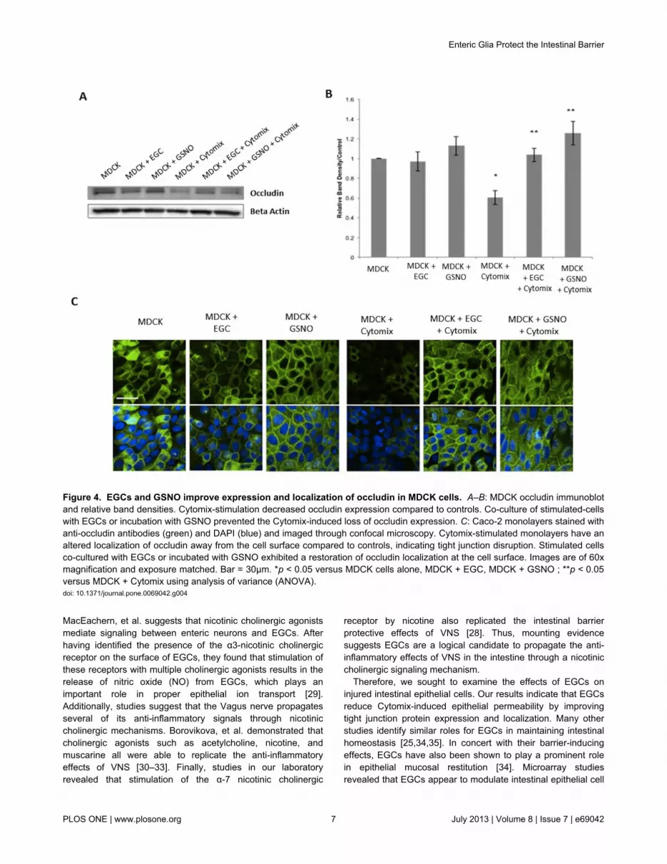
Next, we examined the tight junction protein occludin in bothepithelial cell lines by both immunoblot andimmunohistochemistry. Stimulation of monolayers with Cytomixcaused a 45% ± 6.0 and a 41% ± 7.2 decrease in occludinexpression as compared with controls in Caco-2 and MDCKcells, respectively (Figures 3A-B, 4A-B). Co-culture with EGCsprevented Cytomix-induced loss of occludin expression.Further, incubation with the secreted product, GSNO, alonealso prevented the Cytomix-induced loss of occludinexpression.
To determine the localization of occludin in the epithelialcells, confocal images were examined in both epithelial celllines (Figures 3C, 4C). Control samples showed a pattern ofproper occludin organization and localization at the cellsurface. Cytomix-stimulated epithelial monolayers showed analtered localization of occludin away from the cell surface,suggesting tight junction disruption. However, co-culture ofstimulated epithelial cells with EGCs restored occludinlocalization to the cell surface. Further, epithelial cellsincubated with GSNO alone exhibited a similar restoration ofnormal occludin localization.
EGCs decrease phosphorylated Myosin Light Chain (P-MLC) expression in Cytomix-stimulated epithelialmonolayers
It has been shown that immunostimulation of intestinalepithelial cells results in an increase in Myosin Light ChainKinase (MLCK) activity [2,3,8]. Increased expression of MLCKcauses an increase in phosphorylation of MLC, which isassociated with actin cytoskeletal contraction, tight junctiondisruption, and epithelial barrier breakdown [8]. Thus, we
Enteric Glia Protect the Intestinal Barrier
PLOS ONE | www.plosone.org 3 July 2013 | Volume 8 | Issue 7 | e69042

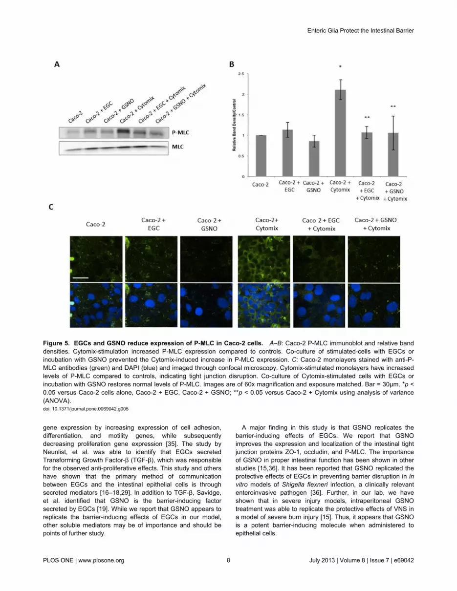
decided to examine P-MLC by both immunoblot andimmunohistochemistry in both epithelial cell lines. Stimulationof monolayers with Cytomix caused a 2-fold and a 2.5-foldincrease in P-MLC expression as compared with controls inCaco-2 and MDCK cells, respectively (Figures 5A-B, 6A-B).Co-culture with EGCs prevented Cytomix-induced increase inP-MLC expression. Further, incubation with the secretedproduct, GSNO, alone also prevented the Cytomix-inducedincrease in P-MLC expression.
To further visualize P-MLC levels, confocal images wereexamined in both epithelial cell lines. Control samplesestablished low, basal levels of P-MLC (Figures 5C, 6C), whichis expected in normal conditions. Cytomix-stimulated epithelialmonolayers had increased P-MLC levels as shown byincreased fluorescence staining, which is associated with tightjunction disruption. Epithelial cells that were co-cultured withEGCs, however, demonstrated reduced staining for P-MLC,with levels comparable to control. Further, epithelial cellsincubated with GSNO alone demonstrated a similar reductionin P-MLC levels, indicating barrier protection.
Discussion
The intestinal barrier plays a critical role in preventing gutinflammation in patients following severe injury. Breakdown ofthe gut barrier is associated with increases in systemicinflammation, distant organ injury, and death [4,6,7,14,20].Studies have shown that modulation of intestinal epithelial tightjunction proteins, through VNS, may be a potential strategy forattenuating gut barrier failure after injury [5,6,13–15]. In thisstudy, we explore the signaling pathway by which VNSmaintains gut barrier integrity after injury, specifically focusingon the role of EGCs. We first showed the importance of EGCsand one of their secreted products, GSNO, in preventingepithelial barrier breakdown in response to pro-inflammatorycytokines in an in vitro co-culture model with EGCs andepithelial cells. The importance of GSNO was further confirmedby incubating EGCs with the NOS inhibitor L-NAME to inhibitthe effects of GSNO. L-NAME prevented EGC-mediated barrierprotection in monolayers exposed to Cytomix, supporting datafrom prior studies demonstrating that blocking GSNO limits thebarrier-protective effects of EGCs [19]. EGCs and GSNO both
Figure 1. EGCs and GSNO attenuate Cytomix-induced monolayer permeability in Caco-2 cells. Caco-2 cells were grown ineither the presence or absence of EGCs or GSNO and incubated with Cytomix (TNF-α, IFN-γ, IL-1β) or PBS for 24 hours. L-NAMEwas used to block GSNO activity from EGCs. Caco-2 monolayer permeability to 4kDa FITC-Dextran was measured (n ≥ 4 samplesper group). Cytomix-stimulation results in an increase in monolayer permeability, indicating barrier dysfunction. The presence ofeither EGCs or GSNO significantly reduces permeability levels. *p < 0.05 versus the controls Alone, + EGC, + GSNO ; **p < 0.01versus + Cytomix, and +EGC +L-NAME +Cytomix.doi: 10.1371/journal.pone.0069042.g001
Enteric Glia Protect the Intestinal Barrier
PLOS ONE | www.plosone.org 4 July 2013 | Volume 8 | Issue 7 | e69042

appear to modulate their effects on intestinal epithelial cellsthrough improved expression and localization of tight junctionproteins ZO-1, occludin, and P-MLC.
This in vitro co-culture model affords us the ability tocompletely isolate intestinal epithelial cells and EGCs toexamine potential signaling mechanisms, which areresponsible for modulation of epithelial barrier function.However, the challenge arises on how to properly simulateinjury and barrier breakdown that is comparable to in vivomodels. We chose the pro-inflammatory cytokines TNF-α, IFN-γ, and IL-1β, collectively referred to as Cytomix, as they havebeen well validated in multiple studies to induce epithelialbarrier breakdown in vitro and simulate the effects ofinflammation-induced barrier failure [21–24]. Additionally, whileMDCKs are not intestinal epithelial cells, they have widelyrecognized as a good epithelial cell model for use as
corroborating evidence in intestinal barrier studies [19,25].Thus, they were used in this study primarily to corroborate thedata found in the Caco-2 cell line.
Studies have shown that gut-derived inflammatory mediatorsare carried into the mesenteric lymph following injury, whichcan contribute to the development of distant organ injury [4].Thus, breakdown of intestinal tight junction proteins, causingincreased permeability, is associated with a systemicinflammatory response and significant morbidity and mortalityin injured patients. In these experiments, we showed thatinjury-induced altered localization of the proteins ZO-1 andoccludin, as well as increases in P-MLC levels, are associatedwith increased epithelial permeability in vitro. Other studieshave shown similar correlations between intestinal permeabilityand tight junction breakdown in vivo. In a murine model oftraumatic brain injury (TBI) [20] or severe burn injury [6,7,14],
Figure 2. EGCs and GSNO improve localization of ZO-1 in Caco-2 and MDCK cells. Epithelial cells were grown in either thepresence or absence of EGCs or GSNO and incubated with Cytomix (TNF-α, IFN-γ, IL-1β) for PBS for 24 hours. A: Caco-2monolayers stained with anti-ZO-1 antibodies (green) and DAPI (blue) and imaged through confocal microscopy. Cytomix-stimulated monolayers have an altered localization of ZO-1 away from the cell surface compared to controls, indicating tight junctiondisruption. Stimulated cells co-cultured with EGCs or incubated with GSNO exhibited a restoration of ZO-1 localization at the cellsurface. B: MDCK cells stained with anti-ZO-1 antibodies (green) and DAPI (blue) and imaged through confocal microscopy.Cytomix-stimulated monolayers have an altered localization of ZO-1 indicating tight junction disruption. Stimulated cells co-culturedwith EGCs or incubated with GSNO demonstrated normal distribution of ZO-1. Images are of 60x magnification and exposurematched. Bar = 30µm.doi: 10.1371/journal.pone.0069042.g002
Enteric Glia Protect the Intestinal Barrier
PLOS ONE | www.plosone.org 5 July 2013 | Volume 8 | Issue 7 | e69042

increases in intestinal barrier failure, as evidenced byincreased paracellular permeability and histologic gut injury,were associated with decreased expression or alteredlocalization of both occludin and ZO-1.
Therapies aimed at protecting intestinal barrier integrity mayhave important clinical relevance as a means to decrease thegut barrier failure after injury. While the focus in our lab hasremained on the ability of VNS to signal to EGCs, severalstudies have shown that enteric neurons may also be involvedin maintaining intestinal epithelial barrier integrity. Studies byboth Conlin, et al. and Neunlist, et al. have revealed theimportance of the enteric neuron-secreted mediator, vasoactiveintestinal peptide (VIP), which reduces intestinal barrier
permeability and improves tight junction protein expression[26,27]. Given this data and the well-established connectionsbetween the Vagus nerve and the ENS, it is conceivable thatVNS may also be directly innervating enteric neurons tosecrete barrier-inducing mediators such as VIP. Examining theeffects of the Vagus nerve on enteric neurons are points offurther study in our laboratory to help further define theintestinal barrier-protective effects of VNS.
However, our laboratory has focused on the ability of VNS toactivate EGCs, altering the intestinal inflammatory response bymodulating local gut injury [15,28]. While this suggests aconnection between the Vagus nerve and EGCs, the signalingmechanism remains unknown. However, the study by
Figure 3. EGCs and GSNO improve expression and localization of occludin in Caco-2 cells. A–B: Caco-2 occludinimmunoblot and relative band densities. Cytomix-stimulation decreased occludin expression compared to controls. Co-culture ofstimulated-cells with EGCs or incubation with GSNO prevented the Cytomix-induced loss of occludin expression. C: Caco-2monolayers stained with anti-occludin antibodies (green) and DAPI (blue) and imaged through confocal microscopy. Cytomix-stimulated monolayers have an altered localization of occludin away from the cell surface compared to controls, indicating tightjunction disruption. Stimulated cells co-cultured with EGCs or incubated with GSNO exhibited a restoration of occludin localization atthe cell surface. Images are of 60x magnification and exposure matched. Bar = 30µm. *p < 0.05 versus Caco-2 cells alone, Caco-2+ EGC, Caco-2 + GSNO; **p < 0.05 versus Caco-2 + Cytomix using analysis of variance (ANOVA).doi: 10.1371/journal.pone.0069042.g003
Enteric Glia Protect the Intestinal Barrier
PLOS ONE | www.plosone.org 6 July 2013 | Volume 8 | Issue 7 | e69042
MacEachern, et al. suggests that nicotinic cholinergic agonistsmediate signaling between enteric neurons and EGCs. Afterhaving identified the presence of the α3-nicotinic cholinergicreceptor on the surface of EGCs, they found that stimulation ofthese receptors with multiple cholinergic agonists results in therelease of nitric oxide (NO) from EGCs, which plays animportant role in proper epithelial ion transport [29].Additionally, studies suggest that the Vagus nerve propagatesseveral of its anti-inflammatory signals through nicotiniccholinergic mechanisms. Borovikova, et al. demonstrated thatcholinergic agonists such as acetylcholine, nicotine, andmuscarine all were able to replicate the anti-inflammatoryeffects of VNS [30–33]. Finally, studies in our laboratoryrevealed that stimulation of the α-7 nicotinic cholinergic
receptor by nicotine also replicated the intestinal barrierprotective effects of VNS [28]. Thus, mounting evidencesuggests EGCs are a logical candidate to propagate the anti-inflammatory effects of VNS in the intestine through a nicotiniccholinergic signaling mechanism.
Therefore, we sought to examine the effects of EGCs oninjured intestinal epithelial cells. Our results indicate that EGCsreduce Cytomix-induced epithelial permeability by improvingtight junction protein expression and localization. Many otherstudies identify similar roles for EGCs in maintaining intestinalhomeostasis [25,34,35]. In concert with their barrier-inducingeffects, EGCs have also been shown to play a prominent rolein epithelial mucosal restitution [34]. Microarray studiesrevealed that EGCs appear to modulate intestinal epithelial cell
Figure 4. EGCs and GSNO improve expression and localization of occludin in MDCK cells. A–B: MDCK occludin immunoblotand relative band densities. Cytomix-stimulation decreased occludin expression compared to controls. Co-culture of stimulated-cellswith EGCs or incubation with GSNO prevented the Cytomix-induced loss of occludin expression. C: Caco-2 monolayers stained withanti-occludin antibodies (green) and DAPI (blue) and imaged through confocal microscopy. Cytomix-stimulated monolayers have analtered localization of occludin away from the cell surface compared to controls, indicating tight junction disruption. Stimulated cellsco-cultured with EGCs or incubated with GSNO exhibited a restoration of occludin localization at the cell surface. Images are of 60xmagnification and exposure matched. Bar = 30µm. *p < 0.05 versus MDCK cells alone, MDCK + EGC, MDCK + GSNO ; **p < 0.05versus MDCK + Cytomix using analysis of variance (ANOVA).doi: 10.1371/journal.pone.0069042.g004
Enteric Glia Protect the Intestinal Barrier
PLOS ONE | www.plosone.org 7 July 2013 | Volume 8 | Issue 7 | e69042
gene expression by increasing expression of cell adhesion,differentiation, and motility genes, while subsequentlydecreasing proliferation gene expression [35]. The study byNeunlist, et al. was able to identify that EGCs secretedTransforming Growth Factor-β (TGF-β), which was responsiblefor the observed anti-proliferative effects. This study and othershave shown that the primary method of communicationbetween EGCs and the intestinal epithelial cells is throughsecreted mediators [16–18,29]. In addition to TGF-β, Savidge,et al. identified that GSNO is the barrier-inducing factorsecreted by EGCs [19]. While we report that GSNO appears toreplicate the barrier-inducing effects of EGCs in our model,other soluble mediators may be of importance and should bepoints of further study.
A major finding in this study is that GSNO replicates thebarrier-inducing effects of EGCs. We report that GSNOimproves the expression and localization of the intestinal tightjunction proteins ZO-1, occludin, and P-MLC. The importanceof GSNO in proper intestinal function has been shown in otherstudies [15,36]. It has been reported that GSNO replicated theprotective effects of EGCs in preventing barrier disruption in invitro models of Shigella flexneri infection, a clinically relevantenteroinvasive pathogen [36]. Further, in our lab, we haveshown that in severe injury models, intraperitoneal GSNOtreatment was able to replicate the protective effects of VNS ina model of severe burn injury [15]. Thus, it appears that GSNOis a potent barrier-inducing molecule when administered toepithelial cells.
Figure 5. EGCs and GSNO reduce expression of P-MLC in Caco-2 cells. A–B: Caco-2 P-MLC immunoblot and relative banddensities. Cytomix-stimulation increased P-MLC expression compared to controls. Co-culture of stimulated-cells with EGCs orincubation with GSNO prevented the Cytomix-induced increase in P-MLC expression. C: Caco-2 monolayers stained with anti-P-MLC antibodies (green) and DAPI (blue) and imaged through confocal microscopy. Cytomix-stimulated monolayers have increasedlevels of P-MLC compared to controls, indicating tight junction disruption. Co-culture of Cytomix-stimulated cells with EGCs orincubation with GSNO restores normal levels of P-MLC. Images are of 60x magnification and exposure matched. Bar = 30µm. *p <0.05 versus Caco-2 cells alone, Caco-2 + EGC, Caco-2 + GSNO; **p < 0.05 versus Caco-2 + Cytomix using analysis of variance(ANOVA).doi: 10.1371/journal.pone.0069042.g005
Enteric Glia Protect the Intestinal Barrier
PLOS ONE | www.plosone.org 8 July 2013 | Volume 8 | Issue 7 | e69042

While this is certainly an important finding, subsequentstudies should attempt to examine the molecular signaling ofGSNO that results in improved barrier function. GSNO is apotent nitric oxide (NO) donor, which can function to S-nitrosylate proteins, a post-translational modification. Studieshave suggested that proteins can be S-nitrosylated on cysteineresidues, which can dramatically alter their function [37–39].GSNO may alter the gut inflammatory response through itsability to alter Nuclear Factor κB (NF-κB) inflammatorysignaling, where it has been shown that S-nitrosylation ofinhibitory κB kinase (IKK) by GSNO inhibits its ability tophosphorylate inhibitor of κB (IκB). Further, S-nitrosylation ofNF-κB has been shown to inhibit its ability to bind DNA toinitiate transcription of pro-inflammatory mediators [39]. Altering
NF-κB inflammatory signaling may have important effects ongut barrier failure, as binding of the NF-κB transcriptional factorin intestinal epithelial cells is associated with tight junctiondisruption [40]. In our laboratory, we observed a similarmechanism, in which burn injury results in increasedphosphorylation of IκB and NF-κB, which is associated withincreased intestinal tight junction breakdown [8]. With thesuggested connections between VNS, GSNO, and improvedtight junction protein expression in this study, it is conceivablethat GSNO may act to inhibit the NF-κB pathway throughprotein S-nitrosylation.
Taken together, our data suggest that EGCs, potentiallythrough their ability to secrete GSNO, improves barrier integrityby preventing inflammation-induced changes in intestinal tight
Figure 6. EGCs and GSNO reduce expression of P-MLC in MDCK cells. A–B: MDCK P-MLC immunoblot and relative banddensities. Cytomix-stimulation increased P-MLC expression compared to controls. Co-culture of stimulated-cells with EGCs orincubation with GSNO prevented the Cytomix-induced increase in P-MLC expression. C: MDCK monolayers stained with anti-P-MLC antibodies (green) and DAPI (blue) and imaged through confocal microscopy. Cytomix-stimulated monolayers have increasedlevels of P-MLC compared to controls, indicating tight junction disruption. Co-culture of Cytomix-stimulated cells with EGCs orincubation with GSNO restores normal levels of P-MLC. Images are of 60x magnification and exposure matched. Bar = 30µm. *p <0.05 versus MDCK cells alone, MDCK + EGC, MDCK + GSNO; **p < 0.05 versus MDCK + Cytomix using analysis of variance(ANOVA).doi: 10.1371/journal.pone.0069042.g006
Enteric Glia Protect the Intestinal Barrier
PLOS ONE | www.plosone.org 9 July 2013 | Volume 8 | Issue 7 | e69042
junction protein expression. The signaling mechanism definedin this project has set the groundwork for developing targeteddrug therapies for intestinal barrier breakdown. Such therapiesmay be a novel treatment strategy aimed at limiting thesystemic inflammatory response and late deaths in patientsfollowing severe injury.
Acknowledgements
The authors would like to thank Ann-Marie Hageny and JamesPutnam for their technical assistance with this project.
Author Contributions
Conceived and designed the experiments: GC TC RC.Performed the experiments: GC NL. Analyzed the data: GC TCBE VB RC. Contributed reagents/materials/analysis tools: TCBE RC. Wrote the manuscript: GC TC RC.
References
1. Musch MW, Walsh-Reitz MM, Chang EB (2006) Roles of ZO-1,occludin, and actin in oxidant-induced barrier disruption. Am J PhysiolGastrointest Liver Physiol 290: G222-G231. doi:10.1152/ajpgi.00301.2005. PubMed: 16239402.
2. Madara JL, Moore R, Carlson S (1987) Alteration of intestinal tightjunction structure and permeability by cytoskeletal contraction. Am JPhysiol 253: C854-C861. PubMed: 3425707.
3. Shen L, Black ED, Witkowski ED, Lencer WI, Guerriero V et al. (2006)Myosin light chain phosphorylation regulates barrier function byremodeling tight junction structure. J Cell Sci 119: 2095-2106. doi:10.1242/jcs.02915. PubMed: 16638813.
4. Deitch EA, Shi HP, Lu Q, Feketeova E, Skurnick J et al. (2004)Mesenteric lymph from burned rats induces endothelial cell injury andactivates neutrophils. Crit Care Med 32: 533-538. doi:10.1097/01.CCM.0000109773.00644.F4. PubMed: 14758175.
5. Krzyzaniak MJ, Peterson CY, Cheadle G, Loomis W, Wolf P et al.(2011) Efferent vagal nerve stimulation attenuates acute lung injuryfollowing burn: The importance of the gut-lung axis. Surgery 150:379-389. doi:10.1016/j.surg.2011.06.008. PubMed: 21783215.
6. Costantini TW, Bansal V, Peterson CY, Loomis WH, Putnam JG et al.(2010) Efferent vagal nerve stimulation attenuates gut barrier injuryafter burn: modulation of intestinal occludin expression. J Trauma 68:1349-1354. doi:10.1097/TA.0b013e3181dccea0. PubMed: 20539179.
7. Costantini TW, Loomis WH, Putnam JG, Drusinsky D, Deree J et al.(2009) Burn-induced gut barrier injury is attenuated byphosphodiesterase inhibition: effects on tight junction structuralproteins. Shock 31: 416-422. doi:10.1097/SHK.0b013e3181863080.PubMed: 18791495.
8. Costantini TW, Loomis WH, Putnam JG, Kroll L, Eliceiri BP et al. (2009)Pentoxifylline modulates intestinal tight junction signaling after burninjury: effects on myosin light chain kinase. J Trauma 66: 17-24. doi:10.1097/TA.0b013e3181937925. PubMed: 19131801.
9. Costantini TW, Peterson CY, Kroll L, Loomis WH, Putnam JG et al.(2009) Burns, inflammation, and intestinal injury: protective effects ofan anti-inflammatory resuscitation strategy. J Trauma 67: 1162-1168.doi:10.1097/TA.0b013e3181ba3577. PubMed: 20009662.
10. Samonte VA, Goto M, Ravindranath TM, Fazal N, Holloway VM et al.(2004) Exacerbation of intestinal permeability in rats after a two-hitinjury: burn and Enterococcus faecalis infection. Crit Care Med 32:2267-2273. PubMed: 15640640.
11. Hollander D (2003) Inflammatory bowel diseases and brain-gut axis. JPhysiol Pharmacol 54 Suppl 4 Suppl 4: 183-190. PubMed: 15075459
12. Shiou SR, Yu Y, Chen S, Ciancio MJ, Petrof EO et al. (2011)Erythropoietin protects intestinal epithelial barrier function and lowersthe incidence of experimental neonatal necrotizing enterocolitis. J BiolChem 286: 12123-12132. doi:10.1074/jbc.M110.154625. PubMed:21262973.
13. Bansal V, Costantini T, Ryu SY, Peterson C, Loomis W et al. (2010)Stimulating the central nervous system to prevent intestinal dysfunctionafter traumatic brain injury. J Trauma 68: 1059-1064. doi:10.1097/TA.0b013e3181d87373. PubMed: 20453760.
14. Krzyzaniak M, Peterson C, Loomis W, Hageny AM, Wolf P et al. (2011)Postinjury vagal nerve stimulation protects against intestinal epithelialbarrier breakdown. J Trauma 70: 1168-1176. doi:10.1097/TA.0b013e318216f754. PubMed: 21610431.
15. Costantini TW, Bansal V, Krzyzaniak M, Putnam JG, Peterson CY et al.(2010) Vagal nerve stimulation protects against burn-induced intestinalinjury through activation of enteric glia cells. Am J Physiol GastrointestLiver Physiol 299: G1308-G1318. doi:10.1152/ajpgi.00156.2010.PubMed: 20705905.
16. Rühl A, Nasser Y, Sharkey KA (2004) Enteric glia. NeurogastroenterolMotil 16 Suppl 1 Suppl 1: 44-49. doi:10.1111/j.1743-3150.2004.00474.x. PubMed: 15066004
17. Savidge TC, Sofroniew MV, Neunlist M (2007) Starring roles forastroglia in barrier pathologies of gut and brain. Lab Invest 87: 731-736.doi:10.1038/labinvest.3700600. PubMed: 17607301.
18. Neunlist M, Van Landeghem L, Bourreille A, Savidge T (2008) Neuro-glial crosstalk in inflammatory bowel disease. J Intern Med 263:577-583. doi:10.1111/j.1365-2796.2008.01963.x. PubMed: 18479256.
19. Savidge TC, Newman P, Pothoulakis C, Ruhl A, Neunlist M et al.(2007) Enteric glia regulate intestinal barrier function and inflammationvia release of S-nitrosoglutathione. Gastroenterology 132: 1344-1358.doi:10.1053/j.gastro.2007.01.051. PubMed: 17408650.
20. Bansal V, Costantini T, Kroll L, Peterson C, Loomis W et al. (2009)Traumatic brain injury and intestinal dysfunction: uncovering the neuro-enteric axis. J Neurotrauma 26: 1353-1359. doi:10.1089/neu.2008.0858. PubMed: 19344293.
21. Wang Q, Guo XL, Wells-Byrum D, Noel G, Pritts TA et al. (2008)Cytokine-induced epithelial permeability changes are regulated by theactivation of the p38 mitogen-activated protein kinase pathway incultured Caco-2 cells. Shock 29: 531-537. PubMed: 17724435.
22. Chavez AM, Menconi MJ, Hodin RA, Fink MP (1999) Cytokine-inducedintestinal epithelial hyperpermeability: role of nitric oxide. Crit Care Med27: 2246-2251. doi:10.1097/00003246-199910000-00030. PubMed:10548215.
23. Boivin MA, Ye D, Kennedy JC, Al-Sadi R, Shepela C et al. (2007)Mechanism of glucocorticoid regulation of the intestinal tight junctionbarrier. Am J Physiol Gastrointest Liver Physiol 292: G590-G598.PubMed: 17068119.
24. Costantini TW, Deree J, Loomis W, Putnam JG, Choi S et al. (2009)Phosphodiesterase inhibition attenuates alterations to the tight junctionproteins occludin and ZO-1 in immunostimulated Caco-2 intestinalmonolayers. Life Sci 84: 18-22. doi:10.1016/j.lfs.2008.10.007. PubMed:18992758.
25. Neunlist M, Aubert P, Bonnaud S, Van Landeghem L, Coron E et al.(2007) Enteric glia inhibit intestinal epithelial cell proliferation partlythrough a TGF-beta1-dependent pathway. Am J Physiol GastrointestLiver Physiol 292: G231-G241. PubMed: 16423922.
26. Conlin VS, Wu X, Nguyen C, Dai C, Vallance BA et al. (2009)Vasoactive intestinal peptide ameliorates intestinal barrier disruptionassociated with Citrobacter rodentium-induced colitis. Am J PhysiolGastrointest Liver Physiol 297: G735-G750. doi:10.1152/ajpgi.90551.2008. PubMed: 19661153.
27. Neunlist M, Toumi F, Oreschkova T, Denis M, Leborgne J et al. (2003)Human ENS regulates the intestinal epithelial barrier permeability and atight junction-associated protein ZO-1 via VIPergic pathways. Am JPhysiol Gastrointest Liver Physiol 285: G1028-G1036. PubMed:12881224.
28. Costantini TW, Krzyzaniak M, Cheadle GA, Putnam JG, Hageny AM etal. (2012) Targeting α-7 nicotinic acetylcholine receptor in the entericnervous system: A cholinergic agonist prevents gut barrier failure aftersevere burn injury. Am J Pathol 181: 478-486. doi:10.1016/j.ajpath.2012.04.005. PubMed: 22688057.
29. MacEachern SJ, Patel BA, McKay DM, Sharkey KA (2011) Nitric oxideregulation of colonic epithelial ion transport: a novel role for enteric gliain the myenteric plexus. J Physiol 589: 3333-3348. doi:10.1113/jphysiol.2011.207902. PubMed: 21558161.
30. Borovikova LV, Ivanova S, Zhang M, Yang H, Botchkina GI et al.(2000) Vagus nerve stimulation attenuates the systemic inflammatoryresponse to endotoxin. Nature 405: 458-462. doi:10.1038/35013070.PubMed: 10839541.
Enteric Glia Protect the Intestinal Barrier
PLOS ONE | www.plosone.org 10 July 2013 | Volume 8 | Issue 7 | e69042

31. Tracey KJ (2002) The inflammatory reflex. Nature 420: 853-859. doi:10.1038/nature01321. PubMed: 12490958.
32. Tracey KJ (2007) Physiology and immunology of the cholinergicantiinflammatory pathway. J Clin Invest 117: 289-296. doi:10.1172/JCI30555. PubMed: 17273548.
33. Rosas-Ballina M, Olofsson PS, Ochani M, Valdés-Ferrer SI, Levine YAet al. (2011) Acetylcholine-synthesizing T cells relay neural signals in avagus nerve circuit. Science 334: 98-101. doi:10.1126/science.1209985. PubMed: 21921156.
34. Van Landeghem L, Chevalier J, Mahé MM, Wedel T, Urvil P et al.(2011) Enteric glia promote intestinal mucosal healing via activation offocal adhesion kinase and release of proEGF. Am J PhysiolGastrointest Liver Physiol 300: G976-G987. doi:10.1152/ajpgi.00427.2010. PubMed: 21350188.
35. Van Landeghem L, Mahé MM, Teusan R, Léger J, Guisle I et al. (2009)Regulation of intestinal epithelial cells transcriptome by enteric glialcells: impact on intestinal epithelial barrier functions. BMC Genomics10: 507. doi:10.1186/1471-2164-10-507. PubMed: 19883504.
36. Flamant M, Aubert P, Rolli-Derkinderen M, Bourreille A, Neunlist MR etal. (2011) Enteric glia protect against Shigella flexneri invasion inintestinal epithelial cells: a role for S-nitrosoglutathione. Gut 60:473-484. doi:10.1136/gut.2010.229237. PubMed: 21139062.
37. Jaffrey SR, Erdjument-Bromage H, Ferris CD, Tempst P, Snyder SH(2001) Protein S-nitrosylation: a physiological signal for neuronal nitricoxide. Nat Cell Biol 3: 193-197. doi:10.1038/35055104. PubMed:11175752.
38. Marshall HE, Hess DT, Stamler JS (2004) S-nitrosylation: physiologicalregulation of NF-kappaB. Proc Natl Acad Sci U S A 101: 8841-8842.doi:10.1073/pnas.0403034101. PubMed: 15187230.
39. Reynaert NL, Ckless K, Korn SH, Vos N, Guala AS et al. (2004) Nitricoxide represses inhibitory kappaB kinase through S-nitrosylation. ProcNatl Acad Sci U S A 101: 8945-8950. doi:10.1073/pnas.0400588101.PubMed: 15184672.
40. Al-Sadi R, Ye D, Dokladny K, Ma TY (2008) Mechanism of IL-1beta-induced increase in intestinal epithelial tight junction permeability. JImmunol 180: 5653-5661. PubMed: 18390750.
Enteric Glia Protect the Intestinal Barrier
PLOS ONE | www.plosone.org 11 July 2013 | Volume 8 | Issue 7 | e69042


