Embed Size (px)
Citation preview
British journal of Dermatology {\994) 130. 307-111.
Epidermal Langerhans cells, HIV-1 infection and psoriasis
V.ZEMHLMAN. F.VAN NEER. N.ROBERTS. P.PATEL. J.LANGTRY ANDR.C.D.STAUGHTONSkin and Therapeutics Research Ijiboratorii. Chelsea and Westminster Hospital. London SWIG 9i\'H. U.K.
A c c e p t e d for p u b l i c a t i o n 5 A u g u s t I 9 9 J
Summary Langerhans cells (LCs) subserve an important antigen-presenting function in the skin immunesystem. 'I'hey bear CD4 receptors, which make them potential targets for infection with the humanimmunodeficiency virus (HIV-1}.
The observation of reduced numbers of LCs in the skin of patients with the acquiredimmunodeficiency syndrome (AIDS), and the association of severe psoriasis with HIV-1 infection,raise interesting questions regarding the role of LCs in the skin of HIV-1-positive psoriatic patients.
In this study. LCs were quantified in the lesional and non-lesional skin of seven HIV-1-positivepsoriatic patients, and the results were compared with age-, sex- and site-matched HIV-1-negativepsoriatic patients. The number of LCs was determined by staining skin sections with S-l()(l poiyclonalantibody, using the three-step avidin-biotin immunoperoxidase method. The S-lOO-positive cellsabove the basal layer were quantified in two ways: cells/mm^ of epidermal area, and cells/mm oflength of basement membrane.
HIV-1-positive psoriatic patients showed a reduction in the number of epidermal LCs comparedwith HIV-1 -negative psoriatic patients using both methods of quantification, in both lesional and non-lesional skin (P<{)-05. Mann-Whitney test). In addition, a reduction in the number of LCs in lesionalcompared with non-lesional skin was observed in both HIV-1-positive and -negative patients whenLCs were quantified per mm- ol epidermal area |P<{)-()5. Wilcoxon test). This reduction was alsoobserved when LCs were quantified per mm length of basement membrane, but the reduction was notstatistically significant in the control group of HIV-1-negative psoriatic patients. Our findings of areduced number of LCs in the epidermis of HIV-1-positive psoriatic patients may be associated with theclinical deterioration of psoriasis in these patients.
Langerhans cells (LCs) play an important role in theimmune system of tbe skin as antigen-presenting cells.They are bone marrow-derived dendritic cells, andpreferentially populate stratified squamous epithefia.Langerhans cells hear CD4 receptors whicb make thempotential targets for the HlV-1 virus.
The involvement of LCs in HIV-1 infection remainscontroversial. Confiicting results have appeared in theliterature regarding the presence of HIV-1 within LCs.and despite apparently strong evidence for HIV-1infection witbin LCs' " these findings have not beensupported by other reports.^"' However. Dreno et ai^have suggested that skin LC density may be of prognosticsignificance in HIV infection, with a decline in LCnumbers paralleling deterioration in clinical state.Although some investigators have documented a reduc-tion in LC numbers in clinically normal skin of HIV-1-infected patients,'*" others have not found any decreasein their number in the skin' or oral mucosa.^'
One of the curious paradoxes of HIV-1 infection is the
tendency for autoimmune or infiammatory phenomenato arise in the setting of profound immunodeficiency.Several observers have noted that HIV infection cantrigger psoriasis or exacerbate pre-existing psoriasis, andthat effective treatment of psoriasis in HIV-infectedpatients can be very difficult." '
The role of LCs in the pathogenesis of psoriasis remainsunclear. Bos et a/.''' have reported morphological abnor-malities in the lesional LCs of psoriatic patients, in theform of smaller dendrites and abnormal clumping. Muchattention has been focused on the number of epidermalLCs in psoriasis; LC density has been reported to bereduced in psoriatic lesionai skin compared with nor-mal'''"''" or non-lesional skin.'** '* Furthermore. Oxholmet H/.'" have documented a reduction in the LC number inuninvolved skin compared with normal skin fromhealthy controls.
This is the first study of LC density in HIV-1-positivepsoriatic skin. We have previously studied the distribu-tion and population of factor Xllla-positive dermal
307

308 V.ZEMELMAN el ffi.
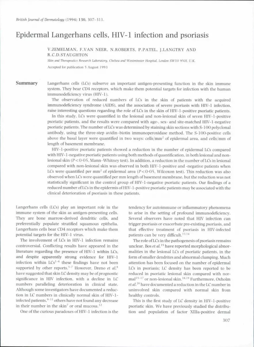
Figure 1. IniinunijhistOL'iieiiiical staining of epidLTinal I.iiiigt*rlianscells identilied with S-HK) polyclonai antibody in lesionai skin ol"psoriatic patients, (a) HIV-1-negative psoriatic skin, and Ib) HIV-1-positive psoriatic skin ( x 100).
Figure 2. Immunohistochemical staining of epiiiernialcells, identified with S-100 poiyclonal antibody in the non-lesional skinof psoriatic patients, (a I HIV-1-negative psoriatic skin, and Ib) HIV-1-positive psoriatic skin ( x 100).
dendrocytes in HIV-related psoriasis. These phagocyticdendritic cells have a possible antigen-presenting func-tion in the dermis. We showed that there were nochanges in the number and distribution of these cells inlesional and non-lesional skin of HIV-positive and-negative individuals.^'
In the present study, we have quantified epidermalLCs in both lesional and non-lesional skin of HIV-1-positive and -negative psoriatic patients. Our aim was toelucidate further the role of LCs in the pathogenesis ofHIV-1-associated psoriasis.
Methods
Patients
After informed consent, punch biopsies were taken fromseven HIV-1 -positive and seven HIV- 1-negative psoriaticpatients. All patients were male, and were aged between25 and 45 years (mean 32).
The patients had not received systemic or topicalcorticosteroid or ultraviolet (UVB or PUVA) therapy forat least 4 weeks before the biopsies were taken.
The HIV-1-positive patients were classified accordingto the Center for Disease Control criteria.^^ and theirdistribution was as follows: stage II (n=2) (asymptoma-tic); stage IV (n= 5). of whom two had AIDS-defininginfection {Pnewnocystis carinii pneumonia and oeso-phageal candidiasis). None of the patients was receivingimmunosuppressive or antiviral therapy at the time thebiopsies were taken.
Biopsies and immunohistochemistry
Two 3-mm punch biopsies were taken from each subjectunder 1% lignocaine anaesthesia. One was taken from alesion on the elbow, the other from clinically normal skinat a distance of 2 cm from the edge of the lesion. Allbiopsies were fixed in formalin for 24 h. before wax

LANGERHANS CELLS. HIV-1 INEECTION AND PSORIASIS 309
2001-
iOO
0Lesionoi (PP) Non-iesional (PN)
f-igure J. Number of suprabasal S-1 OO-positive epidermal LCs/mm^ ofepidermal area in HIV-1-positive compared with HIV-1-negativepsoriatic skin, HIV-1-positive patients showed a significant reductionin the number ofLCs in both lesional and non-lesional psoriatic skin. Inaddition, a significant reduflion in the number of I.Cs in lesionalcompared with non-lesional skin was observed in both groups ofpatients. Values areexpressed as mean ±SRM. • . HIV-positive: B. HIV-negative. *P = O-O38; " P - 0 016.
iO
0
T
Lesional (PP) Non-lesional (PN)
Figure 4. Number of suprabasal S-llK)-positive epidermal IX's/mmbasemeni membrane length in HIV-1-positive compared with HIV-l-negative psoriatic skin, HIV-1-positive patienis showed a significantreduction in the number of LCs in both lesionul and non-lesionalpsoriatic skin. In addition, a signilifanl reduclioii in the number of l,Csin lesional compared with non-le.sional skin was observed in the HIV-i-positive psoriatic patients. Values are expressed as mean±SEM. Q.HIV-positive: H, HIV-negalive. n^7. * P = 0 0 2 6 : " P = 0 0 3 :
embedding. Paraffin sections (4 ^m) were cut. andimmunohistologically stained using the three-step avi-din-biotin immunoperoxidase method. Trypsination(()• 1% in phosphate buffer) was performed for 20 min at57°C. Sections were incubated with normal porcine.serum (Dakopatts X901. High Wycomhe. U.K.) beforeimmunolabelling.
The polyclonai rabbit antibovine S-HK) (DakopattsZ3] ] . High Wycombe, U.K.) was used as primaryantibody at a dilution of 1: ]()(). at room temperature, for60 min. Negative controls were performed by omittingthe primary antiserum. Positive controls were performedusing paraffin sections from melanoma tissue. Peroxi-dase was revealed using 3'3'diaminobenzidine andhydrogen peroxide. Sections were counterstained with1% baematoxylin. and mounted in StyroUte mountingmedium.
In preliminary studies in our laboratory, formalin-calcium-fixed frozen sections were stained for the pres-ence of LCs using GDI monoclonal antibody and S-100poiyclonal antibody in skin serial frozen sections.
mi'thod
Three non-serial sections of each skin biopsy wereexamined. Slides were coded, and counting was per-formed 'blind' by a single observer. Only cells with adistinctive nucleus and with at least one dendriticprocess were identified as LCs. Such cells were countedabove the basal layer, to exclude positive-stainingmelanocytes.
The outline of the epithelium of each section wasdrawn on paper, after projection of the stained section.The paper was mounted on a digitizing tablet, and boththe epidermal area and the length of basement mem-brane were measured using a MOP-Videoplan imageanalyser system.
LCs were expressed as LCs/mm' of epidermal area, andas LCs/mm of basement membrane length.
Statistical analysis of the results was performed usingthe non-parametric two-tailed Mann-Whitney (M-W)and Wilcoxon (W) tests. P-values <0-05 were consid-ered significant.

iU) V.ZEMELMAN etal.
Results
No apparent changes in morphology or localization ofepidermal Langerhans cells were found in the HIV-l-positive psoriatic patients compared with the controlgroupofHIV-1-ncgatlvepsoriaticpatients (Figs 1 and2).
HIV-1-positive psoriatic patients sbow a reducednumber of LCs in both lesional and non-lesional skintompiired with the controls, when LCs were expressedby both metbods of quantification (Figs 3 and 4).
The number of LCs in the lesional skin was reducedcompared with non-lesional skin, in both HIV-positiveand HIV-negative psoriatic patients {Figs 3 and 4).
Discussion
The observation that psoriasis may accompany and beexacerbated by HIV-1 infection raises intriguing ques-tions about the immune system of these patients.
LCs are often identified by the demonstration of CDl,HLA-DR or other cell-surface antigens on unfixed frozentissue. In this study, fixed, paraffin-embedded tissue wasused, and sections were stained with S-l()() protein as amarker for LCs. Several authors have suggested thatcounting of suprabasal S-l()()-positive epidermal cells isa reproducible and reliable method for enumerating LCsin the epidermis of paraffin-embedded sections.'* '•* LCsmay be located occasionally in the basal layer, althoughin our preliminary studies with CDl monoclonal anti-body on frozen sections we observed few positive cells inthe basai layer.
In tbis study, we have confirmed previous reports of areduction in LC numbers in lesional psoriatic skin,compared with non-lesional skin. This reduction wasobserved in the lesions of HIV-1-positive and -negativepsoriatic patients. It has been suggested that T-suppres-sor lymphocytes in the psoriatic epidermis might becytotoxic to LCs,'' or that this reduction in numberrepresents migration of LCs into the dermis.^^
Our observation of a reduction in the number ofepidermal LCs in the non-lesional HIV-positive skin is inagreement with studies using different antibodies (HLA-DR." ATPase.'" and CD1«" - ' ) . We did not see the moresuperficially located epidermal LCs reported by Oxholm
In HIV infection, the reduction in the number of LCs isthought to be due to a direct cytotoxic effect of the HIVvirus.'' However, a recent study has suggested that thereduction in T-helper lymphocytes associated with HTV-1 infection might be the result of the induction ofautoreactivity. rather tban viral cytotoxicity. Thus, thevirus is thought to induce an autoimmune process
similar to graft-versus-host disease, resulting in lympho-penia.'" It is possible that similar effects on Langerhanscells could occur.
Whatever the mechanism, the observation of areduced number of LCs in HIV-1-positive psoriatic skin,measured as S-1 ()()-positive suprabasal dendritic cells, isnot conclusive, as this reduction may be due only to areduction in the expression of S-l 00 by LCs in the skin ofthese patients. Future research should be directed atmeasuring the number of epidermal LCs with severalmarkers, together with functional studies, to clarifyfurther the roie of these cells in the exacerbation ofpsoriasis in HIV-1-infected patients.
References
1 Tschachler E. Groh V, Popovic M cl al. Epidermal Langerhanscells—a target for HTLV-III/t^V infection. / Invest Dermato! 1987:88: 2JJ -7 .
2 Rappersberger K. Gartner S. Schenk P cf al. Langerhani cells are anactual site of HIV-1 replication. Interviroloyn I98S: 29: 1S5-94.
i Hraathen LR. Rmnire/ G. Kiinze RC) cl al. Latent infection ofepidermal Langerhans ceils in HIV-positive individuals. Res Virol1991: 142: 119-21.
4 Zambruno G, Mori L, Marconi A et al. I^etection of HIV-1 inepidermal Langerhans cells of HlV-infet'ted patients using thepolymerase chain reaction. / Invest Dermatol 1991; 96: 979-S2.
5 Kanitakis |. Marchand C. Su H el al Immimohistodiemical study ofnormal skin of HlV-l-infcctcd patients shows no evidence ofinfection of epidermal Langerhans cells by HIV, AiDS Res HumRetroviruses 1989: 5: 293-302.
fl Kalter \X, Gendelman HE, Mettzer MS. Monocytes. dendritic ceilsand Langerhans cells in human immunodeficiency virus infection.Dermatol Clin 1991:9:41 =S-28.
7 Kaiter DC. Greenhouse J|. Orenstein IM et ul. Epidermal Langer-hans cells are not principal reservoirs of virus in HIV disease. /Inummol 1991: 146: iJ9(i-404.
8 DrenoB,MilpicdH. Bignon JDcl id. Prognostic value of l.angerhanscells in the epidermis of HIV patients. Br / Dermcitol 1988: 118:481-6.
9 BelsitoDV. Sanchez MR,'BaerRLclij/. Reduced Langerhans cell laantigen and ATPase activity in patients with the acquiredimmunodeficiency syndrome. ,\'Efi^//Mcif 1984; 310: 1279-82.
11) Belsito DV, Thorbecke GL. Reduced la positive Langerhans cells inAIDS. N Engi] Med 1984: 311:858.
11 Oxholm P, Helweg-Larsen S, Permin H. Immunohistological skininvestigation.^ in patients with the acquired immunodeficiencysyndrome. Acta Path Microbiol Immunol Scand jA] 1986; 84:113-16.
12 Becker J. Ulrich P, Kunze R et al. Immunohistochemical detectionof HIV structural proteins and distrihution of T-lymph(x:ytes andLangerhans cells in the oral mucosa of HIV infected patients.Vircbows Arch I A] 1988; 412: 413-19.
1 3 Duvic M. HIV-associated psoriasis and Reiter's syndrome. ProqDermatol 1990; 24: 1-9.
14 LazarAP.RoenigkHHjr. AlDSandpsoriasis.Cu(i.^l967:39: 347-51.
I 5 Bos JD, Hulsebosch HJ. Krieg SR et al. Immunocompctent ceils in

LANGERHANS CELLS, HIV-1 INFECTION AND PSORIASIS 511
psoriasis. In situ immunophenotyping by monoclonal antibodies.Anh Dermiiti'l Rt-s 198i: 275: 181-9.
16 i,isi P. Investigation on Langerhans cells in pathological humanepidermis. Acta Derm Venereol (Stockh) 1973; 53: 425-8.
17 Ilitftek M, Eaure M. Schmitt D. Thivolet J. Effects of aromaticretinoid treatment on epidermal Langerhans cells in psoriasis. Aquantitative study. / Invest Dermuto! 1 982; 78: 327 (Ahstr.).
18 Torinuki W, Mauduit C Haftek M. Thivolel ], Effect of PDVA andmechlorethamine treatment of psoriatic patients on epidermalLangerhans cells. Aclii Derm Venereol I Stockh I 19K7: 67: 5 52-5.
19 Harper |L Zenielman V. C'yclosporin for psoriasis: changes in T-cells subsets and Langerhans cells In lesiona! and non-lesional skin(during a six month treatment periodi. In: Cticlosporin in PsoriasisIvan loost TH. Heute E. Bos |D. eds|. Sandoz Publications 1988;1(19.
20 Oxhoim A, Oxhotm P. Staberg B, Reduced density of T6-positiVL'epidermal Langerhans cells in uninvolved skin of patients withpsoriasis, Acta Derm Venereol iStockh) 1987: 67: 8-11.
21 Van Neer E, Zemelman V, Cerio R cf ul. The role of factor Xllla-positive dermal dendrocytes in HlV-l-positlve psoriatics. Br /Dermiitol 1993: 128: 29- 3 3.
22 Centers for Disease Control, Classification system for human T-lymphotropic virus type III/O lymphadenopathy associated virusinfections. MMWK 1986; 35: M4-9.
2 3 Halliday GM. McArdle IP. Knight BA, Muller HK. New metho-dology for assessment of the Langerhans cell network. / Pathol1986: 148: 127-34.
24 Maddox PH. Barton SE. Jenkins D. S-l 00sensitivity as a marker forLangerhans cells. Am / Obstet Canecol 1989; 160: 767-8.
25 Shaw PAV, Eletcher A. Counts of S-ll)() positive epidermaldendritic ceils in Kveim test skin blopsie.s. Histopiitholocfii 1991; 18:149-S}.
26 Bieber T, Ring [, Braun-Ealco (). Comparison of different methodsfor enumeration of Langerhans cells in vertical cryosections ofhuman skin. Br I Dermato! 1988: 118: J85-92.
27 Belsito D, Baer R, Thorbecke ). Immunomodulation ofLangerhanscell suriace anligen in various immunodeliciency slates. In;Primar!) Immiinodepcieniii Diseases |Eib! M, Rosen E. eds). Amster-dam; Elsevier, 1986; 255-68.
28 Dalgleish Ati. Wilson S. Compels M et al, T-cell receptor variablegene products and early HIV-i infection. Lancet 1992: i i 9 :824-8.


