Embed Size (px)
Citation preview
Epithelial-to-Mesenchymal Transition in Podocytes Mediated byActivation of NADPH Oxidase in Hyperhomocysteinemia
Chun Zhang1,2, Min Xia1, Krishna M. Boini1, Cai-Xia Li1, Justine M. Abais1, Xiao-Xue Li1,Laura A. Laperle1, and Pin-Lan Li11Department of Pharmacology & Toxicology, Medical College of Virginia, Virginia CommonwealthUniversity, Richmond, VA 232982Department of Nephrology, Union Hospital, Tongji Medical College, Huazhong University ofScience and Technology, 1277 Jie Fang Avenue, Wuhan 430022, China
AbstractThe present study tested the hypothesis that hyperhomocysteinemia (hHcys) induces podocytes toundergo epithelial-to-mesenchymal transition (EMT) through the activation of NADPH oxidase(Nox). It was found that increased homocysteine (Hcys) level suppressed the expression of slitdiaphragm-associated proteins, P-cadherin and zonula occludens-1 (ZO-1) in conditionallyimmortalized mouse podocytes, indicating the loss of their epithelial features. Meanwhile, Hcysremarkably increased the abundance of mesenchymal markers, such as fibroblast specificprotein-1 (FSP-1) and α-smooth muscle actin (α-SMA). These phenotype changes in podocytesinduced by Hcys were accompanied by enhanced superoxide (O2
.−) production, which wassubstantially suppressed by inhibition of Nox activity. Functionally, Hcys significantly enhancedthe permeability of the podocyte monolayer coupled with increased EMT, and this EMT-relatedincrease in cell permeability could be restored by Nox inhibitors. In mice lacking gp91phox
(gp91−/−), an essential Nox subunit gene, hHcys-enhanced podocyte EMT and consequentglomerular injury were examined. In wild-type (gp91+/+) mice, hHcys induced by a folate-free(FF) diet markedly enhanced expression of mesenchymal markers (FSP-1 and α-SMA) butdecreased expression of epithelial markers of podocytes in glomeruli, which were not observed ingp91−/− mouse glomeruli. Podocyte injury, glomerular sclerotic pathology, and markedalbuminuria observed in gp91+/+ mice with hHcys were all significantly attenuated in gp91−/−
mice. These results suggest that hHcys induces EMT of podocytes through activation of Nox,which represents a novel mechanism of hHcys-associated podocyte injury.
KeywordsHomocysteinemia; NADPH oxidase; Podocytes; Epithelial-to-mesenchymal transition; End-stagerenal disease
INTRODUCTIONThere is considerable evidence showing that hyperhomocysteinemia (hHcys) is importantlyimplicated in the progression of end-stage renal disease (ESRD) and in the development of
Send Correspondence and Reprint Request to: Pin-Lan Li, M.D, Ph.D., Department of Pharmacology and Toxicology, MedicalCollege of Virginia, Virginia Commonwealth University, 410 N 12th Street, Richmond, VA 23298, Tel: (804) 828-4793, Fax: (804)828-4794, [email protected] OF INTEREST STATEMENTNo conflicts of interest, financial or otherwise, are declared by the authors.
NIH Public AccessAuthor ManuscriptPflugers Arch. Author manuscript; available in PMC 2012 March 12.
Published in final edited form as:Pflugers Arch. 2011 September ; 462(3): 455–467. doi:10.1007/s00424-011-0981-y.
NIH
-PA Author Manuscript
NIH
-PA Author Manuscript
NIH
-PA Author Manuscript

cardiovascular complications related to ESRD [1–3]. Studies from our laboratory [4, 5] andby others [6] have demonstrated that Hcys induces extracellular matrix (ECM) accumulationand inhibits their degradation in mesangial cells, which ultimately leads toglomerulosclerosis and loss of renal function. Although the precise mechanism of hHcys-induced glomerulosclerosis has not yet been fully elucidated, we and others havedemonstrated that oxidative stress mediated by NADPH oxidase (Nox) is importantlyinvolved in the progression of glomerular injury associated with hHcys [4, 7]. In addition,recent studies have shown that Nox activation is essential in hHcys-induced podocyte injury,which is a crucial early event leading to glomerulosclerosis [8]. However, the molecularmechanism mediating hHcys-induced podocyte injury, in particular, how Nox activationcauses podocyte dysfunction and initiates glomerulosclerosis remains poorly understood.
Podocytes are unique glomerular epithelial cells which comprise the outermost layer of theglomerular filtration barrier [9–11]. Podocyte foot processes interdigitate with thecounterparts of neighboring cells to form the slit diaphragm, which constitutes the finalbarrier to prevent protein loss from vascular to urinary space. It has been reported that anyinjury to podocytes that disrupts the structural and functional integrity of the slit diaphragmwould eventually lead to a defective glomerular filtration, thereby causing proteinuria.Podocyte injury has been considered as the most important early event initiatingglomerulosclerosis in different animal models and humans, such as focal segmentalglomerulosclerosis, membranous nephropathy, diabetic nephropathy, and lupus nephritis[12–14]. Therefore, the molecular mechanism producing podocyte injury becomes one ofthe most important mechanisms in the progression of ESRD due to a variety of causes. InhHcys, podocyte effacement, cytoskeleton reorganization, decreased expression of slitdiaphragm molecules, and podocyte apoptosis were documented [15, 16], indicating theoccurrence of podocyte injury during hHcys. However, the mechanism by which hHcysinduces podocyte injury has not been fully elucidated.
Recently, two major hypotheses have been proposed for podocyte injury under differentconditions. The prevalent view emphasizes the importance of podocyte depletion resultingfrom apoptosis as a causative factor for the onset of proteinuria and glomerulosclerosis [17–19]. In this hypothesis, the reduced podocyte number in glomeruli is attributed to theapoptotic death of these cells. However, recent studies have shown that the detachedpodocytes might be alive and could be presented in the urine of some patients with chronickidney diseases [20–22], Therefore, another hypothesis for podocyte injury proposes thatinjured podocytes could obtain a motile ability facilitating their detachment from theglomerular basement membrane, rather than apoptotic cell death. In this context, emergingevidence suggests that podocytes could undergo a epithelial-to–mesenchymal transition(EMT) process when challenged by different injurious stimuli such as transforming growthfactor-β1 (TGF-β1), high glucose, and adriamycin [23]. Although there is still somedebating concerning the EMT [24], it has been proposed that the EMT process ischaracterized by losing its epithelial features such as nephrin, P-cadherin, and zonulaoccludens-1 (ZO-1) as well as acquiring mesenchymal features, such as increases in theexpression of desmin, fibroblast-specific protein-1 (FSP-1), and α-smooth muscle actin (α-SMA) [25–28]. This phenotype transition process will render podocytes motile andultimately lead to disruption of the delicate architecture of these cells, thereby impairingtheir filtration barrier function and initiating glomerulosclerosis [23]. However, whetherhHcys could also induce podocyte EMT and thereby cause glomerulosclerosis is unknown,and the present study attempted to answer this important question.
Given the important role of Nox activation in Hcys-induced glomerular injury, the presentstudy hypothesized that hHcys may induce podocyte EMT through the activation of Nox andsubsequent O2
.− production, ultimately leading to hHcys-associated podocyte dysfunction
Zhang et al. Page 2
Pflugers Arch. Author manuscript; available in PMC 2012 March 12.
NIH
-PA Author Manuscript
NIH
-PA Author Manuscript
NIH
-PA Author Manuscript

and subsequent glomerulosclerosis. To test this hypothesis, we first used cultured murinepodocytes to examine whether they could undergo EMT upon stimulation of Hcys. Then, theeffects of Nox inhibition on Hcys-induced phenotypic changes and their functionalrelevance were examined. Using mice lacking gp91phox gene, an essential Nox subunit gene,we also tested the role of Nox activation in podocyte EMT in vivo compared with theirgenetic background strain C57BL/6 mice.
MATERIALS AND METHODSCell culture
Conditionally immortalized mouse podocyte cell line, kindly provided by Dr. Klotman PE(Division of Nephrology, Department of Medicine, Mount Sinai School of Medicine, NewYork, NY, USA), were cultured on collagen I-coated flasks or plates in RPMI 1640 mediumsupplemented with recombinant mouse interferon–γ at 33°C. After differentiated at 37°C for10–14 days without interferon–γ, podocytes were used for the proposed experiments. In thepresent study, preparation of L-Hcys (a pathogenic form of Hcys), the concentration andincubation time of L-Hcys treatment were chosen based on our previous studies [16].
gp91phox siRNA transfectiongp91phox siRNA was purchased from Qiagen, which was confirmed to be effective insilencing gp91phox gene in different cells by the company and had been successfully used inour previous studies [15]. The scrambled RNA (Qiagen, Valencia, CA, USA) was confirmedas non-silencing double-strand RNA and used as the control in the present study. Podocyteswere serum-starved for 12 h and then transfected with gp91phox siRNA or scrambled siRNAusing siLentFect Lipid Reagent (Bio-Rad, Hercules, CA, USA). After 18 h of incubation at37 °C, the medium was changed, and L-Hcys (40 μmol/L) added into the medium forindicated time span in different protocols.
Real-time reverse transcription polymerase chain reaction (RT-PCR)Total RNA from cultured podocytes or isolated mouse glomeruli was extracted using TRIzolreagent (Invitrogen, Carlsbad, CA. USA) according to the protocol as described by themanufacturer. Aliquots of total RNA (1 μg) from each sample were reverse-transcribed intocDNA according to the instructions of the first strand cDNA synthesis kit manufacturer(Bio-Rad, Hercules, CA, USA). Equal amounts of the reverse transcriptional products weresubjected to PCR amplification using SYBR Green as the fluorescence indicator on a Bio-Rad iCycler system (Bio-Rad, Hercules, CA, USA). The mRNA levels of target genes werenormalized to the β-actin mRNA levels. The primers used in this study were synthesized byOperon (Huntsville, AL, USA) and the sequences were: P-cadherin senseGTAAGGGCTACCGCTCACTC, antisense TGTGAGGCCAAGTGAAAGAC; ZO-1 senseGAGCTACGCTTGCCACACTGT, antisense TCGGATCTCCAGGAAGACACTT; FSP-1sense GTTACCATGGCAAGACCCTT, antisense AACTTGTCACCCTCTTTGCC; α-SMAsense CAGGATGCAGAAGGAGATCA, antisense TCCACATCTGCTGGAAGGTA; β-actin sense TCGCTGCGCTGGTCGTC, antisense GGCCTCGTCACCCACATAGGA.
Western blot analysisWestern blot analysis was performed as we described previously [29]. In brief, proteins fromthe mouse glomeruli or cultured podocytes were extracted using sucrose buffer. After boiledfor 5 min at 95°C in a 5× loading buffer, 50 μg of total proteins were subjected to SDS-PAGE, transferred onto a PVDF membrane and blocked. Then, the membrane was probedwith primary antibodies of anti-gp91phox (1:500, BD Biosciences, San Jose, CA), anti-P-cadherin (1:200, R&D system, Minneapolis, MN, USA), anti-FSP-1 (1:500, Abcam,
Zhang et al. Page 3
Pflugers Arch. Author manuscript; available in PMC 2012 March 12.
NIH
-PA Author Manuscript
NIH
-PA Author Manuscript
NIH
-PA Author Manuscript

Cambridge, MA, USA), anti-α-SMA (1:200, R&D system, Minneapolis, MN, USA) or anti-β-actin (1:3000, Santa Cruz Biotechnology, Santa Cruz, CA, USA) overnight at 4°Cfollowed by incubation with horseradish peroxidase–labeled IgG (1:5000). The immuno-reactive bands were detected by chemiluminescence methods and visualized on Kodak OmatX-ray films. Densitometric analysis of the images obtained from X-ray films was performedusing the Image J software (NIH, Bethesda, MD, USA).
Indirect immuno-fluorescent stainingThe cells were fixed in 4% PFA for 15 minutes. After rinsed with phosphate-buffer saline(PBS), cells were incubated with goat anti-FSP-1 (1:50, Abcam, Cambridge, MA, USA),goat anti-ZO-1 (1: 50, Santa Cruz Biotechnology, Inc, Santa Cruz, CA, USA), rabbit anti-P-cadherin (1: 50, R&D system, Minneapolis, MN, USA), and mouse anti-α-SMA (1: 100,R&D system, Minneapolis, MN, USA) antibodies. After washing, the slides were incubatedwith Alexa 488-labeled secondary antibodies for 1 h at room temperature. After beingmounted with DAPI-containing mounting solution, the slides were observed under afluorescence microscope and photos were taken and analyzed. The fluorescent intensitieswere quantified by the Image Pro Plus 6.0 software (Media Cybernetics, Bethesda, MD,USA) and the data was normalized to control cells.
Cell permeability assayThe permeability of podocyte monolayer was measured according to the methods as wedescribed previously [30]. Briefly, cells were seeded in the upper chambers of 0.4 μmpolycarbonate Transwell filters of a 24-well filtration microplate (Whatman Inc., FlorhamPark, NJ, USA). After treatment with L-Hcys (40 μmol/L) for different time spans or TGF-β1 (2.5 ng/ml) for 48 h, the culture medium was replaced with fresh phenol red-free RPMI1640 in the presence of Hcys and 70 kD FITC-dextran (2.5 mol/l) in the upper chambers.After 4 h, the filtration microplate was removed, the medium from the lower compartmentwas collected, and then fluorescence was measured in a spectrofluorimeter at an excitationwavelength of 494 nm. The permeable fluorescence intensity was used to represent cellpermeability, and the values were normalized to that of control cells.
Animal proceduresC57BL/6J wild-type (WT; 8 weeks of age, male) and gp91phox knockout (KO) mice (8weeks of age, male) were bred from breeding pairs from The Jackson Laboratory, BarHarbor, ME, USA. All protocols were approved by the Institutional Animal Care and UseCommittee of Virginia Commonwealth University. To speed up the damaging effects ofhHcys on glomeruli, all mice were uninephrectomized as described in previous studies [4,31]. After a 1-week recovery period from the uninephrectomy, KO and WT mice were fed anormal diet or a folate-free (FF) diet (Dyets Inc, Bethlehem, PA, USA) for 4 weeks toinduce hHcys. This model has been demonstrated to induce glomerular damage unrelated tothe uninephrectomy and arterial blood pressure, but specific to hHcys [15]. One day beforethese mice were sacrificed, 24-hour urine samples were collected using mouse metaboliccages. After blood samples were collected, these mice were sacrificed and renal tissues wereharvested for biochemical and molecular analysis as well as morphological examinations.Mouse glomeruli were isolated as described before [32]. Measurement of creatinineclearance (Ccr) was performed as we reported [15].
High performance liquid chromatography (HPLC) analysisPlasma total Hcys levels were measured by HPLC as we described previously [33]. Briefly,blood samples were centrifuged at 1000 g for 10 min at 4°C to obtain plasma. A 100 μLplasma or standard solution mixed with 10 μL of internal standard, thionglycolic acid (2.0
Zhang et al. Page 4
Pflugers Arch. Author manuscript; available in PMC 2012 March 12.
NIH
-PA Author Manuscript
NIH
-PA Author Manuscript
NIH
-PA Author Manuscript

mmol/L) solution, was treated with 10 μL of 10% tri-n-butylphosphine (TBP) solution indimethylformamide at 4°C for 30 minutes. Then, 80 μL of ice-cold 10% trichloroacetic acid(TCA) in 1 mmol/L EDTA were added and centrifuged to remove proteins in the sample.100 μL of the supernatant was transferred into the mixture of 20 μL of 1.55 mol/L sodiumhydroxide, 250 μL of 0.125 mol/L borate buffer (pH 9.5), and 100 μL of 1.0 mg/mL ABD-Fsolution. The resulting mixture was incubated at 60°C for 30 minutes to accomplishderivatization of plasma thiols. HPLC was performed with an HP 1100 series equipped witha binary pump, a vacuum degasser, a thermostated column compartment, and anautosampler (Agilent Technologies, Waldbronn, Germany). Separation was carried out at anambient temperature on an analytical column (Supelco LC-18-DB) with a Supelcosil LC-18guard column. Fluorescence intensities were measured with an excitation wavelength of 385nm and emission wavelength of 515 nm by a Hewlett-Packard Model 1046A fluorescencespectrophotometer. The peak area of chromatographs was quantified with a Hewlett-Packard3392 integrator.
Electromagnetic spin resonance (ESR) analysis of Nox-dependent O2.− productionFor detection of Nox-dependent O2
.− production, proteins from isolated glomeruli of miceor cultured podocytes were extracted using sucrose buffer (250mmol/L sucrose, 20 mmol/LHEPES, 1 mmol/L EDTA, PH=7.4) and then re-suspended with modified Kreb’s-Hepesbuffer containing deferoximine (100 μmol/L, Sigma, St. Louis, MO, USA) anddiethyldithiocarbamate (5 μmol/L, Sigma, St. Louis, MO, USA). The Nox-dependent O2
.−
production was examined by addition of 1 mmol/L NADPH as a substrate in 50 μg proteinand incubated for 15 min at 37 °C in the presence or absence of SOD (200 U/ml), and thensupplied with 1 mM O2
.− specific spin trap 1-hydroxy-3-methoxycarbonyl-2,2,5,5-tetramethylpyrrolidine (CMH, Noxygen, Elzach, Germany) as we described previously [4].The mixture was loaded in glass capillaries and immediately analyzed for O2
.− productionkinetically for 10 min in a Miniscope MS200 ESR spectrometer (Magnettech Ltd, Berlin,Germany). The ESR settings were as follows: biofield, 3350; field sweep, 60 G; microwavefrequency, 9.78 GHz; microwave power, 20 mW; modulation amplitude, 3 G; 4,096 pointsof resolution; receiver gain, 20 for tissue and 50 for cells. The results were expressed as thefold changes of control.
Urinary albumin excretion measurementThe 24-hour urine albumin was detected using a commercially available mouse albuminELISA kit (Bethyl Laboratories, Montgomery, TX, USA) as we described previously [15].
Morphological examinationFor observation of renal morphology using light microscope, pre-fixation perfusion usingphosphate buffer was done prior to fixation to ensure removal of blood. The kidneys werethen perfused using 4% paraformaldehyde (PFA) in situ and renal tissues were post-fixed,paraffin-embedded and stained with periodic acid-Schiff (PAS). Then, glomerularpathological changes were assessed by a blinded observer. Glomerular damage was assessedby a standard semi-quantitative analysis and expressed as glomerular damage index (GDI)as we described before [34]. Fifty glomeruli per slide were counted and graded as 0, 1, 2, 3,or 4, according to 0, <25, 25–50, 51–75, or >75% glomerular damge across a longitudinalkidney section, respectively. The GDI for each mouse was calculated by the formula: (N1 ×1 + N2 × 2 + N3 × 3 + N4 × 4)/n, where N1, N2, N3, and N4 represent the numbers ofglomeruli exhibiting grades 1, 2, 3, and 4, respectively; and n is the total number ofglomeruli graded.
For transmission electron microscopic (TEM) observation of ultrastructural changes inpodocytes, the kidneys were perfused with a fixative containing 3% glutaraldehyde and 4%
Zhang et al. Page 5
Pflugers Arch. Author manuscript; available in PMC 2012 March 12.
NIH
-PA Author Manuscript
NIH
-PA Author Manuscript
NIH
-PA Author Manuscript

paraformaldehyde in 0.1mol/L phosphate buffer. After fixation and dehydration withethanol, the samples were embedded in Durcupan resin for ultra-thin sectioning and TEMexaminations by VCU electron microscopy core facility.
Double-immunofluorescent stainingDouble-immunofluorescent staining was performed using frozen slides from mouse kidneys.After fixation, the slides were incubated with rabbit anti-podocin 1: 200 (Sigma, St. Louis,MO, USA), which was followed by incubation with Alexa 555-labeled goat anti-rabbitsecondary antibody. Then, goat anti-FSP-1 (1:50, Abcam, Cambridge, MA, USA), goat anti-ZO-1 1:50 (Santa Cruz Biotechnology Inc, Santa Cruz, CA, USA), rabbit anti-P-cadherin1:100 (R&D system, Minneapolis, MN, USA), or mouse anti-α-SMA 1:200 (R&D system,Minneapolis, MN, USA) was used to incubate the slides for overnight at 4°C. Afterwashing, the slides were incubated with Alexa 488-labeled secondary antibodies. Finally,the slides were mounted and subjected to examinations using a confocal laser scanningmicroscope (Fluoview FV1000, Olympus, Japan).
Statistical analysisAll of the values are expressed as mean ± SEM. Significant differences among multiplegroups were examined using ANOVA followed by a Student-Newman-Keuls test. χ2 testwas used for testing the significance of ratio and percentage data. P<0.05 was consideredstatistically significant.
RESULTSL-Hcys suppressed epithelial P-cadherin and ZO-1 expression in podocytes
We first examined the expression of P-cadherin, an epithelial marker, in podocytes beforeand after Hcys treatment. As shown in Figure 1A, treatment of podocytes with Hcysmarkedly suppressed P-cadherin mRNA expression in a concentration-dependent manner asdetected by Real-time RT-PCR. P-cadherin mRNA expression started to decrease aftertreatment with 20 μmol/L of L-Hcys for 48 h, and L-Hcys at a concentration of 40 μmol/Lor 80 μmol/L could further decrease P-cadherin mRNA expression in podocytes, which wasalmost comparable to a well-established EMT inducer, TGF-β1 (2.5 ng/ml) [23, 28].Immunofluorescence staining (Figure 1B) further showed that P-cadherin protein expressionwas substantially reduced by treatment of podocytes with L-Hcys (40 μmol/L) for 72 h. Inconsistent with these data, L-Hcys-induced P-cadherin suppression was also confirmed byWestern blot analysis (Figure S1 A).
ZO-1 is a tight junction-associated protein which locates at the slit diaphragm and is linkedto nephrin [35]. This protein is also widely used as an epithelial marker for podocytes. In thepresent study, real-time RT-PCR data showed that ZO-1 mRNA expression was decreasedby the treatment with L-Hcys, with the strongest effect at a concentration of 40 μmol/L(Figure 1C). Immunofluorescence staining showed abundant ZO-1 at the sites of cell-cellcontacts with a characteristic zipper-like pattern between interdigitating processes ofpodocytes. Interestingly, the overall density of ZO-1 staining was markedly decreased in L-Hcys-treated cells, which was similar to that induced by TGF-β1 (Figure 1D).
L-Hcys increased the expression of mesenchymal markers in podocytesWe next investigated whether L-Hcys could induce a mesenchymal transition of podocytes.To this end, the expression of two mesenchymal markers, FSP-1 and α-SMA, wereexamined in podocytes after L-Hcys treatment. As illustrated in Figure 2A and 2C,incubation of these cells with L-Hcys induced the expression of FSP-1 and α-SMA mRNAin a dose-dependent manner, which was similar to the effect of TGF-β1. Immunostaining
Zhang et al. Page 6
Pflugers Arch. Author manuscript; available in PMC 2012 March 12.
NIH
-PA Author Manuscript
NIH
-PA Author Manuscript
NIH
-PA Author Manuscript

showed that podocytes under basal conditions barely expressed FSP-1 and α-SMA.However, after stimulated by L-Hcys (40μmol/L) for 72h, there were clearly increasedFSP-1 and α-SMA expression in the cytoplasm of these cells (Figure 2B and 2D), indicatingthat mesenchymal characteristics of podocytes were obtained under Hcys stimulation.Western blot analysis showed that α-SMA expression was significantly increased upon thestimulation of L-Hcys, although it is less potent as the classic EMT inducer, TGF-β1 (FigureS1B).
Dependency of Hcys-induced phenotype changes on O2.− production via Nox activation inpodocytes
To explore the mechanism mediating Hcys-induced EMT, we performed experiments tomeasure L-Hcys-induced Nox-dependent O2
.− production. In consistent with our previousdata [15], with a 48 h incubation L-Hcys concentration-dependently increased Nox-dependent O2
.− production with a maximal increase of 2.38±0.30 folds at 40μmol/L ofHcys, as compared with control cells (P<0.05, n=5). When podocytes were transfected withgp91phox siRNA, Hcys-induced gp91phox protein expression was substantially inhibited(Figure S2), suggesting a successful knocking down of this gene. Under these conditions,Nox-dependent O2
.− production induced by L-Hcys was also significantly inhibited(1.10±0.22 vs. 2.27±0.27 folds as compared with control cells, P<0.05, n=4), indicating thatO2
.− production in podocytes is mainly derived from the activation of Nox.
Consistent with direct measurement of O2.− production under different treatments, gp91phox
siRNA was found to block Hcys-induced decreases in P-cadherin and ZO-1 expression inpodocytes compared to that observed in vehicle or scrambled siRNA treated cells. Similarly,inhibition of Nox activity using DPI or apocynin also prevented the loss of these epithelialmarkers in podocytes (Figure 3A, two left groups of images). Correspondingly, themesenchymal transition of podocytes, namely, increase in FSP-1 expression induced byHcys was also abolished by gp91phox siRNA transfection or by DPI or apocynin, indicatingpreservation of epithelial phenotype of podocytes. Additionally, inhibition of Nox alsodramatically attenuated Hcys-induced α-SMA expression in podocytes treated with L-Hcys(Figure 3A, two right groups of images). Relative fluorescent intensities of these markerswere summarized in Figure 3B.
Inhibition of EMT improved barrier function of podocytesWe next examined whether podocyte EMT associated with O2
.− production affects thebarrier function of podocyte monolayer. Dextran flux assay was used to assess the filtrationacross the monolayer of cultured differentiated podocytes. As shown in Figure 4A, increaseddextran flux was observed in podocytes treated with L-Hcys for 48 or 72 h, when comparedwith control cells (P<0.05). As expected, TGF-β1 also induced a significant increase in cellpermeability in the podocyte monolayer after 48 h of incubation, which was used as apositive control for this assay. Inhibition of Nox activity using its inhibitors (DPI andapocynin) or gp91 siRNA significantly recovered podocyte permeability (P<0.05, Figure4B).
Expression of epithelial and mesenchymal markers in glomeruli of WT mice on the FF dietHPLC analysis showed that both strains of mice had higher plasma homocysteine levels onFF diet compared to the mice on normal diet (Table 1). Real-time RT-PCR data showed thathHcys induced by the FF diet significantly decreased the mRNA levels of two importantepithelial markers, P-cadherin and ZO-1 in the glomeruli isolated from WT mouse kidneys,but it had no significant effect on the expression of these epithelial markers in glomerulifrom gp91phox KO mice (Figure 5A and 5B). The protein expression of P-cadherin was alsofound to be much lower in WT mice on the FF diet but not KO mice on the same treatment
Zhang et al. Page 7
Pflugers Arch. Author manuscript; available in PMC 2012 March 12.
NIH
-PA Author Manuscript
NIH
-PA Author Manuscript
NIH
-PA Author Manuscript

(Figure 6A). However, the mRNA expression of FSP-1 and α-SMA, two mesenchymalmarkers, were markedly increased in WT mice as compared with gp91phox KO mice whenthese animals suffered from hHcys (P<0.05, Figure 5C and 5D). In consistent with thechanges on mRNA level, the protein expression of FSP-1 and α-SMA in mouse glomeruliwas dramatically induced by hHcys in WT mice, which was much lower in KO mice (Figure6B, 6C). Accompanied with these cellular functional changes, hHcys significantly increasedglomerular O2
.− production in WT mice, but not in KO mice (1.39±0.26 vs. 2.80±0.30 foldsincreases, P<0.05, n=5).
Attenuation of hHcys-associated podocyte EMT in gp91phox gene KO miceTo further confirm podocyte phenotypic changes during hHcys and the role of Noxactivation in this process, fluorescence double-immunostaining was performed usingpodocin as a podocyte marker. As shown in Figure 7A, podocin staining was shown as afine linear-like pattern along the glomerular capillary loop in mice on a normal diet (WT-ND), which was largely colocalized with P-cadherin. When mice were exposed to the FFdiet, the expression of both podocin and P-cadherin showed a dramatic decrease in WT mice(WT-FF), but only a much slighter decrease in gp91phox KO mice (KO-FF). Similar stainingpattern was found for the colocalization of ZO-1 and podocin (Figure 7A, right images). Asshown in Figure 7B, although FSP-1 and α-SMA expression were weak in the glomeruli ofWT mice on the normal diet (WT-ND), it was dramatically increased in the glomeruli ofthese mice on the FF diet (WT-FF), which showed a large degree of colocalization withpodocin. However, the expression of these two mesenchymal markers were substantiallydecreased in podocytes in the KO mice on the same diet (KO-FF), as demonstrated by lowerlevels of expression and less colocalization with podocin (Figure 7B).
Alleviation of hHcys-induced glomerular injury in gp91phox KO miceAs shown in Figure 8A, PAS staining detected marked glomerular sclerotic damages in WTmice on the FF diet, as featured by significant mesangial expansion, glomerular capillarycollapse and fibrosis. In gp91phox gene KO mice, these sclerotic changes in glomeruli weresignificantly suppressed. The average glomerular damage index (GDI) was substantiallyhigher in WT mice compared to gp91phox KO mice on the FF diet (P<0.05, Table 1). Toevaluate the ultrastructural changes of podocytes, TEM experiments were conducted.Compared with the distinct brush-like structures of podocyte foot processes in mice on thenormal diet, evident foot process effacement was observed in WT mice after FF diettreatment. In contrast, podocytes of gp91phox KO mice on the FF diet had relatively normalultrastructures (Figures 8B).
Urinary albumin excretion, moreover, as a parameter for podocyte barrier function andglomerular damage, was found to be significantly increased in WT mice on the FF diet, butthis increase in urinary albumin excretion was not detected in gp91phox KO mice (P<0.05,Table 1). To evaluate the glomerular filtration rate (GFR), Ccr was measured. It was foundthat FF diet treatment induced a decline in Ccr in WT mice, whereas the Ccr was mostlypreserved in gp91phox KO mice on the FF diet (P<0.05, Table 1).
DISCUSSIONThe major goal of this study was to determine whether podocytes could undergo phenotypicchanges and thereby lead to glomerular injury during hHcys and to address whether Nox-dependent O2
.− production is involved in this process. Using in vitro cultured podocytes anda FF diet-induced hHcys animal model in gp91phox gene KO mice, we provided reliableevidence that hHcys directly induced podocyte EMT, podocyte dysfunction and
Zhang et al. Page 8
Pflugers Arch. Author manuscript; available in PMC 2012 March 12.
NIH
-PA Author Manuscript
NIH
-PA Author Manuscript
NIH
-PA Author Manuscript

consequently glomerular damage through gp91phox-containing Nox activation and O2.−
production.
The EMT is referred to as the process of cell transition from epithelial to mesenchymalphenotype, which usually happens during normal developmental process, cancer progressionand metastasis [36]. In the kidney, the EMT process was well documented in tubularepithelial cells in the development of tubulo-interstitial fibrosis [37], where injured proximalor distal tubular epithelial cells could undergo dramatic phenotypic changes, such as the lossof epithelial markers (E-cadherin and cytokeratin) along with the acquisition of somemesenchymal marker (such as α-SMA), being accompanied by related function changes.These transformed cells (i.e. myofibroblasts) have been demonstrated to participate in theinitiation and progression of tubulo-interstitial fibrosis by migrating into the interstitial areato produce abnormal extracellular matrix [38]. Given that podocytes and tubular epithelialcells are developmentally derived from the same origin (metanephric mesenchyme) [39], itis possible that podocytes, similar to tubular epithelial cells, may undergo a phenotypicconversion under specific injurious conditions. Indeed, some recent studies reported thatpodocytes could undergo phenotypic change under some pathological conditions such asdiabetic nephropathy [23, 28], focal segmental glomerulosclerosis [40], and HIV-associatednephropathy [41], although this transition of podocyte is not regarded as classic EMT like incancer-genesis. These studies highlighted the possible involvement of podocyte phenotypicchanges in the pathogenesis of proteinuric diseases, however, its role in hHcys-inducedpodocytes injury and related mechanism activating EMT remain unclear.
In the present study, we first used in vitro cultured podocytes to investigate the possibleeffects of Hcys on podocyte phenotype changes. Our results showed that the epithelialmarkers, P-cadherin and ZO-1 were decreasingly expressed in podocytes when podocyteswere treated with different concentrations of L-Hcys, indicating the loss of their epithelialcharacteristics. It was also found that such loss of podocyte epithelial features after L-Hcysstimulation was accompanied by induction of mesenchymal markers, namely, increases inFSP-1 and α-SMA expression, further suggesting the EMT of podocytes in response toHcys. To our knowledge, this is the first report demonstrating that renal residential cellscould undergo EMT under the stimulation of Hcys. In consistent with our findings, a recentstudy by Sen et. al. has shown that Hcys-mediated TGF-β1 up-regulation triggersendothelial-myofibroblast differentiation in mouse aortic endothelial cells, which wasinvolved in ECM remodeling and participated in vascular thickening and stiffness [42]. Interms of podocyte EMT, recent studies have demonstrated that podocytes could trans-differentiate into myofibroblasts under different stimulators such as TGF-β1, adriamycin,and high glucose [23].
Next, we explored the possible mechanisms involving in Hcys-induced podocyte EMTprocess. Since our previous studies demonstrated that Hcys significantly induces Noxactivation, resulting in O2
.− production, which was considered as a very early mechanismmediating Hcys-induced glomerular injury [16, 30, 43], we wonder whether podocyte EMTis associated with Nox activation. It was indeed found that L-Hcys stimulation for 48 hinduced a significant increase in Nox-dependent O2
.− production in these cells andinhibition of Nox activity or silencing gp91phox gene, a membrane Nox subunit,substantially blocked L-Hcys-induced loss of epithelial markers in podocytes and inhibitedthe conversion of these cells into mesenchymal phenotype. These data suggest that Hcys-induced EMT in vitro is mediated by the activation of Nox and subsequent generation ofreactive oxygen species (ROS). In support of our findings, accumulating evidence showsthat ROS, including Nox-dependent O2
.−, play a central role in mediating the EMT processsuch as findings in cancer cells [44], lung mesenchymal cells [45], alveolar epithelial cells[46], and renal tubular epithelial cells [47].
Zhang et al. Page 9
Pflugers Arch. Author manuscript; available in PMC 2012 March 12.
NIH
-PA Author Manuscript
NIH
-PA Author Manuscript
NIH
-PA Author Manuscript

To address the functional relevance of Hcys-induced EMT, the barrier function of podocytesas the final defense in preventing protein leakage from the plasma into urine [48–50] wasexamined. We found that treatment of podocytes with L-Hcys significantly increaseddextran influx across transdifferentiated podocytes, which was restored by inhibition of Nox,indicating that podocyte barrier function was severely impaired after the phenotypicconversion mediated by Nox activation. Using an albumin influx system, a recent report alsorecorded a similar effect of TGF-β-induced impairment on podocyte permeability throughthe induction of podocyte EMT, although the role of Nox was not addressed in that study[28]. From these results, a conclusion can be drawn that L-Hcys is sufficient to causepodocyte phenotype altercations via Nox activation and this phenotypic change in podocytesis attributed to impaired filtration barrier function.
To further support this view, we used gp91phox KO mice and their genetic background strainto determine the role of Nox-mediated O2
.− production in hHcys-induced podocyte EMTand its functional relevance in vivo. gp91phox, also known as NOX2, is the catalytic subunitof Nox. Data from our laboratory and by other groups have strongly suggested that thegp91phox-containing Nox system is essential in mediating O2
.− production in the kidney inresponse to hHcys or other stimuli [43, 51–53]. In the present study, it was found that the FFdiet treatment increased plasma Hcys levels in both WT and KO mice, indicating asuccessful establishment of hHcys mouse model and also suggests that gp91phox itself is notinvolved in the metabolism of Hcys. gp91phox KO mice on the FF diet exhibited lower levelsof glomerular O2
.− production compared with WT mice, implying that gp91phox genedeficiency prevents hHcys-induced local O2
.− production in glomeruli.
One of the most important findings of the present study is that gp91phox gene deficiency mayprotect podocytes from hHcys-induced EMT, which was supported by restoration of someepithelial markers (such as P-cadherin and ZO-1) and attenuated expression of themesenchymal markers (such as FSP-1 and α-SMA) in the glomeruli isolated from thesemice. These data clearly demonstrate the occurrence of podocyte EMT during hHcys, whichresults from Nox-derived ROS generation locally in the kidney. In accordance withalleviated podocyte EMT in gp91phox KO mice on the FF diet, urine albumin excretion inthese mice was significantly decreased as compared with WT mice on the same diet,suggesting that podocyte barrier function is improved in these KO mice. Additionally, ourpathological studies showed that there was a significant improvement of hHcys-inducedglomerular damage and foot process effacement in these KO mice. In concert, the resultsfrom these in vivo experiments further support the view that Nox-mediated ROS generationis critically involved in mediating podocyte EMT and consequent glomerular functionalimpairment.
In summary, we demonstrated that Hcys directly induced podocytes to undergo EMTprocess through the activation of Nox in vitro, which resulted in damaged podocyte barrierfunction. The amelioration of podocyte EMT in gp91phox KO mice during hHcys furtherstrengthened the conclusion that Nox activation plays an important role in mediating hHcys-induced podocyte EMT and initiating glomerulosclerosis. The findings presented in thisstudy reveal podocyte EMT as a novel mechanism initiating hHcys-induced podocyte injuryand consequent glomerulosclerosis during hHcys and thereby direct the development ofpotential new therapeutic strategies for treatment and prevention of glomerulosclerosisassociated with hHcys.
Supplementary MaterialRefer to Web version on PubMed Central for supplementary material.
Zhang et al. Page 10
Pflugers Arch. Author manuscript; available in PMC 2012 March 12.
NIH
-PA Author Manuscript
NIH
-PA Author Manuscript
NIH
-PA Author Manuscript
AcknowledgmentsThis work was supported by grants DK54927, HL075316, and HL57244 from National Institutes of Health to Dr.Pin-Lan Li and a grant from National Natural Science Foundation of China (31050110433) to Dr. Krishna M.Boini.
References1. Robinson K, Gupta A, Dennis V, Arheart K, Chaudhary D, Green R, Vigo P, Mayer EL, Selhub J,
Kutner M, Jacobsen DW. Hyperhomocysteinemia confers an independent increased risk ofatherosclerosis in end-stage renal disease and is closely linked to plasma folate and pyridoxineconcentrations. Circulation. 1996; 94:2743–8. [PubMed: 8941098]
2. Moustapha A, Gupta A, Robinson K, Arheart K, Jacobsen DW, Schreiber MJ, Dennis VW.Prevalence and determinants of hyperhomocysteinemia in hemodialysis and peritoneal dialysis.Kidney Int. 1999; 55:1470–5. [PubMed: 10201012]
3. Ducloux D, Motte G, Challier B, Gibey R, Chalopin JM. Serum total homocysteine andcardiovascular disease occurrence in chronic, stable renal transplant recipients: a prospective study.J Am Soc Nephrol. 2000; 11:134–7. [PubMed: 10616849]
4. Yi F, Xia M, Li N, Zhang C, Tang L, Li PL. Contribution of guanine nucleotide exchange factorVav2 to hyperhomocysteinemic glomerulosclerosis in rats. Hypertension. 2009; 53:90–6. [PubMed:19029489]
5. Yi F, Zhang AY, Li N, Muh RW, Fillet M, Renert AF, Li PL. Inhibition of ceramide-redoxsignaling pathway blocks glomerular injury in hyperhomocysteinemic rats. Kidney Int. 2006;70:88–96. [PubMed: 16688115]
6. Ingram AJ, Krepinsky JC, James L, Austin RC, Tang D, Salapatek AM, Thai K, Scholey JW.Activation of mesangial cell MAPK in response to homocysteine. Kidney Int. 2004; 66:733–45.[PubMed: 15253728]
7. Tyagi N, Moshal KS, Sen U, Vacek TP, Kumar M, Hughes WM Jr, Kundu S, Tyagi SC. H2Sprotects against methionine-induced oxidative stress in brain endothelial cells. Antioxid RedoxSignal. 2009; 11:25–33. [PubMed: 18837652]
8. Yi F, dos Santos EA, Xia M, Chen QZ, Li PL, Li N. Podocyte injury and glomerulosclerosis inhyperhomocysteinemic rats. Am J Nephrol. 2007; 27:262–8. [PubMed: 17396029]
9. Kriz W. Progression of chronic renal failure in focal segmental glomerulosclerosis: consequence ofpodocyte damage or of tubulointerstitial fibrosis? Pediatr Nephrol. 2003; 18:617–22. [PubMed:12879860]
10. Pavenstadt H, Kriz W, Kretzler M. Cell biology of the glomerular podocyte. Physiol Rev. 2003;83:253–307. [PubMed: 12506131]
11. Kriz W. Ontogenetic development of the filtration barrier. Nephron Exp Nephrol. 2007; 106:e44–50. [PubMed: 17570939]
12. Nagase M, Yoshida S, Shibata S, Nagase T, Gotoda T, Ando K, Fujita T. Enhanced aldosteronesignaling in the early nephropathy of rats with metabolic syndrome: possible contribution of fat-derived factors. J Am Soc Nephrol. 2006; 17:3438–46. [PubMed: 17082236]
13. Matsui I, Hamano T, Tomida K, Inoue K, Takabatake Y, Nagasawa Y, Kawada N, Ito T, KawachiH, Rakugi H, Imai E, Isaka Y. Active vitamin D and its analogue, 22-oxacalcitriol, amelioratepuromycin aminonucleoside-induced nephrosis in rats. Nephrol Dial Transplant. 2009; 24:2354–61. [PubMed: 19297354]
14. Zou J, Yaoita E, Watanabe Y, Yoshida Y, Nameta M, Li H, Qu Z, Yamamoto T. Upregulation ofnestin, vimentin, and desmin in rat podocytes in response to injury. Virchows Arch. 2006;448:485–92. [PubMed: 16418842]
15. Zhang C, Hu JJ, Xia M, Boini KM, Brimson CA, Laperle LA, Li PL. Protection of podocytes fromhyperhomocysteinemia-induced injury by deletion of the gp91phox gene. Free Radic Biol Med.2010; 48:1109–17. [PubMed: 20116427]
16. Zhang C, Hu JJ, Xia M, Boini KM, Brimson C, Li PL. Redox signaling via lipid raft clustering inhomocysteine-induced injury of podocytes. Biochim Biophys Acta. 2010; 1803:482–491.[PubMed: 20036696]
Zhang et al. Page 11
Pflugers Arch. Author manuscript; available in PMC 2012 March 12.
NIH
-PA Author Manuscript
NIH
-PA Author Manuscript
NIH
-PA Author Manuscript

17. Susztak K, Raff AC, Schiffer M, Bottinger EP. Glucose-induced reactive oxygen species causeapoptosis of podocytes and podocyte depletion at the onset of diabetic nephropathy. Diabetes.2006; 55:225–33. [PubMed: 16380497]
18. Wharram BL, Goyal M, Wiggins JE, Sanden SK, Hussain S, Filipiak WE, Saunders TL, DyskoRC, Kohno K, Holzman LB, Wiggins RC. Podocyte depletion causes glomerulosclerosis:diphtheria toxin-induced podocyte depletion in rats expressing human diphtheria toxin receptortransgene. J Am Soc Nephrol. 2005; 16:2941–52. [PubMed: 16107576]
19. Yu D, Petermann A, Kunter U, Rong S, Shankland SJ, Floege J. Urinary podocyte loss is a morespecific marker of ongoing glomerular damage than proteinuria. J Am Soc Nephrol. 2005;16:1733–41. [PubMed: 15829708]
20. Mundel P. Urinary podocytes: lost and found alive. Kidney Int. 2003; 64:1529–30. [PubMed:12969175]
21. Appel D, Kershaw DB, Smeets B, Yuan G, Fuss A, Frye B, Elger M, Kriz W, Floege J, MoellerMJ. Recruitment of podocytes from glomerular parietal epithelial cells. J Am Soc Nephrol. 2009;20:333–43. [PubMed: 19092119]
22. Skoberne A, Konieczny A, Schiffer M. Glomerular epithelial cells in the urine: what has to bedone to make them worthwhile? Am J Physiol Renal Physiol. 2009; 296:F230–41. [PubMed:18842819]
23. Kang YS, Li Y, Dai C, Kiss LP, Wu C, Liu Y. Inhibition of integrin-linked kinase blocks podocyteepithelial-mesenchymal transition and ameliorates proteinuria. Kidney Int. 2010; 78:363–73.[PubMed: 20505657]
24. Kriz W, Kaissling B, Le Hir M. Epithelial-mesenchymal transition (EMT) in kidney fibrosis: factor fantasy? J Clin Invest. 2011; 121:468–74. [PubMed: 21370523]
25. Liu Y. New insights into epithelial-mesenchymal transition in kidney fibrosis. J Am Soc Nephrol.2010; 21:212–22. [PubMed: 20019167]
26. Sam R, Wanna L, Gudehithlu KP, Garber SL, Dunea G, Arruda JA, Singh AK. Glomerularepithelial cells transform to myofibroblasts: early but not late removal of TGF-beta1 reversestransformation. Transl Res. 2006; 148:142–8. [PubMed: 16938652]
27. Bariety J, Hill GS, Mandet C, Irinopoulou T, Jacquot C, Meyrier A, Bruneval P. Glomerularepithelial-mesenchymal transdifferentiation in pauci-immune crescentic glomerulonephritis.Nephrol Dial Transplant. 2003; 18:1777–84. [PubMed: 12937224]
28. Li Y, Kang YS, Dai C, Kiss LP, Wen X, Liu Y. Epithelial-to-mesenchymal transition is a potentialpathway leading to podocyte dysfunction and proteinuria. Am J Pathol. 2008; 172:299–308.[PubMed: 18202193]
29. Zhang DX, Zou AP, Li PL. Ceramide-induced activation of NADPH oxidase and endothelialdysfunction in small coronary arteries. Am J Physiol Heart Circ Physiol. 2003; 284:H605–12.[PubMed: 12424096]
30. Yi F, Jin S, Zhang F, Xia M, Bao JX, Hu J, Poklis JL, Li PL. Formation of lipid raft redoxsignalling platforms in glomerular endothelial cells: an early event of homocysteine-inducedglomerular injury. J Cell Mol Med. 2009; 13:3303–14. [PubMed: 20196779]
31. Sen U, Basu P, Abe OA, Givvimani S, Tyagi N, Metreveli N, Shah KS, Passmore JC, Tyagi SC.Hydrogen sulfide ameliorates hyperhomocysteinemia-associated chronic renal failure. Am JPhysiol Renal Physiol. 2009; 297:F410–9. [PubMed: 19474193]
32. Dai C, Stolz DB, Bastacky SI, St-Arnaud R, Wu C, Dedhar S, Liu Y. Essential role of integrin-linked kinase in podocyte biology: Bridging the integrin and slit diaphragm signaling. J Am SocNephrol. 2006; 17:2164–75. [PubMed: 16837631]
33. Chen YF, Li PL, Zou AP. Effect of hyperhomocysteinemia on plasma or tissue adenosine levelsand renal function. Circulation. 2002; 106:1275–81. [PubMed: 12208805]
34. Iacobini C, Menini S, Oddi G, Ricci C, Amadio L, Pricci F, Olivieri A, Sorcini M, Di Mario U,Pesce C, Pugliese G. Galectin-3/AGE-receptor 3 knockout mice show accelerated AGE-inducedglomerular injury: evidence for a protective role of galectin-3 as an AGE receptor. FASEB J.2004; 18:1773–5. [PubMed: 15361471]
35. Reiser J, Kriz W, Kretzler M, Mundel P. The glomerular slit diaphragm is a modified adherensjunction. J Am Soc Nephrol. 2000; 11:1–8. [PubMed: 10616834]
Zhang et al. Page 12
Pflugers Arch. Author manuscript; available in PMC 2012 March 12.
NIH
-PA Author Manuscript
NIH
-PA Author Manuscript
NIH
-PA Author Manuscript

36. Acloque H, Adams MS, Fishwick K, Bronner-Fraser M, Nieto MA. Epithelial-mesenchymaltransitions: the importance of changing cell state in development and disease. J Clin Invest. 2009;119:1438–49. [PubMed: 19487820]
37. Thiery JP. Epithelial-mesenchymal transitions in development and pathologies. Curr Opin CellBiol. 2003; 15:740–6. [PubMed: 14644200]
38. Deng B, Yang X, Liu J, He F, Zhu Z, Zhang C. Focal adhesion kinase mediates TGF-beta1-induced renal tubular epithelial-to-mesenchymal transition in vitro. Mol Cell Biochem. 2010;340:21–9. [PubMed: 20177740]
39. Lehtonen S, Tienari J, Londesborough A, Pirvola U, Ora A, Reima I, Lehtonen E. CD2-associatedprotein is widely expressed and differentially regulated during embryonic development.Differentiation. 2008; 76:506–17. [PubMed: 18177421]
40. Ohtaka A, Ootaka T, Sato H, Ito S. Phenotypic change of glomerular podocytes in primary focalsegmental glomerulosclerosis: developmental paradigm? Nephrol Dial Transplant. 2002; 17(Suppl9):11–5. [PubMed: 12386275]
41. Lu TC, He JC, Klotman PE. Podocytes in HIV-associated nephropathy. Nephron Clin Pract. 2007;106:c67–71. [PubMed: 17570932]
42. Sen U, Moshal KS, Tyagi N, Kartha GK, Tyagi SC. Homocysteine-induced myofibroblastdifferentiation in mouse aortic endothelial cells. J Cell Physiol. 2006; 209:767–74. [PubMed:16972260]
43. Yi F, Zhang AY, Janscha JL, Li PL, Zou AP. Homocysteine activates NADH/NADPH oxidasethrough ceramide-stimulated Rac GTPase activity in rat mesangial cells. Kidney Int. 2004;66:1977–87. [PubMed: 15496169]
44. Tobar N, Villar V, Santibanez JF. ROS-NFkappaB mediates TGF-beta1-induced expression ofurokinase-type plasminogen activator, matrix metalloproteinase-9 and cell invasion. Mol CellBiochem. 2010; 340:195–202. [PubMed: 20204677]
45. Hecker L, Vittal R, Jones T, Jagirdar R, Luckhardt TR, Horowitz JC, Pennathur S, Martinez FJ,Thannickal VJ. NADPH oxidase-4 mediates myofibroblast activation and fibrogenic responses tolung injury. Nat Med. 2009; 15:1077–81. [PubMed: 19701206]
46. Felton VM, Borok Z, Willis BC. N-acetylcysteine inhibits alveolar epithelial-mesenchymaltransition. Am J Physiol Lung Cell Mol Physiol. 2009; 297:L805–12. [PubMed: 19648289]
47. Rhyu DY, Yang Y, Ha H, Lee GT, Song JS, Uh ST, Lee HB. Role of reactive oxygen species inTGF-beta1-induced mitogen-activated protein kinase activation and epithelial-mesenchymaltransition in renal tubular epithelial cells. J Am Soc Nephrol. 2005; 16:667–75. [PubMed:15677311]
48. Zhang C, Jiang HJ, Chang Y, Fang Z, Sun XF, Liu JS, Deng AG, Zhu ZH. Downregulation ofCD2-associated protein impaired the physiological functions of podocytes. Cell Biol Int. 2009;33:632–9. [PubMed: 19306938]
49. Sun X, Fang Z, Zhu Z, Yang X, He F, Zhang C. Effect of down-regulation of TRPC6 on thepuromycin aminonucleoside-induced apoptosis of mouse podocytes. J Huazhong Univ SciTechnolog Med Sci. 2009; 29:417–22. [PubMed: 19662354]
50. Chen S, Fang Z, Zhu Z, Deng A, Liu J, Zhang C. Protective effect of sulodexide on podocyteinjury in adriamycin nephropathy rats. J Huazhong Univ Sci Technolog Med Sci. 2009; 29:715–9.[PubMed: 20037813]
51. Yi F, Li PL. Mechanisms of homocysteine-induced glomerular injury and sclerosis. Am J Nephrol.2008; 28:254–64. [PubMed: 17989498]
52. Haque MZ, Majid DS. Reduced renal responses to nitric oxide synthase inhibition in mice lackingthe gene for gp91phox subunit of NAD(P)H oxidase. Am J Physiol Renal Physiol. 2008;295:F758–64. [PubMed: 18596078]
53. Haque MZ, Majid DS. Assessment of renal functional phenotype in mice lacking gp91PHOXsubunit of NAD(P)H oxidase. Hypertension. 2004; 43:335–40. [PubMed: 14718366]
Zhang et al. Page 13
Pflugers Arch. Author manuscript; available in PMC 2012 March 12.
NIH
-PA Author Manuscript
NIH
-PA Author Manuscript
NIH
-PA Author Manuscript

Figure 1. L-Hcys suppressed epithelial P-cadherin and ZO-1 expression in podocytesPodocytes were stimulated with different concentrations of L-Hcys or TGF-β1 (2.5 ng/ml)for 48 h. The mRNA level of P-cadherin (A) and ZO-1 (C) were detected using real-timeRT-PCR (n=6 per group. * P<0.05 vs. Ctrl.). Immunofluorescence staining shows theexpression of P-cadherin (B) and ZO-1 (D) in control cells or cells treated with L-Hcys (40μM) or TGF-β1 (2.5 ng/ml) for 72 h. Original magnification, 400×. Images arerepresentative of 5 batches of cells for each group.
Zhang et al. Page 14
Pflugers Arch. Author manuscript; available in PMC 2012 March 12.
NIH
-PA Author Manuscript
NIH
-PA Author Manuscript
NIH
-PA Author Manuscript

Figure 2. L-Hcys induced mesenchymal marker expression in podocytesPodocytes were stimulated with different concentrations of L-Hcys or TGF-β1 (2.5 ng/ml)for 48 h. The mRNA level of FSP-1 (A) and α-SMA (C) were detected using real-time RT-PCR (n=6 per group. * P<0.05 vs. Ctrl). Immunofluorescence staining shows the expressionof FSP-1 (B) and α-SMA (D) in control cells or cells treated with L-Hcys (40 μM) or TGF-β1 (2.5 ng/ml)for 72 h. Original magnification, 400×. Images are representative of 5 batchesof cells for each group.
Zhang et al. Page 15
Pflugers Arch. Author manuscript; available in PMC 2012 March 12.
NIH
-PA Author Manuscript
NIH
-PA Author Manuscript
NIH
-PA Author Manuscript

Figure 3. Reversal of EMT in Hcys-treated podocytes by inhibition of NoxA. Immunofluorescence staining showed that inhibition of Nox activation by gp91phox
siRNA, DPI, or apocynin rescued Hcys-induced loss of epithelial markers, P-cadherin andZO-1. Inhibition of Nox activation also resulted in suppressed expression of mesenchymalmarkers, FSP-1 and α-SMA. Images are representative of 5 batches of cells for each group.Original magnification, 400×. B. Summarized data shows relative fluorescent intensities ofP-cadherin, ZO-1, FSP-1, and α-SMA. Ctrl: control; Veh: vehicle; Scra: scrambled siRNA;gp91si: gp91phox siRNA; Apo: apocynin. n=5 batches of cells. * P<0.05 vs. Ctrl, # P<0.05vs. L-Hcys.
Zhang et al. Page 16
Pflugers Arch. Author manuscript; available in PMC 2012 March 12.
NIH
-PA Author Manuscript
NIH
-PA Author Manuscript
NIH
-PA Author Manuscript
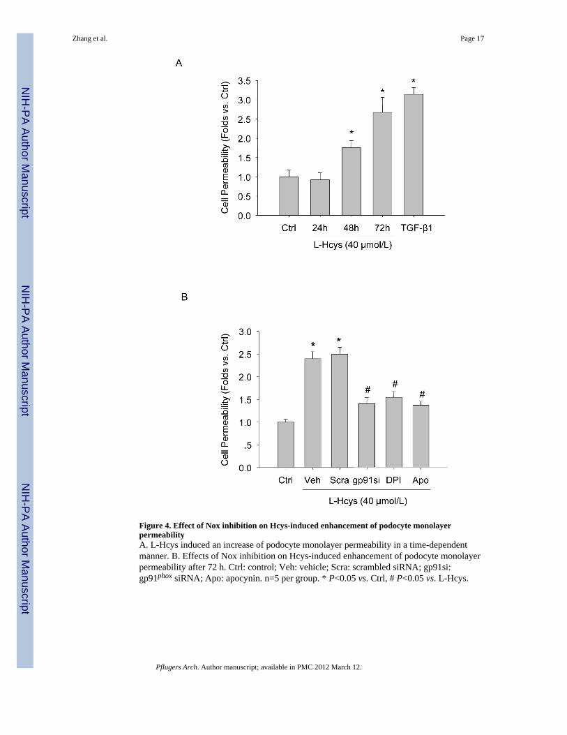
Figure 4. Effect of Nox inhibition on Hcys-induced enhancement of podocyte monolayerpermeabilityA. L-Hcys induced an increase of podocyte monolayer permeability in a time-dependentmanner. B. Effects of Nox inhibition on Hcys-induced enhancement of podocyte monolayerpermeability after 72 h. Ctrl: control; Veh: vehicle; Scra: scrambled siRNA; gp91si:gp91phox siRNA; Apo: apocynin. n=5 per group. * P<0.05 vs. Ctrl, # P<0.05 vs. L-Hcys.
Zhang et al. Page 17
Pflugers Arch. Author manuscript; available in PMC 2012 March 12.
NIH
-PA Author Manuscript
NIH
-PA Author Manuscript
NIH
-PA Author Manuscript

Figure 5. Expression of the mRNA of epithelial and mesenchymal markers in the glomeruliisolated from gp91phox KO and WT miceA and B. Real-time RT-PCR analysis for the expression of epithelial markers (P-cadherinand ZO-1) in the glomeruli of gp91 KO and WT mice (n=4 per group). C and D. RelativemRNA expression of mesenchymal markers (FSP-1 and α-SMA) in the glomeruli of gp91KO and WT mice (n=4 per group). * P<0.05 vs. WT mice on the normal diet; # P<0.05 vs.WT mice on the FF diet.
Zhang et al. Page 18
Pflugers Arch. Author manuscript; available in PMC 2012 March 12.
NIH
-PA Author Manuscript
NIH
-PA Author Manuscript
NIH
-PA Author Manuscript

Figure 6. Western blot analysis of P-cadherin, FSP-1, and α-SMA protein expression in mouseglomeruliA. A representative gel image (upper panel) shows the expression of P-cadherin in mouseglomeruli from different group of mice as indicated and the relative quantization of P-cadherin protein expression was summarized (lower panel). B. A representative gel image(upper panel) shows the expression of FSP-1 in mouse glomeruli from different group ofmice as indicated, and the relative quantization of FSP-1 protein expression was summarizedin the lower panel. C. A. A representative gel image (upper panel) shows the expression ofα-SMA in mouse glomeruli from different group of mice as indicated and the relative
Zhang et al. Page 19
Pflugers Arch. Author manuscript; available in PMC 2012 March 12.
NIH
-PA Author Manuscript
NIH
-PA Author Manuscript
NIH
-PA Author Manuscript

quantization of α-SMA protein expression was summarized in the lower panel. n=4.*P<0.05 vs. WT mice on the normal diet; # P<0.05 vs. WT mice on the FF diet.
Zhang et al. Page 20
Pflugers Arch. Author manuscript; available in PMC 2012 March 12.
NIH
-PA Author Manuscript
NIH
-PA Author Manuscript
NIH
-PA Author Manuscript

Figure 7. Podocyte EMT was attenuated in mice lacking gp91 phox geneA. Frozen kidney sections from WT or gp91phox KO mice were double-immunostained forP-cadherin or ZO-1 (Alexa 488, green color) and podocyte marker podocin (Alexa 555, redcolor). B. Mouse kidney sections were double-immunostained for mesenchymal markersFSP-1 or α-SMA (Alexa 488, green color) and podocin (Alexa 555, red color). n=6 pergroup.
Zhang et al. Page 21
Pflugers Arch. Author manuscript; available in PMC 2012 March 12.
NIH
-PA Author Manuscript
NIH
-PA Author Manuscript
NIH
-PA Author Manuscript

Figure 8. Protection of podocytes and glomeruli from hHcys-induced injury in gp91phox KOmiceA. PAS staining shows glomerular morphological changes (original magnification, ×400).B. gp91phox gene deletion improved podocyte ultrastructure in FF diet-treated mice asshown by TEM examination. Arrow denotes the area of foot process effacement in WT miceon the FF diet. Images are representative of 5 TEM images per kidney from 3 mice pergroup. Original magnification: ×8,000.
Zhang et al. Page 22
Pflugers Arch. Author manuscript; available in PMC 2012 March 12.
NIH
-PA Author Manuscript
NIH
-PA Author Manuscript
NIH
-PA Author Manuscript

NIH
-PA Author Manuscript
NIH
-PA Author Manuscript
NIH
-PA Author Manuscript
Zhang et al. Page 23
Table 1
Plasma total Hcys, urine albumin, creatinine clearance, glomerular damage index data in WT or gp91phox KOmice fed with normal diet or FF diet
ParameterWT-ND KO-ND WT-FF KO-FF
n=10 n=7 n=11 n=8
Total Hcys (μmol/L) 4.25±0.54 4.61±0.77 15.56±1.76* 14.66±2.02*
Urine albumin (μg/24h) 13.21±0.82 15.63±4.37 41.59±4.58* 23.61+2.36#
Ccr (μl/min) 76.09±5.43 74.60±4.80 50.36±2.28* 67.03±5.06#
GDI 0.70±0.15 0.80±0.13 2.80±0.36* 1.30±0.15#
ND: normal diet; FF: FF diet; Ccr: creatinine clearance; GDI: glomerular damage index. The data is expressed as mean ± SEM.
*p<0.05 as compared with WT-ND;
# p<0.05 as compared with WT-FF.
Pflugers Arch. Author manuscript; available in PMC 2012 March 12.


