Embed Size (px)
Citation preview
Theriogeno/ogy40:1107-1116,1993
EQUINE ANTISPERM ANTXBODIES (EASA): PRELIMINARY STUDY OF THE CLINICAL RESPONSE FOLLOWING BREEDING
IN IMMUNIZED MARES
G.J. Nie,a C. Lee, H.W. Momontb and H.S. Joo
Theriogenology Division, Dept. of Clinical and Population Sciences University of Minnesota, St. Paul, MN
Received for publication: January 12, 1993 Accepted: August 19, 7993
ABSTRACT
Maiden mares (n=6), previously injected with stallion sperm cells (SC group, n =2), stallion seminal plasma (SP group, n=2), or phosphate-buffered saline as a control (C group, n=2) were followed through 5 consecutive estrous cycles to evaluate their clinical response when exposed to stallion sperm cells via breeding. Management was similar to that expected on typical breeding farms. The mares were teased daily and bred by artificial insemination (AI) in all 5 cycles. Differences in serum and uterine flushing equine antisperm antibody (EASA) levels, endometrial culture and cytology results, endometrial biopsy score and fertility were evaluated between treatment groups. An enzyme-linked immunosorbant assay (ELISA) was used to determine serum and uterine IgG and IgA levels specific for sperm cell or seminal plasma antigens. Serum IgG specific for sperm cell antigen was higher in the SC group than in the SP and C groups following exposure to sperm cells via breeding (P<O.O5). All other EASA levels were not different between groups (P>O.O5); however, uterine IgA levels in one of the SC treated mares did rise over all 5 cycles. No differences were detected in culture, cytology, biopsy or fertility results between groups (P>O.O5). Changes in EASA levels were detected after breeding mares previously immunized with stallion sperm cells, however an associated clinical response was not apparent.
Key words: antisperm antibodies, EASA, ELISA, mare
Acknowledgments The authors wish to thank N. Kaplan for running the ELISA, Dr. V. King for statistical assistance and the ALG crew for their assistance in the barn when we were shorthanded. aCorrespondence and reprint requests; 385 An ScilVet Med Bldg, 1988 Fitch Ave, St. Paul, MN, USA, 55108 bCurrent address; Dept. of Medical Sciences, University of Wisconsin, 2015 Linden Dr. W., Madison, WI, USA, 53706-1102.
Copyright 0 1993 Butterworth-Heinemann
1108 Theriogenology
INTRODUCTION
Horses have the lowest reproductive efficiency of the common domestic species (12,16,34). There are many factors that play a role in this low efficiency. Poor breeding management, irregular estrons cycles and genital infections are commonly recognized sources of reduced fertility in horses (5); however, many clinically infertile equine cases never have an etiology identified. Undoubtedly there are multiple and IikeIy interrelated factors not yet recognized or understood playing a significant role in the horse’s low reproductive efficiency.
Antisperm antibodies (ASA) are recognized as a significant clinical problem by human fertility experts. A review of the literature indicates ASA is associated with a decreased fertilizing capacity in the rabbit, mouse, pig, cow, baboon and human (11,26,31,33). The role of equine antisperm antibodies (EASA) in fertility has not previously been investigated. To date, the only published reports we are aware of concerning ASA in horses are 2 case reports in which sperm cell immobilizing antibodies were detected in the seminal plasma of two stallions with reduced fertility (30,40). No controlled research has been published concerning the influence of EASA on equine fertility. The lack of information available about EASA along with the influence of ASA on fertility in other species establishes the need for a thorough investigation.
The information presented here is taken from a pilot project conducted as a part of an ongoing study investigating the role of EASA in equine fertility. We used an enzyme-linked immunosorbent assay (ELISA), developed in our laboratory (21), to investigate the clinical response following breeding in mares after previous induction of EASA.
MATERIALS AND METHODS
Experimental Groups
Six maiden, light-horse breed mares between 3 to 4 yr of age were tested for EASA by ELISA (21). The EASA absorbance levels of the project mares were similar to the mean EASA absorbance level in 20 virgin fillies. Each mare was evaluated for breeding soundness (32,36). All mares were housed in a dry lot with a pole-barn shelter. Diet included free-choice alfalfa/grass mixed hay and water.
The 6 mares were randomly assigned to 1 of 3 injection groups: sperm cell (SC), seminal plasma (SP), and control (C) for a total of 2 mares per group. Each mare was injected twice at the end of the 1990 breeding season. The immunogens injected were sonicated whole sperm cells, seminal plasma and phosphate-buffered saline (PBS) in the SC, SP and C groups, respectively. Immunogen preparation is reported by Lee et al. (21). The immunogen was carried in Freund’s complete adjuvant as an initial injection and Freund’s incomplete adjuvant 2 wk later as a booster. All injections were made subcutaneously. Immunogen for the SC and SP
Theriogenology 1109
groups were produced from semen pooled from 12 stallions. Post vaccination EASA levels are reported by Lee et al. (21). This project was conducted during the 1991 breeding season when serum EASA levels in all mares had fallen to near preinjection levels.
Sample/Data Collection
All mares were teased daily for 5 consecutive estrous cycles. Each mare’s uterus and ovaries were examined by palpation and ultrasonography (U/S) daily starting on Day 1 of behavioral estrus and continuing until ovulation was detected by U/S in each cycle. The mares were bred by artificial insemination in each cycle using fresh unextended semen collected from a stallion with proven fertility (this stallion’s semen was included in the immunogen pool). Breeding began when a 230 mm follicle was detected, but no earlier than Day 3 of behavioral estrus and continued every other day until ovulation was detected. Each mare was bred 1 to 7 (mean 3.2) times per cycle with between 0.650and 9.0(mean 4.1) x10’ sperm cells per inseminate. Pregnancy examinations using U/S were conducted on Day 15 following ovulation. Each mare was given a luteolytic dose of prostaglandina immediately following the pregnancy examination. Samples collected from each mare on Day 1 of behavioral estrus included: endomettial tissue specimens (Cycle 1, 3, and 5), 2 swab specimens of the endometrium, and uterine flushings (all 5 cycles).
Endometrial tissue specimens were obtained from the left or right uterine horn-body junction using an equine uterine biopsy forcepsb Swab specimens of the endometrium were obtained from the uterine body using a double guarded uterine culture swab. ’ Uterine flushings were obtained by transcervically infusing 60 ml of PBS into the uterus through a 26-French, 30-in, equine embryo transfer catheter.d The horns and body were massaged for 30 to 60 set before the fluid was recovered through the catheter. Uterine flushing samples were centrifuged to separate cellular debris. The flushing supematant was frozen until analyzed. Venous blood was collected into vaccutainerse on Day 1 of behavioral estrus, 7 d after ovulation, and at the pregnancy examination 15 d after ovulation. After clotting, serum was harvested and frozen until analyzed.
Sample Analysis
One swab specimen of the endometrium from each sampling was submitted
aLutalyse, The Upjohn Company, Kalamazoo, MI, 49001, USA. bNarco Pilling Co., Delaware Dr., Fort Washington, PA 19034, USA. ‘Har-Vet Inc. P.O. Box 6,217 McKay Ave, Spring Valley, WI 54767, USA. dSherwood Medical, St. Louis, MO 63130, USA. eBecton Dickson Vacutainer Systems, Rutherford, NJ.
1110 Theriogenology
for microbial culture. Samples were evaluated as positive or negative for growth of the common equine uterine pathogens (29). The second swab specimen was us
3 to
smear a slide for cytologic evaluation. The slide was stained with Dif Qui for cytologic evaluation. All slides were evaluated by the same individual for inflammatory cell type and quantity. Cell quantity was scored 0 to 4 (O=least, 4=most). The endometrial tissue specimens were fixed in Bouin’s fluid for at least 2 h before submission for slide preparation. Specimens were stained with hematoxylin and eosin (H&E) for evaluation. Endometrial tissue specimens were evaluated and assigned a category using Kenney’s system (20). Serum and uterine flushings were evaluated for EASA levels by enzyme linked immunosorbant assay (ELISA) procedures. The ELISA procedures were the same as described by Lee et al. (21). Uterine fluid samples were diluted 1:20 in PBS. Sperm cells and seminal plasma were used as coating antigens in separate ELISA plates. Semen from 10 stallions (including the breeding stallion used in this project) was pooled to make the antigen. Antigen specific IgG and IgA levels were measured by absorbance at 490 nm with an ELISA reader.g
Statistical Analysis
Contrasts analysis of variance was used to test for an effect of treatment on culture, cytologic and biopsy category results between treatment groups. A Chi- square test for heterogeneity was used to test for an effect of treatment in each cycle on conception rates among groups. Repeated measures analysis of variance was used to test for an effect of treatment and day on the levels of detectable antibody against sperm cells and seminal plasma between groups.
RESULTS
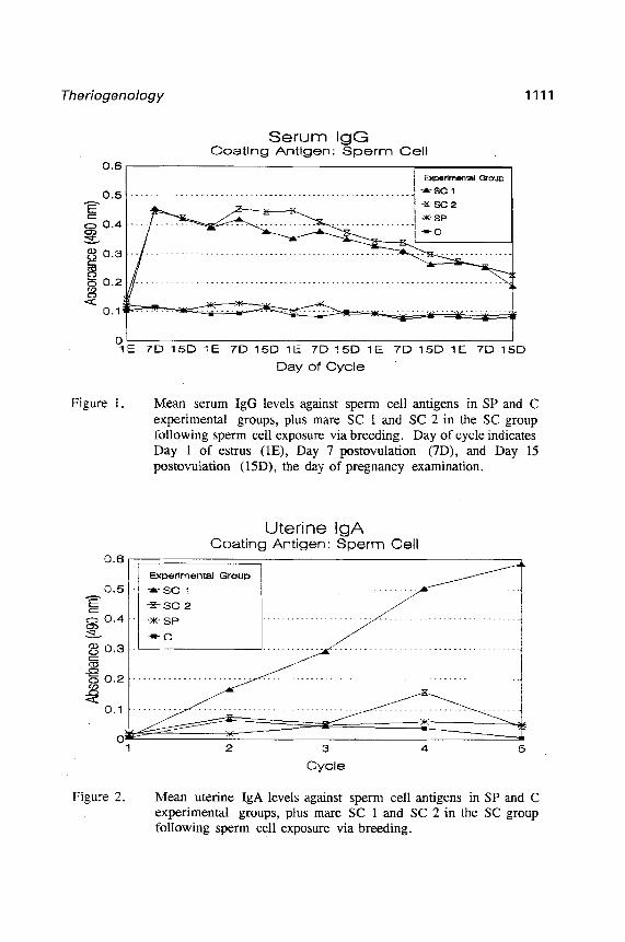
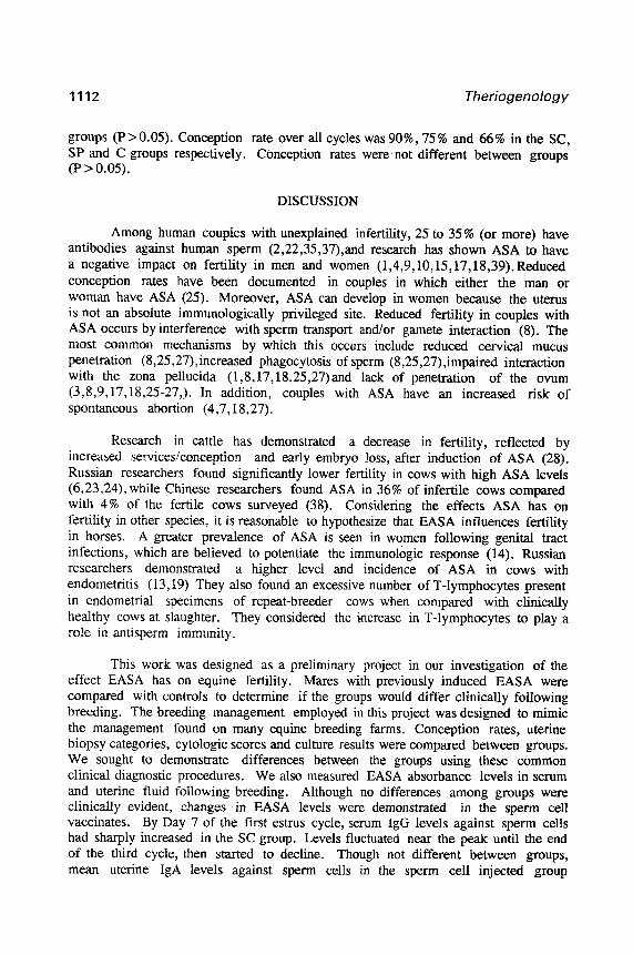
Figure 1 shows serum IgG levels and Figure 2 shows uterine IgA levels against sperm cells over all cycles. The SC group individuals are plotted seperately, while the SP and C group individuals are represented by the mean of each group. There was a difference in mean serum IgG levels (Figure 1) between groups (SC vs C and SC vs SP) for all samples collected after Day 1 of behavioral estrus in Cycle 1 (P<O.O5). Uterine IgA against sperm cells in one SC group individual (SC 1) continued to increase over all cycles (Figure 2). However, mean uterine IgA against sperm cells in the SC group was not different from the SP (P=O.123) or C (P=O.102) groups. There was no difference in serum IgG against seminal plasma, serum IgA against sperm cells or seminal plasma, or uterine IgG against sperm cells or seminal plasma (P>O.O5).
All cultures were negative for growth of uterine pathogens. There was individual cytology and biopsy sample variation but results did not differ between
fBaxter Scientific Products, Miami, FL 33152, USA. gMicroElisa .\utoreader MR 580, Dynatech Laboratories Inc., Chantilly, CA.
Theriogenology 1111
Serum IgG Coating Antigen: Sperm Cell
0.6
0.5
-is
z 0.4
!z-
3 0.3
802 Lo .
$ 0.1:
OL 1E 7D15D 1E 7D15D 1E 7D15D 1E 7D15D 1E 7D15D
Day of Cycle
Figure 1. Mean serum IgG levels against sperm cell antigens in SP and C experimental groups, plus mare SC 1 and SC 2 in the SC group following sperm cell exposure via breeding. Day of cycle indicates Day 1 of estrus (lE), Day 7 postovulation (7D), and Day 15 postovulation (LSD), the day of pregnancy examination.
Uterine IgA Coating Antigen: Sperm Cell
3
Cycle
Figure 2. Mean uterine IgA levels against sperm cell antigens in SP and C experimental groups, plus mare SC 1 and SC 2 in the SC group following sperm cell exposure via breeding.
1112 Theriogenology
groups (P>O.O5). Conception rate over all cycles was 90%, 75% and 66% in the SC, SP and C groups respectively. Conception rates were.not different between groups (P>O.O5).
DISCUSSION
Among human couples with unexplained infertility, 25 to 35% (or more) have antibodies against human sperm (2,22,35,37),and research has shown ASA to have a negative impact on fertility in men and women (1,4,9,10,15,17,18,39).Reduced conception rates have been documented in couples in which either the man or woman have ASA (25). Moreover, ASA can develop in women because the uterus is not an absolute immunologically privileged site. Reduced fertility in couples with ASA occurs by interference with sperm transport and/or gamete interaction (8). The most common mechanisms by which this occurs include reduced cervical mucus penetration (8,25,27),increased phagocytosis of sperm (8,25,27),impaired interaction with the zona pellucida (1,8,17,18,25,27)and lack of penetration of the ovum (3,8,9,17,18,25-27,). In addition, couples with ASA have an increased risk of spontaneous abortion (4,7,18,27).
Research in cattle has demonstrated a decrease in fertility, reflected by increased services/conception and early embryo loss, after induction of ASA (28). Russian researchers found significantly lower fertility in cows with high ASA levels (6,23,24), while Chinese researchers found ASA in 36% of infertile cows compared with 4% of the fertile cows surveyed (38). Considering the effects ASA has on fertility in other species, it is reasonable to hypothesize that EASA influences fertility in horses. A greater prevalence of ASA is seen in women following genital tract infections, which are believed to potentiate the immunologic response (14). Russian researchers demonstrated a higher level and incidence of ASA in cows with endometritis (13,19) They also found an excessive number of T-lymphocytes present in endometrial specimens of repeat-breeder cows when compared with clinically healthy cows at slaughter. They considered the increase in T-lymphocytes to play a role in antisperm immunity.
This work was designed as a preliminary project in our investigation of the effect EASA has on equine fertility. Mares with previously induced EASA were compared with controls to determine if the groups would differ clinically following breeding. The breeding management employed in this project was designed to mimic the management found on many equine breeding farms. Conception rates, uterine biopsy categories, cytologic scores and culture results were compared between groups. We sought to demonstrate differences between the groups using these common clinical diagnostic procedures. We also measured EASA absorbance levels in serum and uterine fluid following breeding. Although no differences among groups were clinically evident, changes in EASA levels were demonstrated in the sperm cell vaccinates. By Day 7 of the first estrus cycle, serum IgG levels against sperm cells had sharply increased in the SC group. Levels fluctuated near the peak until the end of the third cycle, then started to decline. Though not different between groups, mean uterine IgA levels against sperm cells in the sperm ceil injected group
Theriogenology 1113
continued to rise over the first 4 cycles then appeared to level off at the fifth cycle. In Mare 11, uterine IgA levels continued to rise over all cycles. The importance of the serum and uterine Ig rise is not clear from these results. However, the serum and uterine EASA response to uterine insemination with sperm cells, demonstrated in this project, suggest further investigation is needed to define its relationship to fertility. As seen in humans, not every individual with detectable ASA levels has clinically detectable infertility and certainly some women produce very specific ASA against their partner’s sperm cells.
Further investigation is needed to define the importance of EASA in equine fertility. A survey of EASA levels in mares and stallions with known fertility histories would provide some indication of the importance of EASA. All of the mares used in this project were young, healthy and presumably fertile horses. The effect of EASA in older horses and those with other reproductive problems also needs to be investigated.
REFERENCES
1. Alexander NJ, Bearwood BS. An immunosorption assay for antibodies to spermatozoa: comparison with agglutination and immobilization tests. Fertil Steril 1984;41:270-276.
2. Alexander NJ, The effects of antibody sperm-egg interaction. Ann NY Acad Sci 1988;541:317-323.
3. Barratt CL, Havelock LM, Harrison PE, Cooke ID. Antisperm antibodies are more prevalent in men with low sperm motility. Int J Androl 1989;12:2: 110-116.
4. Bronson R, Cooper G, Rosenfeld D. Sperm antibodies: their role in infertility. Fertil Steril 1984;42:171-183.
5. Caslick EA. The sexual cycle and its relation to ovulation with breeding records of the thoroughbred mare. Cornell Vet 1937;27:187-206.
6. Chanturidze RD. Immunological factors in the pathology of pregnancy in the cow. Veterinariya 1975;4:74-75.
7. Cimino C, Barba G, Salerno P, Cittadini E. The effect of female serum antisperm antibodies on in vitro fertilization and embryo transfer. Acta Eur Fertil 1989;20:5:299-303.
8. Clarke GN, Hyne RV, du-Plessis Y, Johnston WIH. Sperm Antibodies and human in vitro fertilization. Fertil Steril 1988;49:6:1018-1025.
9. Cropp CS, Schlaff WD. Antisperm antibodies. Arch Immunol Ther Exp 1990;138: l-2:23-30.
1114 Theriogenology
10. Cunningham DS, Fulgham DL, Ray1 DL, Hansen KA, Alexander NJ. Antisperm antibodies to sperm surface antigens in women with genital tract infection. Am J Obstet Gynecol 1991;164:3:791-796.
11. Fayemi OE, Joo HS, Crab0 BG. Effect of immunization with sperm or seminal plasma on spermatozoal quality in boars. Animal Reprod Sci 1990;23:245-251.
12. Ginther OJ. Reproductive efficiency. In: Ginther OJ (ed), Reproductive Biology of the Mare. Basic and Applied Aspcects. Equiservices, Cross Plains, WI,
13.
14.
15.
16.
17.
18.
19.
20.
-_ 1992;499-562.
Gukov FD. Dynamics of T-Lymphocytes and macrophages in the endometrium of cows at different ages and during infertility of immunological origin. Patomorfologiya Patogenez I Diagnostika Boleznei, Shishkov, 1980;58-59.
Haas GG, Kubota K, Quebbeman JF, Jijon A, Menge AC! Beer AE. Circulating antisperm antibodies in recurrently aborting women. Fertil Steril 1986;45:209- 215.
Haas GG. Antibody-mediated causes of male infertility. Urol Clin North Am 1987; 14:3:539-550.
Hafez ESE. Reproduction in farm animals. Lea & Febiger, Philadelphia, 1980;387-408.
Hare1 W, Nelken D. An enzyme immunoassay for the detection of antisperm antibodies. Am J Reprod Immunol Microbial 1985;8: 137-140.
Ing RM, Wang S, Brennecke AM, Jones WR. An improved indirect enzyme- linked immunosorbent assay (ELISA) for the detection of antisperm antibodies. Am J Reprod Immunol Microbial i!%5;8:15-19.
Ivanov AT, Malinovskii IF, Pankovets EA. Immune reactions to sperm antigens in cattle with puerperal endometritis. Trudy Belorusskii Nauchno Issledovatel’skii Veterinamyi Institut 1975; 13: 168-170.
Kenney RM. Cyclicand pathologic changes of the mare endometrium as detected by biopsy, with a note on early embryonic death. J Am Vet Med Assoc 1978;172:3:241-262.
21. Lee C, Nie GJ, Joo HS, Momont HM. An enzyme-linked immunosorbent assay (ELISA) for detection of antisperm antibodies in horse serum. Theriogenology 1993;40:117-1125.
22. Liu DY, Clarke GN, Baker HW. Inhibition of human sperm-zona pellucida and sperm-oolemma binding by antisperm antibodies. Fertil Steril 1991;55:2:440-442
Theriogenology 1115
23. Luk’yanenko VM. Immunobiological status ‘of infertile cows, with reference to spermatozoa agglutinins. Veterinariya 1978; 10:72-73.
24. Malinovskii IF. Conception rate of cows in relation to titres of sperm antibody in blood serum. Dostizheniya Veterinarnoi Nauki i Peredovogo Opyta Zhivotnovodstvu 1976;2:77-78.
25. Mandelbaum SL, Diamond MP, DeChemey AH. Relationship of antisperm antibodies to oocyte fertilization in in vitro fertilization-embryo transfer. Fertil Steril 1987;47:4:644-651.
26. Mandelbaum SL, Diamond MP, DeChemey AH. The impact of antisperm antibodies on human infertility. J Uroi 1987;138:1:1-8.
27. Menge AC, Beitner 0. Interrelationships among semen characteristics, antisperm antibodies and cervical mucus penetration assays in infertile human couples. Fertil Steril 1989;51:3:486-492.
28. Menge AC. Induced infertility in cattle by iso-immunization with semen and testis. J Reprod Fertil, 1967;13:445-456.
29. Neely DP. Evaluation and therapy of genital disease in the mare. In: Hughes JP (ed), Equine Reproduction. Hoffmann-La Roche Inc, Nutley, NJ, 1983;39-56.
30. Papa FO, Xlvarenga MA, Lopes MD, Campos Filho EP. Infertility of autoimmune origin in a stallion. Eq Vet J, 1990;22:2:145-146.
31. Peterson RN. Rob1 JM, Dziuk PJ, Russel LD. The effects of antisperm plasma membrane antibodies on sperm-egg binding, penetration and fertilization in the pig. J Exp Zoo1 1982;223:1:79-81.
32. Pierson RH. Ward JK, Gloyd J, Meirs DA, Whitherspoon DM, McGee W. Theriogenology and the equine. Part I. The brood mare. J Society For Theriogenology, vol VIII. Society For Theriogenology, Hastings, NE, 1978.
33. Smith M, Peterson RN, Russell LD. Penetration of zona-free hamster eggs by boar sperm treated with the ionophore A23187 and inhibition of penetration by antiplasma membrane antibodies. J Exp Zoo1 1983;225: 1: 157-160.
34. Sullivan JJ. Turner PC, Self LC, Gutteridge HB, Bartlett DE. Survey of reproductive efficiency in the quarter-horse and thoroughbred. J Reprod Fertil 1975;(Suppl ‘3):315-318.
35. Tsukui S, Noda Y, Yano J, Fukuda A, Mori T. Inhibition of sperm penetration through human zona pellucida by antisperm antibodies. Fertil Steril 1986;46: 1:93-96.
1116 Theriogenology
36. Van Camp SD. Breeding soundness examination of the mare and common genital abnormalities encountered. In: Morrow DA (ed), Current Therapy in Theriogenology 2. WB Saunders Company, Philadelphia, 1986;654-661.
37. Vazquez-Levin M, Kaplan P, Guzman I, Grunfeld L, Garrisi GJ, Navot D. The effect of female antisperm antibodies on in vitro fertilization, early embryonic development, and pregnancy outcome. Fertil Steril 1991;56: 1:84-88.
38. Wang GL, Xie CX. The relationship between antisperm antibodies and progesterone in the serum of infertile dairy cows. Acta Vet Zootech Sinica 1990;21:1:26-30.
39. Witkin SS, David SS. Effect of sperm antibodies on pregnancy outcome in a subfertile population. Am J Obstet Gynecol 1988; 158: 1:59-62.
40. Zhang J, Ricketts SW, Tanner SJ. Antisperm antibodies in the semen of a stallion following testicular trauma. Eq Vet J 1990;22:2:138-141.