Embed Size (px)
Citation preview
Proteomic analyses of monocytes obtained from Hispanicwomen with HIV-associated dementia show depressedantioxidants
Stephanie Kraft-Terry1, Yamil Gerena2,3, Valerie Wojna2,4, Marines Plaud-Valentin2,Yolanda Rodriguez2, Pawel Ciborowski1, Raul Mayo2,5, Richard Skolasky6, Howard E.Gendelman1, and Loyda M. Meléndez2,7
1 Department of Pharmacology and Experimental Neuroscience, University of Nebraska MedicalCenter, Omaha, NE2 Specialized Neuroscience Program in NeuroAIDS, University of Puerto Rico Medical SciencesCampus, San Juan, PR3 School of Pharmacy, University of Puerto Rico Medical Sciences Campus, San Juan, PR4 Department of Internal Medicine, Neurology Division, University of Puerto Rico MedicalSciences Campus, San Juan, PR5 Department of Physical Medicine, University of Puerto Rico Medical Sciences Campus, SanJuan, PR6 Department of Orthopedic Surgery, Johns Hopkins University, Baltimore, MD7 Department of Microbiology, School of Medicine, University of Puerto Rico Medical SciencesCampus, San Juan, PR
AbstractMonocyte ingress into the brain during progressive human immunodeficiency virus (HIV-1)infection parallels the severity of cognitive impairments. Although activated monocyte phenotypesemerge during disease, the functional correlates of these cells remain unresolved. To this end, westudied the proteome of blood-derived monocytes obtained from Hispanic women with the mostsevere form of HIV-associated neurocognitive disorder, HIV-associated dementia (HAD).Monocytes isolated from peripheral blood mononuclear cells by CD14+ immunoaffinity columnchromatography were >95% pure. Cells were recovered from five patients without evidence ofcognitive impairment and four with HAD and analyzed by two-dimensional difference gelelectrophoresis and tandem mass spectrometry. Importantly, ADP ribosylhydrolase,myeloperoxidase, thioredoxin, peroxiredoxin 3, NADPH, and GTPase activating protein were alldownregulated in HAD. In regards to myeloperoxidase, thioredoxin, and peroxiredoxin 3 thesechanges were validated in an additional cohort of 30 patients by flow cytometry. We conclude thatdeficits in monocyte antioxidant proteins lead to neuronal damage through the loss of hydrogenperoxide scavenging capabilities, thus exposing neurons to apoptosis-inducing factors. Alteredmonocyte functions therefore may contribute to the development and progression of HAD.
Keywordsmonocytes; proteomics; 2D-DIGE; mass spectrometry; HIV-1; HAND; HAD
*Corresponding Author: Loyda M. Meléndez, PhD, Professor, Department of Microbiology, School of Medicine, Medical SciencesCampus, San Juan, Puerto Rico 00935, Phone: 787-758-6132, Fax: 787-777-0078, [email protected].
NIH Public AccessAuthor ManuscriptProteomics Clin Appl. Author manuscript; available in PMC 2011 May 20.
Published in final edited form as:Proteomics Clin Appl. 2010 September ; 4(8-9): 706–714. doi:10.1002/prca.201000010.
NIH
-PA Author Manuscript
NIH
-PA Author Manuscript
NIH
-PA Author Manuscript
Statement of Clinical Relevance
HIV-associated neurocognitive disorder (HAND) affects up to 50% of virus-infectedpeople during the course of disease. Clinical features of neurocognitive dysfunctionranges from asymptomatic neurocognitive impairment, to minor cognitive motordisorder, to severe cognitive, motor, and behavioral impairments. The latter is calledHIV-associated dementia (HAD) and can affect up to 7% of infected people. Despite theadvent of antiretroviral therapy (ART), the incidence of HAND has remained constant.However, the diagnosis of HAND is made after a battery of neuropsychological tests andafter exclusion of opportunistic infections, psychiatric disorders, and malignancies. Earlybiomarkers are desperately needed to detect, stage, and monitor disease when therapeuticinterventions would prove most helpful. Because HIV-1-infected monocytes pass throughthe blood-brain barrier and differentiate to perivascular macrophages, we chose toanalyze their potential proteomic alterations in a well-characterized Hispanic longitudinalcohort of HIV-1-infected ART-treated women. A significant decrease in specificantioxidant proteins during progressive cognitive impairment were discovered andvalidated by flow cytometry testing. These studies support the significance of the role ofcirculating monocytes in the neuropathogenesis of HIV-1 infection and provide novelblood markers that can readily be applied to the clinic.
IntroductionHIV-associated neurocognitive disorder (HAND) affects up to 50% of HIV-infected peopleduring the course of viral infection. Disease severity for HAND ranges from asymptomaticneurocognitive impairment, to minor cognitive motor disorder, to severe cognitive, motor,and behavioral impairments or HIV-associated dementia (HAD) [1]. HAD can affect 7% ofinfected people during the disease course. Despite the advent of antiretroviral therapy(ART), the incidence of HAND has remained constant albeit in less debilitating forms [2].The underlying mechanisms for neuronal dysfunction or death in disease involveneuroinflammatory responses heralded by infiltrating HIV-1-infected mononuclearphagocytes (MPs; blood-borne monocytes and macrophages and microglia) [3]. InfiltratingMPs carry infection from the periphery to the central nervous system (CNS) andsubsequently initiate a cascade of glial (astrocytes and microglia) autocrine and paracrineimmune events, resulting in the secretion of pro-inflammatory neurotoxic factors and viralproteins [4–6]. MPs also produce chemokines that attract further inflammatory cell CNSinfiltration that perpetuates the neurotoxic cascade [7,8]. Once inside the brain, HIV-1-infected MPs establish a long-lived viral reservoir and serve as a continuous source ofneurotoxins [9].
MPs are responsible for scavenging cellular debris, regulating oxidative stress, andprotecting the host from outside insults. Lack of control of oxidative stress may alsocontribute to neurodegeneration during progressive viral infection. Nonetheless, alterations,if any, that occur in peripheral monocytes that influence their destructive effects as theyingress within the CNS are not known. What is known is that homeostatic maintenancefunctions of blood-borne monocytes are broken and parallel the metabolic changes leadingto an encephalopathy. These metabolic changes are characterized by behavioral, motor, andcognitive impairments [10].
In recent years proteomic analyses of body fluids [plasma and cerebrospinal fluid (CSF)]and monocytes from HIV-1-infected individuals with cognitive impairment have uncoveredprotein changes that reflect CNS disease [11–13]. Parallel studies investigated HIV-1-
Kraft-Terry et al. Page 2
Proteomics Clin Appl. Author manuscript; available in PMC 2011 May 20.
NIH
-PA Author Manuscript
NIH
-PA Author Manuscript
NIH
-PA Author Manuscript
infected monocyte-derived macrophages (MDM) [14–16], yielding a broad range of proteinslinked to cytoskeleton remodeling, motility, oxidative regulation, vesicular trafficking, andimmune responses. Because HIV-1-infected monocytes pass through the blood-brain barrierand differentiate to perivascular macrophages, we chose to analyze the changes induced inmonocytes from patients with HAD in a well-characterized Hispanic longitudinal cohort ofHIV-1-infected ART-treated women. We found a significant decrease in specific antioxidantproteins during progressive cognitive impairment. These studies support the significance ofthe role of circulating monocytes in the neuropathogenesis of HIV-1 infection and providenew therapeutic targets for disease.
Materials and MethodsPatients
The Hispanic/Latino Longitudinal NeuroAIDS cohort of HIV-seropositive women with orwithout cognitive dysfunction was recruited from two well-established primary HIV clinicsin Puerto Rico [17]: the Latin American Center for Sexually Transmitted Diseases at thePuerto Rico Medical Center and the Center of Maternal-Infant HIV Infections at theUniversity of Puerto Rico Medical Sciences Campus (UPR-MSC). The selection criteriawere HIV-1-infected Puerto Rican women at least 18 years of age, with CD4+ T lymphocytecounts of ≤500, at least a 9th grade education, and no evidence of either active systemicinfection or neurodegenerative disease. The educational level was a requirement for theneuropsychological tests. The clinical study was approved by the UPR-MSC InstitutionalReview Board (IRB) and was conducted with the informed consent of the participants.
Evaluations of participantsEvaluations were performed as described previously in Wojna et al., 2006. Followingconsent, individual participants were required to provide demographics and pertinentmedical histories. The information included age at enrollment and the most likely mode ofHIV-1 transmission. Evaluation of cognitive function included history andneuropsychological tests. Plasma and CSF viral loads were determined using UltrasensitiveRNA Roche Amplicor determination, at an AIDS Clinical Trial Group (ACTG) CertifiedLaboratory, of CD4+ and CD8+ T-cell counts by flow cytometry. Lumbar punctures wereperformed once a year. Neurological evaluation consisted of a mental status examination,sensory functions (including response slowing, speed of thought, and language), testing ofbehavior and mood, as well as standard neurologic evaluations of cranial nerves, cerebellar,motor, reflexes, and sensory evaluations. The neuropsychological evaluation included thetests described previously [1,17]. These included tests of verbal memory (subtests 5th trial,memory, and recognition of the Rey Auditory Verbal Learning Test), frontal executivefunction (Stroop word/color and Trail Making B), psychomotor speed (Symbol DigitModalities Test and visual and auditory reaction time non dominant hand), and motor speed(Trail Making A and Grooved Pegboard dominant and nondominant hand). All tests wereconducted on all patients in Spanish. The Beck Depression Index was used to screen fordepression. Both the neurologist and the neuropsychologist were blinded to each other’sfindings. To develop normative values (z-scores) of the neuropsychological tests in PuertoRican women, we identified a group of HIV-1-seronegative women matched for age,education, and sociodemographic status.
Cognitive impairment was determined with the use of the American Academy of NeurologyHIV-associated dementia criteria [1]. In this study 39 HIV-1-seropositive womencharacterized for cognitive function were accrued (20 with normal cognition and 19 withHAD).
Kraft-Terry et al. Page 3
Proteomics Clin Appl. Author manuscript; available in PMC 2011 May 20.
NIH
-PA Author Manuscript
NIH
-PA Author Manuscript
NIH
-PA Author Manuscript
Monocyte isolation from the HIV-infected women cohortCD14+ monocytes were isolated from peripheral blood mononuclear cells (PBMC) of HIV-seropositive patients by immune chromatography columns containing antibodies againstCD14 (Miltenyi Biotech) with high purity (98% CD14+) and protein recovery.
Sample purification and preparationCell lysates were prepared from CD14+ purified monocytes from five HAD and five normalcontrol donors. These lysates were purified using 2D Clean-up (GE Healthcare) andresuspended in 100 μL 0.1% TritonX-100 (Fisher). Acquired protein was quantified using aBiorad Dc protein quantification kit. Five micrograms of protein was aliquoted from eachsample, and another 5 μg from each sample was pooled for an internal standard. Sampleswere dried by speedvac and resuspended in DIGE lysis buffer (30 mM Tris pH 8.0, 7 Murea, 2 M thiourea, 4% CHAPS). Utilizing saturation labeling for low abundant samples(GE Healthcare), these were reduced by treatment with 2 nmol TCEP for 1 hour at 37 ° Cand labeled with 4 nmol Cy5 for experimental samples and Cy3 for the pooled internalstandard for 30 minutes at 37 ° C. The reaction was stopped with the addition of 2x samplebuffer (7 M urea, 2 M thiourea, 4% CHAPS, 2% pharmalyte, 130 mM DTT) [11].
PBMCs were obtained from leukophoresis at the University of Nebraska Medical Center.They were labeled with CD14+ selection micro beads (Miltenyi Biotech) and passedthrough an MS positive selection column (Miltenyi Biotech) for magnetic separation.Monocyte purity was examined by staining with CD14-FITC antibody (Miltenyi Biotech)and analyzed using FACS. Purity was determined to be >99%. Monocytes were lysed usingDIGE lysis buffer, purified using a 2D-Clean-up kit, and quantified using a 2D-Quant kit(GE Healthcare). Four hundred micrograms of protein was reduced with 200 nmol TCEP for1 hour at 37 ° C followed by labeling with 400 nmol Cy 3 for 30 minutes at 37 ° C. Reactionwas stopped by adding 1x sample buffer (7 M urea, 2 M thiourea, 4% CHAPS) to a totalvolume of 445.5 μL, 4.5 μL pharmalytes, and 4.5 mg of DTT.
Isoelectric focusingIn-gel rehydration was performed combining equal volumes of labeled sample and internalstandard for analytical gels or Cy3 labeled monocyte lysates for preparative gelelectrophoresis with rehydration buffer (7 M urea, 2 M thiourea, 4% CHAPS, 1%pharmalytes, 13 mM DTT). Samples were then loaded into the rehydration chamber with apH 3-10, 24-cm immobiline dry strip (GE Healthcare), covered with dry strip cover fluid(GE Healthcare), and incubated at room temperature overnight. Rehydrated strips weretransferred to Ettan IPGphor II for 1st dimension separation (isoelectric focusing, IEF). IEFwas carried out at a constant 20 ° C for 93.7 kVh (500 V for 0.5 kVh, gradient to 1000 V for6 kVh, gradient to 8000 V for 13.5 kVh, 8000 V for 30 kVh, gradient to 10000 V for 16.5kVh, 10000 V for 27.2 kVh) [11].
2D difference gel electrophoresis (2D-DIGE)After IEF, strips were incubated in equilibration solution (6 M urea, 0.1 M Tris pH 8.0, 30%glycerol, 2% SDS, 0.5% DTT) for 15 minutes with gentle agitation. Strips were then loadedonto the top of 10–20% gradient polyacrylamide gels and fixed with 0.5% agarose sealingsolution containing bromophenol blue. Second dimension separation was carried out usingthe Ettan Daltsix Electrophoresis System (GE Healthcare) at 25 ° C. Current was heldconstant at 10 mAmps per gel for the first hour and then at 12 mAmps per gel overnight,until bromophenol blue reached the gel bottom. Visualization of gel spots was performedusing a Typhoon 9410 Variable Mode Imager (GE Healthcare, Piscataway, NJ). Gels werescanned at 100 μm resolution collecting signals for Cy3 and Cy5 at excitation wavelengths
Kraft-Terry et al. Page 4
Proteomics Clin Appl. Author manuscript; available in PMC 2011 May 20.
NIH
-PA Author Manuscript
NIH
-PA Author Manuscript
NIH
-PA Author Manuscript
of 520 and 620 nm, respectively. Images were imported into DeCyder 6.5 (GE Healthcare,Piscataway, NJ) software for spot detection and analysis of differential expression throughbiological variation analysis. One gel from a HAD sample was not used for analysis due topoor protein separation leaving four HAD samples for analysis. Spots with p-values ≤ 0.05were selected for spot picking by means of an automatic Ettan Spot Picker (GE Healthcare,Piscataway, NJ) using a 2.0-mm diameter picking head [11].
In-gel tryptic digestionGel pieces were washed with 200 μL 50% H2O/50% acetonitrile (ACN) for 5 minutes, 200μL 50% ACN/50 mM NH4HCO3 for 30 minutes, 200 μL 50% ACN/ 10 mM NH4HCO3 for30 minutes. Gel pieces were dried by speedvac, treated with trypsin (Promega) and 50μL 10mM NH4HCO3, and incubated overnight at 37 ° C. Peptides were extracted throughincubation of spots with 200 μL 0.1% TFA, 60% ACN for 2 hours. Extracted peptides weredried using a speedvac concentrator. Peptides were resuspended in 20 μL 0.5% TFA andpurified using ZipTips (Millipore, Billerica, CA) following the manufacturer’s protocol.
Protein identificationPeptides were fractionated on a microcapillary RP-C18 column (NewObjectives, Woburn,MA) followed by fragmentation using the ESI LC MS/MS system (ProteomeX System withLCQDecaPlus, ThermoElectron, Inc., San Jose, CA) in a nanospray configuration. Thespectra obtained from mass spectrometric analyses were searched with the use of theSequest search engine (BioWorks 3.2 software from ThermoElectron Inc., San Jose, CA).For high-confidence protein identification, at least two unique peptides sequenced wererequired [11].
Flow cytometryTwo-color flow cytometry was used for intracellular detection of antioxidant expression inmonocytes from frozen PBMC of HIV-positive women. Fifteen samples of each group(normal cognition and HAD) were tested independently for intracellular expression ofantioxidants. PBMC were thawed and incubated with CD14-PE monoclonal antibody (BDBiosciences, CA) for 30 minutes at 4oC. The cells were washed two times with PBS (1X)/FBS (3%) by centrifugation at 1,100 rpm for 5 minutes at 4oC. Cells were permeabilizedwith cytoperm/cytofix solution (BD Biosciences, CA) for 20 minutes followed byincubation with FITC conjugated antibodies against SOD-1, peroxiredoxin, andmyeloperoxidase, thioredoxin for 30 minutes, washed by centrifugation and fixed with 0.5%paraformaldehyde. Cells were analyzed by flow cytometry in a FACS Calibur (BDBiosciences). The Cell Quest software (BD Biosciences, CA) was used for data acquisitionand multivariate analysis. Monocytes were gated in FSC vs. SSC dot plot by size andgranularity, and subsequently the CD14+ cells were identified in FL2 dot plot. Intracellularmarkers were identified in the FL1 channel. Ten thousand events were evaluated for eachsample and the mean fluorescence intensity (MFI) of the cells was determined from themedian peak channel of the histograms. Data on scatter parameters and histograms wereacquired in log mode. Monocyte autofluorescence was subtracted from the fluorescenceintensity values of the stained samples.
Statistical methodsHotelling’s t-squared statistic was used to significance for flow cytometry data (p ≤ 0.05).Hotelliing’s t-squared statistic is analogous to Student’s t-test in that it makes comparisonsbetween two groups. It differs in that it simultaneously adjusts for multiple comparisons,thereby preserving overall experimental Type I error. One-way ANOVA analysis was usedto evaluate significance for 2D-DIGE data (p ≤ 0.05).
Kraft-Terry et al. Page 5
Proteomics Clin Appl. Author manuscript; available in PMC 2011 May 20.
NIH
-PA Author Manuscript
NIH
-PA Author Manuscript
NIH
-PA Author Manuscript
ResultsParticipants
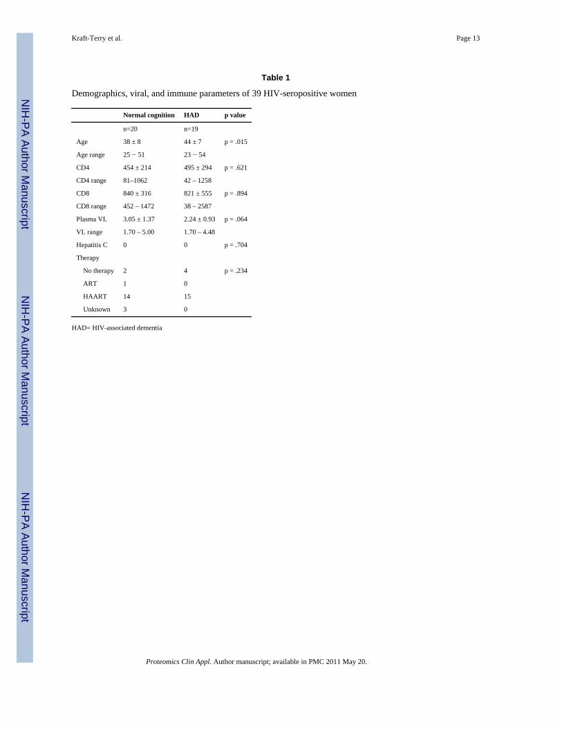
Table 1 lists the demographic, neurologic, immune, and viral statuses of the HIV-1-seropositive Hispanic women included in this study. Those with HAD were older on averagethan those with normal cognition (44 years vs. 38 years, p=.015). There was no differencebetween those with normal cognition and those with HAD with respect to CD4 or CD8 cellcounts, plasma HIV viral load, or type of anti-retroviral therapy use. For this study,individuals with no histories of intravenous drug abuse or co-infections were selected.
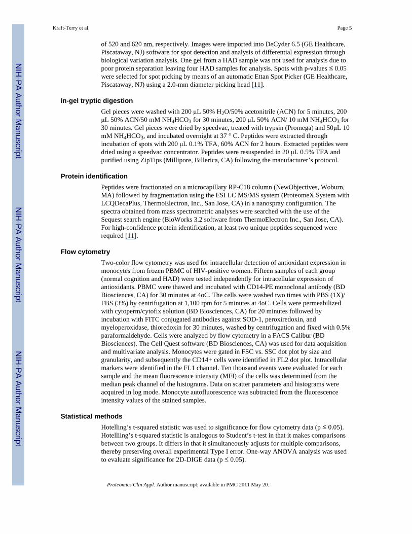
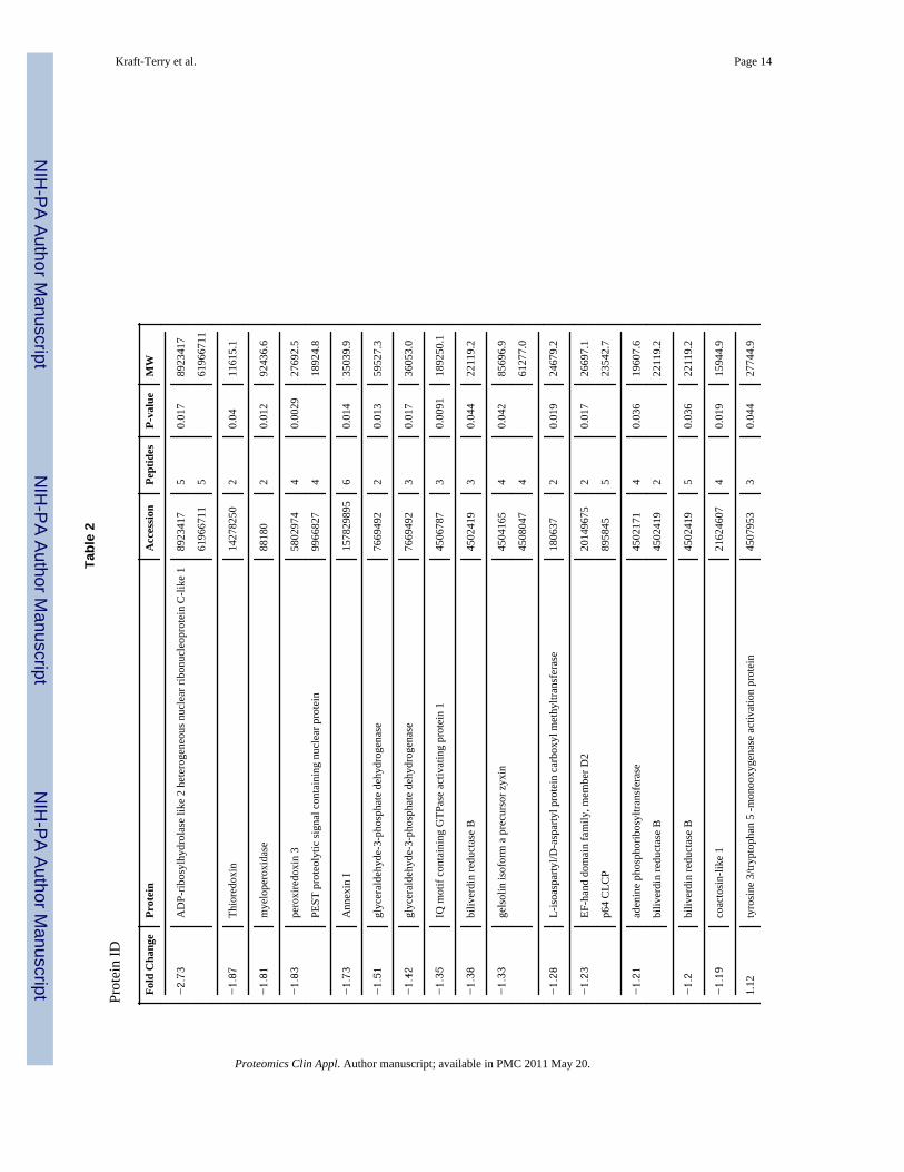
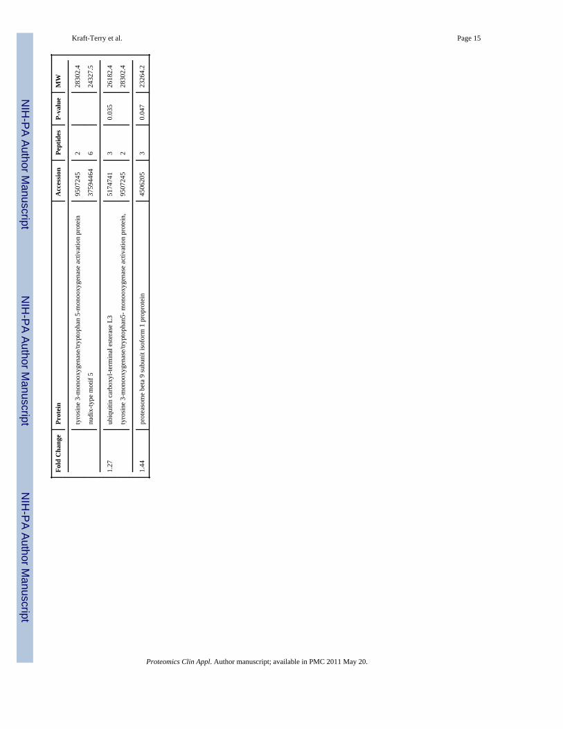
2D-DIGE and protein identification by LC MS MS2D-DIGE analysis identified a between 4221 and 4506 spots in each individual gel. Aftermatching all 9 gels together 18 spots were determined to be significantly differentiallyregulated between normal cognition and HAD with p ≤ 0.05. Six of these significant spotshad a fold change ≥ 1.5 or ≤ −1.5, the arbitrary cut-off value that was selected (Table 2).Because the fold changes observed in this experiment were modest, we chose a slightly lessstringent cut-off with the understanding that we must validate our findings [18]. Three ofthese downregulated spots were identified to be anti-oxidant proteins: myeloperoxidase,peroxiredoxin 3, and thioredoxin (Figure 1). Because we previously identified differences inantioxidant protein levels in individuals with HAD [19], we chose these proteins to bevalidated by flow cytometry from frozen PBMC of an additional 30 participants from thecohort.
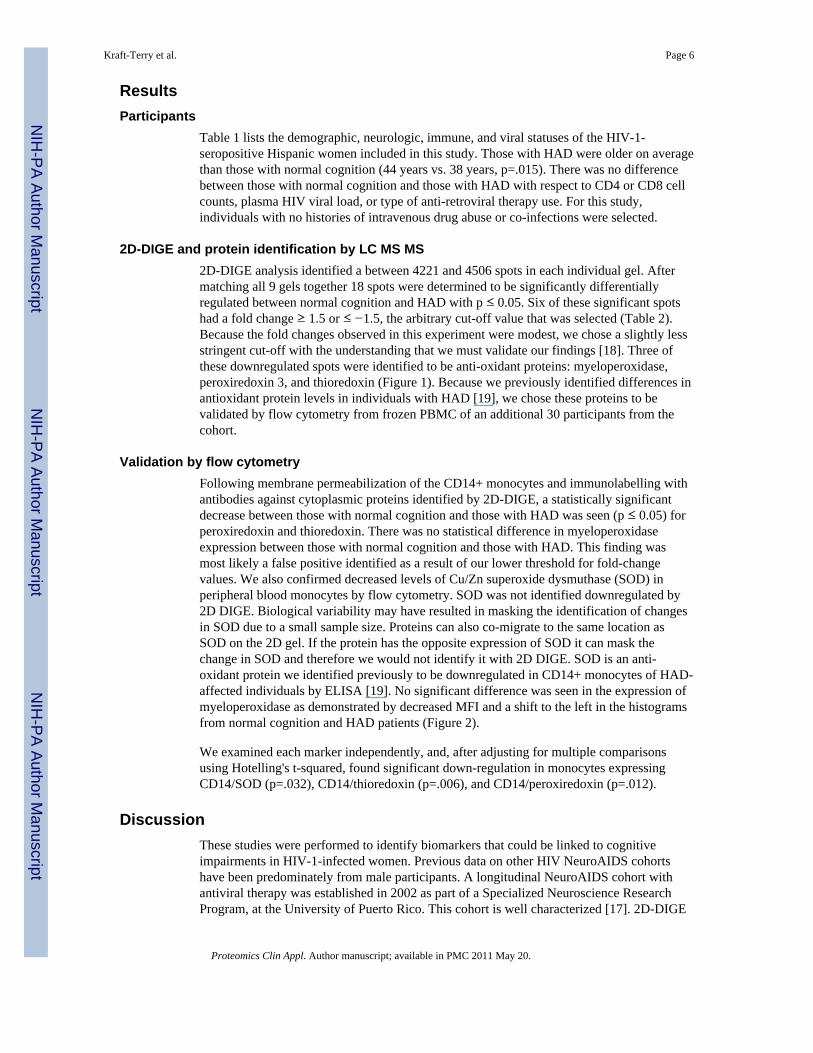
Validation by flow cytometryFollowing membrane permeabilization of the CD14+ monocytes and immunolabelling withantibodies against cytoplasmic proteins identified by 2D-DIGE, a statistically significantdecrease between those with normal cognition and those with HAD was seen (p ≤ 0.05) forperoxiredoxin and thioredoxin. There was no statistical difference in myeloperoxidaseexpression between those with normal cognition and those with HAD. This finding wasmost likely a false positive identified as a result of our lower threshold for fold-changevalues. We also confirmed decreased levels of Cu/Zn superoxide dysmuthase (SOD) inperipheral blood monocytes by flow cytometry. SOD was not identified downregulated by2D DIGE. Biological variability may have resulted in masking the identification of changesin SOD due to a small sample size. Proteins can also co-migrate to the same location asSOD on the 2D gel. If the protein has the opposite expression of SOD it can mask thechange in SOD and therefore we would not identify it with 2D DIGE. SOD is an anti-oxidant protein we identified previously to be downregulated in CD14+ monocytes of HAD-affected individuals by ELISA [19]. No significant difference was seen in the expression ofmyeloperoxidase as demonstrated by decreased MFI and a shift to the left in the histogramsfrom normal cognition and HAD patients (Figure 2).
We examined each marker independently, and, after adjusting for multiple comparisonsusing Hotelling's t-squared, found significant down-regulation in monocytes expressingCD14/SOD (p=.032), CD14/thioredoxin (p=.006), and CD14/peroxiredoxin (p=.012).
DiscussionThese studies were performed to identify biomarkers that could be linked to cognitiveimpairments in HIV-1-infected women. Previous data on other HIV NeuroAIDS cohortshave been predominately from male participants. A longitudinal NeuroAIDS cohort withantiviral therapy was established in 2002 as part of a Specialized Neuroscience ResearchProgram, at the University of Puerto Rico. This cohort is well characterized [17]. 2D-DIGE
Kraft-Terry et al. Page 6
Proteomics Clin Appl. Author manuscript; available in PMC 2011 May 20.
NIH
-PA Author Manuscript
NIH
-PA Author Manuscript
NIH
-PA Author Manuscript
analysis of monocyte samples from this cohort provided six possible protein targets thatappeared to be differentially regulated between normal cognition and HAD. All six of theseproteins were decreased in HAD. Although differences in age were found between thosewith normal cognition and those with HAD, they did not correlate with the expression ofantioxidants (data not shown).
We chose to validate thioredoxin, peroxiredoxin, and myeloperoxidase because of their rolein the control of oxidative stress. Thioredoxin and peroxiredoxin were validated by flowcytometry, but myeloperoxidase did not differ between patients with and those withoutHAD. The downregulation of both thioredoxin and peroxiredoxin in peripheral monocytesimplies decreased antioxidant capabilities of peripheral monocytes in patients with HAD.Because we previously demonstrated a downregulation in SOD-1 in monocytes from womenwith HAD by ELISA, we wanted to validate this finding by flow cytometry as well, asviable cells from the longitudinal cohort and this method do not require physical separationof cells which is best for analysis of different cell populations in a small sample size. Weobtained comparable results, further confirming that peripheral monocytes have decreasedantioxidant proteins in individuals with HAD.
Thioredoxin is an antioxidant protein that facilitates the reduction of proteins by cysteinethiol-disulfide exchange through two redox-active cysteine residues in its active center.Thioredoxin operates with NADPH and thioredoxin reductase to reduce exposed disulfides[20]. Thioredoxin cooperates with peroxiredoxins for an antiapoptotic effect throughscavenging of intracellular hydrogen peroxide [21]. Serum thioredoxin levels are known tobe elevated during HIV-1 infection as compared with levels in uninfected controls [22].Elevated thioredoxin levels during HIV-1 infection demonstrate a mechanism by which cellswork to control oxidative stress levels and prolong cell survival. With a decrease inthioredoxin expression during HAD, there is presumably an increase in oxidative stress,resulting in increased apoptosis and inflammatory signals.
Peroxiredoxins, thioredoxin-dependent peroxidases, protect against apoptosis by scavenginghydrogen peroxide. Peroxiredoxin 3 is expressed within the mitochondria [23]. Natural killerenhancing factors (NKEF)-A and NKEF-B, members of the peroxiredoxin family, areupregulated during HIV-1 infection in CD8+ T-cells and are thought to contribute to theantiviral activity of CD8+ T-cells [24]. Some peroxiredoxins interfere with HIVtranscription by inactivation of HIV-1 long terminal repeat, thereby suppressing levels ofp24 [25]. Decreases in peroxiredoxin levels of individuals with HAD may contribute toincreased HIV replication and intracellular oxidative stress, later translating to neurotoxicity.
Many neurodegenerative disorders are characterized by increased oxidative stress, such asParkinson’s disease, Alzheimer’s disease, and amyotrophic lateral sclerosis to name a few.Recently some oxidative stress-related proteins related to HAD have been elucidated.Hypoxia-inducible factor 1(HIF-1) is activated upon HIV-1 infection and has been found tobe elevated in brains of AIDS patients [26]. The HIV-1 accessory protein, Vpr, has alsobeen implicated in mediating HIF-1 induction [26]. In other studies, CSF and monocytesfrom HAD patients had decreased expression and activity of Cu/Zn superoxide dismutase,indicating that oxidative stress may exacerbate HAD [19].
Traditionally, HIV-1 levels are known to be highest in the basal ganglia and hippocampus[27]. Argawal et al. investigated the loss of dopaminergic neurons in the substantia niagradue to reactive oxygen species (ROS) and GP120 administration, which was attenuated byadministration of SOD or glutathione peroxidase [28]. Degeneration of the substantia nigrais a hallmark of Parkinson’s disease, and therefore has been investigated in HAND due tosome similarities seen between these two neurodegenerative diseases. SOD delivery in
Kraft-Terry et al. Page 7
Proteomics Clin Appl. Author manuscript; available in PMC 2011 May 20.
NIH
-PA Author Manuscript
NIH
-PA Author Manuscript
NIH
-PA Author Manuscript
rodent models of GP120 neurotoxicity results in protection of dopaminergic neurons[28,29]. Recently, we identified SOD downregulation in monocytes of HAND-affectedindividuals [19]. This downregulation was confirmed in our current study in a larger numberof individuals with HAND. We have also identified a down-regulation in SOD in in-vitroassays comparing viral isolates from individuals with HAD to those without neurocognitiveimpairment [30]. These studies prompted further investigation into changes that occur inmonocyte protein expression during HAD. Downregulation of SOD combined with the lossof more specific antioxidant proteins results in unprotected ROS damage to cells.
Decreased antioxidant levels can have detrimental effects on both the affected cell andsurrounding cells. Losing the ability to scavenge reactive oxygen species results indetrimental effects not just on MP, but also on astrocytes and neurons. Astrocytes are knownto compensate for toxic MP secretions in in vitro models of HIV-1 infection [31], but asdisease progresses this careful control of homeostasis erodes and neurotoxicity increasesuncontrolled by these cells [32]. They may become overwhelmed by ROS production andtherefore cannot sufficiently protect neurons in advanced stages of disease.
This study has uncovered antioxidant proteins that are downregulated in HAD. Theseproteins will be further investigated to understand the cause for downregulation and how toprevent or treat this deficiency. While we understand the effects of a decrease in antioxidantcapabilities, what would cause such a decrease in some individuals with HIV-1 infection andnot in others still remains a mystery.
AcknowledgmentsWe would like to thank our patients for supporting this research. We also wish to thank Ms. Tania for patientoutreach, Drs. Rosa Hechavarría and Billy Santiago for neuropsychological testing. We thank the HIV WomenClinic (CEMI) and its Director Dr. Carmen Zorrilla and the Clinic of Sexually Transmitted Diseases (CLETS) andits Director Dr. Hermes García for referring patients to this study and the RCMI-Clinical Research Center forproviding us the clinical facilities, staff, and supplies for laboratory samples. We thank Elizabeth Maldonado forpatient data management. Dr. Edmundo Kraiselburd’s continuous support for the project and the PR SpecializedNeuroscience Program in NeuroAIDS in general are greatly appreciated.
This work was supported by NIH-NINDS grants 1U54NS430 (to L.M.M.), 2 U54 NS43011 (to V.W.), R01MH083516-01, NIH-NCRR-RCMI-CRC-1P20RR11126, and NIH-NCRR-RCMI G12 RR-03051 for the ClinicalProteomics and Flow Cytometry core facilities (to L.M.M.), 5P01 NS31492, 2R37 NS36126, 2R01 NS034239,P20RR15635, U54NS 43011, P01 MH64570, and P01 NS43985 (to H.E.G).
List of abbreviations
HAND HIV-associated neurocognitive disorder
HAD HIV dmentia
two-dimensional image gel electrophoresis
References1. Antinori A, Arendt G, Becker JT, Brew BJ, et al. Updated research nosology for HIV-associated
neurocognitive disorders. Neurology. 2007; 69:1789–1799. [PubMed: 17914061]2. Woods SP, Moore DJ, Weber E, Grant I. Cognitive neuropsychology of HIV-associated
neurocognitive disorders. Neuropsychol Rev. 2009; 19:152–168. [PubMed: 19462243]3. Kramer-Hammerle S, Rothenaigner I, Wolff H, Bell JE, Brack-Werner R. Cells of the central
nervous system as targets and reservoirs of the human immunodeficiency virus. Virus Res. 2005;111:194–213. [PubMed: 15885841]
4. Yadav A, Collman RG. CNS Inflammation and Macrophage/Microglial Biology Associated withHIV-1 Infection. J Neuroimmune Pharmacol. 2009
Kraft-Terry et al. Page 8
Proteomics Clin Appl. Author manuscript; available in PMC 2011 May 20.
NIH
-PA Author Manuscript
NIH
-PA Author Manuscript
NIH
-PA Author Manuscript
5. Kadiu I, Glanzer JG, Kipnis J, Gendelman HE, Thomas MP. Mononuclear phagocytes in thepathogenesis of neurodegenerative diseases. Neurotox Res. 2005; 8:25–50. [PubMed: 16260384]
6. Deshpande M, Zheng J, Borgmann K, Persidsky R, et al. Role of activated astrocytes in neuronaldamage: potential links to HIV-1-associated dementia. Neurotox Res. 2005; 7:183–192. [PubMed:15897153]
7. Persidsky Y, Ghorpade A, Rasmussen J, Limoges J, et al. Microglial and astrocyte chemokinesregulate monocyte migration through the blood-brain barrier in human immunodeficiency virus-1encephalitis. Am J Pathol. 1999; 155:1599–1611. [PubMed: 10550317]
8. Dhillon NK, Williams R, Callen S, Zien C, et al. Roles of MCP-1 in development of HIV-dementia.Front Biosci. 2008; 13:3913–3918. [PubMed: 18508485]
9. Coleman CM, Wu L. HIV interactions with monocytes and dendritic cells: viral latency andreservoirs. Retrovirology. 2009; 6:51. [PubMed: 19486514]
10. Kraft-Terry SD, Buch SJ, Fox HS, Gendelman HE. A coat of many colors: neuroimmune crosstalkin human immunodeficiency virus infection. Neuron. 2009; 64:133–145. [PubMed: 19840555]
11. Rozek W, Ricardo-Dukelow M, Holloway S, Gendelman HE, et al. Cerebrospinal fluid proteomicprofiling of HIV-1-infected patients with cognitive impairment. J Proteome Res. 2007; 6:4189–4199. [PubMed: 17929958]
12. Laspiur JP, Anderson ER, Ciborowski P, Wojna V, et al. CSF proteomic fingerprints for HIV-associated cognitive impairment. J Neuroimmunol. 2007; 192:157–170. [PubMed: 17950469]
13. Kim TA, Avraham HK, Koh YH, Jiang S, et al. HIV-1 Tat-mediated apoptosis in human brainmicrovascular endothelial cells. J Immunol. 2003; 170:2629–2637. [PubMed: 12594291]
14. Ciborowski P, Kadiu I, Rozek W, Smith L, et al. Investigating the human immunodeficiency virustype 1-infected monocyte-derived macrophage secretome. Virology. 2007; 363:198–209.[PubMed: 17320137]
15. Carlson KA, Ciborowski P, Schellpeper CN, Biskup TM, et al. Proteomic fingerprinting of HIV-1-infected human monocyte-derived macrophages: a preliminary report. J Neuroimmunol. 2004;147:35–42. [PubMed: 14741425]
16. Chertova E, Chertov O, Coren LV, Roser JD, et al. Proteomic and biochemical analysis of purifiedhuman immunodeficiency virus type 1 produced from infected monocyte-derived macrophages. JVirol. 2006; 80:9039–9052. [PubMed: 16940516]
17. Wojna V, Skolasky RL, Hechavarria R, Mayo R, et al. Prevalence of human immunodeficiencyvirus-associated cognitive impairment in a group of Hispanic women at risk for neurologicalimpairment. J Neurovirol. 2006; 12:356–364. [PubMed: 17065128]
18. Corzett TH, Fodor IK, Choi MW, Walsworth VL, et al. Statistical analysis of the experimentalvariation in the proteomic characterization of human plasma by two-dimensional difference gelelectrophoresis. J Proteome Res. 2006; 5:2611–2619. [PubMed: 17022632]
19. Velazquez I, Plaud M, Wojna V, Skolasky R, et al. Antioxidant enzyme dysfunction in monocytesand CSF of Hispanic women with HIV-associated cognitive impairment. J Neuroimmunol. 2009;206:106–111. [PubMed: 19101040]
20. Holmgren A, Bjornstedt M. Thioredoxin and thioredoxin reductase. Methods Enzymol. 1995;252:199–208. [PubMed: 7476354]
21. Fujii J, Ikeda Y. Advances in our understanding of peroxiredoxin, a multifunctional, mammalianredox protein. Redox Rep. 2002; 7:123–130. [PubMed: 12189041]
22. Nakamura H, De Rosa S, Roederer M, Anderson MT, et al. Elevation of plasma thioredoxin levelsin HIV-infected individuals. Int Immunol. 1996; 8:603–611. [PubMed: 8671648]
23. Masutani H, Ueda S, Yodoi J. The thioredoxin system in retroviral infection and apoptosis. CellDeath Differ. 2005; 12(Suppl 1):991–998. [PubMed: 15818395]
24. Geiben-Lynn R, Kursar M, Brown NV, Addo MM, et al. HIV-1 antiviral activity of recombinantnatural killer cell enhancing factors, NKEF-A and NKEF-B, members of the peroxiredoxin family.J Biol Chem. 2003; 278:1569–1574. [PubMed: 12421812]
25. Jin DY, Chae HZ, Rhee SG, Jeang KT. Regulatory role for a novel human thioredoxin peroxidasein NF-kappaB activation. J Biol Chem. 1997; 272:30952–30961. [PubMed: 9388242]
26. Deshmane SL, Kremlev S, Amini S, Sawaya BE. Monocyte chemoattractant protein-1 (MCP-1):an overview. J Interferon Cytokine Res. 2009; 29:313–326. [PubMed: 19441883]
Kraft-Terry et al. Page 9
Proteomics Clin Appl. Author manuscript; available in PMC 2011 May 20.
NIH
-PA Author Manuscript
NIH
-PA Author Manuscript
NIH
-PA Author Manuscript
27. Wiley CA, Soontornniyomkij V, Radhakrishnan L, Masliah E, et al. Distribution of brain HIV loadin AIDS. Brain Pathol. 1998; 8:277–284. [PubMed: 9546286]
28. Agrawal L, Louboutin JP, Marusich E, Reyes BA, et al. Dopaminergic neurotoxicity of HIV-1gp120: Reactive oxygen species as signaling intermediates. Brain Res. 2009
29. Louboutin JP, Agrawal L, Reyes BA, Van Bockstaele EJ, Strayer DS. HIV-1 gp120 neurotoxicityproximally and at a distance from the point of exposure: protection by rSV40 delivery ofantioxidant enzymes. Neurobiol Dis. 2009; 34:462–476. [PubMed: 19327399]
30. Toro-Nieves DM, Rodriguez Y, Plaud M, Ciborowski P, et al. Proteomic analyses of monocyte-derived macrophages infected with human immunodeficiency virus type 1 primary isolates fromHispanic women with and without cognitive impairment. J Neurovirol. 2009; 15:36–50. [PubMed:19115125]
31. Wang T, Gong N, Liu J, Kadiu I, et al. Proteomic modeling for HIV-1 infected microglia–astrocytecrosstalk. PLoS ONE. 2008; 3:e2507. [PubMed: 18575609]
32. Genis P, Jett M, Bernton EW, Boyle T, et al. Cytokines and arachidonic metabolites producedduring human immunodeficiency virus (HIV)-infected macrophage-astroglia interactions:implications for the neuropathogenesis of HIV disease. J Exp Med. 1992; 176:1703–1718.[PubMed: 1460427]
Kraft-Terry et al. Page 10
Proteomics Clin Appl. Author manuscript; available in PMC 2011 May 20.
NIH
-PA Author Manuscript
NIH
-PA Author Manuscript
NIH
-PA Author Manuscript
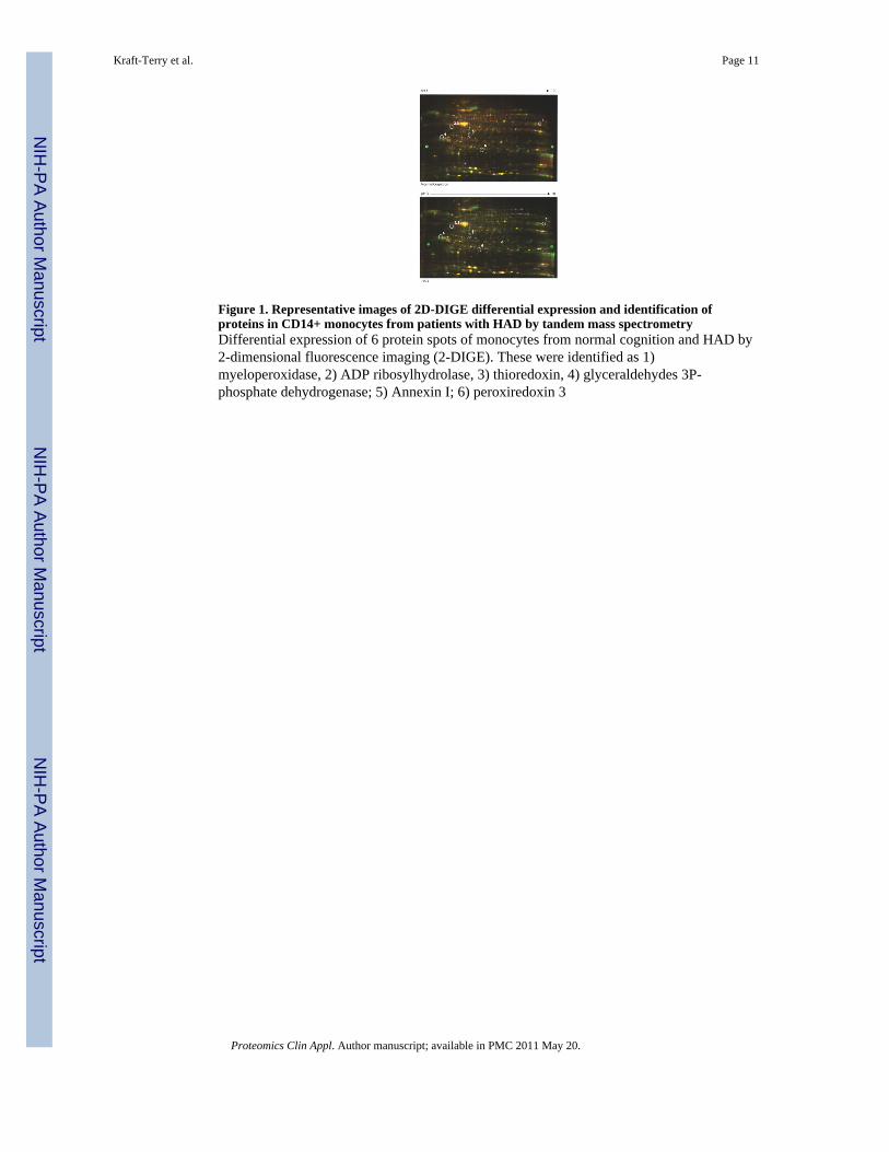
Figure 1. Representative images of 2D-DIGE differential expression and identification ofproteins in CD14+ monocytes from patients with HAD by tandem mass spectrometryDifferential expression of 6 protein spots of monocytes from normal cognition and HAD by2-dimensional fluorescence imaging (2-DIGE). These were identified as 1)myeloperoxidase, 2) ADP ribosylhydrolase, 3) thioredoxin, 4) glyceraldehydes 3P-phosphate dehydrogenase; 5) Annexin I; 6) peroxiredoxin 3
Kraft-Terry et al. Page 11
Proteomics Clin Appl. Author manuscript; available in PMC 2011 May 20.
NIH
-PA Author Manuscript
NIH
-PA Author Manuscript
NIH
-PA Author Manuscript
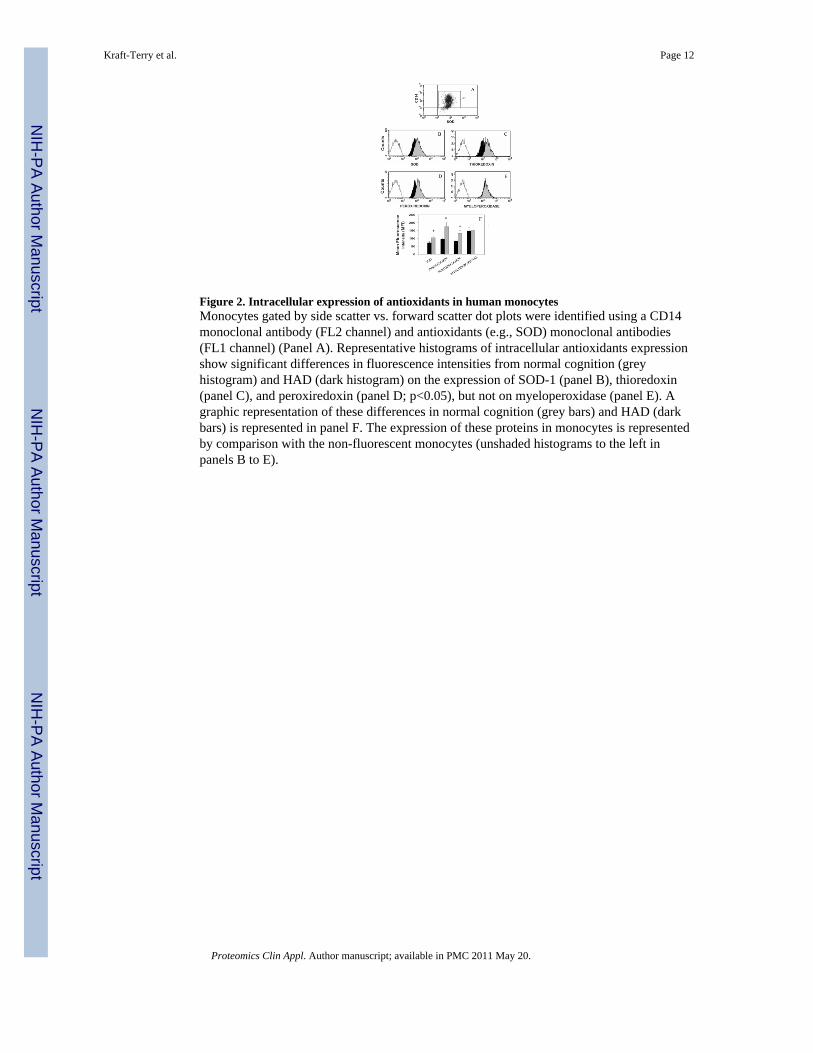
Figure 2. Intracellular expression of antioxidants in human monocytesMonocytes gated by side scatter vs. forward scatter dot plots were identified using a CD14monoclonal antibody (FL2 channel) and antioxidants (e.g., SOD) monoclonal antibodies(FL1 channel) (Panel A). Representative histograms of intracellular antioxidants expressionshow significant differences in fluorescence intensities from normal cognition (greyhistogram) and HAD (dark histogram) on the expression of SOD-1 (panel B), thioredoxin(panel C), and peroxiredoxin (panel D; p<0.05), but not on myeloperoxidase (panel E). Agraphic representation of these differences in normal cognition (grey bars) and HAD (darkbars) is represented in panel F. The expression of these proteins in monocytes is representedby comparison with the non-fluorescent monocytes (unshaded histograms to the left inpanels B to E).
Kraft-Terry et al. Page 12
Proteomics Clin Appl. Author manuscript; available in PMC 2011 May 20.
NIH
-PA Author Manuscript
NIH
-PA Author Manuscript
NIH
-PA Author Manuscript
NIH
-PA Author Manuscript
NIH
-PA Author Manuscript
NIH
-PA Author Manuscript
Kraft-Terry et al. Page 13
Table 1
Demographics, viral, and immune parameters of 39 HIV-seropositive women
Normal cognition HAD p value
n=20 n=19
Age 38 ± 8 44 ± 7 p = .015
Age range 25 − 51 23 − 54
CD4 454 ± 214 495 ± 294 p = .621
CD4 range 81–1062 42 – 1258
CD8 840 ± 316 821 ± 555 p = .894
CD8 range 452 – 1472 38 – 2587
Plasma VL 3.05 ± 1.37 2.24 ± 0.93 p = .064
VL range 1.70 – 5.00 1.70 – 4.48
Hepatitis C 0 0 p = .704
Therapy
No therapy 2 4 p = .234
ART 1 0
HAART 14 15
Unknown 3 0
HAD= HIV-associated dementia
Proteomics Clin Appl. Author manuscript; available in PMC 2011 May 20.
NIH
-PA Author Manuscript
NIH
-PA Author Manuscript
NIH
-PA Author Manuscript
Kraft-Terry et al. Page 14
Tabl
e 2
Prot
ein
ID
Fold
Cha
nge
Prot
ein
Acc
essi
onPe
ptid
esP-
valu
eM
W
−2.73
AD
P-rib
osyl
hydr
olas
e lik
e 2
hete
roge
neou
s nuc
lear
ribo
nucl
eopr
otei
n C
-like
189
2341
75
0.01
789
2341
7
6196
6711
561
9667
11
−1.87
Thio
redo
xin
1427
8250
20.
0411
615.
1
−1.81
mye
lope
roxi
dase
8818
02
0.01
292
436.
6
−1.83
pero
xire
doxi
n 3
5802
974
40.
0029
2769
2.5
PEST
pro
teol
ytic
sign
al c
onta
inin
g nu
clea
r pro
tein
9966
827
418
924.
8
−1.73
Ann
exin
I15
7829
895
60.
014
3503
9.9
−1.51
glyc
eral
dehy
de-3
-pho
spha
te d
ehyd
roge
nase
7669
492
20.
013
5952
7.3
−1.42
glyc
eral
dehy
de-3
-pho
spha
te d
ehyd
roge
nase
7669
492
30.
017
3605
3.0
−1.35
IQ m
otif
cont
aini
ng G
TPas
e ac
tivat
ing
prot
ein
145
0678
73
0.00
9118
9250
.1
−1.38
biliv
erdi
n re
duct
ase
B45
0241
93
0.04
422
119.
2
−1.33
gels
olin
isof
orm
a p
recu
rsor
zyx
in45
0416
54
0.04
285
696.
9
4508
047
461
277.
0
−1.28
L-is
oasp
arty
l/D-a
spar
tyl p
rote
in c
arbo
xyl m
ethy
ltran
sfer
ase
1806
372
0.01
924
679.
2
−1.23
EF-h
and
dom
ain
fam
ily, m
embe
r D2
2014
9675
20.
017
2669
7.1
p64
CLC
P89
5845
523
542.
7
−1.21
aden
ine
phos
phor
ibos
yltra
nsfe
rase
4502
171
40.
036
1960
7.6
biliv
erdi
n re
duct
ase
B45
0241
92
2211
9.2
−1.2
biliv
erdi
n re
duct
ase
B45
0241
95
0.03
622
119.
2
−1.19
coac
tosi
n-lik
e 1
2162
4607
40.
019
1594
4.9
1.12
tyro
sine
3/tr
ypto
phan
5 -m
onoo
xyge
nase
act
ivat
ion
prot
ein
4507
953
30.
044
2774
4.9
Proteomics Clin Appl. Author manuscript; available in PMC 2011 May 20.
NIH
-PA Author Manuscript
NIH
-PA Author Manuscript
NIH
-PA Author Manuscript
Kraft-Terry et al. Page 15
Fold
Cha
nge
Prot
ein
Acc
essi
onPe
ptid
esP-
valu
eM
W
tyro
sine
3-m
onoo
xyge
nase
/tryp
toph
an 5
-mon
ooxy
gena
se a
ctiv
atio
n pr
otei
n95
0724
52
2830
2.4
nudi
x-ty
pe m
otif
537
5944
646
2432
7.5
1.27
ubiq
uitin
car
boxy
l-ter
min
al e
ster
ase
L351
7474
13
0.03
526
182.
4
tyro
sine
3-m
onoo
xyge
nase
/tryp
toph
an5-
mon
ooxy
gena
se a
ctiv
atio
n pr
otei
n,95
0724
52
2830
2.4
1.44
prot
easo
me
beta
9 su
buni
t iso
form
1 p
ropr
otei
n45
0620
53
0.04
723
264.
2
Proteomics Clin Appl. Author manuscript; available in PMC 2011 May 20.


