Embed Size (px)
Citation preview

DEPARTAMENTO DE MICROBIOLOGÍA Y ECOLOGÍA ESTUDIOS SOBRE LA INMUNOGENICIDAD Y LOS MECANISMOS FISIOPATOLÓGICOS DE LA PROTEÍNA NSP4 DE ROTAVIRUS JESÚS RODRÍGUEZ DÍAZ
UNIVERSITAT DE VALENCIA Servei de Publicacions
2005

Aquesta Tesi Doctoral va ser presentada a Valencia el día 10 de Junio de 2005 davant un tribunal format per:
- D. Luis Enjuanes Sánchez - D. Albert Bosch Navarro - D. Nabil Halaihel - D. Franco M. Ruggeri - D. David Navarro Ortega
Va ser dirigida per: D. Javier Blesa Gómez ©Copyright: Servei de Publicacions Jesús Rodríguez Díaz Depòsit legal: I.S.B.N.:84-370-6383-3
Edita: Universitat de València Servei de Publicacions C/ Artes Gráficas, 13 bajo 46010 València Spain Telèfon: 963864115
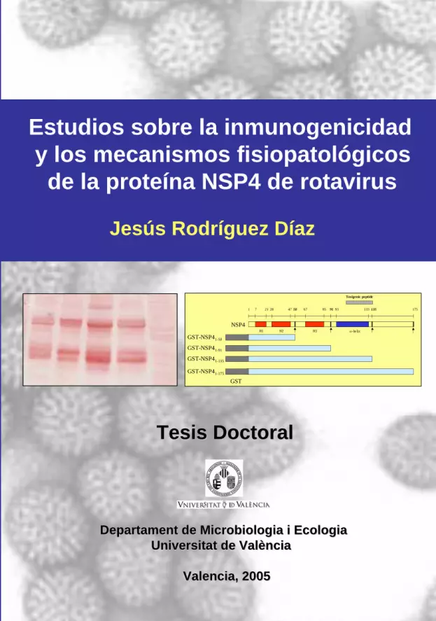
1 7 21 5028 47
H1 H2 H3 α-helix
85 93 133 135 17567 91
NSP4
GST-NSP41-50
GST-NSP41-91
GST-NSP41-135
GST-NSP41-175
Toxigenic peptide
GST
Tesis DoctoralTesis Doctoral
Departament de Microbiologia i Ecologia Departament de Microbiologia i Ecologia UniversitatUniversitat de de ValValèènciancia
Valencia, 2005Valencia, 2005
Estudios sobre la inmunogenicidady los mecanismos fisiopatológicosde la proteína NSP4 de rotavirus
Jesús Rodríguez Díaz
Jesús Rodríguez D
íaz 2005

Estudios sobre la inmunogenicidad y los mecanismos fisiopatológicos de la
proteína NSP4 de rotavirus
Jesús Rodríguez Díaz
Departament de Microbiologia i Ecologia
Universitat de València

Departament de Microbiologia i Ecologia FAX 34 96 386 46 58 Facultat de Medicina i Odontologia Tel. 34 96 386 46 58 Av. Blasco Ibáñez, 17 46010 València
Javier Buesa Gomez, Profesor Titular del Departament de Microbiologia i
Ecologia en la Facultat de Medicina i Odontologia de la Universitat de València,
HACE CONSTAR
que el presente trabajo de investigación titulado “Estudios sobre la inmunogenicidad y los mecanismos fisiopatológicos de la proteína NSP4 de rotavirus”, presentado como recopilación de publicaciones, ha sido
realizado bajo mi dirección por Jesús Rodríguez Díaz, licenciado en Biología,
para optar al título de Doctor en Biología, dentro del Programa de Doctorado
de “Microbiología” (694-275D) de la Universitat de Valencia.
En Valencia, a 7 de febrero de 2005
Prof. Dr. Javier Buesa Gómez

"Siempre que enseñes, enseña a la vez a dudar de lo que enseñas".
(José Ortega y Gasset)
La lógica es un método sistemático para llegar
con confianza a la conclusión errónea.
(Máxima de Manly)


AGRADECIMIENTOS
Hace más de cuatro años que comenzó mi andadura por el mundo de la Virología; en
este tiempo muchas son las personas que han influido positivamente a que este
proyecto vea la luz. En primer lugar quiero agradecer a mis padres y hermanos todo el
apoyo prestado, no sólo durante el periodo que ha durado la elaboración de esta tesis,
sino tambien por la familia tan maravillosa que me ha tocado en suerte. A mi director
Javier Buesa todo el apoyo prestado y la oportunidad brindada, sin la cual esta tesis
hubiera sido imposible. A todos los compañeros del laboratorio de Virología del
Departamento de Microbiología de la Facultad de Medicina de Valencia, donde se
realizó la mayor parte de esta tesis: Pilar, Ana, Beatriz, Rebeca, Rima y los últimos en
llegar: Clara y Juanma; ha sido un verdadero placer haber podido trabajar con
vosotros y compartir no sólo las experiencias del laboratorio.
Pero este proyecto viajó también por diferentes laboratorios. Mi agradecimiento
también a todas las personas de los diferentes grupos de investigación que me
acogieron durante este tiempo, en especial a Gaspar Pérez Martínez y Vicente
Monedero, del grupo de bacterias lácticas del IATA en Burjassot, y a todos los
compañeros que allí tuve durante mis diferentes estancias: Rosa, Raquel, Carlos,
Manolo, Mª Carmen y Mª Jesús. Al grupo de Jesús Zueco en la Facultad de Farmacia
y su ayuda prestada en el análisis de glicoproteínas, especial mención a Isabel Andrés
y su “maña” para transferir geles. Por último, agradecer la ayuda prestada por Lennart
Svensson en mi último destino durante esta tesis, la Universidad de Linköping en
Suecia, así como a los compañeros que allí tuve: Malin y Carolin.
Finalmente, agradecer el apoyo institucional prestado por el Ministerio de Educación y
Ciencia y por el Fondo de Investigación Sanitaria del Instituto de Salud Carlos III, ya
que sin su ayuda económica esta tesis no habría sido posible.
i

ii

INDICE
INTRODUCCIÓN GENERAL...............................................................................1
1. Breve historia y situación taxonómica de los rotavirus.………………...3
2. Características generales de los rotavirus…………….…………………4
3. Clasificación de rotavirus…………………………………………………..5
4. Características estructurales de rotavirus………………………………..6
4.1. Estructura del virión………………………………………………6
4.2. Estructura del genoma…………………………………………...8
4.3. Estructura y función de las proteínas de rotavirus…………..11
4.3.1. Proteína VP4……………………………………………..11
4.3.2. Proteína VP6………………………………………...…..14
4.3.3. Proteína VP7…………………………………………......15
4.3.4. Proteína NSP4……………………………………………17
5. Ciclo replicativo de rotavirus……………………………………………..19
5.1. Adsorción, penetración y decapsidación……………………..20
5.2. Transcripción y replicación…………………..…………………23
5.3. Morfogénesis y liberación de las partículas víricas………….25
6. Fisiopatología de la infección por rotavirus y la proteína NSP4……...27
6.1. Modelo de la inducción de la diarrea por rotavirus………….28
7. Presentación de los trabajos y justificación de la unidad temática…..31
OBJETIVOS…………………………………………………………………………..35
CAPÍTULO 1: Expression and purification of polyhistidine-tagged rotavirus
proteins in insect cells……………………………………………………………….39
Abstract………………………………………………………………………..41
Introduction……………………………………………………………………42
Materials and methods………………………………………………………43
Virus and cells………………………………………………………..43
iii

Cloning of rotavirus NSP4 cDNA into the baculovirus expression
vector………………………………………………………………......43
Expression and purification of NSP4 proteins in insect cells….….45
Scale-up protein production…………………………………………45
Western blotting of NSP4 glycoproteins…….……………………..46
Glycosylation status of NSP4 and deglycosylation analysis……..46
Diarrhea induction by NSP4…………………………………………47
Results…………………………………………………………………………47
Expression and purification of NSP4 proteins in insect cells…….47
Glycosylation status of NSP4 and deglycosylation analysis……..50
Diarrhea induction by NSP4…………………………………………50
Discussion…………………………………………………………………….51
Acknowledgments………..…………………………………………………..53
References……………………………………………………………………53
CAPÍTULO 2: Isolation and characterisation of single-chain variable fragments
(scFv) antibodies against rotavirus NSP4 and VP8* proteins generated by
phage display…………………………………………………………………………57
CAPÍTULO 2A: Single-chain variable fragments (scFv) antibodies against
rotavirus NSP4 enterotoxin generated by phage display……………………..…59
Abstract………………………………………………………………………..61
Introduction……………………………………………………………………62
Materials and methods………………………………………………………64
Expression and purification of NSP4 proteins in insect cells……….64
Cloning expression and purification of GST-NSP4 truncated proteins
in E. coli….…………..…………………………………………………..64
Antibody phage display to the NSP4 protein.…..….………………..65
Screening of individual phage clones by ELISA……..……….……..66
Detection of binding specificity by ELISA.………..…………………67
svFv characterisation……………………...….….……………………67
iv

Results…………………………………………………………………………68
Isolation of svFv against NSP4….………………………………….68
svFv characterisation……………………...…………………………69
Detection of binding specificity by ELISA.…………………………70
Discussion…………………………………………………………………….73
Acknowledgments………..…………………………………………………..75
References……………………………………………………………………75
CAPÍTULO 2B: Selection of single-chain antibodies against the VP8* subunit of
rotavirus VP4 outer capsid protein and their expression in Lactobacillus
casei...…………………………………………………………………………………81
Abstract………………………………………………………………………..83
Isolation of svFv against VP8*………………………………………………85
scFv characterisation……..………………………………………………….86
In vitro inhibition of viral infection in MA 104 cells.………………………..87
Expression of scFv in Lactobacillus casei…………………………………88
Acknowledgments………………………..…………………………………..91
References……………………………………………………………………92
CAPÍTULO 3: Humoral immune response to rotavirus NSP4 enterotoxin in
Spanish children ..……………………………………………………………………97
Abstract………………………………………………………………………..99
Introduction………………………………………………………………….100
Materials and methods…………………………………………………….102
Viruses and cells ………………………………………………….…102
Expression and purification of NSP4 proteins in insect cells……102
Serum samples……………………………..………………………..103
Detection of serum antibodies to Wa and SA11 rotavirus
by ELISA…..…………………………………………………………103
Detection of serum antibodies to NSP4Wa and NSP4SA11 antigens
v
by ELISA……..………………………………………………………104
Statistical analyses………………………...……………………….105
Results………………………………………………………………………105
Antibodies to NSP4 in infected children…………………..……..105
Antibodies to NSP4 in uninfected children and adults..………..106
Antibodies to rotavirus particles……..……………….…………..106
Comparison of the antibody titers between children with rotavirus
diarrhoea, healthy children and adults.……………….………….107
Discussion…………………………………………………………………….108
Acknowledgments……………..……………………………………………..110
References……………………………………………………………………110
CAPÍTULO 4: Nitric oxide production during clinical and experimental infection
with rotavirus…………….…………………………………………………………..115
Abstract……………………………………………………………………...117
Introduction………………………………………………………………….118
Materials and methods…………………………………………………….120
Expression and purification of NSP4 proteins in insect cells……120
NSP4 inoculation and rotavirus infection in HT-29 cells….……..121
Rotavirus infection in mice ………………………………….……..121
Human samples……………………………………………………..122
Detection of rotavirus by ELISA……………………………………122
Nitrite/Nitrate measurements………………………………………122
Quantitative RT-PCR assay for iNOS mRNA……………………123
Statistical analysis…………………………………………………..124
Results………………………………………………………………………124
NSP4 induces a rapid release of NO metabolites from
human intestinal cells.…………………………………………..…..124
Rotavirus stimulates release of NO in vivo…..………....………..125
vi

Children with severe rotavirus gastroenteritis excrete NO in their
urine……………………………..……….………….….……..…..127
Discussion…………………………………………………………………….128
Acknowledgments…………………………..………………………………..130
References……………………………………………………………………130
DISCUSIÓN…………………………………………………………………………137
CONCLUSIONES…………………………………………………………………..149
BIBLIOGRAFIA GENERAL…………..……………………………………………153
vii

viii

Introducción general
INTRODUCCIÓN GENERAL
1

Introducción general_______________________________________________
2

Introducción general
1. Breve historia y situación taxonómica de rotavirus
En 1973 Bishop y colaboradores describieron un virus en forma de rueda tras la
observación al microscopio electrónico de cortes de intestino delgado procedentes de
niños con gastroenteritis (Bishop et al., 1973). No obstante diez años antes, en 1963,
se habían encontrado en la mucosa intestinal de ratones con diarrea unos virus con
una morfología similar. El virus aislado de los ratones fue llamado virus de la diarrea
epizoótica de ratones lactantes (EDIM) (Adams y Kraft, 1963). En ese mismo año,
Malherbe y Harwin (1963) aislaron partículas víricas a partir del tejido rectal de un
mono ‘vervet’ sano. El virus fue llamado SA11, del inglés “simian agent 11”. Este
aislado pudo ser propagado en una línea celular procedente de la misma especie de
simio. Pocos años después Mebus y colaboradores demostraron la presencia de otro
virus de tamaño y morfología similares a los descritos anteriormente en heces de
terneros con diarrea (Mebus et al., 1969). El virus aislado por Mebus también pudo ser
propagado en células fetales bovinas (Mebus et al., 1971).
Debido a su morfología en forma de rueda, estos nuevos virus causantes de
gastroenteritis fueron llamados rotavirus, del término en latín “rota” que significa rueda.
Finalmente, en 1979, Mathews estableció el género rotavirus e incorporó este
nuevo género a la familia Reoviridae. Esta familia está compuesta por virus de
estructura icosaédrica sin envoltura lipídica cuya cápside está formada por capas
concéntricas de proteínas con un diámetro de entre 60 y 80 nm. El genoma está
compuesto por ARN de doble cadena (ARNbc) que se encuentra dividido en 10 a 12
segmentos, lo cual permite que ocurran recombinaciones y reorganizaciones
genéticas. La replicación de los virus pertenecientes a la familia Reoviridae ocurre en
el citoplasma celular, en unas estructuras subcelulares ricas en proteínas víricas
llamadas viroplasma.
3

Introducción general_______________________________________________
Dentro de la familia Reoviridae se agrupan seis géneros diferentes:
Orthoreovirus, Orbivirus, Rotavirus, Coltivirus, Aquareovirus, Cypovirus, Fijivirus,
Phytoreovirus y Oryzavirus. Los cuatro primero géneros infectan mamíferos y aves.
2. Características generales de los rotavirus
Los rotavirus son virus no envueltos con morfología icosaédrica y un tamaño
que oscila entre 60 y 80 nm. La cápside está formada por tres capas proteicas
concéntricas. La capa más interna forma el core que engloba el genoma vírico y los
enzimas encargados de la replicación del ARN.
El genoma de rotavirus está compuesto por 11 segmentos de ARNbc,
numerados del 1 al 11 tras su separación electroforética. Cada segmento codifica al
menos para una proteína. Debido a que las células eucarióticas no poseen ningún
enzima ARN polimerasa ARN dependiente, los segmentos del genoma de rotavirus no
son infectivos en ausencia de la ARN polimerasa viral. Una de las principales
características de rotavirus y debido a la presencia de 11 segmentos genéticos es el
proceso de “reassorting”: cuando una misma célula es infectada por más de una cepa
de rotavirus éstas pueden intercambiar fragmentos genéticos, dando lugar a nuevas
cepas víricas.
La infectividad de rotavirus necesita de la activación por proteasas. Los
rotavirus humanos han sido propagados clásicamente en la línea celular MA104, tras
su activación con tripsina y añadiendo tripsina al medio de cultivo (Estes et al., 2001,
Wyatt et al., 1983). Los rotavirus, al igual que el resto de representantes de la familia
Reoviridae, replican en el citoplasma y pueden formar cuerpos de inclusión
compuestos por proteínas víricas cristalizadas. Otra característica de los rotavirus
durante su morfogénesis es el estado transitorio en el que las partículas virales se
encuentran envueltas. La liberación de la progenie vírica ocurre por lisis celular o
mediante transporte vesicular no convencional en células epiteliales polarizadas.
4

Introducción general 3. Clasificación de rotavirus
Los rotavirus se clasifican en serogrupos, subgrupos y serotipos según sus
características antigénicas. En base a las características antigénicas de la proteína
VP6 se han descrito siete serogrupos o grupos antigénicos. Cada uno de estos grupos
ha sido identificado por una letra de la A a la G. Los grupos A, B y C han sido aislados
tanto en humanos como en animales, mientras que los grupos D, E, F y G han sido
aislados únicamente de animales (Kapikian et al., 2001). No obstante, los rotavirus del
grupo A son los causantes de la mayoría de las infecciones por rotavirus en humanos.
Los rotavirus del grupo A se asocian típicamente a diarrea en niños y animales
jóvenes. El serogrupo B ha producido epidemias anuales de diarrea grave en adultos
en China (Hung et al., 1983, Su et al., 1986, Wang et al., 1985) y en la India (Krishnan
et al., 1999), y brotes de diarrea en recién nacidos en China (Mackow, 1995).
Rotavirus pertenecientes al grupo C se han detectado en casos esporádicos de niños
con diarrea (Jiang et al., 1995, Mackow, 1995, Otsu, 1998, Penaranda et al., 1989,
Rodger et al., 1982) y en varios brotes (Caul et al., 1990, Hamano et al., 1999,
Matsumoto et al., 1989).
Los rotavirus del grupo A se dividen en subgrupos (SG) dependiendo de la
presencia o ausencia de dos epítopos reconocidos por los anticuerpos monoclonales
255/60 y 631/9 (Greenberg et al., 1983). De este modo nos encontramos cuatro SG
diferentes: SG I; SG II; SG I+II y SG ni-I, ni-II. El SG II es el más frecuente entre las
cepas humanas (Arista et al., 1990, Beards y Desselberger, 1989, Georges-Courbot et
al., 1988, Iturriza-Gomara et al., 2001, Mohammed et al., 1994) mientras que el SG I
es más frecuente entre las cepas de origen animal (Lopez et al., 1994, Tang et al.,
1997).
Los rotavirus del grupo A también han sido caracterizados mediante ensayos
de neutralización definiéndose así diferentes serotipos (Hoshino y Kapikian, 1996). Las
proteínas implicadas en esta clasificación serológica son la glicoproteína VP7, que
5
Introducción general_______________________________________________
define los serotipos G, y la hemaglutinina VP4, que define los serotipos P, ambas
proteínas de la cápside externa de rotavirus. Así los rotavirus del grupo A se clasifican
mediante un sistema binario que distingue los distintos serotipos de las proteínas VP7
y VP4 (Graham y Estes, 1985, Hoshino y Kapikian, 1996, Hoshino et al., 1984, Rodger
y Holmes, 1979). Hasta el momento se conocen 15 serotipos G y 13 serotipos P
(Estes, 2001). La denominación G y P son debidas a que la proteína VP7 es
glicosilada (G) y a que la proteína VP4 es sensible a proteasas (P). Debido a las
dificultades intrínsecas de las técnicas serológicas utilizadas para la clasificación de
los rotavirus del grupo A y la introducción de las técnicas moleculares, se estableció
una nueva clasificación basada directamente en las secuencias de los genes VP7 y
VP4. De este modo se distinguen 21 genotipos P en relación a la proteína VP4, de los
cuales solo 16 se corresponden con serotipos conocidos de la proteína (Estes, 2001,
Hoshino y Kapikian, 1996, Rao et al., 2000). Para la proteína VP7 existe una clara
correlación entre genotipos y serotipos (Gouvea et al., 1990). La nomenclatura de los
rotavirus del grupo A se ha consensuado de forma que en primer lugar se describe el
serotipo G y posteriormente el serotipo P y/o el genotipo P entre corchetes. Como
ejemplo, la cepa Wa sería G1 P1A[8].
4. Características estructurales de rotavirus
4.1. Estructura del virión
Los viriones de rotavirus están compuestos por tres capas proteicas que
pueden ser observadas al microscopio electrónico (Figura 1). Las partículas infectivas
completas, con un tamaño aproximado de 75 nm de diámetro, han sido históricamente
llamadas partículas de doble capa, pero los datos estructurales actuales demuestran
que poseen tres capas. La acción de productos quelantes del calcio, como el EDTA o
el EGTA, desestabilizan las proteínas de la cápside externa (VP4 y VP7) pudiéndose
6

Introducción general observar partículas de doble capa. Estas partículas (históricamente llamadas
partículas de simple capa) poseen una morfología rugosa, cuando son observadas al
microscopio electrónico, debido a la disposición trimérica de la proteína VP6. El
tratamiento de los viriones de rotavirus con agentes caotrópicos tales como el cloruro
cálcico permiten observar partículas de simple capa compuestas por las proteínas
VP1, VP2 y VP3. Estas estructuras forman el core del virión y agregan con facilidad.
La estructura tridimensional de las partículas de doble y triple capa ha sido estudiada
mediante técnicas de criomicroscopía y procesado de imágenes en partículas víricas
no tripsinizadas (Prasad y Chiu, 1994, Prasad et al., 1988, Shaw et al., 1993, Yeager
et al., 1994). Estos estudios muestran claramente que el número de triangulación (T)
de rotavirus es 13, confirmando los resultados presentados por Roseto y
colaboradores en 1979 (Roseto et al., 1979). Una característica importante de
rotavirus es la presencia de 132 canales a lo largo de las dos cápsides externas que
comunican el core de la partícula con el exterior.
a b c
Figura 1. Partículas de rotavirus observadas por microscopía electrónica tras tinción con 1% molibdato amónico, la barra representa 100 nm. El panel a muestra las partículas víricas completas, formadas por las proteínas estructurales VP1,VP2, VP3, VP4, VP6 y VP7. El panel b muestra las partículas de doble capa que carecen de las proteínas de cápside externa VP4 y VP7. El panel c muestra las partículas de simple capa o cores formadas por las proteínas VP1, VP2 y VP3 (Estes, 2001).
La función de estos canales no ha sido totalmente aclarada, aunque es probable
que estén implicados en el transporte de los metabolitos necesarios para la
transcripción del ARN viral y para la salida del ARN transcrito que será utilizado en los
procesos posteriores de la replicación viral.
7

Introducción general_______________________________________________
Estos estudios estructurales muestran la presencia de sesenta espículas
(Prasad et al., 1988). Estas espículas están formadas por dímeros de la proteína VP4
y poseen una longitud aproximada de 120 Å. Estudios más recientes han confirmado
que la longitud total de las espículas es de 200 Å y que éstas sobresalen 120 Å de la
superficie viral (Shaw et al., 1993, Yeager et al., 1994). La proteína de la capa externa
VP7 forma trímeros a partir de las 780 moléculas presentes en el virión. Esta proteína
interacciona tanto con los trímeros de la proteína VP6 como con la proteína VP4.
Las partículas de doble capa tienen un radio de 720 Å aproximadamente y
están compuestas por 780 moléculas de VP6 que definen un total de 260 unidades
morfológicas formadas por trímeros de la proteína. Estudios realizados mediante la
expresión de las diferentes proteínas que conforman el virión en el sistema de
baculovirus demuestran que las proteínas estructurales de rotavirus poseen
propiedades intrínsecas de auto-ensamblaje, permitiendo así la producción de
partículas pseudovíricas (Crawford et al., 1994, Labbe et al., 1991).
4.2. Estructura del genoma
El genoma de rotavirus está compuesto por 11 segmentos de ARNbc que se
encuentran localizados en el core del virión. El ARN viral necesita de interacciones
ARN-proteína para adoptar la conformación necesaria y ser empaquetado en el virión
(Kapahnke et al., 1986). Las proteínas implicadas en este proceso no han sido todavía
descritas, aunque las proteínas estructurales presentes en el core del virión (VP1, VP2
y VP3) son claras candidatas. No obstante, proteínas no estructurales también pueden
estar implicadas. Los fragmentos de ARN no son infectivos en ausencia de las
proteínas virales, indicando que necesitan de la replicasa viral y que ésta se encuentra
incluida en el virión.
Los once fragmentos del ARNbc viral poseen características y secuencias
conservadas en todos los rotavirus secuenciados hasta el momento. Los segmentos
8

Introducción general genómicos comienzan siempre con una guanina y están seguidos por secuencias
conservadas en la zona no codificante. Tras la zona codificante de la proteína (ORF),
al menos una por cada segmento genómico, también se encuentran secuencias
conservadas (Figura 1.2). Los ORFs no se encuentran seguidos por secuencias de
poliadenilación. Normalmente el codón de iniciación está precedido por una secuencia
Kozak. Algunos de los genes poseen otros codones de iniciación en fase con la pauta
de lectura a partir del primero (genes 7, 9 y 10) o fuera de fase (gen 11). Las
evidencias de que se disponen indican que todos los genes son monocistrónicos
excepto posiblemente los genes 9 y 11 (Chan et al., 1986, Mattion et al., 1991).
GGCUUUUAAAAA A UU
AUG
ORF
Secuencia no codificante 5’ Secuencia no codificante 3’
Segundo codón de inicio en fase o fuera de fase AUGUGACC
U UGUG
5’ 3’
Figura 1.2. Representación esquemática de la estructura de un fragmento génico de rotavirus. En el esquema se muestran las secuencias consenso de las zonas no codificantes en ambos extremos del gen, así como la posible existencia de un segundo codón de inicio.
Los rotavirus son los únicos virus conocidos que infectan mamíferos y aves que
poseen once segmentos de ARNbc. Esta característica ha permitido utilizar los
patrones electroforéticos de las diferentes cepas de rotavirus para caracterizarlas,
aunque el electroferotipo no puede ser utilizado como único método de clasificación
debido a que cepas muy diferentes pueden compartir el mismo patrón (Estes, 2001).
El número de cada segmento genómico de rotavirus ha sido asignado en referencia a
su capacidad de migración en geles de poliacrilamida, siendo el número 1 el de menor
movilidad electroforética y el 11 el de mayor. En los rotavirus de grupo A, el patrón
9

Introducción general_______________________________________________
electroforético suele estar formado por 4 grupos de bandas (figura 1.3.). Cuatro
segmentos (1-4) de gran peso molecular, dos de peso molecular intermedio (5-6), tres
de pequeño tamaño (7-9) y dos de muy pequeño tamaño molecular (10 y 11). En
algunas cepas aisladas en humanos el segmento que habitualmente codifica para las
proteínas del segmento 11 migra más lentamente que el segmento décimo. Éste
patrón es conocido como electroferotipo corto y casi siempre corresponde a cepas del
subgrupo antigénico I de rotavirus humanos (Hoshino y Kapikian, 1996). El
electroferotipo largo es característico de rotavirus humanos pertenecientes al subgrupo
II.
1
234
5
6
789
10
11
VP1VP2VP3VP4
NSP1
VP6
VP7
NSP2NSP3
NSP4NSP5
GENOME SEGMENTS
ENCODEDPROTEINS
VIRION SCHEMATIC(Protein Locations)
VP1VP2VP3VP4
VP6
VP7
RECONSTRUCTION
NSP6 (Not Visible,off bottom of gel)
Figura 1.3. Asignación de las proteínas codificadas por el genoma de rotavirus. La imagen muestra el fragmento genético que codifica para cada una de las proteínas y la ubicación en el virión de las proteínas estructurales de rotavirus. (Figura reproducida de E. Mossel, M. Estes y F. Ramig, http://www.iah.bbsrc.ac.uk).
Los genes de rotavirus pueden sufrir reordenamientos genómicos, en estos
casos los patrones electroforéticos presentan anomalías, de modo que aparecen
bandas que migran más lentamente y las bandas correspondientes a los segmentos
implicados desaparecen o se observa un brusco descenso de su concentración
10

Introducción general (Hundley et al., 1985). Generalmente las cepas víricas que presentan reordenamientos
en su genoma no son defectivas y los concatémeros resultantes del reordenamiento
sustituyen funcionalmente a los segmentos implicados (Allen y Desselberger, 1985,
Biryahwaho et al., 1987, Graham et al., 1987).
4.3. Estructura y función de las proteínas de rotavirus
Los once fragmentos de rotavirus codifican para un total de 12 proteínas. La
asignación de cada una de estas proteínas al fragmento correspondiente de ARNbc se
ha realizado mediante experimentos de traducción in vitro (Dyall-Smith y Holmes,
1981b, Mason et al., 1980, Mason et al., 1983, McCrae y Faulkner-Valle, 1981, Smith
et al., 1980). La figura 1.3 muestra la relación entre cada segmento de ARNbc, la
proteína que codifica y la posición que cada una de ellas ocupa dentro de la estructura
del virión.
Las principales características de las proteínas de rotavirus se encuentran
resumidas en la tabla 1.1.
Algunas de las proteínas de rotavirus han sido ampliamente estudiadas debido
a su importancia en el ciclo replicativo de rotavirus, sus características antigénicas o
su relevancia en la patogenia vírica.
4.3.1. Proteína VP4
La proteína VP4 es el producto del cuarto segmento genómico, tiene un peso
molecular de 88 KD y dimeriza formando las espículas de la cubierta externa del virión.
Esta proteína posee una longitud de 776 aminoácidos, o 775 en el caso de la mayoría
de las cepas aisladas en humanos, en las cuales falta el aminoácido situado entre las
posiciones 134 y 136. Esta proteína es un importante determinante antigénico, ya que
es capaz de inducir la producción de anticuerpos neutralizantes de la infección por
11

Introducción general_______________________________________________
rotavirus (Hoshino et al., 1995). Esta característica de la proteína VP4 permite la
clasificación de los rotavirus del grupo A en diferentes serotipos P. También es una
proteína clave en la infectividad (Greenberg et al., 1983, Offit et al., 1986). VP4 posee
la capacidad de aglutinar hematíes, actuando como una hemaglutinina viral. Esta
capacidad hemaglutinante no se encuentra presente en las cepas de rotavirus
humanos.
Tabla 1.1. Proteínas codificadas por el genoma de rotavirus
Segmento
Genómico
Producto
proteico
Localización Características y función
1 VP1 core ARN polimerasa, unión a ARN, unión a VP3
2 VP2 core Unión a ARN bc, requerida para la actividad replicasa de VP1
3 VP3 core Guanilil transferasa, metil transferasa, unión a ARNmc, unión a VP1
4 VP4 cápside externa Dimérica, hemaglutinina, antígeno neutralizante, procesada por
proteasas, espícula, adhesión celular, serotipos P
5 NSP1 no estructural Dedos de zinc, unión a ARNsc, unión al citoesqueleto
6 VP6 cápside interna Hidrofóbica, trimérica, antígeno de subgrupo
7 NSP3 no estructural Ácida, unión a ARNm viral, interacciona con elF4G1, inhibe traducción
del hospedador
8 NSP2 no estructural Básica, oligomérica, unión a ARNsc, NTPasa
9 VP7 cápside externa Glicoproteína, trimérica, antígeno neutralizante, unión a Calcio,
serotipos G
10 NSP4 no estructural Glicoproteína, receptor intracelular en el retículo endoplasmático,
morfogénesis, enterotoxina
NSP5 no estructural Básica, fosfoproteína, unión ARNsc, proteína quinasa, interacción con
VP2, NSP2, NSP6
11
NSP6 no estructural Interacción con NSP5, se localiza en el viroplasma
La proteína VP4 de rotavirus es escindida en dos polipéptidos por la acción de
la tripsina, esta escisión ocurre en aminoácidos altamente conservados (figura 1.4),
éstos son la arginina 241 y la arginina 247, aunque la arginina 247 es el lugar de
escisión mayoritario (Lopez et al., 1985). La acción de proteasas sobre la proteína VP4
12

Introducción general potencia la infectividad de rotavirus (Espejo et al., 1981, Estes et al., 1981, Ramia y
Sattar, 1980, Wyatt et al., 1983). Mediante criomicroscopía electrónica se han
demostrado cambios conformacionales en las espículas de rotavirus cuando son
incubados con concentraciones crecientes de tripsina (Crawford et al., 2001), aunque
los cambios estructurales y fisicoquímicos ocurridos en las espículas después de la
proteolisis no están claramente definidos. Los productos de la proteolisis de esta
proteína son las proteínas VP5* y VP8*. La proteína VP8*, de 28 KD, comprende el
fragmento amino terminal de la proteína VP4 mientras que la proteína VP5* de 60 KD
queda contenida en el extremo carboxi terminal de la proteína VP4.
VP8* VP5*
1 71 204 224 236 257 271 384 404 494 554 578 608 776
RVRV RC RC RC
93 208 241 247
Dominio de fusión
HA/SA
Región variable (RV)
Región conservada (RC)Lugares de corte de la tripsina
VP8* VP5*
1 71 204 224 236 257 271 384 404 494 554 578 608 776
RVRV RC RC RC
93 208 241 247
Dominio de fusión
HA/SA
Región variable (RV)
Región conservada (RC)Lugares de corte de la tripsina
Figura 1.4. Representación esquemática de la proteína de VP4 de rotavirus basada en diferentes cepas de rotavirus. Se muestra el dominio de aglutinación y unión a ácido siálico (HA/SA), las zonas de corte de la tripsina y las regiones conservadas y variables de la proteína.
Experimentos realizados con VP8* producida tanto en E. coli (Lizano et al.,
1991) como en células de insecto (Fiore et al., 1991) muestran que la capacidad
hemaglutinante de la proteína VP4 reside dentro de la porción que da lugar a la
proteína VP8*. Estudios posteriores confirmaron que esta región debe encontrarse
entre los aminoácidos 93 y 207 de la proteína VP8* (Fuentes-Panana et al., 1995).
Dentro de la proteína VP5*, entre los aminoácidos 384 y 401 de VP4, se encuentra
una región altamente conservada dentro de todas las cepas de rotavirus. Esta región
comparte una alta homología con los lugares de fusión que se encuentran en los
13

Introducción general_______________________________________________
receptores de otros virus tales como los alphavirus Semliki Forest y el virus Sindbis
(Mackow et al., 1988). De forma que esta zona de la proteína VP5* posiblemente esté
implicada en el proceso de entrada del virus en la célula hospedadora durante el
proceso de internalización del virus.
4.3.2. Proteína VP6
La proteína VP6 es una proteína altamente hidrofóbica e inmunogénica con un
peso molecular de 45 KD y es el principal componente estructural de los viriones. Esta
proteína de la capa intermedia de rotavirus interacciona al mismo tiempo con la
proteína VP2 de la cápside interna y con las proteínas VP4 y VP7 de la cápside
externa de rotavirus (Estes, 2001). La proteína VP6 forma trímeros espontáneamente
y es extremadamente estable (Estes et al., 1987). Estas características junto con la
presencia de epítopos altamente conservados entre diferentes cepas víricas hacen
que sea el antígeno utilizado en la mayoría de las técnicas de diagnóstico de
infecciones por rotavirus (Estes y Cohen, 1989). La proteína VP6 también interacciona
con la proteína NSP4 de rotavirus. Esta interacción es la que dirige a las partículas
víricas inmaduras hacía el interior del retículo endoplasmático (Meyer et al., 1989).
Esta proteína también ha sido relacionada con la maquinaria replicativa de rotavirus.
La adición de agentes caotrópicos a partículas de doble capa disocia las moléculas de
VP6 (figura 1.1) dejando al descubierto los cores víricos. Estas partículas de simple
capa son incapaces de replicar el ARN viral pero en cuanto las condiciones del medio
permiten la reasociación de VP6 a los cores, éstos recuperan la actividad ARN
polimerasa (Bican et al., 1982, Estes y Cohen, 1989, Sandino et al., 1986).
Se han determinado los dominios implicados en las diferentes funciones y
características estructurales de esta proteína mediante análisis realizados con
diferentes variantes virales y proteínas quiméricas (figura 1.5). El dominio de
trimerización de la proteína se encuentra entre los aminoácidos 246 y 314 y la fracción
14

Introducción general necesaria para la formación de las partículas de doble capa ha sido mapeada en los
residuos 353 al 397 (Affranchino y Gonzalez, 1997, Tosser et al., 1994). Los
aminoácidos 296-299 y 305 determinan el epítopo del subgrupo I reconocido por el
anticuerpo monoclonal 255/60 mientras que el residuo 315 es uno de los implicados en
la especificidad del subgrupo II (Lopez et al., 1994, Tang et al., 1997).
Conservada en A y C
O I O
Sitio de trimerización Dominio de ensamblajepartícula doble capa
SGI SGII
1 20 90 105 150 370 397
1 140 246 314 353 397
296 305 315
Región hidrofóbica (O)Región hidrofílica (I)
Conservada en A y C
O I O
Sitio de trimerización Dominio de ensamblajepartícula doble capa
SGI SGII
1 20 90 105 150 370 397
1 140 246 314 353 397
296 305 315
Región hidrofóbica (O)Región hidrofílica (I)
Figura 1.5. Representación esquemática de la proteína de VP6 de rotavirus Se muestran los dominios hidrofóbicos e hidrofílicos de la proteína, así como los epítopos responsables de la división en subgrúpos y las regiones conservadas y variables de la proteína.
4.3.3. Proteína VP7
La proteína VP7 está codificada por el segmento 9 de rotavirus en la mayoría
de las cepas estudiadas, aunque la movilidad electroforética de este segmento puede
variar en determinadas cepas, migrando en posición 7 en la cepa RRV o el octavo
lugar en la cepa UK (Dyall-Smith y Holmes, 1981a).
VP7 es una proteína glicosilada que forma parte de la cápside externa del
virión de rotavirus. La secuencia de glicosilación puede variar de unas cepas a otras,
siendo la secuencia Asn-Ser-Thr, en posiciones 69-71, el lugar de glicosilación
habitual (Gunn et al., 1985, Kouvelos et al., 1984, Nishikawa et al., 1989).
15

Introducción general_______________________________________________
1 6 23 33 44 124 155 192 231 278 309 326
M 30 Q 51 69
H1 H2 Sitio de unión a Ca2+ Secuencia conservada en A, B y C
Secuencia conservada en A, B y C
VR9
235 242
VR8
208 224
VR7
141 150
VR6
119 132
VR1
9 20
VR2
25 32
VR3
37 53
VR4
65 76 87
VR5
100
S
itio de glicosilación
VR Regiones altamente divergentes entre los diferentes serotipos de los rotavirus del grupo A , pero muy conservadas dentro del mismo serotipo
Dominios hidrofóbicos
1 6 23 33 44 124 155 192 231 278 309 326
M 30 Q 51 69
H1 H2 Sitio de unión a Ca2+ Secuencia conservada en A, B y C
Secuencia conservada en A, B y C
VR9
235 242
VR8
208 224
VR7
141 150
VR6
119 132
VR1
9 20
VR2
25 32
VR3
37 53
VR4
65 76 87
VR5
100
Sitio de glicosilación
VR Regiones altamente divergentes entre los diferentes serotipos de los rotavirus del grupo A , pero muy conservadas dentro del mismo serotipo
Dominios hidrofóbicos
Figura 1.6. Representación esquemática de la proteína de VP7 de rotavirus. Se muestran los dominios hidrofóbicos de la proteína, así como los epítopos responsables de la división en serotipos. El segundo codón de iniciación está indicado como M 30 y el sitio de corte del péptido señal como Q 51.
El gen de la proteína VP7 codifica para 326 aminoácidos (figura 1.6). El ORF
comienza con un codón de iniciación precedido por una secuencia de iniciación débil,
seguido por una zona hidrofóbica y un segundo codón de iniciación esta vez precedido
por una secuencia consenso fuerte. Este segundo codón de iniciación también está
seguido por una zona hidrofóbica (figura 1.6). Estas secuencias hidrofóbicas deben ser
las encargadas de dirigir a la proteína VP7 al retículo endoplasmático, donde la
glicosilación es llevada a cabo durante la entrada de la proteína. El péptido señal que
dirige a la proteína VP7 al retículo es escindido en la glicina 51, en la mayoría de las
cepas de rotavirus, de forma que los dos dominios hidrofóbicos no están presentes en
las proteínas maduras (Stirzaker et al., 1987). Mediante estudios realizados con
enzimas proteolíticos se ha demostrado que la proteína VP7 queda insertada en el
retículo endoplasmático orientada hacía el lumen, debido a que una vez asociada a
membranas es resistente a la digestión por estos enzimas.
Las proteínas glicosiladas de rotavirus (VP7 y NSP4) quedan retenidas en el
retículo endoplasmático y no continúan la ruta de secreción seguida por la mayoría de
proteínas glicosiladas. En el caso de la proteína VP7 se han identificado dos regiones
que podrían estar implicadas en esta retención (aminoácidos 51-61 y 61-111), aunque
16

Introducción general el mecanismo no está totalmente aclarado (Poruchynsky y Atkinson, 1988). Estudios
más recientes muestran como la proteína NSP4 es capaz de impedir que las vesículas
nacientes del retículo endoplasmático sigan su ruta hacia el aparato de Golgi (Xu et
al., 2000) pudiendo este mecanismo explicar por qué estas proteínas glicosiladas no
siguen la ruta de secreción habitual.
Para la maduración de las partículas víricas es imprescindible la presencia de
Ca2+. Si el calcio es eliminado de las células infectadas mediante ionóforos o si es
inhibido mediante un competidor como el Mn2+, la maduración de las partículas víricas
es abortada (Poruchynsky et al., 1991) La proteína VP7 forma hetero-oligómeros junto
con NSP4 y VP4 en el retículo endoplasmático. Éstos son estabilizados mediante
calcio que se une a la proteína VP7. Así mismo, si el calcio es eliminado mediante
quelantes, tales como EGTA o EDTA, de las partículas víricas maduras, éstas pierden
su envoltura externa.
VP7 también ha sido utilizada como antígeno para definir los serotipos G de
rotavirus, atendiendo a la presencia de anticuerpos neutralizantes que reconocen esta
proteína.
4.3.4. Proteína NSP4
La proteína NSP4 de rotavirus se encuentra codificada por el segmento 10 del
genoma viral. El estudio sobre esta glicoproteína no estructural se ha intensificado en
los últimos años de forma notable debido a su importancia tanto en la morfogénesis
como en la fisiopatología de rotavirus.
La secuencia de la proteína NSP4 procedente de los rotavirus del gupo A
consta de 175 aminoácidos (figura 1.7.); en la porción amino-terminal de la proteína se
encuentran tres zonas hidrofóbicas (Estes, 2001) que son las encargadas de dirigir la
proteína hacia el retículo endoplasmático donde la proteína será glicosilada. La
17

Introducción general_______________________________________________
porción carboxi-terminal es hidrofílica y queda expuesta hacía el citoplasma celular. En
esta región se encuentran la mayoría de las actividades de la proteína NSP4.
1
Figura 1.7. Representación esquemática de la proteína NSP4 de rotavirus. Se muestran los dominios hidrofóbicos de la proteína, el péptido toxigénico y el sitio de unión a VP6. Los dos sitios de glicosilación se encuentran en la primera región hidrofóbica (H1) que es la única que queda orientada hacia el lumen.
La proteína NSP4 actúa como receptor intracelular en el proceso de
morfogénesis de rotavirus, mediante el cual las partículas víricas inmaduras son
internalizadas en el retículo endoplasmático. La región de la proteína implicada en este
reconocimiento se encuentra en los últimos 20 aminoácidos localizados en la cola
citoplasmática de la proteína. (Au et al., 1989, Au et al., 1993, Meyer et al., 1989,
O'Brien et al., 2000, Olivo y Streckert, 1995, Taylor et al., 1993, Taylor et al., 1996). La
proteína NSP4, al igual que la proteína VP7, queda retenida en el retículo
endoplasmático. No obstante, ninguna de estas proteínas de rotavirus posee
secuencias típicas de retención en el retículo, de forma que deben utilizar un
mecanismo diferente a los descritos hasta ahora para ello. Estudios recientes
muestran que la proteína NSP4 bloquea el tráfico de membranas del retículo
endoplasmático hacia el aparato de Golgi (Xu et al., 2000) y la región implicada en la
retención de NSP4 en el Golgi está comprendida entre los aminoácidos 85 y 123 de la
región citoplasmática de NSP4 (Mirazimi et al., 2003). Otra de las funciones que han
sido otorgadas a la proteína NSP4 de rotavirus durante la morfogénesis viral es su
capacidad para desestabilizar membranas. Esta actividad estaría implicada en la
pérdida de la envuelta lipídica que poseen los rotavirus después de su internalización
7 21 28 47 85 93 133 17567
H1 H2 H3 α-hélice
114 135
155
Unión VP6
Sitio de glicosilación
1
Péptido toxigénico
Desestabilización membranas48 91
85 123
Retención retículo endoplasmático
7 21 28 47 85 93 133 17567
H1 H2 H3 α-hélice
114 135
155
Unión VP6
Sitio de glicosilación
éptido toxigénico
Desestabilización membranas48 91
85 123
Retención retículo endoplasmáticoP
18

Introducción general en el retículo endoplasmático. Esta capacidad se encuentra entre los aminoácidos 48
a 91 de la proteína (Browne et al., 2000, Tian et al., 1996).
Una de las características de la proteína NSP4 es su papel en la patogénesis
viral. En 1996 Ball y colaboradores (Ball et al., 1996) mientras realizaban experimentos
de inmunización en ratones lactantes descubrieron que tanto la proteína completa
como el péptido sintético que codifica los aminoácidos del 114 al 135 de la proteína
NSP4 (NSP4114-135) eran capaces de producir diarrea. De este modo la proteína NSP4
pasó a ser la primera enterotoxina de origen vírico que se describía. El proceso por el
cual la proteína NSP4 o el péptido NSP4114-135 actúan como toxina está asociado a su
capacidad de movilizar calcio del retículo endoplasmático hacia el citoplasma,
estimulando así la salida de iones cloruro y agua al exterior celular (Ball et al., 1996,
Dong et al., 1997, Morris et al., 1999, Tian et al., 1995, Tian et al., 1994).
5. Ciclo replicativo de rotavirus
La replicación de rotavirus ha sido estudiada principalmente en cultivos de
células MA104. En esta línea celular el ciclo replicativo de rotavirus es relativamente
rápido, de forma que se observa un máximo de producción vírica de 10 a 12 horas
post-infección si las células son cultivadas a 37º C, o a las 18 horas si el cultivo celular
se realiza a 33º C. Las características generales del ciclo replicativo de rotavirus en
células MA104 se pueden resumir en los siguientes puntos:
1. El cultivo de la mayoría de las cepas de rotavirus requiere la adición de
proteasas al medio de cultivo. Las proteasas digieren la proteína VP4 de la
cápside externa de rotavirus activando la infectividad del virus.
2. La replicación es totalmente citoplasmática.
3. Las células infectadas no poseen ningún enzima capaz de replicar el
ARNbc que forma el genoma de rotavirus, de forma que el virus debe
proporcionar los enzimas necesarios para la replicación.
19

Introducción general_______________________________________________
4. El ARN transcrito es utilizado tanto para la producción de las proteínas
virales tras su traducción en los ribosomas, como para la producción de la
hebra negativa de ARN que formará el ARNbc. Una vez la hebra negativa
de ARN es sintetizada se mantiene unida a la hebra positiva.
5. Los segmentos de ARNbc se sintetizan en el interior de las partículas
subvirales. Tanto el ARNbc como los segmentos de ARN negativo no se
encuentran libres en el citoplasma de las células infectadas.
6. Las partículas subvirales maduran mediante su internalización en el retículo
endoplasmático. Durante este proceso las partículas adquieren las
proteínas de la cápside externa.
7. Las partículas víricas son liberadas tras la lisis celular.
5.1 Adsorción, penetración y decapsidación
El proceso de unión de rotavirus a la superficie celular es mediado por múltiples
interacciones que varían según la cepa de rotavirus estudiada (Mendez et al., 1999).
La figura 1.7. muestra un resumen de estas interacciones. Para un gran número de
cepas aisladas de animales, una primera etapa de la adsorción vírica es mediada por
la unión de la porción VP8* de la proteína VP4 a ácido siálico (Mackow et al., 1989).
La mayoría de las cepas de rotavirus aisladas en humanos no requieren de la
interacción con ácido siálico (Ciarlet y Estes, 1999); en estas cepas la unión al
gangliósido GM1 parece ser la implicada en esta primera etapa de la adsorción (Guo
et al., 1999). En una segunda etapa de la adsorción para las cepas dependientes de
unión a ácido siálico y en una primera o segunda etapa para las cepas que no
dependen de esta unión, se han identificado interacciones proteína-proteína. La
integrina α2β1 interaccionaría con la proteína VP4, mientras que la integrina α4β1
interaccionaría con la proteína VP7 (Coulson et al., 1997, Hewish et al., 2000). La
secuencia de unión de VP4 a la integrina α2β1 ha sido mapeada en el tripéptido de
20

Introducción general secuencia DGE que se encuentra entre los aminoácidos 308 y 310 de la proteína VP5*
(Zarate et al., 2000). Secuencias peptídicas de unión a integrinas, tales como LDV, LDI
y IDI, están presentes en la proteína VP7 de todas las cepas de rotavirus aisladas de
mamíferos (Coulson et al., 1997). Una de las integrinas implicadas en el proceso de
adsorción y entrada de rotavirus en las células infectadas es la integrina αVβ3. El
motivo de unión a esta integrina se encuentra en la proteína VP4 y estudios con
anticuerpos monoclonales frente a esta integrina son capaces de bloquear la
infectividad de rotavirus, aunque no su adsorción (Guerrero et al., 2000a). La
chaperona hsc70, que forma parte de la familia de las chaperonas hsp70, también se
encuentra implicada en el proceso de adsorción e internalización vírica. La entrada de
rotavirus al interior celular puede ser inhibida incubando los rotavirus con esta
chaperona, de forma que se bloquea la unión de las proteínas VP4 y VP7 a este
receptor celular. Este bloqueo realizado con la proteína hsc70 inhibe la entrada del
virus al interior celular, pero no la unión del virus a las células (Guerrero et al., 2002).
Estudios bioquímicos que muestran la participación de glicoproteínas,
glicolípidos, colesterol y otras proteínas e integrinas relacionadas con la adsorción y
entrada del virus forman parte de microdominios lipídicos (“rafts”) en la membrana
celular. Se ha propuesto que son estas estructuras las que actuarían como receptores
de rotavirus en las células, y no una única proteína (Cuadras y Greenberg, 2003,
Guerrero et al., 2000b).
La entrada de rotavirus al interior celular es un proceso sobre el que todavía no
existe un claro consenso, ya que la entrada puede ocurrir tanto por endocitosis como
por entrada directa del virus. Tanto la proteína VP4 (Ruiz et al., 1997) como la proteína
VP7 (Charpilienne et al., 1997) poseen la capacidad de permeabilizar membranas, de
forma que estas proteínas parecen estar directamente implicadas en la entrada del
virus en la célula. En los primeros estudios realizados sobre la entrada de rotavirus en
las células hospedadoras se describió que tanto la cepa SA11 (Petrie et al., 1983,
21

Introducción general_______________________________________________
Quan y Doane, 1983) como la cepa OSU (Ludert et al., 1987) eran internalizados en
las células mediante endocitosis. Una vez en el interior celular las partículas víricas
son parcialmente decapsidadas debido a las bajas concentraciones de calcio
intracelular, ya que el uso de ionóforos que aumentan la concentración de calcio
intracelular bloquean el proceso de decapsidación (Ludert et al., 1987).
RV dependiente de ácido siálico
RV independiente de ácido siálico
VP8* VP5*
VP5* VP5* ?
VP7 ?
?
Receptor que contiene ácido siálico
Integrina α2β1
Integrina αVβ3
Microestructura de membrana (raft)
hsc70
membrana celular membrana celular
RV dependiente de ácido siálico
RV independiente de ácido siálico
VP8* VP5*
VP5* VP5* ?
VP7 ?
?
Receptor que contiene ácido siálico
Integrina α2β1
Integrina αVβ3
Microestructura de membrana (raft)
hsc70
membrana celular membrana celular
Figura 1.7. Representación esquemática de la adsorción y entrada de rotavirus en la célula infectada. Las cepas dependientes de ácido siálico interaccionan en un primer paso con un receptor de membrana que contiene ácido siálico mediante un dominio localizado en la proteína VP8* y posteriormente con la integrina α2β1. Las cepas de rotavirus independientes de ácido siálico interaccionan directamente con la integrina α2β1 o previamente con el gangliósido GM1. Como muestra la imagen la integrina αVβ3 y la chaperona hsc70 también están implicadas en este proceso.
Estudios realizados con virus humanos tripsinizados y sin tripsinizar muestran
que las partículas activadas mediante proteasas atraviesan la membrana celular de
forma directa, mientras que las partículas no tratadas son fagocitadas. Como resultado
de las dos diferentes vías de entrada de rotavirus, las partículas que fueron
22
Introducción general fagocitadas no fueron capaces de replicar (Suzuki et al., 1985, Suzuki et al., 1986). La
mayoría de los anticuerpos monoclonales con actividad neutralizante que reconocen la
proteína VP4 reconocen la porción VP5* de la proteína, sugiriendo que la entrada de
los rotavirus al interioir celular está mediada por la proteína VP5* (Kirkwood et al.,
1996, Kobayashi et al., 1990, Padilla-Noriega et al., 1995). Estudios recientes
muestran que existen al menos dos dominios en la proteína VP5* necesarios para la
formación del poro por donde entran los rotavirus de forma directa. Un dominio básico
en la región amino terminal de la proteína que interacciona superficialmente con la
membrana celular y un dominio hidrofóbico interno esencial para alterar la
permeabilidad de la membrana (Golantsova et al., 2004).
Una vez las partículas víricas se encuentran en el citoplasma celular las
proteínas de la cápside externa se pierden debido a la baja concentración de calcio,
quedando de este modo libre las partículas parcialmente decapsidadas (VP1, VP2,
VP3, VP6) con capacidad replicativa.
5.2. Transcripción y replicación
La síntesis de los transcritos virales está mediada por el complejo
transcripcional de rotavirus, ya que las células eucariotas no poseen ningún enzima
capaz de sintetizar ARN a partir de un molde de ARN (ARN polimerasa ARN
dependiente). Los primeros estudios realizados tanto “in vitro” (Cohen y Dobos, 1979,
Spencer y Arias, 1981) como “in vivo” (Bass et al., 1992) muestran que la actividad
transcriptasa de rotavirus se encuentra en las partículas de doble capa. Una vez las
partículas de doble capa (VP1, VP2, VP3, VP6) se encuentran en el citoplasma celular
comienza el proceso de transcripción y replicación. Las actividades enzimáticas de la
replicasa de rotavirus se han descrito mediante estudios de transcripción “in vitro”. Las
partículas de rotavirus poseen actividad transcriptrasa, nucleótido fosfohidrolasa,
23

Introducción general_______________________________________________
guanililtransferasa, metilasa y poli(A)-polimerasa (Cohen, 1977, Mason et al., 1980,
McCrae y McCorquodale, 1982, Sandino et al., 1986).
La transcripción y replicación de rotavirus ocurre en unas estructuras
subcelulares localizadas en el citoplasma de las células llamadas viroplasmas. Estas
estructuras están formadas por las proteínas estructurales VP1, VP2, VP3 y VP6, por
ARN vírico y por las proteínas estructurales NSP2 y NSP5 principalmente.
La transcripción en rotavirus es un proceso semiconservativo y todos los
transcritos están compuestos por una hebra de ARN positivo completa sintetizada a
partir de la hebra negativa del ARNbc (McCrae y McCorquodale, 1983). Los ARN
mensajeros nacientes salen al exterior de las partículas de doble capa por los poros
existentes en los ejes de simetría quíntuple de las partículas subvíricas. Los
mensajeros poseen la caperuza, que es incorporada por VP3 en 5’, pero carecen de
colas de poliadenina en su extremo 3’ (Estes, 2001, Jayaram et al., 2004,
Taraporewala y Patton, 2004). Los transcritos de rotavirus, a pesar de carecer de las
colas de poliadenina, son estabilizados en su extremo 3’ mediante la unión de la
proteína no estructural NSP3 a una secuencia de tan sólo cuatro nucleótidos (Poncet
et al., 1993, Poncet et al., 1994). Otras proteínas no estructurales de rotavirus tales
como NSP2 y NSP5 son parte importante del complejo de polimerización del ARN viral
(Taraporewala y Patton, 2004).
La replicación del ARN vírico ocurre en la misma maquinaria replicativa que la
transcripción, pero en este caso el ARNbc se forma a partir de la hebra positiva de
ARN. La síntesis del ARNbc y el empaquetado del mismo en los nuevos viriones
ocurren de forma prácticamente simultánea (Patton y Gallegos, 1988, Patton y
Gallegos, 1990), de forma que no se ha aislado ARN negativo o de doble cadena de
los citoplasmas de las células infectadas.
24

Introducción general 5.3. Morfogénesis y liberación de las partículas víricas
El proceso de morfogénesis de rotavirus se caracteriza por la internalización de
las partículas inmaduras en el retículo endoplasmático y por encontrarse envueltas
transitoriamente.
Las partículas subvirales, que ya contienen el material genético y las proteínas
del core (VP1, VP2 y VP3) y de la cápside intermedia (VP6), son dirigidas desde los
viroplasmas al interior del retículo endoplasmático. Este proceso está mediado por la
interacción de la proteína VP6 con la glicoproteína no estructural NSP4, que actúa
como receptor de las partículas subvirales en el retículo endoplasmático (Au et al.,
1989, Au et al., 1993, Meyer et al., 1989, O'Brien, 2000, Olivo y Streckert, 1995, Taylor
et al., 1993, Taylor et al., 1996). Durante la entrada de las partículas al interior del
retículo endoplasmático, éstas adquieren una envuelta lipídica que desaparece
durante los siguientes procesos de la morfogénesis viral. La alta concentración de
calcio presente en el interior del retículo endoplasmático es importante para el proceso
de morfogénesis. Si el calcio es eliminado del interior del retículo el proceso de
morfogénesis queda bloqueado en el punto en el que las partículas subvirales se
encuentran envueltas (Michelangeli et al., 1995, Ruiz et al., 2000). El proceso de
maduración requiere de la desaparición de la envuelta lipídica y del ensamblaje de las
proteínas estructurales VP7 y VP4 que conforman la cápside externa. Según el
modelo propuesto por Tian y colaboradores en 1996 (Tian et al., 1996) sería la
proteína NSP4 la que actuaría desestabilizando la bicapa lipídica de las partículas
envueltas. Esta teoría se encuentra apoyada por diferentes estudios en los que se
demuestra que la proteína NSP4 posee capacidad de interaccionar con bicapas
lipídicas y desestabilizarlas (Browne et al., 2000, Huang et al., 2004, Huang et al.,
2001, Newton, 1997). La proteína NSP4 también actúa como receptor de glicoproteína
VP7 y la proteína VP4 con las que forma hetero-oligómeros. Finalmente las proteínas
VP4 y VP7 se ensamblan en las partículas víricas nacientes del retículo
25

Introducción general_______________________________________________
endoplasmático formando las partículas víricas completas. En este proceso la
presencia de calcio también resulta importante ya que si el calcio es eliminado de las
células infectadas mediante ionóforos o si es inhibido mediante un competidor como el
Mn2+, la maduración de las partículas víricas es abortada (Poruchynsky et al., 1991).
La liberación de rotavirus de las células infectadas puede ocurrir antes de la
lisis celular si la infección ocurre en células epiteliales polarizadas, tales como las
células Caco-2, en las cuales los virus son secretados por la zona apical de las células
y son transportadas hasta allí mediante un mecanismo de transporte no convencional
en el que el aparato de Golgi no participa (Jourdan et al., 1997). En células no
polarizadas tales como la línea celular MA104 las partículas víricas son liberadas al
exterior celular tras la lisis celular.
Estudios recientes muestran que los rotavirus utilizan las microestructuras de
membrana lipídica (“rafts”) durante su replicación para ser transportadas hasta la
superficie celular tanto en experimentos realizados “in vitro” como “in vivo”. Según esta
teoría los rotavirus utilizarían las mismas rutas de transporte que utilizan los diferentes
componentes de estas microestructuras de membrana en las que se forman zonas
diferenciadas de la membrana celular, explicando de este modo como los rotavirus son
excretados al exterior celular por la zona apical de las células epiteliales polarizadas
(Cuadras y Greenberg, 2003, Sapin et al., 2002).
26
Introducción general 6. Fisiopatología de la infección por rotavirus y la proteína NSP4
Se han sido realizados numerosos esfuerzos para determinar los mecanismos
involucrados en la patogénesis de las infecciones por rotavirus (Estes et al., 2001). La
diarrea inducida por rotavirus se atribuye a tres mecanismos diferenciados, que deben
contribuir a la diarrea producida por rotavirus en diferentes momentos de la infección
vírica (Morris y Estes, 2001):
1) Una reducción en la superficie de absorción del intestino delgado, que
determina una disminución de la capacidad absortiva de agua.
2) Cambios en la permeabilidad osmótica de la mucosa intestinal.
3) Cambios en la secreción de fluidos y electrolitos.
El incremento de calcio que se produce tras la infección por rotavirus provoca
citolisis en células epiteliales no polarizadas (Estes, 2001). Por otra parte la infección
por rotavirus o la adición de la proteína NSP4 produce la pérdida de la integridad del
epitelio en células polarizadas (Tafazoli et al., 2001). Estas actividades deben jugar un
papel importante en la destrucción de los villi del intestino que se observa en la
infección por rotavirus in vivo. La lesión intestinal también puede estar asociada con
una respuesta inmunopatológica. Este mecanismo para la producción de diarrea se
relaciona con un gran componente inflamatorio que no se observa ni en humanos ni en
el modelo de infección murino (Casola et al., 1998, Estes y Morris, 1999, Franco y
Greenberg, 2000). La implicación de la proteína NSP4 en la diarrea inducida por
rotavirus fue postulado por primera vez después de la expresión de las diferentes
proteínas de rotavirus en cultivos celulares. La proteína NSP4 fue la única capaz de
inducir un incremento en la concentración de calcio intracelular mimetizando la
infección por rotavirus (Dong et al., 1997, Tian et al., 1995, Tian et al., 1994). Este
calcio es movilizado desde el retículo endoplasmático al citoplasma celular por medio
de la activación de la fosfolipasa C cuando la proteína es añadida extracelularmente a
27

Introducción general_______________________________________________
las células (Dong et al., 1997, Tian et al., 1995). También se ha propuesto la hipótesis
de que la proteína NSP4 formaría canales en el retículo endoplasmático que mediarían
la salida de calcio al citoplasma cuando la proteína es producida intracelularmente
(Morris y Estes, 2001, Tian et al., 1995, Tian et al., 1994). La importancia de la
proteína NSP4 en la fisiopatología de rotavirus fue confirmada durante experimentos
de inmunización en ratones con la proteína NSP4, que demostró actuar como una
enterotoxina produciendo diarrea en ratones recién nacidos. Esta actividad toxigénica
también fue producida por el péptido sintético conteniendo los aminoácidos 114 a 135
(NSP4114-135) de la proteína (Ball et al., 1996) y confirmada en estudios posteriores
(Horie et al., 1999, Mori et al., 2002a, Morris et al., 1999, Rodríguez-Díaz et al., 2003).
Otro de los factores implicados en la diarrea inducida por rotavirus es la
participación del sistema nervioso entérico. Según los resultados publicados por
Lundgren y colaboradores (Lundgren et al., 2000) más del 60% de la secreción neta
de fluidos durante la diarrea por rotavirus es mediada por el arco reflejo secreto-motor.
Pero hasta el momento no se conoce el nexo entre la infección por rotavirus y la
activación del sistema nervioso entérico. Se ha propuesto que la isquemia producida
por rotavirus puede provocar cambios significativos en la secreción de óxido nítrico en
las células afectadas y que esta molécula actuaría como neurotransmisor activando el
sistema nervioso entérico, aunque este aspecto no ha sido todavía demostrado (Morris
y Estes, 2001). El papel que puede jugar la proteína NSP4 en esta activación no ha
sido investigado todavía, pese a que otras enterotoxinas tales como las toxina
termosensible y termoestable A de E. coli, así como la toxina colérica, ejercen su
capacidad enterotoxigénica después de la activación de las rutas secreto-motoras del
sistema nervioso entérico (Farthing, 2000).
6.1. Modelo de la inducción de la diarrea por rotavirus
La producción de la diarrea por parte de rotavirus es un proceso complejo en el
que se encuentran implicados tanto la propia infección vírica como la respuesta del
28
Introducción general hospedador. La mayoría de los datos que se dispone hasta el momento proviene de
experimentos realizados tanto en animales como en diferentes líneas celulares,
aunque algunos de ellos han sido confirmados en humanos. En la figura 1.8 se
muestra un modelo de los diferentes procesos celulares que ocurren durante la
infeccion por rotavirus y que finalmente son responsables de la diarrea.
Durante el proceso de infección y replicación de rotavirus en los enterocitos
maduros del intestino delgado se produce un aumento de calcio intracelular en el que
la proteína NSP4 se encuentra implicada. Este aumento de calcio intracelular en la
célula infectada activa un gran numero de procesos intracelulares, incluyendo la
desestabilización de la estructura del citoesqueleto celular, la disminución de la
expresión de disacaridasas y otras enzimas en la porción apical de las células, la
inhibición general de los sistemas de cotransporte de solutos acoplados a Na+ y una
reducción de la expresión de las enzimas digestivas en la superficie apical de las
células (Ramig, 2004). La proteína NSP4 es secretada de las células infectadas antes
de que ocurra la lisis celular (Zhang et al., 2000) e interaccionaría con las células
adyacentes no infectadas, produciendo un incremento de calcio intracelular por medio
de la activación de la fosfolipasa C (Dong et al., 1997). Este incremento de calcio
intracelular provoca la salida de iones cloruro y agua al medio extracelular. Al mismo
tiempo, la proteína NSP4 actuaría desestabilizando las uniones célula-célula (“tight
junctions”) provocando cambios en la permeabilidad en el epitelio intestinal (Tafazoli et
al., 2001). Finalmente la proteína NSP4 o alguna otra molécula efectora secretada por
las células infectadas actuarían activando el sistema nervioso entérico, siendo esta
activación la responsable de más del 60% del total de la diarrea inducida por rotavirus
(Lundgren et al., 2000).
29

Introducción general_______________________________________________
Figura 1.8. Representación esquemática de la inducción de la diarrea por rotavirus. El panel A muestra los cambios a nivel celular que ocurren tanto en los entericitos maduros infectados por rotavirus (izquierda) como en entericitos no infectados (derecha) y en células de la cripta (centro). Rotavirus o su toxina NSP4 inhiben el transporte de electrolitos tras atenuar la activación del simportador sodio-glucosa (SGLT1). Al mismo tiempo la actividad de las disacaridasas es atenuada. Probablemente la actividad de la bomba sodio potasio es también atenuada en la pared basolateral de las células. La permeabilidad intercelular también se ve incrementada por la acción de la proteína NSP4 y/o la infección por rotavirus. Simultáneamente el incremento en la concentración de calcio intracelular tras la infección por rotavirus provoca la secreción de citoquinas, prostaglandinas y óxido nítrico en los entericitos maduros así como de aminas y péptidos en las células de la cripta (aunque algunos de estos efectos están aun por demostrar). Todos estos compuestos biológicamente activos actúan activando las dendritas de las neuronas situadas justo en la siguiente capa de la mucosa y por tanto estimular una respuesta del sistema nervioso entérico. El panel B representa una imagen integrada de los eventos que ocurren a nivel de la mucosa intestinal y que desencadenan la diarrea por rotavirus. Tras el incremento de calcio ocurrido en las células de epitelio la toxina NSP4 y/o la infección por rotavirus activan el sistema nervioso entérico produciendo un reflejo que desencadena la producción de diarrea. (Figura tomada de la referencia Lundgren et al., 2000, con permiso de los autores).
30

Introducción general 7. Presentación de los trabajos y justificación de la unidad temática
Las publicaciones incluidas en esta Tesis titulada “Estudios sobre la
inmunogenicidad y los mecanismos fisiopatológicos de la proteína NSP4 de rotavirus”
se dividen en cuatro capítulos:
Capítulo 1. Rodríguez-Díaz, J., López-Andújar, P., García-Díaz, A., Cuenca, J.,
Montava, R. and Buesa, J. (2003). “Expression and purification of polyhistidine-tagged
rotavirus NSP4 proteins in insect cells” Protein Expression and Purification 31: 207-
212.
Capítulo 2. Isolation and Caracteritation Single-chain variable fragment (scFv)
antibodies against rotavirus NSP4 and VP8* proteins generated by phage display.
- Capítulo 2A. Rodríguez-Díaz, J., Monedero,V., Pérez Martínez, G and Buesa,
J. (2004). “Single-chain variable fragment (scFv) antibodies against rotavirus
NSP4 enterotoxin generated by phage display” Journal of Virological Methods
121(2):231-8.
- Capítulo 2B. Monedero, V., Rodríguez-Díaz, J., Viana, R., Buesa, J. and Pérez-
Martínez, G. (2004). “Selection of single-chain antibodies against the VP8*
subunit of rotavirus VP4 outer capsid protein and their expression in
Lactobacillus casei”. Applied and Environmental Microbiology 70(11):6936-9.
Capítulo 3. Rodríguez-Díaz, J., Montava-Vilaplana, R., García-Díaz, A. and Buesa, J.
“Humoral immune response to the NSP4 enterotoxin of rotavirus in Spanish children”.
Journal of Medical Virology, (2005) submitted (JMVir/2005/6047).
Capítulo 4. Rodríguez-Díaz, J., Banasaz, M; Istrate, C; Buesa, J; Lundgren, O;
Espinoza, F; Sundqvist, T; Rottenberg, M and Svensson, L. Nitric oxide production
during clinical and experimental infection with rotavirus. Journal of Virology (2005)
submitted (JVI00211-05).
31

Introducción general_______________________________________________
Todos los trabajos presentados en la presente Tesis “Estudios sobre la
inmunogenicidad y los mecanismos fisiopatológicos de la proteína NSP4 de rotavirus”
se centran en describir tanto aspectos inmunogénicos como fisopatológicos de esta
proteína no estructural de rotavirus.
En el primer capitulo (“Expression and purification of polyhistidine-tagged
rotavirus NSP4 proteins in insect cells”) se describe tanto la producción como la
purificación de la proteína NSP4 perteneciente a cuatro cepas diferentes de rotavirus
en el sistema de expresión de baculovirus. Las proteínas obtenidas de esta forma son
la base de los siguientes trabajos presentados en esta Tesis.
En el segundo capítulo (“Isolation and characterization of single-chain variable
fragment (scFv) antibodies against rotavirus NSP4 and VP8* proteins generated by
phage display”) se presentan dos trabajos. En el primero de ellos (Capítulo 2A:
“Single-chain variable fragment (scFv) antibodies against rotavirus NSP4 enterotoxin
generated by phage display”) la técnica de “phage display” es utilizada para producir
anticuerpos monoclonales de simple cadena frente a la proteína NSP4 de la cepa Wa
de rotavirus. La obtención de anticuerpos monoclonales resulta imprescindible a la
hora de realizar mapeo epitópico de la proteína, así como para estudiar la posible
existencia de epítopos neutralizantes en ella. El primer aspecto se describe en el
trabajo 2A, mientras que en el capitulo 2B (“Selection of single-chain antibodies
against the VP8* subunit of rotavirus VP4 outer capsid protein and their expression in
Lactobacillus casei”) se muestra que anticuerpos producidos mediante la misma
técnica son capaces de neutralizar la infectividad de rotavirus “in vitro”.
En el capítulo 3 (“Humoral response in spanish population to the NSP4
enterotoxin of rotavirus”) se estudia la relevancia de la proteína NSP4 a la hora de
32

Introducción general despertar una respuesta inmune durante la infección de rotavirus “in vivo”, así como la
prevalencia y el título de anticuerpos existentes tanto en niños sanos (susceptibles en
principio a la diarrea por rotavirus) como en adultos sanos (resistentes a la diarrea por
rotavirus).
En el último capítulo de la Tesis (capítulo 4 “Nitric oxide production during
clinical and experimental infection with rotavirus”) se describe un novedoso aspecto en
la fisiopatología de rotavirus como es la participación del óxido nítrico en la
fisiopatología de la infección por rotavirus y cómo la proteína NSP4 regula
directamente su secreción “in vitro”.
Únicamente en el trabajo 2B (“Selection of single-chain antibodies against the
VP8* subunit of rotavirus VP4 outer capsid protein and their expression in
Lactobacillus casei”) Jesús Rodríguez Díaz no aparece como primer autor. Su
contribución en dicho trabajo consistió tanto en la caracterización de los anticuerpos
producidos mediante la técnica de phage display como en la realización de los estudios
de neutralización de la infectividad de rotavirus “in vitro”, así como en la posterior
caracterización de los anticuerpos producidos en Lactobacillus casei.
33

Objetivos _______________________________________________
34

Objetivos
OBJETIVOS
35

Objetivos _______________________________________________
36

Objetivos
Objetivos
La proteína NSP4 de rotavirus ha adquirido un gran protagonismo en los
mecanismos fisiopatológicos que se desarrollan en el curso de las infecciones
entéricas por rotavirus al haber sido descrita como la primera toxina de origen vírico.
Los objetivos de la presente Tesis han sido:
1. Producir y purificar la proteína NSP4 de rotavirus procedente de
diferentes cepas, tanto humanas como animales, en el sistema de
expresión de baculovirus.
2. Obtener anticuerpos monoclonales necesarios para la caracterización de
epítopos de la proteína e identificación de posibles epítopos
neutralizantes.
3. Estudiar la respuesta inmune que la proteína NSP4 de rotavirus induce
en el transcurso de la infección natural por rotavirus y en la población
susceptible y no susceptible a la diarrea por rotavirus.
4. Analizar la capacidad de rotavirus y de la proteína NSP4 de estimular la
síntesis de óxido nítrico, tanto “in vitro” como “in vivo”.
37

Objetivos _______________________________________________
38
Capítulo 1
CAPÍTULO 1
Expresion and purification of polyhistidine-tagged
rotavirus NSP4 proteins in insect cells
Jesús Rodríguez-Díaz, Pilar López-Andújar, Ana García-Díaz, Javier Cuenca,
Rebeca Montava and Javier Buesa*
Department of Microbiology, School of Medicine and Hospital Clínico
Universitario, University of Valencia, 46010 Valencia, Spain
Published in Protein Expression and Purification (2003) 31: 207-212
39

Capítulo 1_______________________________________________________
40

Capítulo 1
Protein Expression and Purification (2003) 31: 207-212
Expresion and purification of polyhistidine-tagged rotavirus NSP4
proteins in insect cells
Jesús Rodríguez-Díaz, Pilar López-Andújar, Ana García-Díaz, Javier Cuenca, Rebeca
Montava, and Javier Buesa*
Department of Microbiology, School of Medicine and Hospital Clínico Universitario, University of
Valencia, 46010 Valencia, Spain
*Corresponding author. Fax: +34-96-3864658
E-mail address: [email protected]
ABSTRACT
The rotavirus nonstructural NSP4 protein, a transmembrane endoplasmic reticulum
specific glycoprotein, has been described as the first viral enterotoxin. Both purified
NSP4 or a peptide corresponding to NSP4 residues 114-135 induce diarrhea in young
mice. NSP4 has a membrane-destabilizing activity and causes an increase in
intracellular calcium levels and chloride secretion by a calcium-dependent signalling
pathway in eucaryotic cells. In this study four recombinant baculoviruses were
generated expressing the rotavirus NSP4 glycoprotein from the human strains Wa and
Ito, the porcine strain OSU, and the simian strain SA11, which belong to two different
NSP4 genotypes, A and B. The recombinant glycoproteins, expressed as polyhistidine-
tagged molecules, were analyzed by Western blotting and immunoprecipitation.
Newborn mice responded with diarrhea after inoculation with each of the recombinant
NSP4 proteins.
Key words: rotavirus; NSP4; baculovirus; insect cells
Abbreviations used: cAMP, cyclic adenosine monophosphate; ER, endoplasmic
reticulum; m.o.i., multiplicity of infection; RT-PCR, reverse transcription and
polymerase chain reaction; VLPs, virus-like-particles.
41
Capítulo 1_______________________________________________________
INTRODUCTION
Rotaviruses are the main etiologic agents of viral gastroenteritis in infants and young
children worldwide, and cause high childhood mortality in developing countries [1].
Despite extensive studies of different animal models, our understanding of the basic
mechanisms of the pathogenesis and immune response in rotavirus infections remains
incomplete. Rotavirus infects the mature enterocytes in the mid and upper part of the
villi of the small intestine, which ultimately causes diarrhea [2]. The rotavirus
nonstructural glycoprotein NSP4 plays a role in viral assembly by acting as an
intracellular receptor that mediates the acquisition of a transient membrane envelope
as subviral double-layered particles bud into the endoplasmic reticulum (ER) [3]. The
17-20 amino acids from the C-terminus extreme are necessary and sufficient for inner
capsid particle binding and this binding is cooperative [4]. NSP4 causes membrane
destabilization associated with a region of the protein located within residues 48-91,
which includes a potential cationic amphipatic helix [5]. It is postulated that NSP4 acts
as a viral enterotoxin and causes diarrhea by triggering a signal transduction pathway
in intestinal epithelial cells which increases intracellular calcium, stimulating cAMP-
dependent Cl- secretion through a Ca+-dependent signalling pathway [6]. This calcium
is mobilized from the ER after activation of phospholipase C [7]. Purified NSP4 or a
peptide corresponding to NSP4 residues 114-135 induce diarrhea in young mice,
suggesting a role for NSP4 in rotavirus pathogenesis [6, 8]. Furthermore, it has been
shown that NSP4 cause diarrhea in newborn mice lacking the cystic fibrosis
transmembrane conductance regulator (CFTR), which is thought to modulate epithelial
sodium channel function, indicating that NSP4 represents a novel secretory agonist [9].
The humoral and cell-mediated immune responses to NSP4 can be important in the
protection against rotavirus infection. Other nonstructural proteins, such as the NSP2
protein, elicit specific antibodies production [10]. It has been shown that NSP4 induces
both humoral and cell-mediated immune responses in humans [11]. When focusing on
42
Capítulo 1
the role that NSP4 can play in the induction of a protective immune response to
rotavirus it is important to compare the deduced amino acid sequences of the different
genotypes and to identify conserved motifs. Recent genetic studies revealed the
presence of four distinct NSP4 alleles, genetic groups or genotypes (A, B, C and D)
including animal and human strains [12, 13, 14, 15]. NSP4 genotype A includes
human, bovine and simian strains; NSP4 genotype B includes human and porcine
strains, genotype C includes human, simian, canine and feline strains, while genotype
D is composed of murine strains [13]. In this study, four recombinant baculoviruses
were generated expressing the NSP4 rotavirus protein from the human Wa and Ito
strains (both belonging to the NSP4 genotype B), from the simian SA11 strain (NSP4
genotype A), and from the porcine OSU strain (NSP4 genotype B). The recombinant
glycoproteins were analyzed by SDS-PAGE and Western blotting and were confirmed
to have enterotoxigenic activity in suckling mice.
MATERIALS AND METHODS
Virus and cells
Simian rotavirus strain SA11 (P5B[2], G3) and human strain Wa (P1A[8], G1) were
obtained from the American Type Culture Collection (ATCC), porcine rotavirus strain
OSU (P9[7], G5) and human rotavirus strain Ito (P1A[8], G3) were kindly provided by
Dr. Albert Bosch, University of Barcelona. These four strains were propagated in
confluent MA104 cells in the presence of 1 µg/ml trypsin (type IX, Sigma). Spodoptera
frugiperda Sf9 insect cells (ATCC-CRL-1711) were grown in Grace’s medium
(Invitrogen Life Technologies) and used to generate recombinant baculoviruses
expressing rotavirus NSP4 proteins by applying the Bac-to-Bac Baculovirus Expression
System (Invitrogen Life Technologies).
Cloning of rotavirus NSP4 cDNA into the baculovirus expression vector
43

Capítulo 1_______________________________________________________
Viral RNA was obtained from rotavirus-infected MA104 cells as previously described
[16]. The full length NSP4 gene (genomic segment 10) was amplified by reverse
transcription-polymerase chain reaction (RT-PCR) using the following primers: sense
(5’ GGC TTT TAA AAG TTC TGT TCC GAG 3’) and antisense (5’ GGT CAC ATC AAG
ACC ATT CC 3’). The attB1 and attB2 sequences were incorporated into the
oligonucleotide primers used for cloning the NSP4 gene in the pDest10 vector using
the Gateway system (Invitrogen Life Technologies). Primers attB1-NSP4-B (5’ GGGG
ACA AGT TTG TAC AAA AAA GCA GGG TTC ATG GAT AAG CTT GCC GAC CTC
3’) and attB2-NSP4-B (5´ GGGG AC CAC TTT GTA CAA GAA AGC TGG GTC TCA
CAT GGA TGC AGT CAC TTC TGA CGG 3’) were used for cloning the NSP4 gene
from the rotavirus strains Wa, Ito and OSU. Primers attB1-NSP4-A (5’ GGGG ACA
AGT TTG TAC AAA AAA GCA GGG TTC ATG GAT AAG CTT ACC GAC CTC 3’) and
attB2-NSP4-A (5´ GGGG AC CAC TTT GTA CAA GAA AGC TGG GTC TTA CAT TGC
TGC TGG TTC TCT TGG TTC 3’) were employed for cloning the NSP4 gene from the
simian rotavirus SA11 strain. A 6xHis tag was added to the N-terminus of each cloned
protein. After confirming the correct insertion of the NSP4 cDNA by sequence analysis
of the recombinant pDest10 donor plasmid (Invitrogen), E. coli Max Efficiency
DH10Bac cells were transformed and plated onto Luria agar plates containing 50 µg/ml
kanamycin, 7 µg/ml gentamicin, 10 µg/ml tetracycline, 40 µg/ml IPTG and 100 µg/ml
Bluo-gal (Invitrogen) following the instructions of the manufacturer (Bac-to-Bac
Baculovirus Expression System Instruction Manual). Recombinant bacmids DNAs were
isolated from the transformed bacterial cells and used to transfect Sf9 cells with
Cellfectin reagent (Invitrogen Life Technologies). Recombinant baculoviruses were
harvested from the cell culture medium at 72 h post-transfection and stored at 4 ºC.
The viral stocks were amplified by infecting Sf9 cells, incubating at 27 ºC and
harvesting virus at 96 h post-infection. Viral titers were determined by plaque assay
following standard procedures [17].
44

Capítulo 1
Expression and purification of NSP4 proteins in insect cells
Sf9 cells were infected with the recombinant baculovirus at a multiplicity of infection
(m.o.i.) of 10. After 48 h incubation, cells from three T75 cell culture flasks (Costar)
were harvested by centrifugation at 500 g for 5 min and resuspended in buffer A
(phosphate buffer 50 mM NaH2PO4, pH 8, 300 mM NaCl) containing 10 mM imidazole,
1% Triton X-100 and 0.1% Protease Inhibitor Cocktail (Sigma) for lysis. The lysates
were diluted two-fold with buffer A and clarified by centrifugation at 22.000 g at 4ºC for
5 min. The recombinant 6xHis-tagged NSP4 proteins produced in the baculovirus-
infected SF9 cells were purified using nickel-nitrile acetate (Ni-NTA) agarose resin
(Qiagen). Clarified lysates were incubated with 0.5 ml Ni-NTA resin 30 min at 4ºC with
orbital shaking at 200 rpm. The resin was then packed into a Poly-Prep
Chromatography Column (Bio-Rad), washed with 3 ml buffer A containing 20 mM
imidazole and afterwards with 3 ml buffer A, 50 mM imidazole. Proteins were eluted
with 3 ml buffer A, 250 mM imidazole, collecting 0.5 ml fractions. Protein
concentrations in these fractions were quantified in a spectrophotometer at 595 nm
using the Bradford reagent (Bio-Rad), analyzed by SDS-PAGE and stained with
Coomassie blue. The NSP4 protein (28 kDa) was collected in the 250 mM fraction.
Fractions containing NSP4 protein were dialyzed against PBS for 24 hours (Membra-
Cel dialysis tubing, Serva) and the dialyzed protein was analyzed by SDS-PAGE and
Western blotting.
Scale-up of protein production
Two different strategies were used to scale-up NSP4 protein production. In “method A”,
increasing amounts of recombinant baculovirus-Sf9 infected cells were lysed as
described above. Clarified cell lysates obtained from 3, 6, 12 and 24 T75 cell culture
45

Capítulo 1_______________________________________________________
flasks were mixed and incubated with 0.5 ml, 1 ml, 2 ml and 4 ml of Ni-NTA resin,
respectively, for 30 min with shaking at 200 rpm at 4ºC. The resin was centrifuged at
120 g for 5 min and the beads were transferred to four chromatography columns.
Beads were washed and protein was eluted as described above. In “method B”,
clarified cell lysates were obtained from sets of three T75 cell culture flasks containing
recombinant baculovirus-Sf9 infected cells and each lysate was incubated with 0.5 ml
Ni-NTA. Beads from lysates were pooled corresponding to the equivalent of 3, 6, 12
and 24 T75 cell culture flasks and the mixtures, consisting of volumes of 0.5, 1 ml, 2
ml, and 4 ml resin suspensions, were packed separately into chromatography columns
for the washing and elution steps.
Western blotting of NSP4 glycoproteins
Eluted fractions were analyzed by SDS-PAGE using 10% gels. The proteins were
transferred onto nitrocellulose using Tris-glycine transfer buffer at 30 V overnight.
NSP4 glycoproteins were detected by an anti-NSP4 (114-134) rabbit polyclonal
antiserum (1/1,000), and with an anti-NSP4 mouse polyclonal antiserum raised in our
laboratory (1/1,000). Peroxidase-labeled anti-rabbit and anti-mouse antibodies were
used as secondary antibodies (1/6,000) and 3-amino-9-ethylcarbazole was employed
as the substrate (Sigma Immunochemicals).
Glycosylation status of NSP4 and deglycosylation analysis
The glycosylation status of the recombinant NSP4Wa protein was confirmed by PAGE
analysis, followed by Western blotting with ConA peroxidase. The nitrocelullose
membrane was blocked with 3% bovine seroalbumin (BSA) in PBST (PBS, 0.2%
Tween 20), and incubated with 5 µg/ml ConA peroxidase in PBST at room temperature
for 1 h. Purified NSP4Wa protein was incubated at 37 ºC for 72 h with 0.01 units of
46

Capítulo 1
endo-β-N-acetylglucosaminidase-H (Endo-H, Roche) in 150 mM sodium citrate buffer,
pH 5.5, to analyse its sensitivity to this enzyme.
Diarrhea induction by NSP4
The ability of the different recombinant NSP4 proteins to induce diarrhea in young mice
was assayed by inoculating intraperitoneally 1 nmol of each protein in 50 µl of sterile
PBS per dose as previously described [6]. Groups of 10 5-day-old Balb/c mice were
inoculated with NSP4 encoded by gene 10 from rotavirus strains Wa, Ito, OSU and
SA11. One group of mice was inoculated with 1 nmol of a Sf9 cell 90 kDa protein in 50
µl of sterile PBS as a negative control. Mice were observed hourly for 8 hours.
RESULTS
Expression and purification of NSP4 proteins in insect cells
We constructed recombinant baculoviruses expressing the NSP4 protein from four
different rotavirus strains representing two distinct NSP4 genotypes, A and B. The
expression of NSP4Wa, NSP4Ito, NSP4OSU and NSP4SA11 by the recombinant
baculoviruses showed low differences in protein production levels, although
immunoblots of cellular lysates revealed a major production of NSP4SA11 (Fig. 1). SDS-
PAGE analysis and Western blotting showed two major bands corresponding to
monomers of NSP4, with molecular wights of 21kD and 28 kD. Oligomers of the protein
were also detected. NSP4 without post-translational modifications has an apparent
molecular weight of 21kD, and of 28 kD when glycosylated. Different patterns of
glycosylation were observed, and there is an incomplete post-translational
glycosylation in insect cells. The NSP4Wa protein was the most efficiently glycosylated,
47

Capítulo 1_______________________________________________________
Fig. 1. Western blot analysis of NSP4 proteins produced in Sf9 cells infected with recombinant baculoviruses. Lane 1, OSU NSP4; lane 2, Wa NSP4; lane 3, SA11 NSP4; lane 4, Ito NSP4, and lane 5, uninfected Sf9 cells. Monomers and dimers of the NSP4 proteins can be seen in each lane. Open arrowheads indicate glycosylated NSP4; closed arrowheads indicate unglycosylated NSP4.The molecular size markers in kDa are indicated.
whereas NSP4OSU and NSP4Ito showed less than 50% glycosylation. NSP4SA11 was
poorly glycosylated (less than 20%) (Fig. 1). Production of NSP4 was in the range of
50-100 µg/ml in the second 250 mM imidazole fractions. These fractions were
quantified by the Bradford method and analyzed by SDS-PAGE and Western blotting
(Fig. 2). Complete purity of the NSP4 proteins could not be achieved in our study
because a 90 kDa cellular protein co-purified in the chromatography eluate.
Glycosylation status of NSP4 and deglycosylation analysis
To confirm that the recombinant NSP4 proteins are glycosylated, they were resolved by
SDS-PAGE, transferred onto nitrocellulose membranes, and probed with ConA
peroxidase. The purified NSP4 proteins expressed by recombinant baculoviruses were
found to be glycosylated (Fig. 3, panel A). Deglycosylation analysis with endo-H
confirmed the glycosylated status of the NSP4SA11 protein (Fig. 3, panel B).
48
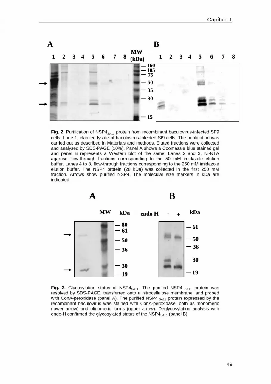
Capítulo 1
1 2 3 4 5 6 7 816010575503530
15
MW (kDa) 1 2 3 4 5 6 7 8
A B1 2 3 4 5 6 7 8
16010575503530
15
MW (kDa) 1 2 3 4 5 6 7 8
A B
Fig. 2. Purification of NSP4SA11 protein from recombinant baculovirus-infected SF9 cells. Lane 1, clarified lysate of baculovirus-infected Sf9 cells. The purification was carried out as described in Materials and methods. Eluted fractions were collected and analysed by SDS-PAGE (10%). Panel A shows a Coomassie blue stained gel and panel B represents a Western blot of the same. Lanes 2 and 3, Ni-NTA agarose flow-through fractions corresponding to the 50 mM imidazole elution buffer. Lanes 4 to 8, flow-through fractions corresponding to the 250 mM imidazole elution buffer. The NSP4 protein (28 kDa) was collected in the first 250 mM fraction. Arrows show purified NSP4. The molecular size markers in kDa are indicated.
A BMW endo H
19
8061
5036
30
+-
50
61
36
kDakDa
19
30
A BMW endo H
19
8061
5036
30
+-
50
61
36
kDakDa
19
30
Fig. 3. Glycosylation status of NSP4SA11. The purified NSP4 SA11 protein was resolved by SDS-PAGE, transferred onto a nitrocellulose membrane, and probed with ConA-peroxidase (panel A). The purified NSP4 SA11 protein expressed by the recombinant baculovirus was stained with ConA-peroxidase, both as monomeric (lower arrow) and oligomeric forms (upper arrow). Deglycosylation analysis with endo-H confirmed the glycosylated status of the NSP4SA11 (panel B).
49

Capítulo 1_______________________________________________________
Scale-up NSP4 protein production
Scaled-up production of NSP4 following two different protein purification strategies
showed a proportional increase of NSP4 recovery with both methods assayed. A
higher protein yield was achieved by incubating the clarified Sf9 cell lysates from 3 T75
cell culture flasks with 0.5 ml of Ni-NTA resin and packing the beads accrued from 3, 6,
12 and 24 flasks (“method B”) than mixing first the clarified Sf9 cell lysates obtained
from the same number of flasks and incubating the mixtures afterwards with
proportionally increased volumes of Ni-NTA resin (“method A”) (Fig. 4).
NSP4 production
050
100150200250300350
6 x 10e7 1,2 x 10e8 2,4 x 10e8 4,8 x 10e8
baculovirus-infected Sf9 cells
µg o
f rec
over
ed
prot
ein
Method AMethod B
Fig. 4. Comparison of NSP4 production by two different procedures: in method A (mixed clarified Sf9 cell lysates) cell lysates from 3, 6, 12 and 24 T75 cell culture flasks containing recombinant baculovirus-Sf9 infected cells were mixed and incubated with 0.5, 1, 2 and 4 ml of Ni-NTA resin, respectively. The resin was centrifuged and transferred to four chromatography columns (Bio-Rad), washed and the protein eluted. In method B (mixed beads), clarified cell lysates from sets of three T75 cell culture flasks containing recombinant baculovirus-Sf9 infected cells were incubated with 0.5 ml Ni-NTA resin. The beads from lysates derived from 3, 6, 12 and 24 T75 cell culture flasks were collected together, and each mixture was packed separately into a chromatography column for the washing and elution steps as described in Materials and methods.
Diarrhea induction by NSP4
The induction of diarrhea by the recombinant NSP4 proteins was studied in newborn
Balb/c mice. All the mice developed diarrhea in the next four hours when they were
given NSP4 corresponding to rotavirus strains Wa, Ito, OSU and SA11. No mice
responded with diarrhea when they were inoculated with the negative control (Table 1).
50

Capítulo 1
Only intraperitoneal inoculation of NSP4, but not oral inoculation, induced diarrhea in
suckling mice, as previously reported [6].
Table 1. Diarrhea induced by intraperitoneal administration of purified NSP4s from different rotavirus strains in newborn mice.
Protein given (a) Diarrhea outcome (b) NSP4Wa 10/10
NSP4Ito 10/10
NSP4OSU 10/10
NSP4SA11 10/10
Sf9 cell protein (c) 0/10
a1 nmol of each recombinant NSP4 protein diluted in 50 µl PBS was given intraperitoneally to newborn Balb/c mice. bNo. of mice suffering diarrhea/No. of challenged mice. c1 nmol of a Sf9 cell 90 kDa protein diluted in 50 µl PBS was given intraperitoneally to newborn Balb/c mice as a negative control.
DISCUSSION
The present paper describes the expression, purification and biological activity of
recombinant rotavirus nonstructural NSP4 proteins from four different viral strains using
the baculovirus expression system. The rotavirus NSP4 protein has been proven to be
a viral protein of great interest according to its biological and enterotoxigenic
properties. This protein consists of 175 amino acids and contains sequences
responsible for the membrane-destabilizing and enterotoxic effects that have been
mapped to different regions of the polypeptide [5]. It has been shown that the
enterotoxin domain is highly conserved among avian and mammalian rotaviruses in
terms of its sequential and structural properties, independently of the NSP4 genotype
[18]. Our goal was to express and purify recombinant NSP4 proteins from the human
Wa and Ito strains (both belonging to the NSP4 genotype B), from the simian SA11
strain (NSP4 genotype A), and from the porcine OSU strain (NSP4 genotype B) to
51

Capítulo 1_______________________________________________________
investigate whether the amino acid differencies in their sequences may be responsible
for differences in their enterotoxigenic activities. In this study we show that all four
recombinant NSP4 proteins have enterotoxigenic activity in suckling mice when given
intraperitoneally, what suggests that this activity must be important as a pathogenetic
mechanism in rotavirus infections.
The baculovirus-insect cell expression system, rather than bacterial expression, was
chosen to express the NSP4 proteins because they are glycosylated proteins, with two
potential N-glycosylation sites in the NSP4 molecule of all mammalian strains.
However, it has been recently reported that purified NSP4s of avian rotaviruses (PO13,
Ty-1, Ty-3 and Ch-1) expressed in E. coli had enterotoxigenic activities in suckling
mice [18].
The expression of NSP4 in Sf9 cells has been previously shown to cause an increase
in intracellular calcium levels [Ca2+]i [7]. Paradoxically, it has been demonstrated that
the expression of NSP4 in Spodoptera cells do not influence the permeability of the
plasma membrane, but alters plasma membrane permeability in mammalian cells [5,
19, 20]. Although very high concentrations of NSP4 are cytotoxic to Sf9 cells, levels
normally found in rotavirus-infected cells do not affect cell viability [20].
Polyhistidine-tagged NSP4 production in insect cells allows the purification of this
protein following a fast and easy procedure. NSP4 is an integral membrane protein
resident in the ER with two N-linked oligosaccharide sites at the amino-terminus, which
is on the luminal side of the ER. It has been reported that the membrane association
properties of NSP4 make purification of this protein difficult, although it can be
achieved by immunoaffinity chromatography [20]. By using the strategy described in
this study, 6xHis-tagged NSP4 can be produced and purified easily with a one-step Ni-
NTA agarose chromathography. Furthermore, histidine tags can be cleaved with rTEV
protease cleavage and removed by affinity chromatography.
Antibodies to NSP4 may play a role in the protection against rotavirus infections. It has
been shown that NSP4 induces both humoral and cellular immune responses in
52

Capítulo 1
humans [11], and elicits systemic and intestinal antibody responses in a gnotobiotic pig
model of human rotavirus disease [21].
The recombinant proteins produced in this study are currently being used as antigens
to detect specific antibodies against NSP4 in human sera from children convalescing
from acute rotavirus infection and in different groups of individuals. They are also
invaluable tools to study their effect on the mouse intestinal mucosa and to analyze the
role that anti-NSP4 antibodies can play in the protection against rotavirus infection in
the mouse model. Many aspects of the function of NSP4 in viral morphogenesis,
pathogenesis and immunity still remain to be clarified.
It has been highlighted the therapeutic potential that synthetic analogs of the active
NSP4114-135 peptide may have for the treatment of neonatal meconium ileus
syndrome or to counteract chronic constipation in cystic fibrosis patients [9]. With these
purposes, further studies on the molecular characterization of this protein are needed
using purified recombinant NSP4 proteins or peptides.
ACKNOWLEDGMENTS
This work was supported by the Fondo de Investigación Sanitaria (F.I.S.) from the
Spanish Ministry of Health (Grant 00/0592). We wish to thank Dr Albert Bosch,
University of Barcelona, for kindly providing rotavirus strains OSU and Ito, and Dr
Lennart Svensson, Swedish Institute for Infectious Disease Control, for providing us
with the anti-NSP4 114-134 rabbit serum. We also thank Andrew McCabe for helpful
discussion and Desmond Donellan for reviewing the English.
REFERENCES
[1] R.I. Glass, J. Gentsch, J.C. Smith, Rotavirus vaccines: Success by reassortment?, Science 265 (1994) 1389-1391.
53

Capítulo 1_______________________________________________________
[2] O. Lundgren, L. Svensson, Pathogenesis of rotavirus diarrhea, Microb Infect 3 (2001) 1145-1156. [3] M. Estes, J. Cohen, Rotavirus gene structure and function, Microbiol Rev 53 (1989) 410-449. [4] J.A. O'Brien, J.A. Taylor, A.R. Bellamy, Probing the structure of rotavirus NSP4: a short sequence at the extreme C terminus mediates binding to the inner capsid particle, J. Virol. 74 (2000) 5388-5394. [5] E.P. Browne, R. Bellamy, J.A. Taylor, Membrane-destabilizing activity of rotavirus NSP4 is mediated by a membrane-proximal amphipatic domain, J. Gen. Virol. 81 (2000) 1955-1959. [6] J.M. Ball, P. Tian, P., C.Q.-Y. Zeng, A.P. Morris, M.K. Estes, Age-dependent diarrhea induced by a rotaviral nonstructural glycoprotein, Science 272 (1996) 101-103. [7] P. Tian, M.K. Estes, Y. Hu, J.M. Ball, C.Q.-Y. Zeng, W.P. Schilling, The rotavirus nonstructural glycoprotein NSP4 mobilizes Ca2+ from the endoplasmic reticulum, J. Virol. 69 (1995) 5763-5772. [8] Y. Horie, O. Nakagomi, Y. Koshimura, Y. Nakagomi, Y. Suzuki, T. Oka, S. Sasaki, Y. Matsuda, S. Watanabe, Diarrhea induction by rotavirus NSP4 in the homologous mouse model system, Virology 262 (1999) 398-407. [9] A.P. Morris, J.K. Scott, J.M. Ball, C.Q.-Y. Zeng, W.K. O’Neal, M.K. Estes, NSP4 elicits age-dependent diarrhea and Ca2+-mediated I- influx into intestinal crypts of CF mice, Am. J. Physiol. 277 (1999) G431-G444. [10] J. Colomina, M.T. Gil, P. Codoñer, J. Buesa, Viral proteins VP2, VP6, and NSP2 are strongly precipitated by serum and fecal antibodies from children with rotavirus symptomatic infection, J. Med. Virol. 56 (1998) 58-65. [11] K. Johansen, J. Hinkula, F. Espinoza, M. Levi, C. Zeng, U. Rudén, T. Vesikari, M. Estes, L. Svensson, Humoral and cell-mediated immune responses in humans to the NSP4 enterotoxin of rotavirus, J. Med. Virol. 59 (1999) 369-377. [12] M. Ciarlet, F. Liprandi, M.E. Conner, M.K. Estes, Species specificity and interspecies relatedness of NSP4 genetic groups by comparative NSP4 sequence analyses of animal rotaviruses, Arch. Virol. 145 (2000) 371-383. [13] N.A. Cunliffe, P.A.Woods, J.P.G. Leite, B.K. Das, M. Ramachandran, M.K. Bhan, C.A. Hart, R.I. Glass, J.R. Gentsch, Sequence analysis of NSP4 gene of human rotavirus allows classification into two main genetic groups, J. Med. Virol. 53 (1997) 41-50. [14] C.D. Kirkwood, E.A. Palombo, Genetic characterization of the rotavirus nonstructural protein, NSP4, Virology 236 (1997) 258-265. [15] X.R. Cao, S. Akihara, Z.Y. Fang, O. Nakagomi, H. Ushijima, Genetic variation in the VP4 and NSP4 genes of human rotavirus serotype 3 (G3 type) isolated in China and Japan, Microbiol. Immunol. 43 (1999) 171-175.
54

Capítulo 1
[16] A.H.-C. Choi, D.R. Knowlton, M.M. McNeal, R.L. Ward, Particle bombardment-mediated DNA vaccination with rotavirus VP6 induces high levels of serum rotavirus IgG but fails to protect mice against challenge, Virology 232 (1997) 129-138. [17] D.R. O'Reilly, L.K. Miller, V.A. Luckow, Baculovirus Expression Vectors. A Laboratory Manual. Oxford University Press, New York, 1994. [18] Y. Mori, M.A. Borgan, N. Ito, M. Sugiyama, N. Minamoto, Sequential analysis of nonstructural protein NSP4s derived from group A avian rotaviruses, Virus Res. 89 (2002) 145-151. [19] K. Newton, J.C. Meyer, R. Bellamy, J.A. Taylor, Rotavirus nonstructural glycoprotein NSP4 alters plama membrane permeability in mammalian cells, J. Virol. 71 (1997) 9458-9465. [20] P. Tian, J.M. Ball, C.Q.Y. Zeng, M.K. Estes, The rotavirus nonstructural glycoprotein NSP4 possesses membrane destabilization activity, J. Virol. 70 (1996) 6973-6981. [21] C. Iosef, K.-O. Chang, M.S.P. Azevedo, L.J. Saif, Systemic and intestinal antibody responses to NSP4 enterotoxin of Wa human rotavirus in a gnotobiotic pig model of human rotavirus disease, J. Med. Virol. 68 (2002) 119-128.
55

Capítulo 1_______________________________________________________
56

Capítulo 2
CAPÍTULO 2
Isolation and characterization of single-chain variable
fragment (scFv) antibodies against rotavirus NSP4 and
VP8* proteins generated by phage display
57

Capítulo 2_______________________________________________________
58

Capítulo 2A
CAPÍTULO 2A
Single-chain variable fragment (scFv) antibodies against
rotavirus NSP4 enterotoxin generated by phage display
Jesús Rodríguez-Díaz a, Vicente Monedero b, Gaspar Pérez-Martínez b
and Javier Buesa a, *
a Department of Microbiology, School of Medicine, University of Valencia,
Hospital Clinico Universitario, and
b Department of Biotechnology, Instituto de Agroquímica y Tecnología de los
Alimentos (IATA), CSIC, Burjassot (Valencia), Spain
Accepted for publication in Journal of Virological Methods (2004)
59

Capítulo 2A______________________________________________________
60

Capítulo 2A
Journal of Virological Methods (2004)
Single-chain variable fragment (scFv) antibodies against rotavirus NSP4
enterotoxin generated by phage display
Jesús Rodríguez-Díaz a, Vicente Monedero b ,Gaspar Pérez-Martínez b
and Javier Buesa a, *
a Department of Microbiology, School of Medicine, University of Valencia, Hospital Clinico
Universitario, and b Department of Biotechnology, Instituto de Agroquímica y Tecnología de los
Alimentos (IATA), CSIC, Burjassot (Valencia), Spain
*Corresponding author. Fax: +34-96-3864658 Fax: +36 96 386 46 58
E-mail address: [email protected]
Abstract
The rotavirus non-structural NSP4 protein causes membrane destabilization as well as
an increase in intracellular calcium levels in eukaryotic cells and induces diarrhea in
young mice, acting as a viral enterotoxin. In this study the phage display technique was
used to generate a panel of single-chain variable fragment (scFv) antibodies specific
for the NSP4 protein of the human rotavirus strain Wa from a human semi-synthetic
scFv library. After several rounds of panning and selection on NSP4 adsorbed to
polystyrene tubes, individual scFv were isolated and characterized by fingerprinting
and by sequencing the VH and VL genes. The isolated scFv antibodies specifically
recognize NSP4 in enzyme immunoassay and in Western blot. Four truncated forms of
the NSP4 protein were constructed which allowed us to map the binding region of the
selected scFv antibodies to the C-terminal portion of NSP4. The isolated scFv
antibodies constitute valuable tools to analyse the mechanisms of NSP4 functions.
Keywords: Rotavirus; NSP4; Phage display library; Single-chain variable fragment
(scFv) antibodies. Abbreviated article title: scFv antibodies against NSP4
61

Capítulo 2A______________________________________________________
1. Introduction
Rotaviruses (RV) are the main etiologic agents of viral gastroenteritis in infants and
young children worldwide, producing high childhood mortality in developing countries
(Parashar et al., 2003). Rotaviruses infect mature enterocytes in the mid- and upper
parts of the villi of the small intestine, which ultimately causes diarrhea (Lundgren and
Svensson, 2001). The rotavirus non-structural glycoprotein NSP4 is an intracellular
receptor that mediates the acquisition of a transient membrane envelope as sub-viral
particles bud into the endoplasmic reticulum. NSP4 acts as an intracellular receptor for
double-layered particles (DLPs) of rotavirus, interacting with VP6 (Estes, 2001; 2003).
Many structural motifs or protein regions have been implicated in the NSP4 biological
function. Amino acids 17 to 20 from the C-terminus extreme are necessary and
sufficient for inner capsid particle binding (O’Brien et al., 2000) and the region involved
in the retention of the NSP4 protein into the endoplasmic reticulum has been mapped
between the amino acids 85 and 123 in the cytoplasmatic region of the protein
(Mirazimi et al., 2003). Residues at positions 48 to 91, a region which includes a
potential cationic amphipatic helix, have been shown to be involved in membrane
destabilization (Tian et al., 1996; Browne et al., 2000). Purified NSP4 or a peptide
corresponding to NSP4 residues 114-135 induce diarrhea in young mice after an
increase in intracellular calcium levels, suggesting a role for NSP4 in rotavirus
pathogenesis (Tian et al., 1994; Ball et al., 1996; Horie et al., 1999).
Six distinct alleles, genotypes or genetic groups (A through F) including animal and
human strains have been described for the NSP4 protein (Ciarlet et al., 2000; Cunliffe
et al., 1997; Kirkwood et al., 1997; Cao et al., 1999; Borgan et al., 2003). Genotype A
includes human, bovine and simian strains; genotype B includes human and porcine
strains; genotype C includes human, simian, canine and feline strains; genotype D
includes murine strains (Ciarlet et al., 2000), while genotypes E and F are composed of
avian strains (Mori et al., 2002).
62

Capítulo 2A
The significance of the humoral and cell-mediated immune responses to NSP4 in the
protection against rotavirus infection in humans is still unknown. Other non-structural
proteins, such as the rotaviral NSP2 protein, elicit the production of specific IgG
antibodies (Colomina et al., 1998). It has been shown that NSP4 induces both humoral
and cell-mediated immune responses in humans (Johansen et al., 1999; Ray et al.,
2003). However, very few studies have been carried out to elucidate the immunologic
determinants in the NSP4 protein. Recently two immunodominant regions have been
described in the NSP4 of group A avian rotavirus PO-13 (P[17], G7, NSP4 genotype
E). The antigenic site I (AS I) (amino acids 151 to 169) and the antigenic site II (AS II)
(amino acids 136 to 150) are the two immunodominant regions and are located in the
cytoplasmatic tail (Borgan et al., 2003).
To increase our knowledge on the NSP4 protein and to elucidate the effects that
blocking of some specific epitopes of this protein can have in rotavirus pathogenesis, it
is necessary to obtain and characterise monoclonal antibodies against NSP4. Single-
chain variable fragment (scFv) antibodies can be particularly useful for performing this
type of studies. scFv are chimeric proteins produced by a fusion of the heavy VH and
the light VL variable regions of immunoglobulins and have epitope recognition sites
similar to monoclonal antibodies. scFv libraries of randomly combined VH and VL chains
from immune or non-immune individuals are usually fused to the minor coat protein of
M13 filamentous phage (pIII). This allows display of the scFv proteins on the M13
surface and selection of scFv directed to a particular antigen by several rounds of
panning on immobilized antigen (Harrison et al., 1996; Griffiths and Duncan, 1998).
This technology is used for the isolation of monoclonal antibody reagents bypassing
hybridoma technology. In this study the phage display technique was used to generate
a panel of scFv antibodies against NSP4 from the Griffin.1 library. Four truncated forms
of NSP4 were constructed in order to elucidate the region recognized by individual
63

Capítulo 2A______________________________________________________
antibodies. The isolated scFv antibodies can be used to further analyse the NSP4
biological functions, particularly its effect as an enterotoxin.
2. Materials and methods
2.1. Expression and purification of NSP4 proteins in insect cells
Two recombinant baculoviruses expressing the NSP4 glycoproteins from the simian
rotavirus strain SA11 (P5B[2], G3, NSP4 genotype A) and the human strain Wa
(P1A[8], G1, NSP4 genotype B) were used to produce the recombinant NSP4
glycoproteins (Rodriguez-Diaz et al., 2003). Spodoptera frugiperda 9 (Sf9) cells were
infected with the recombinant baculoviruses at a multiplicity of infection (m.o.i.) of 10
and incubated for 48 h. The recombinant proteins were purified by affinity
chromatography as previously described (Rodriguez-Diaz et al., 2003). Complete purity
of the NSP4 proteins could not be achieved because a 90 KDa cellular protein co-
purified in the chromatography eluate. Proteins were dialysed and analysed by SDS-
PAGE and their concentrations quantified by the Bradford method.
2.2. Cloning, expression and purification of GST- NSP4 truncated proteins in E.
coli
Four truncated forms of NSP4 from the rotavirus strain Wa were constructed fused to
the glutathione S-transferase (GST) according to the structural motifs of the NSP4
protein: GST-NSP41-50, corresponding to amino acids 1 to 50; GST-NSP41-91,
corresponding to amino acids 1 to 91; GST-NSP41-135, corresponding to amino acids 1
to 135 and GST-NSP41-175, representing the whole NSP4 protein. The NSP4Wa gene
was amplified by PCR with specific primers incorporating NotI and SalI restriction sites
64

Capítulo 2A
for directional cloning into the pGEX-4T-3 expression vector. Primers were: NSP4-1 (5'-
TTT GTC GAC ATG GAT AAG CTT GCC GAC) as the forward primer for all reactions
and NSP4-50 (5'-TTT GC GGC CGC TCA TGA AGC TTT ATG TAA TGT), NSP4-91
(5'-TTT GC GGC CGC TCA TGT AGT AAC CTG CTC TTT), NSP4-135 (5'-TTT GC
GGC CGC TCA TAT CAG GTT GTC ATG TAT), NSP4-175 (5'-TTT GC GGC CGC
TCA CAT GGA TGC AGT CAC), as the reverse primers, respectively. The restriction
sites are underlined and the start and stop codons are indicated in bold. Cultures of E.
coli DH5α were transformed with the pGEX-4T-3 derivatives and were then induced
with 0.1 mM isopropyl-β-D-thiogalactopyranoside (IPTG) for 2 h. Bacterial cells were
harvested and pellets were resuspended in a 1/50 volume of PBS. After sonication,
Triton X-100 was added to the cell lysate at a final concentration of 1%, and cellular
debris was removed by centrifugation at 20.000xg at 4°C. One milliliter of 50%
glutathione-Sepharose 4B (Amersham Biosciences) was added to 10 ml of extract
supernatant, and the resultant mixture was incubated with gentle agitation for 1 h at
room temperature. The mixture was extensively washed with PBS, and proteins were
eluted with an equal volume of 10 mM reduced glutathione in 50 mM Tris-HCl pH 8.0.
2.3. Antibody phage display to the NSP4 protein
The Griffin.1 library, which is a scFv phagemid library made from synthetic V-gene
segments, was generously provided by Dr. A. D. Griffiths (Centre for Protein
Engineering, Medical Research Council, Cambridge, U.K.). The library was made by
recloning the heavy and light chains variable regions from the lox library vectors
(Griffiths et al., 1994) into the phagemid vector pHEN2. Three rounds of selection were
performed following a panning method previously described (Marks et al., 1991).
Immunotubes (Nunc Maxisorp) were coated overnight with 4 ml of purified NSP4Wa
(10 µg/ml) in 50 mM sodium hydrogen carbonate pH 9.6 at room temperature. The
tubes were washed three times with PBS and blocked for 1 h with 2% skim milk in PBS
65

Capítulo 2A______________________________________________________
(M-PBS). Then 1012 phage particles in 4 ml of 2% M-PBS were added and allowed to
bind for 30 min at room temperature by rotating continuously and then without rotation
for at least 90 min. For the first round of selection, tubes were washed 10 times with
PBS containing 0.1% Tween-20 (T-PBS) and 10 times with PBS. The following rounds
were washed 20 times with T-PBS and 20 times with PBS. Bound phages were eluted
by adding 1 ml of 100 mM triethylamine pH 11.6 and rotating 10 min at room
temperature. Eluted phages were neutralized with 0.5 ml of 1 M Tris-HCl pH 9.6 and
used to infect 10 ml of an exponentially growing E. coli TG1 culture for 30 min at 30 ºC
without shaking. The infected culture was pelleted, resuspended in 1 ml of 2xTY, plated
on a large Petri dish (Nalge Nunc) of TYE containing 100 µg/ml ampicilin, 1% glucose,
and grown overnight at 30 ºC. E. coli TG1 cells were recovered from the plate and
infected with VCS-M13 helper phage (Stratagene) at a m.o.i. of 20. Phages were
grown and recovered from supernatant by adding 1/5 volume of 20% PEG 6000, 2.5 M
NaCl followed by 1 h of incubation at 0 ºC and spinning at 10.800 x g for 10 min.
Pellets containing phages were resuspended in 2 ml of PBS.
2.4. Screening of individual phage clones by ELISA
Individual phages obtained in the third round of selection were screened by ELISA.
Ninety-six-well ELISA plates (Costar) were coated with 0.1 µg of purified NSP4Wa per
well. Proteins extracted from Sf9 insect cells were used as a negative control. ELISA
was performed with the total phages obtained in the three rounds of selection and with
individual clones selected from the third round of selection. Coated plates were washed
and blocked with 2% M-PBS 1 h at room temperature. Phages were added in 100 µl of
2% M-PBS and incubated for 2 h. Plates were washed and a horseradish peroxidase
(HRP)-conjugated anti-M13 monoclonal antibody (Amersham Biosciences) 1:5000
diluted in 2% M-PBS was added. After incubation at room temperature for 2 h, plates
66

Capítulo 2A
were washed and developed by adding 3,3’,5,5’-tetramethylbenzidine (TMB, Sigma) as
the substrate. Optical density was measured at 450 nm.
2.5. Detection of binding specificity by ELISA
Immunoplates (Nunc) were coated with 0.1 µg of purified NSP4Wa or NSP4SA11 per
well to analyse the cross-reactivity of the isolated scFv antibodies. Wells were also
coated with 0.1 µg of each of the truncated forms of the NSP4Wa per well. A 90 KDa
protein purified from Sf9 cells or purified GST were used as the negative controls. The
following steps of the ELISA procedure were performed as described above.
2.6. scFv characterisation
The ELISA-positive clones were characterized by BstNI restriction analysis and
fingerprinting as previously described (Clackson et al., 1991). scFv antibody genes
were amplified from single E. coli colonies with primers LMB3 (5`-GAGGAA
ACAGCTATGAC-3´) and Fd seq 1 (5´-GAATTTTCTG TAT GAGG-3´) followed by
digestion of the PCR products with BstNI (Marks et al., 1991). Clones were classified
according to the fingerprinting patterns and sequenced with primer FOR_LinkSeq (5´-
GCCACCTCCGCCTGAACC-3´) to sequence the VH genes and pHEN-SEQ (5´-
CTATGCGGCCCCATTCA-3´) to obtain the sequence of the VL genes. The nucleotide
sequences of the PCR products were analyzed using an ABI PRISM 377 automated
DNA sequencer (Applied Biosystems).
The reactivity of the scFv antibodies against denatured NSP4Wa protein was evaluated
by Western blotting. 100 ng of purified NSP4 were subjected to SDS-PAGE and
transferred onto a nitrocellulose membrane. Phages displaying scFv were used as first
antibodies and HRP-conjugated anti-M13 (Amersham Biosciences) was used as
67

Capítulo 2A______________________________________________________
second antibody. After washings, reactivity was detected with the substrate 3-amino-9-
ethylcarbazole (AEC, Sigma).
3. Results
3.1. Isolation of scFv against NSP4
The Griffin.1 phage display library was used to select phage antibodies against purified
NSP4 from the human rotavirus strain Wa. Phage suspensions (5x10¹² to 1x10¹³ PFU)
were applied, and after each round of panning and selection on NSP4-coated
immunotubes the number of eluted phages was determined by plating. Phage titres
increased after the third round of selection.
Clone
2A1 2A6 2B5 2B11 2B12 2C3 2C4 2D1 2D5 2H11Negative
O.D
. 450
nm
0,0
0,1
0,2
0,3
0,4
0,5
0,6
0,7
NSP4Sf9
Figure 1. ELISA analysis of ten selected clones of M13 phages expressing scFv from the third round. Selected phages specifically reacted against NSP4 but not against a 90 KDa protein from Sf9 cells that co-purified with the baculovirus-expressed NSP4. A negative clone randomly chosen was used as a control. Phage-infected E. coli TG1 culture supernatants containing ~107 phage particles per ml were analysed in the assay.
The enrichment of the phage population in phages specifically recognizing the NSP4
protein was demonstrated by an increase of the reactivity against NSP4 in ELISA with
the pool of phages isolated after each round of selection (results not shown). As the
NSP4 preparations carried a contaminant protein of 90 KDa derived from Sf9 insect
68

Capítulo 2A
cells, 96 independent clones from the third round were simultaneously tested by ELISA
against NSP4 and against the Sf9 purified protein. Ten clones were selected that
specifically reacted against NSP4 but not against the Sf9 protein. These clones were
repurified and their specificity was confirmed in a second ELISA test. As shown in Fig.
1, the ten selected phage clones specifically recognized NSP4.
3.2. scFv characterisation
The scFv gene of the selected clones was amplified by PCR and subjected to BstNI
restriction analysis. Four different banding patterns were detected (Fig. 2). The
presence of four different scFv genes was also confirmed by sequencing the VH and
VL regions, which were analysed with the V-BASE database of human immunoglobulin
genes (Tomlinson et al., 1992).
2A1 2A6 2B5 2B11 2B12 2C3 2C4 2D5 2D1 2H11
516339265247210
MW (bp)
Figure 2. BstNI fingerprinting analysis of the scFv genes present in the selected clones reacting against NSP4. The scFv gene inserts were was amplified by PCR, digested with BstNI and resolved in a 3% agarose gel. Four different banding patterns were detected.
The four established groups contained four different complementarity determining
regions (CDR3) in their VH segments (Table 1). Group I was represented by six clones;
group II by two clones, while groups III and IV had only one representative. One clone
from each groups I and II was chosen (clones 2C3 and 2B5, respectively). The four
groups were also evaluated by Western blotting with NSP4 as antigen using HRP-
69

Capítulo 2A______________________________________________________
conjugated anti-M13 as the second antibody (Fig. 3). Clones 2B5, 2C3, and 2D1 were
positive by Western blot, while 2B12 gave no signal. This result indicated that three
clones recognized linear epitopes that are conserved after protein denaturation,
whereas clone 2B12 recognized most likely a conformational epitope in the NSP4
protein.
2B5 2C3 2D12B12
NSP4
Figure 3. Western blot analysis after SDS-PAGE of NSP4Wa probed with M13 phages bearing anti-NSP4 scFv antibodies fused to the pIII phage capsid protein (i.e., scFv::pIII fusions). The binding was detected with an HRP- conjugated anti-M13 monoclonal antibody.
Table 1. Amino acid sequences of the VH regions CDR3 of 10 isolated scFv genes containing clones specifically reactive with NSP4.
Clone CDR3
2A1, 2A6, 2B11, 2C3, 2D5, 2H11 GSGMER
2B5, 2C4 GSIRDPIK
2B12 RLDI
2D1 YTDSRHGGVG
3.3. Detection of binding specificity by ELISA
The ability of each clone to recognize the NSP4 from two different rotavirus genotypes
was analysed by ELISA. As shown in Fig. 4, three of the clones (2B5, 2B12 and 2C3)
were able to recognize both NSP4 proteins of genotype A (rotavirus strain SA11) and
genotype B (rotavirus strain Wa), indicating that they were interacting with conserved
70

Capítulo 2A
motifs present in the two assayed genotypes. Clone 2D1 only reacted with the NSP4
from genotype B (Fig. 4).
Clone
2B5 2B12 2C3 2D1
O.D
.450
nm
0,0
0,1
0,2
0,3
0,4
NSP4Wa
NSP4SA11
Sf9
Figure 4. Analysis of scFv antibodies binding specificity by ELISA. Recognition of two different NSP4 genotypes, genotype A (strain SA11) and genotype B (strain Wa), by clones 2B5, 2B12, 2C3 and 2D1. Clone 2D1 only reacted with the NSP4 from genotype B. A Sf9 cellular protein of 90 KDa of MW was used as the antigen in the specificity control.
In order to map the NSP4wa region recognized by each scFv clone, four NSP4
truncated forms were constructed, expressed and purified as GST fusions (Fig. 5) and
used as antigens in ELISA tests (Fig. 6). A mouse polyclonal antiserum raised against
the NSP4Wa protein strongly reacted only with the C-terminal region of the protein
(amino acids 135-175). The region containing the amino acids 91 to 135 was poorly
recognized and no signal was detected against the more hydrophobic region of the
protein. The scFv clones were specific to only two of the four regions in NSP4: clones
2B5, 2C3 and 2D1 recognized the truncated proteins GST-NSP41-135 and GST-
NSP41-175, but not the NSP4 region spanning from amino acids 1 to 91 (Fig. 6).
Therefore, these three clones recognized a region of the protein between amino acids
91 and 135, which contains the toxigenic peptide (amino acids 114-135). Clone 2B12
was able to interact only with the full-length NSP4 in the ELISA test (Fig. 6) but did not
71

Capítulo 2A______________________________________________________
recognized this protein by Western blot analysis (Fig. 3). This clone might recognize a
conformational epitope in which residues within the last forty amino acids of the NSP4
might be implicated. Table 2 summarizes the reactivity patterns of the isolated scFv,
the region of the NSP4 recognized by the scFv clones, their binding to NSP4 by
Western blotting and the reactivity against NSP4 genotypes A and B by ELISA.
1 7 21 5028 47
H1 H2 H3 α-helix
85 93 133 135 17567 91
NSP4
GST-NSP41-50
GST-NSP41-91
GST-NSP41-135
GST-NSP41-175
Toxigenic peptide
GST
Figure 5. Schematic representation of the four GST-NSP4Wa fusion proteins (one containing NSP4 full length, three C terminal truncated fragments). The truncated constructions were made taking the structural domains of the protein into consideration. Hydrophobic domains are indicated as H1 to H3. The toxigenic peptide (amino acids 114-135) is contained in the α-helix domain of the protein. The amino acids that mark the C terminal end of each construct are indicated by arrows.
Clone
2B5 2B12 2C3 2D1 Polyclonal serum
O.D
. 450
nm
0,0
0,5
1,0
1,5
2,0
2,5
GST-NSP41-175
GST-NSP41-135
GST-NSP41-91
GST-NSP41-50
GST
Figure 6. Reactivity of the different scFv clones and a polyclonal mouse serum obtained in mice against the four GST-NSP4 constructs (one full length, three truncated, Fig. 5) as detected by ELISA.
72

Capítulo 2A
4. Discussion
In this work the phage display technique was successfully used to select several
human scFv clones that specifically recognize the NSP4 protein. After partial molecular
characterization, four different scFv against the NSP4 protein from the human rotavirus
strain Wa were selected. The four clones differed in their CDR3 sequences, and
Western blot analysis suggested that only one clone (2B12) recognized a
conformational epitope in NSP4. Phage display technology has been used to perform
antigenic analysis of the rotavirus VP7 protein (Huang et al., 2000), to map the domain
in VP4 that binds to cellular receptors (Jolly et al., 2001) and to isolate human anti-VP4
and anti-VP7 monoclonal antibodies that neutralize human rotaviruses (Higo-Moriguchi
et al., 2004). Here this technique has been used to obtain antibodies against the NSP4
protein. The binding region of individual scFv antibodies on NSP4 was mapped by
using four different truncated forms of NSP4wa. Three of the clones mapped to the
NSP4 region between amino acids 91 and 135, which includes the toxigenic peptide
(NSP4114-135).
Table 2. Reactivity patterns of scFv antibodies directed against the NSP4 protein: recognized region of NSP4, binding of scFv to NSP4 on Western blot after SDS-PAGE and reactivity against NSP4 genotypes A and B in ELISA. scFv
clone
recognized
fragmentaWestern blot
binding
NSP4Wa
genotype B
NSP4SA11
genotype A
2B5 aa 91-135 + + +
2B12 aa 135-175 - + +
2C3 aa 91-135 + + +
2D1 aa 91-135 + + - aThe region of NSP4 that is recognized by each clone is indicated in amino acid (aa) positions.
A recent antigenic analysis of the NSP4 of avian rotavirus strain PO-13 demonstrated
four different antigenic sites (AS) (Borgan et al., 2003). Two of these sites are
immunodominant and are placed in regions spanning amino acids 151 to 169 (AS I)
73

Capítulo 2A______________________________________________________
and amino acids 136 to 150 (AS II). Similarly, our results obtained with mouse
polyclonal anti-NSP4 sera showed that the region between amino acids 135 and 175 of
NSP4Wa protein is immunodominant. However, the phage display technique allowed
us to generate scFvs antibodies specifically directed against a non-immunodominant
region (amino acids 91 to 135). This can be explained because in this technique the
immune system is not involved in the selection of the scFv. Therefore, phage display
can be a valuable tool to isolate monoclonal reagents suitable to perform functional
analyses on NSP4. Especially, scFv antibodies reactive with the toxigenic region
(clones 2C3, 2B5 and 2D1) could be particularly useful to study toxicity mechanisms.
Passive and active immunizations against the NSP4 protein have protected mice from
suffering diarrhea after both NSP4 and virulent rotavirus challenges (Ball et al., 1996;
Yu and Langridge, 2001). Neutralizing experiments with scFv antibodies or Fab
fragments against structural viral proteins obtained by the phage display technology
have been successfully carried out with human rotaviruses (Higo-Moriguchi et al.,
2004) as well as with measles virus (de Carvalho Nicacio et al., 2002). Moreover, scFv
antibodies have the advantage that they can be cloned, manipulated and produced in
different hosts (Robin et al., 2003). Therefore, the scFv antibodies specific for NSP4
can be expressed in different cellular hosts in order to perform studies on the NSP4 in
vivo functions. Ongoing studies in our laboratory are trying to elucidate whether these
scFv antibodies can be used to label NSP4 proteins and whether they can reduce in
vivo the ability of NSP4 to produce diarrhea in mice. In addition, it will be necessary to
evaluate whether they can block the increase in intracellular calcium levels, neutralize
the reduction in transepithelial resistance (TER) in polarized cells induced by NSP4
and interfere with rotavirus replication (Tian et al., 1994; Ciarlet et al., 2001; Estes,
2003).
Although only NSP4 from two genotypes (A and B) of NSP4 have been tested in this
study, we were able to obtain scFv clones that differentiated between the two NSP4
74

Capítulo 2A
genotypes. Clone 2D1 only reacted against the protein region between the amino acids
91 to 135 of genotype B, while the rest of clones recognized both A and B genotypes.
These different reactivities can be explained by the diversity of amino acid sequences
in that particular region of the NSP4. Recent studies have shown that NSP4 from
genotypes A and B are the most common genotypes of NSP4 in humans (Iturriza-
Gomara et al., 2003). We consider that future studies on the specificity of the isolated
scFv antibodies against a broader range of NSP4 proteins derived from strains of
different genotypes will show whether these antibodies will be useful to establish a
serotypic classification of NSP4 of human rotaviruses.
Acknowledgments
This work was supported by the Fondo de Investigación Sanitaria (F.I.S.) of the
Spanish Ministry of Health (Grant Contract No 00/0592) and by funds of the Oficina de
Ciencia y Tecnología de la Generalitat Valenciana (Project No CTIDIA/2002/82). J.
Rodriguez-Diaz was the recipient of a fellowship from the Spanish Ministry of
Education and Culture (No AP2000-3623).
References
Ball, J. M., Tian, P., Zeng, C.Q.-Y., Morris, A.P. and Estes, M.K. 1996. Age-dependent
diarrhea induced by a rotaviral nonstructural glycoprotein. Science 272, 101-103.
Borgan, M.A., Mori, Y., Ito, N., Sugiyama, M. and Minamoto, N. 2003. Antigenic
analysis of nonstructural protein (NSP) 4 of group A avian rotavirus strain PO-13.
Microbiol. Immunol. 47, 661-668.
Browne, E. P., Bellamy, R. and Taylor, J.A. 2000. Membrane-destabilizing activity of
rotavirus NSP4 is mediated by a membrane-proximal amphipatic domain. J. Gen.
Virol. 81, 1955-1959.
75

Capítulo 2A______________________________________________________
Cao, X.R., Akihara, S., Fang, Z.Y., Nagacomi, O. and Usijima, H. 1999. Genetic
variation in the VP4 and NSP4 genes of human rotavirus genotype 3 (G3 type)
isolated in China and Japan. Microbiol. Immunol. 43, 171-175.
Ciarlet, M., Crawford, S.E., Estes, M.K., Ciarlet, M., Crawford, S.E., Estes and M.K.
2001. Differential infection of polarized epithelial cell lines by sialic acid-dependent
and sialic acid-independent rotavirus strains. J Virol. 75, 11834-11850.
Ciarlet, M., Liprandi, F., Conner, M.E. and Estes, M.K. 2000. Species specificity and
interspecies relatedness of NSP4 genetic groups by comparative NSP4 sequence
analyses of animal rotaviruses. Arch. Virol. 145, 371-383.
Clackson, T., Hoogenboom, H.R., Griffiths, A.D. and Winter, G. 1991. Making antibody
fragments using phage display libraries. Nature 352, 624-628.
Colomina, J., Gil, M.T., Codoñer, P. and Buesa, J. 1998. Viral proteins VP2, VP6, and
NSP2 are strongly precipitated by serum and fecal antibodies from children with
rotavirus symptomatic infection. J. Med. Virol. 56, 58-65.
Cunliffe, N.A., Woods, P.A., Leite, J.P., Das, B.K., Ramachandran, M., Bhan, M.K.,
Hart, C.A., Glass, R.I. and Gentsch, J.R. 1997. Sequence analysis of NSP4 gene of
human rotavirus allows classification into two main genetic groups. J. Med. Virol. 53,
41-50.
de Carvalho Nicacio, C., Williamson, R.A., Parren, P.W., Lundkvist, A., Burton, D.R.
and Bjorling, E. 2002. Neutralizing human Fab fragments against measles virus
recovered by phage display. J. Virol. 76, 251-258.
Estes, M.K. 2001. Rotaviruses and their replication. In: D.M. Knipe, P.M. Howley et al.
(Eds), Fields Virology, Lippincott Williams and Wilkins, Philadelphia, pp. 1747-1785.
Estes, M.K. 2003. The rotavirus NSP4 enterotoxin: Current status and challenges. In:
Viral Gastroenteritis. U. Desselberger and J. Gray (Eds), Elsevier, Amsterdam, pp.
207-224.
Griffiths, A.D. and Duncan A.R. 1998. Strategies for selection of antibodies by phage
display. Curr. Opin. Biotechnol. 9, 102-108.
76

Capítulo 2A
Griffiths, A.D., Williams, S.C., Hartley, O., Tomlison, I.M., Waterhouse, P., Crosby,
W.L., Konterman, R.E., Jones, P.T., Low, N.M., Alison, T.J., et al. 1994. Isolation of
high affinity human antibodies directly from large synthetic repertories. EMBO J. 13,
3245-3260.
Harrison, J.L., Williams, S.C., Winter, G. and Nissim, A. 1996. Screening of phage
antibody libraries. Methods Enzymol. 267, 83-109.
Higo-Moriguchi, K., Akahori, Y., Iba, Y., Kurosawa, Y., Taniguchi, K. 2004. Isolation of
human monoclonal antibodies that neutralize human rotavirus. J. Virol. 78, 3325-
3332.
Horie, H., Nakagomi, O., Koshimura, Y., Nagakomi, Y., Suzuky, Y., Oka,T., Sasaki, S.,
Matsuda, Y. and Watanabe, S. 1999. Diarrhea induction by rotavirus NSP4 in the
homologous mouse model system. Virology 262, 398-407.
Huang, J.A., Wang, L., Firth, S., Phelps, A., Reeves, P. and Holmes, I. 2000. Rotavirus
VP7 epitope mapping using fragments of VP7 displayed on phages. Vaccine 18,
2257-2265.
Iturriza-Gomara, M., Anderton, E., Kang, G., Gallimore, C., Phillips, W., Desselberger,
U. and Gray, J. 2003. Evidence for genetic linkage between the gene segments
encoding NSP4 and VP6 proteins in common and reassortant human rotavirus
strains. J. Clin. Microbiol. 41, 3566-3573.
Johansen, K., Hinkula, J., Espinoza, F., Levi, M., Zeng, C., Rudén, U., Vesikari, T.,
Estes, M.K. and Svensson, L. 1999. Humoral and cell-mediated immune responses
in humans to the NSP4 enterotoxin of rotavirus. J. Med. Virol. 59, 369-377.
Jolly, C.L., Huang, J.A. and Holmes, I.H. 2001. Selection of rotavirus VP4 cell receptor
binding domains for MA104 cells using a phage display library. J. Virol. Methods 98,
41-51.
Kirkwood, C.D. and Palombo, E.A. 1997. Genetic characterization of the rotavirus
nonstructural protein, NSP4.Virology 236, 258-265.
77

Capítulo 2A______________________________________________________
Lundgren, O. and Svensson, L. 2001. Pathogenesis of rotavirus diarrhea. Microb.
Infect. 3, 1145-1156.
Marks, J.D., Hoogenboom, H.R., Bonnert, T.P., McCafferty, J., Griffiths, A.D. and
Winter G. 1991. By-passing immunization: human antibodies from V-gene libraries
displayed on phage. J. Mol. Biol. 222, 581-597.
Mirazimi, A., Magnusson, K.E. and Svensson, L. 2003. A cytoplasmic region of the
NSP4 enterotoxin of rotavirus is involved in retention in the endoplasmic reticulum.
J. Gen. Virol. 84, 875-883.
Mori, Y., Borgan, M.A., Ito, N., Sugiyama, M., Minamoto, N. 2002. Sequential analysis
of nonstructural protein NSP4s derived from group A avian rotaviruses. Virus Res.
89, 145-151.
O’Brien, J.A., Taylor, J.A. and Bellamy, A.R. 2000. Probing the structure of rotavirus
NSP4: a short sequence at the extreme C terminus mediates binding to the inner
capsid particle. J. Virol. 74, 5388-5394.
Parashar, U.D., Hummelman, E.G., Bresee, J.S., Miller, M.A. and Glass, R.I. 2003.
Global illness and deaths caused by rotavirus disease in children. Emerg. Infect.
Dis. 9, 565-572.
Ray, P., Malik, J., Singh, R.K., Bhatnagar, S., Bahl, R., Kumar, R. and Bhan, M.K.
2003. Rotavirus nonstructural protein NSP4 induces heterotypic antibody responses
during natural infection in children. J. Infect. Dis. 187, 1786-1793.
Robin, S., Petrov, K ., Dintinger,T., Kujumdzieva, A.,Tellier, C. and Dion, M. 2003.
Comparison of three microbial hosts for the expression of an active catalytic scFv.
Mol. Immunol. 39, 729-738.
Rodríguez-Díaz, J., López-Andújar, P., García-Díaz, A., Cuenca, J., Montava, R. and
Buesa, J. 2003. Expression and purification of polyhistidine-tagged rotavirus NSP4
proteins in insect cells. Protein Expr. Purif. 31, 207-212.
78

Capítulo 2A
Tian, P., Ball, J., Zeng, C.Q. and Estes, M.K. 1996. The rotavirus nonstructural
glycoprotein NSP4 possesses membrane destabilization activity. J. Virol. 70, 6973-
6981.
Tian, P., Hu, Y., Schilling, W.P., Lindsay, D.A., Eiden, J., Estes, M.K. 1994. The
nonstructural glycoprotein of rotavirus affects intracellular calcium levels. J. Virol. 68,
251-257.
Tomlinson, I.M., Walter, G., Marks, J.D., Llewelyn, M.B. and Winter, G. 1992. The
repertoire of human germline VH sequences reveals about fifty groups of VH
segments with different hypervariable loops. J. Mol. Biol. 227, 776-798.
Yu, J. and Langridge, W.H. 2001. A plant-based multicomponent vaccine protects mice
from enteric diseases. Nat. Biotechnol. 19, 548 552.
79

Capítulo 2A______________________________________________________
80

Capítulo 2B
CAPÍTULO 2B
Selection of single-chain antibodies against the VP8*
subunit of rotavirus VP4 outer capsid protein and their
expression in Lactobacillus casei
Vicente Monedero1*, Jesús Rodríguez-Díaz2, Rosa Viana1, Javier Buesa2 and
Gaspar Pérez-Martínez1
1 Departamento de Biotecnología, Instituto de Agroquímica y Tecnología de
Alimentos (CSIC), Burjassot, Valencia, Spain. 2 Departamento de Microbiología,
Facultad de Medicina, Universitat de València, Valencia, Spain.
Published in Applied and Enviromental Microbiology. (2004) 70(11):6936-9.
81

Capítulo 2B______________________________________________________
82
Capítulo 2B
Applied and Enviromental Microbiology. (2004) 70(11):6936-9
Selection of single-chain antibodies against the VP8* subunit of rotavirus
VP4 outer capsid protein and their expression in Lactobacillus casei
Vicente Monedero1*, Jesús Rodríguez-Díaz2, Rosa Viana1, Javier Buesa2 and Gaspar Pérez-
Martínez1
1 Departamento de Biotecnología, Instituto de Agroquímica y Tecnología de Alimentos (CSIC),
Burjassot, Valencia, Spain.
2 Departamento de Microbiología, Facultad de Medicina, Universitat de València, Valencia,
Spain.
*Corresponding author:
Vicente Monedero
Instituto de Agroquímica y Tecnología de Alimentos (CSIC)
Apartado de correos 73
46100 Burjassot, Valencia, Spain
Tel.: + 34 963900022
FAX:+ 34 963636301
e-mail: [email protected]
ABSTRACT Single-chain antibodies (scFv) recognizing the VP8* fraction of rotavirus outer capsid
and blocking rotavirus infection in vitro were isolated by phage display. Vectors for the
extracellular expression in Lactobacillus casei of one of the scFv were constructed. L.
casei was able to secrete active scFv to the growth medium, showing the potential of
probiotic bacteria to be engineered to express molecules suitable for in vivo anti-
rotavirus therapies.
Keywords: rotavirus, VP8*, single-chain antibodies, lactic acid bacteria
Running title: Phage antibodies against VP8*
83

Capítulo 2B______________________________________________________
Group A rotaviruses are the main cause of diarrhea in children worldwide and are
estimated to be responsible for more than 800.000 deaths per year in children under
five years of age (12). The incidence of rotavirus disease in developing countries as
well as the lack of an effective vaccine supports the development of new and more
effective antirotaviral strategies. Mucosal immunity against rotavirus infection is
believed to rely mainly on the production of rotavirus-specific IgA antibodies at the
intestinal mucosal surface (12, 17, 26). Moreover, lacteal IgG or monoclonal IgA
against the VP8* portion of rotaviral VP4 outer capsid can protect newborn mice
against rotavirus-induced diarrhea (5, 21, 22). This makes VP8* a good target for the
design of antirotavirus therapies.
Lactic acid bacteria (LAB) are GRAS microorganism present in numerous food
fermentations and are also normal constituents of the intestinal habitat. In addition,
some strains of LAB exhibit probiotic properties. These characteristics have been
exploited for their use as live vectors for the expression of different peptides and its
delivery to mucosal surfaces in animal models. These include: antigens (16),
interleukins (24), enzymes (3) and single chain antibodies (scFv) (1, 13). These last
molecules are chimeric proteins consisting in a fusion of the VH and VL variable regions
of immunoglobulins (19). Specific scFv can be isolated by the phage display technique
after panning of phage scFv libraries on immobilized antigen (7, 9). scFv offer very
interesting clinical perspectives and, although they may not be as powerful as natural
immunoglobulins, many possible applications could be envisaged, since they can be
cloned, manipulated and produced in microbial hosts (20). Two cases of successful
therapeutical application by in vivo delivery of scFv in mucosae by LAB have been
reported (1, 13). We decided to construct Lactobacillus casei strains expressing
extracellular or cell wall attached anti-VP8* scFv, which might be useful to deliver
passive immunity against rotavirus. The use of L. casei might be of particular interest,
since some strains exhibit an intrinsic beneficial effect in the treatment of rotaviral
diarrhea (11, 18)
84

Capítulo 2B
Isolation of scFv against VP8*
The Griffin.1 phage display library (6) was used to select phage antibodies against
purified VP8* from rotaviral SA11 strain. This library is a semisynthetic human scFv
library composed by more than 109 independent clones carrying VH and VL
immunoglobulin variable regions cloned into pHEN2 (8) to produce a scFv fused to the
pIII protein of the M13 viral capsid. Several rounds of panning and selection of VP8*-
binding phages was carried out as described (9) with VP8*-coated immunotubes
(Polysorp, NUNC). Titers of eluted phages and their signals in VP8*-specific ELISA
increased after each round, indicating the enrichment of VP8* specific phages (data not
shown).
A 414n
m
0,0
0,2
0,4
0,6
0,8
1,0
1,2
1,4
1,6VP8*BSA
2A1 2A2 2A4 2B1 2B2 2B3 2E1 2E2 2E3 2E4 2E11 2F6 2F7 2F8 2H6
A 414n
m
0,0
0,2
0,4
0,6
0,8
1,0
1,2
1,4
1,6VP8*BSA
A 414n
m
0,0
0,2
0,4
0,6
0,8
1,0
1,2
1,4
1,6VP8*BSA
2A1 2A2 2A4 2B1 2B2 2B3 2E1 2E2 2E3 2E4 2E11 2F6 2F7 2F8 2H6
A
2A1 2A2 2A4 2B1 2B2 2B3 2E1 2E2 2E3 2E4 2E11 2F6 2F7 2F8 2H6
VP8*
2A1 2A2 2A4 2B1 2B2 2B3 2E1 2E2 2E3 2E4 2E11 2F6 2F7 2F8 2H6
VP8*
B
Figure 1 Isolation of anti-VP8* scFv by phage display. A) ELISA analysis of clones recognizing VP8* and isolated from rounds three and four. Bovine seroalbumine (BSA) was used as negative control. B) Western blot analysis of 50 ng of VP8* probed with 107 M13 phages/ml carrying anti-VP8* scFv::pIII fusions. The blots were developed with anti-M13-HRPO conjugated (Amersham).
85

Capítulo 2B______________________________________________________
Phages were rescued from several individual clones from the third and fourth rounds of
selection and tested for their ability to bind VP8* by ELISA. From 96 assayed clones,
65 turned out to be positive, showing signals ranging from weak (A414 of 0.2) to strong
positives (A414 > 1.5). Fifteen independent clones representing both weak and strong
positives were randomly chosen and assayed in a second ELISA. As can be seen in
Figure 1A, phages derived from all the clones gave a positive ELISA with VP8*, but not
with the BSA negative control.
scFv characterization
Positive clones were assayed for recognition of VP8* by Western blot analysis using
the whole M13 phages as an antibody reagent. Fifty nanograms of VP8* were subject
to SDS-PAGE, electrotransferred to nitrocellulose membranes and probed with M13
phages (107 pfu/ml in PBS plus 2% skim milk). Blots were developed with an anti-M13
antibody conjugated to alkaline phosphatase (Amersham) and NBT/BCIP as substrate
(Roche) (Figure 1B). Only clones 2B3, 2E4, and 2F6 proved to perform well in Western
blot and were able to detect denatured VP8* transferred to nitrocellulose membranes,
indicating the recognition of linear epitopes in VP8*. Clones were subject to PCR
amplification of the scFv coding gene with oligonucleotides FOR_linkseq (5´-
GCCACCTCCGCCTGAACC-3') and pHEN-SEQ (5’-CTATGCGGCCCCATTCA-3’) and
sequenced. Inspection of the complementarity determining region 3 (CDR3) of VH
revealed that the fifteen clones carried 8 different set of sequences (Table 1). Clones
exhibited a high variability in their sequences, although four clones (2A1, 2B3, 2E3 and
2E11) had a biased amino acid composition in their CDR3, with preference for the
basic amino acid arginine and for the presence of tryptophan. However, at this stage it
cannot be stated whether scFv sharing homologous CDR3 are recognizing similar
epitopes in VP8*. Surprisingly, one clone (2H6) was shown to be originated from a
pHEN2 clone of the Griffin.1 library which did not receive the VL region during cloning,
and thus contained an in-frame fusion between the signal peptide carried by pHEN2,
86

Capítulo 2B
the VH region, the linker region and the pIII coding gene. Clones 2E4 and 2F6, which
were functional in Western blot, were identical in sequence. The third positive clone in
Western blot, 2B3, shared the same CDR3 sequence as clone 2E11, although they
differed in their VL set of sequences (Table 1), which might explain the inability of clone
2E11 to recognize denatured VP8* on a membrane.
Table 1. Amino acid sequences of VH CDR3 and usage of VH and VL genes by the isolated scFv against VP8* clone VH
a VLa CDR3 sequence
2A1, 2B1, 2B2, 2E1, 2E2, 2F7, 2F8 DP73 Vλ1 YRRNT 2E4, 2F6 DP49 Vλ3 SIPAP 2A2 DP31 Vλ3 QYSNVRL 2A4 DP12 Vλ3 GAMY 2B3 DP49 Vλ3 WRRAR 2E3 DP14 Vλ3 LWRTL 2E11 DP49 Vκ1 WRRAR 2H6 DP2 - REQDALN a Assignment to different VH and VL germ lines was done according to the V-BASE database of human immunoglobulin genes (25). The 2H6 clone did not contain a VL segment.
In vitro inhibition of viral infection in MA104 cells
VP8* is implicated in the viral recognition of glycoproteins on the surface of mature
enterocytes (4), and it is therefore involved in the first steps of viral entry (14, 16).
VP8*-specific IgA monoclonal antibodies and mouse lacteal IgG antibodies are able to
inhibit in vitro and in vivo rotavirus infections (5, 22), probably by preventing virus
attachment to cells. We therefore tested the ability of our scFvs to inhibit rotavirus
infection in vitro. A representative of each group of clones was chosen (clones 2A1,
2A2, 2A4, 2B3, 2E3, 2E4, 2E11 and 2H6). Phages displaying scFv::pIII fusions (109
pfu) were incubated with trypsin-activated SA11 rotavirus strain (3X10² pfu) in 50 µl of
PBS and the mixture was used to infect MA104 cells monolayers grown in microtiter
plates in MEM medium supplemented with 10% fetal bovine serum. The microtiter
plates were centrifuged for 1 h at 1500 rpm and cells were washed with 100 µl of MEM.
Two-hundred microliters of MEM containing 1 µg/ml trypsin without fetal bovine serum
were added to each well and plates were incubated at 37ºC for 15 h. Then, cells were
87

Capítulo 2B______________________________________________________
washed with PBS and fixed for 10 min with 100 µl of acetone/methanol (1:1). Infection
spots were developed with a goat anti-rotavirus serum (Chemicon) followed by an anti-
goat IgG HRPO-conjugated and counted in a microscope. It could be observed that all
the scFv-carrying phages were able to inhibit viral infection by 40 to 80 % compared to
an M13 phage displaying an unrelated scFv or a control were no phage was added
(Figure 2). According to Ruggeri et al. (21), a percentage of inhibition higher than 60 %
can be considered indicative of a blocking antibody.
0
20
40
60
80
100
120
2A1 2A2 2A4 2B3 2E3 2E4 2E11 2H6 control
Figure 2. In vitro inhibition of SA11 virus infection in MA104 cells. Phages carrying scFv::pIII fusions were incubated with SA11 virus and the mixture was used to infect MA104 cells. SA11 proliferation was visualized by an anti-rotavirus goat serum and an anti-goat IgG HRPO-conjugated. The results are shown as percentage of infection spots relative to an experiment where no phage was added. The control represents a phage expressing an unrelated scFv.
Expression of scFv in Lactobacillus casei
One of the scFv giving a high SA11 strain blocking activity was chosen (2A1 clone) and
its gene cloned in the LAB expression vector pRo266. This vector was obtained by
replacing the usp45 secretion and spaX cell wall-anchoring signals in pT1NX (23) by
the sequence for the signal peptide and cell wall anchor regions from L. casei cell wall
proteinase, PrtP (10). Briefly, the gene fragment from the PrtP cell wall anchor signal
(PrtPAnch) was amplified by PCR with the Expand High Fidelity PCR kit (Roche) and
oligonucleotides 5'-CGAGTGGATCCAAGGTACTTGA-3' and 5'-
ATGTTACAGCCATCGGTACCGCA-3' and L. casei chromosomal DNA as template
88

Capítulo 2B
(restriction sites introduced in the oligonucleotides to facilitate cloning are underlined).
The PCR product obtained was digested with BamHI and cloned into pT1NX digested
with BamHI-SpeI(made blunt with the klenow enzyme). The resulting plasmid was
digested with BamHI and BglII(blunt) and ligated to the PCR product encoding the PrtP
secretion signal (ssPrtP) obtained with oligonucleotides 5'-GGTTCTAGAACTTTTGGG-
3' and 5'-ATGAGGATCCGTCGCCGGCCGAGATAGCCGCCTT-3' and digested with
BamHI, resulting in pRo266. The fragment encoding 2A1 scFv was amplified by PCR
with oligonucleotides SCFV1 (5'-GCGGCCGGCCCGGCCATGC-3') and Fdseq1 (5´-
GAATTTTCTGTATGAGG-3'). Then, it was digested with NgoMIV and cloned into
pRo266 digested with NgoMIV/NcoI(blunt), yielding pScFv3, which contained a
ssPrtP::scFv fusion. Plasmid pScFv4, carrying a ssPrtP::scFv::PrtPAnch fusion was
constructed by cloning the 2A1 scFv coding region, amplified with oligonucleotides
SCFV1 and SCFV3 (5'-CTGCGGCCCCATTCAGATCC-3') and digested with NgoMIV,
into pRo266 digested with NgoMIV/BamHI(blunt). Ligation mixtures were directly used
to transform Lactococcus lactis MG1363 and sequencing of the corresponding
plasmids was carried out to verify the correct sequences. Transformation of L. casei
with the anchoring vector pScFv4 was very inefficient and this plasmid showed
structural instability in this host (not shown). L. casei cells harboring pScFv3 were
grown in 15 ml MRS medium for three hours and then resuspended in 15 ml of M9
medium supplemented with 0.5 % glucose, 0.1 % triptone, 10 mM MgCl2, 1 mM CaCl2,
5 µg/ml erythromycin, buffered with 50 mM sodium carbonate pH 7.4 and grown for a
further 4 hours. Cell supernatants and crude extracts were prepared as described (2).
Samples were resolved by SDS-PAGE and probed in western blot with an anti-c-myc
tag monoclonal antibody (1 µg/ml, Roche), which recognizes the C-terminal myc tag
present in the scFv (8), followed by incubation with anti-mouse IgG conjugated to
alkaline phosphatase and using BCIP/NBT (Roche) as the substrate. As can be seen in
Figure 3A, cells were able to secrete mature scFv to the growth medium, although
approximately one half of the protein remained intracellular, as the unprocessed form
89

Capítulo 2B______________________________________________________
(Figure 3A). The scFv secreted by L. casei cells was 50-fold concentrated from the
growth medium with an Amicon Ultra centrifugal filter device (10 kDa molecular weight
cut-off, Millipore) and used in an ELISA to VP8*. scFv binding was detected with the
anti-c-myc monoclonal antibody (4 µg/ml) and a 1:2000 dilution of anti-mouse IgG
alkaline phosphatase-conjugated (Roche). As shown in Figure 3B, 2A1 scFv was able
to recognize SA11 VP8*. This indicated that the secreted scFv had a correct folding
that would retain its biological activity and demonstrated the potential of L. casei to be
engineered to express such antibodies. Our future work will be aimed to the
optimization of scFv-producing strains and their test in an in vivo rotavirus infection
mice model. In particular, new constructs designed to display the scFv on L. casei
surface would be necessary, as cells might function as multibinding reagents in VP8*
recognition, increasing avidity.
prtPss::scFvscFv
cells
supe
rnat
ant
prtPss::scFvscFv
cells
supe
rnat
ant
9766
45
30
20
Adilutions
A41
4nm
0,0
0,1
0,2
0,3
0,4
0,5
VP8* scFv (2A1)VP8* controlBSA scFv (2A1)BSA control
1/2 1/4 1/8 1/16
B
Figure 3 Expression and functionality of 2A1 scFv in Lactobacillus casei. A) Western blot of cells extract and supernatant from L. casei [pScFv3] equivalent to 1 ml of culture, probed with an anti-c-myc tag which recognized the tagged scFv. Molecular weight markers are indicated in KDa. B) Functionality of 2A1 scFv secreted by L. casei as determined by ELISA. Culture supernatants of L. casei secreting 2A1 scFv were 50-fold concentrated and several dilutions were assayed for VP8* recognition by ELISA. The control represents supernatants of an L. casei strain carrying the cloning vector pRo266. BSA was used as negative control.
90

Capítulo 2B
Acknowledgements
We thank Dr. Sue Ellis from the Centre for Protein Engineering (Cambridge, England)
for the supply of the Griffin.1 library and Dr. Lothar Steidler (University of Ghent,
Belgium) for plasmid pT1NX. This work has been carried out with financial support from
the Commission of the European Communities, QLRT-2000-0146 (DEPROHEALTH). It
does not necessarily reflect its views and in no way anticipates the Commission's future
policy in this area. It was also financed by the Oficina de Ciencia y Tecnología of the
Generalitat Valenciana. J. Rodríguez-Díaz was recipient of a fellowship from the
Spanish Ministerio de Educación y Cultura.
91

Capítulo 2B______________________________________________________
REFERENCES
1. Beninati, C., M. R. Oggioni, M. Boccanera, M. R. Spino sa, T. Maggi, S. Conti
W. Magliani, F. De Bernardis, G. Teti, A. Cassone, G. Pozzi, and L. Polonelli. 2000.
Therapy of mucosal candidiasis by expression of an anti-idiotype in human commensal
bacteria. Nat. Biotechnol. 18:1060-1064.
2. de Oliveira, M. L., V. Monedero, E. N. Miyaji, L. C. C. Leite, P. L. Ho, and G.
Pérez-Martínez. 2003. Expression of Streptococcus pneumoniae antigens, PsaA
(Pneumococcal Surface Antigen A) and PspA (Pneumococcal Surface Protein A) by
Lactobacillus casei. FEMS Microbiol. Lett. 227:25-31
3. Drouault, S., C. Juste, P. Marteau, P. Renault, and G. Corthier. 2002. Oral
treatment with Lactococcus lactis expressing Staphylococcus hyicus lipase enhances
lipid digestion in pigs with induced pancreatic insufficiency. Appl. Environ. Microbiol.
68:3166-3168.
4. Fuentes-Pananá, E. M., S. López, M. Gorziglia, and C. Arias. 1995. Mapping the
hemagglutination domain of rotaviruses. J. Virol. 69:2629-2632.
5. Gil, M. T., C. O. De Souza, M. Asensi, and J. Buesa. 2000. Homotypic protection
against rotavirus-induced diarrhea in infant mice breast-fed by dams immunized with
the recombinant VP8* subunit of the VP4 capsid protein. Viral Immunol. 13:187–200
6. Griffin, H., MRC, Cambridge, UK, unpublished data
7. Griffiths, A. D., and A. R. Duncan. 1998. Strategies for selection of antibodies by
phage display. Curr. Opin. Biotechnol. 9:102-108.
92

Capítulo 2B
8. Griffiths, A. D., S. C. Williams, O. Hartley, I. M. Tomlinson, P. Waterhouse, W. L.
Crosby, R. E. Kontermann, P. T. Jones, N. M. Low, T. J. Allison, et al. 1994.
Isolation of high affinity human antibodies directly from large synthetic repertoires.
EMBO J. 13: 3245-3260.
9. Harrison, J. L., S. C. Williams, G. Winter, and A. Nissim. 1996. Screening of
phage antibody libraries. Meth. Enzymol. 267:83-109.
10. Holck, A., and H. Naes. 1992. Cloning, sequencing and expression of the gene
encoding the cell-envelope-associated proteinase from Lactobacillus paracasei subsp.
paracasei NCDO 151. J. Gen. Microbiol. 138:1353-1364.
11. Isolauri, E., M. Juntunen, T. Rautanen, P. Sillanaukee, and T. Koivula. 1991. A
human Lactobacillus strain (Lactobacillus casei sp strain GG) promotes recovery from
acute diarrhea in children. Pediatrics. 88:90-97.
12. Kapikian, A. Z., Y. Hoshino, and R. M. Chanock. 2001. Rotaviruses. p. 1787-
1883. In D.M. Knipe and P.M. Howley (eds.), Fields Virology, 4th ed., vol. 2, Lippincott,
Williams & Wilkins, Philadelphia, Pa.
13. Krüger, C., Y. Hu, Q. Pan, H. Marcotte, A. Hultberg, D. Delwar, P. J. van Dalen,
P. H. Pouwels, R. J. Leer, C. G. Kelly, C. van Dollenweerd, J. K. Ma, and L.
Hammarstrom. 2002. In situ delivery of passive immunity by lactobacilli producing
single-chain antibodies. Nat. Biotechnol. 20:702-706.
93

Capítulo 2B______________________________________________________
14. Ludert, J. E., N. Feng, J. H. Yu, R. L. Broome, Y. Hoshino, and H. B.
Greenberg. 1996 Genetic mapping indicates that VP4 is the rotavirus cell attachment
protein in vitro and in vivo. J. Virol. 70:487-493.
15. Méndez, E., S. López, M. A. Cuadras, P. Romero, and C. Arias. 1999. Entry of
rotaviruses is a multistep process. Virology 263:450-459.
16. Mercenier, A., H. Muller-Alouf, and C. Grangette. 2000. Lactic acid bacteria as
live vaccines. Curr. Issues Mol. Biol. 2:17-25.
17. Offit, P. A. 1996. Host factors associated with protection against rotavirus disease:
The skies are clearing. J. Infect. Dis. 174:59-64.
18. Pedone, C. A., C. C. Arnaud, E. R. Postaire, C. F. Bouley, and P. Reinert. 2000.
Multicentric study of the effect of milk fermented by Lactobacillus casei on the
incidence of diarrhoea. Int. J. Clin. Pract. 54:568-571.
19. Raag R., and M. Whitlow 1995. Single-chain Fvs. FASEB J. 9:73-80.
20. Robin, S., K. Petrov, T. Dintinger, A. Kujumdzieva, C. Tellier, and M. Dion.
2003. Comparison of three microbial hosts for the expression of an active catalytic
scFv. Mol. Immunol. 39:729-738.
21. Ruggeri, F. M., and H. B. Greenberg. 1991. Antibodies to the trypsin cleavage
peptide VP8* neutralize rotavirus by inhibiting binding of virions to target cells in
culture. J. Virol. 65:2211-2219.
94

Capítulo 2B
22. Ruggeri, F. M., K. Johansen, G. Basile, J. P. Kraehenbuhl, and L. Svensson.
1998. Antirotavirus immunoglobulin A neutralizes virus in vitro after transcytosis
through epithelial cells and protects infant mice from diarrhea. J. Virol. 72:2708-2714.
23. Schotte, L., L. Steidler, J. Vandekerckhove, and E. Remaut. 2000. Secretion of
biologically active murine interleukin-10 by Lactococcus lactis. Enzyme Microb.
Technol. 27:761-765.
24. Steidler, L. 2002. In situ delivery of cytokines by genetically engineered
Lactococcus lactis. Antonie Van Leeuwenhoek. 82:323-331.
25. Tomlinson, I. M., G. Walter, J. D. Marks, M. B. Llewelyn, and G. Winter. 1992.
The repertoire of human germline VH sequences reveals about fifty groups of VH
segments with different hypervariable loops. J. Mol. Biol. 227:776-798.
26. Ward, R.L. 2003. Possible mechanisms of protection elicited by candidate rotavirus
vaccines as determined with the adult mouse model. Viral Immunol. 16:17-24.
95

Capítulo 2B______________________________________________________
96

Capítulo 3
CAPÍTULO 3
Humoral immune response to rotavirus NSP4 enterotoxin
in Spanish children
Jesús Rodríguez-Díaz, Rebeca Montava, Ana García-Díaz, and Javier Buesa*
Department of Microbiology, School of Medicine, University of Valencia,
Hospital Clinico Universitario
Submitted to Journal of Medical Virology (2005)
97

Capítulo 3 ______________________________________________________
98
Capítulo 3
Submitted to Journal of Medical Virology (2005)
Humoral immune response to rotavirus NSP4 enterotoxin
in Spanish children
Jesús Rodríguez-Díaz, Rebeca Montava, Ana García-Díaz, and Javier Buesa*
Department of Microbiology, School of Medicine, University of Valencia, Hospital Clinico
Universitario
*Corresponding author. Fax: +34-96-3864658 Fax: +36 96 386 46 58
E-mail address: [email protected]
ABSTRACT
The rotavirus nonstructural protein 4 (NSP4) has been shown to play a crucial role in
rotavirus-induced diarrhoea, acting as a viral enterotoxin. It has also been
demonstrated that antibody to NSP4 can reduce the severity of rotavirus-induced
diarrhoea in newborn mice. Two recombinant baculoviruses expressing the NSP4
protein from the SA11 and Wa rotavirus strains, genotypes A and B respectively, were
used to produce and purify these glycoproteins, which were applied as antigen in an
enzyme-linked immunosorbent assay (ELISA) to analyse the specific antibody
response to NSP4 in human sera. Serum samples from 30 children convalescing from
a rotavirus infection, from 54 healthy children under 5-years-old and from 49 adults
were tested to determine the presence of antibodies to the viral enterotoxin and to
rotavirus particles. Seventy percent of the sera from rotavirus-infected children showed
an IgG antibody response to either one or both NSP4 proteins used in this study,
although the response was weak. However, IgG antibodies towards either one or both
NSP4 proteins were only detected in 26% of the non-convalescent healthy children and
in only 18% of the adults. No serum IgA antibodies towards NSP4 were found in this
study. IgG antibody recognition of the NSP4 protein from the SA11 and Wa rotavirus
strains was not always heterotypic.
Shortened title: Antibody response to rotavirus NSP4 protein
Key words: rotavirus gastroenteritis, nonstructural protein 4, enterotoxin, antibodies.
99

Capítulo 3 ______________________________________________________
INTRODUCTION
Rotaviruses (RV) are the main etiologic agents of viral gastroenteritis in infants and
young children worldwide, producing high childhood mortality in developing countries
(Parashar et al., 2003b). The importance of the rotavirus diarrhoea has encouraged
several research groups to develop rotavirus vaccines. The vaccine candidates include
the bovine rotavirus UK-based tetravalent vaccine, the rhesus rotavirus-based
tetravalent vaccine and the bovine rotavirus NCDV-RIT4234 vaccine (Bern and Glass,
1994, Kapikian et al., 2001, Parashar et al., 2003a). At present two different rotavirus
vaccines are in a late-stage of development; these vaccines are a monovalent human
rotavirus (P1A(8), G1) vaccine and a pentavalent bovine-human reassortant vaccine
(Barnes et al., 2002, Bern and Glass, 1994, Offit, 2002, Richardson et al., 1993). The
importance of structural proteins, such as VP7 and VP4, which are able to induce
neutralising antibodies has been extensively studied (Conner et al., 1993, Contreras et
al., 1995, Coulson, 1993, Crawford et al., 1999, Gorrell and Bishop, 1997, Herrmann et
al., 1999). However, much controversy exists as to whether these antibodies are
directly involved in protection against rotavirus-induced diarrhoea (Jiang et al., 2002).
The rotavirus nonstructural glycoprotein NSP4, encoded by rotavirus gene 10, is an
intracellular receptor that mediates the acquisition of a transient membrane envelope
as subviral particles are translocated across the endoplasmic reticulum (ER)
membrane (Estes, 2001). Purified NSP4 or a peptide corresponding to NSP4 amino
acids 114-135 induce diarrhoea in young mice, suggesting a role of NSP4 in rotavirus
pathogenesis (Ball et al., 1996, Horie et al., 1999). NSP4 and NSP4 (114-135) peptide
increase chloride secretion by a calcium-dependent signalling pathway acting as a viral
enterotoxin (Ball et al., 1996). A specific fragment of NSP4 (amino acids 112 to 175) is
secreted via a nonclassical pathway early after cellular infection (Zhang et al., 2000),
and NSP4 or this fragment exogenously applied to cells causes an increase in
intracellular calcium via the phospholipase C-inositol 1,3,5-triphosphate cascade that
100
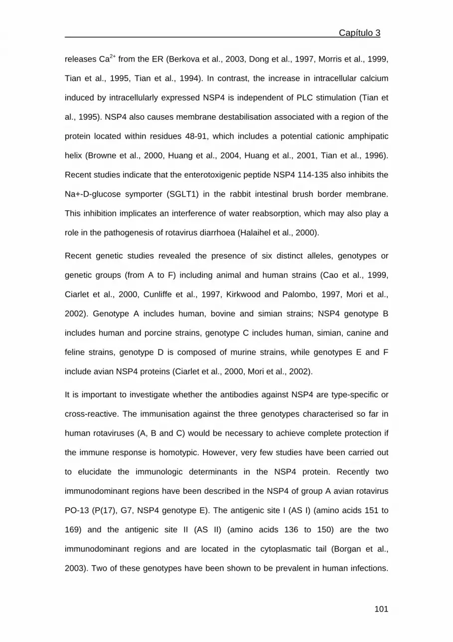
Capítulo 3
releases Ca2+ from the ER (Berkova et al., 2003, Dong et al., 1997, Morris et al., 1999,
Tian et al., 1995, Tian et al., 1994). In contrast, the increase in intracellular calcium
induced by intracellularly expressed NSP4 is independent of PLC stimulation (Tian et
al., 1995). NSP4 also causes membrane destabilisation associated with a region of the
protein located within residues 48-91, which includes a potential cationic amphipatic
helix (Browne et al., 2000, Huang et al., 2004, Huang et al., 2001, Tian et al., 1996).
Recent studies indicate that the enterotoxigenic peptide NSP4 114-135 also inhibits the
Na+-D-glucose symporter (SGLT1) in the rabbit intestinal brush border membrane.
This inhibition implicates an interference of water reabsorption, which may also play a
role in the pathogenesis of rotavirus diarrhoea (Halaihel et al., 2000).
Recent genetic studies revealed the presence of six distinct alleles, genotypes or
genetic groups (from A to F) including animal and human strains (Cao et al., 1999,
Ciarlet et al., 2000, Cunliffe et al., 1997, Kirkwood and Palombo, 1997, Mori et al.,
2002). Genotype A includes human, bovine and simian strains; NSP4 genotype B
includes human and porcine strains, genotype C includes human, simian, canine and
feline strains, genotype D is composed of murine strains, while genotypes E and F
include avian NSP4 proteins (Ciarlet et al., 2000, Mori et al., 2002).
It is important to investigate whether the antibodies against NSP4 are type-specific or
cross-reactive. The immunisation against the three genotypes characterised so far in
human rotaviruses (A, B and C) would be necessary to achieve complete protection if
the immune response is homotypic. However, very few studies have been carried out
to elucidate the immunologic determinants in the NSP4 protein. Recently two
immunodominant regions have been described in the NSP4 of group A avian rotavirus
PO-13 (P(17), G7, NSP4 genotype E). The antigenic site I (AS I) (amino acids 151 to
169) and the antigenic site II (AS II) (amino acids 136 to 150) are the two
immunodominant regions and are located in the cytoplasmatic tail (Borgan et al.,
2003). Two of these genotypes have been shown to be prevalent in human infections.
101

Capítulo 3 ______________________________________________________
NSP4 proteins corresponding to genotypes A and B have been detected in a recent
study in almost all the rotavirus strains infecting humans (Iturriza-Gomara et al., 2003).
Although several studies have analysed the importance of the immune response
against NSP4, the role that both humoral and cell-mediated immune responses to
NSP4 may play in the protection against rotavirus infection is still unknown. NSP4 has
been shown to induce both humoral and cell-mediated immune responses in humans
(Johansen et al., 1999, Ray et al., 2003, Yuan et al., 2004). Passive and active
immunisations against the NSP4 protein have protected mice from suffering diarrhoea
after both NSP4 and virulent rotavirus challenges (Ball et al., 1996, Yu and Langridge,
2001). It has been predicted that antibody to NSP4 should reduce rotavirus-induced
diarrhoea, inducing a similar protection to that elicited by antibodies to bacterial
enterotoxins against bacteria-induced diarrhoea (Estes, 2003).
In this study, two recombinant baculoviruses expressing the NSP4 rotavirus proteins
from the simian strain SA11 (genotype A) and from the human rotavirus strain Wa
(genotype B) were employed. The recombinant proteins were purified and used to coat
microtiter ELISA plates to determine antibodies to NSP4 in human sera. Sera from
rotavirus-infected children, uninfected children and adults were tested to analyse the
prevalence and the titers of both IgG and IgA antibodies against NSP4. The results
indicate a high prevalence of anti-NSP4 antibodies in infected children though at low or
very low titer levels. A low prevalence of anti-NSP4 antibodies was found among non-
convalescent healthy children and adults.
MATERIALS AND METHODS
Viruses and cells
Simian rotavirus strain SA11 (P5B(2), G3) and Wa (P1A(8), G1) were obtained from
the American Type Culture Collection (ATCC ref. 899-VR and 2018-VR, respectively).
Both strains were propagated in confluent MA104 cells in the presence of 1 µg/ml
102

Capítulo 3
trypsin (type IX, Sigma Chemical Co., St. Louis, MO). Spodoptera frugiperda Sf9 insect
cells (ATCC ref. CRL-1711) were grown in Grace’s medium (Gibco Invitrogen Corp.,
Paisley, Scotland, UK) supplemented with 10% foetal bovine serum.
Expression and purification of NSP4 proteins in insect cells
Recombinant baculoviruses expressing the NSP4s proteins from SA11 and Wa strains
were used to produce and purify the recombinant NSP4s from genotypes A and B as
previously described (Rodriguez-Diaz et al., 2003). Briefly, Sf9 cells were infected with
the recombinant baculoviruses at a multiplicity of infection (m.o.i.) of 10. After 48 h of
incubation, cells were harvested by centrifugation at 500 g for 5 min and resuspended
in buffer A (phosphate buffer 50 mM NaH2PO4, pH 8, 300 mM NaCl, 10 mM imidazole)
containing 1% Triton X-100 and 0.1% protease inhibitor cocktail (Sigma, St. Louis, MO)
for lysis. The lysates were diluted in the same volume of buffer A (1:1) and centrifuged
at 22.000 g at 4ºC for 5 min. Clarified lysates were incubated with 1 ml Ni-NTA
(Qiagen) for 30 min at 4ºC with shaking. The resin was then washed with 3 ml buffer A,
20 mM imidazole and with 3 ml of buffer A, 50 mM imidazole. The proteins were eluted
with 3 ml buffer A, 250 mM imidazole, and 0.5 ml fractions were collected. Proteins
were quantified by the Bradford method, analysed by SDS-PAGE and Coomasie blue
stained.
Subjects and serum samples
Sera were collected from 30 children less than 5 years of age convalescent of an acute
rotavirus infection, 15 to 20 days after the onset of diarrhoea; from 54 non-
convalescent healthy children under 5-years-old and from 49 adults 20-35 years of
age.
103

Capítulo 3 ______________________________________________________
Detection of serum IgG antibodies to Wa and SA11 rotavirus by ELISA
Titers of serum IgG to rotavirus strains Wa and SA11 were determined by ELISA as
described (Colomina et al., 1998). Briefly, 96-well microtiter plates were coated
overnight at 4ºC with CsCl density gradient-purified rotavirus particles (SA11 or Wa
strains) previously treated with 1.5 M sodium thiocyanate and diluted in carbonate-
bicarbonate buffer (pH 9.6) at a final concentration of 2 µg/ml. Serum samples were
serially diluted, starting at 1/100, in PBS-Tween (PBS-T) containing 1% bovine serum
albumin (PBS-T-BSA). 100 µl were added and incubated for 2 h at 37ºC, and the plates
were washed three times with PBS-T. Horseradish peroxidase-conjugated anti-human
IgG goat antibody (Sigma Immunochemicals, St. Louis, MO) diluted 1/2000 in PBS-T-
BSA was added. The plates were incubated 2 h at 37ºC, and washed three times with
PBS-T. Reactions were developed by adding o-phenylendiamine (Sigma
Immunochemicals) in citrate-phosphate buffer (pH 5) with 0.1% H2O2. The reaction was
stopped with 3M H2SO4. Plates were read at 492 nm in a spectrophotometer. As a
control for specificity, wells were coated with the same amount of protein from MA104
mock-infected cells. A sample was considered positive when its OD was three times
higher than the absorbance value of the same sample in the specificity control. The
titer of each sample was considered the inverse of the last dilution showing a positive
reaction.
Detection of serum antibodies to NSP4Wa and NSP4SA11 antigens by ELISA
ELISA tests were performed with purified NSP4 proteins from the SA11 and Wa
rotavirus strains produced by recombinant baculoviruses to detect antibodies to
NSP4Wa and NSP4SA11 following a similar procedure to that described above. Mock-
infected Sf9 cells were processed in the same way as the baculovirus-infected Sf9 cells
producing the recombinant proteins, and were used as specificity controls. Quantitation
of IgA/IgG was carried out by assaying each serum sample at different dilutions,
starting at 1/25 for both Ig isotypes. Horseradish peroxidase-conjugated goat anti-
104
Capítulo 3
human IgA (α-chain specific) and anti-human IgG antibodies (Sigma
Immunochemicals, St. Louis, MO) were used to detect human IgA and IgG antibodies,
respectively. A test sample was considered positive when the OD was three times
higher than the absorbance value of the same sample in the specificity control. As in
the ELISA against the rotavirus particles, the titer of each sample was considered the
inverse of the last dilution that showed a positive result.
Statistical analyses
Comparisons between the mean titers of antibodies to NSP4 and to whole rotavirus
obtained in the three different groups (convalescent children, non-convalescent healthy
children and adults) were carried out with the t-student analysis. Categorical variables
were compared by χ2 test. P<0,05 was considered significant. The SPSS software
(Lead Technologies, Inc.) was used to perform these analyses.
RESULTS
Antibodies to NSP4 in rotavirus-infected children
Seventy per cent of convalescent serum samples collected from rotavirus-infected
children were found to contain IgG antibodies against at least one of the two NSP4
proteins used in this study (Table I). Twelve sera (40 %) recognised both NSP4Wa and
NSP4SA11 proteins. This result indicates a heterotypic recognition by the antibodies to
NSP4 in these 12 samples or the event of previous infections with rotavirus carrying
NSP4s from the two genotypes used in this study. Five sera (16.6 %) recognised only
NSP4SA11 (genotype A) but failed to recognise NSP4Wa (genotype B), and 4 (13.3 %)
reacted only to NSP4Wa (Table I). Statistical analyses of these data demonstrated a
significant difference in the recognition between NSP4Wa and NSP4SA11 (P = 0.03),
suggesting that recognition of both NSP4 proteins is not heterotypic. Comparison of the
105

Capítulo 3 ______________________________________________________
mean values of IgG antibody titers in all the convalescent serum samples was
established with the titers found in the samples from the uninfected children and the
adults (Fig. 1A). No IgA antibodies to either of the recombinant NSP4 proteins were
detected.
Table 1. Percentage of serum samples from convalescent children of an acute rotavirus infection, non-convalescent children and adults containing IgG antibodies to NSP4Wa (genotype B), NSP4SA11 (genotype A) or to both NSP4 proteins. Convalescent children
Uninfected children Adults
NSP4Wa/NSP4SA11 12 (40%) 7 (12.9%) 3 (6.1%)
NSP4Wa 4 (13.3%) 4 (7.4%) 6 (12.2%)
NSP4SA11 5 (16.6%) 3 (5.5%) 0 (0%)
Negative 9 (30%) 40 (74%) 40 (81.6 %)
Total 30 54 49
Antibodies to NSP4 in uninfected children and adults
Fourteen serum samples (26 %) from the uninfected children recognised at least one of
the NSP4 proteins (Table 1). The distribution of the positive samples was as follows: 7
sera (12.9 %) reacted to both proteins, 4 samples (7.4 %) were positive against
NSP4Wa and 3 samples (5.5 %) against NSP4SA11. Nine serum samples (18.4 %) from
the adults were found positive for antibodies against at least one of the NSP4s
proteins: 3 of them (6.1 %) recognised the NSP4 from both SA11 and Wa strains and 6
(12.3 %) were only reactive towards the NSP4 from the human Wa strain.
Antibodies to rotavirus particles
All the serum samples showed high titers of IgG antibodies against complete rotavirus
particles, both viral strains Wa and SA11, indicating that all the rotavirus-infected
children had seroconverted. The mean titers were compared among the three groups
(Fig. 1B).
106

Capítulo 3
A high prevalence of serum IgG antibodies to whole rotavirus was found both in the
uninfected children and in the adults; this seroprevalence was higher than 80% in the
non-convalescent children indicating previous rotavirus infection(s). The percentage of
sera from the adults reacting against rotavirus particles was higher than 90%. All the
samples recognising the NSP4 protein also reacted with the rotaviral particles.
0
50
100
150
200
250
300
350
400
Convalescenchildren
Non-covalescenchildren
Adults
Ant
ibod
y tit
er
NSP4Wa
NSP4SA11*
0
50
100
150
200
250
300
350
400
Convalescenchildren
Non-covalescenchildren
Adults
Ant
ibod
y tit
er
NSP4Wa
NSP4SA11*
A
B
0
1000
2000
3000
4000
5000
6000
7000
8000
9000
Convalescen children Non-covalescenchildren
Adults
Antib
ody
titer
RVWa
RVSA11
Figure 1. IgG antibody mean titers to NSP4 proteins (panel A) and to whole rotavirus particles (panel B) in convalescent children, non-convalescent children and adults. Panel A shows the average antibody titers against both NSP4 proteins NSP4Wa and NSP4SA11.The asterisks show the presence of significative differences. Panel B represents the average titers of the sera recognising the complete rotavirus particles of strains Wa and SA11. The error bars are indicating the standard deviation (SD).
Comparison of the antibody titers between children with rotavirus diarrhoea,
healthy children and adults
Comparing the mean values of serum IgG antibody titers against the NSP4 proteins
between the three groups showed that the infected children presented higher titer
levels towards the NSP4 from the Wa or SA11 strains than the adults or the uninfected
children (P<0.001). The comparison of the mean titers obtained from the uninfected
107

Capítulo 3 ______________________________________________________
children and the adults did not show significant differences (P> 0.05). When the
comparison was established between the serum titers obtained against the full
rotavirus particles no differences were detected (P>0.05).
The number of positive and negative serum samples against both NSP4 genotypes A
and B were also compared between the three groups. The infected children presented
a higher percentage of positive samples to either of the NSP4s proteins than the
uninfected children or the adults (P<0.001). No statistical differences were found
between the uninfected children and adults (P>0.05). The number of positive serum
samples against rotavirus particles did not differ among the three groups (P>0.05).
DISCUSSION
This study describes the antibody response to the NSP4 protein in children
convalescing from acute rotavirus diarrhoea, in non-convalescent healthy children and
in healthy adults. In the convalescent children we found a very high prevalence of IgG
antibodies to the genotypes of NSP4 that are more common in human infections
(Iturriza-Gomara et al., 2003). We have found that human serum antibodies can
discriminate between NSP4 genotype A and NSP4 genotype B in naturally infected
children. This result does not accord with those reported from rotavirus-infected
children in India by (Ray et al., 2003), who postulated a cross-reactivity of IgG
antibodies to NSP4 genotypes A, B and C. Our results are in accordance with those
obtained by Yuan et al. (2004), who detected both homotypic and heterotypic
responses to NSP4 in children vaccinated against rotavirus. We found that in naturally
infected children the IgG titer levels against the NSP4 proteins are low or very low, as
previously described by Johansen et al. (1999) as opposed to the high titers described
by Ray et al. (2003). In our study, no IgA response to the NSP4 proteins was detected
in the convalescent sera. By contrast, Ray et al. (2003) found by ELISA IgA
108

Capítulo 3
seroconversions to NSP4 in children with acute rotavirus gastroenteritis. The main
difference between these IgA ELISA protocols is that Ray et al. used biotin-conjugated
anti-human IgA and horseradish peroxidase-conjugated avidin-biotin, which may have
increased the sensitivity of the technique. However, similar to our results, Johansen et
al. (1999) did not detect either IgA antibodies to NSP4.
The results in convalescent children show that the nonstructural protein NSP4 elicits
IgG antibodies that can play a role in the protection against the enterotoxic function of
this protein, as demonstrated in mice (Ball et al., 1996). It is important to investigate
whether the antibodies against NSP4 are type-specific or cross-reactive. The fact that
the antibodies to NSP4 can discriminate between both genotypes A and B enables us
to develop serotyping procedures to determine the NSP4 genotype of rotavirus strains.
With this purpose we have developed single-chain variable fragment (scFv) antibodies
against NSP4 and some of these antibodies discriminate both NSP4 genotypes A and
B (Rodriguez-Diaz et al., 2004).
Antibodies are generally considered to be a proxy for protection against rotavirus
reinfection because they are a marker for past exposure, although it remains unclear
which viral proteins might induce a protective antibody response (Jiang et al., 2002). In
this study we found that a very high proportion of non-convalescent healthy children
and adults have suffered previous rotavirus infections, due to the high prevalence of
antibodies to whole rotaviral particles. Comparison of the average titers of IgG
antibodies against the NSP4 proteins in these two groups did not showed statistical
differences. Moreover, we found a low prevalence of IgG antibodies against the NSP4
proteins in both groups, in spite of having previously experienced rotavirus infections.
This result does not accord with those obtained among Swedish and Nicaraguan adults
(Johansen et al., 1999), in which the prevalence of IgG antibodies against NSP4
genotype A is high in the adult population. However, the antibody titers were always
low or very low, as observed in our study.
109

Capítulo 3 ______________________________________________________
We found that 74% percent of the healthy children and 81.6% of the adults have no
antibodies against NSP4. Thus, one can hypothesize that reinfection with rotavirus
would probably provoke diarrhoea in them, an assumption that could be mistaken. It is
well known that in humans rotavirus infections happen throughout life but the resulting
disease is mild and often asymptomatic (Bishop, 1996). Besides, NSP4 activity as an
enterotoxin in the murine model shows an age dependent pattern (Ball et al., 1996),
inducing diarrhoea only during the first 15 days of age.
In conclusion, we show that the NSP4 protein can elicit an IgG antibody response in
humans after natural rotavirus infections and that the recognition of different NSP4s by
these antibodies does not seem to be always heterotypic. We have also found that the
prevalence of antibodies against NSP4 is very low among non-convalescent healthy
children below 5 years of age, as well as in adults supposed to be protected from
rotavirus disease upon reinfection.
ACKNOWLEDGMENTS
This work was supported by the Fondo de Investigación Sanitaria (F.I.S.) of the
Spanish Ministry of Healt Grant Contract No 00/0592. J. Rodríguez-Díaz was the
recipent of a fellowship from the Spanish Ministry of Education and Culture (No.
AP2000-3623).
REFERENCES
Ball JM, Tian P, Zeng CQ, Morris AP Estes MK. 1996. Age-dependent diarrhoea
induced by a rotaviral nonstructural glycoprotein. Science 272:101-104.
Barnes GL, Lund JS, Mitchell SV, De Bruyn L, Piggford L, Smith AL, Furmedge J, Masendycz PJ, Bugg HC, Bogdanovic-Sakran N, Carlin JB, Bishop RF. 2002. Early phase II trial of human rotavirus vaccine candidate RV3. Vaccine 20:2950-2956.
110

Capítulo 3
Berkova Z, Morris AP, Estes MK. 2003. Cytoplasmic calcium measurement in rotavirus enterotoxin-enhanced green fluorescent protein (NSP4-EGFP) expressing cells loaded with Fura-2. Cell Calcium 34:55-68.
Bern C, Glass RI. 1994. Viral infections of gastrointestinal tract. In: Impact of diarrhoeal deseases worldwide, Kapikian A, editor. New York: Marcel Dekker, Inc. pp. 1-26.
Bishop RF. 1996. Natural history of human rotavirus infections. Arch Virol Suppl. 12:119-128.
Borgan MA, Mori Y, Ito N, Sugiyama M, Minamoto N. 2003. Antigenic analysis of nonstructural protein (NSP) 4 of group A avian rotavirus strain PO-13. Microbiol Immunol 47:661-668.
Browne EP, Bellamy AR, Taylor JA. 2000. Membrane-destabilizing activity of rotavirus NSP4 is mediated by a membrane-proximal amphipathic domain. J Gen Virol 81:1955-1959.
Cao XR, Akihara S, Fang ZY, Nakagomi O, Ushijima H. 1999. Genetic variation in the VP4 and NSP4 genes of human rotavirus serotype 3 (G3 type) isolated in China and Japan. Microbiol Immunol 43:171-175.
Ciarlet M, Liprandi F, Conner ME, Estes MK. 2000. Species specificity and interspecies relatedness of NSP4 genetic groups by comparative NSP4 sequence analyses of animal rotaviruses. Arch Virol 145:371-383.
Colomina J, Gil MT, Codoñer P, Buesa J. 1998. Viral proteins VP2, VP6 and NSP2 are strongly precipitated by serum and fecal antibodies from children with rotavirus infection. J Med Virol 56:58-65.
Conner ME, Crawford SE, Barone C, Estes MK. 1993. Rotavirus vaccine administered parenterally induces protective immunity. J Virol 67:6633-6641.
Contreras JF, Menchaca GE, Padilla-Noriega L, Tamez RS, Greenberg HB, Lopez S, Arias CF. 1995. Heterogeneity of VP4 neutralization epitopes among serotype P1A human rotavirus strains. Clin Diagn Lab Immunol 2:506-508.
Coulson BS. 1993. Typing of human rotavirus VP4 by an enzyme immunoassay using monoclonal antibodies. J Clin Microbiol 31:1-8.
Crawford SE, Estes MK, Ciarlet M, Barone C, O'Neal CM, Cohen J, Conner ME. 1999. Heterotypic protection and induction of a broad heterotypic neutralization response by rotavirus-like particles. J Virol 73:4813-4822.
111

Capítulo 3 ______________________________________________________
Cunliffe NA, Woods PA, Leite JP, Das BK, Ramachandran M, Bhan MK, Hart CA, Glass RI, Gentsch JR. 1997. Sequence analysis of NSP4 gene of human rotavirus allows classification into two main genetic groups. J Med Virol 53:41-50.
Dong Y, Zeng CQ, Ball JM, Estes MK, Morris AP. 1997. The rotavirus enterotoxin NSP4 mobilizes intracellular calcium in human intestinal cells by stimulating phospholipase C-mediated inositol 1,4,5-trisphosphate production. Proc Natl Acad Sci USA 94:3960-3965.
Estes MK. 2001. Rotaviruses and their replication. In: Knipe DM, Howley PM, editors. Fields Virology, 4th edn. Philadelphia: Lippincott, Williams and Wilkins. pp 1747-1785.
Estes MK. 2003. The rotavirus NSP4 enterotoxin: Current status and challenges. In: Viral Gastroenteritis, Gray J, Desselberger U, editors. Amsterdam: Elsevier, pp 207-224.
Gorrell RJ, Bishop RF. 1997. Production of reassortant viruses containing human rotavirus VP4 and SA11 VP7 for measuring neutralizing antibody following natural infection. Clin Diagn Lab Immunol 4:509-514.
Halaihel N, Lievin V, Ball JM, Estes MK, Alvarado F, Vasseur M. 2000. Direct inhibitory effect of rotavirus NSP4(114-135) peptide on the Na(+)-D-glucose symporter of rabbit intestinal brush border membrane. J Virol 74:9464-9470.
Herrmann JE, Chen SC, Jones DH, Tinsley-Bown A, Fynan EF, Greenberg HB, Farrar GH. 1999. Immune responses and protection obtained by oral immunization with rotavirus VP4 and VP7 DNA vaccines encapsulated in microparticles. Virology 259:148-153.
Horie Y, Nakagomi O, Koshimura Y, Nakagomi T, Suzuki Y, Oka T, Sasaki S, Matsuda Y, Watanabe, S. 1999. Diarrhoea induction by rotavirus NSP4 in the homologous mouse model system. Virology 262:398-407.
Huang H, Schroeder F, Estes MK, McPherson T, Ball JM. 2004. The interaction(s) of rotavirus NSP4 C-terminal peptides with model membranes. Biochem J 380:723-133.
Huang H, Schroeder F, Zeng C, Estes MK, Schoer JK, Ball JM. 2001. Membrane interactions of a novel viral enterotoxin: rotavirus nonstructural glycoprotein NSP4. Biochemistry 40:4169-4180.
Iturriza-Gomara M, Anderton E, Kang G, Gallimore C, Phillips W, Desselberger U, Gray J. 2003. Evidence for genetic linkage between the gene segments encoding
112

Capítulo 3
NSP4 and VP6 proteins in common and reassortant human rotavirus strains. J Clin Microbiol 41:3566-3573.
Jiang B, Gentsch JR, Glass RI. 2002. The role of serum antibodies in the protection against rotavirus disease: An overview. Clin Infect Dis 34:1351-1361.
Johansen K, Hinkula J, Espinoza F, Levi M, Zeng C, Ruden U, Vesikari T, Estes M, Svensson L. 1999. Humoral and cell-mediated immune responses in humans to the NSP4 enterotoxin of rotavirus. J Med Virol 59:369-377.
Kapikian AK, Hoshino Y, Chanock RM. 2001. Rotaviruses. In: Knipe DM, Howley PM, editors. Fields Virology, 4th edn. Philadelphia: Lippincott, Williams and Wilkins. pp 1787-1883.
Kirkwood CD, Palombo, EA. 1997. Genetic characterization of the rotavirus nonstructural protein, NSP4. Virology 236:258-265.
Mori Y, Borgan MA, Ito N, Sugiyama M, Minamoto N. 2002. Sequential analysis of nonstructural protein NSP4s derived from group A avian rotaviruses. Virus Res 89:145-151.
Morris AP, Scott JK, Ball JM, Zeng CQ, O'Neal WK, Estes MK. 1999. NSP4 elicits age-dependent diarrhoea and Ca(2+)mediated I(-) influx into intestinal crypts of CF mice. Am J Physiol 277:G431-G444.
Offit PA. 2002. The future of rotavirus vaccines. Semin Pediatr Infect Dis 13:190-195.
Parashar U, Bresee J, Glass R. 2003a. The global burden of diarrhoeal disease in children. Bull World Health Organ 81: 236.
Parashar UD, Hummelman EG, Bresee JS, Miller MA, Glass, RI. 2003b. Global illness and deaths caused by rotavirus disease in children. Emerg Infect Dis 9:565-572.
Ray P, Malik J, Singh RK, Bhatnagar S, Bahl R, Kumar R, Bhan MK. 2003. Rotavirus nonstructural protein NSP4 induces heterotypic antibody responses during natural infection in children. J Infect Dis 187:1786-1793.
Richardson SC, Grimwood K, Bishop RF. 1993. Analysis of homotypic and heterotypic serum immune responses to rotavirus proteins following primary rotavirus infection by using the radioimmunoprecipitation technique. J Clin Microbiol 31:377-385.
113
Capítulo 3 ______________________________________________________
Rodríguez-Díaz J, López-Andújar P, García-Díaz A, Cuenca J, Montava R, Buesa J. 2003. Expression and purification of polyhistidine-tagged rotavirus NSP4 proteins in insect cells. Protein Expr Purif 31:207-212.
Rodríguez-Díaz J, Monedero V, Pérez-Martínez G, Buesa J. 2004. Single-chain variable fragment (scFv) antibodies against rotavirus NSP4 enterotoxin generated by phage display. J Virol Methods 121:231-238.
Tian, P, Ball JM, Zeng CQ, Estes MK. 1996. The rotavirus nonstructural glycoprotein NSP4 possesses membrane destabilization activity. J Virol 70:6973-6981.
Tian P, Estes MK, Hu Y, Ball JM, Zeng CQ, Schilling WP. 1995. The rotavirus nonstructural glycoprotein NSP4 mobilizes Ca2+ from the endoplasmic reticulum. J Virol 69:5763-5772.
Tian P, Hu Y, Schilling WP, Lindsay DA, Eiden J, Estes MK. 1994. The nonstructural glycoprotein of rotavirus affects intracellular calcium levels. J Virol 68:251-217.
Yu J, Langridge WH. 2001. A plant-based multicomponent vaccine protects mice from enteric diseases. Nat Biotechnol 19:548-552.
Yuan L, Ishida SI, Honma S, Patton JT, Hodgins DC, Kapikian AZ. 2004. Homotypic and heterotypic serum isotype-specific antibody responses to rotavirus nonstructural protein 4 and viral protein (VP) 4, VP6 and VP7 in infants who received selected live oral rotavirus vaccines. J Infect Dis 189:1834-1845.
Zhang M, Zeng CQ-Y, Morris MP, Estes MK. 2000. A functional NSP4 enterotoxin
peptide secreted from rotavirus-infected cells. J Virol 74:11663-11670.
114

Capítulo 4
CAPÍTULO 4
Nitric oxide production during clinical and experimental
infection with rotavirus
Jesús Rodríguez-Díaz1,8, Mahanez Banasaz2, Claudia Istrate3, Javier Buesa1,
Ove Lundgren4, Felix Espinoza5, Tommy Sundqvist6, Martin Rottenberg7 and
Lennart Svensson8*
1Department of Microbiology, School of Medicine, University of Valencia, Spain;
2Deparment of Virology, Swedish Institute for Infectious control, Sweden; 3Instituto de
Biologia Experimental e Tecnologica and Instituto de Tecnologia Quimica e Biologica,
Oeiras, Portugal; 4Division of Physiology, University of Gothenburg; 5Department of
Microbiology, UNAN, León, Nicaragua; 6Division of Medical Microbiology, University of
Linköping, Sweden; 7Microbiology and Tumor Biology Center, Karolinska Institute,
Sweden; 8Division of Molecular Virology, University of Linköping, Sweden
Submitted to Journal of Virology (2005)
115

Capítulo 4 ______________________________________________________
116

Capítulo 4
Submitted to Journal of Virology (2005)
Nitric oxide production during clinical and experimental infection with
rotavirus
Jesús Rodríguez-Díaz1,8, Mahanez Banasaz2, Claudia Istrate3, Javier Buesa1, Ove Lundgren4,
Felix Espinoza5, Tommy Sundqvist6, Martin Rottenberg7 and Lennart Svensson8*
1Department of Microbiology, School of Medicine, University of Valencia, Spain; 2Deparment of
Virology, Swedish Institute for Infectious control, Sweden; 3Instituto de Biologia Experimental e
Tecnologica and Instituto de Tecnologia Quimica e Biologica, Oeiras, Portugal; 4Division of
Physiology, University of Gothenburg; 5Department of Microbiology, UNAN, León, Nicaragua;
6Division of Medical Microbiology, University of Linköping, Sweden; 7Microbiology and Tumor
Biology Center, Karolinska Institute, Sweden; 8Division of Molecular Virology, University of
Linköping, Sweden
*Corresponding author: Lennart Svensson, Division of Molecular Virology, University of
Linköping, SE-581 85 Linköping, Sweden. e-mail: [email protected]
Phone: +46 13228803
Fax : +46 13 22 47 89
ABSTRACT
The pathophysiological mechanisms behind rotavirus-induced diarrhoea still remain
incomplete. Current views suggest that the non-structural protein 4 (NSP4) of rotavirus
and the enteric nervous system (ENS) participate in water secretion and diarrhoea. In
the present work the role of nitric oxide (NO) in rotavirus infection and disease have
been studied in vitro, mice and humans. Incubation of human intestinal epithelial cells
(HT-29) with purified NSP4 but not with infectious virus produced NO2/NO3
accumulation in the incubation media. The NSP4-induced release of NO metabolites
occurred within the first minutes after the addition of the toxin. Mice infected with
murine rotavirus (strain EDIM) accumulated NO2/NO3 in their urines; newborn pups
(n=10) at the onset for diarrhoea and adult mice (n=10) at the onset for virus shedding.
117

Capítulo 4 ______________________________________________________
Following rotavirus infection, iNOS mRNA was upregulated in ileum, but not in
duodenum or jejunum of newborn pups within five days post infection. A prospective
clinical study including 46 children with acute rotavirus infection and 19 age-matched
controls concluded that rotavirus infection stimulates a strong NO metabolites
production (p<0.001) during the course of the disease. Altogether these observations
identify NO as an important mediator of host responses during rotavirus infection.
Key words: rotavirus, NSP4, enterotoxin, nitric oxide.
Running Title: Rotavirus stimulates nitric oxide production.
INTRODUCTION
Rotaviruses are the main etiologic agents of viral gastroenteritis in infants and
young children worldwide, and are responsible for significant mortality in developing
countries and high morbidity in developed countries (27). Rotavirus infects the mature
enterocytes located in the upper half of intestinal villi that ultimetly leads to cell death. A
striking observation in both animals and humans is that only a few percent of the
mature villous epithelial cells and no crypts seem to be infected (24, 25, 28, 33), all
suggesting that there is no absolute correlation between histological lesions and clinical
symptoms. Mechanisms proposed to explain the rotavirus induced fluid and electrolyte
secretion includes villous ischemia (25, 26), the participation of the NSP4 enterotoxin
(4) and activation of the enteric nervous system (18, 19). Direct evidence for
participation of the enteric nervous system (ENS) in rotavirus diarrhoea in mice and
humans has been reported (6, 14, 17, 32). While these studies indicated that a
significant part of the reported net fluid secretion is mediated by the secromotor reflex,
including serotonin, acetylcholine and VIP (6, 14, 17, 32), the specific details of how
the secretor nervous reflex is activated are currently unknown. It has been
hypothesised that rotavirus or its toxin may influence enteroendocrine cells to release
118

Capítulo 4
peptides/amines which, in turn, may activate nervous dendrites located just underneath
the intestinal epithelium. Another proposal infers that enterocytes in response to
luminal rotavirus release nitric oxide (NO), which, directly or indirectly may stimulate
enteric afferents. None of the two hypotheses has been examined and in this study we
have investigated the possible role of NO (19, 23, 28).
NO is a major secretory product of mammalian cells that participates in host
defence, homeostatic and development functions. Activation of the immune system is
associated with an increase in macrophage NO production. NO is also a
neurotransmitter in the peripheral nervous system controlling e.g. smooth muscles in
the gastrointestinal tract and in the vasculature (9, 10). The enzyme NO synthesase
(NOS) exist in two main forms, a constitutive form (cNOS) and an isoform that is
inducible (iNOS). The isoforms of the cNOS were named after the tissue in which they
were found, neuronal NOS (nNOS) and endothelial NOS (eNOS) (2). The activity of
both cNOS is regulated by the intracellular calcium concentration influenced by
physiological stimuli (12, 21). iNOS, on the other hand, is calcium independent (34). In
the immunological system iNOS is induced by cytokines at the transcriptional level
primarily in macrophages, where NO is produced in high concentrations for long
periods of time (43).
In the mid 1990’s, the non-structural protein 4 (NSP4) of rotavirus and a 22
amino acid synthetic peptide of the protein (NSP4114-135) were reported to induce an
age-dependent diarrhoea in mice in the absence of histological alterations (4). Further
studies showed that NSP4 and the NSP4114-135 peptide exogenously applied to
mammalian cells caused an increase in intracellular calcium. This calcium mobilisation
from the endoplasmic reticulum (ER) was found to occur after the activation of the C
phospholipase (7, 40). These observations in conjunction with the calcium dependence
of cNOS synthesis and the possibility that NO may activate a secretory nervous reflex
prompted the present study.
119

Capítulo 4 ______________________________________________________
In the present work, we show that human intestinal epithelial cells (HT-29)
responded with release of NO metabolites from the NSP4 toxin of rotavirus but not
from productive rotavirus infection possibly through the calcium-dependent cNOS.
Furthermore, animal experiments revealed that newborn mice responded with release
of NO derivatives that correlated with onset of diarrhoea and adult mice with onset of
virus shedding. Furthermore, iNOS mRNA from ileum, but not jejunum and duodenum
was upregulated within five days post-infection in newborn mice. A most novel
observation was that children with severe rotavirus disease released significant NO
derivatives, all suggesting that NO play an important role during rotavirus infection.
MATERIALS AND METHODS
Expression and purification of NSP4 protein in insect cells
Recombinant baculovirus expressing the NSP4 protein from SA11 (G3, P5B[2])
strain was used to produce and purify the recombinant NSP4 as previously described
(30). Briefly, Sf9 cells were infected with the recombinant baculovirus at a multiplicity of
infection (m.o.i.) of 10. After 48 h of incubation, cells were harvested by centrifugation
at 500 X g for 5 min and resuspended in buffer A (phosphate buffer 50 mM NaH2PO4,
pH 8, 300 mM NaCl, 10 mM imidazole) containing 1% Triton X-100 and 0.1% protease
inhibitor cocktail (Sigma) for lysis. The lysates were then diluted in the same volume of
buffer A (1:1) and centrifuged at 22.000 X g at 4ºC for 5 min. Clarified lysates were
incubated with 1 ml Ni-NTA (Qiagen) for 30 min at 4ºC with shaking. The resin was
then washed with 3 ml buffer A, containing 20 mM imidazole and with 3 ml of buffer A,
containing 50 mM imidazole. The protein was finally eluted with 3 ml buffer A,
containing 250 mM imidazole, in 0.5 ml fractions. The protein was extensively dialysed
against PBS, quantified by the Bradford method and analysed by SDS-PAGE.
120

Capítulo 4
NSP4 inoculation and rotavirus infection in HT-29 cells
HT-29 human intestinal epithelial cells were placed in 24 well plates (Nunc) and
incubated at 37°C 5% CO2, with 1 ml of media in each well (MEM Eagle Medium 25mM
HEPES 10% foetal calf serum). After the cells reached confluence the monolayers
were washed 3 times with Dulbecco's modified Eagle medium (DMEM) without phenol
red and different concentrations of purified NSP4 were added to the cells (from 1 nM to
500 nM) in 400 µl of total serum free DMEM without phenol red. Several wells were
used as negative control by adding Sf9 antigen from non infected Sf9 cells (30) in the
same volume of media. The cells were incubated with the toxin from 5 minutes to 48
hours at 37°C 5% CO2. After the incubation the supernatants were carefully recovered
and stored at -80°C.
In other experiments HT-29 cells growing in 24 well plates as described above
were infected with trypsin-activated rotavirus SA11 strain. Prior to infection (m.o.i. 0.01-
10), monolayers were washed 3 times with serum free D-MEM without phenol red.
Virus was incubated for 1 h at 37ºC 5%CO2 and the monolayers were subsequently
washed and 400 µl D-MEM without phenol red were added to each well for 24, 48 and
72 hours. After each incubation the supernatants were carefully recovered and stored
at -80ºC.
Rotavirus infection in mice
Rotavirus naïve adult and newborn BALB/c mice were infected with the murine
rotavirus strain EDIM (G3 P10 [16]) as described previously (14) .Two groups of 10
adult BALB/ mice each group were orally administered at the age of 8 weeks, the first
group with 10 µl of PBS containing 5 X 10⁴ fluorescent forming units (FFUs) of EDIM
rotavirus and the second group was mock infected. Two groups of 10 newborn Balb/c
121

Capítulo 4 ______________________________________________________
mice (7 days old) were treated as the adult mice. All the mice were followed for two
weeks after infection. Urine and stool samples were collected daily and stored at -80ºC.
Human samples
A total number of 46 urine and stool samples were collected during the months of
February to May of 2004 in the hospitals of Leon and Chilandega in Nicaragua from
children under 2 years of age suffering from severe rotavirus diarrhoea. The study
included children of mothers who agreed to participate after information about the
study. The urine and stool samples were collected the next day after the hospitalization
of the children. Clinical data obtained from an attending paediatrician included
temperature, presence or absence of vomiting and hydratation status, evaluated
according to standarized scheme of signs and symptoms. Three different states of
dehydration were assigned, mild, moderate and severe (8). The samples were stored
at –20º C until further use. Also 19 samples of urine and stools were collected from
healthy age-matched children from the same geographic localisation.
Detection of rotavirus by ELISA
Detection of rotavirus in the supernatants of the infected cells or in the stool
specimens from mice and humans were performed by ELISA as previously described
(37).
Nitrite/Nitrate measurements
To analyze the concentration of the stable oxidation products of NO in the
supernatants and the urine samples, the total concentration of nitrate and nitrite
(NO2/NO3) was calculated following the protocol designed by Verdon et al (42). Briefly
50 µl of the cell supernatants or from a dilution 1/50 of the urine samples were placed
in each well of a 96 well plate. Then 10 µl of NADPH 10 µM were added (final
concentration 1 µM) and 40 µl of a master mix (100 µl of glucose-6-phosphate 50 mM,
122

Capítulo 4
40 µl of Glucose-6-phosphate dehydrogenase 40 U/ml, 80 µl of nitrate reductase from
Aspergillus niger 10 U/ml and PBS to 4 ml) were added to each well. All the products
for the master mix were purchased from SIGMA. Furthermore, two standard curves
were made with known amounts of nitrite and nitrate to determine the efficiency of the
conversion from nitrate to nitrite and to quantify the concentrations of the samples. The
reaction was developed for 45 minutes at room temperature and then the Griess
reaction was performed by adding 100 µl of 1% (w/v) sulfanilamide in 5% phosphoric
acid followed by 100 µl of 0,1% (w/v) N-(1-naphtyl)ethylenediamine HCl (NED), after 10
minutes of incubation at room temperature the absorbance was read at 540 nm. NO
production was estimated from the concentration of nitrite and nitrate in the incubation
medium and the urine samples.
Quantitive RT-PCR assay for iNOS mRNA
iNOS and hypoxanthine-guanine phosphoribosyltransferase (HPRT) transcript
mRNA from freshly extracted duodenum, jejunum and ileum was obtained using
guanidinium acid-thiocyanate and phenol-chloroform extraction (31). 2 µg of RNA were
denatured and reverse transcribed using random hexanucleotides (Pharmacia,
Uppsala, Sweden). The real time PCR was performed in duplicate in a volume of 25 µl
reactions containing Platinum® SYBR® Green qPCR Supermix-UDG (Invitrogen),
forward and reverse primers, and 0.5 µl of cDNA on an ABI Prism® 7500 sequence
detection system (Applied Biosystems). The following primer sequences and
concentrations (determined following guidelines from Applied Biosystems) were used:
Sense iNOS: 5´ GGC AGC CTG TGA GAC CTT TG 3´ 500 nM
Antisense iNOS: 5´ GCA TTG GAA GTG AAG CGT TTC 3´ 500 nM
Sense HPRT 5´ GCT TTT CGC CGC TTG CT 3´ 500 nM
Antisense HPRT 5´ CTC GTC ATC GGC CGT GAT 3´ 500 nM
123
Capítulo 4 ______________________________________________________
Serial-fold dilutions of a cDNA sample were amplified to control amplification
efficiency for each primer pair. Thereafter the Ct values for all cDNA samples were
obtained. HPRT was used as a control gene to calculate the ∆Ct values for individual
samples. The relative amount of iNOS/ HPRT transcripts was calculated using the
2(∆∆Ct) method as described previously (16). These values were then used to calculate
the mean of the relative expression (fold induction) of iNOS mRNA in tissues from
uninfected and infected mice.
Statistical analysis
In those experiments in which statistical analysis was required the non-
parametric Mann-Whitney U test was used to determine the presence or absence of
differences between different groups and significant level of P < 0,05 was used. All the
analyses were performed by using the SPSS package for Macintosh computers (LEAD
Technologies Inc., USA).
RESULTS
NSP4 induces a rapid release of NO metabolites from human intestinal cells.
To investigate if human intestinal cells respond with release of NO following
incubation with NSP4, confluent monolayers of HT-29 were incubated with various
concentrations of NSP4. As shown in figure 1A, the NO2/NO3 concentration in the
supernatants was increased within 5 minutes after administration of 100 nM of NSP4.
Figure 1B shows that the effect of NSP4 was dose-dependent and reached a plateau
>100nM. On the contrary, rotavirus infection of HT-29 cells did not result in a
detectable NO release, independently of virus concentration or time (data not shown).
124

Capítulo 4
Figure 1. NO2/NO3 concentration in the supernatants of HT-29 cells after incubation with the NSP4 toxin. Panel A shows the time-dependent response after incubation with the NSP4 enterotoxin at a concentration of 100 nM. As it can be observed the increase of NO2/NO3 in the supernatants occurs within the first five minutes after the addition of the toxin. Panel B shows the dosis-dependent response curve of NO2/NO3 after the addition of different concentrations of NSP4. In this experiments the incubation time was 5 minutes in all the cases in base of the previous results. For the negative control purified antigen from Sf9 mock infected cells was used.
Rotavirus stimulates release of NO in vivo
To what extent rotavirus infected release NO was studied in two types of
experiments. In a first series of experiments, 7 days old pups were infected with EDIM
and diarrhoea and NO2/NO3 levels in the urine were recorded. All infected pups
responded with diarrhoea following inoculation with EDIM. The results obtained with
the newborn mice show two different peaks of NO metabolites release, the first less
pronounced approximately at the onset of diarrhoea (figure 2A) and the second more
pronounced on day 9 post-infection. Mock infected mice show a steady increase of NO
derivatives release during the first 4 days, most likely due to ageing. Watery diarrhoea
was starting at day 2 after infection and was resolved at day 11 after infection (figure
2A). The observation that NO metabolites not only were rapidly released at the onset of
diarrhoea, but also increased between days 6 and 9, suggest participation of iNOS.
Therefore, iNOS mRNA was measured 5 days after infection in the duodenum, jejunum
and ileum. While a 3 fold-increase of iNOS mRNA levels in the ileum of the infected
125

Capítulo 4 ______________________________________________________
mice was noted no iNOS mRNA increase could be detected in jejunum or duodenum
(figure 3).
Figure 2. NO2/NO3 concentration measured in the urine of the infected mice. Panel A shows the NO2/NO3 concentration in the urine of newborn mice (n=10) after rotavirus infection. The arrows show the beginning and the end of the diarrhoea induced by rotavirus and the (*) show the sampling points where statistical differences were observed (P<0,05). Two different maximum points can be observed. The first one occurred at 24 h post infection (p.i.) just before the starting of the symptoms at 24 h p.i. A second maximum point occurred at day 9 p.i. very close to the ending of the diarrhoea at day 11 p.i. Panel B shows the NO2/NO3 concentration measured in the urine of the adult infected mice (n=10). The arrows show the first and the last day where rotavirus could be detected in the faces and the (*) shows the presence of significant differences (P<0,05). A single maximum point occurred at day 4 p.i. and the virus shedding was finally resolved at day 7 p.i.
To study if NO correlated exclusively with clinical symptoms and thus only
occurred in newborn pups, adult mice were infected and NO2/NO3 and viral shedding
recorded in a second type of experiments. Figure 2B shows not only that adult mice
respond with release of NO metabolites following infection but also that the
concentration in urine increased from day 1 to day 4. The levels of NO2/NO3 recovered
their basal levels at day 9 after infection. Notably, the kinetics of NO2/NO3 levels in
urine and of viral shedding (arrows in figure 2B) are parallel (figure 2B).
Table 1. Correlation between NO2/NO3 concentration and clinical symptoms from rotavirus disease. aNumber of children in each group.bTotal number of children.
126

Capítulo 4
0
0,5
1
1,5
2
2,5
3
3,5
Ileum Jejunum Duodenum
iNO
S m
RN
A f
old
in
cre
ase Infected mice
Control mice
Figure 3. The figure shows the activation of the iNOS mRNA synthesis in the intestine of the newborn mice measured as the fold increase of iNOS mRNA concentration compared to HPRT mRNA in the same sample. A three times fold increase of iNOS mRNA can be observed in ileum, but not in jejenum and duodenum at 5 days post infection.
Figure 4. NO2/NO3 concentration measured in the urine of rotavirus infected (n=46) and non-infected children (n=19). The (*) shows the presence of significant differences (p<0,001) between both groups The bars indicate the standard deviation
Children with severe rotavirus gastroenteritis excrete NO in their urine
In vitro and animal studies suggested an important role of NO during rotavirus
infection and disease. To strengthen these observations, a prospective clinical study
was performed in Leon, Nicaragua to investigate if also children hospitalized for severe
rotavirus disease would release NO. Urine and stool samples from 46 children, less
than 2 years of age and 19 healthy age- matched controls were investigated. Figure 4
shows that children with clinical symptoms of rotavirus released significantly more NO
derivatives than healthy uninfected children (P<0.001). Efforts were made to correlate
the concentrations of NO2/NO3 with sex, clinical symptoms or age (table 1). Table 1
127

Capítulo 4 ______________________________________________________
shows that the NO2/NO3 concentration in the urine within the different groups of
children was not related to sex, fever, dehydration status or vomiting.
DISCUSSION
In the present study, we have investigated the role of NO during clinical and
experimental infections with rotavirus.
The release of NO metabolites in HT-29 cells after NSP4 exposure exhibited a
time-kinetic similar as that of intracellular calcium reported by Dong et al (7). The
increase of intracellular calcium in HT-29 cells occurs within seconds after stimulation
with NSP4 (7) and is followed by release of NO metabolites as reported in this study.
These observations together with the fact that iNOS needs several hours to produce
NO after its activation (15) suggest an activation of calcium-dependent cNOS (12, 21).
While this had been postulated previously, this is the first time it has been
demonstrated that NSP4 can induce secretion of NO in human intestinal epithelial
cells.
The effect of NO production by NSP4 was in sharp contrast to the lack of effects
after infection of the HT-29 cells with rotavirus. It has been reported that rotavirus
infection increases the intracellular calcium concentration (22) and that NSP4 may be
implicated in this mechanism (40, 41). However it has also been shown that the
intracellular calcium pathway of NSP4 is different depending on administration route.
Extracellularly administered NSP4 activate the phospholipase C signalling pathway that
release calcium from the ER, while this pathway does not seem to operate when NSP4
is expressed intracellularly (7, 40, 41). The different signalling pathways can possibly
explain why extracellulary added NSP4 stimulates secretion of NO in HT-29 cells but
not infectious rotavirus.
It has previously been reported that purified NSP4 perturbed polarized epithelia
through effects on tight junction (38) and that integrity of tight junction is both calcium
128

Capítulo 4
and NO-dependent (44). It is thus reasonable to assume that the effect of NSP4 on
polarized epithelial cells include both calcium and NO responses.
As indicated in the introduction an activation of enteric nervous secretory
reflex(es) may explain part of the fluid secretion seen in rotavirus infection. It has been
proposed that NO released from the intestinal epithelial cells, directly or indirectly, may
stimulate enteric nerves (19). The effects of NO on enteric nerve cells have been
investigated on myenteric neurons (39). These experiments showed that NO, if
anything, hyperpolarizes myenteric neurons, observations that are not compatible with
NO directly activating the afferent limbs of secretory reflexes. It seems possible,
however, that NO together with e.g. prostaglandins, may activate neurons, since
several studies, e.g. reference (3) indicate that NO in conjunction with prostaglandins
E2 may be responsible for inflammatory hyperalgesia. Indeed a role for prostaglandins
in rotavirus infection have previously been reported (36) thus suggesting that
prostaglandin and NO may activate neurons and induce an inflammatory immune
response.
The calcium-independent iNOS is absent in resting cells, but can be induced in
macrophages and epithelial cells (1) during infection. Evidence for such mechanisms
was also obtained in the present study by showing that intestinal iNOS mRNA was
increased, in particular in the ileum, indicating a restricted and not a general activation
of iNOS. The results obtained with the murine model showed that rotavirus infection
stimulated excretion of NO metabolites in the urine of both adult and newborn mice.
The results from the newborn mice suggest that NO may be involved in the
pathophysiology of rotavirus, due to the rapid NO production observed at the onset of
the diarrhoea. While the observation from adult mice seems to indicate that there is
also a participation of NO in the innate immunity as previously reported for other
viruses (5, 13, 20, 29). However, further work needs to be done to elucidate whether
the activation of NO in the small intestine is involved in the innate immunity against
rotavirus or has a direct implication for the pathophysiology of rotavirus induced
129

Capítulo 4 ______________________________________________________
secretion by stimulating nervous afferents in the ENS or both. It should be underlined,
however, that NO alone was not able to stimulate clinical symptoms, as adult mice did
not respond with diarrhoea although the urinary NO2/NO3 concentration was increased.
The results from the prospective study strongly suggests a clinical relevance of
NO during rotavirus infection. The patients enrolled in this study had all severe
gastroenteritis and were hospitalized upon presentation. The concentration of NO2/NO3
in the urine (site of accumulation) was high compared to controls and reached similar
levels described as for other intestinal diseases (35). Attempts were made to associate
sex, age, and clinical symptoms including dehydration with NO2/NO3 levels, but no
such correlation could be found. A previous report found that children suffering of
neuronal diseases linked to rotavirus infections had high levels of NO derivatives in
serum and cerebrospinal fluid (11) further supporting a role of NO in rotavirus disease.
In conclusion, the results from in vitro, animal and human studies suggest that
NO is an important mediator during rotavirus disease and most likely participate both in
the virus pathophysiology and in the immune response.
ACKNOWLEDGMENTS J. Rodriguez-Diaz was the recipient of a fellowship from the Spanish Ministry of
Education and Culture (No AP2000-3623). Claudia Istrate was recipient of a fellowship
from the Gulbenkian Fundation. This work was supported by grant 8266 from the
Swedish Research Council and from SIDA/SAREK
REFERENCES
1. Aktan, F. 2004. iNOS-mediated nitric oxide production and its regulation. Life Sci 75:639-53.
130

Capítulo 4
2. Alderton, W. K., C. E. Cooper, and R. G. Knowles. 2001. Nitric oxide synthases: structure, function and inhibition. Biochem J 357:593-615.
3. Aley, K. O., G. McCarter, and J. D. Levine. 1998. Nitric oxide signaling in pain and nociceptor sensitization in the rat. J Neurosci 18:7008-14.
4. Ball, J. M., P. Tian, C. Q. Zeng, A. P. Morris, and M. K. Estes. 1996. Age-dependent diarrhea induced by a rotaviral nonstructural glycoprotein. Science 272:101-4.
5. Bi, Z., and C. S. Reiss. 1995. Inhibition of vesicular stomatitis virus infection by nitric oxide. J Virol 69:2208-13.
6. Cezard, J. P., J. F. Duhamel, M. Meyer, I. Pharaon, M. Bellaiche, C. Maurage, J. L. Ginies, J. M. Vaillant, J. P. Girardet, T. Lamireau, A. Poujol, A. Morali, J. Sarles, J. P. Olives, C. Whately-Smith, S. Audrain, and J. M. Lecomte. 2001. Efficacy and tolerability of racecadotril in acute diarrhea in children. Gastroenterology 120:799-805.
7. Dong, Y., C. Q. Zeng, J. M. Ball, M. K. Estes, and A. P. Morris. 1997. The rotavirus enterotoxin NSP4 mobilizes intracellular calcium in human intestinal cells by stimulating phospholipase C-mediated inositol 1,4,5-trisphosphate production. Proc Natl Acad Sci U S A 94:3960-5.
8. Espinoza, F., M. Paniagua, H. Hallander, K. O. Hedlund, and L. Svensson. 1997. Prevalence and characteristics of severe rotavirus infections in Nicaraguan children. Ann Trop Paediatr 17:25-32.
9. Furchgott, R. F. 1990. The 1989 Ulf von Euler lecture. Studies on endothelium-dependent vasodilation and the endothelium-derived relaxing factor. Acta Physiol Scand 139:257-70.
10. Garthwaite, J. 1991. Glutamate, nitric oxide and cell-cell signalling in the nervous system. Trends Neurosci 14:60-7.
11. Kawashima, H., Y. Inage, M. Ogihara, Y. Kashiwagi, K. Takekuma, A. Hoshika, T. Mori, and Y. Watanabe. 2004. Serum and cerebrospinal fluid
131
Capítulo 4 ______________________________________________________
nitrite/nitrate levels in patients with rotavirus gastroenteritis induced convulsion. Life Sci 74:1397-405.
12. Knowles, R. G., M. Palacios, R. M. Palmer, and S. Moncada. 1989. Formation of nitric oxide from L-arginine in the central nervous system: a transduction mechanism for stimulation of the soluble guanylate cyclase. Proc Natl Acad Sci U S A 86:5159-62.
13. Komatsu, T., D. D. Ireland, N. Chen, and C. S. Reiss. 1999. Neuronal expression of NOS-1 is required for host recovery from viral encephalitis. Virology 258:389-95.
14. Kordasti, S., H. Sjovall, O. Lundgren, and L. Svensson. 2004. Serotonin and vasoactive intestinal peptide antagonists attenuate rotavirus diarrhoea. Gut 53:952-7.
15. Li, Y., N. Ito, T. Suzuki, D. J. Stechschulte, and K. N. Dileepan. 1995. Dexamethasone inhibits nitric oxide-mediated cytotoxicity via effects on both macrophages and target cells. Immunopharmacology 30:177-86.
16. Livak, K. J., and T. D. Schmittgen. 2001. Analysis of relative gene expression data using real-time quantitative PCR and the 2(-Delta Delta C(T)) Method. Methods 25:402-8.
17. Lundgren, O., A. T. Peregrin, K. Persson, S. Kordasti, I. Uhnoo, and L. Svensson. 2000. Role of the enteric nervous system in the fluid and electrolyte secretion of rotavirus diarrhea. Science 287:491-5.
18. Lundgren, O., and L. Svensson. 2003. The enteric nervous system and infectious diarrhea, p. 51-67. In J. Gray (ed.), Viral gastroenteritis, vol. 9. Elsevier, London.
19. Lundgren, O., and L. Svensson. 2001. Pathogenesis of rotavirus diarrhea. Microbes Infect 3:1145-56.
20. MacLean, A., X. Q. Wei, F. P. Huang, U. A. Al-Alem, W. L. Chan, and F. Y. Liew. 1998. Mice lacking inducible nitric-oxide synthase are more susceptible to
132

Capítulo 4
herpes simplex virus infection despite enhanced Th1 cell responses. J Gen Virol 79 ( Pt 4):825-30.
21. Mayer, B., K. Schmidt, P. Humbert, and E. Bohme. 1989. Biosynthesis of endothelium-derived relaxing factor: a cytosolic enzyme in porcine aortic endothelial cells Ca2+-dependently converts L-arginine into an activator of soluble guanylyl cyclase. Biochem Biophys Res Commun 164:678-85.
22. Michelangeli, F., M. C. Ruiz, J. R. del Castillo, J. E. Ludert, and F. Liprandi. 1991. Effect of rotavirus infection on intracellular calcium homeostasis in cultured cells. Virology 181:520-7.
23. Morris, A. P., and M. K. Estes. 2001. Microbes and microbial toxins: paradigms for microbial-mucosal interactions. VIII. Pathological consequences of rotavirus infection and its enterotoxin. Am J Physiol Gastrointest Liver Physiol 281:G303-10.
24. Offit, P. A., H. F. Clark, M. J. Kornstein, and S. A. Plotkin. 1984. A murine model for oral infection with a primate rotavirus (simian SA11). J Virol 51:233-6.
25. Osborne, M. P., S. J. Haddon, A. J. Spencer, J. Collins, W. G. Starkey, T. S. Wallis, G. J. Clarke, K. J. Worton, D. C. Candy, and J. Stephen. 1988. An electron microscopic investigation of time-related changes in the intestine of neonatal mice infected with murine rotavirus. J Pediatr Gastroenterol Nutr 7:236-48.
26. Osborne, M. P., S. J. Haddon, K. J. Worton, A. J. Spencer, W. G. Starkey, D. Thornber, and J. Stephen. 1991. Rotavirus-induced changes in the microcirculation of intestinal villi of neonatal mice in relation to the induction and persistence of diarrhea. J Pediatr Gastroenterol Nutr 12:111-20.
27. Parashar, U. D., E. G. Hummelman, J. S. Bresee, M. A. Miller, and R. I. Glass. 2003. Global illness and deaths caused by rotavirus disease in children. Emerg Infect Dis 9:565-72.
28. Ramig, R. F. 2004. Pathogenesis of intestinal and systemic rotavirus infection. J Virol 78:10213-20.
133

Capítulo 4 ______________________________________________________
29. Reiss, C. S., and T. Komatsu. 1998. Does nitric oxide play a critical role in viral infections? J Virol 72:4547-51.
30. Rodriguez-Diaz, J., P. Lopez-Andujar, A. Garcia-Diaz, J. Cuenca, R. Montava, and J. Buesa. 2003. Expression and purification of polyhistidine-tagged rotavirus NSP4 proteins in insect cells. Protein Expr Purif 31:207-12.
31. Rottenberg, M. E., E. Castanos-Velez, R. de Mesquita, O. G. Laguardia, P. Biberfeld, and A. Orn. 1996. Intracellular co-localization of Trypanosoma cruzi and inducible nitric oxide synthase (iNOS): evidence for dual pathway of iNOS induction. Eur J Immunol 26:3203-13.
32. Salazar-Lindo, E., J. Santisteban-Ponce, E. Chea-Woo, and M. Gutierrez. 2000. Racecadotril in the treatment of acute watery diarrhea in children. N Engl J Med 343:463-7.
33. Starkey, W. G., J. Collins, T. S. Wallis, G. J. Clarke, A. J. Spencer, S. J. Haddon, M. P. Osborne, D. C. Candy, and J. Stephen. 1986. Kinetics, tissue specificity and pathological changes in murine rotavirus infection of mice. J Gen Virol 67 ( Pt 12):2625-34.
34. Stuehr, D. J., H. J. Cho, N. S. Kwon, M. F. Weise, and C. F. Nathan. 1991. Purification and characterization of the cytokine-induced macrophage nitric oxide synthase: an FAD- and FMN-containing flavoprotein. Proc Natl Acad Sci U S A 88:7773-7.
35. Sundqvist, T., P. Laurin, K. Falth-Magnusson, K. E. Magnusson, and L. Stenhammar. 1998. Significantly increased levels of nitric oxide products in urine of children with celiac disease. J Pediatr Gastroenterol Nutr 27:196-8.
36. Superti, F., C. Amici, A. Tinari, G. Donelli, and M. G. Santoro. 1998. Inhibition of rotavirus replication by prostaglandin A: evidence for a block of virus maturation. J Infect Dis 178:564-8.
37. Svensson, L., I. Uhnoo, M. Grandien, and G. Wadell. 1986. Molecular epidemiology of rotavirus infections in Uppsala, Sweden, 1981: disappearance of a predominant electropherotype. J Med Virol 18:101-11.
134
Capítulo 4
38. Tafazoli, F., C. Q. Zeng, M. K. Estes, K. E. Magnusson, and L. Svensson. 2001. NSP4 enterotoxin of rotavirus induces paracellular leakage in polarized epithelial cells. J Virol 75:1540-6.
39. Tamura, K., M. Schemann, and J. D. Wood. 1993. Actions of nitric oxide-generating sodium nitroprusside in myenteric plexus of guinea pig small intestine. Am J Physiol 265:G887-93.
40. Tian, P., M. K. Estes, Y. Hu, J. M. Ball, C. Q. Zeng, and W. P. Schilling. 1995. The rotavirus nonstructural glycoprotein NSP4 mobilizes Ca2+ from the endoplasmic reticulum. J Virol 69:5763-72.
41. Tian, P., Y. Hu, W. P. Schilling, D. A. Lindsay, J. Eiden, and M. K. Estes. 1994. The nonstructural glycoprotein of rotavirus affects intracellular calcium levels. J Virol 68:251-7.
42. Verdon, C. P., B. A. Burton, and R. L. Prior. 1995. Sample pretreatment with nitrate reductase and glucose-6-phosphate dehydrogenase quantitatively reduces nitrate while avoiding interference by NADP+ when the Griess reaction is used to assay for nitrite. Anal Biochem 224:502-8.
43. Vodovotz, Y., N. S. Kwon, M. Pospischil, J. Manning, J. Paik, and C. Nathan. 1994. Inactivation of nitric oxide synthase after prolonged incubation of mouse macrophages with IFN-gamma and bacterial lipopolysaccharide. J Immunol 152:4110-8.
44. Wu, Z., D. Milton, P. Nybom, A. Sjo, and K. E. Magnusson. 1996. Vibrio cholerae hemagglutinin/protease (HA/protease) causes morphological changes in cultured epithelial cells and perturbs their paracellular barrier function. Microb Pathog 21:111-23.
135

Capítulo 4 ______________________________________________________
136

______ Discusión
DISCUSIÓN
137

Discusión________ _______________________________________________
138

______ Discusión
Los objetivos de la presente Tesis Doctoral se han centrado principalmente en
el estudio de los aspectos inmunológicos y fisiopatológicos de la glicoproteína NSP4
de rotavirus. Esta proteína ha despertado un gran interés desde que en 1996 fue
descrita su actividad enterotoxigénica en el modelo de ratón lactante (Ball et al., 1996).
Debido a la trascendencia epidemiológica de las infecciones por rotavirus, tanto en los
países de baja renta como en los países desarrollados, es importante conocer cuáles
son los determinantes inmunológicos capaces de inducir protección, así como
determinar las principales causas fisiopatológicas de la diarrea por rotavirus.
El primero de los objetivos de este estudio fue la producción y purificación de la
glicoproteína NSP4 procedente de cuatro cepas diferentes de rotavirus, aisladas tanto
de humanos como de animales. En el capítulo 1 (“Expression and purification of
polyhistidine-tagged rotavirus NSP4 proteins in insect cells”) se describe la clonación,
producción y purificación de las proteínas NSP4 procedentes de la cepas de rotavirus
humanos ITO y Wa (genotipo B), de la cepa porcina OSU (genotipo B) y de la cepa de
simio SA11 (genotipo A) en el sistema de expresión de baculovirus. Este sistema fue
elegido, en lugar de sistemas más habituales como el de E. coli, debido a que la
proteína NSP4 posee dos sitios de glicosilación en su extremo amino-terminal.
Mediante el sistema de expresión de baculovirus en células de insecto fue posible la
producción de las cuatro proteínas en su forma glicosilada. El grado de glicosilación de
las proteínas recombinantes no fue en ninguno de los casos del 100%, pero se
consiguieron niveles de glicosilación que variaron entre el 40 y el 80% según la
proteína producida. Las diferentes proteínas fueron sintetizadas con una cola de 6
histidinas para facilitar su purificación mediante cromatografía de afinidad en columnas
de niquel. Con anterioridad ya se había procedido a la clonación, producción y
purificación de la proteína NSP4 de la cepa SA11 en baculovirus (Ball et al., 1996),
pero el proceso de purificación requería de múltiples pasos de cromatografía. Las
proteínas producidas en células de insecto y purificadas mediante cromatografía de
139

Discusión________ _______________________________________________
afinidad mostraron conservar sus funciones biológicas, ya que en experimentos de
inoculación “in vivo” todas ellas fueron capaces de inducir diarrea en ratones lactantes
de 6 días de edad. No obstante, una proteína celular de 90 KD que copurificaba con la
proteína NSP4 no fue capaz de inducir diarrea cuando se inoculaba a los ratones
recién nacidos. Los resultados obtenidos en este primer trabajo nos permitieron
disponer de los reactivos necesarios para continuar nuestro estudio sobre la
inmunogenicidad y distintos aspectos fisiopatológicos de la proteína NSP4 de
rotavirus.
El segundo objetivo de la presente Tesis fue analizar la estructura antigénica
de la proteína NSP4 y abordar la producción de anticuerpos monoclonales que nos
permitieran neutralizar la actividad toxigénica de la proteína NSP4. La proteína NSP4
purificada fue utilizada para inmunizar ratones Balb/c de 4 semanas de edad. La
inmunización se realizó utilizando protocolos estándar y se obtuvieron varios sueros
hiperinmunes frente a la proteína NSP4 de las diferentes cepas utilizadas. Pese a que
los títulos de los sueros de los diferentes ratones inmunizados fueron elevados en
todos los animales inmunizados con distintas proteínas, y a que se obtuvieron varios
hibridomas productores de anticuerpos monoclonales frente a NSP4, durante el
proceso de purificación por dilución límite se comprobó la inestabilidad de todos ellos.
Existe una nueva metodología que permite la producción de anticuerpos monoclonales
denominada la técnica de “phage display”. En el capítulo 2 de la presente Tesis
(“Isolation and characterization of single-chain variable fragment (scFv) antibodies
against rotavirus NSP4 and VP8* proteins generated by phage display”) se describe la
producción de anticuerpos de simple cadena frente a las proteínas NSP4 y VP8* de
rotavirus mediante la técnica de “phage display”, consiguiendo de este modo la
producción y caracterización de anticuerpos monoclonales a pesar de los resultados
negativos obtenidos utilizando la técnica de producción de hibridomas. Los anticuerpos
monoclonales de simple cadena (scFv, del inglés “single chain variable fragments”)
140

______ Discusión
son proteínas quiméricas que se producen tras fusionar las zonas variables da las
cadenas pesadas (VH) y ligeras (VL) de las inmunoglobulinas y poseen capacidad de
reconocimiento de epítopos idéntico al de las inmunoglobulinas naturales, aunque
carecen del fragmento cristalizable y de las propiedades que confiere el hecho de
poseer una estructura multimérica. A pesar de las carencias que estos anticuerpos
presentan con respecto a las inmunoglobulinas naturales, estos reactivos
monoclonales han demostrado ser útiles a la hora de bloquear la infectividad de otros
virus (de Carvalho Nicacio et al., 2002).
En el primero de los trabajos presentados en el capítulo 2 (capítulo 2A “Single-chain
variable fragment (scFv) antibodies against rotavirus NSP4 enterotoxin generated by
phage display”) la técnica de “phage display” fue utilizada con éxito y se obtuvieron
diferentes anticuerpos de simple cadena frente a la proteína NSP4wa. Tras la
caracterización molecular de los diferentes clones, se identificaron un total de 4
anticuerpos en base al análisis de restricción y a las diferencias en la secuencia de los
diferentes clones. Se analizó su capacidad de reconocer epítopos lineales en la
proteína NSP4 mediante western blot. Tres de los clones (2B5, 2C3 y 2D1) fueron
capaces de reaccionar en western blot, lo que indica que reaccionan frente a epítopos
lineales, mientras que el clon 2B12 fue negativo, sugiriendo que el epítopo reconocido
por este clon es aparentemente conformacional. El siguiente paso en el análisis de los
diferentes clones fue determinar qué región de la proteína NSP4 reconocía cada uno
de ellos. Con este propósito se produjeron en E. coli cuatro construcciones de la
proteína NSP4Wa como proteínas de fusión a GST para permitir su purificación por
cromatografía de afinidad. Las construcciones consistían en tres formas truncadas de
la proteína NSP4Wa en su extremo carboxi-terminal y una cuarta con la proteína
completa. Tres de los clones obtenidos (2B5, 2C3 y 2D1) mostraron ser capaces de
reconocer la región de la proteína que se encuentra delimitada entre los aminoácidos
91 y 135, que contienen la fracción toxigénica de la proteína (aminoácidos 114-135). El
clon 2B12 sólo fue capaz de reconocer la proteína completa expresada en E. coli,
141

Discusión________ _______________________________________________
indicando que los aminoácidos 135 a 175 de la proteína se encuentran implicados en
la formación del posible epítopo conformacional reconocido por este anticuerpo. Este
resultado contrasta con los resultados obtenidos con el suero policlonal de ratón
producido frente a la misma proteína en nuestro laboratorio, ya que este suero posee
una reactividad muy pobre frente a la región delimitada entre los aminoácidos 91 y 135
de la proteína, siendo la región carboxi-terminal de la proteína (aminoácidos 135-175)
la más inmunogénica. Los resultados obtenidos con nuestro suero policlonal están en
concordancia con los obtenidos por Borgan y colaboradores con la proteína NSP4 de
la cepa aviar de rotavirus PO13, en la que tras estudios de inmunización demostraron
que la región carboxi-terminal de la proteína que se encuentra a continuación del
péptido toxigénico es inmunodominante (Borgan et al., 2003). Los resultados
obtenidos con la técnica de “phage display”, en la que el 75% de los clones obtenidos
reaccionan frente a una región no inmunodominante de la proteína, pueden deberse a
que en el proceso de selección de los scFv no existe ningún sistema inmunológico
implicado, de forma que los clones que no podrían ser obtenidos “in vivo” son
obtenidos mediante esta técnica. Finalmente, y para completar la caracterización de
los scFv obtenidos, se estudió su capacidad para reconocer proteínas NSP4 de
diferentes genotipos. Se utilizó la técnica de ELISA para determinar la reactividad de
los 4 clones obtenidos frente a las proteínas NSP4 procedentes de la cepa Wa
(genotipo B y proteína con la cual se obtuvieron los scFv) y de la cepa SA11 (genotipo
A). Tres de los clones (2B5, 2B12 y 2C3) reaccionaron frente a ambas proteínas,
indicando el reconocimiento de epítopos conservados en ambas proteínas de
genotipos A y B. No obstante, el clon 2D1 sólo fue capaz de reconocer la proteína
NSP4Wa, indicando que reconoce un epítopo no conservado en ambos genotipos. Este
último resultado plantea la posibilidad de establecer una clasificación antigénica de las
proteínas NSP4, aunque sería necesaria una caracterización más detallada con
proteínas de los diferentes genotipos de NSP4 descritos hasta el momento.
142

______ Discusión
Uno de los principales objetivos planteados al comienzo de la presente Tesis fue
analizar la presencia de epítopos neutralizantes en la proteína NSP4. Pese al esfuerzo
realizado en esta dirección y debido a la falta de un buen modelo “in vitro” y a la
dificultad que implica el modelo “in vivo” para probar la capacidad neutralizante de los
reactivos monoclonales obtenidos, no fue posible determinar la capacidad
neutralizante de los mismos. No obstante, y prácticamente en paralelo, obtuvimos una
colección de scFv frente a la fracción VP8* de la proteína VP4 de rotavirus en
colaboración con el Departamento de Biotecnología del Instituto de Agroquímica y
Tecnología de los Alimentos (IATA, CSIC). El segundo trabajo presentado en el
capítulo 2 (capítulo 2B “Selection of single-chain antibodies against the VP8* subunit of
rotavirus VP4 outer capsid protein and their expression in Lactobacillus casei”) muestra
la selección, caracterización y expresión en L. casei de scFv también obtenidos
mediante la técnica de “phage display” y procedentes de la misma genoteca utilizada
en el capítulo 2A. La principal razón por la que se presenta este trabajo en esta Tesis,
pese a que no se centra en el estudio de la proteína NSP4, es porque en él se
demuestra que los anticuerpos de simple cadena producidos mediante la técnica de
“phage display” pueden poseer capacidad neutralizante, como indican los resultados
obtenidos en el capítulo 2B. Un total de 8 scFv diferentes frente a la proteína VP8* de
rotavirus fueron producidos y caracterizados de forma similar a los scFv obtenidos
frente a la proteína NSP4. Todos ellos fueron analizados para conocer su capacidad
para neutralizar la infectividad por rotavirus. Con ese propósito se utilizó un ensayo de
neutralización “in vitro”. Los resultados indican que todos los scFv obtenidos fueron
capaces de neutralizar la infección por rotavirus en células MA104, no así un fago M13
que expresaba un scFv sin capacidad de reconocimiento para la proteína VP8*. El
nivel de neutralización fue diferente en cada uno de los clones obtenidos, pero los
clones 2A1 y 2A12 presentaron porcentajes de inhibición superiores al 70%. Por este
motivo el clon 2A1 (porcentaje de inhibición superior al 80%) fue elegido para su
expresión en la bacteria láctica Lactobacillus casei. La expresión de la proteína en L.
143

Discusión________ _______________________________________________
casei tuvo como principal inconveniente que la concentración de scFv producido en
ella no era suficiente para realizar estudios de inhibición “in vivo”. No obstante, los
resultados obtenidos muestran que el scFv producido por L. casei presenta la
conformación adecuada, ya que mantiene su capacidad de reconocimiento frente a la
proteína VP8* de rotavirus. A pesar del pobre nivel de expresión, los resultados
obtenidos en este trabajo abren la posibilidad de utilizar bacterias consideradas
seguras como vectores para inducir inmunidad pasiva y bloquear la infectividad de
rotavirus “in vivo.”
El tercero de los objetivos de la presente Tesis fue determinar la importancia de
la respuesta inmune frente a la proteína NSP4 en humanos para analizar la posible
relación entre la presencia o ausencia de anticuerpos frente a esta proteína vírica y la
protección frente a la enfermedad producida por la infección por rotavirus. En el
capítulo 3 (“Humoral immune response to the NSP4 enterotoxin of rotavirus in Spanish
children”) se aborda el estudio en el que se determina la presencia y el título de
anticuerpos frente a las proteínas NSP4Wa y NSP4SA11 (genotipos B y A
respectivamente) presentes en el suero de niños convalecientes de una infección por
rotavirus, niños sanos (supuestamente susceptibles a padecer diarrea por rotavirus) y
adultos sanos (en principio protegidos frente a la diarrea por rotavirus). Los resultados
obtenidos en el trabajo muestran que existe una alta prevalencia (70%) de anticuerpos
de clase IgG, pero no de clase IgA, frente a NSP4 en los sueros de los niños
convalecientes con títulos bajos o muy bajos. No obstante tanto en los niños sanos
como en los adultos sanos el porcentaje de sueros con capacidad de reaccionar frente
a la proteína NSP4 fue muy bajo en ambos casos (26 y 18% respectivamente) y
siempre a títulos bajos o muy bajos. En estos dos grupos tampoco fueron detectados
anticuerpos séricos de clase IgA específicos de NSP4. Estos resultados contrastan
con los resultados obtenidos frente a la partícula completa de rotavirus, en los que en
todos los grupos estudiados la prevalencia de anticuerpos fue superior al 80%
144

______ Discusión
indicando seroconversión en los niños convalecientes y contacto previo con el virus en
los niños y adultos sanos. Otro de los aspectos importantes sobre la inmunidad
conferida por la proteína NSP4 en infecciones naturales es conocer si existe reacción
cruzada entre los diferentes genotipos de la proteína NSP4. En este estudio la
proteína NSP4 de los dos genotipos más comunes en humanos (A y B) (Iturriza-
Gomara et al., 2003) fueron estudiados para determinar la reactividad cruzada entre
ambos genotipos. Los resultados indican que el 40% de los sueros de los niños
convalecientes fueron capaces de reconocer la proteína NSP4 de ambos genotipos A
y B. Pese al alto porcentaje de reactividad cruzada observada, el análisis estadístico
muestra que sí existen diferencias significativas en el reconocimiento de ambas
proteínas por los diferentes sueros analizados. Los resultados obtenidos en este
estudio pueden ser comparados con los obtenidos en estudios similares en diferentes
países. Nuestros resultados concuerdan en casi todos los aspectos con los obtenidos
por Johansen y cols. en 1999, con la única diferencia de que ellos detectan una alta
prevalencia de anticuerpos frente a NSP4 en la población adulta. No obstante un
trabajo más reciente realizado en niños hospitalizados en la India (Ray et al., 2003)
presenta resultados muy diferentes de los nuestros. En el estudio presentado por Ray
y cols. los títulos de anticuerpos de clase IgG son mucho más elevados que los
hallados por nosotros y por Johansen y cols.; así mismo en dicho trabajo se muestran
títulos de anticuerpos de clase IgA similares a los de anticuerpos clase IgG, mientras
que nosotros no encontramos anticuerpos IgA, al igual que Johansen y colaboradores.
La siguiente diferencia entre nuestro trabajo y el trabajo presentado por Ray y
colaboradores es que ellos demuestran que existe reconocimiento cruzado entre los
diferentes genotipos de la proteína NSP4.
En el cuarto capítulo, “Nitric oxide production during clinical and experimental
infection with rotavirus” se describe un nuevo aspecto de la actividad fisiopatológica de
la proteína NSP4. En este capítulo se realiza un estudio que comienza con los
145
Discusión________ _______________________________________________
experimentos “in vitro” realizados sobre células epiteliales humanas HT-29 y termina
con experimentos realizados con muestras clínicas. Hasta el momento, varios trabajos
habían propuesto la participación del óxido nítrico (ON) en la patogénesis de rotavirus
(Lundgren y Svensson, 2001; Morris y Estes, 2001), hipotetizando que las células
epiteliales infectadas por rotavirus podrían sintetizar dicha molécula que actuaría
estimulando el sistema nervioso entérico (SNE). Los experimentos realizados “in vitro”
muestran que la proteína NSP4 posee la capacidad de estimular la secreción de
productos derivados del ON al medio de cultivo. Esta secreción ocurre durante los
primeros minutos tras la adición de NSP4 y se trata de una respuesta dependiente de
la concentración. Estos resultados coinciden con el incremento de calcio intracelular
que se observa en las mismas células HT-29 en respuesta a la toxina NSP4 (Dong et
al., 1997) indicando que la enzima implicada es la óxido nítrico sintasa constitutiva
(cNOS) dependiente de calcio. Se realizaron experimentos infectando con rotavirus las
células HT-29, pero paradójicamente en las células infectadas no se observó una
respuesta de ON. De esta forma, el incremento de calcio intracelular inducido por la
toxina NSP4 cuando actúa extracelularmente produciría, además de los demás efectos
descritos hasta el momento, una activación de la enzima cNOS, produciéndose así un
aumento en los niveles de ON secretados por las células. En un segundo grupo de
experimentos se estudió la secreción de productos derivados del ON “in vivo” en el
modelo murino. Los resultados obtenidos indican que “in vivo” rotavirus es capaz de
incrementar la concentración de los derivados del ON en la orina de los ratones
infectados. Este incremento se observa tanto en el modelo de ratón lactante como en
los ratones adultos, y en ambos casos existe una relación directa con la diarrea y con
la excreción de rotavirus, respectivamente. Este incremento de ON “in vivo” está
relacionado con la activación de la enzima óxido nítrico sintasa inducible (iNOS), como
demuestran los resultados por PCR a tiempo real para detectar los niveles de ARN
mensajero de la enzima iNOS en el intestino de ratones lactantes infectados.
Finalmente, se utilizaron muestras de orina provenientes de niños con infección por
146

______ Discusión
rotavirus y de niños sanos. Los resultados obtenidos con dichas muestras indican que
existe un incremento en la secreción de los derivados del ON en la orina de los niños
infectados por rotavirus. Estos resultados confirman la implicación del ON en la
fisiopatología de la infección por rotavirus, no sólo en el modelo murino sino también
en humanos.
147

Conclusiones _______________________________________________
148

Conclusiones
CONCLUSIONES
149

Conclusiones _______________________________________________
150

Conclusiones
Conclusiones
1. El sistema de expresión de baculovirus en células de insecto es apropiado
para la producción y purificación de la proteína NSP4 de diferentes cepas de
rotavirus humanas y de animales en forma biológicamente activa.
2. Los sueros obtenidos de ratones inmunizados con la proteína NSP4
reconocen principalmente el fragmento carboxi-terminal de la proteína
(aminoácidos 114 a 175), por lo que esta región es inmunodominante.
3. Los anticuerpos producidos mediante la técnica de “phage display” son
reactivos útiles para identificar epítopos en la proteína NSP4 y
probablemente para realizar estudios de inhibición de sus funciones
biológicas.
4. La utilización de la técnica de “phage display” permite el aislamiento de
anticuerpos monoclonales de simple cadena frente a regiones no
inmunodominantes de la proteína NSP4.
5. La proteína NSP4 provoca una respuesta inmune humoral tras la infección
natural por rotavirus en niños, constituída por anticuerpos séricos de clase
IgG. Esta respuesta no parece ser de larga duración, según los resultados
obtenidos con sueros de niños y adultos sanos.
6. Los anticuerpos IgG séricos desarrollados frente a NSP4 en niños con
gastroenteritis aguda por rotavirus presentan en el 40% de los casos
estudiados reactividad cruzada frente a más de un genotipo de dicha
proteína, por lo que este reconocimento de los genotipos A y B de NSP4 no
es siempre heterotípico.
151
Conclusiones _______________________________________________
7. La proteína NSP4 provoca un incremento de la secreción de óxido nítrico en
cultivos de células epiteliales humanas HT-29. Este incremento ocurre de
forma paralela al incremento de calcio intracelular observado en la misma
línea celular en respuesta a NSP4, indicando una posible participación de la
enzima óxido nítrico sintasa constitutiva.
8. Las infecciones por rotavirus en ratones Balb/c provocan un aumento en la
concentración de los productos derivados del óxido nítrico en la orina de los
animales infectados, con la participación de la enzima óxido nítrico sintasa
inducible en el modelo murino. Este incremento se observa también en la
orina de niños con gastroenteritis por rotavirus.
152

Bibliografía general
BIBLIOGRAFÍA GENERAL
153

Bibliografía general _______________________________________________
154

Bibliografía general
Adams, W. R. y Kraft, L. M. (1963). Epizootic diarrhea of infant mice: indentification of the etiologic agent. Science 141, 359-360.
Affranchino, J. L. y Gonzalez, S. A. (1997). Deletion mapping of functional domains in the rotavirus capsid protein VP6. J Gen Virol 78 (8), 1949-55.
Aktan, F. (2004). iNOS-mediated nitric oxide production and its regulation. Life Sci 75, 639-53.
Alderton, W. K., Cooper, C. E. y Knowles, R. G. (2001). Nitric oxide synthases: structure, function and inhibition. Biochem J 357, 593-615.
Aley, K. O., McCarter, G. y Levine, J. D. (1998). Nitric oxide signaling in pain and nociceptor sensitization in the rat. J Neurosci 18, 7008-14.
Allen, A. M. y Desselberger, U. (1985). Reassortment of human rotaviruses carrying rearranged genomes with bovine rotavirus. J Gen Virol 66 (12), 2703-14.
Arista, S., Giovannelli, L., Pistoia, D., Cascio, A., Parea, M. y Gerna, G. (1990). Electropherotypes, subgroups and serotypes of human rotavirus strains causing gastroenteritis in infants and young children in Palermo, Italy, from 1985 to 1989. Res Virol 141, 435-48.
Au, K. S., Chan, W. K., Burns, J. W. y Estes, M. K. (1989). Receptor activity of rotavirus nonstructural glycoprotein NS28. J Virol 63, 4553-62.
Au, K. S., Mattion, N. M. y Estes, M. K. (1993). A subviral particle binding domain on the rotavirus nonstructural glycoprotein NS28. Virology 194, 665-73.
Ball, J. M., Tian, P., Zeng, C. Q., Morris, A. P. y Estes, M. K. (1996). Age-dependent diarrhea induced by a rotaviral nonstructural glycoprotein. Science 272, 101-4.
Barnes, G. L., Lund, J. S., Mitchell, S. V., De Bruyn, L., Piggford, L., Smith, A. L., Furmedge, J., Masendycz, P. J., Bugg, H. C., Bogdanovic-Sakran, N., Carlin, J. B. y Bishop, R. F. (2002). Early phase II trial of human rotavirus vaccine candidate RV3. Vaccine 20, 2950-6.
Bass, D. M., Baylor, M. R., Chen, C., Mackow, E. M., Bremont, M. y Greenberg, H. B. (1992). Liposome-mediated transfection of intact viral particles reveals that plasma membrane penetration determines permissivity of tissue culture cells to rotavirus. J Clin Invest 90, 2313-20.
155

Bibliografía general _______________________________________________
Beards, G. M. y Desselberger, U. (1989). Determination of rotavirus serotype-specific antibodies in sera by competitive enhanced enzyme immunoassay. J Virol Methods 24, 103-10.
Beninati, C., Oggioni, M. R., Boccanera, M., Spinosa, M. R., Maggi, T., Conti, S., Magliani, W., De Bernardis, F., Teti, G., Cassone, A., Pozzi, G. y Polonelli, L. (2000). Therapy of mucosal candidiasis by expression of an anti-idiotype in human commensal bacteria. Nat Biotechnol 18, 1060-4.
Berkova, Z., Morris, A. P. y Estes, M. K. (2003). Cytoplasmic calcium measurement in rotavirus enterotoxin-enhanced green fluorescent protein (NSP4-EGFP) expressing cells loaded with Fura-2. Cell Calcium 34, 55-68.
Bern, C. y Glass, R. I. (1994). Viral infections of gastrointestinal tract. En Impact of diarrheal deseases worldwide., págs. 1-26. Editado por A. Kapikian. New York: Marcel Dekker.
Bi, Z. y Reiss, C. S. (1995). Inhibition of vesicular stomatitis virus infection by nitric oxide. J Virol 69, 2208-13.
Bican, P., Cohen, J., Charpilienne, A. y Scherrer, R. (1982). Purification and characterization of bovine rotavirus cores. J Virol 43, 1113-7.
Biryahwaho, B., Hundley, F. y Desselberger, U. (1987). Bovine rotavirus with rearranged genome reassorts with human rotavirus. Brief report. Arch Virol 96, 257-64.
Bishop, R. F. (1996). Natural history of human rotavirus infections. Arch Virol Suppl. 12, 119-128.
Bishop, R. F., Davidson, B. L., Holmes, I. H. y Ruck, B. J. (1973). Virus particles in epithelial cells of duodenal mucosal from children with viral gastroenteritis. Lancet 2, 1281-1283.
Borgan, M. A., Mori, Y., Ito, N., Sugiyama, M. y Minamoto, N. (2003). Antigenic analysis of nonstructural protein (NSP) 4 of group A avian rotavirus strain PO-13. Microbiol Immunol 47, 661-8.
Browne, E. P., Bellamy, A. R. y Taylor, J. A. (2000). Membrane-destabilizing activity of rotavirus NSP4 is mediated by a membrane-proximal amphipathic domain. J Gen Virol 81, 1955-9.
Cao, X. R., Akihara, S., Fang, Z. Y., Nakagomi, O. y Ushijima, H. (1999). Genetic variation in the VP4 and NSP4 genes of human rotavirus serotype 3 (G3 type) isolated in China and Japan. Microbiol Immunol 43, 171-5.
156

Bibliografía general
Casola, A., Estes, M. K., Crawford, S. E., Ogra, P. L., Ernst, P. B., Garofalo, R. P. y Crowe, S. E. (1998). Rotavirus infection of cultured intestinal epithelial cells induces secretion of CXC and CC chemokines. Gastroenterology 114, 947-55.
Caul, E. O., Ashley, C. R., Darville, J. M. y Bridger, J. C. (1990). Group C rotavirus associated with fatal enteritis in a family outbreak. J Med Virol 30, 201-5.
Cezard, J. P., Duhamel, J. F., Meyer, M., Pharaon, I., Bellaiche, M., Maurage, C., Ginies, J. L., Vaillant, J. M., Girardet, J. P., Lamireau, T., Poujol, A., Morali, A., Sarles, J., Olives, J. P., Whately-Smith, C., Audrain, S. y Lecomte, J. M. (2001). Efficacy and tolerability of racecadotril in acute diarrhea in children. Gastroenterology 120, 799-805.
Chan, W. K., Penaranda, M. E., Crawford, S. E. y Estes, M. K. (1986). Two glycoproteins are produced from the rotavirus neutralization gene. Virology 151, 243-52.
Charpilienne, A., Abad, M. J., Michelangeli, F., Alvarado, F., Vasseur, M., Cohen, J. y Ruiz, M. C. (1997). Solubilized and cleaved VP7, the outer glycoprotein of rotavirus, induces permeabilization of cell membrane vesicles. J Gen Virol 78 (6), 1367-71.
Choi, A. H., Knowlton, D. R., McNeal, M. M. y Ward, R. L. (1997). Particle bombardment-mediated DNA vaccination with rotavirus VP6 induces high levels of serum rotavirus IgG but fails to protect mice against challenge. Virology 232, 129-38.
Ciarlet, M., Crawford, S. E. y Estes, M. K. (2001). Differential infection of polarized epithelial cell lines by sialic acid-dependent and sialic acid-independent rotavirus strains. J Virol 75, 11834-50.
Ciarlet, M. y Estes, M. K. (1999). Human and most animal rotavirus strains do not require the presence of sialic acid on the cell surface for efficient infectivity. J Gen Virol 80 (4), 943-8.
Ciarlet, M., Liprandi, F., Conner, M. E. y Estes, M. K. (2000). Species specificity and interspecies relatedness of NSP4 genetic groups by comparative NSP4 sequence analyses of animal rotaviruses. Arch Virol 145, 371-83.
Clackson, T., Hoogenboom, H. R., Griffiths, A. D. y Winter, G. (1991). Making antibody fragments using phage display libraries. Nature 352, 624-8.
Cohen, J. (1977). Ribonucleic acid polymerase activity associated with purified calf rotavirus. J Gen Virol 36, 395-402.
157

Bibliografía general _______________________________________________
Cohen, J. y Dobos, P. (1979). Cell free transcription and translation of rotavirus RNA. Biochem Biophys Res Commun 88, 791-6.
Colomina, J., M.T. Gil, P. Codoñer, and J. Buesa. (1998). Viral proteins VP2, VP6, and NSP2 are strongly precipitated by serum and fecal antibodies from children with rotavirus symptomatic infection. J Med Virol 56, 58-65.
Conner, M. E., Crawford, S. E., Barone, C. y Estes, M. K. (1993). Rotavirus vaccine administered parenterally induces protective immunity. J Virol 67, 6633-41.
Contreras, J. F., Menchaca, G. E., Padilla-Noriega, L., Tamez, R. S., Greenberg, H. B., Lopez, S. y Arias, C. F. (1995). Heterogeneity of VP4 neutralization epitopes among serotype P1A human rotavirus strains. Clin Diagn Lab Immunol 2, 506-8.
Coulson, B. S. (1993). Typing of human rotavirus VP4 by an enzyme immunoassay using monoclonal antibodies. J Clin Microbiol 31, 1-8.
Coulson, B. S., Londrigan, S. L. y Lee, D. J. (1997). Rotavirus contains integrin ligand sequences and a disintegrin-like domain that are implicated in virus entry into cells. Proc Natl Acad Sci USA 94, 5389-94.
Crawford, S. E., Estes, M. K., Ciarlet, M., Barone, C., O'Neal, C. M., Cohen, J. y Conner, M. E. (1999). Heterotypic protection and induction of a broad heterotypic neutralization response by rotavirus-like particles. J Virol 73, 4813-22.
Crawford, S. E., Labbe, M., Cohen, J., Burroughs, M. H., Zhou, Y. J. y Estes, M. K. (1994). Characterization of virus-like particles produced by the expression of rotavirus capsid proteins in insect cells. J Virol 68, 5945-52.
Crawford, S. E., Mukherjee, S. K., Estes, M. K., Lawton, J. A., Shaw, A. L., Ramig, R. F. y Prasad, B. V. (2001). Trypsin cleavage stabilizes the rotavirus VP4 spike. J Virol 75, 6052-61.
Cuadras, M. A. y Greenberg, H. B. (2003). Rotavirus infectious particles use lipid rafts during replication for transport to the cell surface in vitro and in vivo. Virology 313, 308-21.
Cunliffe, N. A., Woods, P. A., Leite, J. P., Das, B. K., Ramachandran, M., Bhan, M. K., Hart, C. A., Glass, R. I. y Gentsch, J. R. (1997). Sequence analysis of NSP4 gene of human rotavirus allows classification into two main genetic groups. J Med Virol 53, 41-50.
158

Bibliografía general
de Carvalho Nicacio, C., Williamson, R. A., Parren, P. W., Lundkvist, A., Burton, D. R. y Bjorling, E. (2002). Neutralizing human Fab fragments against measles virus recovered by phage display. J Virol 76, 251-8.
Dong, Y., Zeng, C. Q., Ball, J. M., Estes, M. K. y Morris, A. P. (1997). The rotavirus enterotoxin NSP4 mobilizes intracellular calcium in human intestinal cells by stimulating phospholipase C-mediated inositol 1,4,5-trisphosphate production. Proc Natl Acad Sci USA 94, 3960-5.
Drouault, S., Juste, C., Marteau, P., Renault, P. y Corthier, G. (2002). Oral treatment with Lactococcus lactis expressing Staphylococcus hyicus lipase enhances lipid digestion in pigs with induced pancreatic insufficiency. Appl Environ Microbiol 68, 3166-8.
Dyall-Smith, M. L. y Holmes, I. H. (1981). Gene-coding assignments of rotavirus double-stranded RNA segments 10 and 11. J Virol 38, 1099-103.
Espejo, R. T., Lopez, S. y Arias, C. (1981). Structural polypeptides of simian rotavirus SA11 and the effect of trypsin. J Virol 37, 156-60.
Espinoza, F., Paniagua, M., Hallander, H., Hedlund, K. O. y Svensson, L. (1997). Prevalence and characteristics of severe rotavirus infections in Nicaraguan children. Ann Trop Paediatr 17, 25-32.
Estes, M. K. (2001). Rotaviruses and their replication. En Fields Virology, 4ª ed., págs. 1747-1785. Editado por D. M. Knipe y P. M. Howley. Philadelphia: Lippincott, Williams y Wilkins.
Estes, M. K. (2003). The rotavirus NSP4 enterotoxin: Current status and challenges. En Viral Gastroenteritis, págs. 207-224. Editado por U. Desselberger y J. Gray. Amsterdam: Elsevier.
Estes, M. K. y Cohen, J. (1989). Rotavirus gene structure and function. Microbiol Rev 53, 410-49.
Estes, M. K., Crawford, S. E., Penaranda, M. E., Petrie, B. L., Burns, J. W., Chan, W. K., Ericson, B., Smith, G. E. y Summers, M. D. (1987). Synthesis and immunogenicity of the rotavirus major capsid antigen using a baculovirus expression system. J Virol 61, 1488-94.
Estes, M. K., Graham, D. Y. y Mason, B. B. (1981). Proteolytic enhancement of rotavirus infectivity: molecular mechanisms. J Virol 39, 879-88.
159

Bibliografía general _______________________________________________
Estes, M. K., Kang, G., Zeng, C. Q., Crawford, S. E. y Ciarlet, M. (2001). Pathogenesis of rotavirus gastroenteritis. Novartis Found Symp 238, 82-96; discussion 96-100.
Estes, M. K. y Morris, A. P. (1999). A viral enterotoxin. A new mechanism of virus-induced pathogenesis. Adv Exp Med Biol 473, 73-82.
Farthing, M. J. (2000). Enterotoxins and the enteric nervous system - a fatal atraction. Int Jj Med Microbiol 290, 491-496.
Fiore, L., Greenberg, H. B. y Mackow, E. R. (1991). The VP8 fragment of VP4 is the rhesus rotavirus hemagglutinin. Virology 181, 553-63.
Franco, M. A. y Greenberg, H. B. (2000). Immunity to homologous rotavirus infection in adult mice. Trends Microbiol 8, 50-2.
Fuentes-Panana, E. M., Lopez, S., Gorziglia, M. y Arias, C. F. (1995). Mapping the hemagglutination domain of rotaviruses. J Virol 69, 2629-32.
Furchgott, R. F. (1990). The 1989 Ulf von Euler lecture. Studies on endothelium-dependent vasodilation and the endothelium-derived relaxing factor. Acta Physiol Scand 139, 257-70.
Garthwaite, J. (1991). Glutamate, nitric oxide and cell-cell signalling in the nervous system. Trends Neurosci 14, 60-7.
Georges-Courbot, M. C., Beraud, A. M., Beards, G. M., Campbell, A. D., Gonzalez, J. P., Georges, A. J. y Flewett, T. H. (1988). Subgroups, serotypes, and electrophoretypes of rotavirus isolated from children in Bangui, Central African Republic. J Clin Microbiol 26, 668-71.
Gil, M. T., de Souza, C. O., Asensi, M. y Buesa, J. (2000). Homotypic protection against rotavirus-induced diarrhea in infant mice breast-fed by dams immunized with the recombinant VP8* subunit of the VP4 capsid protein. Viral Immunol 13, 187-200.
Glass, R. I., J. Gentsch, and J.C. Smith. (1994). Rotavirus vaccines: Success by reassortment? Science 265, 1389-1391.
Golantsova, N. E., Gorbunova, E. E. y Mackow, E. R. (2004). Discrete domains within the rotavirus VP5* direct peripheral membrane association and membrane permeability. J Virol 78, 2037-44.
160

Bibliografía general
Gorrell, R. J. y Bishop, R. F. (1997). Production of reassortant viruses containing human rotavirus VP4 and SA11 VP7 for measuring neutralizing antibody following natural infection. Clin Diagn Lab Immunol 4, 509-14.
Gouvea, V., Glass, R. I., Woods, P., Taniguchi, K., Clark, H. F., Forrester, B. y Fang, Z. Y. (1990). Polymerase chain reaction amplification and typing of rotavirus nucleic acid from stool specimens. J Clin Microbiol 28, 276-82.
Graham, A., Kudesia, G., Allen, A. M. y Desselberger, U. (1987). Reassortment of human rotavirus possessing genome rearrangements with bovine rotavirus: evidence for host cell selection. J Gen Virol 68 ( Pt 1), 115-22.
Graham, D. Y. y Estes, M. K. (1985). Rotavirus-like agent, rats, and man. Lancet 2, 886.
Greenberg, H., McAuliffe, V., Valdesuso, J., Wyatt, R., Flores, J., Kalica, A., Hoshino, Y. y Singh, N. (1983). Serological analysis of the subgroup protein of rotavirus, using monoclonal antibodies. Infect Immun 39, 91-9.
Griffiths, A. D. y Duncan, A. R. (1998). Strategies for selection of antibodies by phage display. Curr Opin Biotechnol 9, 102-8.
Griffiths, A. D., Williams, S. C., Hartley, O., Tomlinson, I. M., Waterhouse, P., Crosby, W. L., Kontermann, R. E., Jones, P. T., Low, N. M., Allison, T. J. y et al. (1994). Isolation of high affinity human antibodies directly from large synthetic repertoires. EMBO J 13, 3245-60.
Guerrero, C. A., Bouyssounade, D., Zarate, S., Isa, P., Lopez, T., Espinosa, R., Romero, P., Mendez, E., Lopez, S. y Arias, C. F. (2002). Heat shock cognate protein 70 is involved in rotavirus cell entry. J Virol 76, 4096-102.
Guerrero, C. A., Mendez, E., Zarate, S., Isa, P., Lopez, S. y Arias, C. F. (2000a). Integrin alpha(v)beta(3) mediates rotavirus cell entry. Proc Natl Acad Sci USA 97, 14644-9.
Guerrero, C. A., Zarate, S., Corkidi, G., Lopez, S. y Arias, C. F. (2000b). Biochemical characterization of rotavirus receptors in MA104 cells. J Virol 74, 9362-71.
Gunn, P. R., Sato, F., Powell, K. F., Bellamy, A. R., Napier, J. R., Harding, D. R., Hancock, W. S., Siegman, L. J. y Both, G. W. (1985). Rotavirus neutralizing protein VP7: antigenic determinants investigated by sequence analysis and peptide synthesis. J Virol 54, 791-7.
Guo, C. T., Nakagomi, O., Mochizuki, M., Ishida, H., Kiso, M., Ohta, Y., Suzuki, T., Miyamoto, D., Hidari, K. I. y Suzuki, Y. (1999). Ganglioside GM(1α) on the cell
161

Bibliografía general _______________________________________________
surface is involved in the infection by human rotavirus KUN and MO strains. J Biochem (Tokyo) 126, 683-8.
Halaihel, N., Lievin, V., Ball, J. M., Estes, M. K., Alvarado, F. y Vasseur, M. (2000). Direct inhibitory effect of rotavirus NSP4(114-135) peptide on the Na(+)-D-glucose symporter of rabbit intestinal brush border membrane. J Virol 74, 9464-70.
Hamano, M., Kuzuya, M., Fujii, R., Ogura, H., Mori, T., Nakayama, T., Yuen, E., Katayama, K., Mitsunobu, Y. y Inoue, K. (1999). Outbreak of acute gastroenteritis caused by human group C rotavirus in a primary school. Jpn J Infect Dis 52, 170-1.
Harrison, J. L., Williams, S. C., Winter, G. y Nissim, A. (1996). Screening of phage antibody libraries. Methods Enzymol 267, 83-109.
Herrmann, J. E., Chen, S. C., Jones, D. H., Tinsley-Bown, A., Fynan, E. F., Greenberg, H. B. y Farrar, G. H. (1999). Immune responses and protection obtained by oral immunization with rotavirus VP4 and VP7 DNA vaccines encapsulated in microparticles. Virology 259, 148-53.
Hewish, M. J., Takada, Y. y Coulson, B. S. (2000). Integrins alpha2beta1 and alpha4beta1 can mediate SA11 rotavirus attachment and entry into cells. J Virol 74, 228-36.
Higo-Moriguchi, K., Akahori, Y., Iba, Y., Kurosawa, Y. y Taniguchi, K. (2004). Isolation of human monoclonal antibodies that neutralize human rotavirus. J Virol 78, 3325-32.
Holck, A. y Naes, H. (1992). Cloning, sequencing and expression of the gene encoding the cell-envelope-associated proteinase from Lactobacillus paracasei subsp. paracasei NCDO 151. J Gen Microbiol 138, 1353-64.
Horie, Y., Nakagomi, O., Koshimura, Y., Nakagomi, T., Suzuki, Y., Oka, T., Sasaki, S., Matsuda, Y. y Watanabe, S. (1999). Diarrhea induction by rotavirus NSP4 in the homologous mouse model system. Virology 262, 398-407.
Hoshino, Y. y Kapikian, A. Z. (1996). Classification of rotavirus VP4 and VP7 serotypes. Arch Virol Suppl 12, 99-111.
Hoshino, Y., Saif, L. J., Kang, S. Y., Sereno, M. M., Chen, W. K. y Kapikian, A. Z. (1995). Identification of group A rotavirus genes associated with virulence of a porcine rotavirus and host range restriction of a human rotavirus in the gnotobiotic piglet model. Virology 209, 274-80.
162

Bibliografía general
Hoshino, Y., Wyatt, R. G., Greenberg, H. B., Flores, J. y Kapikian, A. Z. (1984). Serotypic similarity and diversity of rotaviruses of mammalian and avian origin as studied by plaque-reduction neutralization. J Infect Dis 149, 694-702.
Huang, H., Schroeder, F., Estes, M. K., McPherson, T. y Ball, J. M. (2004). The interaction(s) of rotavirus NSP4 C-terminal peptides with model membranes. Biochem J 380, 723-33.
Huang, H., Schroeder, F., Zeng, C., Estes, M. K., Schoer, J. K. y Ball, J. M. (2001). Membrane interactions of a novel viral enterotoxin: rotavirus nonstructural glycoprotein NSP4. Biochemistry 40, 4169-80.
Huang, J. A., Wang, L., Firth, S., Phelps, A., Reeves, P. y Holmes, I. (2000). Rotavirus VP7 epitope mapping using fragments of VP7 displayed on phages. Vaccine 18, 2257-65.
Hundley, F., Biryahwaho, B., Gow, M. y Desselberger, U. (1985). Genome rearrangements of bovine rotavirus after serial passage at high multiplicity of infection. Virology 143, 88-103.
Hung, T., Chen, G. M., Wang, C. G., Chou, Z. Y., Chao, T. X., Ye, W. W., Yao, H. L. y Meng, K. H. (1983). Rotavirus-like agent in adult non-bacterial diarrhoea in China. Lancet 2, 1078-9.
Iosef, C., Chang, K.-O., Azevedo, M.S.P., Saif, L. J. (2002). Systemic and intestinal antibody responses to NSP4 enterotoxin of Wa human rotavirus in a gnotobiotic pig model of human rotavirus disease. J Med Virol 68, 119-128.
Isolauri, E., Juntunen, M., Rautanen, T., Sillanaukee, P. y Koivula, T. (1991). A human Lactobacillus strain (Lactobacillus casei sp strain GG) promotes recovery from acute diarrhea in children. Pediatrics 88, 90-7.
Iturriza-Gomara, M., Anderton, E., Kang, G., Gallimore, C., Phillips, W., Desselberger, U. y Gray, J. (2003). Evidence for genetic linkage between the gene segments encoding NSP4 and VP6 proteins in common and reassortant human rotavirus strains. J Clin Microbiol 41, 3566-73.
Iturriza-Gomara, M., Isherwood, B., Desselberger, U. y Gray, J. (2001). Reassortment in vivo: driving force for diversity of human rotavirus strains isolated in the United Kingdom between 1995 and 1999. J Virol 75, 3696-705.
Jayaram, H., Estes, M. K. y Prasad, B. V. (2004). Emerging themes in rotavirus cell entry, genome organization, transcription and replication. Virus Res 101, 67-81.
163

Bibliografía general _______________________________________________
Jiang, B., Dennehy, P. H., Spangenberger, S., Gentsch, J. R. y Glass, R. I. (1995). First detection of group C rotavirus in fecal specimens of children with diarrhea in the United States. J Infect Dis 172, 45-50.
Jiang, B., Gentsch, J. R. y Glass, R. I. (2002). The role of serum antibodies in the protection against rotavirus disease: an overview. Clin Infect Dis 34, 1351-61.
Johansen, K., Hinkula, J., Espinoza, F., Levi, M., Zeng, C., Ruden, U., Vesikari, T., Estes, M. y Svensson, L. (1999). Humoral and cell-mediated immune responses in humans to the NSP4 enterotoxin of rotavirus. J Med Virol 59, 369-77.
Jolly, C. L., Huang, J. A. y Holmes, I. H. (2001). Selection of rotavirus VP4 cell receptor binding domains for MA104 cells using a phage display library. J Virol Methods 98, 41-51.
Jourdan, N., Maurice, M., Delautier, D., Quero, A. M., Servin, A. L. y Trugnan, G. (1997). Rotavirus is released from the apical surface of cultured human intestinal cells through nonconventional vesicular transport that bypasses the Golgi apparatus. J Virol 71, 8268-78.
Kapahnke, R., Rappold, W., Desselberger, U. y Riesner, D. (1986). The stiffness of dsRNA: hydrodynamic studies on fluorescence-labelled RNA segments of bovine rotavirus. Nucleic Acids Res 14, 3215-28.
Kapikian, A., Hoshino, Y. y Chancock, R. M. (2001). Rotaviruses. En Fields Virology, 4ª ed., págs. 1787-1883. Editado por D. M. Knipe y P. M. Howley. Philadelphia: Lippincott, Williams y Wilkins.
Kawashima, H., Inage, Y., Ogihara, M., Kashiwagi, Y., Takekuma, K., Hoshika, A., Mori, T. y Watanabe, Y. (2004). Serum and cerebrospinal fluid nitrite/nitrate levels in patients with rotavirus gastroenteritis induced convulsion. Life Sci 74, 1397-405.
Kirkwood, C. D., Bishop, R. F. y Coulson, B. S. (1996). Human rotavirus VP4 contains strain-specific, serotype-specific and cross-reactive neutralization sites. Arch Virol 141, 587-600.
Kirkwood, C. D. y Palombo, E. A. (1997). Genetic characterization of the rotavirus nonstructural protein, NSP4. Virology 236, 258-65.
Knowles, R. G., Palacios, M., Palmer, R. M. y Moncada, S. (1989). Formation of nitric oxide from L-arginine in the central nervous system: a transduction mechanism for stimulation of the soluble guanylate cyclase. Proc Natl Acad Sci USA 86, 5159-62.
164

Bibliografía general
Kobayashi, N., Taniguchi, K. y Urasawa, S. (1990). Identification of operationally overlapping and independent cross-reactive neutralization regions on human rotavirus VP4. J Gen Virol 71 (11), 2615-23.
Komatsu, T., Ireland, D. D., Chen, N. y Reiss, C. S. (1999). Neuronal expression of NOS-1 is required for host recovery from viral encephalitis. Virology 258, 389-95.
Kordasti, S., Sjovall, H., Lundgren, O. y Svensson, L. (2004). Serotonin and vasoactive intestinal peptide antagonists attenuate rotavirus diarrhoea. Gut 53, 952-7.
Kouvelos, K., Petric, M. y Middleton, P. J. (1984). Comparison of bovine, simian and human rotavirus structural glycoproteins. J Gen Virol 65 (7), 1211-4.
Krishnan, T., Sen, A., Choudhury, J. S., Das, S., Naik, T. N. y Bhattacharya, S. K. (1999). Emergence of adult diarrhoea rotavirus in Calcutta, India. Lancet 353, 380-1.
Kruger, C., Hu, Y., Pan, Q., Marcotte, H., Hultberg, A., Delwar, D., van Dalen, P. J., Pouwels, P. H., Leer, R. J., Kelly, C. G., van Dollenweerd, C., Ma, J. K. y Hammarstrom, L. (2002). In situ delivery of passive immunity by lactobacilli producing single-chain antibodies. Nat Biotechnol 20, 702-6.
Labbe, M., Charpilienne, A., Crawford, S. E., Estes, M. K. y Cohen, J. (1991). Expression of rotavirus VP2 produces empty corelike particles. J Virol 65, 2946-52.
Li, Y., Ito, N., Suzuki, T., Stechschulte, D. J. y Dileepan, K. N. (1995). Dexamethasone inhibits nitric oxide-mediated cytotoxicity via effects on both macrophages and target cells. Immunopharmacology 30, 177-86.
Livak, K. J. y Schmittgen, T. D. (2001). Analysis of relative gene expression data using real-time quantitative PCR and the 2(-Delta Delta C(T)) Method. Methods 25, 402-8.
Lizano, M., Lopez, S. y Arias, C. F. (1991). The amino-terminal half of rotavirus SA114fM VP4 protein contains a hemagglutination domain and primes for neutralizing antibodies to the virus. J Virol 65, 1383-91.
Lopez, S., Arias, C. F., Bell, J. R., Strauss, J. H. y Espejo, R. T. (1985). Primary structure of the cleavage site associated with trypsin enhancement of rotavirus SA11 infectivity. Virology 144, 11-9.
Lopez, S., Espinosa, R., Greenberg, H. B. y Arias, C. F. (1994). Mapping the subgroup epitopes of rotavirus protein VP6. Virology 204, 153-62.
165

Bibliografía general _______________________________________________
Ludert, J. E., Feng, N., Yu, J. H., Broome, R. L., Hoshino, Y. y Greenberg, H. B. (1996). Genetic mapping indicates that VP4 is the rotavirus cell attachment protein in vitro and in vivo. J Virol 70, 487-93.
Ludert, J. E., Michelangeli, F., Gil, F., Liprandi, F. y Esparza, J. (1987). Penetration and uncoating of rotaviruses in cultured cells. Intervirology 27, 95-101.
Lundgren, O., Peregrin, A. T., Persson, K., Kordasti, S., Uhnoo, I. y Svensson, L. (2000). Role of the enteric nervous system in the fluid and electrolyte secretion of rotavirus diarrhea. Science 287, 491-5.
Lundgren, O. y Svensson, L. (2001). Pathogenesis of rotavirus diarrhea. Microbes Infect 3, 1145-56.
Lundgren, O. y Svensson, L. (2003). The enteric nervous system and infectious diarrhea. En Viral gastroenteritis, págs. 51-67. Editado por U. Desselberger y J. Gray. London: Elsevier.
Mackow, E. R. (1995). Group B and C rotaviruses. En Infectious of gastrointestinal tract. Editado por M. J. Blaster, P. D. Smith y J. I. Ravdin. New York: Raven Press.
Mackow, E. R., Barnett, J. W., Chan, H. y Greenberg, H. B. (1989). The rhesus rotavirus outer capsid protein VP4 functions as a hemagglutinin and is antigenically conserved when expressed by a baculovirus recombinant. J Virol 63, 1661-8.
Mackow, E. R., Shaw, R. D., Matsui, S. M., Vo, P. T., Dang, M. N. y Greenberg, H. B. (1988). The rhesus rotavirus gene encoding protein VP3: location of amino acids involved in homologous and heterologous rotavirus neutralization and identification of a putative fusion region. Proc Natl Acad Sci USA 85, 645-9.
MacLean, A., Wei, X. Q., Huang, F. P., Al-Alem, U. A., Chan, W. L. y Liew, F. Y. (1998). Mice lacking inducible nitric-oxide synthase are more susceptible to herpes simplex virus infection despite enhanced Th1 cell responses. J Gen Virol 79 (4), 825-30.
Malherbe, H. y Harwin, R. (1963). The cytopathic effects of vervet monkey viruses. S Afr Med J 37, 407-411.
Marks, J. D., Hoogenboom, H. R., Bonnert, T. P., McCafferty, J., Griffiths, A. D. y Winter, G. (1991). By-passing immunization. Human antibodies from V-gene libraries displayed on phage. J Mol Biol 222, 581-97.
166

Bibliografía general
Mason, B. B., Graham, D. Y. y Estes, M. K. (1980). In vitro transcription and translation of simian rotavirus SA11 gene products. J Virol 33, 1111-21.
Mason, B. B., Graham, D. Y. y Estes, M. K. (1983). Biochemical mapping of the simian rotavirus SA11 genome. J Virol 46, 413-23.
Matsumoto, K., Hatano, M., Kobayashi, K., Hasegawa, A., Yamazaki, S., Nakata, S., Chiba, S. y Kimura, Y. (1989). An outbreak of gastroenteritis associated with acute rotaviral infection in schoolchildren. J Infect Dis 160, 611-5.
Mattion, N. M., Mitchell, D. B., Both, G. W. y Estes, M. K. (1991). Expression of rotavirus proteins encoded by alternative open reading frames of genome segment 11. Virology 181, 295-304.
Mayer, B., Schmidt, K., Humbert, P. y Bohme, E. (1989). Biosynthesis of endothelium-derived relaxing factor: a cytosolic enzyme in porcine aortic endothelial cells Ca2+-dependently converts L-arginine into an activator of soluble guanylyl cyclase. Biochem Biophys Res Commun 164, 678-85.
McCrae, M. A. y Faulkner-Valle, G. P. (1981). Molecular biology of rotaviruses. I. Characterization of basic growth parameters and pattern of macromolecular synthesis. J Virol 39, 490-6.
McCrae, M. A. y McCorquodale, J. G. (1982). The molecular biology of rotaviruses. II. Identification of the protein-coding assignments of calf rotavirus genome RNA species. Virology 117, 435-43.
McCrae, M. A. y McCorquodale, J. G. (1983). Molecular biology of rotaviruses. V. Terminal structure of viral RNA species. Virology 126, 204-12.
Mebus, C. A., Kono, M., Underdahl, N. R. y Twiehaus, M. J. (1971). Cell culture propagation of neonatal calf diarrhea (scours) virus. Can Vet J 12, 69-72.
Mebus, C. A., Underdahl, N. R., Rhodes, M. B. y Twiehaus, M. J. (1969). Further studies on neonatal calf diarrhea virus. Proc Annu Meet USA Anim Health Assoc. 73, 97-99.
Mendez, E., Lopez, S., Cuadras, M. A., Romero, P. y Arias, C. F. (1999). Entry of rotaviruses is a multistep process. Virology 263, 450-9.
Mercenier, A., Muller-Alouf, H. y Grangette, C. (2000). Lactic acid bacteria as live vaccines. Curr Issues Mol Biol 2, 17-25.
167

Bibliografía general _______________________________________________
Meyer, J. C., Bergmann, C. C. y Bellamy, A. R. (1989). Interaction of rotavirus cores with the nonstructural glycoprotein NS28. Virology 171, 98-107.
Michelangeli, F., Liprandi, F., Chemello, M. E., Ciarlet, M. y Ruiz, M. C. (1995). Selective depletion of stored calcium by thapsigargin blocks rotavirus maturation but not the cytopathic effect. J Virol 69, 3838-47.
Michelangeli, F., Ruiz, M. C., del Castillo, J. R., Ludert, J. E. y Liprandi, F. (1991). Effect of rotavirus infection on intracellular calcium homeostasis in cultured cells. Virology 181, 520-7.
Mirazimi, A., Magnusson, K. E. y Svensson, L. (2003). A cytoplasmic region of the NSP4 enterotoxin of rotavirus is involved in retention in the endoplasmic reticulum. J Gen Virol 84, 875-83.
Mohammed, K. A., el Assouli, S. M. y Banjar, Z. M. (1994). Human rotavirus subgroups and serotypes in children with acute gastroenteritis in Saudi Arabia from 1988 to 1992. J Med Virol 44, 237-42.
Mori, Y., Borgan, M. A., Ito, N., Sugiyama, M. y Minamoto, N. (2002a). Diarrhea-inducing activity of avian rotavirus NSP4 glycoproteins, which differ greatly from mammalian rotavirus NSP4 glycoproteins in deduced amino acid sequence in suckling mice. J Virol 76, 5829-34.
Mori, Y., Borgan, M. A., Ito, N., Sugiyama, M. y Minamoto, N. (2002b). Sequential analysis of nonstructural protein NSP4s derived from Group A avian rotaviruses. Virus Res 89, 145-51.
Morris, A. P. y Estes, M. K. (2001). Microbes and microbial toxins: paradigms for microbial-mucosal interactions. VIII. Pathological consequences of rotavirus infection and its enterotoxin. Am J Physiol Gastrointest Liver Physiol 281, G303-10.
Morris, A. P., Scott, J. K., Ball, J. M., Zeng, C. Q., O'Neal, W. K. y Estes, M. K. (1999). NSP4 elicits age-dependent diarrhea and Ca(2+)mediated I(-) influx into intestinal crypts of CF mice. Am J Physiol 277, G431-44.
Newton, K., Meyer, J.C., Bellamy, R., Taylor, J.A. (1997). Rotavirus nonstructural glycoprotein NSP4 alters plama membrane permeability in mammalian cells. J Virol 71, 9458-9465.
Nishikawa, K., Hoshino, Y., Taniguchi, K., Green, K. Y., Greenberg, H. B., Kapikian, A. Z., Chanock, R. M. y Gorziglia, M. (1989). Rotavirus VP7 neutralization epitopes of serotype 3 strains. Virology 171, 503-15.
168

Bibliografía general
O'Brien, J. A., Taylor, J. A. y Bellamy, A. R. (2000). Probing the structure of rotavirus NSP4: a short sequence at the extreme C terminus mediates binding to the inner capsid particle. J Virol 74, 5388-94.
Offit, P. A. (1996). Host factors associated with protection against rotavirus disease: the skies are clearing. J Infect Dis 174 Suppl 1, S59-64.
Offit, P. A. (2002). The future of rotavirus vaccines. Semin Pediatr Infect Dis 13, 190-5.
Offit, P. A., Blavat, G., Greenberg, H. B. y Clark, H. F. (1986). Molecular basis of rotavirus virulence: role of gene segment 4. J Virol 57, 46-9.
Offit, P. A., Clark, H. F., Kornstein, M. J. y Plotkin, S. A. (1984). A murine model for oral infection with a primate rotavirus (simian SA11). J Virol 51, 233-6.
Oliveira, M. L., Monedero, V., Miyaji, E. N., Leite, L. C., Lee Ho, P. y Perez-Martinez, G. (2003). Expression of Streptococcus pneumoniae antigens, PsaA (pneumococcal surface antigen A) and PspA (pneumococcal surface protein A) by Lactobacillus casei. FEMS Microbiol Lett 227, 25-31.
Olivo, M. y Streckert, H. J. (1995). Studies on the single-shelled rotavirus receptor with a synthetic peptide derived from the cytoplasmic domain of NS28. Arch Virol 140, 2151-61.
O'Reilly, D. R., Miller, L.K., Luckow, V.A. (1994). Baculovirus Expression Vectors. A Laboratory Manual. New York: Oxford University Press.
Osborne, M. P., Haddon, S. J., Spencer, A. J., Collins, J., Starkey, W. G., Wallis, T. S., Clarke, G. J., Worton, K. J., Candy, D. C. y Stephen, J. (1988). An electron microscopic investigation of time-related changes in the intestine of neonatal mice infected with murine rotavirus. J Pediatr Gastroenterol Nutr 7, 236-48.
Osborne, M. P., Haddon, S. J., Worton, K. J., Spencer, A. J., Starkey, W. G., Thornber, D. y Stephen, J. (1991). Rotavirus-induced changes in the microcirculation of intestinal villi of neonatal mice in relation to the induction and persistence of diarrhea. J Pediatr Gastroenterol Nutr 12, 111-20.
Otsu, R. (1998). A mass outbreak of gastroenteritis associated with group C rotaviral infection in schoolchildren. Comp Immunol Microbiol Infect Dis 21, 75-80.
Padilla-Noriega, L., Dunn, S. J., Lopez, S., Greenberg, H. B. y Arias, C. F. (1995). Identification of two independent neutralization domains on the VP4 trypsin cleavage products VP5* and VP8* of human rotavirus ST3. Virology 206, 148-54.
169

Bibliografía general _______________________________________________
Parashar, U., Bresee, J. y Glass, R. (2003a). The global burden of diarrhoeal disease in children. Bull World Health Organ 81, 236.
Parashar, U. D., Hummelman, E. G., Bresee, J. S., Miller, M. A. y Glass, R. I. (2003b). Global illness and deaths caused by rotavirus disease in children. Emerg Infect Dis 9, 565-72.
Patton, J. T. y Gallegos, C. O. (1988). Structure and protein composition of the rotavirus replicase particle. Virology 166, 358-65.
Patton, J. T. y Gallegos, C. O. (1990). Rotavirus RNA replication: single-stranded RNA extends from the replicase particle. J Gen Virol 71 (5), 1087-94.
Pedone, C. A., Arnaud, C. C., Postaire, E. R., Bouley, C. F. y Reinert, P. (2000). Multicentric study of the effect of milk fermented by Lactobacillus casei on the incidence of diarrhoea. Int J Clin Pract 54, 568-71.
Penaranda, M. E., Cubitt, W. D., Sinarachatanant, P., Taylor, D. N., Likanonsakul, S., Saif, L. y Glass, R. I. (1989). Group C rotavirus infections in patients with diarrhea in Thailand, Nepal, and England. J Infect Dis 160, 392-7.
Petrie, B. L., Estes, M. K. y Graham, D. Y. (1983). Effects of tunicamycin on rotavirus morphogenesis and infectivity. J Virol 46, 270-4.
Poncet, D., Aponte, C. y Cohen, J. (1993). Rotavirus protein NSP3 (NS34) is bound to the 3' end consensus sequence of viral mRNAs in infected cells. J Virol 67, 3159-65.
Poncet, D., Laurent, S. y Cohen, J. (1994). Four nucleotides are the minimal requirement for RNA recognition by rotavirus non-structural protein NSP3. EMBO J 13, 4165-73.
Poruchynsky, M. S. y Atkinson, P. H. (1988). Primary sequence domains required for the retention of rotavirus VP7 in the endoplasmic reticulum. J Cell Biol 107, 1697-706.
Poruchynsky, M. S., Maass, D. R. y Atkinson, P. H. (1991). Calcium depletion blocks the maturation of rotavirus by altering the oligomerization of virus-encoded proteins in the ER. J Cell Biol 114, 651-6.
Prasad, B. V. y Chiu, W. (1994). Structure of rotavirus. Curr Top Microbiol Immunol 185, 9-29.
170

Bibliografía general
Prasad, B. V., Wang, G. J., Clerx, J. P. y Chiu, W. (1988). Three-dimensional structure of rotavirus. J Mol Biol 199, 269-75.
Quan, C. M. y Doane, F. W. (1983). Ultrastructural evidence for the cellular uptake of rotavirus by endocytosis. Intervirology 20, 223-31.
Raag, R. y Whitlow, M. (1995). Single-chain Fvs. FASEB J 9, 73-80.
Ramia, S. y Sattar, S. A. (1980). Rotavirus concentration from cell culture harvests: trypsin treatment followed by hydroextraction. Appl Environ Microbiol 40, 1133-5.
Ramig, R. F. (2004). Pathogenesis of intestinal and systemic rotavirus infection. J Virol 78, 10213-20.
Rao, C. D., Gowda, K. y Reddy, B. S. (2000). Sequence analysis of VP4 and VP7 genes of nontypeable strains identifies a new pair of outer capsid proteins representing novel P and G genotypes in bovine rotaviruses. Virology 276, 104-13.
Ray, P., Malik, J., Singh, R. K., Bhatnagar, S., Bahl, R., Kumar, R. y Bhan, M. K. (2003). Rotavirus nonstructural protein NSP4 induces heterotypic antibody responses during natural infection in children. J Infect Dis 187, 1786-93.
Reiss, C. S. y Komatsu, T. (1998). Does nitric oxide play a critical role in viral infections? J Virol 72, 4547-51.
Richardson, S. C., Grimwood, K. y Bishop, R. F. (1993). Analysis of homotypic and heterotypic serum immune responses to rotavirus proteins following primary rotavirus infection by using the radioimmunoprecipitation technique. J Clin Microbiol 31, 377-85.
Robin, S., Petrov, K., Dintinger, T., Kujumdzieva, A., Tellier, C. y Dion, M. (2003). Comparison of three microbial hosts for the expression of an active catalytic scFv. Mol Immunol 39, 729-38.
Rodger, S. M., Bishop, R. F. y Holmes, I. H. (1982). Detection of a rotavirus-like agent associated with diarrhea in an infant. J Clin Microbiol 16, 724-6.
Rodger, S. M. y Holmes, I. H. (1979). Comparison of the genomes of simian, bovine, and human rotaviruses by gel electrophoresis and detection of genomic variation among bovine isolates. J Virol 30, 839-46.
171

Bibliografía general _______________________________________________
Rodriguez-Diaz, J., Lopez-Andujar, P., Garcia-Diaz, A., Cuenca, J., Montava, R. y Buesa, J. (2003). Expression and purification of polyhistidine-tagged rotavirus NSP4 proteins in insect cells. Protein Expr Purif 31, 207-12.
Rodriguez-Diaz, J., Monedero, V., Perez-Martinez, G. y Buesa, J. (2004). Single-chain variable fragment (scFv) antibodies against rotavirus NSP4 enterotoxin generated by phage display. J Virol Methods 121, 231-8.
Roseto, A., Escaig, J., Delain, E., Cohen, J. y Scherrer, R. (1979). Structure of rotaviruses as studied by the freeze-drying technique. Virology 98, 471-5.
Rottenberg, M. E., Castanos-Velez, E., de Mesquita, R., Laguardia, O. G., Biberfeld, P. y Orn, A. (1996). Intracellular co-localization of Trypanosoma cruzi and inducible nitric oxide synthase (iNOS): evidence for dual pathway of iNOS induction. Eur J Immunol 26, 3203-13.
Ruggeri, F. M. y Greenberg, H. B. (1991). Antibodies to the trypsin cleavage peptide VP8 neutralize rotavirus by inhibiting binding of virions to target cells in culture. J Virol 65, 2211-9.
Ruggeri, F. M., Johansen, K., Basile, G., Kraehenbuhl, J. P. y Svensson, L. (1998). Antirotavirus immunoglobulin A neutralizes virus in vitro after transcytosis through epithelial cells and protects infant mice from diarrhea. J Virol 72, 2708-14.
Ruiz, M. C., Abad, M. J., Charpilienne, A., Cohen, J. y Michelangeli, F. (1997). Cell lines susceptible to infection are permeabilized by cleaved and solubilized outer layer proteins of rotavirus. J Gen Virol 78 (11), 2883-93.
Ruiz, M. C., Cohen, J. y Michelangeli, F. (2000). Role of Ca2+in the replication and pathogenesis of rotavirus and other viral infections. Cell Calcium 28, 137-49.
Salazar-Lindo, E., Santisteban-Ponce, J., Chea-Woo, E. y Gutierrez, M. (2000). Racecadotril in the treatment of acute watery diarrhea in children. N Engl J Med 343, 463-7.
Sandino, A. M., Jashes, M., Faundez, G. y Spencer, E. (1986). Role of the inner protein capsid on in vitro human rotavirus transcription. J Virol 60, 797-802.
Sapin, C., Colard, O., Delmas, O., Tessier, C., Breton, M., Enouf, V., Chwetzoff, S., Ouanich, J., Cohen, J., Wolf, C. y Trugnan, G. (2002). Rafts promote assembly and atypical targeting of a nonenveloped virus, rotavirus, in Caco-2 cells. J Virol 76, 4591-602.
172

Bibliografía general
Schotte, L., Steidler, L., Vandekerckhove, J. y Remaut, E. (2000). Secretion of biologically active murine interleukin-10 by Lactococcus lactis. Enzyme Microb Technol 27, 761-765.
Shaw, A. L., Rothnagel, R., Chen, D., Ramig, R. F., Chiu, W. y Prasad, B. V. (1993). Three-dimensional visualization of the rotavirus hemagglutinin structure. Cell 74, 693-701.
Smith, M. L., Lazdins, I. y Holmes, I. H. (1980). Coding assignments of double-stranded RNA segments of SA 11 rotavirus established by in vitro translation. J Virol 33, 976-82.
Spencer, E. y Arias, M. L. (1981). In vitro transcription catalyzed by heat-treated human rotavirus. J Virol 40, 1-10.
Starkey, W. G., Collins, J., Wallis, T. S., Clarke, G. J., Spencer, A. J., Haddon, S. J., Osborne, M. P., Candy, D. C. y Stephen, J. (1986). Kinetics, tissue specificity and pathological changes in murine rotavirus infection of mice. J Gen Virol 67 (12), 2625-34.
Steidler, L. (2002). In situ delivery of cytokines by genetically engineered Lactococcus lactis. Antonie Van Leeuwenhoek 82, 323-31.
Stirzaker, S. C., Whitfeld, P. L., Christie, D. L., Bellamy, A. R. y Both, G. W. (1987). Processing of rotavirus glycoprotein VP7: implications for the retention of the protein in the endoplasmic reticulum. J Cell Biol 105, 2897-903.
Stuehr, D. J., Cho, H. J., Kwon, N. S., Weise, M. F. y Nathan, C. F. (1991). Purification and characterization of the cytokine-induced macrophage nitric oxide synthase: an FAD- and FMN-containing flavoprotein. Proc Natl Acad Sci USA 88, 7773-7.
Su, C. Q., Wu, Y. L., Shen, H. K., Wang, D. B., Chen, Y. H., Wu, D. M., He, L. N. y Yang, Z. L. (1986). An outbreak of epidemic diarrhoea in adults caused by a new rotavirus in Anhui Province of China in the summer of 1983. J Med Virol 19, 167-73.
Sundqvist, T., Laurin, P., Falth-Magnusson, K., Magnusson, K. E. y Stenhammar, L. (1998). Significantly increased levels of nitric oxide products in urine of children with celiac disease. J Pediatr Gastroenterol Nutr 27, 196-8.
Superti, F., Amici, C., Tinari, A., Donelli, G. y Santoro, M. G. (1998). Inhibition of rotavirus replication by prostaglandin A: evidence for a block of virus maturation. J Infect Dis 178, 564-8.
173

Bibliografía general _______________________________________________
Suzuki, H., Kitaoka, S., Konno, T., Sato, T. y Ishida, N. (1985). Two modes of human rotavirus entry into MA 104 cells. Arch Virol 85, 25-34.
Suzuki, H., Kitaoka, S., Sato, T., Konno, T., Iwasaki, Y., Numazaki, Y. y Ishida, N. (1986). Further investigation on the mode of entry of human rotavirus into cells. Arch Virol 91, 135-44.
Svensson, L., Uhnoo, I., Grandien, M. y Wadell, G. (1986). Molecular epidemiology of rotavirus infections in Uppsala, Sweden, 1981: disappearance of a predominant electropherotype. J Med Virol 18, 101-11.
Tafazoli, F., Zeng, C. Q., Estes, M. K., Magnusson, K. E. y Svensson, L. (2001). NSP4 enterotoxin of rotavirus induces paracellular leakage in polarized epithelial cells. J Virol 75, 1540-6.
Tamura, K., Schemann, M. y Wood, J. D. (1993). Actions of nitric oxide-generating sodium nitroprusside in myenteric plexus of guinea pig small intestine. Am J Physiol 265, G887-93.
Tang, B., Gilbert, J. M., Matsui, S. M. y Greenberg, H. B. (1997). Comparison of the rotavirus gene 6 from different species by sequence analysis and localization of subgroup-specific epitopes using site-directed mutagenesis. Virology 237, 89-96.
Taraporewala, Z. F. y Patton, J. T. (2004). Nonstructural proteins involved in genome packaging adn replication of rotaviruses and other members of the Reoviridae. Virus Res 101, 57-66.
Taylor, J. A., O'Brien, J. A., Lord, V. J., Meyer, J. C. y Bellamy, A. R. (1993). The RER-localized rotavirus intracellular receptor: a truncated purified soluble form is multivalent and binds virus particles. Virology 194, 807-14.
Taylor, J. A., O'Brien, J. A. y Yeager, M. (1996). The cytoplasmic tail of NSP4, the endoplasmic reticulum-localized non-structural glycoprotein of rotavirus, contains distinct virus binding and coiled coil domains. EMBO J 15, 4469-76.
Tian, P., Ball, J. M., Zeng, C. Q. y Estes, M. K. (1996). The rotavirus nonstructural glycoprotein NSP4 possesses membrane destabilization activity. J Virol 70, 6973-81.
Tian, P., Estes, M. K., Hu, Y., Ball, J. M., Zeng, C. Q. y Schilling, W. P. (1995). The rotavirus nonstructural glycoprotein NSP4 mobilizes Ca2+ from the endoplasmic reticulum. J Virol 69, 5763-72.
174

Bibliografía general
Tian, P., Hu, Y., Schilling, W. P., Lindsay, D. A., Eiden, J. y Estes, M. K. (1994). The nonstructural glycoprotein of rotavirus affects intracellular calcium levels. J Virol 68, 251-7.
Tomlinson, I. M., Walter, G., Marks, J. D., Llewelyn, M. B. y Winter, G. (1992). The repertoire of human germline VH sequences reveals about fifty groups of VH segments with different hypervariable loops. J Mol Biol 227, 776-98.
Tosser, G., Delaunay, T., Kohli, E., Grosclaude, J., Pothier, P. y Cohen, J. (1994). Topology of bovine rotavirus (RF strain) VP6 epitopes by real-time biospecific interaction analysis. Virology 204, 8-16.
Verdon, C. P., Burton, B. A. y Prior, R. L. (1995). Sample pretreatment with nitrate reductase and glucose-6-phosphate dehydrogenase quantitatively reduces nitrate while avoiding interference by NADP+ when the Griess reaction is used to assay for nitrite. Anal Biochem 224, 502-8.
Vodovotz, Y., Kwon, N. S., Pospischil, M., Manning, J., Paik, J. y Nathan, C. (1994). Inactivation of nitric oxide synthase after prolonged incubation of mouse macrophages with IFN-gamma and bacterial lipopolysaccharide. J Immunol 152, 4110-8.
Wang, C. A., Yang, R. J., Chen, G. M., Wang, J. L., Xiong, J. Y., Liu, H. M. y Hung, T. (1985). [Molecular epidemiological study of ADRV from 12 outbreaks]. Wei Sheng Wu Xue Bao 25, 153-7.
Ward, R. L. (2003). Possible mechanisms of protection elicited by candidate rotavirus vaccines as determined with the adult mouse model. Viral Immunol 16, 17-24.
Wu, Z., Milton, D., Nybom, P., Sjo, A. y Magnusson, K. E. (1996). Vibrio cholerae hemagglutinin/protease (HA/protease) causes morphological changes in cultured epithelial cells and perturbs their paracellular barrier function. Microb Pathog 21, 111-23.
Wyatt, R. G., James, H. D., Jr., Pittman, A. L., Hoshino, Y., Greenberg, H. B., Kalica, A. R., Flores, J. y Kapikian, A. Z. (1983). Direct isolation in cell culture of human rotaviruses and their characterization into four serotypes. J Clin Microbiol 18, 310-7.
Xu, A., Bellamy, A. R. y Taylor, J. A. (2000). Immobilization of the early secretory pathway by a virus glycoprotein that binds to microtubules. EMBO J 19, 6465-74.
Yeager, M., Berriman, J. A., Baker, T. S. y Bellamy, A. R. (1994). Three-dimensional structure of the rotavirus haemagglutinin VP4 by cryo-electron microscopy and difference map analysis. EMBO J 13, 1011-8.
175
Bibliografía general _______________________________________________
Yu, J. y Langridge, W. H. (2001). A plant-based multicomponent vaccine protects mice from enteric diseases. Nat Biotechnol 19, 548-52.
Yuan, L., Ishida, S. I., Honma, S., Patton, J. T., Hodgins, D. C. y Kapikian, A. Z. (2004). Homotipic and heterotipic serum isotype-specific antibody responses to rotavirus nonstructural protein 4 and viral protein (VP) 4, VP6 and VP7 in infants who recived selected live oral rotavirus vaccines. J Infect Dis 189, 1834-1845.
Zarate, S., Espinosa, R., Romero, P., Guerrero, C. A., Arias, C. F. y Lopez, S. (2000). Integrin alpha2beta1 mediates the cell attachment of the rotavirus neuraminidase-resistant variant nar3. Virology 278, 50-4.
Zhang, M., Zeng, C. Q., Morris, A. P. y Estes, M. K. (2000). A functional NSP4 enterotoxin peptide secreted from rotavirus-infected cells. J Virol 74, 11663-70.
176


