Embed Size (px)
Citation preview
RESEARCH ARTICLE Open Access
Evaluation of HIV protease and nucleosidereverse transcriptase inhibitors on proliferation,necrosis, apoptosis in intestinal epithelial cellsand electrolyte and water transport and epithelialbarrier function in miceManuel B Braga Neto1, Carolina V Aguiar1, Jamilly G Maciel1, Bruna MC Oliveira1, Jesus E Sevilleja2,Reinaldo B Oriá1,2,3, Gerly AC Brito3,4, Cirle A Warren2, Richard L Guerrant1,2*, Aldo AM Lima1,2,4
Abstract
Background: Protease inhibitors (PI’s) and reverse transcriptase drugs are important components of highly activeantiretroviral therapy (HAART) for treating human acquired immunodeficiency syndrome (AIDS). Long-term clinicaltherapeutic efficacy and treatment compliance of these agents have been limited by undesirable side-effects, suchas diarrhea. This study aims to investigate the effects of selected antiretroviral agents on intestinal histopathologyand function in vivo and on cell proliferation and death in vitro.
Methods: Selected antiretroviral drugs were given orally over 7 days, to Swiss mice, as follows: 100 mg/kg ofnelfinavir (NFV), indinavir (IDV), didanosine (DDI) or 50 mg/kg of zidovudine (AZT). Intestinal permeability measuredby lactulose and mannitol assays; net water and electrolyte transport, in perfused intestinal segments; and smallintestinal morphology and cell apoptosis were assessed in treated and control mice. In vitro cell proliferation wasevaluated using the WST-1 reagent and apoptosis and necrosis by flow cytometry analysis.
Results: NFV, IDV, AZT and DDI caused significant reductions in duodenal and in jejunal villus length (p < 0.05).IDV and AZT increased crypt depth in the duodenum and AZT increased crypt depth in the jejunum. NFV, AZT andDDI significantly decreased ileal crypt depth. All selected antiretroviral drugs significantly increased net watersecretion and electrolyte secretion, except for DDI, which did not alter water or chloride secretion. Additionally,only NFV significantly increased mannitol and lactulose absorption. NFV and IDV caused a significant reduction incell proliferation in vitro at both 24 h and 48 h. DDI and AZT did not alter cell proliferation. There was a significantincrease in apoptosis rates in IEC-6 cells after 24 h with 70 ug/mL of NFV (control: 4.7% vs NFV: 22%) while IDV,AZT and DDI did not show any significant changes in apoptosis compared to the control group. In jejunal sections,IDV and NFV significantly increased the number of TUNEL positive cells.
Conclusion: The PI’s, NFV and IDV, increased cell apoptosis in vivo, water and electrolyte secretion and intestinalpermeability and decreased villus length and cell proliferation. NFV was the only drug tested that increased cellapoptosis in vitro. The nucleoside reverse transcriptase inhibitors, AZT and DDI, did not affect cell apoptosis orproliferation. These findings may partly explain the intestinal side-effects associated with PI’s.
* Correspondence: [email protected] of Biomedicine and Clinical Research Unit-University Hospital,Federal University of Ceará, Fortaleza, BrazilFull list of author information is available at the end of the article
Braga Neto et al. BMC Gastroenterology 2010, 10:90http://www.biomedcentral.com/1471-230X/10/90
© 2010 Neto et al; licensee BioMed Central Ltd. This is an Open Access article distributed under the terms of the Creative CommonsAttribution License (http://creativecommons.org/licenses/by/2.0), which permits unrestricted use, distribution, and reproduction inany medium, provided the original work is properly cited.
BackgroundProtease inhibitors (PI’s) and reverse transcriptase drugs(RTs) are important components of highly active antire-troviral therapy (HAART) for treating human acquiredimmunodeficiency syndrome (AIDS) and have signifi-cantly changed the natural history of AIDS, increasinglife expectancy and quality of life [1,2]. These drugs areknown to reduce human immunodeficiency virus (HIV)load and increase circulating CD4 T cells, thus resultingin fewer gastrointestinal conditions, ultimately reducinghospitalization, and enhancing life expectancy in HIV-infected individuals [3,4]. However, long-term clinicaltherapeutic efficacy and treatment compliance of theseagents have been limited by undesirable side-effects,such as diarrhea, most commonly seen with ritonavir-boosted PI’s, and enteric infections, which have beenreported to occur in up to 62% of patients [4-6].Although life-saving benefits of HAART are well-
known, antiretroviral intestinal side-effects have notbeen explored in animal models and the mechanisms ofHAART-induced intestinal epithelial damage in vitro arestill poorly understood. There is a scarcity of in vivo andin vitro studies that evaluate the effects of these agentson intestinal barrier function. Bode et al, 2005, reporteddecreased transepithelial resistance in HT-29 mono-layers after treatment with PI’s, accompanied by massivecell apoptosis but not necrosis [7]. Since these initialfindings, several studies have been conducted both invitro and in vivo, suggesting the potential benefits ofHIV PI’s in chemotherapy [8]. Nelfinavir has beendemonstrated to induce endoplasmic reticulum (ER)stress, autophagy and apoptosis both in vivo and in vitroand the authors have, therefore, suggested it could berepositioned as a chemotherapy agent [9]. Anotherstudy demonstrated that PI’s induce secretory diarrhea,by potentiating muscarinic chloride secretion in T84cells through amplification and prolongation of an apicalmembrane Ca2+-dependent chloride conductance [10].Our group has demonstrated that intestinal epithelial
barrier breakdown due to enteric infections and diarrheamight lead to antiretroviral drug malabsorption andincreased drug resistance [11]. Improvements of gastro-intestinal symptoms and antiretroviral drug levels werefound with oral glutamine derivatives in a randomizedclinical trial enrolling hospitalized AIDS patients afterseven days of intervention [12]. However, it is of greatimportance to further understand the mechanismsinvolved in this process in order to optimize possiblefuture interventions that can lead to a decrease in sideeffects such as diarrhea.The intestinal epithelial barrier is composed of an
extremely dynamic cell population, which behaves dif-ferently during intestinal adaptation following mucosal
injury [13]. This epithelial lining is renewed with ahighly active cell turn over by means of their surround-ing crypts, from where stem cells migrate and differenti-ate towards the villus tip [14-17]. This current studyassessed changes in the intestinal barrier function fromselected anti-retroviral drugs in mice and in intestinalepithelial cells, thus shedding light on specific drug-hostinteractions and intestinal side effects of targeted antire-troviral therapy.
MethodsReagents and drugsMellibiose and chemicals for enteric perfusion, includingNaCl, KCl, CaCl2, NaHCO3, and NaH2PO4, and sodiumcitrate were obtained from Sigma-Aldrich, St. Louis,MO. Lactulose and mannitol were purchased from Luit-pold Produtos Farmacêuticos Ltda., Barueri, SP, andfrom Henrifarma Produtos Químicos e Farmacêuticos,São Paulo, SP, Brazil, respectively. Nelfinavir (NFV),indinavir (IDV), zidovudine (AZT), and didanosine(DDI) were kindly granted by the Infectious Diseases’State Hospital São José, Fortaleza, CE, Brazil, solely forthe purpose of rodent experiments. Tetrazolium saltWST-1 reagent was obtained from Roche (Mannheim,Germany). Mitomycin C was obtained from Roche(Mannheim, Germany). Annexin V Apoalert kit withBinding buffer was obtained from BD Biosciences (Clo-netech, Palo Alto, CA). ApopTag Plus Peroxidase InSitu Detection Kit (Serologicals Corp., Norcross, GA).AZT, DDI, IDV and NFV used for cell culture com-pounds were obtained through the AIDS Research andReference Reagent Program, Division of AIDS, NIAID,NIH.
AnimalsMale Swiss mice weighing 30 to 40 g were obtainedfrom the Clinical Research Unit & Institute of Biomedi-cine animal facility (Federal University of Ceara), andwere housed in temperature-controlled rooms prior toanimal experiments. All animals received standard micechow diet and water ad libitum. Surgical procedures,animal handling and treatment were reviewed andapproved by the Animal Care and Use Committee atthe Federal University of Ceara, according to the Brazi-lian College for Animal Experimentation guidelines(COBEA).
Treatment regimenAnimals were weighed and randomized into differenttreatment groups. NFV, IDV, AZT, and DDI werediluted in sterile phosphate buffer (PBS), as vehicle, anda control group treated only with PBS. Each group con-sisted of six or more animals. NFV (100 mg/kg), IDV
Braga Neto et al. BMC Gastroenterology 2010, 10:90http://www.biomedcentral.com/1471-230X/10/90
Page 2 of 13
(100 mg/kg), AZT (50 mg/kg) and DDI (100 mg/kg) orPBS, were given orally by gavage on days 1 through 7.In order to find an evaluable regimen for testing, weconducted a pilot study to evaluate survival rates usingthe following concentrations: NFV (100 and 300 mg/kg);IDV (300 mg/kg); AZT (100 and 300 mg/kg) and DDI(150 and 100 mg/kg). Doses and the experimentalcourse (one week-treatment) were defined based on thesurvival data. All animals were weighed and clinicallyexamined daily for diarrhea until the study end point.Data on nutritional intake was not collected during thetime of the experiment. Therefore, we cannot excludedifferences in nutritional intake between the groups.
Histology and Intestinal MorphometryMice were sacrificed by a lethal injection of a euthanasiasolution containing chloral hydrate (250 mg/kg, i.p),under anesthesia, on day 8. Immediately after euthana-sia, 0.5 cm-samples were harvested from different intest-inal segments, as follows: duodenum, jejunum, andileum, based on anatomical hallmarks. Tissue specimenswere fixed in 10% neutral buffered formalin, and dehy-drated for 12 h. On the following day, specimens werecut with a razor blade and then stored in 70% ethanolfor paraffin embedding. 5 um-thick cross-sections wereprepared for hematoxylin-eosin staining (HE). Cryptdepth and villus height were measured from HE stainedslides on a light microscope equipped with a digitalcamera, and a computer-aided image capture system.Villus height was measured from the tip to the villus-crypt junction. The crypt depth was measured from thevillus-crypt junction to the crypt bottom. At least 10clear longitudinal sections of the villi and the cryptswere selected randomly from each sample, measuredwith an eyepiece ruler and averaged after proper calibra-tion. All morphometric analyses were conducted blindlyregarding experimental groups and diarrheal outcomes.Morphologic analyses were carried out by only oneinvestigator in a blinded manner; hence inter or intra-individual variability was not assessed.
In vivo Analysis for Cell DeathAnalysis of apoptosis or necrosis was performed usingApopTag Plus Peroxidase In Situ Detection Kit (Serolo-gicals Corp., Norcross, GA) for TUNEL (terminal deoxy-nucleotidyltransferase-mediated dUTP-biotin nick endlabeling). The ApopTag Plus Peroxidase. In Situ Detec-tion Kit distinguishes apoptosis from necrosis by specifi-cally detecting DNA cleavage and chromatincondensation associated with apoptosis. However, theremay be some instances where cells exhibiting necroticmorphology my stain lightly or in rare instance, DNAfragmentation can be absent or incomplete in induced
apoptosis. Thus, the results were presented as TUNELpositive cells as recommended [18]. Pariffin-embeddedintestinal tissue samples sections were hydrated andincubated with 20 μg/ml of proteinase K (Sigma, NewYork) for 15 minutes at room temperature (RT). Endo-genous peroxidase was blocked by treatment with 3%(wt/vol) hydrogen peroxide in PBS for 5 minutes at RT.Slides were then washed with PBS and sections wereincubated in a humidified chamber at 37°C for 1 h withTdT buffer containing TdT enzyme and reaction buffer.Afterwards, samples were incubated for 10 min at RTwith a stop/wash buffer and then incubated in a humidi-fied chamber for 30 min with anti-digoxigenin-peroxi-dase conjugate at RT. Samples were then washed severaltimes in PBS, the slides were covered with peroxidasesubstrate to develop color and then wash in threechanges of distilled H2O and counterstained in 0.5%(vol/vol) methyl green for 10 minutes at RT. Cell apop-tosis was measured by counting under a light micro-scope the number TUNEL positive cells, whichrepresent apoptotic cells and possibly some necroticcells. At least 10 randomly selected sections from eachsample were counted and averaged.
Intestinal Absorption and PermeabilityThe lactulose/mannitol ratio (L:M) was used in this studyas the primary parameter to evaluate the integrity of theintestinal barrier function. Mannitol, a monosaccharide,is considered a biological marker of total intestinalabsorptive area, since it is absorbed transcellularly. Incontrast, lactulose, a disaccharide, is a marker of mucosaldamage, since it is only absorbed paracellularly. Animalswere fed with a low carbohydrate-chow diet on days 5, 6and 7 of the treatment regimen. On day 7, mice werefasted overnight (8-12 h) and placed in metabolic cages(3 animals/cage). On day 8, 0.25 ml of a solution contain-ing lactulose (20 mg/mL) and mannitol (50 mg/mL) wasgiven to the experimental mice by gavage. After 1 h theanimals regained access to food and water ad libitum.The urine was collected during the next 24 hours in aflask and mixed with a 25 uL-solution containing chlor-hexidine (40 mg/mL). Total urine volumes were col-lected, aliquoted to 50 μl and stored at -20°C for furtheranalyses. Each urine sample (50 ul) was mixed with50 uL of a solution containing mellibiose (3.6 mM) anddiluted in 2.9 ml of doubled-distilled and deionizedwater. After centrifugation and filtration via a Milliporemembrane (0.22 um), a 50 uL-filtered urine solution wasemployed for sugar determination using high-perfor-mance liquid chromatography (HPLC) with pulsedamperometric detection, as previously described [19].Urinary recovery of both lactulose and mannitol was cal-culated as a percentage of the dose ingested.
Braga Neto et al. BMC Gastroenterology 2010, 10:90http://www.biomedcentral.com/1471-230X/10/90
Page 3 of 13
Intestinal Net Fluid and Electrolyte TransportOn day 7, experimental mice treated either with NFV,IDV, AZT, DDI or PBS, were fasted for 12 h, with freeaccess to water ad libitum. Animals were submitted tointestinal perfusion to evaluate net fluid and electrolytetransport. After ketamine (35 mg/kg, i.m.) and xylazine(5 mg/kg, i.m.) anesthesia, a median 3- to 5-cm laparot-omy was performed for visualization of the small intes-tine. An approximately 15-cm ileal segment was selectedand washed with 1 ml of phosphate-buffered saline (pH7.4), and the proximal and distal ends were ligated bymeans of polyvinyl cannulas for tissue perfusion. Polyvi-nyl cannulas (internal diameter 0.08 cm.; outer diameter0.26 cm; Cole-Parmer Instrument Company, VernonHills, IL) were inserted approximately 5 cm distal to theligament of Treitz and 5 cm proximal to the ileocecalvalve (internal diameter, 0.085 in.; outer diameter 0.128in.; Becton Dickinson, Sparks, MD). Ringer’s solutionswere pre-warmed to 37°C, maintained at pH 7.4, andintroduced through the proximal cannula with the aidof a motorized pump (Masterflex C/L Pump System,Model 77120-62; Cole-Parmer Instrument Co.). Perfu-sion was maintained at the slow rate of 0.16 ml/minthroughout the experiment. Samples were taken every15 min throughout the 75-min study period for electro-lyte and osmolarity measurements, as well as for deter-mination of phenolsulfonphthalein (PSP) concentration.At the end of the perfusion, the animals were sacrificed,and the dry weight (after desiccation at 90°C for 72 h)of the intestinal segment was used to calculate the per-fusate flow and net water and electrolyte transport.
Biochemical AnalysesPSP (50 ug/ml) was used as a non-absorbable markerfor sodium, potassium, chloride and water net flux cal-culation. PSP was measured spectrophotometrically(Spectrophotometer Model C382; Microsonal S.A, SãoPaulo, SP, Brazil) according to the method developed bySchedl and Clifton [20]. Sodium and potassium concen-trations in the perfusate were measured by flame photo-metry (Flame Photometer Model 443; InstrumentationLaboratory, Lexington, MA). The colorimetric methodfor chloride detection (Labtest Bio. Diagnostics, BeloHorizonte, MG, Brazil) was used according to the man-ufacturer’s instructions. The osmolarity of the perfusionsamples was measured with a vapor pressure osmometer(Model 5100C; Wescor, Logan, UT).
Cell cultureRat intestinal jejunal crypt cells (IEC-6, passages 8-14)were purchased from American Type Culture Collection(Rockville, MD) and were cultured at 37°C in a 5% CO2incubator. The maintenance cell media was Dulbecco’sModified Eagle Media (DMEM; Gibco BRL, Grand
Island, NY) supplemented with 5% Fetal Bovine Serum(FBS), 5 mg bovine insulin, 50 ug/ml of penicillin/strep-tomycin (DMEM; Gibco BRL, Grand Island, NY) and afinal concentration of 1 mM of sodium pyruvate. Themedia was changed thrice a week, according to standardculture protocols. The cultured cells were trypsinizedwith 0.05% EDTA trypsin when 90-95% confluence wasachieved.
Cell proliferationCell proliferation was measured indirectly using the tetra-zolium salt WST-1 (4-[3-(4-iodophenyl)-2H-5-tetrazolio]-1-3-benzene disulfonate), according to the manufacturerrecommendations. A 96-well plate was seeded with IEC-6cells in a total concentration of 4 × 10³ cells/well in100 uL of DMEM media. Cells were allowed to attach for48 hours, when the wells were washed with 100 uL ofDMEM media. The cells were then incubated for 24 and48 h with either DMEM media or DMEM media incu-bated with NFV (7 ug/mL, 10 ug/mL, 70 ug/mL and100 ug/mL), DDI (5 ug/mL, 10 ug/mL, 50 ug/mL and100 ug/mL), IDV (5 ug/mL, 10 ug/mL, 50 ug/mLand 100 ug/mL) and AZT (5 ug/mL, 10 ug/mL, 50 ug/mland 100 ug/mL). After 24 and 48 hours, wells were incu-bated for 4 hours with 10 uL of the tetrazolium salt andthe absorbance was measured using an ELISA microplatereader at 450 nm (reference range 420-480 nm). Tetrazo-lium salts are cleaved to formazan by mitochondrialenzymes in viable cells. Enhancement of the number ofviable cells will result in an increase of the amount of theformazan dye, which is detectable by the ELISA reader.Therefore, this model indirectly measures cell proliferationin a time-dependent-manner. The experiments were car-ried out separately with individual control groups for eachexperiment, using different cell passages. Therefore, theanalyses were done comparing values within each indivi-dual experiment.
Flow Cytometry for Apoptosis and NecrosisApoptosis and necrosis were measured by flow cytome-try analyses using the ApoAlert annexin V kit. AnnexinV is a molecule that binds to phosphatidylserine (PS)and when conjugated to a fluorochrome detects apopto-tic cells expressing PS on the reversed membrane sur-face. For this protocol, propidium iodide was also usedto detect necrotic and late apoptotic cells, which expresspropidium iodide inside the membrane. The cells wereseeded on 12-well plates in a concentration of 5 ×10⁵cells/well. These cells were allowed to attach on theplate surface for 24 hours. Afterwards, cells were washedwith DMEM media and incubated with NFV (70 u/mL),IDV (100 ug/mL), DDI (100 ug/mL), AZT (100 ug/mL).After 24 h of incubation, cells were trypsinized, centri-fuged, and washed with serum-containing media, before
Braga Neto et al. BMC Gastroenterology 2010, 10:90http://www.biomedcentral.com/1471-230X/10/90
Page 4 of 13
incubation with annexin V. Cells were counted anddiluted to 105-106 cells and rinsed with 1× Binding Buf-fer, and re-suspended in 200 uL of Binding Buffer. 5 uLof annexin V and 10 uL of propidium iodide wereadded and incubated for 5-15 min in the dark. The sam-ples were then processed at the University of Virginia’sFlow Cytometry Core, using a FACS Calibur dual laser(Becton Dickinson).
Statistical AnalysesResults are expressed as mean ± standard error (SEM), asgenerated by GraphPad Prism version 4.0 (GraphPad soft-ware, San Diego, CA). The differences between the experi-mental groups were compared by one-way ANOVA,corrected by Bonferroni’s multiple comparison tests.
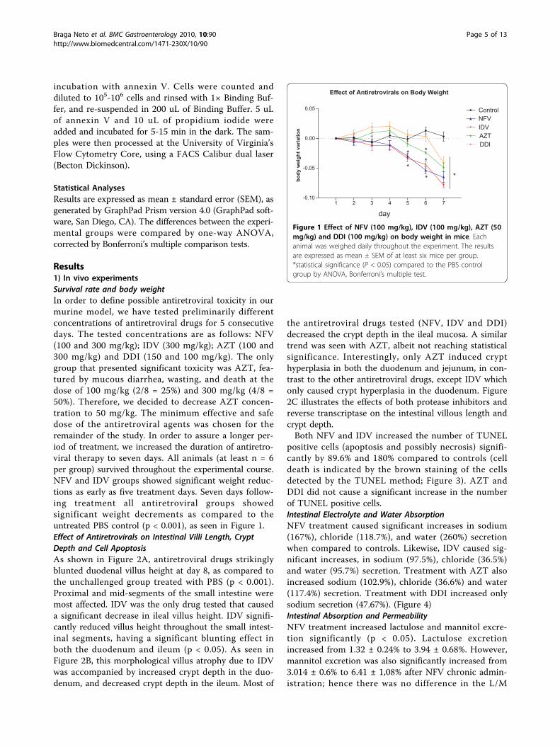
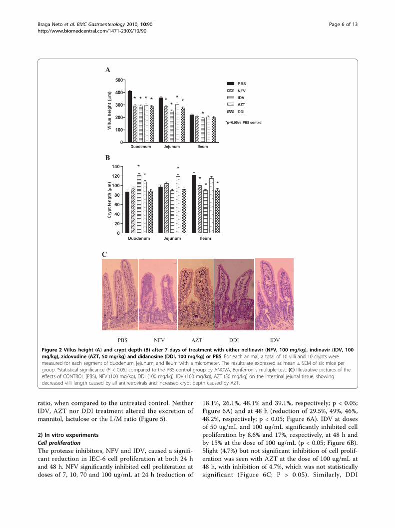
Results1) In vivo experimentsSurvival rate and body weightIn order to define possible antiretroviral toxicity in ourmurine model, we have tested preliminarily differentconcentrations of antiretroviral drugs for 5 consecutivedays. The tested concentrations are as follows: NFV(100 and 300 mg/kg); IDV (300 mg/kg); AZT (100 and300 mg/kg) and DDI (150 and 100 mg/kg). The onlygroup that presented significant toxicity was AZT, fea-tured by mucous diarrhea, wasting, and death at thedose of 100 mg/kg (2/8 = 25%) and 300 mg/kg (4/8 =50%). Therefore, we decided to decrease AZT concen-tration to 50 mg/kg. The minimum effective and safedose of the antiretroviral agents was chosen for theremainder of the study. In order to assure a longer per-iod of treatment, we increased the duration of antiretro-viral therapy to seven days. All animals (at least n = 6per group) survived throughout the experimental course.NFV and IDV groups showed significant weight reduc-tions as early as five treatment days. Seven days follow-ing treatment all antiretroviral groups showedsignificant weight decrements as compared to theuntreated PBS control (p < 0.001), as seen in Figure 1.Effect of Antiretrovirals on Intestinal Villi Length, CryptDepth and Cell ApoptosisAs shown in Figure 2A, antiretroviral drugs strikinglyblunted duodenal villus height at day 8, as compared tothe unchallenged group treated with PBS (p < 0.001).Proximal and mid-segments of the small intestine weremost affected. IDV was the only drug tested that causeda significant decrease in ileal villus height. IDV signifi-cantly reduced villus height throughout the small intest-inal segments, having a significant blunting effect inboth the duodenum and ileum (p < 0.05). As seen inFigure 2B, this morphological villus atrophy due to IDVwas accompanied by increased crypt depth in the duo-denum, and decreased crypt depth in the ileum. Most of
the antiretroviral drugs tested (NFV, IDV and DDI)decreased the crypt depth in the ileal mucosa. A similartrend was seen with AZT, albeit not reaching statisticalsignificance. Interestingly, only AZT induced crypthyperplasia in both the duodenum and jejunum, in con-trast to the other antiretroviral drugs, except IDV whichonly caused crypt hyperplasia in the duodenum. Figure2C illustrates the effects of both protease inhibitors andreverse transcriptase on the intestinal villous length andcrypt depth.Both NFV and IDV increased the number of TUNEL
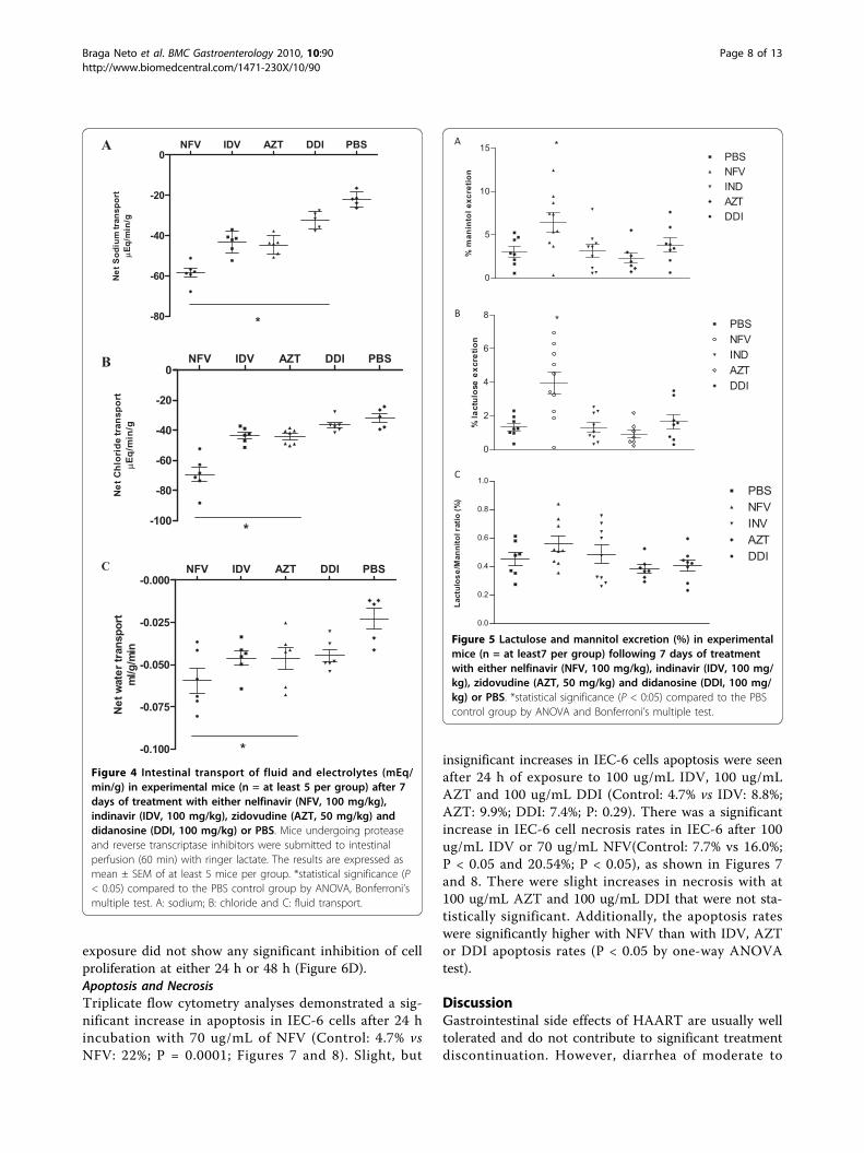
positive cells (apoptosis and possibly necrosis) signifi-cantly by 89.6% and 180% compared to controls (celldeath is indicated by the brown staining of the cellsdetected by the TUNEL method; Figure 3). AZT andDDI did not cause a significant increase in the numberof TUNEL positive cells.Intestinal Electrolyte and Water AbsorptionNFV treatment caused significant increases in sodium(167%), chloride (118.7%), and water (260%) secretionwhen compared to controls. Likewise, IDV caused sig-nificant increases, in sodium (97.5%), chloride (36.5%)and water (95.7%) secretion. Treatment with AZT alsoincreased sodium (102.9%), chloride (36.6%) and water(117.4%) secretion. Treatment with DDI increased onlysodium secretion (47.67%). (Figure 4)Intestinal Absorption and PermeabilityNFV treatment increased lactulose and mannitol excre-tion significantly (p < 0.05). Lactulose excretionincreased from 1.32 ± 0.24% to 3.94 ± 0.68%. However,mannitol excretion was also significantly increased from3.014 ± 0.6% to 6.41 ± 1,08% after NFV chronic admin-istration; hence there was no difference in the L/M
Effect of Antiretrovirals on Body Weight
1 2 3 4 5 6 7-0.10
-0.05
0.00
0.05 ControlNFVIDVAZTDDI
**
*
day
*
* *
body
wei
ght v
aria
tion
Figure 1 Effect of NFV (100 mg/kg), IDV (100 mg/kg), AZT (50mg/kg) and DDI (100 mg/kg) on body weight in mice. Eachanimal was weighed daily throughout the experiment. The resultsare expressed as mean ± SEM of at least six mice per group.*statistical significance (P < 0.05) compared to the PBS controlgroup by ANOVA, Bonferroni’s multiple test.
Braga Neto et al. BMC Gastroenterology 2010, 10:90http://www.biomedcentral.com/1471-230X/10/90
Page 5 of 13
ratio, when compared to the untreated control. NeitherIDV, AZT nor DDI treatment altered the excretion ofmannitol, lactulose or the L/M ratio (Figure 5).
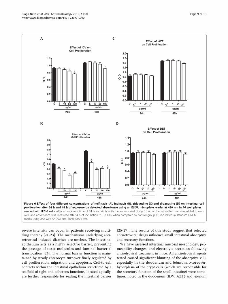
2) In vitro experimentsCell proliferationThe protease inhibitors, NFV and IDV, caused a signifi-cant reduction in IEC-6 cell proliferation at both 24 hand 48 h. NFV significantly inhibited cell proliferation atdoses of 7, 10, 70 and 100 ug/mL at 24 h (reduction of
18.1%, 26.1%, 48.1% and 39.1%, respectively; p < 0.05;Figure 6A) and at 48 h (reduction of 29.5%, 49%, 46%,48.2%, respectively; p < 0.05; Figure 6A). IDV at dosesof 50 ug/mL and 100 ug/mL significantly inhibited cellproliferation by 8.6% and 17%, respectively, at 48 h andby 15% at the dose of 100 ug/mL (p < 0.05; Figure 6B).Slight (4.7%) but not significant inhibition of cell prolif-eration was seen with AZT at the dose of 100 ug/mL at48 h, with inhibition of 4.7%, which was not statisticallysignificant (Figure 6C; P > 0.05). Similarly, DDI
Duodenum Jejunum Ileum0
100
200
300
400
500PBS
NFV
AZT
DDI
IDV* ** * * **
*
* *
*p<0.05vs PBS control
Villu
s he
ight
(µm
)
Duodenum Jejunum Ileum0
20
40
60
80
100
120
140
*** *
* *
Cry
pt le
ngth
( µm
)
C
PBS NFV AZT DDI IDV
A
B
Figure 2 Villus height (A) and crypt depth (B) after 7 days of treatment with either nelfinavir (NFV, 100 mg/kg), indinavir (IDV, 100mg/kg), zidovudine (AZT, 50 mg/kg) and didanosine (DDI, 100 mg/kg) or PBS. For each animal, a total of 10 villi and 10 crypts weremeasured for each segment of duodenum, jejunum, and ileum with a micrometer. The results are expressed as mean ± SEM of six mice pergroup. *statistical significance (P < 0.05) compared to the PBS control group by ANOVA, Bonferroni’s multiple test. (C) Illustrative pictures of theeffects of CONTROL (PBS), NFV (100 mg/kg), DDI (100 mg/kg), IDV (100 mg/kg), AZT (50 mg/kg) on the intestinal jejunal tissue, showingdecreased villi length caused by all antiretrovirals and increased crypt depth caused by AZT.
Braga Neto et al. BMC Gastroenterology 2010, 10:90http://www.biomedcentral.com/1471-230X/10/90
Page 6 of 13
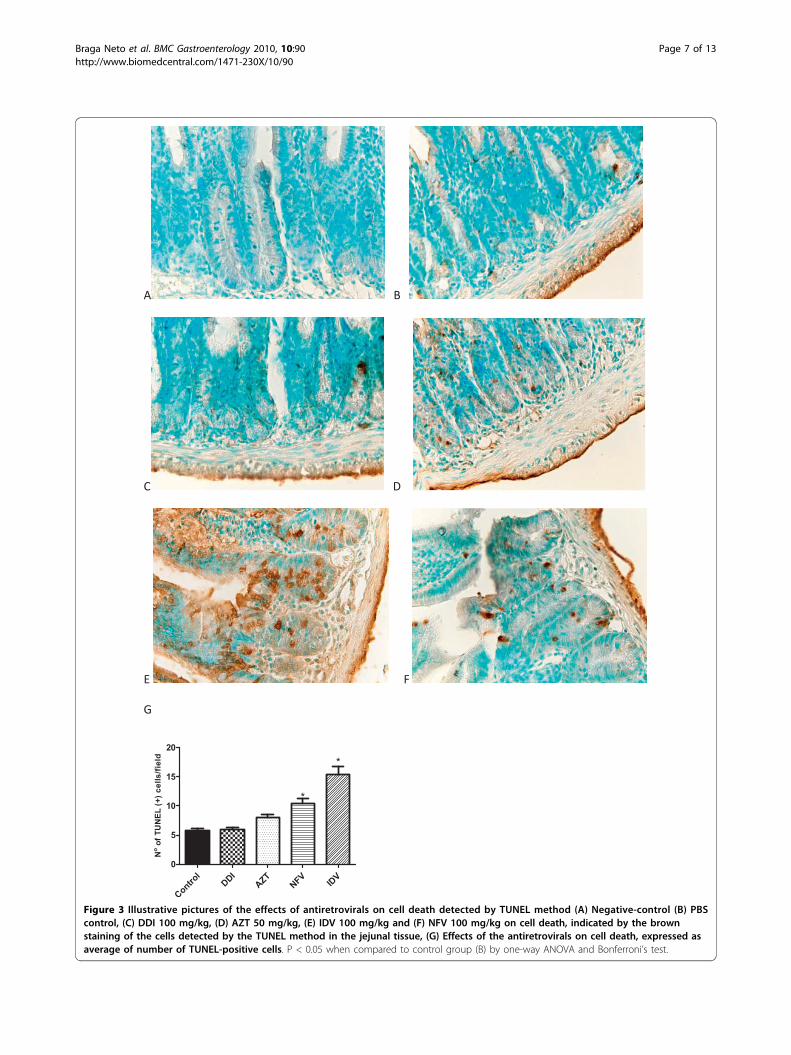
Figure 3 Illustrative pictures of the effects of antiretrovirals on cell death detected by TUNEL method (A) Negative-control (B) PBScontrol, (C) DDI 100 mg/kg, (D) AZT 50 mg/kg, (E) IDV 100 mg/kg and (F) NFV 100 mg/kg on cell death, indicated by the brownstaining of the cells detected by the TUNEL method in the jejunal tissue, (G) Effects of the antiretrovirals on cell death, expressed asaverage of number of TUNEL-positive cells. P < 0.05 when compared to control group (B) by one-way ANOVA and Bonferroni’s test.
Braga Neto et al. BMC Gastroenterology 2010, 10:90http://www.biomedcentral.com/1471-230X/10/90
Page 7 of 13
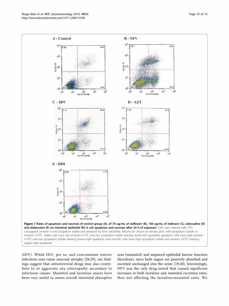
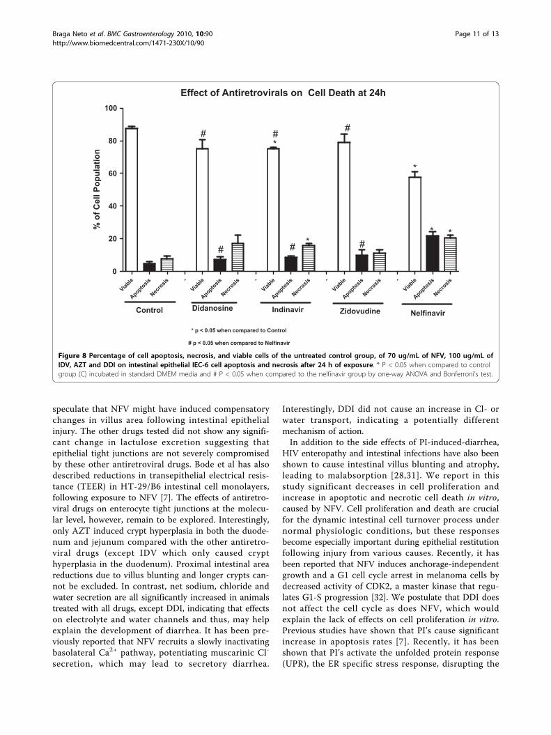
exposure did not show any significant inhibition of cellproliferation at either 24 h or 48 h (Figure 6D).Apoptosis and NecrosisTriplicate flow cytometry analyses demonstrated a sig-nificant increase in apoptosis in IEC-6 cells after 24 hincubation with 70 ug/mL of NFV (Control: 4.7% vsNFV: 22%; P = 0.0001; Figures 7 and 8). Slight, but
insignificant increases in IEC-6 cells apoptosis were seenafter 24 h of exposure to 100 ug/mL IDV, 100 ug/mLAZT and 100 ug/mL DDI (Control: 4.7% vs IDV: 8.8%;AZT: 9.9%; DDI: 7.4%; P: 0.29). There was a significantincrease in IEC-6 cell necrosis rates in IEC-6 after 100ug/mL IDV or 70 ug/mL NFV(Control: 7.7% vs 16.0%;P < 0.05 and 20.54%; P < 0.05), as shown in Figures 7and 8. There were slight increases in necrosis with at100 ug/mL AZT and 100 ug/mL DDI that were not sta-tistically significant. Additionally, the apoptosis rateswere significantly higher with NFV than with IDV, AZTor DDI apoptosis rates (P < 0.05 by one-way ANOVAtest).
DiscussionGastrointestinal side effects of HAART are usually welltolerated and do not contribute to significant treatmentdiscontinuation. However, diarrhea of moderate to
A NFV IDV AZT DDI PBS
-80
-60
-40
-20
0
*
Net
Sod
ium
tran
spor
tµ
Eq/m
in/g
B NFV IDV AZT DDI PBS
-100
-80
-60
-40
-20
0
*
Net
Chl
orid
etr
ansp
ort
µEq
/min
/g
C NFV IDV AZT DDI PBS
-0.100
-0.075
-0.050
-0.025
-0.000
*
Net
wat
er tr
ansp
ort
ml/g
/min
Figure 4 Intestinal transport of fluid and electrolytes (mEq/min/g) in experimental mice (n = at least 5 per group) after 7days of treatment with either nelfinavir (NFV, 100 mg/kg),indinavir (IDV, 100 mg/kg), zidovudine (AZT, 50 mg/kg) anddidanosine (DDI, 100 mg/kg) or PBS. Mice undergoing proteaseand reverse transcriptase inhibitors were submitted to intestinalperfusion (60 min) with ringer lactate. The results are expressed asmean ± SEM of at least 5 mice per group. *statistical significance (P< 0.05) compared to the PBS control group by ANOVA, Bonferroni’smultiple test. A: sodium; B: chloride and C: fluid transport.
Figure 5 Lactulose and mannitol excretion (%) in experimentalmice (n = at least7 per group) following 7 days of treatmentwith either nelfinavir (NFV, 100 mg/kg), indinavir (IDV, 100 mg/kg), zidovudine (AZT, 50 mg/kg) and didanosine (DDI, 100 mg/kg) or PBS. *statistical significance (P < 0:05) compared to the PBScontrol group by ANOVA and Bonferroni’s multiple test.
Braga Neto et al. BMC Gastroenterology 2010, 10:90http://www.biomedcentral.com/1471-230X/10/90
Page 8 of 13
severe intensity can occur in patients receiving multi-drug therapy [21-23]. The mechanisms underlying anti-retroviral-induced diarrhea are unclear. The intestinalepithelium acts as a highly selective barrier, preventingthe passage of toxic molecules and luminal bacterialtranslocation [24]. The normal barrier function is main-tained by steady enterocyte turnover finely regulated bycell proliferation, migration, and apoptosis. Cell-to-cellcontacts within the intestinal epithelium structured by ascaffold of tight and adherens junctions, located apically,are further responsible for sealing the intestinal barrier
[25-27]. The results of this study suggest that selectedantiretroviral drugs influence small intestinal absorptiveand secretory functions.We have assessed intestinal mucosal morphology, per-
meability changes, and electrolyte secretion followingantiretroviral treatment in mice. All antiretroviral agentstested caused significant blunting of the absorptive villi,especially in the duodenum and jejunum. Moreover,hyperplasia of the crypt cells (which are responsible forthe secretory function of the small intestine) were some-times, noted in the duodenum (IDV, AZT) and jejunum
A C
Effect of IDV on Cell Proliferation
C 5 10 50 100 - C 5 10 50 1000.0
0.2
0.4
0.6
0.8
1.0
1.2
* **
µg/mL µg/mL
24h 48h
O.D
Effect of AZTon Cell Proliferation
C 0.1 1 10 100 - C 0.
1 1 10 100
0.0
0.2
0.4
0.6
0.8
1.0
1.2
1.4
1.6
1.8
2.0
ug/ml ug/mlh84h42
O.D
B D
Effect of NFV onCell Proliferation
C 7 10 70 100 - C 7 10 70 1000.0
0.1
0.2
0.3
0.4
0.5
0.6
0.7
0.8
0.9
1.0
** *
*
*
*
**
µg/ml µg/ml24h 48h
O.D
Effect of DDIon Cell Proliferation
C 5 10 50 100 - C 5 10 50 10
00.0
0.2
0.4
0.6
0.8
1.0
1.2
1.4
24h 48h
µg/mL µg/mL
O.D
Figure 6 Effect of four different concentrations of nelfinavir (A), indinavir (B), zidovudine (C) and didanosine (D) on intestinal cellproliferation after 24 h and 48 h of exposure by detected absorbance using an ELISA microplate reader at 420 nm in 96 well platesseeded with IEC-6 cells. After an exposure time of 24 h and 48 h, with the antiretroviral drugs, 10 uL of the tetrazolium salt was added to eachwell, and absorbance was measured after 4 h of incubation. * P < 0.05 when compared to control group (C) incubated in standard DMEMmedia using one-way ANOVA and Bonferroni’s test.
Braga Neto et al. BMC Gastroenterology 2010, 10:90http://www.biomedcentral.com/1471-230X/10/90
Page 9 of 13
(IDV). While HIV, per se, and concomitant entericinfections may cause mucosal atrophy [28,29], our find-ings suggest that antiretroviral drugs may also contri-bute to or aggravate any enteropathy secondary toinfectious causes. Mannitol and lactulose assays havebeen very useful to assess overall intestinal absorptive
area (mannitol) and impaired epithelial barrier function(lactulose), since both sugars are passively absorbed andexcreted unchanged into the urine [19,30]. Interestingly,NFV was the only drug tested that caused significantincreases in both lactulose and mannitol excretion rates,thus not affecting the lactulose:mannitol ratio. We
A - Control B - NFV
C – IDV D - AZT
E - DDI
Figure 7 Rates of apoptosis and necrosis of control group (A), of 70 ug/mL of nelfinavir (B), 100 ug/mL of indinavir (C), zidovudine (D)and didanosine (E) on intestinal epithelial IEC-6 cell apoptosis and necrosis after 24 h of exposure. Cells were stained with FITC-conjugated annexin V and propidium iodide and analyzed by flow cytometry. Results are shown as density plots with propidium iodide vs.annexin V-FITC. Viable cells have low annexin-V-FITC and low propidium iodide staining (lower-left quadrant), apoptotic cells have high annexinV-FITC and low propidium iodide staining (lower-right quadrant), and necrotic cells have high propidium iodide and annexin V-FITC staining(upper-right quadrant).
Braga Neto et al. BMC Gastroenterology 2010, 10:90http://www.biomedcentral.com/1471-230X/10/90
Page 10 of 13
speculate that NFV might have induced compensatorychanges in villus area following intestinal epithelialinjury. The other drugs tested did not show any signifi-cant change in lactulose excretion suggesting thatepithelial tight junctions are not severely compromisedby these other antiretroviral drugs. Bode et al has alsodescribed reductions in transepithelial electrical resis-tance (TEER) in HT-29/B6 intestinal cell monolayers,following exposure to NFV [7]. The effects of antiretro-viral drugs on enterocyte tight junctions at the molecu-lar level, however, remain to be explored. Interestingly,only AZT induced crypt hyperplasia in both the duode-num and jejunum compared with the other antiretro-viral drugs (except IDV which only caused crypthyperplasia in the duodenum). Proximal intestinal areareductions due to villus blunting and longer crypts can-not be excluded. In contrast, net sodium, chloride andwater secretion are all significantly increased in animalstreated with all drugs, except DDI, indicating that effectson electrolyte and water channels and thus, may helpexplain the development of diarrhea. It has been pre-viously reported that NFV recruits a slowly inactivatingbasolateral Ca2+ pathway, potentiating muscarinic Cl-
secretion, which may lead to secretory diarrhea.
Interestingly, DDI did not cause an increase in Cl- orwater transport, indicating a potentially differentmechanism of action.In addition to the side effects of PI-induced-diarrhea,
HIV enteropathy and intestinal infections have also beenshown to cause intestinal villus blunting and atrophy,leading to malabsorption [28,31]. We report in thisstudy significant decreases in cell proliferation andincrease in apoptotic and necrotic cell death in vitro,caused by NFV. Cell proliferation and death are crucialfor the dynamic intestinal cell turnover process undernormal physiologic conditions, but these responsesbecome especially important during epithelial restitutionfollowing injury from various causes. Recently, it hasbeen reported that NFV induces anchorage-independentgrowth and a G1 cell cycle arrest in melanoma cells bydecreased activity of CDK2, a master kinase that regu-lates G1-S progression [32]. We postulate that DDI doesnot affect the cell cycle as does NFV, which wouldexplain the lack of effects on cell proliferation in vitro.Previous studies have shown that PI’s cause significantincrease in apoptosis rates [7]. Recently, it has beenshown that PI’s activate the unfolded protein response(UPR), the ER specific stress response, disrupting the
Effect of Antiretrovirals on Cell Death at 24h
Viable
Apoptosis
Necro
sis-
Viable
Apoptosis
Necro
sis-
Viable
Apoptosis
Necro
sis-
Viable
Apoptosis
Necro
sis-
Viable
Apoptosis
Necro
sis0
20
40
60
80
100
*
*
*
**
Control Didanosine Indinavir Zidovudine Nelfinavir
###
###
* p < 0.05 when compared to Control
# p < 0.05 when compared to Nelfinavir
% o
f Cel
l Pop
ulat
ion
Figure 8 Percentage of cell apoptosis, necrosis, and viable cells of the untreated control group, of 70 ug/mL of NFV, 100 ug/mL ofIDV, AZT and DDI on intestinal epithelial IEC-6 cell apoptosis and necrosis after 24 h of exposure. * P < 0.05 when compared to controlgroup (C) incubated in standard DMEM media and # P < 0.05 when compared to the nelfinavir group by one-way ANOVA and Bonferroni’s test.
Braga Neto et al. BMC Gastroenterology 2010, 10:90http://www.biomedcentral.com/1471-230X/10/90
Page 11 of 13
intestinal barrier integrity, and that reduction in CHOPexpression, which is a UPR-induced transcription factorthat mediates apoptosis, significantly reduced apoptosisboth in vitro and in vivo [33]. These findings furtherhelp elucidate the different mechanisms by which HIVPI’s can induce cell death in different cell lines. Indeed,our study showed a similar increase in cell apoptosis ingroups treated with NFV both in vivo and in vitro. Inaddition, we also report a significant increase in necrosisrates caused by IDV and NFV, which had not been pre-viously reported [7].Our in vitro and in vivo models do not take into
account other host and environmental variables includ-ing HIV infection itself or opportunistic intestinalpathogens that can certainly contribute to impairedintestinal function and responses to antiretroviral agents.HIV associated diarrhea may be partly related to mus-carinic induced-chloride secretion [10]. Mucosal HIVreplication might induce a pro-inflammatory and Th1-mediated cytokine over-expression [34], which couldadditionally disrupt the intestinal barrier with increasedsecretion with electrolyte and water loss. Depletion oflymphocytes in the lamina propria by HIV could disruptthe immune system and increase susceptibility to oppor-tunistic infections [29,35]. Drug absorption may befurther compromised by opportunistic enteric infectionslike Cryptosporidium spp. and Micromonospora spp.,the former has been found in HIV-immunocompro-mised patients in Brazilian settings, with and withoutdiarrhea [11,36,37]. Thus, a wide range of secretoryintestinal transport disruption may occur in vivo due todirect antiretroviral therapy and both systemic andintestinal infections.
ConclusionIn conclusion, this study focused on understanding theeffects of protease inhibitors and reverse transcriptionantiretroviral drugs on the intestinal epithelium. Gaininginsight into how antiretroviral agents cause diarrhea isessential in developing interventions that could poten-tially reduce these side effects, avoid therapy disconti-nuation and increase efficacy of the antiretroviral drugs.We have found that both PI’s, NFV and IDV, decreasedproliferation and increased necrosis in vitro and thatonly NFV increased apoptosis in vitro, in contrast to thereverse transcriptase inhibitors which did not have suchan effect. Additionally, both PI’s and reverse transcrip-tase inhibitors altered intestinal morphology in vivo,reducing villous height in the duodenum and jejunumand induced sodium secretion. Furthermore, NFV andIDV induced cell apoptosis in vivo and chloride andwater secretion. NFV was the only drug tested thatcaused significant changes in intestinal permeability.Further studies are needed to elucidate molecular
mechanisms involved in HIV PI-induced epithelialdamage, including cell cycle studies, WNT-signaling andR-spondin-1, specific cell death pathways as well as stu-dies to examine whether cell migration and cellularcytoskeletal F-actin are affected and contribute to dis-rupting the intestinal barrier. Additionally, thesemechanisms might be modulated by important gut-trophic nutrients, such as glutamine, alanyl-glutamine,arginine and zinc [38], which could reduce intestinaldamage, increase drug absorption and decrease undesir-able side effects, such as diarrhea that can be a limitingfactor for treatment adherence and efficacy.
AcknowledgementsThe project described was supported by award number D43 TW006578from the Fogarty International Center and award number U01 AI026512from the National Institute of Allergy and Infectious Diseases and in part bytwo Brazilian funding agencies, CNPq and CAPES and Grant # 55000645from the HHMI International Infectious Disease Scholarship. The followingreagents used in the in vitro experiments were obtained through the AIDSResearch and Reference Reagent Program, Division of AIDS, NIAID, NIH:nelfinavir, indinavir, zidovudine and didanosine. Additionally, we would liketo thank Joanne Lennigan for her assistance on flow cytometry.
Author details1Institute of Biomedicine and Clinical Research Unit-University Hospital,Federal University of Ceará, Fortaleza, Brazil. 2Center for Global Health,Division of Infectious Diseases and International Health, University of Virginia,Charlottesville, USA. 3Department of Morphology, Federal University of Ceará,Fortaleza, Brazil. 4Department of Physiology and Pharmacology, FederalUniversity of Ceará, Fortaleza, Brazil.
Authors’ contributionsMB: Participated in the in vitro and in vivo experiments, performed dataanalysis and drafted the manuscript. CV: Participated in the in vivoexperiments, data storage, analysis and helped drafting the manuscript. JG:Participated in the in vivo experiments and data analysis. JE: Participated inthe in vitro experiments and data analysis. CA: Participated in the studydesign and writing the paper. RB: Participated in statistical analysis anddrafting and reviewing of the manuscript. GB and BO: Participated in the invivo experiments and critical reviewing. AL and RG: Participated in thecoordination, conception, experiments design and critical reviewing andwriting. All authors read and approved the final manuscript.
Competing interestsThe authors declare that they have no competing interests.
Received: 11 February 2010 Accepted: 11 August 2010Published: 11 August 2010
References1. Moreno S, López Aldeguer J, Arribas JR, Domingo P, Iribarren JA, Ribera E,
Rivero A, Pulido F, HIV 2020 Project: The future of antiretroviral therapy:challenges and needs. J Antimicrob Chemother 2010, 65(5):827-35.
2. El SY, Vivet-Boudou V, Marquet R: HIV-1 reverse transcriptase inhibitors.Appl Microbiol Biotechnol 2007, 75:723-37.
3. McKinnon JE, Mellors JW, Swindells S: Simplification strategies to reduceantiretroviral drug exposure: progress and prospects. Antivir Ther 2009,14(1):1-12.
4. Hill A, Balkin A: Risk factors for gastrointestinal adverse events in HIVtreated and untreated patients. AIDS Rev 2009, 11(1):30-8.
5. Guarino A, Bruzzese E, De MG, Buccigrossi V: Management ofgastrointestinal disorders in children with HIV infection. Paediatr Drugs2004, 6:347-62.
Braga Neto et al. BMC Gastroenterology 2010, 10:90http://www.biomedcentral.com/1471-230X/10/90
Page 12 of 13
6. Tramarin A, Parise N, Campostrini S, Yin DD, Postma MJ, Lyu R, Grisetti R,Capetti A, Cattelan AM, Di Toro MT, Mastroianni A, Pignattari E,Mondardini V, Calleri G, Raise E, Starace F, Palladio Study Group:Association between diarrhea and quality of life in HIV-infected patientsreceiving highly active antiretroviral therapy. Qual Life Res 2004,13:243-50.
7. Bode H, Lenzner L, Kraemer OH, Kroesen AJ, Bendfeldt K, Schulzke JD,Fromm M, Stoltenburg-Didinger G, Zeitz M, Ullrich R: The HIV proteaseinhibitors saquinavir, ritonavir, and nelfinavir induce apoptosis anddecrease barrier function in human intestinal epithelial cells. Antivir Ther2005, 10:645-55.
8. Chow WA, Jiang C, Guan M: Anti-HIV drugs for cancer therapeutics: backto the future? Lancet Oncol 2009, 10(1):61-71.
9. Gills JJ, Lopiccolo J, Tsurutani J, Shoemaker RH, Best CJ, Abu-Asab MS,Borojerdi J, Warfel NA, Gardner ER, Danish M, Hollander MC, Kawabata S,Tsokos M, Figg WD, Steeg PS, Dennis PA: Nelfinavir, a lead HIV proteaseinhibitor, is a broad-spectrum, anticancer agent that inducesendoplasmic reticulum stress, autophagy, and apoptosis in vitro and invivo. Clin Cancer Res 2007, 13(17):5183-94.
10. Rufo PA, Lin PW, Andrade A, Jiang L, Rameh L, Flexner C, Alper SL,Lencer WI: Diarrhea-associated HIV-1 APIs potentiate muscarinicactivation of Cl- secretion by T84 cells via prolongation of cytosolic Ca2+ signaling. Am J Physiol Cell Physiol 2004, 286:C998-C1008.
11. Wuhib T, Silva TM, Newman RD, Garcia LS, Pereira ML, Chaves CS,Wahlquist SP, Bryan RT, Guerrant RL, Sousa A de Q, Queiroz TRB, Sears CL:Cryptosporidial and microsporidial infections in humanimmunodeficiency virus-infected patients in northeastern Brazil. J InfectDis 1994, 170:494-97.
12. Bushen OY, Davenport JA, Lima AB, Piscitelli SC, Uzgiris AJ, Silva TM, Leite R,Kosek M, Dillingham RA, Girao A, Lima AA, Guerrant RL: Diarrhea andreduced levels of antiretroviral drugs: improvement with glutamine oralanyl-glutamine in a randomized controlled trial in northeast Brazil. ClinInfect Dis 2004, 38:1764-70.
13. Berkes J, Viswanathan VK, Savkovic SD, Hecht G: Intestinal epithelialresponses to enteric pathogens: effects on the tight junction barrier, iontransport, and inflammation. Gut 2003, 52:439-51.
14. Bjerknes M, Cheng H: Clonal analysis of mouse intestinal epithelialprogenitors. Gastroenterology 1999, 116:7-14.
15. Potten CS: Stem cells in gastrointestinal epithelium: numbers,characteristics and death. Philos Trans R Soc Lond B Biol Sci 1998,353:821-30.
16. Clatworthy JP, Subramanian V: Stem cells and the regulation ofproliferation, differentiation and patterning in the intestinal epithelium:emerging insights from gene expression patterns, transgenic and geneablation studies. Mech Dev 2001, 101:3-9.
17. Fuchs BC, Bode BP: Stressing Out Over Survival: Glutamine as anApoptotic Modulator. J Surg Res 2006, 131(1):26-40.
18. Kroemer G, Galluzzi L, Vandenabeele P, Abrams J, Alnemri ES, Baehrecke EH,Blagosklonny MV, El-Deiry WS, Golstein P, Green DR, Hengartner M,Knight RA, Kumar S, Lipton SA, Malorni W, Nuñez G, Peter ME, Tschopp J,Yuan J, Piacentini M, Zhivotovsky B, Melino G, Nomenclature Committee onCell Death 2009: Classification of cell death: recommendations of theNomenclature Committee on Cell Death 2009. Cell Death Differ 2009,16(1):3-11.
19. Bao Y, Silva TM, Guerrant RL, Lima AM, Fox JW: Direct analysis of mannitol,lactulose and glucose in urine samples by high-performance anion-exchange chromatography with pulse amperometric detection. Clinicalevaluation of intestinal permeability in human immunodeficiency virusinfection. J Chromatogr B Biomed Appl 1996, 685:105-12.
20. Schedl HP, Clifton JA: Solute and water absorption by the human smallintestine. Nature 1963, 199:1264-7.
21. Bardsley-Elliot A, Plosker GL: Nelfinavir: an update on its use in HIVinfection. Drugs 2000, 59:581-620.
22. Lemberg DA, Palasanthiran P, Goode M, Ziegler JB: Tolerabilities ofantiretrovirals in paediatric HIV infection. Drug Saf 2002, 25:973-91.
23. Jarvis B, Nelfinavir Faulds D: A review of its therapeutic efficacy in HIVinfection. Drugs 1998, 56:147-67.
24. Guttman JA, Li Y, Wickham ME, Deng W, Vogl AW, Finlay BB: Attaching andeffacing pathogen-induced tight junction disruption in vivo. Cell Microbiol2006, 8:634-45.
25. Utech M, Bruwer M, Nusrat A: Tight junctions and cell-cell interactions.Methods Mol Biol 2006, 341:185-95.
26. Laukoetter MG, Bruewer M, Nusrat A: Regulation of the intestinalepithelial barrier by the apical junctional complex. Curr Opin Gastroenterol2006, 22:85-9.
27. Gasbarrini G, Montalto M: Structure and function of tight junctions. Rolein intestinal barrier. Ital J Gastroenterol Hepatol 1999, 31:481-8.
28. Ullrich R, Zeitz M, Heise W, L’age M, Hoffken G, Riecken EO: Small intestinalstructure and function in patients infected with humanimmunodeficiency virus (HIV): evidence for HIV-induced enteropathy.Ann Intern Med 1989, 111:15-21.
29. Mehandru S, Poles MA, Tenner-Racz K, Horowitz A, Hurley A, Hogan C,Boden D, Racz P, Markowitz M: Primary HIV-1 infection is associated withpreferential depletion of CD4+ T lymphocytes from effector sites in thegastrointestinal tract. J Exp Med 2004, 200:761-70.
30. Barboza Junior MS, Silva TM, Guerrant RL, Lima AA: Measurement ofintestinal permeability using mannitol and lactulose in children withdiarrheal diseases. Braz J Med Biol Res 1999, 32:1499-504.
31. Gianotti N, Soria A, Lazzarin A: Antiviral activity and clinical efficacy ofatazanavir in HIV-1-infected patients: a review. New Microbiol 2007,30:79-88.
32. Jiang W, Mikochik PJ, Ra JH, Lei H, Flaherty KT, Winkler JD, Spitz FR: HIVprotease inhibitor nelfinavir inhibits growth of human melanoma cellsby induction of cell cycle arrest. Cancer Res 2007, 1; 67(3):1221-7.
33. Wu X, Sun L, Zha W, Studer E, Gurley E, Chen L, Wang X, Hylemon PB,Pandak WM Jr, Sanyal AJ, Zhang L, Wang G, Chen J, Wang JY, Zhou H: HIVprotease inhibitors induce endoplasmic reticulum stress and disruptbarrier integrity in intestinal epithelial cells. Gastroenterology 2010,138(1):197-209.
34. McGowan I, Elliott J, Fuerst M, Taing P, Boscardin J, Poles M, Anton P:Increased HIV-1 mucosal replication is associated with generalizedmucosal cytokine activation. J Acquir Immune Defic Syndr 2004, 37:1228-36.
35. Ullrich R, Zeitz M, Riecken EO: Enteric immunologic abnormalities inhuman immunodeficiency virus infection. Semin Liver Dis 1992, 12:167-74.
36. Nannini EC, Okhuysen PC: HIV1 and the gut in the era of highly activeantiretroviral therapy. Curr Gastroenterol Rep 2002, 4:392-8.
37. Lima AA, Silva TM, Gifoni AM, Barrett LJ, McAuliffe IT, Bao Y, Fox JW,Fedorko DP, Guerrant RL: Mucosal injury and disruption of intestinalbarrier function in HIV-infected individuals with and without diarrheaand cryptosporidiosis in northeast Brazil. Am J Gastroenterol 1997,92:1861-6.
38. Carneiro-Filho BA, Bushen OY, Brito GA, Lima AA, Guerrant RL: Glutamineanalogues as adjunctive therapy for infectious diarrhea. Curr Infect DisRep 2003, 5:114-9.
Pre-publication historyThe pre-publication history for this paper can be accessed here:http://www.biomedcentral.com/1471-230X/10/90/prepub
doi:10.1186/1471-230X-10-90Cite this article as: Braga Neto et al.: Evaluation of HIV protease andnucleoside reverse transcriptase inhibitors on proliferation, necrosis,apoptosis in intestinal epithelial cells and electrolyte and watertransport and epithelial barrier function in mice. BMC Gastroenterology2010 10:90.
Submit your next manuscript to BioMed Centraland take full advantage of:
• Convenient online submission
• Thorough peer review
• No space constraints or color figure charges
• Immediate publication on acceptance
• Inclusion in PubMed, CAS, Scopus and Google Scholar
• Research which is freely available for redistribution
Submit your manuscript at www.biomedcentral.com/submit
Braga Neto et al. BMC Gastroenterology 2010, 10:90http://www.biomedcentral.com/1471-230X/10/90
Page 13 of 13


