Embed Size (px)
Citation preview

JOURNAL OF BACTERIOLOGY,0021-9193/01/$04.0010 DOI: 10.1128/JB.183.11.3499–3505.2001
June 2001, p. 3499–3505 Vol. 183, No. 11
Copyright © 2001, American Society for Microbiology. All Rights Reserved.
Evidence against an Interaction between the mRNA DownstreamBox and 16S rRNA in Translation Initiation
ISABELLA MOLL,1 MICHAEL HUBER,1 SONJA GRILL,1 POONEH SAIRAFI,1 FLORIAN MUELLER,2
RICHARD BRIMACOMBE,2 PAOLA LONDEI,3 AND UDO BLASI1*
Institute of Microbiology and Genetics, University of Vienna, Vienna Biocenter, 1030 Vienna, Austria1;Max-Planck Institute of Molecular Genetics, 14195 Berlin, Germany2; and Department of
Medical Biochemistry, University of Bari, 70124 Bari, and Department of CellularBiotechnologies and Hematology, University of Rome, 00161 Rome, Italy3
Received 29 November 2000/Accepted 7 March 2001
Based on the complementarity of the initial coding region (downstream box [db]) of several bacterial andphage mRNAs to bases 1469 to 1483 in helix 44 of 16S rRNA (anti-downstream box [adb]), it has beenproposed that db-adb base pairing enhances translation in a way that is similar to that of the Shine-Dalgarno(SD)/anti-Shine-Dalgarno (aSD) interaction. Computer modeling of helix 44 on the 30S subunit shows that thetopography of the 30S ribosome does not allow a simultaneous db-adb interaction and placement of the ini-tiation codon in the ribosomal P site. Thus, the db-adb interaction cannot substitute for the SD-aSD inter-action in translation initiation. We have always argued that any contribution of the db-adb interaction shouldbe most apparent on mRNAs devoid of an SD sequence. Here, we show that 30S ribosomes do not bind to leaderlessmRNA in the absence of initiator tRNA, even when the initial coding region shows a 15-nucleotide complemen-tarity (optimal fit) with the putative adb. In addition, an optimized db did not affect the translational efficiencyof a leaderless l cI-lacZ reporter construct. Thus, the db-adb interaction can hardly serve as an initial re-cruitment signal for ribosomes. Moreover, we show that different leaderless mRNAs are translated in heter-ologous systems although the sequence of the putative adb’s within helix 44 of the 30S subunits of the corre-sponding bacteria differ largely. Taken our data together with those of others (M. O’Connor, T. Asai, C. L.Squires, and A. E. Dahlberg, Proc. Natl. Acad. Sci. USA 96:8973–8978, 1999; A. La Teana, A. Brandi, M. O’Connor,S. Freddi, and C. L. Pon, RNA 6:1393–1402, 2000), we conclude that the db does not base pair with the adb.
The downstream box (db) located 8 to 13 nucleotides (nt)downstream of the start codon has been originally proposed tobase pair with the anti-downstream box (adb) spanning nt 1469to 1483 within helix 44 of the 16S rRNA (Fig. 1A; 37). The dbelement was identified first in the highly expressed genes 0.3and 10 of Escherichia coli phage T7 and has been reported tostimulate translation per se or in combination with the Shine-Dalgarno (SD) sequence (37, 38). Since the original proposal,there has been much publicity in favor of the proposed db-adbinteraction (3, 6, 7, 8, 9, 16, 21, 23, 26, 33, 36, 43, 44). In somecases, the importance of the db-adb interaction in translationinitiation was proposed solely based on computer analysis inthe complete absence of any experimental data. The support-ing experimental evidence for the db-adb base pairing restsentirely on manipulations of mRNAs containing a putative dband the observations that, in general, increases in complemen-tarity of the db with the adb resulted in an increased expressionand decreases in the db-adb complementarity had the corre-sponding downward effects on expression (9, 36, 38).
Biochemical evidence for the db-adb interaction is lacking.Chemical protection studies on l cI mRNA-70S initiationcomplexes (31) and on phage T4 gene 32 mRNA-30S and -70Scomplexes (15) have failed to show protection of the putativedb. Mutational studies revealed that alterations on either side
of the adb-containing helix 44 that disrupt the helical continu-ity had deleterious effects on ribosome function. These effectscould be reversed upon introduction of compensatory muta-tions that restored base pairing within the helix (10). Thesestudies indicated that a stable helix rather than a particularprimary sequence is important for ribosome function. Re-cently, O’Connor et al. (28) performed a landmark experimentby reversing all 12 bp of the stem containing the putative adb,thereby creating a mutant 16S rRNA with a radically alteredbase pairing potential. This 16S rRNA allele with the adb-fliphas been expressed in an E. coli strain in which all of the sevenrrn operons had been deleted (2). The expression rates ofseveral previously described db-containing reporter constructswere found to be indistinguishable in both the adb-flip mutantand the isogenic wild-type (wt) strain. These genetic studiesshowed that any db-associated enhancer activity does not in-volve db-adb base pairing (28). The db-adb interaction hasbeen suggested to be instrumental for the translation of E. colicspA mRNA during cold shock, and it seemed conceivable thatthe adb is particularly exposed in cold-shocked ribosomes (8, 21).Using the same approach as O’Connor et al. (28), La Teana etal. (18) have recently shown that 30S db-flip mutants translatedthe cspA mRNA with the same efficiency as wt ribosomesunder cold shock as well as under non-cold shock conditions,suggesting again that the db-adb base pairing is irrelevant.
It has been suggested that the db serves as an independenttranslation initiation signal (38). In another report, it has beensuggested that the db-adb and the SD/anti-SD (aSD) interac-tions act synergistically to enhance translation initiation (7).
* Corresponding author. Mailing address: Institute of Microbiologyand Genetics, University of Vienna, Vienna Biocenter, Dr. Bohrgasse9, 1030 Vienna, Austria. Phone: 43–1-4277–54609. Fax: 43–1-4277–9546. E-mail: [email protected].
3499

However, no direct in vitro experiments have been performedto address the question of whether an mRNA with an optimalfit to the adb has an increased affinity for the 30S subunit aswould be expected regardless of whether the db provides per sea ribosomal recruitment signal, whether it acts in concert withthe SD sequence, or whether it acts transiently to increase theconcentration of the start codon close to the decoding center(39). Here, we present genetic and biochemical studies as wellas topographical information on the ribosome which, takentogether with previously performed genetic studies (18, 28),add strong evidence against the db-adb interaction.
MATERIALS AND METHODS
Bacterial strains and plasmids. The E. coli strains MS59l (31) and MC4100F9(40) as well as the Bacillus subtilis strain 168 (1) have been described. They weregrown in Luria-Bertani medium (20) supplemented with ampicillin (100 mg/ml)where appropriate to maintain selection of plasmids. Growth of the liquid cul-tures was monitored photometrically by measuring the optical density at a wave-length of 600 nm (OD600).
Plasmid pAXL7 harbors the full-length cI gene under transcriptional controlof a T7 promoter (41).
Construction of plasmids used in this study. Plasmids pRB381-1 andpRB381-3 are derivatives of plasmid pRB381 (4). They were constructed asfollows. Plasmid pMCcI(63) (31) was digested with XbaI and SmaI, and thefragment was inserted into the corresponding sites of plasmid pUC18 (19). Theresulting pUC18 derivative was then cleaved with XbaI and BamHI, and thefragment containing the l pRM promoter and the first 63 codons of the cI genewas inserted into the corresponding sites of pRB381, resulting in pRB381-1.
The putative db in l cI was optimized in plasmid pRB381-3 by using thePCR-based site-directed mutagenesis procedure described by Hall and Emery(14). Briefly, the immediate 59-coding region of the cI gene was modified asshown in Fig. 3A, using complementary oligonucleotides comprising this regionin conjunction with biotinylated oligonucleotides for the upstream and down-stream region of the part of the cI gene present in plasmid pMCcI(63) (31). Afterhybridization of the two strands and the fill-in reaction with T4 DNA polymerase,a PCR with nonbiotinylated oligonucleotides was performed. The resulting frag-ment was cleaved with XbaI and BamHI and cloned into the corresponding sitesof the vector pRB381, resulting in plasmid pRB381-3. The plasmids pRB381-1and pRB381-3 are isogenic except for the initial coding region of the cI gene(optimized db*; see Fig. 3A).
Plasmid pRB381cI is a derivative of the E. coli-B. subtilis shuttle vectorpRB381 and was constructed as follows. First, a PCR fragment containing the lacpromoter (from nt 260 to 11) from plasmid pUHE21-2 (17) and the first 189 ntof the l cI gene were inserted into the XbaI-SmaI sites of plasmid pKT35 (30).In the resulting plasmid, pKTplaccI, the lac promoter abutted the start codon ofthe cI gene via an NcoI site, which ensured that transcription of cI mRNA startsat the adenosine of the start codon. Plasmid pKTplaccI was then used as atemplate with an XbaI forward primer and a BamHI reverse primer. The PCRproduct was cleaved with XbaI and BamHI, and the fragment was inserted intothe same sites of pRB381. In the resulting plasmid pRB381cI, the cI-lacZmRNA, which contains the first 63 codons of the cI gene, is transcribed from themodified lacpo. The transcript starts with the A of the initiating codon of the cIgene. All DNA manipulations were verified by sequencing.
Filter-binding assay. Filter-binding assays were performed using a Schleicherand Schuell SRC 072/0 Minifold II Slot Blot apparatus. The cI34 mRNA, cIOptdb
mRNA, and cI34SD mRNA (Fig. 2) were obtained by hybridization of cDNAnucleotides containing the T7 f10 promoter and either the first 34 bp (cI34 andcI34SD mRNAs, including the 59 extension) of the cI gene or the first 33 bp ofcI with the optimized db (cIOptdb mRNA) and subsequent T7 RNA polymerase-directed mRNA synthesis. All mRNAs were labeled with [a-32P]CTP. In areaction volume of 50 ml, 0.5 pmol of 32P-labeled mRNA was incubated with 30Sribosomes at a molar ratio of 1:10 (Fig. 2). fMet-tRNAf
Met was added in atwofold excess over 30S ribosomes. The reaction mixtures were incubated for 10min at 37°C. Samples were added to the filter under vacuum and washed with 25volumes of VD1 reaction buffer as previously described (41). The filters wereexposed to a Molecular Dynamics PhosphorImager screen for quantitation of theretained mRNA. The total counts per minute values were corrected for thenumber of C’s in each mRNA.
b-galactosidase assays. The b-galactosidase activities were determined asdescribed by Miller (20). Triplicate aliquots were taken of each culture at anOD600 of 0.8.
In vitro translation. Full-length Lactococcus lactis phage r1t rro mRNA wasobtained as described previously (22) except that the reverse “PCR-oligonucle-otide” was complementary to a region downstream of the rro stop codon. Forfull-length synthesis of cI mRNA, plasmid pAXL7 was cleaved with PvuII. ThePCR fragment and the linearized plasmid served as templates for in vitro tran-scription reactions with T7 RNA polymerase. The in vitro translation reactionswith Sulfolobus solfataricus extracts were performed as described by Condo et al.(5). The samples contained the following in a final volume of 50 ml: 10 mM KCl,20 mM Tris-HCl (pH 7.0), 18 mM MgOAc, 7 mM b-mercaptoethanol, 3 mMATP, 1 mM GTP, 5 mg of bulk S. solfataricus tRNA, 20 mM amino acids (exceptfor methionine), 2 ml of [35S]methionine (1,200 Ci/mmol), 20 ml of S. solfataricusS30 extract (preincubated for 10 min at 73°C), and 10 pmol of mRNA. Thesamples were incubated at 73°C for 40 min. The labeled proteins were separatedon a sodium dodecyl sulfate (SDS)–12% polyacrylamide gel. The gel was driedunder vacuum and exposed to an X-ray film.
FIG. 1. Localization of the putative adb in the penultimate stem ofE. coli 16S rRNA and spatial localization of the adb in the 30S subunitof T. thermophilus. (A) Helices 44 and 45 of the E. coli 16S rRNA areenlarged. The adb sequence shown boxed (nt 1469 to 1483) in helix 44has been suggested to base pair with the db in mRNA, i.e., with mRNAsequences downstream of the start codon. (B) Placement of themRNA (nt 26 to 112; green tube) in the T. thermophilus 16S rRNAbased on cross-linking studies (35). The positions of the aSD sequenceand the adb in the 16S rRNA are shown. The aSD sequence and theputative adb region are depicted in magenta. The mRNA is shown asa green tube from nt 26 to 112 with regard to the A (11) of the startcodon. The P-site tRNA is shown in orange. (C) Stereoview of thestructure shown in panel B added to the contour of the 30S subunit(see text).
3500 MOLL ET AL. J. BACTERIOL.
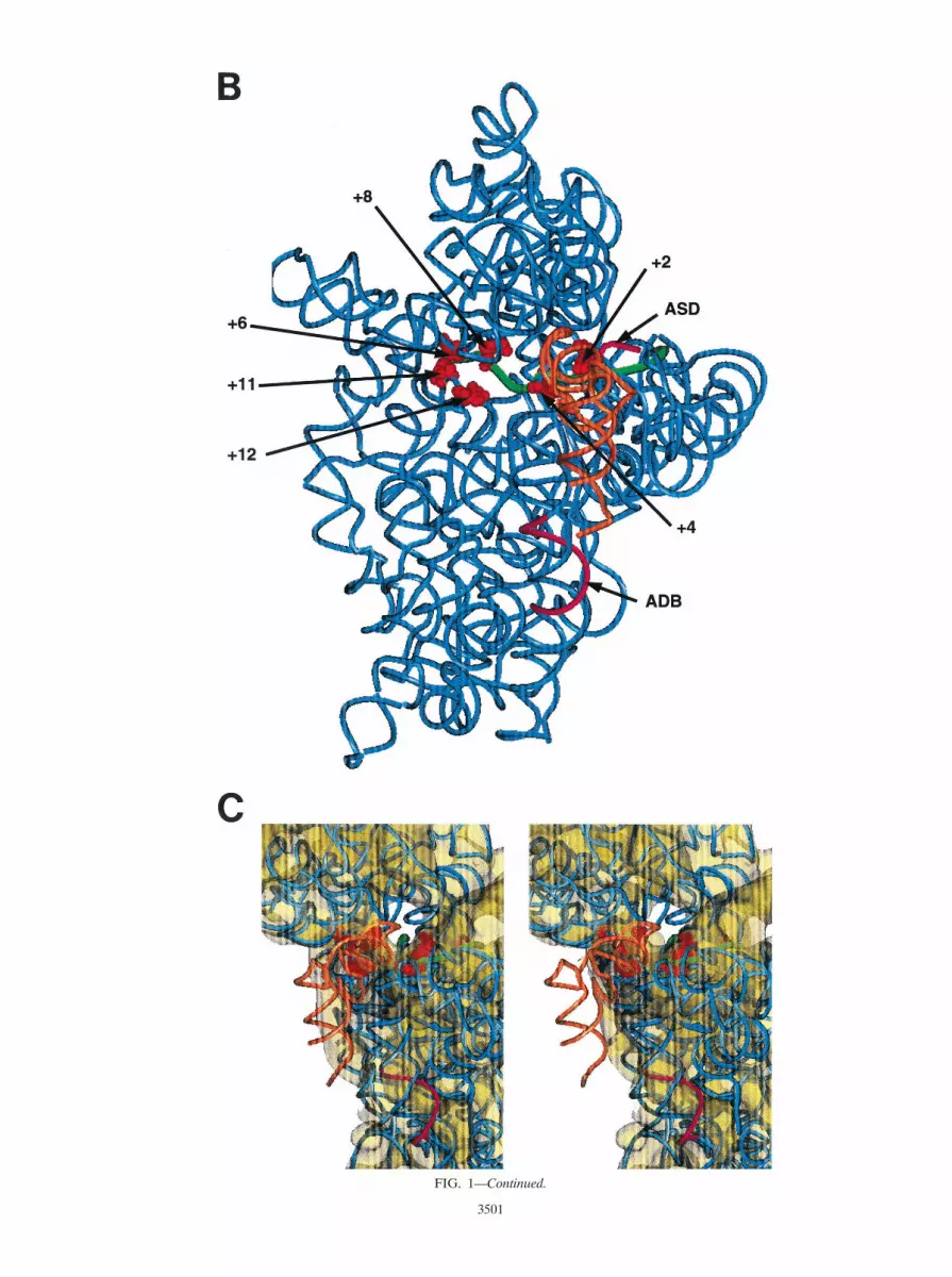
FIG. 1—Continued.
3501

Computer graphics and modeling of helix 44 on the 30S subunit. All of therelevant procedures for modeling of helix 44 on the 30S subunit have beendescribed in detail by Mueller and Brimacombe (24, 25).
RESULTS AND DISCUSSION
Spatial organization of mRNA-db and adb on the 30S sub-unit. The recently published crystal structure of Thermus ther-mophilus 16S rRNA (42) is shown in Fig. 1B. Nucleotides inthe E. coli 16S rRNA which have been cross-linked to down-stream positions (12, 14, etc.) in the mRNA (35) are high-lighted in red as CPK models. It should be noted that attemptsto cross-link the putative adb to mRNA have failed (R. Bri-macombe, unpublished results).
In Fig. 1C, the structure shown in Fig. 1B was added to thecontour of the 30S subunit obtained by electron cryomicros-copy (24). The stereo views show that helix 44 is far from thedecoding site and runs down to the bottom of the interface siteof the E. coli 30S subunit with the adb situated approximatelyin the middle of the body. In the translation initiation complex,the mRNA cross-links are grouped around the “hole,” and partof the putative db on mRNA would pass through the hole.Thus, the putative db is exposed to the solvent site and thewhole shoulder of the body of the 30S subunit is situatedbetween the putative db on the mRNA and the adb in helix 44(Fig. 1C). Taken together with other topographical informa-tion on the 30S ribosome (11, 34, 42), models where the startcodon can be placed in the ribosomal P site while the adjacentdb interacts simultaneously with the adb (38) or where the dbacts synergistically with the SD/aSD interaction (7) are nottenable. These topographical constraints are supported by pre-vious chemical probing experiments which showed that theputative db on l cI mRNA is not protected from modifyingreagents in 70S initiation complexes (31).
An optimal db does not result in 30S subunit binding toleaderless l cI mRNA. Although the topography of the ribo-some apparently excludes that the db can mechanistically sub-stitute for the SD sequence, it seemed worth testing the hy-pothesis that the db-adb interaction could contribute to initialinteractions between the 30S subunit and mRNA prior to for-mation of the translation initiation complex (39). We haveargued that a contribution of the putative db-adb interaction inribosome mRNA binding may be best assessed in the absenceof an SD/aSD interaction (31). Therefore, the relative affinitiesof 30S subunits were determined for leaderless l cI wt mRNAand for a derivative thereof containing an “optimized db” aswell as for a cI mRNA derivative with a 59-terminal extensioncontaining an SD sequence (Fig. 2). l cI34 RNA contains thefirst 34 nt, and 8 consecutive nucleotides are complementary tothe putative adb (Fig. 2). The l cIOptdb comprises 33 nt, ofwhich nt 1 to 15 are fully complementary to the putative adb(Fig. 2). Both were added to a 10-fold molar excess of 30Ssubunits (conditions which reflect the relative affinity constantsof the corresponding complexes). As shown in Fig. 2, only0.37% of cI34 mRNA and 0.30% of cIOptdb mRNA formed abinary complex with 30S subunits. When the cI34SD mRNA(Fig. 2) was used as a binding substrate, approximately 46% ofthis mRNA formed a binary complex with ribosomes. Thus, incontrast to the SD/aSD interaction, an optimal base pairingpotential did not increase the affinity of ribosomes for thecIOptdb mRNA. In other words, an optimized db cannot pro-vide sufficient interactions with the 30S ribosome to stimulateinitial binding of the mRNA to the ribosome. The addition ofa 2-fold molar excess of fMet-tRNAf
Met over ribosomes in-creased the amount of bound cI34 mRNA approximately4-fold, that of cIOptdb mRNA 3-fold, and that of cI34SDmRNA 1.6-fold. Since the topography of the 30S subunits (Fig.
FIG. 2. The cI34, cIOptdb, and cI34SD mRNAs are depicted. The putative db sequences as well as the putative adb in E. coli 16S rRNA areshown in bold. The start codons and the SD sequence in cI34SD mRNA are underlined. The total counts per minute values corrected for thenumber of C’s in each mRNA were set at 100% (mRNA input). Ternary complexes were allowed to form in the presence of a twofold molar excessof fMet-tRNAf
Met over ribosomes.
3502 MOLL ET AL. J. BACTERIOL.
1) should restrict a simultaneous interaction of both the startcodon and anti-codon in the P site and the db-adb, it was notsurprising that the presence of the optimal db did not enhanceternary complex formation (Fig. 2).
The failure of 30S ribosomes to form a binary complex withl cIOptdb mRNA is consistent with kinetic toeprint experi-ments (31) and with studies performed by La Teana et al. (18),who have recently shown that the adb is not accessible to acDNA oligonucleotide while the anti-SD sequence, whichserved as a control, was fully accessible. Ever since the db-adbbase pairing was first proposed, it has been perplexing that itwould require extensive unwinding of the 16S rRNA stemcomprising the adb. Again, La Teana et al. (18) have shown,using chemical probing, that the adb is in a double-strandedconformation and therefore is not available for base pairing.
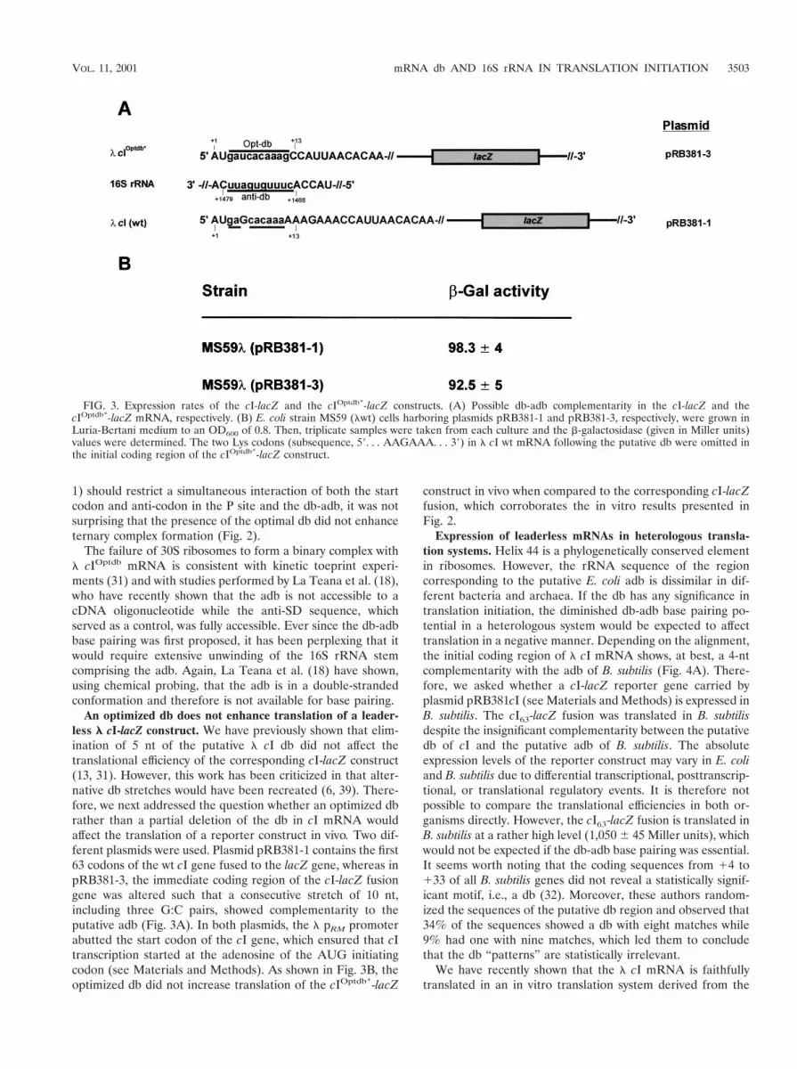
An optimized db does not enhance translation of a leader-less l cI-lacZ construct. We have previously shown that elim-ination of 5 nt of the putative l cI db did not affect thetranslational efficiency of the corresponding cI-lacZ construct(13, 31). However, this work has been criticized in that alter-native db stretches would have been recreated (6, 39). There-fore, we next addressed the question whether an optimized dbrather than a partial deletion of the db in cI mRNA wouldaffect the translation of a reporter construct in vivo. Two dif-ferent plasmids were used. Plasmid pRB381-1 contains the first63 codons of the wt cI gene fused to the lacZ gene, whereas inpRB381-3, the immediate coding region of the cI-lacZ fusiongene was altered such that a consecutive stretch of 10 nt,including three G:C pairs, showed complementarity to theputative adb (Fig. 3A). In both plasmids, the l pRM promoterabutted the start codon of the cI gene, which ensured that cItranscription started at the adenosine of the AUG initiatingcodon (see Materials and Methods). As shown in Fig. 3B, theoptimized db did not increase translation of the cIOptdb*-lacZ
construct in vivo when compared to the corresponding cI-lacZfusion, which corroborates the in vitro results presented inFig. 2.
Expression of leaderless mRNAs in heterologous transla-tion systems. Helix 44 is a phylogenetically conserved elementin ribosomes. However, the rRNA sequence of the regioncorresponding to the putative E. coli adb is dissimilar in dif-ferent bacteria and archaea. If the db has any significance intranslation initiation, the diminished db-adb base pairing po-tential in a heterologous system would be expected to affecttranslation in a negative manner. Depending on the alignment,the initial coding region of l cI mRNA shows, at best, a 4-ntcomplementarity with the adb of B. subtilis (Fig. 4A). There-fore, we asked whether a cI-lacZ reporter gene carried byplasmid pRB381cI (see Materials and Methods) is expressed inB. subtilis. The cI63-lacZ fusion was translated in B. subtilisdespite the insignificant complementarity between the putativedb of cI and the putative adb of B. subtilis. The absoluteexpression levels of the reporter construct may vary in E. coliand B. subtilis due to differential transcriptional, posttranscrip-tional, or translational regulatory events. It is therefore notpossible to compare the translational efficiencies in both or-ganisms directly. However, the cI63-lacZ fusion is translated inB. subtilis at a rather high level (1,050 6 45 Miller units), whichwould not be expected if the db-adb base pairing was essential.It seems worth noting that the coding sequences from 14 to133 of all B. subtilis genes did not reveal a statistically signif-icant motif, i.e., a db (32). Moreover, these authors random-ized the sequences of the putative db region and observed that34% of the sequences showed a db with eight matches while9% had one with nine matches, which led them to concludethat the db “patterns” are statistically irrelevant.
We have recently shown that the l cI mRNA is faithfullytranslated in an in vitro translation system derived from the
FIG. 3. Expression rates of the cI-lacZ and the cIOptdb*-lacZ constructs. (A) Possible db-adb complementarity in the cI-lacZ and thecIOptdb*-lacZ mRNA, respectively. (B) E. coli strain MS59 (lwt) cells harboring plasmids pRB381-1 and pRB381-3, respectively, were grown inLuria-Bertani medium to an OD600 of 0.8. Then, triplicate samples were taken from each culture and the b-galactosidase (given in Miller units)values were determined. The two Lys codons (subsequence, 59. . . AAGAAA. . . 39) in l cI wt mRNA following the putative db were omitted inthe initial coding region of the cIOptdb*-lacZ construct.
VOL. 11, 2001 mRNA db AND 16S rRNA IN TRANSLATION INITIATION 3503
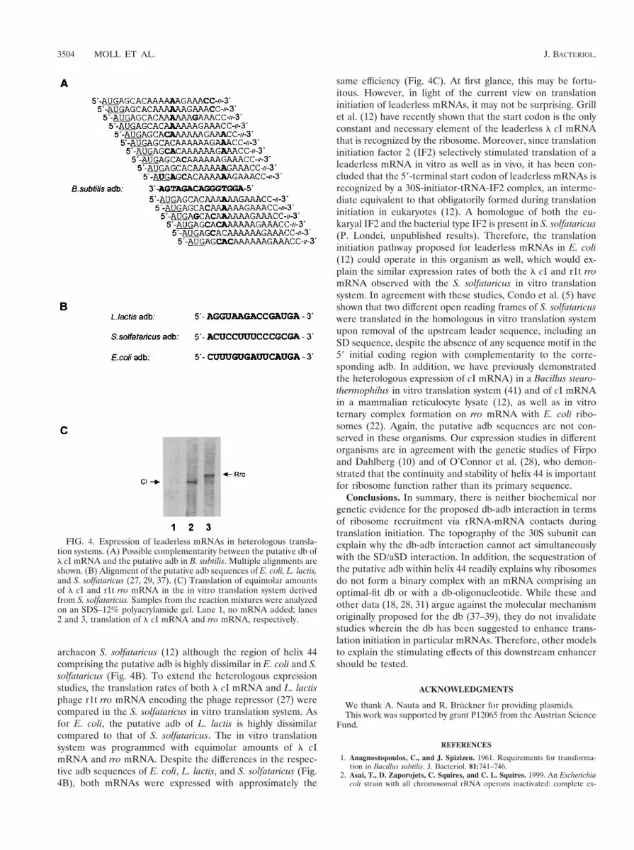
archaeon S. solfataricus (12) although the region of helix 44comprising the putative adb is highly dissimilar in E. coli and S.solfataricus (Fig. 4B). To extend the heterologous expressionstudies, the translation rates of both l cI mRNA and L. lactisphage r1t rro mRNA encoding the phage repressor (27) werecompared in the S. solfataricus in vitro translation system. Asfor E. coli, the putative adb of L. lactis is highly dissimilarcompared to that of S. solfataricus. The in vitro translationsystem was programmed with equimolar amounts of l cImRNA and rro mRNA. Despite the differences in the respec-tive adb sequences of E. coli, L. lactis, and S. solfataricus (Fig.4B), both mRNAs were expressed with approximately the
same efficiency (Fig. 4C). At first glance, this may be fortu-itous. However, in light of the current view on translationinitiation of leaderless mRNAs, it may not be surprising. Grillet al. (12) have recently shown that the start codon is the onlyconstant and necessary element of the leaderless l cI mRNAthat is recognized by the ribosome. Moreover, since translationinitiation factor 2 (IF2) selectively stimulated translation of aleaderless mRNA in vitro as well as in vivo, it has been con-cluded that the 59-terminal start codon of leaderless mRNAs isrecognized by a 30S-initiator-tRNA-IF2 complex, an interme-diate equivalent to that obligatorily formed during translationinitiation in eukaryotes (12). A homologue of both the eu-karyal IF2 and the bacterial type IF2 is present in S. solfataricus(P. Londei, unpublished results). Therefore, the translationinitiation pathway proposed for leaderless mRNAs in E. coli(12) could operate in this organism as well, which would ex-plain the similar expression rates of both the l cI and r1t rromRNA observed with the S. solfataricus in vitro translationsystem. In agreement with these studies, Condo et al. (5) haveshown that two different open reading frames of S. solfataricuswere translated in the homologous in vitro translation systemupon removal of the upstream leader sequence, including anSD sequence, despite the absence of any sequence motif in the59 initial coding region with complementarity to the corre-sponding adb. In addition, we have previously demonstratedthe heterologous expression of cI mRNA) in a Bacillus stearo-thermophilus in vitro translation system (41) and of cI mRNAin a mammalian reticulocyte lysate (12), as well as in vitroternary complex formation on rro mRNA with E. coli ribo-somes (22). Again, the putative adb sequences are not con-served in these organisms. Our expression studies in differentorganisms are in agreement with the genetic studies of Firpoand Dahlberg (10) and of O’Connor et al. (28), who demon-strated that the continuity and stability of helix 44 is importantfor ribosome function rather than its primary sequence.
Conclusions. In summary, there is neither biochemical norgenetic evidence for the proposed db-adb interaction in termsof ribosome recruitment via rRNA-mRNA contacts duringtranslation initiation. The topography of the 30S subunit canexplain why the db-adb interaction cannot act simultaneouslywith the SD/aSD interaction. In addition, the sequestration ofthe putative adb within helix 44 readily explains why ribosomesdo not form a binary complex with an mRNA comprising anoptimal-fit db or with a db-oligonucleotide. While these andother data (18, 28, 31) argue against the molecular mechanismoriginally proposed for the db (37–39), they do not invalidatestudies wherein the db has been suggested to enhance trans-lation initiation in particular mRNAs. Therefore, other modelsto explain the stimulating effects of this downstream enhancershould be tested.
ACKNOWLEDGMENTS
We thank A. Nauta and R. Bruckner for providing plasmids.This work was supported by grant P12065 from the Austrian Science
Fund.
REFERENCES
1. Anagnostopoulos, C., and J. Spizizen. 1961. Requirements for transforma-tion in Bacillus subtilis. J. Bacteriol. 81:741–746.
2. Asai, T., D. Zaporojets, C. Squires, and C. L. Squires. 1999. An Escherichiacoli strain with all chromosomal rRNA operons inactivated: complete ex-
FIG. 4. Expression of leaderless mRNAs in heterologous transla-tion systems. (A) Possible complementarity between the putative db ofl cI mRNA and the putative adb in B. subtilis. Multiple alignments areshown. (B) Alignment of the putative adb sequences of E. coli, L. lactis,and S. solfataricus (27, 29, 37). (C) Translation of equimolar amountsof l cI and r1t rro mRNA in the in vitro translation system derivedfrom S. solfataricus. Samples from the reaction mixtures were analyzedon an SDS–12% polyacrylamide gel. Lane 1, no mRNA added; lanes2 and 3, translation of l cI mRNA and rro mRNA, respectively.
3504 MOLL ET AL. J. BACTERIOL.

change of rRNA genes between bacteria. Proc. Natl. Acad. Sci. USA 96:1971–1976.
3. Bibb, M. J., J. White, J. M. Ward, and G. R. Janssen. 1994. The mRNA forthe 23S rRNA methylase encoded by the ermE gene of Saccharopolysporaerythraea is translated in the absence of a conventional ribosome-binding site.Mol. Microbiol. 14:533–545.
4. Bruckner, R. 1992. A series of shuttle vectors for Bacillus subtilis and Esch-erichia coli. Gene 122:187–192.
5. Condo, I., A. Ciammaruconi, D. Benelli, D. Ruggero, and P. Londei. 1999.Cis-acting signals controlling translational initiation in the thermophilic ar-chaeon Sulfolobus solfataricus. Mol. Microbiol. 34:377–384.
6. Etchegaray, J. P., and M. Inouye. 1999. DB or not DB in translation? Mol.Microbiol. 33:438–439.
7. Etchegaray, J. P., and M. Inouye. 1999. Translational enhancement by anelement downstream of the initiation codon in Escherichia coli. J. Biol.Chem. 274:10079–10085.
8. Etchegaray, J. P., and M. Inouye. 1999. A sequence downstream of theinitiation codon is essential for cold shock induction of cspB in Escherichiacoli. J. Bacteriol. 181:5852–5854.
9. Faxen, M., J. Plumbridge, and L. A. Isakson. 1991. Codon choice andpotential complementarity between mRNA downstream of the initiationcodon and bases 1471–1480 in 16S ribosomal RNA affects expression of glnS.Nucleic Acids Res. 19:5247–5251.
10. Firpo, M. A., and A. E. Dahlberg. 1998. The importance of base pairing in thepenultimate stem of Escherichia coli 16S rRNA for ribosomal subunit asso-ciation. Nucleic Acids Res. 26:2156–2160.
11. Gabashvili, I. S., K. A. Agrawal, C. M. T. Spahn, R. Grassucci, D. I. Svergun,J. Frank, and P. Penczek. 1999. Solution structure of the E. coli 70S ribo-some at 11.5A resolution. Cell 100:537–549.
12. Grill, S., C. O. Gualerzi, P. Londei, and U. Blasi. 2000. Selective stimulationof translation of leaderless mRNA by initiation factor 2: evolutionary impli-cations for translation. EMBO J. 19:4101–4110.
13. Grundling, A. 1997. Diploma thesis. University of Vienna, Vienna, Austria.14. Hall, L., and D. C. Emery. 1991. A rapid and efficient method for site-
directed mutagenesis by PCR, using biotinylated universal primers andstreptavidin-coated magnetic beads. Protein Eng. 4:601.
15. Huttenhofer, A., and H. F. Noller. 1994. Footprinting mRNA-ribosome com-plexes with chemical probes. EMBO J. 13:3892–3901.
16. Ito, K., K. Kawakami, and Y. Nakamura. 1993. Multiple control of Esche-richia coli lysyl-tRNA synthetase expression involves a transcriptional repres-sor and a translational enhancer element. Proc. Natl. Acad. Sci. USA 90:302–306.
17. Lanzer, M., and H. Bujard. 1988. Promoters determine largely the efficiencyof repressor action. Proc. Natl. Acad. Sci. USA 85:8973–8977.
18. La Teana, A., A. Brandi, M. O’Connor, S. Freddi, and C. L. Pon. 2000.Translation during cold adaptation does not involve mRNA-rRNA basepairing through the downstream box. RNA 6:1393–1402.
19. Messing, J., R. Crea., and P. H. Seeburg. 1981. A system for shot gun DNAsequencing. Nucleic Acids Res. 9:309–321.
20. Miller, J. H. 1972. Experiments in molecular genetics. Cold Spring HarborLaboratory Press, Cold Spring Harbor, N.Y.
21. Mitta, M., L. Fang, and M. Inouye. 1997. Deletion analysis of cspA ofEscherichia coli: requirement of the AT-rich UP element for cspA transcrip-tion and the downstream box in the coding region for its cold shock induc-tion. Mol. Microbiol. 26:321–335.
22. Moll, I., A. Resch, and U. Blasi. 1998. Discrimination of 59-terminal startcodons by translation initiation factor 3 is mediated by ribosomal protein S1.FEBS Lett. 436:213–217.
23. Morita, M., M. Kanemori, H. Yanagi, and T. Yura. 1999. Heat-inducedsynthesis of sigma32 in Escherichia coli: structural and functional dissectionof rpoH secondary structure. J. Bacteriol. 181:401–410.
24. Mueller, F., and R. Brimacombe. 1997. A new model for the three-dimen-sional folding of E. coli 16S rRNA. I. Fitting the RNA to a 3D electronmicroscopic map at 20A. J. Mol. Biol. 271:524–544.
25. Mueller, F., and R. Brimacombe. 1997. A new model for the three-dimen-sional folding of E. coli 16S rRNA. II. The RNA-protein interaction data. J.Mol. Biol. 271:545–565.
26. Nagai, H., H. Yuzawa, and T. Yura. 1991. Interplay of two cis-acting mRNAregions in translational control of sigma32 synthesis during the heat shockresponse of E. coli. Proc. Natl. Acad. Sci. USA 88:10515–10519.
27. Nauta, A., D. van Sinderen, H. Karsens, E. Smit, G. Venema, and J. Kok.1996. Inducible gene expression mediated by a repressor-operator systemisolated from Lactococcus lactis bacteriophage r1t. Mol. Microbiol. 19:1331–1341.
28. O’Connor, M., T. Asai, C. L. Squires, and A. E. Dahlberg. 1999. Enhance-ment of translation by the downstream box does not involve mRNA-rRNAbase pairing of mRNA with the penultimate stem sequence of 16S rRNA.Proc. Natl. Acad. Sci. USA 96:8973–8978.
29. Olsen, G. J., N. R. Pace, M. Nuell, B. P. Daine, R. Gupta, and C. R. Woese.1985. Sequence of the 16S rRNA gene from the thermoacidophilic archae-bacterium Sulfolobus solfataricus and its evolutionary implications. J. Mol.Evol. 22:301–307.
30. Resch, A., K. Tedin, A. Graschopf, E. Haggård-Ljungquist, and U. Blasi.1995. Ternary complex formation on leaderless phage mRNA. FEMS Mi-crobiol. Rev. 17:151–157.
31. Resch, A., K. Tedin, A. Grundling, A. Mundlein, and U. Blasi. 1996. Down-stream/anti-downstream-box interactions are dispensable for translation ini-tiation of leaderless mRNAs. EMBO J. 15:4740–4748.
32. Rocha, E. P. C., A. Danchin, and A. Viari. 1999. Translation in B. subtilis:roles and trends of initiation and termination, insights from a genome anal-ysis. Nucleic Acids Res. 27:3567–3576.
33. Sanchez, R., M. Roovers, and N. Glansdorff. 2000. Organization and expres-sion of a Thermus thermophilus arginine cluster: presence of unidentifiedopen reading frames and absence of a Shine-Dalgarno sequence. J. Bacte-riol. 182:5911–5915.
34. Schluenzen, F., A. Tocilj, R. Zarivach, J. Harms, M. Gluehman, D. Janell, A.Bashan, H. Bartels, I. Agmon, F. Franceschi, and A. Yonath. 2000. Structureof functionally activated small ribosomal subunit at 3.3 A resolution. Cell102:615–623.
35. Sergiev, P. V., I. N. Lavrik, V. A. Wlasoff, S. S. Dokudovskaya, O. A.Dontsova, A. A. Bogdanov, and R. Brimacombe. 1997. The path of mRNAthrough the bacterial ribosome: a site-directed cross-linking study using newphotoreactive derivatives of guanosine and uridine. RNA 3:464–475.
36. Shean, C. S., and M. Gottesman. 1992. Translation of the prophage l cItranscript. Cell 70:513–522.
37. Sprengart, M. L., H. P. Fatscher, and E. Fuchs. 1990. The initiation oftranslation in E. coli: apparent base pairing between the 16S rRNA anddownstream sequences of the mRNA. Nucleic Acids Res. 18:1719–1723.
38. Sprengart, M. L., E. Fuchs, and A. G. Porter. 1996. The downstream box: anefficient and independent translation initiation signal in Escherichia coli.EMBO J. 15:665–674.
39. Sprengart, M. L., and A. G. Porter. 1997. Functional importance of RNAinteractions in selection of translation initiation codons. Mol. Microbiol.24:19–28.
40. Steiner, M., W. Lubitz, and U. Blasi. 1993. The missing link in phage lysis ofGram positive bacteria: gene 14 of B. subtilis phage f29 encodes the func-tional homolog of l S protein. J. Bacteriol. 175:1038–1042.
41. Tedin, K., A. Resch, and U. Blasi. 1997. Requirements for ribosomal proteinS1 for translation initiation of mRNAs with and without a 59-leader se-quence. Mol. Microbiol. 25:189–199.
42. Wimberley, B. T., D. E. Brodersen, W. M. Clemons, R. J. Morgan-Warren,A. P. Carter, C. Vornhelm, T. Hartsch, and V. Ramakrishnan. 2000. Struc-ture of the 30S ribosomal subunit. Nature 407:327–339.
43. Winzeler, E., and L. Shapiro. 1997. Translation of the leaderless CaulobacterdnaX mRNA. J. Bacteriol. 179:3981–3988.
44. Wu, C. J., and G. R. Janssen. 1996. Translation of vph mRNA in Strepto-myces lividans and Escherichia coli after removal of the 59-untranslatedleader. Mol. Microbiol. 22:339–355.
VOL. 11, 2001 mRNA db AND 16S rRNA IN TRANSLATION INITIATION 3505


