Embed Size (px)
Citation preview
Evidence for a Xer/dif System for ChromosomeResolution in ArchaeaDiego Cortez1¤, Sophie Quevillon-Cheruel2, Simonetta Gribaldo1, Nicole Desnoues1, Guennadi
Sezonov1,3, Patrick Forterre1,4., Marie-Claude Serre4.*
1 Institut Pasteur, Unite Biologie Moleculaire du Gene chez les Extremophiles, Paris, France, 2 Institut de Biochimie et de Biophysique Moleculaire et Cellulaire, UMR8619-
CNRS, Universite Paris-Sud 11, IFR115, Orsay, France, 3 Universite Pierre et Marie Curie, Paris, France, 4 Institut de Genetique et Microbiologie, Universite Paris-Sud 11,
UMR8621-CNRS, IFR115, Orsay, France
Abstract
Homologous recombination events between circular chromosomes, occurring during or after replication, can generate dimersthat need to be converted to monomers prior to their segregation at cell division. In Escherichia coli, chromosome dimers areconverted to monomers by two paralogous site-specific tyrosine recombinases of the Xer family (XerC/D). The Xerrecombinases act at a specific dif site located in the replication termination region, assisted by the cell division protein FtsK.This chromosome resolution system has been predicted in most Bacteria and further characterized for some species. Archaeahave circular chromosomes and an active homologous recombination system and should therefore resolve chromosomedimers. Most archaea harbour a single homologue of bacterial XerC/D proteins (XerA), but not of FtsK. Therefore, the role ofXerA in chromosome resolution was unclear. Here, we have identified dif-like sites in archaeal genomes by using acombination of modeling and comparative genomics approaches. These sites are systematically located in replicationtermination regions. We validated our in silico prediction by showing that the XerA protein of Pyrococcus abyssi specificallyrecombines plasmids containing the predicted dif site in vitro. In contrast to the bacterial system, XerA can recombine dif sitesin the absence of protein partners. Whereas Archaea and Bacteria use a completely different set of proteins for chromosomereplication, our data strongly suggest that XerA is most likely used for chromosome resolution in Archaea.
Citation: Cortez D, Quevillon-Cheruel S, Gribaldo S, Desnoues N, Sezonov G, et al. (2010) Evidence for a Xer/dif System for Chromosome Resolution inArchaea. PLoS Genet 6(10): e1001166. doi:10.1371/journal.pgen.1001166
Editor: William F. Burkholder, Agency for Science, Technology and Research, Singapore
Received March 26, 2010; Accepted September 17, 2010; Published October 21, 2010
Copyright: � 2010 Cortez et al. This is an open-access article distributed under the terms of the Creative Commons Attribution License, which permitsunrestricted use, distribution, and reproduction in any medium, provided the original author and source are credited.
Funding: This work was supported by funds from the Centre National de la Recherche Scientifique and the University Paris-Sud 11 (UMR8619 and UMR8621). DC wasthe recipient of a fellowship from CONACYT. The funders had no role in study design, data collection and analysis, decision to publish, or preparation of the manuscript.
Competing Interests: The authors have declared that no competing interests exist.
* E-mail: [email protected]
. These authors are joint senior authors on this work.
¤ Current address: Universite de Lausanne, Centre Integratif de Genomique, Le Genopode, Quartier UNIL-Sorge, Lausanne, Switzerland
Introduction
In Bacteria, homologous recombination is essential during DNA
replication to resume stalled replication forks and to repair DNA
double and single strand breaks. Odd numbers of homologous
recombination events between circular chromosomes generate
dimers, which need to be resolved to ensure proper segregation in
daughter cells. In Escherichia coli two paralogous site-specific
tyrosine recombinases XerC and XerD were shown to convert
chromosome dimers to monomers [1] by acting at a specific DNA
recombination site, dif, located close to the replication termination
region [2–4]. Homologues of XerCD are widespread in the
bacterial domain, and dif sites have been characterized in several
Proteobacteria and Firmicutes [5–10]. dif sites are semi-conserva-
tive inverted repeats formed by two arms (Xer protein binding
sites) of 11 base pairs [11], separated by a spacer of 6 bp and are
fairly conserved among Bacteria [5]. In Proteobacteria the XerCD
activity is tightly regulated by the cell division protein FtsK, a
DNA translocase anchored at the division septum [12–15]. In E.
coli, 8 bp G-rich polar sequence elements (KOPS) direct FtsK
translocation on DNA [16–19]. KOPS are oriented from the
origin of replication towards dif where their polarity is precisely
inverted. FtsK DNA translocation is therefore always oriented
towards the dif site and dif sites carried on a chromosome dimer
are brought together at midcell. FtsK further controls chromo-
some dimer resolution by activating XerD activity through
protein-protein interactions [20,21]. In several Lactococcus and
Streptococcus strains, the canonical bacterial XerCD-dif system has
been replaced by a single tyrosine recombinase, XerS (distantly
related to XerCD) whose gene is located next to its specific dif-like
site and localized at the terminus of replication [22]. Strikingly,
this XerS-dif-like system still depends on the KOPS-oriented FtsK
activity to form the synaptic complex for recombination [22].
Archaea harbour circular chromosomes and have an active
homologous recombination system [23]. Therefore, they are
expected to resolve chromosomal dimers to ensure proper
chromosome segregation. It was previously reported that most
archaeal genomes encode a single protein homologous to bacterial
XerCD [24]; however, none encode a FtsK homologue. It is thus
unclear whether archaeal Xer-like proteins (hereafter called
XerA) are involved in chromosome resolution in Archaea, as in
Bacteria.
In order to determine whether XerA is involved in chromosome
resolution, we performed an in silico search for XerA specific
PLoS Genetics | www.plosgenetics.org 1 October 2010 | Volume 6 | Issue 10 | e1001166
recombination dif-like sites in four closely related archaeal
genomes from Thermococcales. We identified a highly conserved
28 bp sequence that shares 14 out of 28 bases with characterized
bacterial dif sites. The predicted dif sites are systematically located
in the replication termination regions of the four genomes. The
same analysis performed on three Sulfolobales genomes revealed
that a similar site is also present in this crenarchaeotal species. We
further identified short polarized sequences that point towards the
predicted dif sites in Thermococcales genomes. We validated the in
silico predictions by showing that a purified recombinant XerA
protein from Pyrococcus abyssi specifically recombines plasmids
carrying the predicted dif site of this archaeon. The recombination
activity did not require the presence of any protein partner, in
contrast to bacterial Xer-mediated recombination. Our data
strongly suggest that XerA is most likely used for chromosome
resolution in Archaea.
Results
The majority (88%) of archaeal genomes sequenced so far
(KEGG database [25]) harbour single orthologues of the bacterial
XerCD recombinases. Alignments of several bacterial XerCD
proteins with XerA proteins from different Archaea revealed that
they share a conserved C-terminal domain where the catalytic site
(Figure S1) is located. The six catalytic residues (R-K-H-R-[H/
W]-Y) characteristic of tyrosine recombinases [26] are perfectly
conserved in archaeal XerA proteins [24].
The more variable outer sequences of the bacterial dif sites are
the place of specific amino-acids/bases contacts that drive protein-
DNA interaction specificity. Several amino acids residues involved
in these contacts were identified, which led to the definition of a
dif-binding region within the C-terminal domain of the Xer
recombinases [27–29]. The dif binding motif is also conserved in
XerA proteins. Notably, the key residues that define binding
specificity for the XerC or XerD binding sites are distinct from
XerC or XerD in XerA (Figure 1A and Figure S1).
In silico identification of dif-like sites in archaeal genomesTo search for conserved putative archaeal dif sites, we selected
as first candidates four closely related genomes of Thermococcales
since their XerA proteins [Pyrococcus abyssi (Pab0255), Pyrococcus
horikoshii (PH1826), Pyrococcus furiosus (PF1868) and Thermococcus
kodakaraensis (TK0777)] are the most similar to bacterial XerCD
(35%–39% identity; Figure S2) among archaeal XerA. The
putative dif-binding motif of these XerA proteins shares numerous
conserved positions with both XerC (10 out of 19 positions) and
XerD (11 out of 19 positions) proteins (Figure 1A). These
remarkable sequence similarities suggest that one may expect to
identify conserved dif-like sequences in these four closely related
archaeal genomes based on known properties of bacterial dif sites.
Finally, XerA proteins are well conserved between these four
species (above 85% similarity, Figure S2). Thermococcales XerA
proteins are thus expected to recognize similar dif sites.
We built an algorithm to search for any potential tyrosine
recombinase-binding site. We searched for imperfect inverted
repeats of 11 to 15 bp separated by spacers ranging from 4 to 10 bp.
A total of 481,319 sequences were recovered after this analysis. In
order to reduce the sequences to one single most likely dif candidate,
we selected only sequences that were conserved above 80%
similarity in the four genomes. We found six sequences fulfilling
this criterion: two were shawn to be spacer sequences of CRISPRs
[30,31], three were imperfect inverted repeats of 11 bp separated by
long spacers (one of 8 bp and two of 10 bp), and only one sequence
in each genome was composed of 11 bp imperfect inverted repeats
separated by a 6 bp spacer (Figure 1B). Strikingly, these sequences
are 100% conserved between P. horikoshii and P. furiosus, have three
mismatches with that of Pyrococcus abyssi and seven with that of T.
kodakaraensis. Moreover, these four predicted sites share many
positions with the bacterial dif consensus sites (Figure 1B). The three
Pyrococcus sites show 14 out of 28 conserved positions of the bacterial
dif-consensus, and the T. kodakaraensis site shows 18 out of 28
(Figure 1B). The same site search was performed on the T.
gammatolerans, T. onnurineus and T. sibiricus genomes and led to the
identification of unique sites showing the same level of conservation
with the bacterial dif-consensus (Figure S3). Further analysis of the
dif sites environment in Thermococcales genomes revealed that all dif
sites are surrounded by conserved flanking sequences (Figure S3).
As in the canonical bacterial model, Thermococcales dif sites
predicted by our analysis were not located next to the xerA genes
(Figure 2 and Figure S4). The position of the xerA genes relative to
the replication origins (oriC) was highly variable (Table S1),
whereas the predicted dif sites were located within the second
quarter of the genome for P. horikoshi, P. furiosus and T. kodakaraensis
(135u, 122u and 130u from oriC, respectively) and in the third
quarter (2142u from oriC) for P. abyssi (Figure 2). In the latter case,
the difference in position could be a consequence of the large
fragment inversion containing oriC that recently occurred in this
species [32]. The conservation of dif site positions relative to oriC in
the four Thermococcales (between 122u and 142u) is especially
striking since these genomes have been extensively rearranged by
chromosome recombination, as indicated by the patterns obtained
from whole genome alignments (Figure S5).
The dif-like sites identified in our analyses do not localize precisely
at 180u from oriC. However, the predicted dif site of P. abyssi is
located into a late replicating fragment of the genome [33]. dif sites
positions are therefore compatible with a localization in the
terminus region of chromosome replication. It is not known if the
two replication forks always meet at the same point in Thermococcales.
A precise site for the terminus of DNA replication in Thermococcales
genomes cannot be predicted by using GC skew analysis because, in
contrast to the sharp peak observed at oriC, the potential termination
Author Summary
Bacteria with circular chromosome and active homologousrecombination systems have to resolve chromosomaldimers before segregation at cell division. In Escherichiacoli, the Xer site-specific recombination system, composedof two recombinases and a specific chromosomal site (dif),is involved in the correct inheritance of the chromosome.The recombination event is tightly regulated by thechromosome translocase FtsK. This chromosome resolu-tion system has been predicted in most bacteria andfurther characterized for some species. Intriguingly, mostarchaea possess a gene coding for a recombinasehomologous to bacterial Xers, but none have homologuesof the bacterial FtsK. We identified the specific target sitesfor archaeal Xer. This site, present in one copy perchromosome, is located in the replication terminationregion and shows sequence similarities with bacterial difsites. In vitro, the archaeal Xer recombines this site in theabsence of protein partner. It has been shown thatDNA–related proteins from Archaea and Eukarya sharea common origin, whereas their analogues in Bacteriahave evolved independently. In this context, Eukarya andArchaea would represent sister groups. Therefore, thepresence of a shared Xer-dif system between Bacteriaand Archaea illustrates the complex origin of modern DNAgenomes.
Xer Recombination in Archaea
PLoS Genetics | www.plosgenetics.org 2 October 2010 | Volume 6 | Issue 10 | e1001166
region appears as a broad distribution [33,34]. The terminus region
appears to be especially prone to chromosomal rearrangement in
Thermococcales [32], possibly explaining this lack of resolution.
We next extended our analysis to archaeal genomes outside of
the Thermococcales group. Using the dif sites and flanking sequences
found in Thermococcales, we constructed a Hidden Markov Model
with HMMER2 [35] and searched other archaeal genomes for
potential dif sequences. As an example, a single statistical-
significant sequence matching the Thermococcales dif sites was found
in the Methanosphaera stadtmanae genome. This site is located at
Figure 1. Alignments of Xer dif-binding motifs and dif sites from Archaea and Bacteria. A. Alignments of the dif binding motifs frombacterial XerC and XerD and archaeal XerA proteins. Amino acids shared between XerD dif binding motifs, XerC dif binding motifs and putative difbinding motifs of archaeal XerA proteins from Thermococcales are boxed. Conserved positions in XerD dif binding motifs are indicated by ‘{’ andconserved positions in XerC dif binding motifs are indicated by ‘{’. The putative dif binding motifs of XerA proteins share several positions with XerDand/or XerC proteins. The key motif for each family (XRX for XerC, XKX for XerA and and RQX for XerD) are shown in bold and shaded in dark gray. B.Predicted Thermococcales dif sites are highly conserved, show very few or no mismatches between the two arms of the inverted repeat and shareseveral positions with the bacterial dif consensus (bold letters). Consensus sequences are indicated according to the IUPAC code.doi:10.1371/journal.pgen.1001166.g001
Xer Recombination in Archaea
PLoS Genetics | www.plosgenetics.org 3 October 2010 | Volume 6 | Issue 10 | e1001166
about 180uC from oriC (Figure S6), and is surrounded by imperfect
inverted repeats. We then selected three Sulfolobales genomes to
search for dif sites in Crenarchaeota. Sulfolobus species possess
multiple replication origins [36,37] raising the possibility that an
alternative to the canonical Xer recombination system may occur
in these organisms. We used the same initial methodology that was
applied to Thermococcales genomes. Unique dif sites were found for
S. acidocaldarius and S. tokodaı, whereas two copies of this site were
found at the same chromosomal location in the S. solfataricus
genome (Figure S7). As opposed to Euryarchaeota, the predicted
dif sites localized close to xerA genes, and were flanked on only one
side by a short conserved sequence of 13 bp.
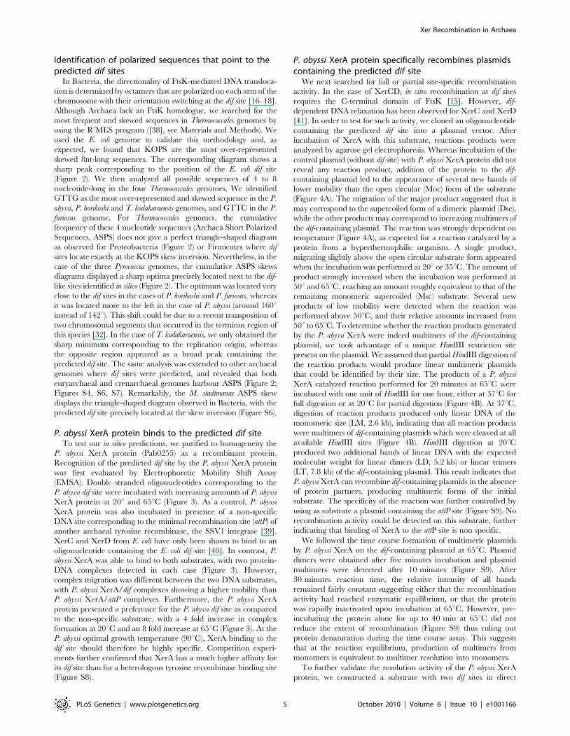
Figure 2. Genomic localization of dif sites and xer genes. The E. coli KOPS cumulative skew produces a pyramidal shaped graphic with the difsite (black and white rectangle) located at the summit (178u from oriC). xerC, xerD and ftsK genes are located in the oriC region. ASPS skew graphicsfrom Thermococcales show rounded shapes where oriC (dark gray diamond) can be localized precisely and where putative regions of replicationtermination (light gray rectangles) can be broadly determined. GTTG is the most skewed 4 nt sequence in P. abyssi, P. horikoshii and T. kodakaraensisgenomes whereas GTTC is the most skewed 4 nt sequence in P. furiosus genome. Thermococcales dif sites (black rectangles) are all located at thesummit of the ASPS skews graphics, at 120–142u from oriC, in putative replication termination regions. The xerA gene (white rectangles) positions aremore variable. Genomic coordinates of oriC, dif and xerA genes can be found in Table S1.doi:10.1371/journal.pgen.1001166.g002
Xer Recombination in Archaea
PLoS Genetics | www.plosgenetics.org 4 October 2010 | Volume 6 | Issue 10 | e1001166
Identification of polarized sequences that point to thepredicted dif sites
In Bacteria, the directionality of FtsK-mediated DNA transloca-
tion is determined by octamers that are polarized on each arm of the
chromosome with their orientation switching at the dif site [16–18].
Although Archaea lack an FtsK homologue, we searched for the
most frequent and skewed sequences in Thermococcales genomes by
using the R’MES program ([38], see Materials and Methods). We
used the E. coli genome to validate this methodology and, as
expected, we found that KOPS are the most over-represented
skewed 8nt-long sequences. The corresponding diagram shows a
sharp peak corresponding to the position of the E. coli dif site
(Figure 2). We then analyzed all possible sequences of 4 to 8
nucleotide-long in the four Thermococcales genomes. We identified
GTTG as the most over-represented and skewed sequence in the P.
abyssi, P. horikoshi and T. kodakaraensis genomes, and GTTC in the P.
furiosus genome. For Thermococcales genomes, the cumulative
frequency of these 4 nucleotide sequences (Archaea Short Polarized
Sequences, ASPS) does not give a perfect triangle-shaped diagram
as observed for Proteobacteria (Figure 2) or Firmicutes where dif
sites locate exactly at the KOPS skew inversion. Nevertheless, in the
case of the three Pyrococcus genomes, the cumulative ASPS skews
diagrams displayed a sharp optima precisely located next to the dif-
like sites identified in silico (Figure 2). The optimum was located very
close to the dif sites in the cases of P. horikoshi and P. furiosus, whereas
it was located more to the left in the case of P. abyssi (around 160uinstead of 142u). This shift could be due to a recent transposition of
two chromosomal segments that occurred in the terminus region of
this species [32]. In the case of T. kodakaraensis, we only obtained the
sharp minimum corresponding to the replication origin, whereas
the opposite region appeared as a broad peak containing the
predicted dif site. The same analysis was extended to other archaeal
genomes where dif sites were predicted, and revealed that both
euryarchaeal and crenarchaeal genomes harbour ASPS (Figure 2;
Figures S4, S6, S7). Remarkably, the M. stadtmanae ASPS skew
displays the triangle-shaped diagram observed in Bacteria, with the
predicted dif site precisely located at the skew inversion (Figure S6).
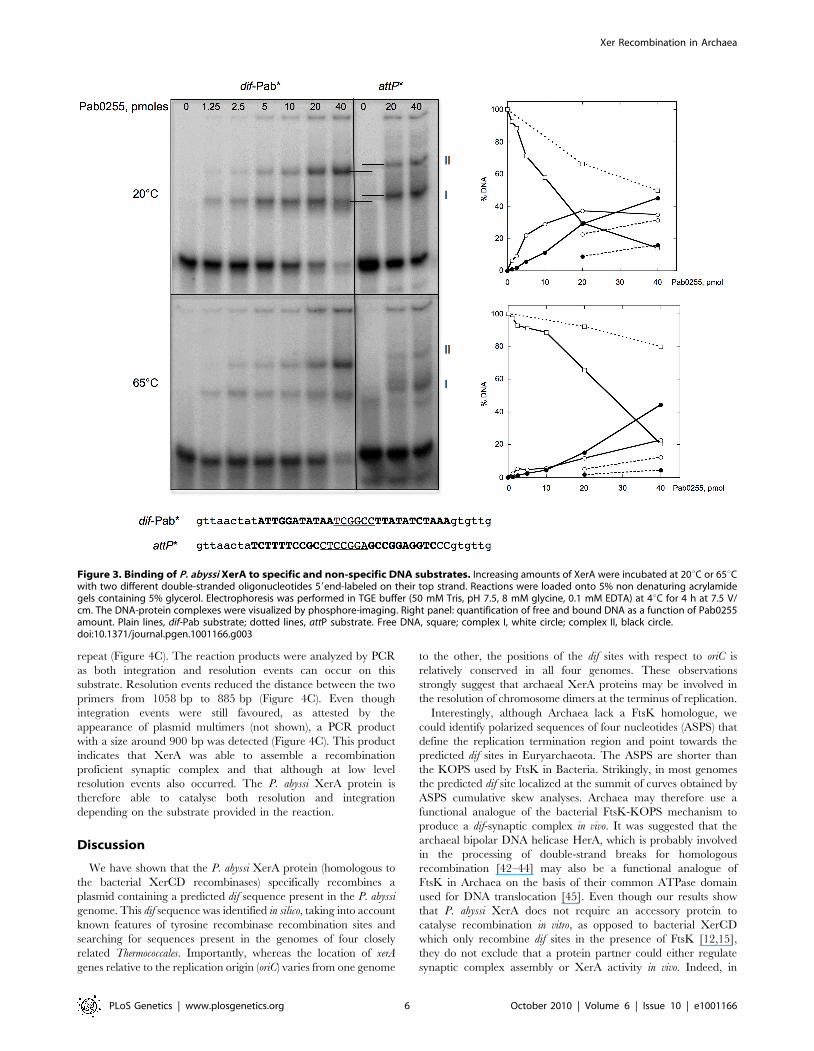
P. abyssi XerA protein binds to the predicted dif siteTo test our in silico predictions, we purified to homogeneity the
P. abyssi XerA protein (Pab0255) as a recombinant protein.
Recognition of the predicted dif site by the P. abyssi XerA protein
was first evaluated by Electrophoretic Mobility Shift Assay
(EMSA). Double stranded oligonucleotides corresponding to the
P. abyssi dif site were incubated with increasing amounts of P. abyssi
XerA protein at 20u and 65uC (Figure 3). As a control, P. abyssi
XerA protein was also incubated in presence of a non-specific
DNA site corresponding to the minimal recombination site (attP) of
another archaeal tyrosine recombinase, the SSV1 integrase [39].
XerC and XerD from E. coli have only been shawn to bind to an
oligonucleotide containing the E. coli dif site [40]. In contrast, P.
abyssi XerA was able to bind to both substrates, with two protein-
DNA complexes detected in each case (Figure 3). However,
complex migration was different between the two DNA substrates,
with P. abyssi XerA/dif complexes showing a higher mobility than
P. abyssi XerA/attP complexes. Furthermore, the P. abyssi XerA
protein presented a preference for the P. abyssi dif site as compared
to the non-specific substrate, with a 4 fold increase in complex
formation at 20uC and an 8 fold increase at 65uC (Figure 3). At the
P. abyssi optimal growth temperature (90uC), XerA binding to the
dif site should therefore be highly specific. Competition experi-
ments further confirmed that XerA has a much higher affinity for
its dif site than for a heterologous tyrosine recombinase binding site
(Figure S8).
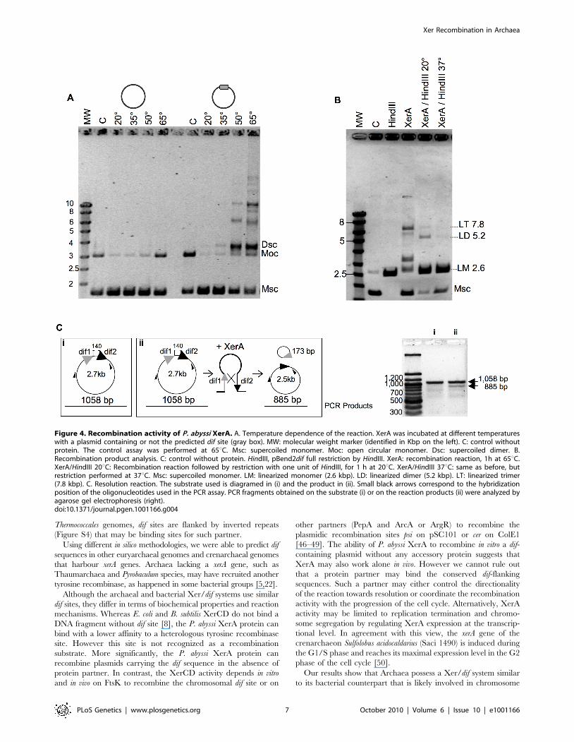
P. abyssi XerA protein specifically recombines plasmidscontaining the predicted dif site
We next searched for full or partial site-specific recombination
activity. In the case of XerCD, in vitro recombination at dif sites
requires the C-terminal domain of FtsK [15]. However, dif-
dependent DNA relaxation has been observed for XerC and XerD
[41]. In order to test for such activity, we cloned an oligonucleotide
containing the predicted dif site into a plasmid vector. After
incubation of XerA with this substrate, reactions products were
analyzed by agarose gel electrophoresis. Whereas incubation of the
control plasmid (without dif site) with P. abyssi XerA protein did not
reveal any reaction product, addition of the protein to the dif-
containing plasmid led to the appearance of several new bands of
lower mobility than the open circular (Moc) form of the substrate
(Figure 4A). The migration of the major product suggested that it
may correspond to the supercoiled form of a dimeric plasmid (Dsc),
while the other products may correspond to increasing multimers of
the dif-containing plasmid. The reaction was strongly dependent on
temperature (Figure 4A), as expected for a reaction catalyzed by a
protein from a hyperthermophilic organism. A single product,
migrating slightly above the open circular substrate form appeared
when the incubation was performed at 20u or 35uC. The amount of
product strongly increased when the incubation was performed at
50u and 65uC, reaching an amount roughly equivalent to that of the
remaining monomeric supercoiled (Msc) substrate. Several new
products of low mobility were detected when the reaction was
performed above 50uC, and their relative amounts increased from
50u to 65uC. To determine whether the reaction products generated
by the P. abyssi XerA were indeed multimers of the dif-containing
plasmid, we took advantage of a unique HindIII restriction site
present on the plasmid. We assumed that partial HindIII digestion of
the reaction products would produce linear multimeric plasmids
that could be identified by their size. The products of a P. abyssi
XerA catalyzed reaction performed for 20 minutes at 65uC were
incubated with one unit of HindIII for one hour, either at 37uC for
full digestion or at 20uC for partial digestion (Figure 4B). At 37uC,
digestion of reaction products produced only linear DNA of the
monomeric size (LM, 2.6 kb), indicating that all reaction products
were multimers of dif-containing plasmids which were cleaved at all
available HindIII sites (Figure 4B). HindIII digestion at 20uCproduced two additional bands of linear DNA with the expected
molecular weight for linear dimers (LD, 5.2 kb) or linear trimers
(LT, 7.8 kb) of the dif-containing plasmid. This result indicates that
P. abyssi XerA can recombine dif-containing plasmids in the absence
of protein partners, producing multimeric forms of the initial
substrate. The specificity of the reaction was further controlled by
using as substrate a plasmid containing the attP site (Figure S9). No
recombination activity could be detected on this substrate, further
indicating that binding of XerA to the attP site is non specific.
We followed the time course formation of multimeric plasmids
by P. abyssi XerA on the dif-containing plasmid at 65uC. Plasmid
dimers were obtained after five minutes incubation and plasmid
multimers were detected after 10 minutes (Figure S9). After
30 minutes reaction time, the relative intensity of all bands
remained fairly constant suggesting either that the recombination
activity had reached enzymatic equilibrium, or that the protein
was rapidly inactivated upon incubation at 65uC. However, pre-
incubating the protein alone for up to 40 min at 65uC did not
reduce the extent of recombination (Figure S9) thus ruling out
protein denaturation during the time course assay. This suggests
that at the reaction equilibrium, production of multimers from
monomers is equivalent to multimer resolution into monomers.
To further validate the resolution activity of the P. abyssi XerA
protein, we constructed a substrate with two dif sites in direct
Xer Recombination in Archaea
PLoS Genetics | www.plosgenetics.org 5 October 2010 | Volume 6 | Issue 10 | e1001166
repeat (Figure 4C). The reaction products were analyzed by PCR
as both integration and resolution events can occur on this
substrate. Resolution events reduced the distance between the two
primers from 1058 bp to 885 bp (Figure 4C). Even though
integration events were still favoured, as attested by the
appearance of plasmid multimers (not shown), a PCR product
with a size around 900 bp was detected (Figure 4C). This product
indicates that XerA was able to assemble a recombination
proficient synaptic complex and that although at low level
resolution events also occurred. The P. abyssi XerA protein is
therefore able to catalyse both resolution and integration
depending on the substrate provided in the reaction.
Discussion
We have shown that the P. abyssi XerA protein (homologous to
the bacterial XerCD recombinases) specifically recombines a
plasmid containing a predicted dif sequence present in the P. abyssi
genome. This dif sequence was identified in silico, taking into account
known features of tyrosine recombinase recombination sites and
searching for sequences present in the genomes of four closely
related Thermococcales. Importantly, whereas the location of xerA
genes relative to the replication origin (oriC) varies from one genome
to the other, the positions of the dif sites with respect to oriC is
relatively conserved in all four genomes. These observations
strongly suggest that archaeal XerA proteins may be involved in
the resolution of chromosome dimers at the terminus of replication.
Interestingly, although Archaea lack a FtsK homologue, we
could identify polarized sequences of four nucleotides (ASPS) that
define the replication termination region and point towards the
predicted dif sites in Euryarchaeota. The ASPS are shorter than
the KOPS used by FtsK in Bacteria. Strikingly, in most genomes
the predicted dif site localized at the summit of curves obtained by
ASPS cumulative skew analyses. Archaea may therefore use a
functional analogue of the bacterial FtsK-KOPS mechanism to
produce a dif-synaptic complex in vivo. It was suggested that the
archaeal bipolar DNA helicase HerA, which is probably involved
in the processing of double-strand breaks for homologous
recombination [42–44] may also be a functional analogue of
FtsK in Archaea on the basis of their common ATPase domain
used for DNA translocation [45]. Even though our results show
that P. abyssi XerA does not require an accessory protein to
catalyse recombination in vitro, as opposed to bacterial XerCD
which only recombine dif sites in the presence of FtsK [12,15],
they do not exclude that a protein partner could either regulate
synaptic complex assembly or XerA activity in vivo. Indeed, in
Figure 3. Binding of P. abyssi XerA to specific and non-specific DNA substrates. Increasing amounts of XerA were incubated at 20uC or 65uCwith two different double-stranded oligonucleotides 59end-labeled on their top strand. Reactions were loaded onto 5% non denaturing acrylamidegels containing 5% glycerol. Electrophoresis was performed in TGE buffer (50 mM Tris, pH 7.5, 8 mM glycine, 0.1 mM EDTA) at 4uC for 4 h at 7.5 V/cm. The DNA-protein complexes were visualized by phosphore-imaging. Right panel: quantification of free and bound DNA as a function of Pab0255amount. Plain lines, dif-Pab substrate; dotted lines, attP substrate. Free DNA, square; complex I, white circle; complex II, black circle.doi:10.1371/journal.pgen.1001166.g003
Xer Recombination in Archaea
PLoS Genetics | www.plosgenetics.org 6 October 2010 | Volume 6 | Issue 10 | e1001166
Thermococcales genomes, dif sites are flanked by inverted repeats
(Figure S4) that may be binding sites for such partner.
Using different in silico methodologies, we were able to predict dif
sequences in other euryarchaeal genomes and crenarchaeal genomes
that harbour xerA genes. Archaea lacking a xerA gene, such as
Thaumarchaea and Pyrobaculum species, may have recruited another
tyrosine recombinase, as happened in some bacterial groups [5,22].
Although the archaeal and bacterial Xer/dif systems use similar
dif sites, they differ in terms of biochemical properties and reaction
mechanisms. Whereas E. coli and B. subtilis XerCD do not bind a
DNA fragment without dif site [8], the P. abyssi XerA protein can
bind with a lower affinity to a heterologous tyrosine recombinase
site. However this site is not recognized as a recombination
substrate. More significantly, the P. abyssi XerA protein can
recombine plasmids carrying the dif sequence in the absence of
protein partner. In contrast, the XerCD activity depends in vitro
and in vivo on FtsK to recombine the chromosomal dif site or on
other partners (PepA and ArcA or ArgR) to recombine the
plasmidic recombination sites psi on pSC101 or cer on ColE1
[46–49]. The ability of P. abyssi XerA to recombine in vitro a dif-
containing plasmid without any accessory protein suggests that
XerA may also work alone in vivo. However we cannot rule out
that a protein partner may bind the conserved dif-flanking
sequences. Such a partner may either control the directionality
of the reaction towards resolution or coordinate the recombination
activity with the progression of the cell cycle. Alternatively, XerA
activity may be limited to replication termination and chromo-
some segregation by regulating XerA expression at the transcrip-
tional level. In agreement with this view, the xerA gene of the
crenarchaeon Sulfolobus acidocaldarius (Saci 1490) is induced during
the G1/S phase and reaches its maximal expression level in the G2
phase of the cell cycle [50].
Our results show that Archaea possess a Xer/dif system similar
to its bacterial counterpart that is likely involved in chromosome
Figure 4. Recombination activity of P. abyssi XerA. A. Temperature dependence of the reaction. XerA was incubated at different temperatureswith a plasmid containing or not the predicted dif site (gray box). MW: molecular weight marker (identified in Kbp on the left). C: control withoutprotein. The control assay was performed at 65uC. Msc: supercoiled monomer. Moc: open circular monomer. Dsc: supercoiled dimer. B.Recombination product analysis. C: control without protein. HindIII, pBend2dif full restriction by HindIII. XerA: recombination reaction, 1h at 65uC.XerA/HindIII 20uC: Recombination reaction followed by restriction with one unit of HindIII, for 1 h at 20uC. XerA/HindIII 37uC: same as before, butrestriction performed at 37uC. Msc: supercoiled monomer. LM: linearized monomer (2.6 kbp). LD: linearized dimer (5.2 kbp). LT: linearized trimer(7.8 kbp). C. Resolution reaction. The substrate used is diagramed in (i) and the product in (ii). Small black arrows correspond to the hybridizationposition of the oligonucleotides used in the PCR assay. PCR fragments obtained on the substrate (i) or on the reaction products (ii) were analyzed byagarose gel electrophoresis (right).doi:10.1371/journal.pgen.1001166.g004
Xer Recombination in Archaea
PLoS Genetics | www.plosgenetics.org 7 October 2010 | Volume 6 | Issue 10 | e1001166
dimer resolution. However, the archaeal Xer system displays
differences, such as the involvement of a unique protein and the
ability to perform site-specific recombination in vitro in the absence
of accessory proteins. To get a better view of the evolution of the
Xer system, we performed a phylogenetic analysis including
bacterial and archaeal Xer proteins and a subset of bacteriophage
encoded tyrosine recombinases (Figure 5). Unfortunately, the
resulting tree is not resolved at most basal nodes, preventing a
clear view of the evolutionary relationships of these proteins.
However, it shows that bacterial and archaeal homologues are not
intermixed, indicating that no recent horizontal gene transfer has
occurred between domains. Our data thus suggest that a Xer/dif
system was present in the common ancestor of Archaea and
Bacteria, suggesting that this ancestor had a circular double-
stranded DNA genome. However, this raises further issues such as
why the replication machinery of Archaea and Bacteria are now
composed of non homologous proteins [51]. Alternatively,
homologous viral tyrosine recombinases may have been recruited
independently in Archaea and Bacteria to be used as Xer/dif
systems after transition from RNA to DNA genomes [52]. In any
case, the presence of a shared Xer-dif system in Bacteria and
Archaea illustrates the complex origin of modern DNA genomes.
Further studies of Xer-dif systems in different archaeal and
bacterial groups will be now necessary to test alternative scenarios
for the origin and evolution of Xer proteins.
Materials and Methods
Identification of dif candidatesA model searched for all inverted repeats (11 to 15bp) separated
by a spacer (4 to 10bp) present in non-coding genomic regions. A
consensus sequence was deduced from the alignment of predicted
dif sites and represented as a sequence logo [53].
Skew analysesStatistical significant skewed words in the four Thermococcales
genomes were determined using the R’MES program (http://mig.
jouy.inra.fr/logiciels/rmes/) [38]. Since R’MES reads only one
single strand of DNA at time, we artificially defined several ends of
replication, starting at 120u, and then moving by 5u steps up to
200u from oriC. From these artificially-defined ends of replication
to oriC we reverse complemented the genomic sequence. The most
skewed word for each analyzed genome was selected by
comparing the R’MES results. Cumulative skews were calculated
using the formula:
Skew~X
word{word complementð Þ=
wordzword complementð Þ:
Genome alignmentsGenomes of the four Thermococcales where aligned by dot-plot
analyses based on BlastP searches (e-value of 1610210).
Homology searches and phylogenetic analysisXer homologues were searched by BLASTP at the NCBI
(http://www.ncbi.nlm.nih.gov/) against complete sequenced ar-
chaeal genomes using E. coli XerC and XerD proteins as query
(threshold of 16e210). The retrieved homologues were aligned using
Muscle and a specific HMM profile was calculated for exhaustive
detection of homologues. Using the HMMER program (http://
hmmer.janelia.org/) we performed iterative searches until no new
homologues were detected in the archaeal genomes. From the
resulting dataset, a preliminary phylogenetic analysis allowed to
select 62 representative XerC and XerD sequences from several
bacterial species and several tyrosine recombinases from plasmids/
viruses or from mobile elements integrated into cellular genomes.
The final alignment was trimmed to remove ambiguously aligned
positions, leading to 222 conserved residues for phylogenetic
analysis. A maximum likelihood tree was obtained by using PHYML
[54], with the WAG evolutionary model including correction for
heterogeneity of evolutionary rates (4 categories+invariant) and
statistical support at nodes was calculated by non parametric
bootstrap on 100 resampling of the original dataset by PHYML.
Cloning, expression, and purification of Pab0255The Pab0255 gene was amplified by PCR using P. abyssi geno-
mic DNA as a template, Phusion high-fidelity DNA polymerase
(Finnzyme) and the following primers:
59-GGGAACATATGCACCATCACCATCACCATGAGGA-
GAGGGAGGAGAGAGTGAGGGATGATACAATTG-39
59-TTTTTGCGGCCGCTTAGGAACCCCCGATG-39
The forward primer allows the addition of six histidine codons
in frame with the ATG start codon (underlined). The PCR
product was digested by NdeI and NotI and cloned into a derivative
of a pET vector (Novagen). The resulting recombinant plasmid
was sequenced prior to being transformed into the E. coli
expression strain Rosetta(DE3)pLys (Novagen).
Cells were grown in 2xYT medium (BIO101Inc.) at 37uC to
A600nm = 1 and expression of Pab0255 was induced by the
addition of 0.5 mM IPTG (final concentration). Four hours after
induction, cells were harvested by centrifugation, and the pellets
resuspended in 40 ml of 50 mM Tris-HCl, pH 8.0, 1 M NaCl and
5 mM b-mercaptoethanol and stored at 220uC. Cells were lysed
by sonication and centrifuged at 13, 0006 g for 30 min at 25uC.
The supernatant was collected and heated for 15 minutes at 70uC.
After centrifugation at 13,0006 g, the supernatant was loaded
onto a Ni2+ affinity column (Ni-NTA agarose, Qiagen) pre-
equilibrated in the same buffer. The His-tagged Pab0255 was
eluted at 20 mM imidazole, and loaded onto a 2 ml HiTrap
Heparin (Amersham Biosciences) column pre-equilibrated in a
buffer containing 50 mM Tris-HCl pH 7.0, 200 mM NaCl,
1 mM DTT. A NaCl linear gradient (200 mM to 2 M) was
developed, and the protein eluted at about 800 mM NaCl. Finally,
the protein was loaded onto a cation-exchange SP Sepharose
column (Amersham Biosciences) pre-equilibrated in the same
buffer, and eluted by a NaCl linear gradient. The purified protein
was dialysed against 50 mM Tris pH7.0, 300 mM NaCl, 50%
glycerol prior to being stored at 220uC.
Substrate preparationThe following 43 nt long oligonucleotides containing the top
and bottom strands of the predicted dif site from P. abyssi (Pab) or
minimal attP site from SSV1 [39] were purchased from
Eurogentec:
Pab-Dif-Top 59-gttaactatATTGGATATAATCGGCCTTA-
TATCTAAAgtgttg-39
Pab-Dif-Bottom 59-caacacTTTAGATATAAGGCCGATTA
TATCCAATatagttaac-39
attP-Top 59-gttaactaTCTTTTCCGCCTCCGGAGCCGGA
GGTCCCgtgttg-39
attP-Bottom 59-caacacGGGACCTCCGGCTCCGGAGGC
GGAAAAGAtagttaac-39
For binding assays, top strand oligonucleotides were 59-end
labeled by using [g-32P]ATP and T4 polynucleotide kinase.
Unincorporated nucleotides were removed by spin dialysis, and
the labeled oligonucleotide was then hybridized with a 2-fold
Xer Recombination in Archaea
PLoS Genetics | www.plosgenetics.org 8 October 2010 | Volume 6 | Issue 10 | e1001166
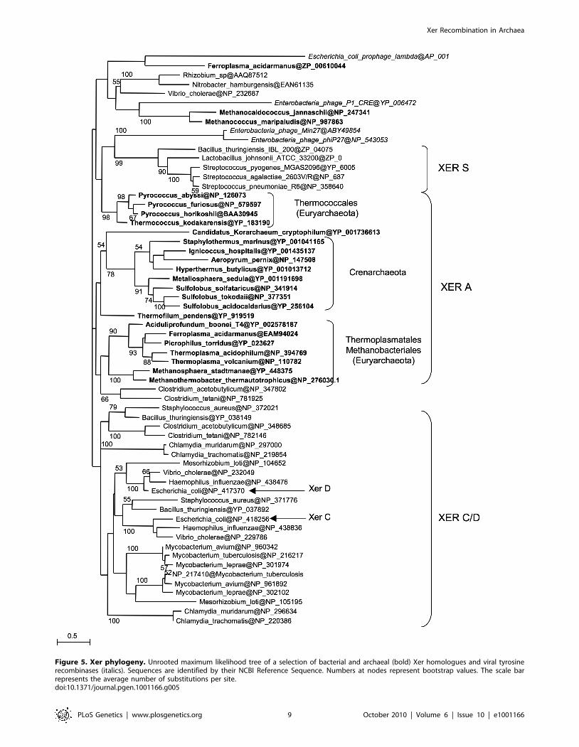
Figure 5. Xer phylogeny. Unrooted maximum likelihood tree of a selection of bacterial and archaeal (bold) Xer homologues and viral tyrosinerecombinases (italics). Sequences are identified by their NCBI Reference Sequence. Numbers at nodes represent bootstrap values. The scale barrepresents the average number of substitutions per site.doi:10.1371/journal.pgen.1001166.g005
Xer Recombination in Archaea
PLoS Genetics | www.plosgenetics.org 9 October 2010 | Volume 6 | Issue 10 | e1001166
excess of unlabeled complementary strand in TE buffer (10 mM
Tris, pH 8.0, 1 mM EDTA).
Electrophoretic mobility shift assaysThe DNA binding reactions were carried out in 20 ml of a
mixture composed of 0.5 mM 59-end labeled dif substrate or attP
substrate, increasing amounts of Pab0255 in a binding buffer
composed of 50 mM Tris pH 7.5, 30 mM NaCl and 0.5 mg
poly(dIdC).poly(dIdC). Incubation was performed for 30 min at
either 20uC or 65uC, and then 5 ml of 56 loading buffer (10 mM
Tris pH 7.5, 1 mM EDTA, 20% glycerol, 0.1 mg/ml BSA, 0.1%
xylene cyanol) was added to the binding reactions. The samples
were loaded onto 8% polyacrylamide gels (30:0.5 acrylamide:bi-
sacrylamide), and electrophoresis performed in 16 TGE buffer
(50 mM Tris, 8 mM Glycine, 0.1 mM EDTA) at 4uC for 4 h at
7 V/cm. The DNA-protein complexes were visualized by
autoradiography and phosphorimaging.
Recombination assaysThe 43 bp double stranded oligonucleotide containing the
predicted P. abyssi dif and the 43 bp double stranded oligonucleotide
containing the attP site were cloned into the pBend2 (2.6 Kbp)
vector [55]. pBend2, pBend2-dif and pBend2-attP plasmids were
purified on CsCl gradients. Recombination reactions were
performed in 20 ml of reaction mixture consisting of 30 mM Tris
pH 7.5, 50 mg/ml bovine serum albumin, 50 mM NaCl, 500 ng of
plasmid and 40 pmol of XerA protein. The reaction mixture was
incubated at 65uC (unless otherwise stated) and at the times
indicated, quenched with SDS (0.5% final) and 106 loading buffer
(100 mM EDTA, 5% SDS, 40% glycerol, 0.35% Bromphenol blue)
was added. Reaction mixes were loaded on a 1.2% agarose gel, and
electrophoresis performed in 16TAE buffer at room temperature
for 3 hr at 4 V/cm with buffer circulation. DNA was visualized by
staining with ethidium bromide.
Supporting Information
Figure S1 Alignment of the C-terminal domain of Xer proteins
from the XerD, XerC, XerA and XerS subfamilies. Left panel: dif
binding motif alignment. The XerA putative dif binding motif
show high residues conservation with both XerC and XerD motifs.
Thermococcales XerA harbour the XerC ‘XRX’ motif signature.
XerS proteins show very few residues conserved, meaning that
they belong to other tyrosine recombinase subfamily. Right panel:
catalytic domain of tyrosine recombinases. Catalytic residues are
highlighted in white bold lettering. Note that two catalytic residues
apart from the highly conserved motif are not represented here.
Found at: doi:10.1371/journal.pgen.1001166.s001 (0.09 MB PDF)
Figure S2 Xer similarity scores. A. Top five E. coli XerC and
XerD matches in complete sequenced archaeal genomes. B.
Similarities between XerA from Thermococcales.
Found at: doi:10.1371/journal.pgen.1001166.s002 (0.06 MB PDF)
Figure S3 Thermococcales predicted dif sites and conserved
flanking regions. Alignment of predicted dif sites and conserved
flanking sequences. The flanking sequences are approximately
23 bp long and AT rich. A consensus sequence was deduced and is
represented as a sequence logo (see [53] in the main text).
Found at: doi:10.1371/journal.pgen.1001166.s003 (0.08 MB PDF)
Figure S4 Genomic localization of dif sites and xer genes. ASPS
skew graphics from T. sibiricus, T. onnurineus and T. gammatolerans.
TGGT is the most skewed sequence (ASPS) for all species.
Symbols are as in Figure 2. Genomic coordinates of oriC, dif and
xerA genes can be found in Table S1.
Found at: doi:10.1371/journal.pgen.1001166.s004 (0.11 MB PDF)
Figure S5 Whole genome alignments of the four Thermococcales
genomes. A. Alignment of P. horikoshii (X-axis) and P. abyssi (Y-axis)
genomes shows that they share several regions with conserved
gene order. dif sites (circle) are located in a relatively well-
conserved region at 135u from oriC (triangle) in P. horikoshii and at
142u from oriC in P. abyssi. The xerA gene (square) genomic position
is indicated. Regions where replication may end are indicated by
dark rectangles on the axes and delimited by doted lines. B,C.
Alignments of P. furiosus and P. abyssi genomes (B) and of T.
kodakaraensis and P. abyssi genomes (C) reveal an extensive gene
order loss. However dif relative positions with respect to oriC are
maintained in all these genomes (respectively 122u and 130u from
oriC in the P. furiosus and T. kodakaraensis genomes).
Found at: doi:10.1371/journal.pgen.1001166.s005 (0.31 MB PDF)
Figure S6 Identification and localization of the M. stadtmanae dif
site. A single statistical-significant sequence matching the Thermo-
coccales dif sites was found in M. stadtmanae by using HMM search.
The dif candidate localizes at the ASPS skew inversion.
Found at: doi:10.1371/journal.pgen.1001166.s006 (0.09 MB PDF)
Figure S7 Sulfolobales dif sites. By using the methodology
described in the main text of this article on S. solfataricus, S.
acidocaldarius and S. tokodaii genomes, one single sequence that fits
all of the requirements (two inverted repeats separated by a spacer
of 4–8 base pairs, highly conserved between the three genomes
and located inside intergenic regions) was found. This potential dif
candidate is present only once in S. acidocaldarius and S. tokodaii, but
has two copies (only one highly conserved), at the same
chromosomal location in S. solfataricus genome.
Found at: doi:10.1371/journal.pgen.1001166.s007 (0.10 MB PDF)
Figure S8 Binding specificity of P. abyssi XerA to specific and
non-specific DNA substrates. 40 pmoles of XerA were incubated
with dif-Pab or attP substrates at 20uC with increasing amounts of
non specific competitor poly(dIdC)2. Bottom panel: quantification
of free and bound DNA as a function of poly(dIdC)2 amount. Plain
lines, dif-Pab substrate; dotted lines, attP substrate. Free DNA,
square; complex I, white circle; complex II, black circle.
Found at: doi:10.1371/journal.pgen.1001166.s008 (0.14 MB PDF)
Figure S9 XerA enzymatic properties. A. XerA substrate
specificity. The three substrates were incubated for 1 hr at 65uCwith or without 10 pmol of XerA. Recombination products are
only observed on the pBend2-dif substrate. B: time course of XerA-
mediated recombination at 65uC. C: XerA was pre-incubated at
different times at 65uC and then mixed with the dif-containing
plasmid for one hour at 65uC. No difference in activity is observed
between the different lanes, indicating that XerA is stable for more
than one hour at 65uC.
Found at: doi:10.1371/journal.pgen.1001166.s009 (0.25 MB PDF)
Table S1 Specific genomic positions of oriC-cdc6, xerA genes and
dif sites in Thermococcales genomes.
Found at: doi:10.1371/journal.pgen.1001166.s010 (0.06 MB PDF)
Acknowledgments
We thank Sophie Schbath, Meriem El Karoui, Tamara Basta, and Didier
Mazel for helpful discussions.
Author Contributions
Conceived and designed the experiments: DC SQC SG PF MCS.
Performed the experiments: DC SQC SG ND GS MCS. Analyzed the
data: DC SQC SG GS PF MCS. Wrote the paper: DC PF MCS.
Xer Recombination in Archaea
PLoS Genetics | www.plosgenetics.org 10 October 2010 | Volume 6 | Issue 10 | e1001166
References
1. Blakely G, May G, McCulloch R, Arciszewska LK, Burke M, et al. (1993) Tworelated recombinases are required for site-specific recombination at dif and cer in
E. coli K12. Cell 75: 351–361.2. Blakely G, Colloms S, May G, Burke M, Sherratt D (1991) Escherichia coli XerC
recombinase is required for chromosomal segregation at cell division. New Biol3: 789–798.
3. Clerget M (1991) Site-specific recombination promoted by a short DNA segment
of plasmid R1 and by a homologous segment in the terminus region of theEscherichia coli chromosome. New Biol 3: 780–788.
4. Kuempel PL, Henson JM, Dircks L, Tecklenburg M, Lim DF (1991) dif, a recA-independent recombination site in the terminus region of the chromosome of
Escherichia coli. New Biol 3: 799–811.
5. Carnoy C, Roten CA (2009) The dif/Xer recombination systems inproteobacteria. PLoS One 4: e6531. doi:10.1371/journal.pone.0006531.
6. Jensen RB (2006) Analysis of the terminus region of the Caulobacter crescentus
chromosome and identification of the dif site. J Bacteriol 188: 6016–6019.
7. Neilson L, Blakely G, Sherratt DJ (1999) Site-specific recombination at dif by
Haemophilus influenzae XerC. Mol Microbiol 31: 915–926.8. Sciochetti SA, Piggot PJ, Blakely GW (2001) Identification and characterization
of the dif Site from Bacillus subtilis. J Bacteriol 183: 1058–1068.9. Val ME, Kennedy SP, El Karoui M, Bonne L, Chevalier F, et al. (2008) FtsK-
dependent dimer resolution on multiple chromosomes in the pathogen Vibrio
cholerae. PLoS Genet 4: e1000201. doi:10.1371/journal.pgen.1000201.
10. Yen MR, Lin NT, Hung CH, Choy KT, Weng SF, et al. (2002) oriC region and
replication termination site, dif, of the Xanthomonas campestris pv. campestris 17chromosome. Appl Environ Microbiol 68: 2924–2933.
11. Blakely GW, Sherratt DJ (1994) Interactions of the site-specific recombinasesXerC and XerD with the recombination site dif. Nucleic Acids Res 22:
5613–5620.
12. Aussel L, Barre FX, Aroyo M, Stasiak A, Stasiak AZ, et al. (2002) FtsK is a DNAmotor protein that activates chromosome dimer resolution by switching the
catalytic state of the XerC and XerD recombinases. Cell 108: 195–205.13. Barre FX, Aroyo M, Colloms SD, Helfrich A, Cornet F, et al. (2000) FtsK
functions in the processing of a Holliday junction intermediate during bacterialchromosome segregation. Genes Dev 14: 2976–2988.
14. Ip SC, Bregu M, Barre FX, Sherratt DJ (2003) Decatenation of DNA circles by
FtsK-dependent Xer site-specific recombination. EMBO J 22: 6399–6407.15. Steiner W, Liu G, Donachie WD, Kuempel P (1999) The cytoplasmic domain of
FtsK protein is required for resolution of chromosome dimers. Mol Microbiol31: 579–583.
16. Bigot S, Saleh OA, Lesterlin C, Pages C, El Karoui M, et al. (2005) KOPS:
DNA motifs that control E. coli chromosome segregation by orienting the FtsKtranslocase. EMBO J 24: 3770–3780.
17. Corre J, Louarn JM (2002) Evidence from terminal recombination gradients thatFtsK uses replichore polarity to control chromosome terminus positioning at
division in Escherichia coli. J Bacteriol 184: 3801–3807.18. Levy O, Ptacin JL, Pease PJ, Gore J, Eisen MB, et al. (2005) Identification of
oligonucleotide sequences that direct the movement of the Escherichia coli FtsK
translocase. Proc Natl Acad Sci U S A 102: 17618–17623.19. Pease PJ, Levy O, Cost GJ, Gore J, Ptacin JL, et al. (2005) Sequence-directed
DNA translocation by purified FtsK. Science 307: 586–590.20. Sivanathan V, Allen MD, de Bekker C, Baker R, Arciszewska LK, et al. (2006)
The FtsK gamma domain directs oriented DNA translocation by interacting
with KOPS. Nat Struct Mol Biol 13: 965–972.21. Yates J, Zhekov I, Baker R, Eklund B, Sherratt DJ, et al. (2006) Dissection of a
functional interaction between the DNA translocase, FtsK, and the XerDrecombinase. Mol Microbiol 59: 1754–1766.
22. Le Bourgeois P, Bugarel M, Campo N, Daveran-Mingot ML, Labonte J, et al.(2007) The unconventional Xer recombination machinery of Streptococci/
Lactococci. PLoS Genet 3: e117. doi:10.1371/journal.pgen.0030117.
23. Haldenby S, White MF, Allers T (2009) RecA family proteins in archaea: RadAand its cousins. Biochem Soc Trans 37: 102–107.
24. Serre MC, Duguet M (2003) Enzymes that cleave and religate DNA at hightemperature: the same story with different actors. Prog Nucleic Acid Res Mol
Biol 74: 37–81.
25. Kanehisa M, Goto S, Kawashima S, Nakaya A (2002) The KEGG databases atGenomeNet. Nucleic Acids Res 30: 42–46.
26. Sherratt DJ, Wigley DB (1998) Conserved themes but novel activities inrecombinases and topoisomerases. Cell 93: 149–152.
27. Cao Y, Hallet B, Sherratt DJ, Hayes F (1997) Structure-function correlations in
the XerD site-specific recombinase revealed by pentapeptide scanningmutagenesis. J Mol Biol 274: 39–53.
28. Spiers AJ, Sherratt DJ (1997) Relating primary structure to function in theEscherichia coli XerD site-specific recombinase. Mol Microbiol 24: 1071–1082.
29. Subramanya HS, Arciszewska LK, Baker RA, Bird LE, Sherratt DJ, et al. (1997)Crystal structure of the site-specific recombinase, XerD. EMBO J 16:
5178–5187.
30. Lillestol RK, Redder P, Garrett RA, Brugger K (2006) A putative viral defence
mechanism in archaeal cells. Archaea 2: 59–72.
31. Makarova KS, Grishin NV, Shabalina SA, Wolf YI, Koonin EV (2006) A
putative RNA-interference-based immune system in prokaryotes: computational
analysis of the predicted enzymatic machinery, functional analogies with
eukaryotic RNAi, and hypothetical mechanisms of action. Biol Direct 1: 7.
32. Zivanovic Y, Lopez P, Philippe H, Forterre P (2002) Pyrococcus genome
comparison evidences chromosome shuffling-driven evolution. Nucleic Acids
Res 30: 1902–1910.
33. Myllykallio H, Lopez P, Lopez-Garcia P, Heilig R, Saurin W, et al. (2000)
Bacterial mode of replication with eukaryotic-like machinery in a hyperthermo-
philic archaeon. Science 288: 2212–2215.
34. Lopez P, Philippe H, Myllykallio H, Forterre P (1999) Identification of putative
chromosomal origins of replication in Archaea. Mol Microbiol 32: 883–886.
35. Eddy SR (1998) Profile hidden Markov models. Bioinformatics 14: 755–763.
36. Lundgren M, Andersson A, Chen L, Nilsson P, Bernander R (2004) Three
replication origins in Sulfolobus species: synchronous initiation of chromosome
replication and asynchronous termination. Proc Natl Acad Sci U S A 101:
7046–7051.
37. Robinson NP, Dionne I, Lundgren M, Marsh VL, Bernander R, et al. (2004)
Identification of two origins of replication in the single chromosome of the
archaeon Sulfolobus solfataricus. Cell 116: 25–38.
38. Hoebeke M, Schbath S (2006) R’MES: Finding Exceptional Motifs, version 3.
User guide http://genome.jouy.inra.fr/ssb/rmes.
39. Serre MC, Letzelter C, Garel JR, Duguet M (2002) Cleavage properties of an
archaeal site-specific recombinase, the SSV1 integrase. J Biol Chem 277:
16758–16767.
40. Hayes F, Sherratt DJ (1997) Recombinase binding specificity at the chromosome
dimer resolution site dif of Escherichia coli. J Mol Biol 266: 525–537.
41. Cornet F, Hallet B, Sherratt DJ (1997) Xer recombination in Escherichia coli. Site-
specific DNA topoisomerase activity of the XerC and XerD recombinases. J Biol
Chem 272: 21927–21931.
42. Constantinesco F, Forterre P, Koonin EV, Aravind L, Elie C (2004) A bipolar
DNA helicase gene, herA, clusters with rad50, mre11 and nurA genes in
thermophilic archaea. Nucleic Acids Res 32: 1439–1447.
43. Quaiser A, Constantinesco F, White MF, Forterre P, Elie C (2008) The Mre11
protein interacts with both Rad50 and the HerA bipolar helicase and is recruited
to DNA following gamma irradiation in the archaeon Sulfolobus acidocaldarius.
BMC Mol Biol 9: 25.
44. Zhang S, Wei T, Hou G, Zhang C, Liang P, et al. (2008) Archaeal DNA helicase
HerA interacts with Mre11 homologue and unwinds blunt-ended double-
stranded DNA and recombination intermediates. DNA Repair (Amst) 7:
380–391.
45. Iyer LM, Makarova KS, Koonin EV, Aravind L (2004) Comparative genomics
of the FtsK-HerA superfamily of pumping ATPases: implications for the origins
of chromosome segregation, cell division and viral capsid packaging. Nucleic
Acids Res 32: 5260–5279.
46. Colloms SD, Alen C, Sherratt DJ (1998) The ArcA/ArcB two-component
regulatory system of Escherichia coli is essential for Xer site-specific recombination
at psi. Mol Microbiol 28: 521–530.
47. Cornet F, Mortier I, Patte J, Louarn JM (1994) Plasmid pSC101 harbors a
recombination site, psi, which is able to resolve plasmid multimers and to
substitute for the analogous chromosomal Escherichia coli site dif. J Bacteriol 176:
3188–3195.
48. Stirling CJ, Colloms SD, Collins JF, Szatmari G, Sherratt DJ (1989) xerB, an
Escherichia coli gene required for plasmid ColE1 site-specific recombination, is
identical to pepA, encoding aminopeptidase A, a protein with substantial
similarity to bovine lens leucine aminopeptidase. EMBO J 8: 1623–1627.
49. Stirling CJ, Szatmari G, Stewart G, Smith MC, Sherratt DJ (1988) The arginine
repressor is essential for plasmid-stabilizing site-specific recombination at the
ColE1 cer locus. EMBO J 7: 4389–4395.
50. Lundgren M, Bernander R (2007) Genome-wide transcription map of an
archaeal cell cycle. Proc Natl Acad Sci U S A 104: 2939–2944.
51. Forterre P (1999) Displacement of cellular proteins by functional analogues from
plasmids or viruses could explain puzzling phylogenies of many DNA
informational proteins. Mol Microbiol 33: 457–465.
52. Forterre P (2002) The origin of DNA genomes and DNA replication proteins.
Curr Opin Microbiol 5: 525–532.
53. Crooks GE, Hon G, Chandonia JM, Brenner SE (2004) WebLogo: a sequence
logo generator. Genome Res 14: 1188–1190.
54. Guindon S, Gascuel O (2003) A simple, fast, and accurate algorithm to estimate
large phylogenies by maximum likelihood. Syst Biol 52: 696–704.
55. Kim J, Zwieb C, Wu C, Adhya S (1989) Bending of DNA by gene-regulatory
proteins: construction and use of a DNA bending vector. Gene 85: 15–23.
Xer Recombination in Archaea
PLoS Genetics | www.plosgenetics.org 11 October 2010 | Volume 6 | Issue 10 | e1001166


