Embed Size (px)
Citation preview

Article
Evidence that C9ORF72 D
ipeptide Repeat ProteinsAssociate with U2 snRNP to Cause Mis-splicing inALS/FTD PatientsGraphical Abstract
Highlights
d GR and PR toxic DPR peptides block spliceosome assembly
and splicing in vitro
d GR and PR toxic DPR peptides specifically associate with U2
snRNP in nuclear extracts
d U2 snRNP mislocalizes to the cytoplasm in C9ORF72-
patient-iPSC-derived motor neurons
d DPR-mediated dysfunction of U2 snRNP may explain mis-
splicing in ALS/FTD patients
Yin et al., 2017, Cell Reports 19, 2244–2256June 13, 2017 ª 2017 The Author(s).http://dx.doi.org/10.1016/j.celrep.2017.05.056
Authors
Shanye Yin, Rodrigo Lopez-Gonzalez,
Ryan C. Kunz, ..., Steven P. Gygi,
Fen-Biao Gao, Robin Reed
In Brief
Yin et al. report that GR and PR DPR
peptides associate with U2 snRNP and
block spliceosome assembly and splicing
in vitro. U2 snRNP mis-localizes to the
cytoplasm in C9ORF72-patient-iPSC-
derived motor neurons. DPR-mediated
dysfunction of U2 snRNP may explain a
large portion of the mis-splicing in
C9ORF72 patients.

Cell Reports
Article
Evidence that C9ORF72 DipeptideRepeat Proteins Associate with U2 snRNPto Cause Mis-splicing in ALS/FTD PatientsShanye Yin,1 Rodrigo Lopez-Gonzalez,2 Ryan C. Kunz,1 Jaya Gangopadhyay,1 Carl Borufka,1,3 Steven P. Gygi,1
Fen-Biao Gao,2 and Robin Reed1,4,*1Department of Cell Biology, Harvard Medical School, 240 Longwood Ave, Boston, MA 02115, USA2Department of Neurology, University of Massachusetts Medical School, Worcester, MA 01605, USA3Present address: Department of Life Sciences, Imperial College London, Exhibition Road, London SW7 2AZ, UK4Lead Contact*Correspondence: [email protected]
http://dx.doi.org/10.1016/j.celrep.2017.05.056
SUMMARY
Hexanucleotide repeat expansion in the C9ORF72gene results in production of dipeptide repeat(DPR) proteins that may disrupt pre-mRNA splicingin amyotrophic lateral sclerosis (ALS) and fronto-temporal dementia (FTD) patients. At present, themechanisms underlying this mis-splicing are notunderstood. Here, we show that addition of proline-arginine (PR) and glycine-arginine (GR) toxic DPRpeptides to nuclear extracts blocks spliceosomeassembly and splicing, but not other types of RNAprocessing. Proteomic and biochemical analysesidentified the U2 small nuclear ribonucleoproteinparticle (snRNP) as a major interactor of PR and GRpeptides. In addition, U2 snRNP, but not othersplicing factors, mislocalizes from the nucleus tothe cytoplasm both in C9ORF72 patient inducedpluripotent stem cell (iPSC)-derived motor neuronsand in HeLa cells treated with the toxic peptides.Bioinformatic studies support a specific role for U2-snRNP-dependent mis-splicing in C9ORF72 patientbrains. Together, our data indicate that DPR-medi-ated dysfunction of U2 snRNP could account for asmuch as �44% of the mis-spliced cassette exonsin C9ORF72 patient brains.
INTRODUCTION
Expansion of a hexanucleotide repeat (G4C2) in the first intron of the
C9ORF72 gene is the most frequent cause of amyotrophic lateral
sclerosis (ALS) and frontotemporal dementia (FTD) (DeJesus-
Hernandez et al., 2011; Renton et al., 2011; Rutherford et al.,
2012). The expansion generates both sense and anti-sense
repeat RNAs, which form aggregates in patient brains (Cooper-
Knock et al., 2014; DeJesus-Hernandez et al., 2011; Renton
et al., 2011). The repeat RNAs also generate five dipeptide repeat
2244 Cell Reports 19, 2244–2256, June 13, 2017 ª 2017 The Author(This is an open access article under the CC BY-NC-ND license (http://
(DPR) proteins via translation of both the sense and anti-sense
transcripts (Taylor et al., 2016). These DPR proteins (glycine-argi-
nine [GR], proline-arginine [PR], glycinealanine [GA], glycineproline
[GP], and proline alanine [PA]) are found in both the nucleus and the
cytoplasm of C9ORF72 patient tissues, including brain and spinal
cord, as well as in patient induced pluripotent stem cell (iPSC)-
derived motor neurons (Ash et al., 2013; Mori et al., 2013; Wen
et al., 2014). Of the DPR proteins, GR and PR are toxic in human,
yeast, and Drosophila model systems (Freibaum et al., 2015;
Jovi�ci�c et al., 2015; Mizielinska et al., 2014; Tran et al., 2015;
Wenet al., 2014; Yanget al., 2015; Zhanget al., 2015). Understand-
ing the mechanisms by which the repeat RNAs and/or the DPRs
cause neurodegeneration is a focus of intense investigation.
Among the possibilities being examined are that the repeat RNAs
or DPR proteins interfere with essential cellular processes (Taylor
et al., 2016), such as nucleocytoplasmic trafficking (Freibaum
et al., 2015; Jovi�ci�c et al., 2015; Zhang et al., 2015), protein synthe-
sis (Kanekura et al., 2016), mitochondria function (Lopez-Gonzalez
et al., 2016), and pre-mRNA splicing (Conlon et al., 2016; Cooper-
Knock et al., 2014; Kwon et al., 2014; Prudencio et al., 2015). In
addition, pathogenesismay involve DPR-protein-mediated disrup-
tion of the dynamics and assembly of membrane-less organelles
such as nucleoli, stress granules, and nuclear speckle domains
(Lee et al., 2016).
The focus of our study is to understand the mechanisms
by which the C9ORF72 repeat expansion causes mis-splicing.
Several mechanisms have been proposed. For example, the
repeat RNAs may sequester essential splicing factors and
thereby block splicing (Conlon et al., 2016; Cooper-Knock
et al., 2014, 2015; Haeusler et al., 2014). In addition, �5,000
mis-splicing events were observed when cultured astrocytes
were treated with PR toxic peptides, indicating that DPR pro-
teins may play a role in mis-splicing (Kwon et al., 2014). It was
also reported that synthetic DPR proteins expressed in tissue
culture cells interact with RNA-binding proteins and, in partic-
ular, with those containing low-complexity domains (Lee et al.,
2016; Lin et al., 2016). The low complexity domain proteins are
thought to mediate the assembly of membrane-less cellular
compartments, including nuclear speckle domains, which are
where splicing components normally localize (Li et al., 2013;
s).creativecommons.org/licenses/by-nc-nd/4.0/).

A
B
C
D
Figure 1. GR and PR Toxic Peptides Inhibit
Splicing In Vitro
(A) Schematic of CMV-Ftz DNA template used for
transcription-coupled splicing showing the CMV
promoter and sizes of the exons and intron.
(B) CMV-Ftz DNA was incubated in transcription-
coupled splicing reaction mixtures for 15 min with
no peptide or in the presence of 10 mMGR, PR, or
FLAG peptides. a-Amanitin was then added to
stop further transcription, and incubation was
continued for 30 min to allow splicing. RNA spe-
cies are indicated. The line below the intron marks
intron breakdown products.
(C) Schematic of CMV-Ftz-let-7a DNA template
which contains a pri-let-7a sequence in the Ftz
intron.
(D) CMV-Ftz DNA was incubated in transcription/
splicing/pri-miRNAprocessing reactionmixtures for
15 min with 10 mM of the indicated peptides. After
adding a-amanitin, incubation was continued for
45min. The asterisks on the gel indicate the spliced
mRNA. Markers in nucleotides (nts) are shown.
See also Figure S1.
Ramaswami et al., 2013; Taylor et al., 2016). Thus, mis-splicing
in C9ORF72 patient cells could be caused by disruption of
nuclear speckles and/or to loss of RNA-binding proteins essen-
tial for splicing due to their low complexity domain-mediated
aggregation.
Here, we report that both GR and PR toxic peptides block spli-
ceosome assembly and splicing in nuclear extracts, and both
peptides specifically associate with the U2 small nuclear ribonu-
cleoprotein particle (snRNP). In C9ORF72 patient iPSC-motor
neurons, U2 snRNP is depleted from nuclear speckle domains
and is mislocalized to the cytoplasm. This mislocalization may
be directly caused by the DPR proteins, as we observe U2
snRNP mislocalization to the cytoplasm in HeLa cells treated
with PR peptide. In addition, bioinformatic analyses indicate
that the toxic peptides interfere with U2 snRNP to cause mis-
splicing in C9ORF72 patient brains. Together, these results pro-
vide evidence that DPR protein-mediated dysfunction of a major
spliceosomal snRNP is a central mechanism underlying mis-
splicing inC9ORF72 patients. The genes disrupted by thismech-
anism include those with mitochondrial, neuronal, and gene
expression functions and are candidate genes for contributing
to ALS/FTD pathogenesis.
RESULTS
GR and PR Toxic Dipeptides Inhibit Splicing In VitroTo investigate the mechanisms by which the DPR proteins and/
or repeat RNAs encoded by the C9ORF72 expansion inhibit
splicing, we used an RNAP II transcription-coupled splicing sys-
tem (Das et al., 2006, 2007; Folco et al., 2011). For this system,
we used a DNA template driven by the cytomegalovirus (CMV)
promoter and encoding the well-characterized Ftz splicing sub-
strate (Das et al., 2006, 2007; Folco et al., 2011) (Figure 1A). This
CMV-Ftz DNA template was incubated in HeLa cell nuclear
extracts for 15 min to generate an RNAP II transcript, which is
then spliced by the 45 min time point (Figure 1B, lanes 1 and
2). As shown in Figure S1A, increasing amounts of an RNA con-
taining�60 copies of theC9ORF72 repeats does not affect tran-
scription-coupled splicing. In contrast, a positive control RNA
containing exonic splicing enhancers (Das et al., 2007) efficiently
blocked splicing in this assay (Figure S1A).
We next tested the effects of the toxic repeat dipeptides on
splicing in our system using synthetic FLAG-tagged peptides
containing 20 repeats of GR or PR. FLAG peptide alone was
used as a negative control. When these peptides were added
to nuclear extracts, transcription occurred as efficiently as add-
ing no peptide at all (Figure 1B, lanes 1, 3, 5, and 7). Strikingly,
however, splicing was potently blocked byGR and PR toxic pep-
tides, but not by FLAG peptide (Figure 1B, lanes 2, 4, 6, and 8).
Moreover, splicing intermediates (exon 1 and lariat-exon 2)
were not detected, indicating that the block occurs prior to or
at the first catalytic step of splicing. We obtained the same re-
sults with another splicing substrate (CMV-AdML; Figures S1B
and S1C), suggesting that the inhibitory effect of the toxic pep-
tides on splicing is general.
The observation that the GR and PR peptides block splicing is
consistent with previous data showing that these peptides cause
extensive mis-splicing in cultured cells (Kwon et al., 2014). The
inhibitory effect of GR and PR on splicing in vitro is dose depen-
dent, as we observed no effect with 0.1 or 1 mM of either GR or
PR, �50% inhibition with 5 mM of either peptide, and complete
inhibition with 10 mM of the peptides (Figure S1D). Although
high levels of the peptides are required to block splicing in the
in vitro system, whereas only low levels of DPR proteins are pre-
sent inC9ORF72 patient cells, there are reasonable explanations
for how these low levels might interfere with splicing. Specif-
ically, mis-splicing in C9ORF72 patient cells occurs in alterna-
tively spliced exons (Prudencio et al., 2015). Such exons typically
contain weak splicing signals, which are known to be sensitive to
minor dysfunction in splicing factors (Fu and Ares, 2014). Thus,
low levels of DPR proteins in patient cells might be sufficient to
cause disruptions of alternative splicing. In contrast to patient
Cell Reports 19, 2244–2256, June 13, 2017 2245

cells, in vitro systems require the use of highly efficient splicing
substrates in order for splicing to occur, and splicing of these
efficient substrates is not affected by small alterations in the
functions/levels of splicing factors. Thus, it is not unexpected
that low levels of GR/PR peptides have no effect on splicing
the efficient substrates used for in vitro studies. Another notable
difference between the patient cells and our in vitro model sys-
tem is that patient cells typically harbor DPR proteins containing
hundreds to thousands of repeats whereas the peptides that we
use contain only 20 repeats. Longer peptides could not be tested
because of the technical difficulties in synthesizing them (Kwon
et al., 2014; Lin et al., 2016; Shi et al., 2017).
To determine whether other types of RNA processing are
affected by GR or PR peptides, we added them to our transcrip-
tion-coupled primary-microRNA (pri-miRNA) processing system
(Yin et al., 2015). As shown in Figures S1E and S1F, no effect on
pri-miRNA processing was observed when the peptides were
present at the same levels that block splicing. We also devel-
oped an in vitro transcription/splicing/pri-miRNA processing
system in which the pri-let-7a is located in the Ftz intron (Fig-
ure 1C). In the absence of peptides or in the presence of the
FLAGpeptide, both splicing and pri-miRNA processing products
were observed (Figure 1D, lanes 2 and 4). However, when the
toxic peptides were added to the reaction, splicing was blocked,
whereas no apparent effect on pri-miRNA processing was
observed (Figure 1D, lanes 6 and 8). The peptides also did not
affect other gene expression steps, including transcription, U6
snRNA processing, or tRNA processing. (We note that process-
ing of U6 and tRNAs results in their labeling by the 32P-UTP
added to the nuclear extract [Das et al., 2007], and we do not
detect any effect on the levels of either labeled RNA species
when the toxic peptides were added to the nuclear extract [Fig-
ures 1B and S1].) We conclude that GR and PR specifically block
splicing in vitro.
GR and PR Associate with U2 snRNPTo investigate the mechanisms by which the toxic peptides
block splicing, we identified their interacting proteins in HeLa
nuclear extract. As a first purification step, we added GR, PR,
or FLAG peptides to the extract and then size-separated these
samples by gel filtration. This was followed by western blotting
(Figure 2A) or ethidium bromide staining (Figure 2B) to detect
the proteins and RNAs, respectively. Both GR and PR eluted in
fractions containing high-molecular-weight RNP complexes,
such as U1 and U2 snRNPs (Figures 2A and 2B). The toxic pep-
tides also co-eluted with the ALS-causative protein FUS, but not
with TDP-43 (Figure 2A). In contrast, FLAGpeptide eluted in frac-
tions containing free proteins. To identify proteins that interact
with GR or PR, we individually pooled the fractions containing
these peptides and used them for immunoprecipitations (IPs)
with an antibody against FLAG. We found that these FLAG IPs
were inefficient and thus tested pull-downs using avidin affinity
selection of biotinylated peptides. As shown in Figure S1G, the
biotinylated peptides inhibited splicing with similar efficiency
as the non-biotinylated peptides. We then used the biotinylated
peptides for pull-downs from the gel filtration fractions, and the
set of proteins bound to them is shown on a silver-stained gel
(Figure 2C). To identify the GR- or PR-interacting proteins, we
2246 Cell Reports 19, 2244–2256, June 13, 2017
carried out quantitative mass spectrometry of the total proteins
in each pull-down (Table S1). Gene Ontology (GO) and gene
set enrichment analysis (GSEA) analyses of the proteins in both
pull-downs revealed RNA splicing as themost enriched category
(Figure S2; Tables S2 and S3). Strikingly, further examination of
the proteomic data revealed that six U2 snRNP components
were among the highest hits shared between the GR and PR
pull-downs (Figure 2D, red dots). The U2 snRNP component
SF3A1 was the highest specific hit in the GR pull-down, and
five out of the top seven hits in this pull-down were U2 snRNP
proteins (Table S1). Moreover, all of the other known U2 snRNP
proteins, including the full sets of SF3a and SF3b sub-com-
plexes, were present in both pull-downs (Figure 2E; Table S1).
In contrast to U2 snRNP, components of other snRNPs were
not enriched in the pull-downs, and their complete sets of
cognate proteins were not detected (Table S1). For example,
U5 snRNP is known to contain eight specific components. We
only detected three of them, two of which are �200 kDa and
are found as common contaminants in many non-related pull-
downs. These results suggest that U2 snRNP specifically asso-
ciates with GR and PR. In addition to U2 snRNP, an enrichment
of the barrier-to-autointegration factor (BAF) chromatin remodel-
ing complex was seen in both pull-downs (Figure 2D, green dots;
Table S1). We also detected a group of aminoacyl-tRNA
synthetases that was specifically enriched in the PR, but not in
the GR, pull-down (Figure 2D, purple dots; Table S1). We note
that our silver-stained gel of the pull-downs revealed different
patterns of proteins associatedwith the twopeptides (Figure 2C).
These differences can be ascribed to different levels of each
protein pulled down by GR and PR (e.g., see GR and PR ranks
in Table S1).
Importantly, only a few components of other multi-component
complexes, such as the transcription export (TREX) or survival of
motor neuron (SMN) complexes, were detected, indicating that
GR and PR are not generally associated with all cellular com-
plexes. Previous studies found that the translation machinery
was the most abundant interactor of GR and PR expressed in
HEK293 cells as GFP fusions from C9ORF72-repeat-containing
RNA (Kanekura et al., 2016; Lee et al., 2016; Lopez-Gonzalez
et al., 2016). We also detected most of the components of the
large and small ribosomal subunits in our GR and PR pull-downs,
but they were not top hits. One of the possible reasons for this
is that we used nuclear extract whereas the other studies used
whole-cell lysates, which are rich in ribosomes (Kanekura
et al., 2016; Lee et al., 2016; Lopez-Gonzalez et al., 2016).
GR and PR Bind to U2 snRNP and Disrupt SpliceosomeAssemblyWe next analyzed the RNAs present in the pull-downs on an
ethidium-bromide-stained gel (Figure 3A). Consistent with the
proteomics results, U2 snRNA, but not the other spliceosomal
snRNAs, was enriched in both GR and PR pull-downs, support-
ing a specific interaction between GR/PR and U2 snRNP. This
was further confirmed by western blotting, which showed that
the SF3a subunits (SF3A1–3) and SNRPB2 were enriched in
the GR and PR pull-downs (Figure 3B). In contrast, the pri-
miRNA processing factor DROSHAwas not detected bywestern
in the pull-downs, consistent with the observation that GR/PR

A
B
C
D
E
Figure 2. GR and PR Associate with U2 snRNP
(A) Nuclear extract reaction mixtures were incubated with 10 mM of GR, PR, or FLAG peptide followed by separation on Sephacryl-S500 columns. The indicated
fractions from each columnwere used for western blots with an antibody against FLAG (rowsGR, PR, and FLAG). TheGR gel filtration fractionswere then used for
western blotting analysis with antibodies against the U1 snRNP component SNRPC, the U2 snRNP components SNRPB2, SF3A1, SF3A2, and SF3A3 (the latter
two proteins co-migrate on the SDS gel), FUS, and TARDBP. We obtained similar results when the PR or FLAG gel filtration fractions were used for the western
blotting (data not shown). Fraction 25 is the void volume, and low-molecular-weight factors, such as free proteins, elute in fractions �70–80.
(B) Same as (A), except that total RNA from the GR fractions was run on an 8% denaturing gel and stained with ethidium bromide. RNA species are indicated.
(C) Silver stained gel showing total proteins in GR, PR, or FLAG pull-downs from gel filtration fractions 40–60.
(D) Scatterplot depicting the protein intensity identified in GR or PR pull-downs. Red, purple, and green dots indicate U2 snRNP components, tRNA synthetases,
and BAF complex components, respectively.
(E) Table showing quantitative mass spectrometry data for the U2 snRNP components in GR or PR pull-downs. The rank of each protein in the total pull-downs
sorted by mass spectrometry intensity, the calculated mass, and the mass spectrometry intensity are shown.
See also Figure S2 and Tables S1, S2, and S3.
does not affect pri-miRNA processing (Figures 1C, 1D, S1E, and
S1F). We also performed reverse IPs using antibodies to SF3a or
SNRPB2, which confirmed that GR/PR associate with U2 snRNP
(Figure 3C).
Previous studies showed that the functional form of U2 snRNP
in nuclear extracts is a 17S particle consisting of the SF3a and
SF3b sub-complexes and a 12S U2 snRNP core particle, which
contains U2 snRNA, the Sm proteins, SNRPB2, and SNRPA1
(Wahl et al., 2009). Both the 12S and 17S U2 snRNP can be de-
tected on a native agarose gel using a 32P-labeled 20 O-methyl
oligonucleotide (U2 oligo) that base pairs to the 50 portion of
U2 snRNA (Figures 3D and 3E, lane 1) (Folco et al., 2011; Ruby
et al., 1993). We next used this assay to examine the effect of
GR and PR on U2 snRNP. This analysis revealed that addition
of either toxic peptide resulted in a quantitative mobility shift of
the 12S and 17S U2 snRNP particles (Figure 3E, lanes 2 and
3), consistent with the observation that both peptides associated
with this snRNP in the pull-down assay. These results raise the
Cell Reports 19, 2244–2256, June 13, 2017 2247

D
E
A
B
C
G
F
H
Figure 3. GR and PR Bind to U2 snRNP and
Inhibit Spliceosome Assembly
(A) Ethidium-bromide-stained gel showing total
RNA isolated from GR, PR, FLAG, or beads-alone
(mock) pull-down from gel filtration fractions. A
low level of the RNA component of signal recog-
nition particle, 7SL RNA, was seen in the GR pull-
down. However, the protein components of this
complex were either low hits or not detected
(Table S1). Thus, SRP is unlikely to be a genuine
GR interactor.
(B) Pull-downs were analyzed by western blotting
using the indicated antibodies.
(C) Antibodies against SF3A1-3, SNRPB2, or IgG
were used for IPs from nuclear extract followed by
western blotting with antibodies against GR or PR.
(D) Schematic of 12S and 17S U2 snRNP showing
the U2 20 O-methyl oligo that base pairs to the 50
end of U2 snRNA. The gray boxes indicate the Sm
core, SNRPB2 and SNRPA1, and the gray circle
indicates the SF3a and SF3b complexes.
(E) Nuclear extracts were incubated with 10 mM of
the indicated peptides under splicing conditions
for 5 min followed by a 5-min incubation with32P-labeled U2 20 O-methyl oligo. Samples were
fractionated on a native agarose gel and detected
by PhosphorImager. The 17S and 12SU2 snRNPs
are indicated. Ori marks the gel origin.
(F and G) CMV-Ftz DNA template was incubated
in transcription-coupled splicing mixtures in the
presence of FLAGorGR for the indicated times. An
aliquot from each time point was analyzed on an
8%denaturing polyacrylamide gel (F) or on a native
agarose gel (G) to detect total RNA or RNP com-
plexes, respectively. The spliceosome (ssome) and
spliced mRNP complex are indicated, and Ori
marks the gel origin.
(H) CMV-Ftz DNA template was incubated for
15 min in transcription-coupled splicing mixtures
followed by addition of a-amanitin and continued
incubation for 30 min. The indicated peptides
were added either prior to (bef) or after (aft) tran-
scription.
possibility that GR and PR disrupt splicing by affecting normal
U2 snRNP function.
As 17S U2 snRNP is essential for spliceosome assembly, we
next examined the effects of the toxic peptides on this pro-
cess. To do this, either FLAG or GR was added to nuclear
extract followed by a transcription-coupled splicing reaction
using the CMV-Ftz DNA substrate. The RNAs within the spli-
ceosomal complexes were analyzed on a denaturing gel (Fig-
ure 3F), and the complexes were analyzed on a native agarose
2248 Cell Reports 19, 2244–2256, June 13, 2017
gel (Figure 3G). In the presence of FLAG
peptide, the kinetics of splicing and
spliceosome assembly were the same
as with no peptide at all. Specifically, af-
ter 10min of transcription, nascent tran-
scripts were detected (Figure 3F, lane 1)
and efficiently assembled into the
spliceosome (Figure 3G, lane 1). Over
time, the spliced mRNA and the spliced
messenger ribonucleoprotein (mRNP) complex continued to
accumulate (Figure 3F and 3G, lanes 2–4). In contrast, when
GR was included in the reaction, splicing was inhibited (Fig-
ure 3F, lanes 5–8), and a complex with slower mobility than
the spliceosome was detected by 10 min of incubation
and was not significantly changed over time (Figure 3G, lanes
5–8; note that a low level of splicing [Figure 3F, lane 8] and
spliced mRNP [Figure 3G, lane 8] are detected by 60 min,
because the reaction is leaky by this late time point). We
B
A
C
D
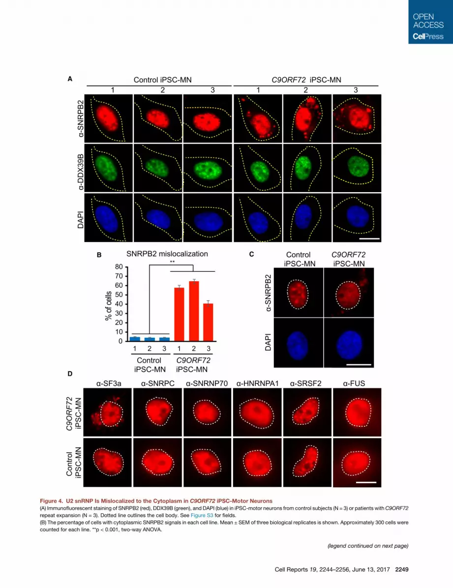
Figure 4. U2 snRNP Is Mislocalized to the Cytoplasm in C9ORF72 iPSC-Motor Neurons
(A) Immunofluorescent staining of SNRPB2 (red), DDX39B (green), and DAPI (blue) in iPSC-motor neurons from control subjects (N = 3) or patients withC9ORF72
repeat expansion (N = 3). Dotted line outlines the cell body. See Figure S3 for fields.
(B) The percentage of cells with cytoplasmic SNRPB2 signals in each cell line. Mean ± SEM of three biological replicates is shown. Approximately 300 cells were
counted for each line. **p < 0.001, two-way ANOVA.
(legend continued on next page)
Cell Reports 19, 2244–2256, June 13, 2017 2249

conclude that GR inhibits spliceosome assembly, thereby re-
sulting in defective splicing. Previously, we showed that the
spliceosome assembles on nascent transcripts as rapidly as
they are synthesized by RNAP II (Das et al., 2006). Consistent
with these data, we observed that addition of GR or PR only
blocked splicing when added before, but not after, transcrip-
tion (Figure 3H). This order of addition experiment further dem-
onstrates that the effect of the toxic peptides is specific, as
they only cause a block to splicing at a specific step in the
splicing pathway and do not interfere with splicing after tran-
scription and spliceosome assembly occur.
U2 snRNP Mislocalizes to the Cytoplasm in C9ORF72
Patient iPSC-Motor NeuronsA critical unanswered question is whether U2 snRNP is affected in
C9ORF72 patient cells. To investigate this possibility, we exam-
ined high-yield motor neuron cultures differentiated from 3
C9ORF72 patient-derived iPSC lines (Lopez-Gonzalez et al.,
2016). These iPSC-motor neurons are known to contain the
C9ORF72 repeat expansion as well as DPRs generated by trans-
lation of the repeat RNAs (Almeida et al., 2013; Ash et al., 2013;
Donnelly et al., 2013; Lopez-Gonzalez et al., 2016; Mori et al.,
2013; Su et al., 2014). iPSC-motor neurons derived from
three non-C9ORF72 subjects were used as controls. Immunoflu-
orescent staining showed that, in the controls, SNRPB2 and
DDX39B were properly localized to the nucleus (Figure 4A). In
contrast, we found a striking mislocalization of SNRPB2 to the
cytoplasm in the C9ORF72 iPSC-motor neurons. This was not
the case for DDX39B, whichwas properly localized to the nucleus
(Figure 4A). Notably, mislocalization of SNRPB2 in patient iPSC-
motor neurons occurs in the same cells in which DDX39B is prop-
erly localized, indicating that the effect is on SNRPB2 rather than
a general disruption of the nucleus. Moreover, quantitation re-
vealedmislocalization of SNRPB2 in a large fraction of the patient
iPSC-motor neurons (�40%–60%) (Figure 4B), and this was not
seen in controls (p < 0.001, two-way ANOVA; see Figure S3 for
fields). Notably, lighter exposures of our iPSC-motor neuron
immunofluorescent staining data indicate that in C9ORF72 pa-
tient cells in which SNRPB2 was mislocalized to the cytoplasm,
the level of SNRPB2 in nuclear speckle domains was lower than
in controls, and the nucleoplasmic level of SNRPB2 was likewise
reduced (Figure 4C; see Figure S4 for fields). Together, these data
raise the possibility that U2 snRNP is mislocalized from its normal
cellular location in C9ORF72 patient cells.
To further investigate the significance of SNRPB2 mislocaliza-
tion, we carried out additional immunofluorescent staining in the
iPSC-motor neurons. As shown in Figure 4D, SF3a is mislocal-
ized to the cytoplasm only in patient iPSC-motor neurons and
not in control iPSC-motor neurons. In contrast, the U1 snRNP
components, SNRPC and SNRNP70, are properly localized to
the nucleus in both patient and control iPSC-motor neurons. In
addition, HNRNPA1, SRSF2, and FUS remain properly localized
(C) Light exposure of immunofluorescent staining using SNRPB2 (red) and DAPI (b
Figure S4 for fields. ‘
(D) Immunofluorescent staining in patient or control iPSC-motor neurons using ant
line outlines the cell nucleus.
MN, motor neuron. Scale bars, 20 mm.
2250 Cell Reports 19, 2244–2256, June 13, 2017
to the nucleus in the patient and control cells. Together, these
data support the conclusion that U2 snRNP, a major component
of the splicing machinery, is mislocalized to the cytoplasm in
C9ORF72 iPSC-motor neurons, and this mislocalization may
play a role in the mis-splicing observed in these patient cells.
U2 snRNP Mislocalizes to the Cytoplasm in HeLa CellsTreated with PRTo determinewhether the toxic peptides could cause themisloc-
alization of U2 snRNP, we used immunofluorescent staining to
examine HeLa cells treated with PR or FLAG peptides (GR pep-
tide is unstable in cell culture media [Kwon et al., 2014] and was
not tested in our study). As shown in Figure S5, both PR and
FLAG peptides were distributed in both the nucleus and cyto-
plasm. Significantly, U2 snRNP components SNRPB2 and
SF3a were mislocalized to the cytoplasm in the PR-treated,
but not control-treated, cells (Figures S5A and S5B; see Fig-
ure S5E for fields). In contrast, DDX39B and SRSF2 were prop-
erly localized to the nucleus in both the PR-treated and
control-treated cells (Figures S5C and S5D; see Figure S5E for
fields). These data are consistent with our results showing that
U2 snRNP is mislocalized in C9ORF72 patient iPSC-motor
neurons and suggest a direct role of C9ORF72 toxic peptides
in causing U2 snRNP mislocalization.
U2-Dependent Exons Are Preferentially Mis-spliced inC9ORF72 Patient BrainsPrevious RNA-sequencing (RNA-seq) analyses found that 23%
of cassette exons out of the whole genome (9,878 out of
42,216) were skipped when the U2 snRNP component SF3B1
was knocked down in HeLa cells (Kfir et al., 2015). If U2 snRNP
dysfunction plays an important role in mis-splicing in C9ORF72
patients, these ‘‘U2-dependent’’ exons might be preferentially
mis-spliced in patients. To investigate this question, we reana-
lyzed RNA-seq datasets for cassette exons in C9ORF72, spo-
radic ALS (sALS), and control brains (Prudencio et al., 2015). In
their study, they examined eight cerebellums and eight frontal
cortexes, and we used these data for our analysis. In conjunc-
tion, we performed a neutral simulation to estimate the extent
of U2 dependency expected by chance in the whole genome.
This analysis revealed that in the random sampling, the highest
frequency of the percentage of U2-dependent exons in the
genome was 23% (marked by red dot in Figures 5A and 5B). In
contrast, in C9ORF72 cerebellums, U2-dependent exons
account for 35% (540/1,578) of the total skipped cassette exons,
significantly higher than expected by chance (c2 = 98.3, p <
10�10, c2 test; Figure 5A). Notably, as shown in Figure 5A, an
even greater enrichment of U2-dependent exons (42%, 83/
200) was observed in the top 200 most significantly mis-spliced
exons inC9ORF72 cerebellum (c2 = 36.2, p < 10�10, c2 test). This
was not the case for mis-spliced exons in sALS cerebellums,
of which only 27% were U2 dependent (46 out of 170; c2 = 1.
lue) inC9ORF72 iPSC-motor neurons. Dotted line outlines the cell nucleus. See
ibodies against SF3a, SNRPC, SNRNP70, HNRNPA1, SRSF2, and FUS. Dotted

A B
C D
Figure 5. U2-Dependent Exons Are Preferentially Mis-spliced in C9ORF72 Patient Brain
(A and B) Histogram showing U2-dependent exons present in 10,000 random samplings. The red dot indicates the percentage (23%) of the U2-dependent exons
in the genome that occur with the highest frequency. Lines indicate the percentage of U2-dependent exons that are mis-spliced in C9ORF72 or sALS patient
cerebellums (A) or frontal cortex (B). For C9ORF72, U2-dependent exons for total mis-spliced (1,578) and top 200 mis-spliced cassette exons (FDR ranging from
7E-124 to 2E-12) are shown.
(C and D) Venn diagrams showing U2- and HNRNPH-dependent mis-spliced exons in C9ORF72 cerebellums (top 200 exons) (C) and frontal cortex (107
exons) (D).
See also Tables S4, S5, and S6.
3, p = 0.26, c2 test; Figure 5A). We obtained similar results when
we reanalyzed the RNA-seq data from the frontal cortex in which
a total of 107 cassette exons were skipped (Prudencio et al.,
2015). In this case, our analysis revealed that U2-dependent
exons were significantly enriched in C9ORF72 (44%, 47/107,
c2 = 25.1, p = 1.0E-6, c2 test; Figure 5B), but not in sALS
(25%, 12/48, c2 = 0.07, p = 0.8, c2 test; Figure 5B). Together,
our results lend further support to our proteomic, biochemical,
and U2 snRNP cellular localization studies, indicating that U2
snRNP plays an important role in the mis-splicing in C9ORF72
patients.
To further investigate whether mis-splicing in C9ORF72 was
specific to U2 snRNP, we repeated the same analysis for ‘‘U1-
dependent exons’’ using data from knockdown of the U1 snRNP
component SNRNPC in HeLa cells (Rosel-Hillgartner et al.,
2013). Our results showed that 15% (232/1,578) of all cassette
exons or 17% of the top 200 cassette exons altered in
C9ORF72 patient cerebellums were U1 dependent, which was
not significantly different from that of the global average of total
exons (13%, p = 0.11 and p = 0.12, c2 test). Similarly, no enrich-
ment of U1-dependent exons was observed for C9ORF72 pa-
tient frontal cortex (14%, 15/107, c2 = 0.05, p = 0.8, c2 test).
In a recent study, it was reported that HNRNPH plays a role in
mis-splicing in C9ORF72 patient brains (Conlon et al., 2016).
Accordingly, we next examined the contribution of U2 snRNP
versus HNRNPH to mis-splicing using the top 200 mis-spliced
exons from C9ORF72 patient cerebellum and the 107 exons
from frontal cortex (Figures 5C and 5D). This analysis revealed
that mis-splicing of 83 exons was U2 dependent, 25 exons
were HNRNPH dependent, and 10 exons were both U2 and
HNRNPH dependent in cerebellum (Figure 5C). Similar results
were obtained in the frontal cortex (Figure 5D). These data indi-
cate that U2 snRNP dysfunction has a greater contribution than
HNRNPH to mis-splicing in C9ORF72 patient brain.
U2-Dependent Mis-splicing Occurs in PR-Treated CellsIn light of our multiple lines of evidence showing that GR/PR pep-
tides associate with and affect U2 snRNP, we next investigated
whether the U2-dependentmis-splicing observed in the bioinfor-
matics analyses could be caused by the toxic peptides. A previ-
ous RNA-seq analysis showed that treatment of cultured
astrocytes with PR resulted in splicing dysfunction, involving
mis-splicing events in 4,298 genes (Kwon et al., 2014). We
used these data to compare PR-dependent mis-spliced genes
with the U2-dependent genes that we identified in C9ORF72
patient cerebellum and frontal cortex. This analysis revealed
that 39% and 40% of the U2-dependent genes altered in the
C9ORF72 cerebellum and frontal cortex, respectively, were
also PR dependent, significantly more than expected by chance
(20%, p < 10�10 and p = 1.0E-6, c2 test). These data support the
model that the toxic peptides directly lead to dysfunction of
U2 snRNP and mis-splicing in C9ORF72 patient cells. Further
Cell Reports 19, 2244–2256, June 13, 2017 2251

evidence for the importance of the toxic peptides in causing
U2-dependent mis-splicing came from a genome-wide compar-
ison of U2-dependent genes with PR-dependent genes. We
found that 42% of the genes affected by PR are U2 dependent
(1,797 out of 4,298), significantly higher than expected by
chance (24%, c2 = 292.8, p < 10�10, c2 test). Thus, there is a
high level of correlation between U2-dependent mis-splicing
and PR-dependent mis-splicing events in the whole genome.
Together, these analyses provide evidence that DPR proteins
disrupt normal U2 snRNP function, and this could be an impor-
tant mechanism underlying mis-splicing in C9ORF72 patient
cells.
DISCUSSION
Previous studies reported that mis-splicing occurs in C9ORF72
patient brains, but the underlying mechanisms are not under-
stood. Here, we obtainedmultiple lines of evidence that dysfunc-
tion of the general splicing factor U2 snRNP plays an important
role in this mis-splicing. Our in vitro studies showed that
C9ORF72 toxic dipeptides (GR and PR) block spliceosome
assembly and splicing and specifically associate and interfere
with U2 snRNP. To investigate whether the effects on U2 snRNP
are relevant to the mis-splicing observed in C9ORF72 patient
cells, we examined iPSC-motor neurons generated from these
patients. We observed a dramatic mislocalization of U2 snRNP
components, but not other snRNPs or splicing factors, to the
cytoplasm in C9ORF72 patient iPSC-motor neurons. Our data
indicate that this mislocalization is due to the toxic peptides
because we observed specific mislocalization of U2 snRNP
components to the cytoplasm in HeLa cells treated with PR
peptide. We also carried out bioinformatic analyses, which re-
vealed that U2-dependent exons are preferentially mis-spliced
in C9ORF72 patient brain tissues, accounting for as much
as �44% of the most significantly mis-spliced exons. Of these,
PR-dependent mis-splicing accounts for �40% of them.
Together, these data are consistent with the possibility that the
mis-splicing observed in patients with C9ORF72 repeat expan-
sion is due, in part, to DPRs interfering with U2 snRNP. An impor-
tant question for future work is to determine whether and how
these U2-dependent mis-splicing events are involved in the
pathogenesis of C9ORF72 repeat expansion in ALS/FTD
patients.
One obvious mechanism by which mis-splicing of U2-depen-
dent exons could contribute to disease is by affecting genes that
are essential for normal motor function. We listed the 81 U2-
dependent genes that are affected by mis-splicing in C9ORF72
cerebellum in Table S4 and the 46 U2-dependent genes affected
by mis-splicing in the frontal cortex in Table S5. (Note that there
are fewer genes than exons with mis-splicing, because some of
the genes have more than one mis-splicing event.) We extracted
potentially relevant information about the genes from the litera-
ture, and this information is shown in the tables. One of the
largest categories of mis-spliced U2-dependent genes hasmito-
chondrial functions (labeled red in Tables S4 and S5), an espe-
cially interesting finding in light of the known links between
mitochondrial dysfunction and ALS resulting from multiple
different causes (Cozzolino and Carrı, 2012; Lopez-Gonzalez
2252 Cell Reports 19, 2244–2256, June 13, 2017
et al., 2016). It was also recently reported that C9ORF72 patient
DPRs compromise mitochondrial function and cause oxidative
stress (Lopez-Gonzalez et al., 2016). The U2-dependent genes
related to mitochondria include HIF1A, UQCRH, DNM1L,
COX16, TMEM126, NDUFAF5, PDHA1, and TIMM9, and two
mitochondrial ribosomal protein genes, MRPL52 and MRPS31
(Tables S4 and S5). HIF1A is interesting as it is a transcription
factor that has a role in the distribution of mitochondria in axons.
Genes with some relationship to neurons or neuronal disease
were another large category in the U2-dependent mis-spliced
genes (designated in blue in Tables S4 and S5). Notable among
these are PARD3 andMARK2, which modulate neuronal polarity
and neurite outgrowth, respectively. Finally, we found that
several U2-dependent genes have functions in different steps
of gene expression. These include transcription and splicing fac-
tors (marked in gray and yellow, respectively, in Tables S4 and
S5). Three of the splicing factors aremembers of the U2AF family
(RBM39, U2AF1, and RBM23). These proteins are essential for
recruiting U2 snRNP to the pre-mRNA, and it is possible that their
mis-splicing further exacerbates the mis-splicing observed in
C9ORF72 patient brains. We also found that GEMIN7 was mis-
spliced (Table S5). This protein is a component of the SMN com-
plex, which functions in snRNP biogenesis (Battle et al., 2006).
Thus, defective snRNP biogenesis could also contribute to the
mis-splicing observed in C9ORF72 patients. Notably, mutation
of the SMN protein causes the childhood motor neuron disease
spinal muscular atrophy, and previous work revealed that this
disease shares a biochemical pathway with ALS (Gama-Car-
valho et al., 2017; Shan et al., 2010; Yamazaki et al., 2012).
Our bioinformatic study also revealed that 40%of the U2-depen-
dent genes in C9ORF72 patient brains are also PR dependent.
These genes are listed in Table S6, which reveals that they are
enriched in genes with mitochondrial and pre-mRNA splicing
functions. Together, our results raise the possibility that DPR-
mediated dysfunction of U2 snRNP, a major component of the
splicing machinery, causes mis-splicing of genes involved in
cellular functions that have previously been associated with
ALS/FTD pathogenesis.
Several mechanisms may underlie DPR-mediated U2-depen-
dent mis-splicing in C9ORF72 patients. For example, our in vitro
data showing that U2 snRNP binds to GR/PR toxic peptides and
disrupts splicing and spliceosome assembly raise the possibility
that interactions between the DPRs and U2 snRNP directly
disrupt the function of this essential splicing factor. Another pos-
sibility is that the loss of U2 snRNP to the cytoplasm that we
observed in C9ORF72 patient-iPSC derived motor neurons
may decrease the levels of nuclear U2 snRNP required for proper
splicing. In addition, we observed loss of U2 snRNP from nuclear
speckle domains, and this may disrupt normal U2 snRNP
function. In previous studies, overexpression of tagged GR/PR
showed that it is primarily in the cytoplasm (Kwon et al., 2014;
Lee et al., 2016), and we found that PR peptide localizes to the
cytoplasm in HeLa cells. In addition, immunohistochemistry of
C9ORF72 patient brains showed that DPRs are present in both
the nucleus and cytoplasm (Mori et al., 2013; Wen et al., 2014).
These observations may explain how U2 snRNP is mislocalized
to the cytoplasm in C9ORF72 patient cells. Specifically, it is
known that snRNP biogenesis takes place in the cytoplasm
(Battle et al., 2006). Thus, it is possible that interactions between
the DPRs and U2 snRNP may sequester U2 snRNP in the
cytoplasm.
In recent studies,GRor PRpeptides overexpressed in cultured
cells were found to associate with numerous cellular proteins
(Lee et al., 2016; Lin et al., 2016; Lopez-Gonzalez et al., 2016).
Among these were RNA-binding proteins that contain low
complexity domains, and of these, several are known ALS-caus-
ative proteins (e.g., FUS, TARDBP, HNRNPA1, and MATR3).
Interactions betweenGR/PR and the low complexity domain pro-
teins disrupt the assembly and dynamics of membrane-less
organelles, which may contribute to C9ORF72 pathogenesis
(Lee et al., 2016). Consistent with these studies, our proteomic
analysis of GR/PR-interacting proteins revealed a large overlap
with numerous ALS-causative proteins, including those known
to contain low complexity domains (Figure 6A). In addition, the
GR/PR interactors identified in our study overlap significantly
with those identified in previous studies (Lee et al., 2016;
Lopez-Gonzalez et al., 2016) (Figure 6B). Several observations
suggest that at least some of the GR/PR interactors are physio-
logically relevant. Specifically, multiple U2 snRNP proteins
were identified as GR/PR interactors not only in our work but
also in the recent studies (Lee et al., 2016; Lopez-Gonzalez
et al., 2016) (Figure 6B). In addition, these common interactors
were identified using a variety of cell types and pull-down strate-
gies and are independent of repeat protein lengths (Figure 6C).
Many of these U2 snRNP proteins contain low complexity
domains (Figures 6D and S6), and the interaction between GR/
PR and these low complexity domain-containing U2 snRNP
proteins may explain why U2 snRNP is lost from its normal loca-
tion in the membrane-less organelle (nuclear speckle domains)
and is mis-localized to the cytoplasm.
Our data indicate that U2-dependent mis-splicing accounts
for a large fraction of the mis-splicing observed in patients with
C9ORF72 repeat expansion, which is themost frequentmutation
in familial ALS/FTD. In addition, mis-splicing is present in other
forms of familial ALS (e.g., those due to FUS or TDP-43 muta-
tion). Sporadic ALS, which is the most common form of all types
of ALS, also exhibits mis-splicing. Thus, correction of mis-
splicing is a potential therapeutic approach for multiple forms
of ALS. In this regard, splicing modulator compounds that can
correct mis-splicing are emerging as potential therapies for other
diseases, and these or related compoundsmay have efficacy for
the treatment of ALS/FTD.
EXPERIMENTAL PROCEDURES
Plasmids
CMV-Ftz plasmid was described previously (Das et al., 2006). CMV-Ftz-let-7a
plasmid was constructed by inserting a 395-nt pri-let-7a fragment into the
BamHI site in the Ftz intron. CMV-DNA templates were amplified by PCR using
forward (50-TGGAGGTCGCTGAGTAGTGC-30) and reverse (50- TAGAAGGCA
CAGTCGAG GCT-30) primers.
Transcription-Coupled RNA Processing
For in vitro studies, we used HeLa cell nuclear extracts prepared according to
Krainer et al. (1984). Transcription-coupled splicing reactions were performed
as described elsewhere (Das et al., 2007; Folco et al., 2011), except that pre-
initiation complexes (PICs) were assembled on the CMV-DNA templates prior
to transcription (Yu et al., 2010). Briefly, CMV-DNA templates were incubated
in 15 mL nuclear extract containing 3.2mMMgCl2 and 5 mL polyvinyl alcohol for
20 min at 30�. The indicated peptides (10 mM) were then added in a final
reaction mixture volume of 25 mL containing 0.5 mM ATP, 20 mM creatine
phosphate (di-Tris salt), and 1 mL 32P-UTP (250 Ci/mmol; Perkin Elmer Life
Sciences). Reaction mixtures were incubated for 15 min at 30�C to allow tran-
scription followed by addition of a-amanitin (200 ng). Incubation was
continued at 30�C for times indicated. For transcription/splicing/pri-miRNA
processing, PICs were assembled using the same conditions as for transcrip-
tion-coupled splicing. No peptide or 10 mM of the indicated peptides was then
added and incubation was continued for 15 min to allow transcription in the
presence of 0.5 mM ATP, 20 mM creatine phosphate (di-Tris salt), and 1 mL32P-UTP, and an additional 3.2mMMgCl2was added to bring the final concen-
tration to 6.4 mM in a final reaction mixture volume of 25 mL. a-Amanitin
(200 ng) was added and incubation was continued for 45min to allow process-
ing. Total RNA was fractionated on 8% denaturing polyacrylamide gels and
detected by PhosphorImager.
Proteomic Analysis of GR and PR Interacting Proteins
GR, PR, or FLAG peptides were biotinylated using the EZ-Link TFPA-PEG3-
Biotin kit (Thermo Fisher). Splicing reactionmixtures containing these peptides
(10 mM) were separated on Sephacryl-S500 gel filtration columns in a buffer
containing 20 mM Tris (pH 7.8), 60 mM KCl, 0.1% Triton X-100, and 0.2 mM
PMSF. Proteins in the gel filtration fractions were analyzed on NuPAGE Novex
4%–12% Bis-Tris Gels (Thermo Fisher) followed by western blotting. Total
RNAs from the fractions were analyzed on an 8% denaturing polyacrylamide
gel stained with ethidium bromide. The data for rows FLAG, SNRPC, SNRPB2,
SF3A1, SF3A2/3, FUS, and TARDBP shown in Figure 2A and the RNA gel
shown in Figure 2B were from the gel filtration column containing the GR pep-
tide. Similar results were obtained with gel filtration fractions containing PR
and FLAG peptides. Pull-downs were carried out from fractions 33–69 in the
gel filtration buffer using streptavidin magnetic particles (Roche) at 4�C for
2 hr and washed five times with 13 PBS/0.1% Triton X-100. Proteins associ-
ated with biotinylated peptides were labeled using tandem mass tag (TMT)
(McAlister et al., 2012) and analyzed with an Orbitrap Fusion mass spectrom-
eter coupled to a Proxeon EASY-nLC 1000 liquid chromatography (LC) pump
(Thermo Fisher Scientific).
Immunoprecipitations
A rabbit polyclonal antibody that recognizes the three SF3a complex subunits
or a mouse monoclonal antibody against SNRPB2 were described previously
(Das et al., 2000). Rabbit polyclonal antibodies against DROSHA and a control
immunoglobulin G were from Abcam. For protein IPs, antibodies were cross-
linked to protein Sepharose beads with dimethylpymelimidate (Sigma). Prior to
IPs, 250-mL reaction mixtures containing 75 mL nuclear extract, 3.2 mMMgCl2,
0.5 mM ATP, and 20 mM creatine phosphate (di-Tris salt) were incubated for
20 min at 30� and then spun at 14,000 rpm in a microfuge for 5 min at 4�C.The supernatant was mixed with 150 mL PBS/0.1% Triton X-100/0.2 mM
PMSF), protease inhibitor EDTA-free (Roche), and spun in a microfuge at
14,000 rpm for 5 min at 4�C. IPs were carried out at 4�C for 2 hr and washed
five times with 13 PBS/0.1% Triton X-100. IPs were mixed with protein gel
loading buffer without DTT at room temperature to elute the proteins. DTT
(2 mM final) was then added, and samples were boiled for 2 min and run on
4%–12% SDS-PAGE gels.
U2 snRNP and Spliceosome Assembly Assays
A U2 20OMethyl oligo that base pairs to U2 snRNA (50-mCmAmGmAmU
mAmCmUmAmCmAmCmUm UmGmA-30) was 32P-labeled with g-ATP
(250 Ci/mmol; Perkin Elmer Life Sciences) and T4 polynucleotide kinase.
Reaction mixtures (25 mL) containing 15 mL nuclear extract were incubated
in the presence of 0.5 mM ATP, 3.2 mM MgCl2, 20 mM creatine phosphate
(di-Tris salt), 10 mM peptides, and 0.4 mM U2 oligo for 5 min. For spliceo-
some assembly, transcription-coupled splicing reactions with or without
the peptides were incubated for the times indicated and then run on
G-50 micro columns (Amersham Biosciences) to remove unincorporated32P-UTP. Heparin (0.65 mg/mL final concentration) was added to the
G-50 column-purified reactions before loading on a 1.2% low-melting-point
agarose gel.
Cell Reports 19, 2244–2256, June 13, 2017 2253

A
B
D
C
Figure 6. Comparison of Toxic Peptide Interactomes in Different Datasets
(A) Venn diagram showing overlap between GR- and PR-interacting proteins identified in this study. Known ALS proteins identified in both datasets are shown.
(B) Venn diagrams showing overlap between interacting proteins identified in this study and in four published datasets (the Mcknight_1 and Mcknight_2 datasets
are described in Lin et al., 2016; the Taylor dataset is described in Lee et al., 2016; and the Gao dataset is described in Lopez-Gonzalez et al., 2016).
(C) Table showing methods used to generate the DPR interactome datasets.
(D) Examples of U2 snRNP proteins showing their low complexity domains.
See Figure S6 for this analysis with all of the U2 snRNP components.
2254 Cell Reports 19, 2244–2256, June 13, 2017

Generation of iPSC-Derived Motor Neurons
Generation of iPSC-derived neurons was described previously (Lopez-
Gonzalez et al., 2016). Briefly, iPSCs were expanded in Matrigel-coated
wells and at 60% confluency were split with Accutase into Matrigel-coated
wells. 24 hr after plating, culture medium was replaced with neuroepithelial
progenitor (NEP) medium, DMEM/F12, Neurobasal medium at 1:1,
0.53 N2, 0.53 B27, 0.1 mM ascorbic acid (Santa Cruz Biotechnology),
1X Glutamax (Invitrogen), 3 mM CHIR99021 (Tocris Bioscience), 2 mM
DMH1 (Tocris Bioscience), and 2 mM SB431542 (Stemgent) for 6 days.
NEPs were dissociated with dispase and split 1:6 into Matrigel-coated
wells. NEPs were cultured in motor neuron progenitor (MNP) induction
medium (the same medium as described above with 0.1 mM RA [Stemgent]
and 0.5 mM purmorphamine [Stemgent]) for 6 days. MNPs were dissociated
with dispase to generate suspension cultures. After 6 days in culture, cells
were dissociated into single cells, plated on laminin-coated plates/cover-
slips in motor neuron differentiation medium (containing 0.5 mM RA,
0.1 mM purmorphamine, and 0.1 mM compound E; Calbiochem), and
cultured for 1 month.
Immunofluorescent Staining
For immunofluorescent staining, iPSC-motor neurons were fixed with 4%
paraformaldehyde in PBS for 15 min and permeabilized with 0.1% Triton
X-100 in PBS for 15 min. After incubation in 5% fetal bovine serum (FBS) for
1 hr at room temperature, cells were incubated overnight at 4�C in custom
primary antibodies SNRPB2 (1:100), DDX39B (1:1,000), SF3a (1:1,000), and
FUS (1:1,000) or in commercial antibodies SNRPC (Santa Cruz Biotechnology,
1:200), SNRNP70 (Millipore, 1:200), HNRNPA1 (Santa Cruz Biotechnology,
1:200), and SRSF2 (1:1000, Invitrogen). After three washes in PBS, cells
were incubated with mouse Alexa Fluor 647 (for SNRPB2, SNRNP70, and
SRSF2) or rabbit Alexa 488 (for HNRNPA1, DDX39B, SNRPC, SF3a, and
FUS) secondary antibodies (Invitrogen, 1:1000) for 1 hr at room temperature,
followed by three washes in PBS. Images were captured with a Nikon
TE2000U inverted microscope.
Bioinformatic Analysis and Statistics
Brain transcriptome profiles ofC9ORF72 ALS and sALS are described in GEO:
GSE67196 (Prudencio et al., 2015). Processed RNA-seq data were provided
by the authors, and a cutoff of Bonferroni-corrected false discovery rate
(FDR) < 0.05 was used to specify significantly altered exons. RNA-seq data
of SF3B1 knockdown was from GEO: GSE65644 (Kfir et al., 2015). RNA-seq
data of HNRNPH or control knockdown in 293T cells was from GEO:
GSE16642 (Xiao et al., 2009). RNA-seq data of SNRNPC or control knockdown
in HeLa cells was from GEO: GSE42485 (Rosel-Hillgartner et al., 2013). Mis-
spliced genes in cultured astrocytes treated with PR peptide were from
(Kwon et al., 2014). Low-complexity domain prediction was done using
SMART online tool (Letunic et al., 2015). Statistical analyses were done with
GraphPad Prism (GraphPad Software). Differences between groups were
analyzed with ANOVA, Student’s t test, or c2 test. p < 0.05 was considered
significant.
SUPPLEMENTAL INFORMATION
Supplemental Information includes Supplemental Experimental Procedures,
six figures, and six tables and can be found with this article online at http://
dx.doi.org/10.1016/j.celrep.2017.05.056.
AUTHOR CONTRIBUTIONS
S.Y. and R.R. conceived the project, and S.Y. carried out most of the experi-
ments. F.-B.G. directed iPSC-motor neuron generation, which was carried out
by R.L.-G. Imaging analyses were carried out by S.Y. and J.G. Splicing and U2
snRNP native gel analysis were done C.B. Mass spectrometry was performed
by R.K. The data were analyzed by R.K., S.P.G., S.Y., and R.R. Themanuscript
waswritten by S.Y. and R.R. with input from F.-B.G. and R.L.-G and assistance
from all authors.
ACKNOWLEDGMENTS
We are grateful to the Nikon Imaging Center and the Image and Data Analysis
Core at Harvard Medical School for help with microscopy and image analysis.
GR and PR peptides were provided by A.D. Gitler. We thank R. Batra for the
RNA-seq data from C9ORF72 and sALS patients. This work was supported
by NIH grants GM043375 (to R.R.) and NS057553 and NS079725 (to
F.-B.G.), ALS Therapy Alliance grant 2013-S-006 (to R.R.), and ALS Associa-
tion grant 2179 (to F.-B.G.).
Received: January 24, 2017
Revised: April 4, 2017
Accepted: May 16, 2017
Published: June 13, 2017
REFERENCES
Almeida, S., Gascon, E., Tran, H., Chou, H.J., Gendron, T.F., Degroot, S.,
Tapper, A.R., Sellier, C., Charlet-Berguerand, N., Karydas, A., et al. (2013).
Modeling key pathological features of frontotemporal dementia with
C9ORF72 repeat expansion in iPSC-derived human neurons. Acta Neuropa-
thol. 126, 385–399.
Ash, P.E., Bieniek, K.F., Gendron, T.F., Caulfield, T., Lin, W.L., Dejesus-Her-
nandez, M., van Blitterswijk, M.M., Jansen-West, K., Paul, J.W., 3rd, Rade-
makers, R., et al. (2013). Unconventional translation of C9ORF72 GGGGCC
expansion generates insoluble polypeptides specific to c9FTD/ALS. Neuron
77, 639–646.
Battle, D.J., Kasim, M., Yong, J., Lotti, F., Lau, C.K., Mouaikel, J., Zhang, Z.,
Han, K.,Wan, L., andDreyfuss, G. (2006). The SMN complex: an assemblyma-
chine for RNPs. Cold Spring Harb. Symp. Quant. Biol. 71, 313–320.
Conlon, E.G., Lu, L., Sharma, A., Yamazaki, T., Tang, T., Shneider, N.A., and
Manley, J.L. (2016). The C9ORF72 GGGGCC expansion forms RNA G-quad-
ruplex inclusions and sequesters hnRNP H to disrupt splicing in ALS brains.
eLife 5, e17820.
Cooper-Knock, J., Walsh, M.J., Higginbottom, A., Robin Highley, J., Dickman,
M.J., Edbauer, D., Ince, P.G., Wharton, S.B., Wilson, S.A., Kirby, J., et al.
(2014). Sequestration of multiple RNA recognition motif-containing proteins
by C9orf72 repeat expansions. Brain 137, 2040–2051.
Cooper-Knock, J., Bury, J.J., Heath, P.R., Wyles, M., Higginbottom, A., Gels-
thorpe, C., Highley, J.R., Hautbergue, G., Rattray, M., Kirby, J., and Shaw, P.J.
(2015). C9ORF72 GGGGCC expanded repeats produce splicing dysregulation
which correlates with disease severity in amyotrophic lateral sclerosis. PLoS
ONE 10, e0127376.
Cozzolino, M., and Carrı, M.T. (2012). Mitochondrial dysfunction in ALS. Prog.
Neurobiol. 97, 54–66.
Das, R., Zhou, Z., and Reed, R. (2000). Functional association of U2 snRNP
with the ATP-independent spliceosomal complex E. Mol. Cell 5, 779–787.
Das, R., Dufu, K., Romney, B., Feldt, M., Elenko, M., and Reed, R. (2006).
Functional coupling of RNAP II transcription to spliceosome assembly. Genes
Dev. 20, 1100–1109.
Das, R., Yu, J., Zhang, Z., Gygi, M.P., Krainer, A.R., Gygi, S.P., and Reed, R.
(2007). SR proteins function in coupling RNAP II transcription to pre-mRNA
splicing. Mol. Cell 26, 867–881.
DeJesus-Hernandez, M., Mackenzie, I.R., Boeve, B.F., Boxer, A.L., Baker, M.,
Rutherford, N.J., Nicholson, A.M., Finch, N.A., Flynn, H., Adamson, J., et al.
(2011). Expanded GGGGCC hexanucleotide repeat in noncoding region of
C9ORF72 causes chromosome 9p-linked FTD and ALS. Neuron 72, 245–256.
Donnelly, C.J., Zhang, P.W., Pham, J.T., Haeusler, A.R., Mistry, N.A., Viden-
sky, S., Daley, E.L., Poth, E.M., Hoover, B., Fines, D.M., et al. (2013). RNA
toxicity from the ALS/FTD C9ORF72 expansion is mitigated by antisense inter-
vention. Neuron 80, 415–428.
Folco, E.G., Coil, K.E., and Reed, R. (2011). The anti-tumor drug E7107 reveals
an essential role for SF3b in remodeling U2 snRNP to expose the branch point-
binding region. Genes Dev. 25, 440–444.
Cell Reports 19, 2244–2256, June 13, 2017 2255

Freibaum, B.D., Lu, Y., Lopez-Gonzalez, R., Kim, N.C., Almeida, S., Lee, K.H.,
Badders, N., Valentine, M., Miller, B.L., Wong, P.C., et al. (2015). GGGGCC
repeat expansion in C9orf72 compromises nucleocytoplasmic transport. Na-
ture 525, 129–133.
Fu, X.D., and Ares, M., Jr. (2014). Context-dependent control of alternative
splicing by RNA-binding proteins. Nat. Rev. Genet. 15, 689–701.
Gama-Carvalho, M., L Garcia-Vaquero, M., R Pinto, F., Besse, F., Weis, J.,
Voigt, A., Schulz, J.B., and De Las Rivas, J. (2017). Linking amyotrophic lateral
sclerosis and spinal muscular atrophy through RNA-transcriptome homeosta-
sis: a genomics perspective. J. Neurochem. 141, 12–30.
Haeusler, A.R., Donnelly, C.J., Periz, G., Simko, E.A., Shaw, P.G., Kim, M.S.,
Maragakis, N.J., Troncoso, J.C., Pandey, A., Sattler, R., et al. (2014).
C9orf72 nucleotide repeat structures initiate molecular cascades of disease.
Nature 507, 195–200.
Jovi�ci�c, A., Mertens, J., Boeynaems, S., Bogaert, E., Chai, N., Yamada, S.B.,
Paul, J.W., 3rd, Sun, S., Herdy, J.R., Bieri, G., et al. (2015). Modifiers of C9orf72
dipeptide repeat toxicity connect nucleocytoplasmic transport defects to FTD/
ALS. Nat. Neurosci. 18, 1226–1229.
Kanekura, K., Yagi, T., Cammack, A.J., Mahadevan, J., Kuroda, M., Harms,
M.B., Miller, T.M., and Urano, F. (2016). Poly-dipeptides encoded by the
C9ORF72 repeats block global protein translation. Hum. Mol. Genet. 25,
1803–1813.
Kfir, N., Lev-Maor, G., Glaich, O., Alajem, A., Datta, A., Sze, S.K., Meshorer, E.,
and Ast, G. (2015). SF3B1 association with chromatin determines splicing out-
comes. Cell Rep. 11, 618–629.
Krainer, A.R., Maniatis, T., Ruskin, B., and Green, M.R. (1984). Normal and
mutant human beta-globin pre-mRNAs are faithfully and efficiently spliced
in vitro. Cell 36, 993–1005.
Kwon, I., Xiang, S., Kato, M., Wu, L., Theodoropoulos, P., Wang, T., Kim, J.,
Yun, J., Xie, Y., and McKnight, S.L. (2014). Poly-dipeptides encoded by the
C9orf72 repeats bind nucleoli, impede RNA biogenesis, and kill cells. Science
345, 1139–1145.
Lee, K.H., Zhang, P., Kim, H.J., Mitrea, D.M., Sarkar, M., Freibaum, B.D., Cika,
J., Coughlin, M., Messing, J., Molliex, A., et al. (2016). C9orf72 dipeptide re-
peats impair the assembly, dynamics, and function of membrane-less organ-
elles. Cell 167, 774–788.
Letunic, I., Doerks, T., and Bork, P. (2015). SMART: recent updates, new de-
velopments and status in 2015. Nucleic Acids Res. 43, D257–D260.
Li, Y.R., King, O.D., Shorter, J., and Gitler, A.D. (2013). Stress granules as cru-
cibles of ALS pathogenesis. J. Cell Biol. 201, 361–372.
Lin, Y., Mori, E., Kato, M., Xiang, S., Wu, L., Kwon, I., and McKnight, S.L.
(2016). Toxic PR poly-dipeptides encoded by the C9orf72 repeat expansion
target LC domain polymers. Cell 167, 789–802.
Lopez-Gonzalez, R., Lu, Y., Gendron, T.F., Karydas, A., Tran, H., Yang, D., Pet-
rucelli, L., Miller, B.L., Almeida, S., andGao, F.B. (2016). Poly(GR) in C9ORF72-
related ALS/FTD compromisesmitochondrial function and increases oxidative
stress and DNA damage in iPSC-derived motor neurons. Neuron 92, 383–391.
McAlister, G.C., Huttlin, E.L., Haas, W., Ting, L., Jedrychowski, M.P., Rogers,
J.C., Kuhn, K., Pike, I., Grothe, R.A., Blethrow, J.D., and Gygi, S.P. (2012).
Increasing the multiplexing capacity of TMTs using reporter ion isotopologues
with isobaric masses. Anal. Chem. 84, 7469–7478.
Mizielinska, S., Gronke, S., Niccoli, T., Ridler, C.E., Clayton, E.L., Devoy, A.,
Moens, T., Norona, F.E., Woollacott, I.O., Pietrzyk, J., et al. (2014). C9orf72
repeat expansions cause neurodegeneration in Drosophila through arginine-
rich proteins. Science 345, 1192–1194.
Mori, K., Weng, S.M., Arzberger, T., May, S., Rentzsch, K., Kremmer, E.,
Schmid, B., Kretzschmar, H.A., Cruts, M., Van Broeckhoven, C., et al.
(2013). The C9orf72 GGGGCC repeat is translated into aggregating dipep-
tide-repeat proteins in FTLD/ALS. Science 339, 1335–1338.
Prudencio, M., Belzil, V.V., Batra, R., Ross, C.A., Gendron, T.F., Pregent, L.J.,
Murray, M.E., Overstreet, K.K., Piazza-Johnston, A.E., Desaro, P., et al. (2015).
Distinct brain transcriptome profiles in C9orf72-associated and sporadic ALS.
Nat. Neurosci. 18, 1175–1182.
2256 Cell Reports 19, 2244–2256, June 13, 2017
Ramaswami, M., Taylor, J.P., and Parker, R. (2013). Altered ribostasis: RNA-
protein granules in degenerative disorders. Cell 154, 727–736.
Renton, A.E., Majounie, E., Waite, A., Simon-Sanchez, J., Rollinson, S., Gibbs,
J.R., Schymick, J.C., Laaksovirta, H., van Swieten, J.C., Myllykangas, L., et al.;
ITALSGEN Consortium (2011). A hexanucleotide repeat expansion in
C9ORF72 is the cause of chromosome 9p21-linked ALS-FTD. Neuron 72,
257–268.
Rosel-Hillgartner, T.D., Hung, L.H., Khrameeva, E., Le Querrec, P., Gelfand,
M.S., and Bindereif, A. (2013). A novel intra-U1 snRNP cross-regulation mech-
anism: alternative splicing switch links U1C and U1-70K expression. PLoS
Genet. 9, e1003856.
Ruby, S.W., Chang, T.H., and Abelson, J. (1993). Four yeast spliceosomal pro-
teins (PRP5, PRP9, PRP11, and PRP21) interact to promote U2 snRNP binding
to pre-mRNA. Genes Dev. 7, 1909–1925.
Rutherford, N.J., Heckman, M.G., Dejesus-Hernandez, M., Baker, M.C., Soto-
Ortolaza, A.I., Rayaprolu, S., Stewart, H., Finger, E., Volkening, K., Seeley,
W.W., et al. (2012). Length of normal alleles of C9ORF72 GGGGCC repeat
do not influence disease phenotype. Neurobiol. Aging 33, 2950–2957.
Shan, X., Chiang, P.M., Price, D.L., and Wong, P.C. (2010). Altered distribu-
tions of Gemini of coiled bodies and mitochondria in motor neurons of
TDP-43 transgenic mice. Proc. Natl. Acad. Sci. USA 107, 16325–16330.
Shi, K.Y., Mori, E., Nizami, Z.F., Lin, Y., Kato, M., Xiang, S., Wu, L.C., Ding, M.,
Yu, Y., Gall, J.G., and McKnight, S.L. (2017). Toxic PRn poly-dipeptides en-
coded by the C9orf72 repeat expansion block nuclear import and export.
Proc. Natl. Acad. Sci. USA 114, E1111–E1117.
Su, Z., Zhang, Y., Gendron, T.F., Bauer, P.O., Chew, J., Yang, W.Y., Fostvedt,
E., Jansen-West, K., Belzil, V.V., Desaro, P., et al. (2014). Discovery of a
biomarker and lead small molecules to target r(GGGGCC)-associated defects
in c9FTD/ALS. Neuron 83, 1043–1050.
Taylor, J.P., Brown, R.H., Jr., and Cleveland, D.W. (2016). Decoding ALS: from
genes to mechanism. Nature 539, 197–206.
Tran, H., Almeida, S., Moore, J., Gendron, T.F., Chalasani, U., Lu, Y., Du, X.,
Nickerson, J.A., Petrucelli, L., Weng, Z., and Gao, F.B. (2015). Differential
toxicity of nuclear RNA foci versus dipeptide repeat proteins in a Drosophila
model of C9ORF72 FTD/ALS. Neuron 87, 1207–1214.
Wahl, M.C., Will, C.L., and L€uhrmann, R. (2009). The spliceosome: design prin-
ciples of a dynamic RNP machine. Cell 136, 701–718.
Wen, X., Tan, W., Westergard, T., Krishnamurthy, K., Markandaiah, S.S., Shi,
Y., Lin, S., Shneider, N.A., Monaghan, J., Pandey, U.B., et al. (2014). Antisense
proline-arginine RAN dipeptides linked to C9ORF72-ALS/FTD form toxic nu-
clear aggregates that initiate in vitro and in vivo neuronal death. Neuron 84,
1213–1225.
Xiao, X., Wang, Z., Jang, M., Nutiu, R., Wang, E.T., and Burge, C.B. (2009).
Splice site strength-dependent activity and genetic buffering by poly-G runs.
Nat. Struct. Mol. Biol. 16, 1094–1100.
Yamazaki, T., Chen, S., Yu, Y., Yan, B., Haertlein, T.C., Carrasco, M.A., Tapia,
J.C., Zhai, B., Das, R., Lalancette-Hebert, M., et al. (2012). FUS-SMN protein
interactions link the motor neuron diseases ALS and SMA. Cell Rep. 2,
799–806.
Yang, D., Abdallah, A., Li, Z., Lu, Y., Almeida, S., and Gao, F.B. (2015). FTD/
ALS-associated poly(GR) protein impairs the Notch pathway and is recruited
by poly(GA) into cytoplasmic inclusions. Acta Neuropathol. 130, 525–535.
Yin, S., Yu, Y., and Reed, R. (2015). Primary microRNA processing is function-
ally coupled to RNAP II transcription in vitro. Sci. Rep. 5, 11992.
Yu, Y., Das, R., Folco, E.G., andReed, R. (2010). Amodel in vitro system for co-
transcriptional splicing. Nucleic Acids Res. 38, 7570–7578.
Zhang, K., Donnelly, C.J., Haeusler, A.R., Grima, J.C., Machamer, J.B., Stein-
wald, P., Daley, E.L., Miller, S.J., Cunningham, K.M., Vidensky, S., et al. (2015).
The C9orf72 repeat expansion disrupts nucleocytoplasmic transport. Nature
525, 56–61.


![Mikro Tik Certified Network Associate 1[1]](https://img.pdfslide.net/doc/110x75/634539bcf474639c9b04bf17/mikro-tik-certified-network-associate-11.jpg)


