Embed Size (px)
Citation preview

Evolutionary insights into the unique electromotility motor ofmammalian outer hair cel
Oseremen E. Okoruwa, Michael D. Weston, Divvya C. Sanjeevi, Amanda R. Millemon, BerndFritzsch, Richard Hallworth, and Kirk W. Beisel*Department of Biomedical Sciences, Creighton University School of Medicine, Omaha, NE 68178,USA
SUMMARYPrestin (SLC26A5) is the molecular motor responsible for cochlear amplification by mammaliancochlea outer hair cells and has the unique combined properties of energy-independent motility,voltage sensitivity, and speed of cellular shape change. The ion transporter capability, typical ofSLC26A members, was exchanged for electromotility function and is a newly derived feature of thetherian cochlea. A putative minimal essential motif for the electromotility motor (meEM) wasidentified through the amalgamation of comparative genomic, evolution, and structuraldiversification approaches. Comparisons were done among nonmammalian vertebrates, eutherianmammalian species, and the opossum and platypus. The opossum and platypus SLC26A5 proteinswere comparable to the eutherian consensus sequence. Suggested from the point-accepted mutationanalysis, the meEM motif spans all the transmembrane segments and represented residues 66–503.Within the eutherian clade, the meEM was highly conserved with a substitution frequency of only39/7497 (0.5%) residues, compared with 5.7% in SLC26A4 and 12.8% in SLC26A6 genes. Clade-specific substitutions were not observed and there was no sequence correlation with low or highhearing frequency specialists. We were able to identify that within the highly conserved meEM motiftwo regions, which are unique to all therian species, appear to be the most derived features in theSLC26A5 peptide.
INTRODUCTIONThe molecular evolution of a protein family involves a combination of gene expansion eventsthat includes gene duplication and duplication of the whole genome (Haldane 1932; Ohno1970, 1993; Taylor and Raes 2004; Domazet-Loso et al. 2007). The eutherian genome haspresumably undergone two rounds of whole genome duplication (2R Hypothesis) occurringin early chordate evolution (Urochordata–Craniata) (Garcia-Fernandez and Holland 1994;Dehal and Boore 2005; Hallbook et al. 2006). Expansion and contraction of the number ofgene family members can occur by a number of mechanisms, including but not limited to theacquisition of transposable elements, independent gene duplication through cis- or trans-events, and alternation of chromosomal number with subsequent deletion or translocation ofgenes, chromosomal segments or whole chromosomes (Furlong and Holland 2002). Afterduplication these isogenic copies, under selective pressure, can be routed along a number ofpossible evolutionary pathways that diversify the isogenes through point mutations, insertionsor deletions (Indels), and/or conversion events within the coding, noncoding, and/or cis-regulatory regions (King et al. 2007; Prud’homme et al. 2007; Roth et al. 2007; Zheng et al.2007). These pathways are defined as nonfunctionalization, neofunctionalization, andsubfunctionalization of the evolving isogenes (Ohno 1970; Force et al. 1999; Lynch and Conery
*Author for correspondence (email: [email protected]).
NIH Public AccessAuthor ManuscriptEvol Dev. Author manuscript; available in PMC 2009 April 8.
Published in final edited form as:Evol Dev. 2008 ; 10(3): 300–315. doi:10.1111/j.1525-142X.2008.00239.x.
NIH
-PA Author Manuscript
NIH
-PA Author Manuscript
NIH
-PA Author Manuscript

2000). There is evidence that formation of paralogs is often due to an initial acceleration of thenonsynonymous substitution rate leading to functional divergence that is then followed bypurifying selection, which excludes substitutions that prevent or interfere with refinement ofthe protein structure–function relationship (Li et al. 1985; Ohta 1993; Lynch and Conery2000). Metazoan genomic comparisons have recently identified punctuated protein evolutionin metazoan lineages that identify evolutionary innovations in protein structure and function(Domazet-Loso, Brajkovic, and Tautz 2007; Putnam et al. 2007). Comparative analysis ofproteins and their paralogs provide insights into the diversity of protein structures and,combined with functional studies on engineered proteins, reveal structure–functionrelationships that contribute to phenotypic and morphological diversification (Marsh andGriffiths 2005). Recently, Ortlund et al. (2007) have combined phylogenic and functionalapproaches with X-ray crystallography to elucidate the generation of new functional paradigmsof proteins.
Here we examined the evolution of an anion transporter molecule to a unique ATP-independentelectromotility motor protein, prestin or SLC26A5, that provides sound amplification of thecochlea with rapid conformational changes that occur in kilohertz ranges (Dallos and Fakler2002; Dallos et al. 2006). Prestin (SLC26A5) is a protein critical for the function of theperipheral auditory system in mammals. Its presence results in outer hair cell (OHC)electromotility, allowing OHCs to elongate and contract in synchronization with sound wavesto amplify sounds (Zheng et al. 2000). Electromotility in OHC is characterized by a somaticshape change in length of up to 5%, which occurs with alterations of the membrane potential(Brownell et al. 1985; Ashmore 1987; Santos-Sacchi and Dilger 1988). This motility is voltagedependent and a nonlinear capacitance (NLC) is thought to initiate a bimodal conformationalchange in the protein itself (Santos-Sacchi 1991; Oliver et al. 2001; Santos-Sacchi et al.2001). Removal of prestin by targeted mutagenesis in mice (Liberman et al. 2002) results in a40–60 dB loss of cochlear sensitivity in vivo. In humans a spontaneous 5′-noncoding region(NCR) splice acceptor mutation (IVS2-2A>G) in prestin exon 3 is associated with congenitalnonsyndromic deafness in DFNB61 patients (Liu et al. 2003). Thus, prestin is necessary forelectromotility and mediate the active amplification, boosting cochlear sensitivity by 100–1000-fold (40–60dB).
The SLC26A family members of anion exchangers are expressed in the luminal or apicalmembranes of epithelial tissue and are primarily involved in transport of Cl− and (Mountand Romero 2004). However, each member has a distinctive expression distribution inepithelial cells and anion specificity (Mount and Romero 2004; Kere 2006). Cochlearexpression analysis of the other SLC26A3–6 transporters has demonstrated that SLC26A4–6genes are expressed, whereas SLC26A3 is not present. SLC26A4 is present in the spindle cellsof stria vascularis, spiral prominence, outer sulcus cells (along with their root processes), andthe Claudius and Deiter’s cells of the organ of Corti (Everett et al. 1999; Yoshino et al.2004). SLC26A6 transcripts are found in the organ of Corti and likely are expressed in thesupporting cells. In comparison, SLC26A5 has a limited expression profile that is primarilyrestricted to OHCs (Zheng et al. 2000), but transcripts can be detected in brain, testis, andvestibular tissue (Adler et al. 2003; Zheng et al. 2003). The biological importance of these fourgenes is further exemplified by human SLC26A inherited diseases (Kere 2006). Four geneticdiseases associated with the SLC26A family are congenital chloridorrhea (SLC26A3), Pendredsyndrome (SLC26A4), nonsyndromic deafness (SLC26A5), and calcium oxalate nephrolithiasis(SLC26A6), and are based on their respective unique functional and expression profiles.Genetically engineered null mutant mice exhibit similar diseases (Everett et al. 2001; Liu etal. 2003; Wang et al. 2005b; Jiang et al. 2006; Schweinfest et al. 2006). The physiologicalimportance of these transporters and the integrity of their associated functional protein motifsare demonstrated by the high frequency of pathogenic mutant alleles associated with thesefunctional domains (Kere 2006).
Okoruwa et al. Page 2
Evol Dev. Author manuscript; available in PMC 2009 April 8.
NIH
-PA Author Manuscript
NIH
-PA Author Manuscript
NIH
-PA Author Manuscript

Two major protein motifs represented in prestin were identified as the sulfate transporter andthe sulfate transporter and antisigma-factor antagonists (STAS) domains (Zheng et al. 2000).The presence of a sulfate transporter domain placed prestin within the family of sulfatepermeases and related transporters (SUL1) and the major facilitator super-family. The SUL1domain is highly conserved in the SLC26A gene family (Zheng et al. 2000; Weber et al.2003; Franchini and Elgoyhen 2006; Rajagopalan et al. 2006). Unlike the other members ofthe SLC26A family, mammalian SLC26A5 has the unique combined properties of the voltage-dependent conformational changes, NLC and the absence of apparent anion transportercapabilities (Zheng et al. 2000; Oliver et al. 2001; Dallos and Fakler 2002; Mount and Romero2004). NLC represents the “voltage sensor” of prestin, whereas the conformational changesare mediated by the “actuator” (Dallos and Fakler 2002). The voltage sensor was found tointeract with chloride and bicarbonate ions at the cytoplasmic side of the membrane (Oliver etal. 2001).
The structural–functional relationship of the SLC26A5 protein with electromotility and NLChas been explored using numerous strategies, including mutational and bioinformaticsapproaches. Mutational studies have targeted nonconserved charged amino acids in the sulfatetransporter motif (Oliver et al. 2001), glycosylation sites (Matsuda et al. 2004), phosphorylationsites (Deak et al. 2005), cysteine residues (McGuire et al. 2007), chimeric (protein segmentsderived from other Slc26a family members) or domain swapping (Zheng et al. 2005), andterminal deletions of the amino- and carboxy-cytoplasmic tails (Navaratnam et al. 2005; Zhenget al. 2005). Protein sequence comparisons of the Slc26a5 orthologs and paralogs have alsopermitted the identification of putative amino acid residues that may be involved inelectromotile and NLC characteristics (Rajagopalan et al. 2006; Albert et al. 2007). Generally,the results of alteration of the SLC26A5 sequence falls into three categories. These are (1) littleto no change in NLC function or electrophysiological properties, (2) the maintenance of NLCwith shift in the membrane potential (V1/2) where the two conformation states are equal inproportion, and (3) the loss of NLC (Dallos, Zheng, and Cheatham 2006). Loss of NLC wassuggested to be due to failure of SLC26A5 to insert into the plasma membrane and is observedin mutant proteins without the carboxy-cytoplasmic tails (Zheng et al. 2005). Loss of membraneexpression is also observed with mutated STAS domains of other SLC26A family members(Taylor et al. 2002; Karniski 2004). Truncation of either the amino- or carboxy-cytoplasmictails were found to eliminate NLC, in spite of the surface expression of the mutated SLC26A5(Navaratnam et al. 2005). Attempts to document electromotility in nonmammalian hair cellshave been unsuccessful (Adler et al. 2003; He et al. 2003; Weber et al. 2003; Boekhoff-Falk2005; Albert et al. 2007). Recently the zebrafish (Danio rerio, Dreri) slc26a5 was found to beexpressed in hair cells, but displayed a weaker voltage dependence, and slower kinetics (Albertet al. 2007). In addition, no electromotility was observed and the subcellular immunodetectionpattern was similar to that described for rodent vestibular hair cells (Adler et al. 2003).Therefore, it appears the evolution of SLC26A5 voltage-dependent capacitance and plasmamembrane-based electro-motility parallels that of the mammalian cochlea. This evolutionshould be reflected in the associated prestin protein sequences.
Comparative genome analysis represents a powerful technique for functional inference ofgenes (Domazet-Loso, Brajkovic, and Tautz 2007; Putnam et al. 2007). Its foundation is theability to identify orthologous genes. Several methods have been developed for mappingorthologs through sequence comparison utilizing a combined approach of BLAST searchingfollowed by more accurate sequence-alignment schemes (e.g., CLUSTALW and PAML)(Sonnhammer and Hollich 2005; Cantarel et al. 2006). One of the key issues with all thesemethods is their underlying assumption that sequence similarity alone contains sufficientinformation for prediction of orthologs among species. Other additional criteria are needed todistinguish between SLC26A5 orthologs and other SLC26A family members (paralogs),especially in evolutionarily distant nonmammalian species. Herein, we have established more
Okoruwa et al. Page 3
Evol Dev. Author manuscript; available in PMC 2009 April 8.
NIH
-PA Author Manuscript
NIH
-PA Author Manuscript
NIH
-PA Author Manuscript

stringent criteria for identification of the SLC26A5 orthologs. These were: (1) maintenance ofchromosomal linkage; (2) similar gene organization of exon and exon–intron boundaries; (3)nucleotide and amino acid homology; and (4) maintenance of the SUL and STAS structuralmotifs. Using this approach we have established the evolution of the SLC26A family of sulfatetransporters and the conservation of prestin peptide sequences within the mammalian taxa.Using these criteria our genomic and bioinformatic research objectives were also able toidentify and sequence the prestin ortholog of the platypus (Ornithorhynchus anatinus[Oanat]) and the opossum (Monodelphis domestica [Mdome]), belonging to monotremes andmarsupials, respectively. Because the modern marsupials and monotremes are the survivors ofearly branching of the mammalian evolutionary tree (~110 Myr, respectively), we chose theegg-laying prototherian platypus and the meta-therian oppossum as representative of the earlydivergence from the eutherian mammals. Our initial steps were to identify the platypus andopossum SlC26A5 orthologs and their associated genomic sequence and structure. With therecent expansion of genomic sequencing to include a large number of mammalian andnonmammalian vertebrate (NMV) species, we hypothesized that a significant degree ofhomology would be preserved among the SLC26A5 orthologs and its electromotility propertieswould also remain highly conserved among mammalian species, including the monotremesand marsupials.
MATERIALS AND METHODSIdentification and characterization of SLC26A5 containing Oanat BAC clones
BAC Library OA__Bb (Platypus) filters were obtained from the Clemson University GenomicsInstitute (CUGI) BAC/EST Resource Center (Clemson, SC, USA). The OA_Bb clones in thelibrary had an average insert size of 143 kb and represented 11 genome equivalents. Southernblot analysis was done with the Oanat genomic BAC filters using standard conditions andP-32-labeled probes were derived from cloned, mouse Slc26a5 coding region cDNA that werehybridized overnight at 50°C. Following hybridization, blots were washed under moderatelystringent conditions (0.5 × SSC/0.1% SDS at 50°C for 30min) and exposed to Kodak XAR5film at room temperature for 4–48 h with intensifying screens. Positive clones were obtainedfrom CUGI and BAC DNA was obtained using large-scale BAC purification kit following themanufacturer’s protocol. All BAC genomic fragments were characterized by restrictionenzyme digestion, sequence analysis, and Southern blotting analyses. BamHI, EcoRI, andHindIII restriction enzyme digestions were done overnight at 37°C and analyzed by agarosegel electrophoresis.
Gene-specific oligodeoxynucleotide primers (designed with the assistance of the Oligo 4.0program, Natl. Biosciences, Plymouth, MN, USA) were prepared so that the Oanat genomicsequence could be confirmed. PCR reactions were performed in an MJR thermocycler using3.0 U Taq Polymerase (PE Biosystems, Foster City, CA, USA), BAC DNA preparation for 35cycles (94°C for 30 sec, 50–55°C for 30 sec, and 72°C for 3.5 min). The amplified productswere purified by gel electrophoresis, and subcloned pCRII-TOPO (Invitrogen Corporation,Carlsbad, CA, USA).
SLC26A paralog identification and sequence annotationIdentification of the genomic sequences of the SLC26A5 orthologs was done using BLASTanalysis on Ensembl and NCBI genomic databases. Ortholog and paralog comparisons(Thompson et al., 1994) were done using CLUSTALW and the Clc protein workbench (version3 by CLC Bio, Cambridge, MA, USA). The parameters used for these alignments were: Gapopen cost 1/4 5.0, Gap extension cost 1/4 1.0, and End gap cost 1/4 cheap (all end gaps aretreated as gap extensions and any gaps past 10 were free). Additional manual annotation andalignment were performed by the investigators. Bidirectional Blast similarity searching using
Okoruwa et al. Page 4
Evol Dev. Author manuscript; available in PMC 2009 April 8.
NIH
-PA Author Manuscript
NIH
-PA Author Manuscript
NIH
-PA Author Manuscript

the tBlastn algorithms was applied to find additional orthologs in the genomic sequencesobtained from NCBI (http://www.ncbi.nlm.nih.gov/Genomes/), Ensembl(http://www.ensembl.org/index.html) and UCSC Genome Bioinformatics(http://genome.cse.ucsc.edu/cgi-bin/hgBlat) websites. Ensembl Release 45 was used as theprimary database, except platypus and Ciona intestinalis (Cinte) genomic sequences werederived from Release 46 (August, 2007). NCBI and Ensembl annotated SLC26A paralogprotein sequences were confirmed by manual inspection of the corresponding BLAST or Blatsearches. Unannotated or incorrectly annotated genes were manually determined. Conservationof synteny was determined by comparing adjacent orthologous gene pairs allowing forinversions in gene order between species. If there was a match, the two orthologous gene pairswere considered to be part of a syntenous region. Such regions were extended by directionalscanning along the genome. Gene order and inversions were also noted.
Phylogenetic analysisOrthologous genes were grouped using all-against-all, pairwise BLAST similarity searches atthe level of predicted proteins keeping reciprocally best-matching genes. Phylogenetic analyseswere carried out in MEGA 4.1 (Tamura et al. 2007) using maximum likelihood, neighbor-joining, minimum evolution, maximum parsimony, and unweighted pair group method witharithmetic mean algorithms with pairwise deletion. Comparisons were made with models usingDayoff PAM and the James, Taylor, Thorton (JTT) matrices. Reliable bootstrap values wereobtained for all nodes of the tree except for the terminal nodes linking. Stability of clades wasevaluated by 500–1000 bootstrap rearrangements.
RESULTSEvolution of the SLC26A gene family
We have used a comparative genomic approach to determine the evolution of SLC26A genefamily nucleotide and amino acid homologies and the maintenance of SUL and STAS motifs(Zheng et al. 2000; Weber et al. 2003; Franchini and Elgoyhen 2006; Rajagopalan et al.2006). Examination of the chromosomal synteny and genomic organization, including exon–intron boundaries, was included to solidify the identification of each ortholog. Based ongenomic analysis, the mammalian SLC26A family consists of 9–11 genes (Mount and Romero2004; Franchini and Elgoyhen 2006). As depicted in Fig. 1A, the mouse (Mus musculus,Mmusc) genome contains 11 genes that can be generally divided into two major groups, whichare SLC26A4/11 (nine members) and SLC26A5/6 (two members). The SLC26A4/11 groupcan be further subdivided into two subfamilies based on sequence homology and genomicorganization. The SLC26A4 subfamily is represented by the prototypic Slc26a4 gene andincludes a3, a4, a8, and a9, whereas, the SLC26A11 subfamily contains the prototypicSlc26a11 along with a1, a2, and a10 genes.
Similar to the bioinformatic analyses of Franchini and Elgoyhen (2006), we were able todetermine that the initial paralogs of the SLC26A family could be separated based on theirsimilarity with the sulfate permeases or transporters, which were represented by the prototypicSLC26A11 and SLC26A4 proteins, respectively. Fig. 1B summarizes the results of theevolution of SLC26 in eukyarotes. Analysis of the yeast (Saccharomyces cerevisiae, Scere)genome identified only one SLC26A-like gene, which had a homology with the SLC26A4prototype. The Choanoflagellates were selected for analysis, because they are among theclosest unicellular relatives of animals (King and Carroll 2001). From the available, but limitedchoanoflagellate (Monosiga brevicollis, Mbrev) genomic sequence(http://genome.jgi-psf.org/Monbr1/Monbr1.home.html), a single SLC26A gene with 13putative exons was identified in the Monbr1/scaffold_11:371728-374996 and encoded a 556amino acid protein (Monbr1:25685). The deduced amino acid sequence had the greatest
Okoruwa et al. Page 5
Evol Dev. Author manuscript; available in PMC 2009 April 8.
NIH
-PA Author Manuscript
NIH
-PA Author Manuscript
NIH
-PA Author Manuscript
similarity with the Mmusc SLC26A11 (expected = 2e – 91). Paralogs representing the twomajor grouping of SLC26A genes, SLC26A11-like and SLC26A4-like, were present in slimemolds (Dictyostelium discoideum, Ddisc), worm (Caenorhabditis elegans, Celeg), and avariety of insects (fruit fly, Drosophila melanogaster [Dmela]; honey bee, Apis mellifera[Amell]; mosquito, Anopheles gambiae [Agamb] and Aedes aegypti [Aaegy]; and red flourbeetle, Tribolium castaneum [Tcast]). A common feature in the genomic organization of theseSLC26A family members was the relatively small number of exons (~ 2–5 exons). BLASTanalyses using mouse SLC26A4 and SLC26A5 polypeptide sequences ascertained that thededuced SLC26A4-like proteins in slime mold, worms, and insects have a slightly lowerhomology with SLC26A5, but were not significantly different in homology with SLC26A4.Annotation of these ancestral genes as orthologs or paralogs of Slc26a4, Slc26a5, or evenSlc26a6 genes is at best difficult. This is reflected in the inconsistencies in SLC26A orthologsdesignations in both the Ensembl and NCBI databases. Variations in both the number ofparalogs and the degree of identity and similarity were observed among these species. Thedegree of completion of the associated genomes also contributed to some of the observedvariations. Similar to other gene families, the number and sequence differences in paralogsappear to expand and contract through the evolution of each of these eukaryotic species.
Distinct paralogs of the SLC26A3/4 and SLC26A5/6 ancestral genes became first apparent indeuterostomes, as suggested by genomic analyses of sea urchin (Strongylocentrotuspurpuratus [Spurp]) (see Fig. 1B). However, in the invertebrate chordates, represented by twospecies of sea squirt (Ciona intestinalis [Cinte] and Ciona savignyi [Csavi]), there is anexpansion of the SLC26A3/4 and SLC26A5/6 paralogs as well as an increase in exon numbers.Two genes located on chromosome 7q are linked in a head to head orientation, similar to thelinkage found with vertebrate SLC26A3 and SLC26A4 loci. The other two paralogs are moresimilar to the ancestral SLC26A5/6 gene. In the vertebrates the number of exons in each paralogincreased to that presently observed in all extant vertebrate species with the exon–intronboundaries, for the most part, remaining consistent among the SLC26A3/4 and SLC26A5/6orthologs. Distinct SLC26A3 and SLC26A6 genes first appeared in bony fish with theirassociated linkage relationships. An additional feature in the bony fish was an expansion ofthe number of SLC26A6-like paralogs. Because SLC26A3 has remained in close linkage withthe SLC26A4 locus in all vertebrate species, SLC26A3 gene likely arose from a tandemduplication of the ancestral SLC26A3/4 gene. However, SLC26A6 has neither a linkage withSCL26A5 nor a parallel chromosomal segment synteny. Therefore, genomic duplication event,such as those reflected in other genes, may have led to the eventual divergence of these twogenes from the SLC26A5/6 ancestral gene.
In order to compare only the SLC26A5 orthologs we used chromosomal synteny as our initialstep in the classification and exclusion of other SLC26A paralogs. As shown in Fig. 2, we wereable to identify that four genes were consistently linked with SLC26A5 and retained syntenyin all vertebrates, except for bony fish. Reelin (RELN) was always 5′ to SLC26A5 and both lociwere always in the same 5′–3′ orientation. Three genes were present downstream (3′) andretained the same relative order and orientation. The most adjacent was proteasome (prosome,macropain) 26S subunit, ATPase, 2 (PSMC2), next was zuotin related factor 1 (ZFR1), whichis also designated as DnaJ (Hsp40) homolog, subfamily C, member 2 (DNAJC2), and peptidase(mitochondrial processing) beta (PMPCB) being the most 3′. Both PSMC2 and PMPCB werein an opposite orientation to SCL26A5, whereas ZFR1 had the same tail to head orientation andin general was physically overlapping with the 5′ PSMC2 and 3′ PMPCB loci. Except forRELN, synteny was maintained in the corresponding chromosomal segments of the Japanesepufferfish Fugu (Takifugu rubripes, Trubr), green spotted pufferfish (Tetraodon nigroviridis,Tnigr), Japanese medaka (Oryzias latipes, Olati) and Stickleback (Gasterosteus aculeatus,Gacul). However, linkage of SLC26A5 with only the PSMC2 locus was observed in zebrafish(Dreri). Chromosomal synteny made misidentification of orthologs of the aminotic SLC26A5
Okoruwa et al. Page 6
Evol Dev. Author manuscript; available in PMC 2009 April 8.
NIH
-PA Author Manuscript
NIH
-PA Author Manuscript
NIH
-PA Author Manuscript
impossible. There were no linkage relationships observed in either the sea urchin or sea squirtgenomes.
In general, the vertebrate SLC26A3 and SLC26A4 loci were linked with orthologs representingthe BCAP29, CBLL1 and DLD loci, whereas the SLC26A6 locus was linked with CELSR3 andTMEM89 (data not shown). If linkage data was unavailable, then genomic organization of thecoding exons for the SLC26A3–6 genes within a BAC clone or contig was used foridentification of the respective orthologs. As such, the orthologs for each of the four vertebrateparalogs could be readily identified by the organization and encoded sequences present at theexon 5′ and 3′ boundaries.
Evolution of the vertebrate SLC26A5 peptideAfter we had documented the orthologs of the SLC26A5 genes, the amino acid homologiesamong the vertebrata were then examined. Three regions of heterogeneity were identified,which were in the amino- and carboxy-termini and a highly negatively charged region withinor adjacent to the STAS domain. Eight NMV species were evaluated; chicken (Gallus gallus,Ggall), frog (Xenopus tropicalis, Xtrop), zebrafish (Danio rerio, Dreri), fugu (Takifugurubripes, Trubr), mefugu (Takifugu obscurus, Tobsc), medaka (Oryzias latipes, Olati),stickleback (Gasterosteus aculeatus, Gacul), and tetradon (Tetraodon nigroviridis, Tnigr). Inthe NMV SLC26A5 peptides, these three regions have a higher degree of sequence variabilitycompared with the SUL and STAS domains (Fig. 3). Because of the increased availability ofsequenced mammalian genomes (Hubbard et al. 2007), we were able to compare full and partialprotein sequences of up to 22 eutherian species; the armadillo (Dasypus novemcinctus,Dnove), bushbaby (Otolemur garnettii, Ogami), cat (Felis catus, Fcatu), cow (Bos taurus,Btaru), chimpanzee (Pan troglodytes, Ptrog), dog (Canis familiaris, Cfami), elephant(Loxodonta africana, Lafri), gerbil (Meriones unguiculatus, Mungu), guinea-pig (Caviaporcellus, Cproc), European and Madagascar hedgehogs (Echinops telfairi, Etefa andErinaceus europaeus, Eeuro), horse (Equus caballus, Ecaba), human (Homo sapiens,Hsapi), microbat (Myotis lucifugus, Mluci), mouse (Mus musculus, Mmusc), pig (Sus scrofa,Sscro), rabbit (Oryctolagus cuniculus, Ocuni), rat (Rattus norvegicus, Rnorv), rhesus monkey(Macaca mulatta, Mmula), European shrew (Sorex araneus, Saran), ground squirrel(Spermophilus tridecemlineatus, Strid), and treeshrew (Tupaia belangeri, Tbela). The proteinalignment for SLC26A5 is provided in Fig. 4. The eutherian mammal isofunctional SLC26A5family members were highly conserved (see Fig. 4). Likewise, little if any divergence wasobserved in the transmembrane encompassing region (SulPtp) of the eutherian species. Thishigh degree of conservation in SulPtp was in stark contrast with the variability in the deducedamino acid sequences from the chicken (Ggall), frog (Xtrop), and the six bony fish SLC26A5(see Fig. 3). Comparison of the eutherian and NMV SLC26A5 amino acid sequencesdemonstrated Indels within prestin were primarily restricted to the amino- and carboxy-termini,two regions within SulPtp, and the negatively charged segment. As depicted in Fig. 5, thetransition of the NMV to the eutherian sequences within SulPtp differed by deletions withincoding exon 4 that reduced the length of a hydrophilic loop and by insertion of a putativetransmembrane hydrophobic α-helix within exon 6. Among the SLC26A5 orthologs, theeutherian coding exons 6 and 7 were represented by a single exon in the NMVs (Fig. 5). Besidesingle residue variations within each paralogs, Indels were observed within the coding exons,whereas the exon boundaries remained conserved. Among the vertebrate SLC26A5 genes,Indels were predominately confined to five coding exons: 1, 4, 6, 16, and 18 (Fig. 3–Fig. 5).Coding exons 1 and 18 represented the amino-terminus and carboxy-terminus, respectively,coding exons 4 and 6 were within the SulPtp region, and exon 16 represented the chargedcluster and STAS domains. Indels within coding exon 16 were primarily associated with thecharged cluster motif. The prevalence of Indels associated with exons 4 and 6 was also found
Okoruwa et al. Page 7
Evol Dev. Author manuscript; available in PMC 2009 April 8.
NIH
-PA Author Manuscript
NIH
-PA Author Manuscript
NIH
-PA Author Manuscript

when the eutherian SLC26A5 consensus protein was compared with the eutherian consensusproteins of SLC26A4 and SLC26A6.
Identification of the platypus and opossum SLC26A5 orthologsDifferences in the consensus peptides of eutherian and NMV SLC26A5 clearly demonstratedthat Indels in exons 6 and 8 appear to be a mammalian innovation (see Fig. 5). We predictedthat SLC26A5 peptides from primitive prototherian (monotremes) and metatherian(marsupials) species should also exhibit these mammalian adaptations in SLC26A5 peptidesequences. Inner ear morphological data support monotremes as the sister group of extantmarsupials and eutherian mammals by the presence of a recognizable organ of Corti (Fernandezand Schmidt 1963;Jorgensen and Locket 1995;Ladhams and Pickles 1996) compared with thebasilar papilla of avians, amphibians, reptiles, and bony fish. Like the latter, monotremes havea lagena at the tip of the cochlea that is absent in eutherian cochlea (Ladhams and Pickles1996;Fritzsch et al. 2002). This cochlear phenotype is shared by both the platypus and theechidna (Ladhams and Pickles 1996).
In order to determine the genomic sequence of the SLC26A5 gene in the platypus(Ornithorhynchus anatinus, Oanat), we had identified and obtained OA_Bb BAC genomicclones that hybridized with a mouse prestin cDNA probe. Four clones, 435A11, 449F06,506D22, and 556D19, were selected and found to containing the full complement of OanatSLC26A5 coding exons (see supplementary Fig. S1). Subsequent PCR and sequencing analysesconfirmed and corrected the genomic organization and sequence of the platypus prestinprovided by Ensembl.
Comparisons of the Oanat and Mdome SLC26A5 proteins showed a homology of 597/724(82.4%) identity and 652/724 (90.0%) similarity with 3/724 gaps (Fig. 5). Both taxa were moredivergent from the NMV consensus sequence (373/743 [50.0%] identity, 483/743 [65.0%]similarity, 70/743 gaps and 376/732 [51.4%] identity, 484/732 [65.0%] similarity, 70/743 gaps,respectively) compared with the eutherian consensus protein (584/747 [78.2%] identity,652/747 [87.2%] similarity, 10/747 gaps and 614/746 [82.3%] identity, 669/746 [89.7%]similarity, 7/746 gaps, respectively). Within the SulPtp domain, an amino acid homology of91–93% was found among Oanat, Mdome, and the eutherian consensus proteins. Gaps betweenthe eutherian and the Oanat and Mdome proteins were primarily associated with the amino-and carboxy-cytoplasmic tails. Coding exon 4 has small Indels with the Oanat with one fewerresidue (DDMF-AGGMGSTN) and the Mdomi (DDIVIPGGGGNSTN) with an additionalresidue compared with the shortened eutherian amino acid sequence (DDIVIPGG-VNATN).No sequence gaps among these three proteins were found in the inserted “eutherian”transmembrane hydrophobic α-helix of the exon 6 with Oanat and Mdome having the highesthomology and Oanat and the eutherians exhibiting the greatest diversity. A third region ofheterogeneity was also observed, but corresponded to the diverse nonhelical segment (residues308–320) found among the eutherian clades. Based on the homology among the platypus,opossum, and the eutherian SLC26A5 proteins, we conclude that the Indel alternations in exons4 and 6 were the major changes that likely created the final evolutionary steps in formation ofthe minimal essential motif for the electromotility motor (meEM) in the mammalian prestin.
Comparison of the mammalian SLC26A4, SLC26A5, and SLC26A6 orthologsThe opossum SLC26A5 genomic sequence was also available for analysis and provided anexcellent outgroup of eutherian mammals for phylogenic comparisons (see Fig. 5 and Fig. 6).We selected the extant marsupial clade, since this taxon branched from the eutherian lineage~ 124–138 Myr ago, as depicted in Fig. 6A. The opossum inner ear has similar anatomical andphysiological properties of the eutherian inner ear. The opossum cochlea exhibits a flexiblebasilar membrane, parallel cellular organization of the organ of Corti (Fernandez and Schmidt
Okoruwa et al. Page 8
Evol Dev. Author manuscript; available in PMC 2009 April 8.
NIH
-PA Author Manuscript
NIH
-PA Author Manuscript
NIH
-PA Author Manuscript

1963), OHC electromotility (R. Hallworth, unpublished data), and an auditory upper frequencysensitivity limit beyond those observed for NMVs (Frost and Masterton 1994;Reimer 1995).
High protein sequence conservation among orthologs corresponds with the functional motifsor domains, whereas greater variability is observed outside of these domains (Table 1). Theproteins representing each gene were divided into six contiguous topological regions,representing the amino-terminus, a segment containing all the putative transmembranespanning (TM) helices (i.e., SulPtp), the amino portion of the intracellular tail (designated asSUL1′), a charged cluster motif within the intracellular tail, the STAS domain and the carboxy-terminus (Zheng, et al. 2000;Oliver et al. 2001;Deak et al. 2005;Zheng et al. 2005). Assummarized in Table 1 and depicted in Fig. 6B, the total variability differed among themammalian orthologs of these three genes. SLC26A5 orthologs have the lowest frequency ofamino acid substitutions with a highly significant increase of 2-fold (P>0.001) in orthologs ofSLC26A4 (see supplementary Fig. S2) and 4-fold in SLC26A6 (see supplementary Fig. S3).Of the six comparable regions, only the SUL1′ regions paralleled the increasing change in theamino acid sequence variations among the three paralogs. The greatest variability was observedin the charged cluster and carboxy-terminus regions. In contrast, the substitution rates in theSTAS domains were similar for all three paralogs, suggesting this motif is necessary forfunction of these transporters. Most striking was the low frequency (39/7497 residues, 0.5%)of substitution associated with the SLC26A5 SulPtp region, which was significantly lower thanthe conserved SUL′ (32/1390 residues, 2.3%) and STAS (52/1411 residues, 5.1%) regions. Inaddition, the SulPtp region in SLC26A4 and SLC26A6, respectively, had ~11- and 25-foldgreater substitution frequencies. Of the 39 residues differing from the consensus SLC26A5SulPtp sequence, 16 substitutions were confined to a nonhelical stretch, residues 308–320, withthe observed K310H and N314S substitutions being primarily associated with the Glires clade(Fig. 3), represented by Ocuni (rabbit), Strid (squirrel), Cporc (guinea-pig), Mungu (gerbil),Rnorv (rat), and Mmusc (mouse). Three additional variants, F1206L, I132V, and L380I, werealso observed. Of the remaining substitutions 10/13 had neutral to positive PAML scores withonly 3/7497 residues (0.04%) being nonconservative substitutions. This extremely high degreeof conservation likely reflects stringent conformational and functional properties associatedthe SLC26A5 SulPtp region. Of the mammalian SLC26A5 genes 9 of 21 species hadincomplete sequence data available and limited the identification of all amino acid variationswithin prestin. Despite the partial protein sequence information, this sequence data set suggeststhat the prestin meEM motif comprises the entire SulPtp region.
DISCUSSIONWe have used the combined approaches of comparative evolutionary biology and proteinstructural and functional relationships to help predict the polypeptide motif responsible for thepolypeptide motif responsible for electromotility. Evolution of OHC electromotility hasrequired both the molecular evolution of an ATP-independent electrical motor protein and thestructural context in which this novel protein can function, the mammalian cochlea. Thisrequired anatomical framework includes the evolution of both the unique structure of the OHClateral wall and the cytoarchitecture of both the organ of Corti and the tectorial membrane. Itis within this cellular scaffolding that prestin can mediate the OHC cochlear amplifier function(Dallos and Fakler 2002; Dallos, Zheng, and Cheatham 2006). Use of electromotility as amechanism for sound amplification is only found in extant eutherian mammals, which includemarsupials. The monotremes (echidna and platypus) are unique among vertebrates by sharingmany features common to the eutherian mammals and to ancestral mammals, birds and reptiles.Although the monotreme middle ear is typically mammalian and the cochlea has a flexiblebasilar membrane, it has an atypical organ of Corti. The monotreme organ of Corti has threerows of both inner hair cells (IHCs) and pillar cells and five or more rows of OHCs. Also, ithas a comma-shaped, rather than coiled, cochlea that contains at its end a lagenar macula
Okoruwa et al. Page 9
Evol Dev. Author manuscript; available in PMC 2009 April 8.
NIH
-PA Author Manuscript
NIH
-PA Author Manuscript
NIH
-PA Author Manuscript

(Fernandez and Schmidt 1963; Ladhams and Pickles 1996). The ancestral mammalian cochleahad already begun the transition from the basilar papilla to the mammalian cochlea around163–191 Myr with the divergence of the prototherian clade (Benton and Donoghue 2007;Nikolaev et al. 2007). The transition to a eutherian cochlea was completed during the next 39–53 Myr as suggested by the opossum cochlea with the marsupial divergence from the eutherianlineage by 124–138 Myr. The cladogram of the eutherian SulPtp domain demonstrated thestability of the sequence with an extremely low substitution rate with the absence of clade-specific substitution (Fig. 6B) compared with SulPtp domains of SLC26A4 and SLC26A6. Inaddition there was no correlation in the SulPtp polypeptide sequence variation with high(Mluci [2–120 kHz], Mmusc [1–91 kHz]) or low frequency (Lafri [0.02–12 kHz], Hsapi [0.06–23 kHz]), Ptrog (0.04–28 kHz) specialist (Fay and Popper 1994; Heffner 2004). Our phylogenicanalysis supports the idea that two major Indel changes in the SLC26A5 SulPtp motif occurredduring the separation of the NMVs from the ancestral mammals. These sites within the SulPtpdomain were further modified by substitution mutations to become completely conserved withno additional substitutions occurring in the eutherian lineage, which underwent rapid expansion95–113 Myr into Afrotheria, Xenarthra, and especially the Boreoeutheria taxon, whichsubsequently branched into the Laurasiatheria and Euarchontoglires clades (Benton andDonoghue 2007).
Evolution of the meEM motif is related to the improved and subsequently unaltered molecularfunction of OHC electromotility and this function is in turn directly related to the tertiary andquaternary structure of the protein (Choi and Kim 2006; Fornasari et al. 2007; Ortlund et al.2007; Sitbon and Pietrokovski 2007). It is our supposition that the meEM motif, because of itsstructural and functional requirements, must be highly conserved and exhibit little to nosequence variation. In the SulPtp motif of the SLC26A family members, what appears to be athe purifying selection process (Marsh and Griffiths 2005; Choi and Kim 2006), hasincorporated both Indel changes and missense substitutions to alter the NMV SLC26A5 proteinto provide OHC fast motility. The stringency of the meEM motif is demonstrated by theextremely low substitution rate in the putative meEM motif of eutherian SLC26A5 (see Fig.6C). Two major functional constraints are: (1) the 10–12 membrane spanning segments in totothat permit the fast transition between two electromotile conformations that affect theintramembrane dimensions; and (2) the interactions among the tightly packed prestinhomooligomeric structures in the OHC lateral wall (Zheng et al. 2006; Wu et al. 2007; Zhanget al. 2007). Thus, the major nonsynonymous evolutionary steps, represented by the two SulPtpIndels, and the subsequent purifying selection in the therian SLC26A5 molecular structure isbased on the biophysics of its thermodynamic stability and folding to provide the necessaryelectrogenic conformational changes associated with OHC electromotility (Schaechinger andOliver 2007; Sitbon and Pietrokovski 2007). Based on our comparisons,the meEM motif spansthe amino acid residues 66–503 and encompasses all the TM regions.
At least two topological models of the SLC26A5 have been suggested based on the predictedTM α-helices (Oliver et al. 2001; Santos-Sacchi et al. 2001; Zheng et al. 2001; Deak et al.2005; Rajagopalan et al. 2006). Secondary structure algorithms predicted that the number ofTM regions vary from 10 to 12 regions. Because both the amino- and carboxy-tails arecytoplasmic (Oliver et al. 2001; Zheng et al. 2001; Navaratnam et al. 2005), an even numberof TM motifs are required. Presently, a 12 TM topology is the most prevalent model (Oliveret al. 2001; Zheng et al. 2001; Rajagopalan et al. 2006). Deak et al. (2005) further refined thismodel by proposing the presence of two short, distorted helices that intercalate into the plasmamembrane to bring the S238 residue intracellularly to permit phosphorylation bycytoplasmically localized kinases and phosphatases. Short helices that interdigitate into themembrane are present in many ion channels, transporter, and exchanger proteins, and arerequired elements in the topological and functional properties (Durell et al. 1998; Gouaux andMackinnon 2005). Presently, the models of prestin topology are controversial and will require
Okoruwa et al. Page 10
Evol Dev. Author manuscript; available in PMC 2009 April 8.
NIH
-PA Author Manuscript
NIH
-PA Author Manuscript
NIH
-PA Author Manuscript

X-ray crystallographic analyses to elucidate the compressed and expanded structural forms(Dallos, Zheng, and Cheatham 2006; Muallem and Ashmore 2006).
A predicted hallmark of the ancestral SLC26A4/5 gene would be regulation of Cl− and ion in the gastrointestinal tract or its equivalent. It appears that SLC26A3 has retained
this function and is also electrogenic (Shcheynikov et al. 2006). SLC26A4 has evolved both aunique expression in thyrocytes and an anion specificity for iodine, chloride, formate, andnitrate (Scott et al. 1999; Scott and Karniski 2000). Although SLC26A6 has the widest tissueexpression pattern, with the highest transcript levels in the intestine, kidney, and pancreas, thistransporter has evolved a high affinity for Cl−/oxalate anion exchange along with sulfate andCl−/formate transport (Jiang et al. 2002). In vitro expression analyses demonstrated that mouseSLC26A6 exhibits electrogenic properties, whereas the human ortholog is an electroneutralCl−/ exchanger (Chernova et al. 2005). Human and mouse SLC26A6 have an amino acidhomology of 76% identity and 86% similarity. SLC26A5 is more highly derived, with thefunctional characteristics of electrogenicity, NLC, voltage-dependent conformationalmovement, and a transducer of intracellular Cl− and/or without mediating aniontransport (Dallos and Fakler 2002). However, the supposition that prestin does not function asa transporter is controversial and recent experimental data suggest that the mammalianSLC26A5 is a functional anion antiporter (Muallem and Ashmore 2006; Schaechinger andOliver 2007).
SLC26A5 and SLC26A6 have diverged from the SLC26A5/6 ancestral gene in both theirassociated functional properties and their expression profiles. Not only does this necessitatechanges in the protein sequences, with modification of the associated SulPtp and STASstructure motifs, but would also require changes in the transcription regulation and regulatoryelements in the 5′ promoter region. The tail to head orientation and syntenic relationship ofReln with SLC26A5 is maintained in all mammals and provides a stable platform for evolution,likely including primary promoter elements. SLC26A5 and SLC26A6 are differentiallyexpressed in the organ of Corti with restricted expression in OHCs and supporting cells,respectively (Everett et al. 1999; Yoshino et al. 2004). Because both cochlear hair cells andtheir supporting cells are derived from the same cell lineage, the transcript regulatorymechanism must provide for this dichotomy in cellular expression (Fekete et al. 1998; Fritzschet al. 2006). Expression of genes in cochlear hair cells is mediated through a gene networkinvolving the bHLH gene, Atoh1, and the other important transcription regulatory factors,BarHL, Pou4f3, and Gfi1, which are involved in cochlear hair cell maturation and maintenance(Bermingham et al. 1999; Keithley et al. 1999; Li et al. 2002; Hertzano et al. 2004; Matei etal. 2005). The developmental upregulation of prestin occurring in prenatal mice (Zheng et al.2000) parallels maturation of hair cells and suggests it is downstream of Atoh1 regulatoryproteins. An additional regulatory element would be required to eliminate SLC26A5 in IHCs.Evidence for a negative regulatory element was the identification of a mouse Slc26a5 promoterfragment that controlled IHC expression (Li et al. 2004). Promoter evolution of SLC26A5should require positive regulatory elements that are mediated via Atoh1 network process anda negative element to suppress expression in IHCs. Similarly, cochlear expression of SLC26A6should be mediated by transcription regulatory factors associated with supporting cells cellfate acquisition, maturation, and maintenance. The Delta/Notch/Hes signaling pathway isknown to play a prominent role in supporting cell development, maturation, and maintenance(Adam et al. 1998; Haddon et al. 1998; Lewis et al. 1998; Fritzsch, Beisel, and Hansen2006), but is not involved in cell fate specification (Daudet et al. 2007). Interestingly, extrarows of hair cells are not unique to monotremes, but are found in genetically engineered mutantsaffecting Delta/Notch/Hes signaling pathway, the PCP pathway for convergent extension,Neurog1, and Foxg1 (Ma et al. 2000; Zine et al. 2001; Matei et al. 2005; Wang et al. 2005a;Brooker et al. 2006; Pauley et al. 2006). Both the Atoh1 and Delta/Notch/Hes gene networks
Okoruwa et al. Page 11
Evol Dev. Author manuscript; available in PMC 2009 April 8.
NIH
-PA Author Manuscript
NIH
-PA Author Manuscript
NIH
-PA Author Manuscript

are ancient gene mechanisms involved in neurosensory systems (Fritzsch and Beisel 2001) andit is the transcription factors and elements in which the SLC26A5 promoter evolved for OHCexpression. Bioinformatic approaches are now being employed along with promoter dissectionto identify these regulatory elements.
With the rapid increase in a plethora of genomic sequences, phylogenic analysis is nowproviding a viable approach for evolutionary biology, genomic, developmental, proteomic, andbiomedical research (Birney et al. 2007; Huttley et al. 2007; Nikolaev et al. 2007). Thevertebrate inner ear represents a rich evolutionary change in the morphology and sensoryproperties to form a single organ comprising of three sensory systems for detection of soundpressure (cochlea), gravity and linear acceleration (saccular and utricular maculae), and angularacceleration (cristae ampullares of the semicircular canals) (Fritzsch and Beisel 2001; Fritzschet al. 2007). Both novel and co-opted transcription regulatory factors have facilitated theseevolving morphological changes along with evolution of novel and co-opted physiological andfunctional genes to facilitate mechanosensory detection apparati. The present cladogram ofSLC26A5 is still incomplete and requires the elucidation of the genomic sequences of severalextant aquatic chordates, such as hagfish (Eptatretus burgeri [Eburg]), lamprey (Petromyzonmarinus [Pmari]), and latimeria (Latimeria menadoensis [Lmend]). Sequencing efforts arecurrently in progress (Danke et al. 2004; Miyake and Amemiya 2004; Suzuki et al. 2004).Currently, Cinte is being used as the invertebrate-chordate outgroup. However, Lmena wouldbe the most compatible outgroup for the mammals, including the prototheria and metatheria,because the coel-acanth genome is thought to be stable and evolving neutrally with few majorrearrangements and is the most representative of the ancestral tetrapod genomes (Danke et al.2004; Noonan et al. 2004). In addition, latimeria has a tetrapod-like basilar papilla with haircells connected to a tectorial membrane like structure (Fritzsch 1988, 2003). We are presentlypursuing this avenue of phylogenetic investigation into the sequence and linkage of the LmenaSLC26A3–6 paralogs.
In summary, our investigation suggests that there was a corresponding evolution of theSLC26A5-associated electro-motility with the formation of the mammalian organ of Corti andthat the functional requirements of the meEM motif leads to an extraordinarily, highlyconserved polypeptide sequence. Evolution of the mammalian SLC26A5 gene was associatedwith the nonsynonymous Indels alternations of coding exons 4 and 6 and the subsequentfixation of the meEM amino acid sequence in the eutherians. An additional outcome of ouranalyses is that two major Indel sites within the putative meEM motif need to be verified byphysiological investigation of the expressed platypus SLC26A5 protein. We are currentlycreating a full coding cDNA construct and will use a combined approach of site-directedmutagenesis and chimeric engineered proteins with the platypus, chicken, frog, and gerbilprestin to recapitulate the evolution of the meEM motif. These latter studies would also begreatly assisted by the determination of the crystallographic structure of SLC26A5.
Supplementary MaterialRefer to Web version on PubMed Central for supplementary material.
AcknowledgmentsWe wish to thank Lilian Calisto for her technical assistance and expertise in the associated BAC Southern blothybridizations and sequence analysis of the platypus genome fragments. This investigation was supported in part byresearch grants from the USPHS National Institute on Deafness and Other Communication Disorders R01 DC05009(K. W. B.), R01 DC05590 (B. F.), F32 DC08253 (M. W.), and from NSF/EPSCoR (R. H.).
Okoruwa et al. Page 12
Evol Dev. Author manuscript; available in PMC 2009 April 8.
NIH
-PA Author Manuscript
NIH
-PA Author Manuscript
NIH
-PA Author Manuscript

REFERENCESAdam J, et al. Cell fate choices and the expression of Notch, Delta and Serrate homologues in the chick
inner ear: parallels with Drosophila sense-organ development. Development 1998;125:4645–4654.[PubMed: 9806914]
Adler HJ, Belyantseva IA, Merritt RC Jr, Frolenkov GI, Dougherty GW, Kachar B. Expression of prestin,a membrane motor protein, in the mammalian auditory and vestibular periphery. Hear. Res2003;184:27–40. [PubMed: 14553901]
Albert JT, et al. Voltage-sensitive prestin orthologue expressed in zebrafish hair cells. J. Physiol2007;580:451–461. [PubMed: 17272340]
Ashmore JF. A fast motile response in guinea-pig outer hair cells: the cellular basis of the cochlearamplifier. J. Physiol 1987;388:323–347. [PubMed: 3656195]
Benton MJ, Donoghue PC. Paleontological evidence to date the tree of life. Mol. Biol. Evol 2007;24:26–53. [PubMed: 17047029]
Bermingham NA, et al. Math1: an essential gene for the generation of inner ear hair cells. Science1999;284:1837–1841. [PubMed: 10364557]
Birney E, et al. Identification and analysis of functional elements in 1% of the human genome by theENCODE pilot project. Nature 2007;447:799–816. [PubMed: 17571346]
Boekhoff-Falk G. Hearing in Drosophila: development of Johnston’s organ and emerging parallels tovertebrate ear development. Dev. Dyn 2005;232:550–558. [PubMed: 15704117]
Brooker R, Hozumi K, Lewis J. Notch ligands with contrasting functions: Jagged1 and Delta1 in themouse inner ear. Development 2006;133:1277–1286. [PubMed: 16495313]
Brownell WE, Bader CR, Bertrand D, de Ribaupierre Y. Evoked mechanical responses of isolatedcochlear outer hair cells. Science 1985;227:194–196. [PubMed: 3966153]
Cantarel BL, Morrison HG, Pearson W. Exploring the relationship between sequence similarity andaccurate phylogenetic trees. Mol. Biol. Evol 2006;23:2090–2100. [PubMed: 16891377]
Chernova MN, et al. Functional comparison of mouse slc26a6 anion exchanger with human SLC26A6polypeptide variants: differences in anion selectivity, regulation, and electrogenicity. J. Biol. Chem2005;280:8564–8580. [PubMed: 15548529]
Choi IG, Kim SH. Evolution of protein structural classes and protein sequence families. Proc. Natl. Acad.Sci. USA 2006;103:14056–14061. [PubMed: 16959887]
Dallos P, Fakler B. Prestin, a new type of motor protein. Nat. Rev. Mol. Cell. Biol 2002;3:104–111.[PubMed: 11836512]
Dallos P, Zheng J, Cheatham MA. Prestin and the cochlear amplifier. J. Physiol 2006;576(Pt 1):37–42.[PubMed: 16873410]
Danke J, et al. Genome resource for the Indonesian coelacanth, Latimeria menadoensis. J. Exp. Zool. AComp. Exp. Biol 2004;301:228–234.
Daudet N, Ariza-McNaughton L, Lewis J. Notch signalling is needed to maintain, but not to initiate, theformation of prosensory patches in the chick inner ear. Development 2007;134:2369–2378.[PubMed: 17537801]
Deak L, et al. Effects of cyclic nucleotides on the function of prestin. J. Physiol 2005;563(Pt 2):483–496.[PubMed: 15649974]
Dehal P, Boore JL. Two rounds of whole genome duplication in the ancestral vertebrate. PLoS Biol2005;3:e314. [PubMed: 16128622]
Domazet-Loso T, Brajkovic J, Tautz D. A phylostratigraphy approach to uncover the genomic history ofmajor adaptations in metazoan lineages. Trends Genet 2007;23:533–539. [PubMed: 18029048]
Durell SR, Hao Y, Guy HR. Structural models of the transmembrane region of voltage-gated and otherK + channels in open, closed, and inactivated conformations. J. Struct. Biol 1998;121:263–284.[PubMed: 9615442]
Everett LA, et al. Targeted disruption of mouse Pds provides insight about the inner-ear defectsencountered in Pendred syndrome. Hum. Mol. Genet 2001;10:153–161. [PubMed: 11152663]
Okoruwa et al. Page 13
Evol Dev. Author manuscript; available in PMC 2009 April 8.
NIH
-PA Author Manuscript
NIH
-PA Author Manuscript
NIH
-PA Author Manuscript

Everett LA, Morsli H, Wu DK, Green ED. Expression pattern of the mouse ortholog of the Pendred’ssyndrome gene (Pds) suggests a key role for pendrin in the inner ear. Proc. Natl. Acad. Sci. USA1999;96:9727–9732. [PubMed: 10449762]
Fay, RR.; Popper, AN. Comparative Hearing: Mammals. New York: Springer-Verlag; 1994.Fekete DM, Muthukumar S, Karagogeos D. Hair cells and supporting cells share a common progenitor
in the avian inner ear. J. Neurosci 1998;18:7811–7821. [PubMed: 9742150]Fernandez C, Schmidt RS. The opossum ear and evolution of the coiled cochlea. J. Comp. Neurol
1963;121:151–159. [PubMed: 14051841]Force A, Lynch M, Pickett FB, Amores A, Yan YL, Post-lethwait J. Preservation of duplicate genes by
complementary, degenerative mutations. Genetics 1999;151:1531–1545. [PubMed: 10101175]Fornasari MS, Parisi G, Echave J. Quaternary structure constraints on evolutionary sequence divergence.
Mol. Biol. Evol 2007;24:349–351. [PubMed: 17124181]Franchini LF, Elgoyhen AB. Adaptive evolution in mammalian proteins involved in cochlear outer hair
cell electromotility. Mol. Phylogenet. Evol 2006;41:622–635. [PubMed: 16854604]Fritzsch B. The amphibian octavo-lateralis system and its regressive and progressive evolution. Acta
Biol. Hung 1988;39:305–322. [PubMed: 3077009]Fritzsch B. The ear of Latimeria chalumnae revisited. Zoology (Jena) 2003;106:243–248. [PubMed:
16351908]Fritzsch B, Beisel KW. Evolution and development of the vertebrate ear. Brain Res. Bull 2001;55:711–
721. [PubMed: 11595355]Fritzsch B, Beisel KW, Hansen LA. The molecular basis of neurosensory cell formation in ear
development: a blueprint for hair cell and sensory neuron regeneration? Bioessays 2006;28:1181–1193. [PubMed: 17120192]
Fritzsch B, et al. Development and evolution of inner ear sensory epithelia and their innervation. J.Neurobiol 2002;53:143–156. [PubMed: 12382272]
Fritzsch B, Beisel KW, Pauley S, Soukup G. Molecular evolution of the vertebrate mechanosensory celland ear. Int. J. Dev. Biol 2007;51:663–678. [PubMed: 17891725]
Frost SB, Masterton RB. Hearing in primitive mammals: Monodelphis domestica and Marmosaelegans. Hear. Res 1994;76:67–72. [PubMed: 7928716]
Furlong RF, Holland PW. Were vertebrates octoploid? Philos. Trans. R. Soc. Lond. B Biol. Sci2002;357:531–544. [PubMed: 12028790]
Garcia-Fernandez J, Holland PW. Archetypal organization of the amphioxus hox gene cluster. Nature1994;370:563–566. [PubMed: 7914353]
Gouaux E, Mackinnon R. Principles of selective ion transport in channels and pumps. Science2005;310:1461–1465. [PubMed: 16322449]
Haddon C, Jiang YJ, Smithers L, Lewis J. Delta-Notch signalling and the patterning of sensory celldifferentiation in the zebra-fish ear: evidence from the mind bomb mutant. Development1998;125:4637–4644. [PubMed: 9806913]
Haldane, JBS. The Causes of Evolution. New York: Cornell University Press; 1932.Hallbook F, Wilson K, Thorndyke M, Olinski RP. Formation and evolution of the chordate neurotrophin
and Trk receptor genes. Brain Behav. Evol 2006;68:133–144. [PubMed: 16912467]He DZ, et al. Chick hair cells do not exhibit voltage-dependent somatic motility. J. Physiol 2003;546(Pt
2):511–520. [PubMed: 12527737]Heffner RS. Primate hearing from a mammalian perspective. Anat. Rec. A Discov. Mol. Cell Evol. Biol
2004;281:1111–1122. [PubMed: 15472899]Hertzano R, et al. Transcription profiling of inner ears from Pou4f3(ddl/ddl) identifies Gfi1 as a target
of the Pou4f3 deafness gene. Hum. Mol. Genet 2004;13:2143–2153. [PubMed: 15254021]Hubbard TJ, et al. Ensembl 2007. Nucleic Acids Res 2007;35(Database issue):D610–D617. [PubMed:
17148474]Huttley GA, Wakefield MJ, Easteal S. Rates of genome evolution and branching order from whole
genome analysis. Mol. Biol. Evol 2007;24:1722–1730. [PubMed: 17494028]Jiang Z, et al. Calcium oxalate urolithiasis in mice lacking anion transporter Slc26a6. Nat. Genet
2006;38:474–478. [PubMed: 16532010]
Okoruwa et al. Page 14
Evol Dev. Author manuscript; available in PMC 2009 April 8.
NIH
-PA Author Manuscript
NIH
-PA Author Manuscript
NIH
-PA Author Manuscript
Jiang Z, Grichtchenko II, Boron WF, Aronson PS. Specificity of anion exchange mediated by mouseSlc26a6. J. Biol. Chem 2002;277:33963–33967. [PubMed: 12119287]
Jorgensen JM, Locket NA. The inner ear of the echidna Tachyglossus aculeatus: the vestibular sensoryorgans. Proc. Biol. Sci 1995;260:183–189. [PubMed: 7784438]
Karniski LP. Functional expression and cellular distribution of diastrophic dysplasia sulfate transporter(DTDST) gene mutations in HEK cells. Hum. Mol. Genet 2004;13:2165–2171. [PubMed: 15294877]
Keithley EM, Erkman L, Bennett T, Lou L, Ryan AF. Effects of a hair cell transcription factor, Brn-3.1,gene deletion on ho-mozygous and heterozygous mouse cochleas in adulthood and aging. Hear. Res1999;134:71–76. [PubMed: 10452377]
Kere J. Overview of the SLC26 family and associated diseases. Novartis Found. Symp 2006;273:2–11.[PubMed: 17120758]discussion 11–8, 261–4.
King DC, et al. Finding cis-regulatory elements using comparative genomics: some lessons fromENCODE data. Genome Res 2007;17:775–786. [PubMed: 17567996]
King N, Carroll SB. A receptor tyrosine kinase from choano-flagellates: molecular insights into earlyanimal evolution. Proc. Natl. Acad. Sci. USA 2001;98:15032–15037. [PubMed: 11752452]
Ladhams A, Pickles JO. Morphology of the monotreme organ of Corti and macula lagena. J. Comp.Neurol 1996;366:335–347. [PubMed: 8698891]
Lewis AK, Frantz GD, Carpenter DA, de Sauvage FJ, Gao WQ. Distinct expression patterns of notchfamily receptors and ligands during development of the mammalian inner ear. Mech. Dev1998;78:159–163. [PubMed: 9858718]
Li M, Tian Y, Fritzsch B, Gao J, Wu X, Zuo J. Inner hair cell Cre-expressing transgenic mouse. Genesis2004;39:173–177. [PubMed: 15282743]
Li S, Price SM, Cahill H, Ryugo DK, Shen MM, Xiang M. Hearing loss caused by progressivedegeneration of cochlear hair cells in mice deficient for the Barhl1 homeobox gene. Development2002;129:3523–3532. [PubMed: 12091321]
Li WH, Wu CI, Luo CC. A new method for estimating synonymous and nonsynonymous rates ofnucleotide substitution considering the relative likelihood of nucleotide and codon changes. Mol.Biol. Evol 1985;2:150–174. [PubMed: 3916709]
Liberman MC, Gao J, He DZ, Wu X, Jia S, Zuo J. Prestin is required for electromotility of the outer haircell and for the cochlear amplifier. Nature 2002;419:300–304. [PubMed: 12239568]
Liu XZ, et al. Prestin, a cochlear motor protein, is defective in non-syndromic hearing loss. Hum. Mol.Genet 2003;12:1155–1162. [PubMed: 12719379]
Lynch M, Conery JS. The evolutionary fate and consequences of duplicate genes. Science2000;290:1151–1155. [PubMed: 11073452]
Ma Q, Anderson DJ, Fritzsch B. Neurogenin 1 null mutant ears develop fewer, morphologically normalhair cells in smaller sensory epithelia devoid of innervation. J. Assoc. Res. Otolaryngol 2000;1:129–143. [PubMed: 11545141]
Marsh L, Griffiths CS. Protein structural influences in rhodopsin evolution. Mol. Biol. Evol 2005;22:894–904. [PubMed: 15647521]
Matei V, et al. Smaller inner ear sensory epithelia in Neurog 1 null mice are related to earlier hair cellcycle exit. Dev. Dyn 2005;234:633–650. [PubMed: 16145671]
Matsuda K, Zheng J, Du GG, Klocker N, Madison LD, Dallos P. N-linked glycosylation sites of the motorprotein prestin: effects on membrane targeting and electrophysiological function. J. Neurochem2004;89:928–938. [PubMed: 15140192]
McGuire, RM.; Pereira, FA.; Raphael, RM. Modulation of prestin function due to cysteine point mutation.30th ARO Midwinter meeting; 2007. 41 Abs
Miyake T, Amemiya CT. BAC libraries and comparative genomics of aquatic chordate species. Comp.Biochem. Physiol. C Toxicol. Pharmacol 2004;138:233–244. [PubMed: 15533781]
Mount DB, Romero MF. The SLC26 gene family of multifunctional anion exchangers. Pflugers Arch2004;447:710–721. [PubMed: 12759755]
Muallem D, Ashmore J. An anion antiporter model of prestin, the outer hair cell motor protein. Biophys.J 2006;90:4035–4045. [PubMed: 16565043]
Okoruwa et al. Page 15
Evol Dev. Author manuscript; available in PMC 2009 April 8.
NIH
-PA Author Manuscript
NIH
-PA Author Manuscript
NIH
-PA Author Manuscript

Navaratnam D, Bai JP, Samaranayake H, Santos-Sacchi J. N-terminal-mediated homomultimerizationof prestin, the outer hair cell motor protein. Biophys. J 2005;89:3345–3352. [PubMed: 16113116]
Nikolaev S, Montoya-Burgos JI, Margulies EH, Rougemont J, Nyffeler B, Antonarakis SE. Early historyof mammals is elucidated with the ENCODE multiple species sequencing data. PLoS Genet2007;3:e2. [PubMed: 17206863]
Noonan JP, et al. Coelacanth genome sequence reveals the evolutionary history of vertebrate genes.Genome Res 2004;14:2397–2405. [PubMed: 15545497]
Ohno, S. Evolution by Gene Duplication. Berlin: Springer-Verlag; 1970.Ohno S. Patterns in genome evolution. Curr. Opin. Genet. Dev 1993;3:911–914. [PubMed: 8118217]Ohta T. Pattern of nucleotide substitutions in growth hormone-prolactin gene family: a paradigm for
evolution by gene duplication. Genetics 1993;134:1271–1276. [PubMed: 8375661]Oliver D, et al. Intracellular anions as the voltage sensor of prestin, the outer hair cell motor protein.
Science 2001;292:2340–2343. [PubMed: 11423665]Ortlund EA, Bridgham JT, Redinbo MR, Thornton JW. Crystal structure of an ancient protein: evolution
by conformational epistasis. Science 2007;317:1544–1548. [PubMed: 17702911]Pauley S, Lai E, Fritzsch B. Foxg1 is required for morphogenesis and histogenesis of the mammalian
inner ear. Dev. Dyn 2006;235:2470–2482. [PubMed: 16691564]Prud’homme B, Gompel N, Carroll SB. Emerging principles of regulatory evolution. Proc. Natl. Acad.
Sci. USA 2007;104:8605–8612. [PubMed: 17494759]Putnam NH, et al. Sea anemone genome reveals ancestral eumetazoan gene repertoire and genomic
organization. Science 2007;317:86–94. [PubMed: 17615350]Rajagopalan L, et al. Essential helix interactions in the anion transporter domain of prestin revealed by
evolutionary trace analysis. J. Neurosci 2006;26:12727–12734. [PubMed: 17151276]Reimer K. Hearing in the marsupial Monodelphis domestica as determined by auditory-evoked brainstem
responses. Audiology 1995;34:334–342. [PubMed: 8833313]Roth C, et al. Evolution after gene duplication: models, mechanisms, sequences, systems, and organisms.
J. Exp. Zool. B Mol. Dev. Evol 2007;308:58–73. [PubMed: 16838295]Santos-Sacchi J. Reversible inhibition of voltage-dependent outer hair cell motility and capacitance. J.
Neurosci 1991;11:3096–3110. [PubMed: 1941076]Santos-Sacchi J, Dilger JP. Whole cell currents and mechanical responses of isolated outer hair cells.
Hear. Res 1988;35:143–150. [PubMed: 2461927]Santos-Sacchi J, Shen W, Zheng J, Dallos P. Effects of membrane potential and tension on prestin, the
outer hair cell lateral membrane motor protein. J. Physiol 2001;531(Pt 3):661–666. [PubMed:11251048]
Sayle RA, Milner-White EJ. RASMOL: biomoleculer graphics for all. Trends Biochem. Sci 1995;20:374.[PubMed: 7482707]
Schaechinger TJ, Oliver D. Nonmammalian orthologs of prestin (SLC26A5) are electrogenic divalent/chloride anion exchangers. Proc. Natl. Acad. Sci. USA 2007;104:7693–7698. [PubMed: 17442754]
Schweinfest CW, et al. slc26a3 (dra)-deficient mice display chloride-losing diarrhea, enhanced colonicproliferation, and distinct up-regulation of ion transporters in the colon. J. Biol. Chem2006;281:37962–37971. [PubMed: 17001077]
Scott DA, Karniski LP. Human pendrin expressed in Xenopus laevis oocytes mediates chloride/formateexchange. Am. J. Physiol. Cell. Physiol 2000;278:C207–C211. [PubMed: 10644529]
Scott DA, Wang R, Kreman TM, Sheffield VC, Karniski LP. The Pendred syndrome gene encodes achloride–iodide transport protein. Nat. Genet 1999;21:440–443. [PubMed: 10192399]
Shcheynikov N, et al. Coupling modes and stoichiometry of Cl−/HCO3− exchange by slc26a3 andslc26a6. J. Gen. Physiol 2006;127:511–524. [PubMed: 16606687]
Sitbon E, Pietrokovski S. Occurrence of protein structure elements in conserved sequence regions. BMCStruct. Biol 2007;7:3. [PubMed: 17210087]
Sonnhammer EL, Hollich V. Scoredist: a simple and robust protein sequence distance estimator. BMCBioinformatics 2005;6:108. [PubMed: 15857510]
Okoruwa et al. Page 16
Evol Dev. Author manuscript; available in PMC 2009 April 8.
NIH
-PA Author Manuscript
NIH
-PA Author Manuscript
NIH
-PA Author Manuscript

Suzuki T, Ota T, Fujiyama A, Kasahara M. Construction of a bacterial artificial chromosome library fromthe inshore hagfish, Eptatretus burgeri: a resource for the analysis of the agnathan genome. GenesGenet. Syst 2004;79:251–253. [PubMed: 15514445]
Tamura K, Dudley J, Nei M, Kumar S. MEGA4: Molecular evolutionary genetics analysis (MEGA)software version 4.0. Mol. Biol. Evol 2007;24:1596–1599. [PubMed: 17488738]
Taylor JP, Metcalfe RA, Watson PF, Weetman AP, Trembath RC. Mutations of the PDS gene, encodingpendrin, are associated with protein mislocalization and loss of iodide efflux: implications for thyroiddysfunction in Pendred syndrome. J. Clin. Endocrinol. Metab 2002;87:1778–1784. [PubMed:11932316]
Taylor JS, Raes J. Duplication and divergence: the evolution of new genes and old ideas. Annu. Rev.Genet 2004;38:615–643. [PubMed: 15568988]
Thompson JD, Higgins DG, Gibson TJ. CLUSTAL W: improving the sensitivity of progressive multiplesequence alignment through sequence weighting, position-specific gap penalties and weight matrixchoice. Nucleic Acids Res 1994;22:4673–4680. [PubMed: 7984417]
Wang J, et al. Regulation of polarized extension and planar cell polarity in the cochlea by the vertebratePCP pathway. Nat. Genet 2005a;37:980–985. [PubMed: 16116426]
Wang Z, et al. Renal and intestinal transport defects in Slc26a6-null mice. Am. J. Physiol. Cell. Physiol2005b;288:C957–C965. [PubMed: 15574486]
Weber T, et al. Expression of prestin-homologous solute carrier (SLC26) in auditory organs ofnonmammalian vertebrates and insects. Proc. Natl. Acad. Sci. USA 2003;100:7690–7695.[PubMed: 12782792]
Wu X, Currall B, Yamashita T, Parker LL, Hallworth R, Zuo J. Prestin–prestin and prestin–GLUT5interactions in HEK293T cells. Dev. Neurobiol 2007;67:483–497. [PubMed: 17443803]
Yoshino T, et al. The immunohistochemical analysis of pendrin in the mouse inner ear. Hear. Res2004;195:9–16. [PubMed: 15350275]
Zhang R, Qian F, Rajagopalan L, Pereira FA, Brownell WE, Anvari B. Prestin modulates mechanics andelectromechanical force of the plasma membrane. Biophys. J 2007;93:L07–L09. [PubMed:17468166]
Zheng D, et al. Pseudogenes in the ENCODE regions: consensus annotation, analysis of transcription,and evolution. Genome Res 2007;17:839–851. [PubMed: 17568002]
Zheng J, Du GG, Anderson CT, Keller JP, Orem A, Dallos P, Cheatham M. Analysis of the oligomericstructure of the motor protein prestin. J. Biol. Chem 2006;281:19916–19924. [PubMed: 16682411]
Zheng J, et al. The C-terminus of prestin influences nonlinear capacitance and plasma membranetargeting. J. Cell. Sci 2005;118(Pt 13):2987–2996. [PubMed: 15976456]
Zheng J, Long KB, Matsuda KB, Madison LD, Ryan AD, Dallos PD. Genomic characterization andexpression of mouse prestin, the motor protein of outer hair cells. Mamm. Genome 2003;14:87–96. [PubMed: 12584604]
Zheng J, Long KB, Shen W, Madison LD, Dallos P. Prestin topology: localization of protein epitopes inrelation to the plasma membrane. Neuroreport 2001;12:1929–1935. [PubMed: 11435925]
Zheng J, Shen W, He DZ, Long KB, Madison LD, Dallos P. Prestin is the motor protein of cochlear outerhair cells. Nature 2000;405:149–155. [PubMed: 10821263]
Zine A, et al. Hes1 and Hes5 activities are required for the normal development of the hair cells in themammalian inner ear. J. Neurosci 2001;21:4712–4720. [PubMed: 11425898]
Okoruwa et al. Page 17
Evol Dev. Author manuscript; available in PMC 2009 April 8.
NIH
-PA Author Manuscript
NIH
-PA Author Manuscript
NIH
-PA Author Manuscript

Fig. 1.Evolution of the SLC26A paralogs as revealed by mouse protein comparisons. (A) Therelationship of the paralogous SLC26A proteins from the nonredundant protein sequencedatabase from Mmusc is depicted. A distance tree was determined using the fast minimumevolution algorithm of BLAST pairwise alignments with a maximum sequence difference of0.75. Members of the SLC26A4/5 (red dot) and SLC26A1/11 (blue and green dots) subfamiliesare indicated. Aliases for SLC26A3–6 members are provided. (B) The evolutionary expansionof the SLC26A4/5 paralogs is illustrated with the number of genes represented in each taxonbeing provided. The difference shades of red and purple indicate a common ancestral precursor.The number of coding exons associated with each gene is provided. The paralogs designated
Okoruwa et al. Page 18
Evol Dev. Author manuscript; available in PMC 2009 April 8.
NIH
-PA Author Manuscript
NIH
-PA Author Manuscript
NIH
-PA Author Manuscript

with the asterisk (*) are associated with the permease-like SLCA1/11 subfamily. The taxawithin the chordate are demarcated. The paralogs of the sea squirt (Cinte), as indicated by thegray box, have a high-sequence homology among themselves making precise identification ofthe orthologous genes difficult. There are no linkage associations to assist in the orthologassignments.
Okoruwa et al. Page 19
Evol Dev. Author manuscript; available in PMC 2009 April 8.
NIH
-PA Author Manuscript
NIH
-PA Author Manuscript
NIH
-PA Author Manuscript

Fig. 2.Chromosomal synteny among different vertebrate SLC26A5 loci. The linkage relationship ofSLC26A5 (red) with the Reelin (RELN) (blue), proteasome (prosome, macropain) 26S subunit,ATPase, 2 (PSMC2) (green), zuotin related factor 1 (ZFR1) (dark blue) and peptidase(mitochondrial processing) beta (PMPCB) (purple) loci is depicted for 12 vertebrate specieswith an emphasis on mammals. The chromosomal orientations (dot) and the associated physicallength of the corresponding chromosomal segments are indicated.
Okoruwa et al. Page 20
Evol Dev. Author manuscript; available in PMC 2009 April 8.
NIH
-PA Author Manuscript
NIH
-PA Author Manuscript
NIH
-PA Author Manuscript
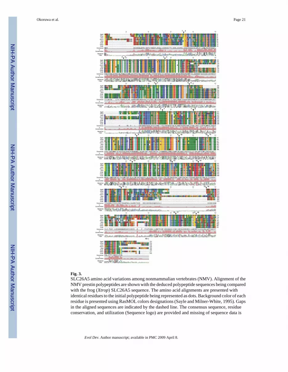
Fig. 3.SLC26A5 amino acid variations among nonmammalian vertebrates (NMV). Alignment of theNMV prestin polypeptides are shown with the deduced polypeptide sequences being comparedwith the frog (Xtrop) SLC26A5 sequence. The amino acid alignments are presented withidentical residues to the initial polypeptide being represented as dots. Background color of eachresidue is presented using RasMOL colors designations (Sayle and Milner-White, 1995). Gapsin the aligned sequences are indicated by the dashed line. The consensus sequence, residueconservation, and utilization (Sequence logo) are provided and missing of sequence data is
Okoruwa et al. Page 21
Evol Dev. Author manuscript; available in PMC 2009 April 8.
NIH
-PA Author Manuscript
NIH
-PA Author Manuscript
NIH
-PA Author Manuscript

indicated by X. Residue number and the location of each exon boundary ( ) are provided atthe top of each row of sequence.
Okoruwa et al. Page 22
Evol Dev. Author manuscript; available in PMC 2009 April 8.
NIH
-PA Author Manuscript
NIH
-PA Author Manuscript
NIH
-PA Author Manuscript
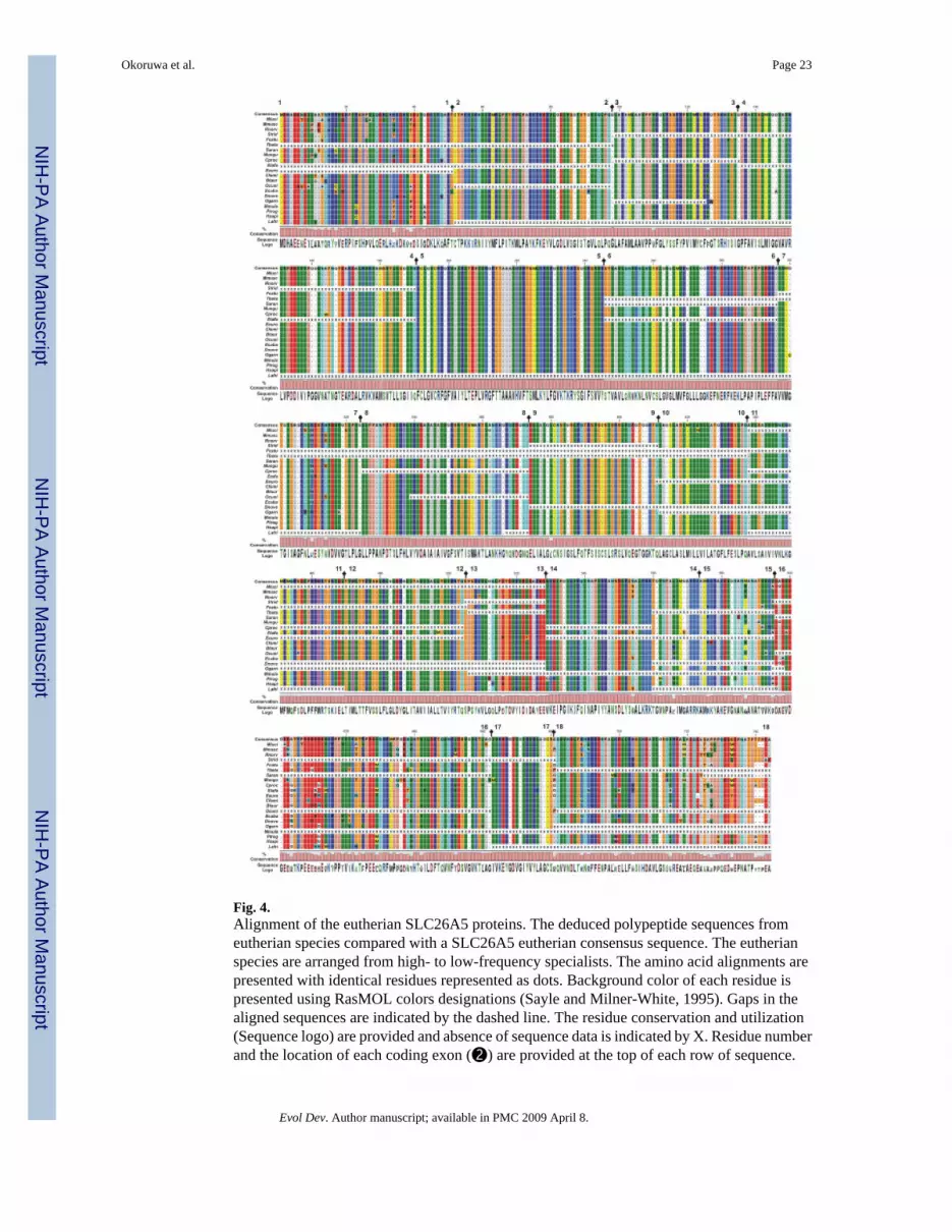
Fig. 4.Alignment of the eutherian SLC26A5 proteins. The deduced polypeptide sequences fromeutherian species compared with a SLC26A5 eutherian consensus sequence. The eutherianspecies are arranged from high- to low-frequency specialists. The amino acid alignments arepresented with identical residues represented as dots. Background color of each residue ispresented using RasMOL colors designations (Sayle and Milner-White, 1995). Gaps in thealigned sequences are indicated by the dashed line. The residue conservation and utilization(Sequence logo) are provided and absence of sequence data is indicated by X. Residue numberand the location of each coding exon (➋) are provided at the top of each row of sequence.
Okoruwa et al. Page 23
Evol Dev. Author manuscript; available in PMC 2009 April 8.
NIH
-PA Author Manuscript
NIH
-PA Author Manuscript
NIH
-PA Author Manuscript

Fig. 5.Alignment of the eutheria and nonmammalian vertebrate SLC26A5 consensus sequences withopossum and platypus polypeptides. The amino acid alignments are presented with identicalresidues to the initial polypeptide being shown as dots. Background color of each residue ispresented using RasMOL colors designations (Sayle and Milner-White, 1995). Gaps in thealigned sequences are indicated by the dashed line. The residue conservation and utilization(Sequence logo) are provided and absence of sequence data is indicated by X. The coding exoncontribution to the deduced amino acid sequences are indicated by ↕ and the number of theexon is indicated. The protein motifs were obtained from Ensembl protein features for mouse(Mmusc) SLC26A5. These motifs, prosites, domains are demarcated by: gray (▪)—
Okoruwa et al. Page 24
Evol Dev. Author manuscript; available in PMC 2009 April 8.
NIH
-PA Author Manuscript
NIH
-PA Author Manuscript
NIH
-PA Author Manuscript

hydrophobic transmembrane regions; red (▪)—SUL1 [InterPro: IPR001902], magenta (▪)—Sulph_transpt [Pfam: PF00916; InterPro: IPR011547], orange (▪)—SulP_transpt [Prositepatterns: PS01130; InterPro: IPR001902]; black (▪) [Pfam: PF01740; InterPro: IPR002645]and dark blue (▪) [Prosite profiles: PS50801; InterPro: IPR002645]—STAS motifs; and brown(▪)—Sodium:dicarboxylate symporter [PRINTS: PR00173 and InterPro: IPR001991].
Okoruwa et al. Page 25
Evol Dev. Author manuscript; available in PMC 2009 April 8.
NIH
-PA Author Manuscript
NIH
-PA Author Manuscript
NIH
-PA Author Manuscript
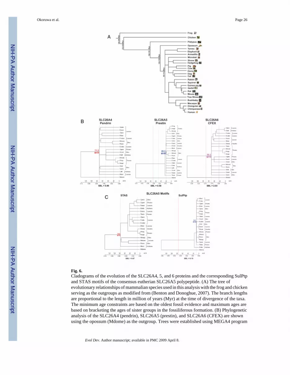
Fig. 6.Cladograms of the evolution of the SLC26A4, 5, and 6 proteins and the corresponding SulPtpand STAS motifs of the consensus eutherian SLC26A5 polypeptide. (A) The tree ofevolutionary relationships of mammalian species used in this analysis with the frog and chickenserving as the outgroups as modified from (Benton and Donoghue, 2007). The branch lengthsare proportional to the length in million of years (Myr) at the time of divergence of the taxa.The minimum age constraints are based on the oldest fossil evidence and maximum ages arebased on bracketing the ages of sister groups in the fossiliferous formation. (B) Phylogeneticanalysis of the SLC26A4 (pendrin), SLC26A5 (prestin), and SLC26A6 (CFEX) are shownusing the opossum (Mdome) as the outgroup. Trees were established using MEGA4 program
Okoruwa et al. Page 26
Evol Dev. Author manuscript; available in PMC 2009 April 8.
NIH
-PA Author Manuscript
NIH
-PA Author Manuscript
NIH
-PA Author Manuscript

(Tamura et al. 2007) with the following parameters: neighbor-joining, pairwise deletion,constant substitution rate, and the amino acid JTT matrix. The sums of the branch lengths (SBL)are given. The time scale (Myr) is based on the divergence of the marsupials from the eutherianlineage and are represented by red line (71.2 Myr—SLC26A4), a dashed blue line (20.3 Myr—SLC26A5), and a dotted purple line (50.2 Myr—SLC26A6). (C) Phyogenetic trees areshown for the SLC26A5 protein motifs, STAS (red line—76.6 Myr) and SulPtp (blue dottedline—9.0 Myr). Opossum was used as the outgroup and a time scale (Myr) and SBL areindicated.
Okoruwa et al. Page 27
Evol Dev. Author manuscript; available in PMC 2009 April 8.
NIH
-PA Author Manuscript
NIH
-PA Author Manuscript
NIH
-PA Author Manuscript

NIH
-PA Author Manuscript
NIH
-PA Author Manuscript
NIH
-PA Author Manuscript
Okoruwa et al. Page 28Ta
ble
1R
egio
nal v
aria
tions
in a
min
o ac
id su
bstit
utio
ns a
mon
g th
e SL
C26
A4–
6 or
thol
ogs
Prot
ein
dom
ain
Gen
ePa
ram
eter
sN
-term
inus
SulP
tpSU
L1′
Cha
rged
clu
ster
STA
SC
-term
inus
Tot
al
SLC
26A
4D
omai
n Si
tes
1–69
70–5
1751
8–61
061
1–62
862
9–73
373
4–78
01–
780
(Pen
drin
)%
subs
titut
ions
12.6
(125
/101
6)5.
7 (4
07/7
180)
5.6
(84/
1513
)7.
1 (2
2/31
1)6.
3 (1
01/1
601)
10.8
(75/
694)
6.6
(814
/123
15)
PAM
L0.
217±
0.03
90.
102±
0.00
90.
124±
0.01
90.
132±
0.04
60.
105±
0.01
80.
181±
0.03
90.
115±
0.00
7
SLC
26A
5D
omai
n Si
tes
1–65
66–5
0350
4–58
758
8–63
063
1–72
371
4–74
41–
744
(Pre
stin
)%
subs
titut
ions
7.3
(77/
1056
)0.
5 (3
9/74
97)
2.3
(32/
1390
)11
.7 (8
4/71
8)5.
1 (5
2/14
11)
15.9
(76/
477)
3.0
(380
/126
49)
PAM
L0.
120±
0.02
50.
012±
0.00
30.
079±
0.01
70.
179±
0.03
60.
086±
0.02
10.
176±
0.05
20.
048±
0.00
3
SLC
26A
6D
omai
n Si
tes
1–11
211
3–50
850
9–63
063
1–65
165
2–73
873
9–75
91–
759
(CFE
X)
% su
bstit
utio
ns7.
8 (1
39/1
774)
12.8
(811
/633
0)15
.8 (2
52/1
594)
26.5
(89/
336)
7.3
(105
/143
4)24
.7 (8
8/35
5)12
.6 (1
485/
1182
3)
PAM
L0.
288±
0.03
60.
192±
0.01
50.
240±
0.03
60.
783±
0.21
20.
163±
0.02
90.
285±
0.07
20.
234±
0.07
2
Evol Dev. Author manuscript; available in PMC 2009 April 8.


