Embed Size (px)
Citation preview
BioMed CentralBMC Musculoskeletal Disorders
BMC Musculoskeletal Disorders 2002, 3 xResearch articleExperimental arthritis induced by a clinical Mycoplasma fermentans isolateAntonio Rivera1, Antonio Yáñez*2, Gloria León-Tello3, Constantino Gil1, Silvia Giono4, Eduardo Barba5 and Lilia Cedillo1
Address: 1Centro de Investigaciones en Ciencias Microbiológicas, Instituto de Ciencias, Benemérita Universidad Autónoma de Puebla, Puebla, Pue. México, 2Laboratorio de Microbiología Oral, Facultad de Estomatología, Benemérita Universidad Autónoma de Puebla, Puebla, Pue, México, 3Facultad de Ciencias Químicas, Benemérita Universidad Autónoma de Puebla, Puebla, Pue, México, 4Escuela Nacional de Ciencias Biológicas, Instituto Politécnico Nacional and 5Escuela de Veterinaria, Benemérita Universidad Autónoma de Puebla, Puebla, Pue, México
E-mail: Antonio Rivera - [email protected]; Antonio Yáñez* - [email protected]; Gloria León-Tello - [email protected]; Constantino Gil - [email protected]; Silvia Giono - [email protected]; Eduardo Barba - [email protected]; Lilia Cedillo - [email protected]
*Corresponding author
AbstractBackground: Mycoplasma fermentans has been associated with rheumatoid arthritis. Recently, itwas detected in the joints and blood of patients with rheumatoid arthritis, but it is not clear yethow the bacteria enter the body and reach the joints. The purpose of this study was to determinethe ability of M. fermentans to induce experimental arthritis in rabbits following inoculation of thebacteria in the trachea and knee joints.
Methods: P-140 and PG-18 strains were each injected in the knee joints of 14 rabbits in order toevaluate and compare their arthritogenicity. P-140 was also injected in the trachea of 14 rabbits inorder to test the ability of the bacteria to reach the joints and induce arthritis.
Results: M. fermentans produced an acute arthritis in rabbits. Joint swelling appeared first in rabbitsinjected with P-140, which caused a more severe arthritis than PG-18. Both strains were able tomigrate to the uninoculated knee joints and they were detected viable in the joints all along theduration of the experiment. Changes in the synovial tissue were more severe by the end of theexperiment and characterized by the infiltration of neutrophils and substitution of adipose tissueby connective tissue. Rabbits intracheally injected with P-140 showed induced arthritis and thebacteria could be isolated from lungs, blood, heart, kidney, spleen, brain and joints.
Conclusion: M. fermentans induced arthritis regardless of the inoculation route. These findingsmay help explain why mycoplasmas are commonly isolated from the joints of rheumatic patients.
BackgroundNumerous studies have demonstrated that some virusesand bacteria including mycoplasmas could be relatedwith the pathogenesis of arthritis [1–4]. The primary hab-
itats of human and animal mycoplasmas are the mucoussurface of the respiratory and urogenital tracts, the eyes,mammary glands and joints [5]. Mycoplasmas have beenincriminated as causative agents of arthritis since they
Published: 3 June 2002
BMC Musculoskeletal Disorders 2002, 3:15
Received: 6 February 2002Accepted: 3 June 2002
This article is available from: http://www.biomedcentral.com/1471-2474/3/15
© 2002 Rivera et al; licensee BioMed Central Ltd. Verbatim copying and redistribution of this article are permitted in any medium for any purpose, provided this notice is preserved along with the article's original URL.
Page 1 of 7(page number not for citation purposes)
BMC Musculoskeletal Disorders 2002, 3 http://www.biomedcentral.com/1471-2474/3/15
were found in rats and mice with natural arthritis 50 yearsago [6,7]. Bartholomew isolated M. hominis, M. hyorhinisand M. arthritidis from the synovial fluid in 14 of 17 pa-tients with rheumatoid arthritis, systemic lupus erythema-tosus and Reiter's Syndrome in 1965 [8].
Williams et al. reported the presence of Mycoplasma fer-mentans in the synovial fluid of patients with rheumatoidarthritis [7], but these results could not be reproduced infurther investigations probably because of the fastidiousgrowth requirements of the microorganism [9]. Recently,in 1997, Schaeverbeke et al. using molecular biology toolsdetected and also isolated M. fermentans in joints of pa-tients with rheumatoid arthritis and other joint disorders[10,11]. Further, Johnson et al. detected M. fermentans byPCR in 88% of synovial fluid samples from patients withdifferent arthritis [2].
Schaeverbeke et al. studied and compared the genotypiccharacteristics of seven strains isolated from the synovialfluid of patients with arthritis with three reference strainsof the bacteria [12]. Recent studies have also shown thepresence of M. fermentans DNA and specific antibodies tothe bacteria in the synovial fluid of patients with rheuma-toid arthritis [13]. This mycoplasma has been detected inblood of patients with rheumatoid arthritis, producingsystemic mycoplasmal infections [14], these results sug-gest that M. fermentans may play a role in the developmentof the disease. Some mycoplasmas may act as unspecificmitogens of B and T cells, while other mycoplasma (M. ar-thritidis) produce MAM, which shows superantigenic ac-tivity [5,15]. M. fermentans possesses a potentimmunomodulator product named macrophage activat-ing lipopeptide 2 (MALP-2) which activates macrophagesto release cytokines which may in turn increase the neu-trophilic infiltrate into the joint. MALP-2 could induce therelease of macrophage-derived 5'-nucleotidase, this en-zyme may occur in joints in excessive amounts when M.fermentans is present in patients with rheumatoid arthritis[16]. These abilities are used by mycoplasmas during theinduction of natural arthritis in animals.
Mycoplasmas are common inhabitants of the human res-piratory and genitourinary tracts where they producechronic infections, they also are able to invade other tis-sues and in some cases induce an autoimmune response[5]. Pathogenesis of some human mycoplasmal infectionsis poorly understood, that is the case of Mycoplasma fer-mentans. Even this microorganism has been isolated in thejoints of rheumatic patients; it is not clear how the bacte-ria enter the body and how they reach the joints. On thebasis of these observations we attempted to experimental-ly reproduce an arthritic disease in rabbits employing a M.fermentans strain isolated from the respiratory tract of hu-mans. The second purpose of this study was to test if this
strain when injected in the respiratory tract was able toreach the joints and induce arthritis.
MethodsBacterial strains and growth conditionsM. fermentans P-140 was isolated from the respiratory tractof asthmatic patient in our laboratory [17] and M. fermen-tans PG-18 was a kind gift of Dr. Gail H. Cassell (Univer-sity of Alabama at Birmingham).
The strains were grown at 37°C in l liter of E broth consist-ing of 2.1% (wt/vol.) PPLO broth base (Difco), 20%(vol./vol.) horse serum, 0.002% (wt/vol.) phenol red,0.25% (wt/vol.) glucose and 10% (vol./vol.) yeast extract.
Animal modelForty-nine New Zealand-White rabbits weighing 3 to 4 kgwere used. The animal protocol complied with all relevantand institutional policies including the Helsinki Declara-tion. Blood and throat swabs of animals were checked forthe genus mycoplasma by culture and PCR in order to de-termine that rabbits were mycoplasma free.
Arthritogenic ability of M. fermentansThe right knee joints of 14 rabbits were injected with 0.1ml of a broth culture that contained 1 × 10⁶ colony form-ing units per milliliter (CFU/ml) of Mycoplasma fermentansP-140. Also 14 rabbits were injected with 0.1 ml of brothcultures that contained 1 × 10⁶ CFU/ml of Mycoplasma fer-mentans PG-18. The left joint received no treatment andwas considered a control. As negative controls seven rab-bits were injected with 0.1 ml of E broth (mycoplasmafree) in the right knee joint.
An increase in the joint diameter was considered an indi-cator of joint swelling. The diameters were measured twiceevery third day during the experiment. Two rabbits of eachexperimental group and one from the control group weresacrificed on day 3,5,8,13,20,27 and 34 after the injection.Both the left and right synovia received the same treat-ment; the synovial membrane was divided in two parts.The presence of viable mycoplasmas was determined byinoculating one half of the synovial membrane in l ml ofE broth and ten fold serial dilutions were also cultured aninoculated onto agar [18]. Blood cultures were performed.The presence of aerobic flora in synovium was determinedby plating sheep blood agar plates. Knee synovia were re-moved and immediately fixed in 10% formalin pH 7.0.Samples were embedded in paraffin, sectioned andstained with hematoxilin and eosin.
Arthritis induced by M. fermentans after tracheal injectionFourteen New Zealand white rabbits were intracheally in-jected with 0.1 ml of a broth culture that contained 1 ×10⁶ CFU/ml of Mycoplasma fermentans P-140. Valuation of
Page 2 of 7(page number not for citation purposes)
BMC Musculoskeletal Disorders 2002, 3 http://www.biomedcentral.com/1471-2474/3/15
joint swelling and sacrifice of the animals were performedin the same way as in the experiment about M. fermentansarthritogenic ability. Five milliliters of blood were extract-ed from heart and cultured before sacrifice. Animals weresacrificed by the injection of 5 ml of Anesthesal. Brain, tra-chea, lungs, heart, spleen, kidney and knee joints were ex-tracted, 1 cm3 of tissue or half of the synovia weredeposited in 0.9 ml of E broth and ten fold serial dilutionswere also done [18]. Tissue and synovial cultures, and his-topathological study of the synovia were performed as de-scribed in the experiment of arthritogenic ability of M.fermentans.
DNA amplificationA Polymerase Chain Reaction (PCR) test was used to con-firm the presence of M. fermentans in synovial tissues. Theoligonucleotide primers used for PCR detection wereRW004 5'(GGACTATTGTCTAAACAATTTCCC) 3' andRW005 5'(GGTTATTCGATTTCTAAATCGCCT) 3' de-signed by Wang, which amplified a 206-nucleotide specif-ic gene sequence within the insertion sequence-likeelement that exist in multiple copies only in the M. fer-mentans genome [19]. Before we used this PCR based testto detect M. fermentans, we confirmed the specificity ofthese primers.
The reaction mixture contained 50 mM KCl, 1.5 mMMgCl2, 10 mM Tris-HCL (pH 8.3), 0.2 mM of each deox-ynucleotide triphosphate, 6 µM of each primer and 1 unitof AmpliTaq® (Perkin Elmer Cetus, Emerville, CA.) in a to-tal volume of 50 µl. The sample to be analyzed (5 µl) wasalways added last. A diluted lysate of M. fermentans PG-18corresponding to 100 CCU and sterile water were used aspositive and negative controls respectively. The amplifica-tion involved 40 cycles, each consisted of denaturation at95°C for 25 s, primers annealing at 60°C for 60 s and ex-tension at 72°C for 60 s. The amplified products were an-alyzed by electrophoresis in 2% agarose gels andvisualized by UV light after ethidium bromide staining.
StatisticsMany researchers use at least three animals for each timepoint for a better statistical and experimental reproduc-tion. Although, animal models offer several advantages,one disadvantage is the cost. We used 49 rabbits in ourstudy and it represented a considerable cost so we werenot able to use a greater amount of animals. In previousstudies where we used the rabbit model to test the arthri-togenic ability of M. pneumoniae, M. pulmonis and M. ar-thritidis[22] we injected 6 animals with each strain and wehad a small standard deviations and small animal-animalvariations. These facts gave us the confidence to use only2 rabbits in each time point. However, the use of only twoanimals may lower the statistical power of the results. TheMann Whitney U-statistic was used to assess the statistical
difference between articular diameters of M. fermentansPG-18 and P-140 and to compare the recovery of myco-plasmas in the knee synovial samples. The Kruskall-Wallistest was used to compare M. fermentans PG-18, P-140 in-traarticular injected, P-140 intracheally injected and con-trol.
ResultsWhen animal models are used it is important to check thatthey are mycoplasma free because the presence of thesemicroorganisms would change the results. The rabbitsthat we used were free of any genus of mycoplasma in thesites tested.
Arthritogenic ability of M. fermentansOne goal of this study was to experimentally reproduce ar-thritis in rabbits employing a clinical isolate from the hu-man respiratory tract and compare it with the arthritisproduced by a type strain. We used a strain isolated fromthe respiratory tract instead of one from joints because wewanted to test that a mycoplasma isolated from the hu-man respiratory tract was able to induce arthritis. Wefound that rabbits inoculated with Mycoplasma fermentansP-140 showed increases of five percent in the knee jointdiameter during the first seven days postinoculation. Thena decreased was observed, during the days 11 and 15, jointdiameters increased to 3.5% and other increase (4 %) involume of the joint was observed on days 17 and 19. (Ta-ble 1).
Joint increments induced by Mycoplasma fermentans PG-18were lesser than those induced by M. fermentans P-140.The only significant increment of the joint diameter(3.8%) produced by M. fermentans PG-18 was observedon day 23. This increase was similar in magnitude to thesecond increase induced by M. fermentans P-140 (Table 1).After 27 days a slow decrease of the joint diameter was ob-served in both groups of rabbits.
The group of rabbits that received E broth showed a min-imum increment of the joint diameter (0.7%), starting inthe day three and on day 15 the inflammation disap-peared. Kruskall-Wallis test showed statistical differences(P < 0.0001) in the increases of the joint diameter inducedby experimental group respect to control group. No statis-tical difference was observed in joint diameters betweenM. fermentans PG-18 and P-140. M. fermentans P-140 in-duced more severe inflammation in right knee joints thanPG-18 (p < 0.005).
Detection and quantitation of MycoplasmasSynovial samples obtained from knee joints injected withM. fermentans P-140 and PG18 showed 197 and 95 CFU/ml of the organisms respectively, after three days postin-oculation (p < 0.05). At the end of the experiments the
Page 3 of 7(page number not for citation purposes)
BMC Musculoskeletal Disorders 2002, 3 http://www.biomedcentral.com/1471-2474/3/15
number of mycoplasmas isolated dropped to 37 and 29respectively (p > 0.05). In the left knee joints (which werenot inoculated) 38 CFU/ml of P-140 were recovered dur-ing the first three days and 25 CFU/ml of PG-18, by theend of the experiment 43 CFU/ml and 35 CFU/ml, respec-tively were recovered. A significant difference (P = 0.01)was observed in the recovery of M. fermentans PG-18, be-tween right and left synovium (Table 2). Aerobic bacteriawere not isolated from the culture in blood agar. Bothstrains were recovered from blood cultures, suggestingthat mycoplasmas reach the blood stream and this could
explain in part why the uninoculated knee joints con-tained mycoplasmas by the end of the experiment. PCRdetection showed 206-bp products in the synovium sam-ples. Control rabbits did not show amplified fragments.
Arthritis induced by M. fermentans after intracheal injec-tionOnce we observed that M. fermentans P-140 was able to in-duce arthritis when it was intraarticular injected, our goalwas to test if M. fermentans P-140 was able to induce ar-
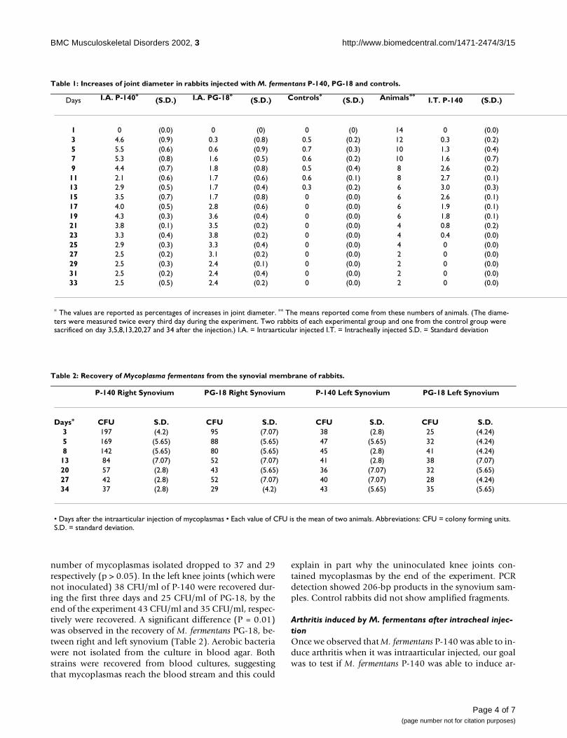
Table 1: Increases of joint diameter in rabbits injected with M. fermentans P-140, PG-18 and controls.
Days I.A. P-140*(S.D.) I.A. PG-18*
(S.D.) Controls*(S.D.) Animals**
I.T. P-140 (S.D.)
1 0 (0.0) 0 (0) 0 (0) 14 0 (0.0)3 4.6 (0.9) 0.3 (0.8) 0.5 (0.2) 12 0.3 (0.2)5 5.5 (0.6) 0.6 (0.9) 0.7 (0.3) 10 1.3 (0.4)7 5.3 (0.8) 1.6 (0.5) 0.6 (0.2) 10 1.6 (0.7)9 4.4 (0.7) 1.8 (0.8) 0.5 (0.4) 8 2.6 (0.2)11 2.1 (0.6) 1.7 (0.6) 0.6 (0.1) 8 2.7 (0.1)13 2.9 (0.5) 1.7 (0.4) 0.3 (0.2) 6 3.0 (0.3)15 3.5 (0.7) 1.7 (0.8) 0 (0.0) 6 2.6 (0.1)17 4.0 (0.5) 2.8 (0.6) 0 (0.0) 6 1.9 (0.1)19 4.3 (0.3) 3.6 (0.4) 0 (0.0) 6 1.8 (0.1)21 3.8 (0.1) 3.5 (0.2) 0 (0.0) 4 0.8 (0.2)23 3.3 (0.4) 3.8 (0.2) 0 (0.0) 4 0.4 (0.0)25 2.9 (0.3) 3.3 (0.4) 0 (0.0) 4 0 (0.0)27 2.5 (0.2) 3.1 (0.2) 0 (0.0) 2 0 (0.0)29 2.5 (0.3) 2.4 (0.1) 0 (0.0) 2 0 (0.0)31 2.5 (0.2) 2.4 (0.4) 0 (0.0) 2 0 (0.0)33 2.5 (0.5) 2.4 (0.2) 0 (0.0) 2 0 (0.0)
* The values are reported as percentages of increases in joint diameter. ** The means reported come from these numbers of animals. (The diame-ters were measured twice every third day during the experiment. Two rabbits of each experimental group and one from the control group were sacrificed on day 3,5,8,13,20,27 and 34 after the injection.) I.A. = Intraarticular injected I.T. = Intracheally injected S.D. = Standard deviation
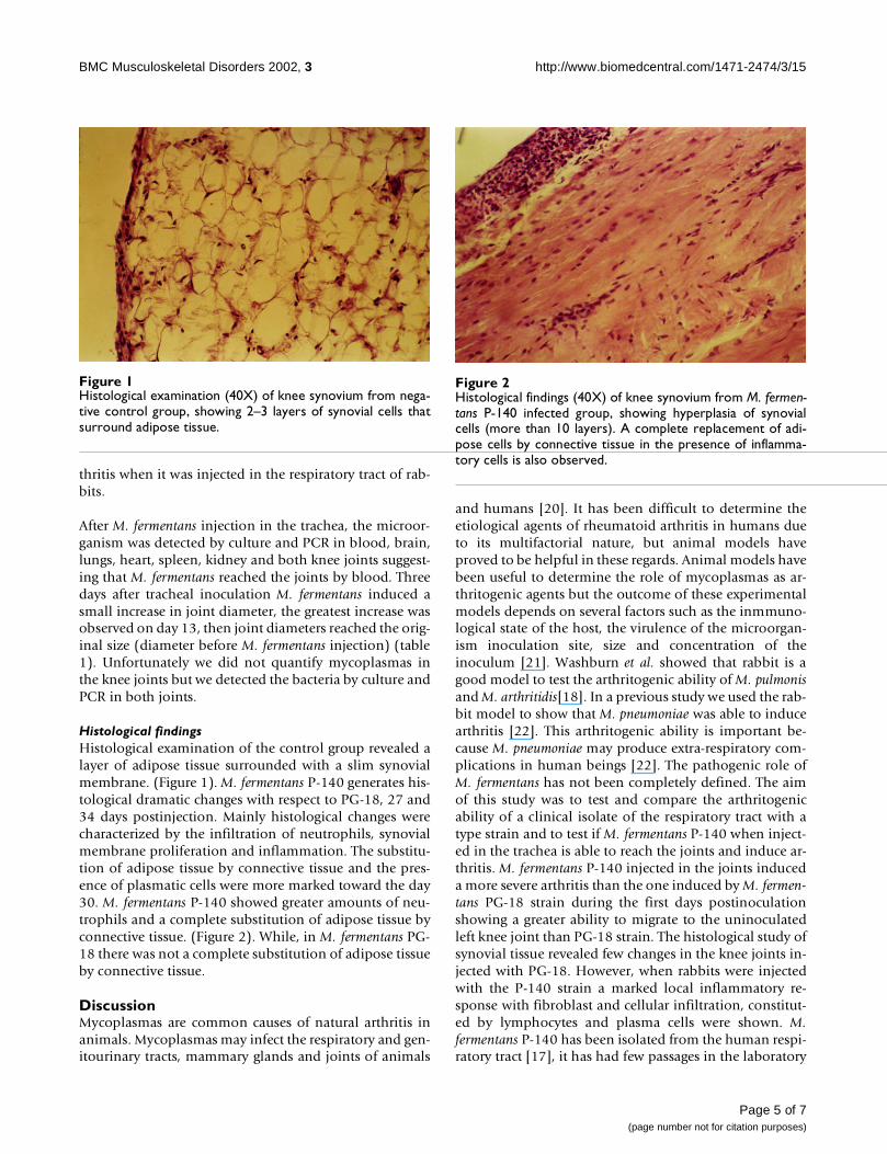
Table 2: Recovery of Mycoplasma fermentans from the synovial membrane of rabbits.
P-140 Right Synovium PG-18 Right Synovium P-140 Left Synovium PG-18 Left Synovium
Days* CFU S.D. CFU S.D. CFU S.D. CFU S.D.3 197 (4.2) 95 (7.07) 38 (2.8) 25 (4.24)5 169 (5.65) 88 (5.65) 47 (5.65) 32 (4.24)8 142 (5.65) 80 (5.65) 45 (2.8) 41 (4.24)
13 84 (7.07) 52 (7.07) 41 (2.8) 38 (7.07)20 57 (2.8) 43 (5.65) 36 (7.07) 32 (5.65)27 42 (2.8) 52 (7.07) 40 (7.07) 28 (4.24)34 37 (2.8) 29 (4.2) 43 (5.65) 35 (5.65)
• Days after the intraarticular injection of mycoplasmas • Each value of CFU is the mean of two animals. Abbreviations: CFU = colony forming units. S.D. = standard deviation.
Page 4 of 7(page number not for citation purposes)
BMC Musculoskeletal Disorders 2002, 3 http://www.biomedcentral.com/1471-2474/3/15
thritis when it was injected in the respiratory tract of rab-bits.
After M. fermentans injection in the trachea, the microor-ganism was detected by culture and PCR in blood, brain,lungs, heart, spleen, kidney and both knee joints suggest-ing that M. fermentans reached the joints by blood. Threedays after tracheal inoculation M. fermentans induced asmall increase in joint diameter, the greatest increase wasobserved on day 13, then joint diameters reached the orig-inal size (diameter before M. fermentans injection) (table1). Unfortunately we did not quantify mycoplasmas inthe knee joints but we detected the bacteria by culture andPCR in both joints.
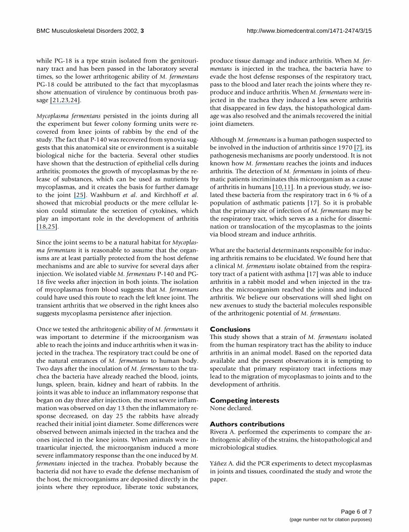
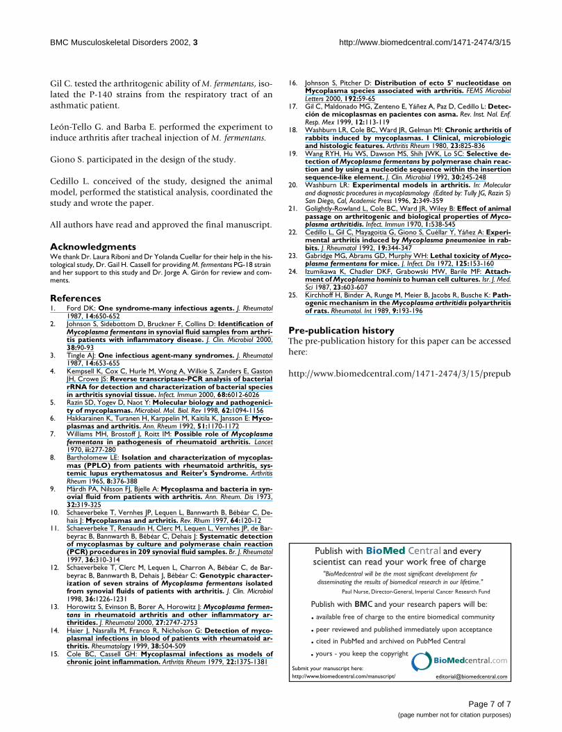
Histological findingsHistological examination of the control group revealed alayer of adipose tissue surrounded with a slim synovialmembrane. (Figure 1). M. fermentans P-140 generates his-tological dramatic changes with respect to PG-18, 27 and34 days postinjection. Mainly histological changes werecharacterized by the infiltration of neutrophils, synovialmembrane proliferation and inflammation. The substitu-tion of adipose tissue by connective tissue and the pres-ence of plasmatic cells were more marked toward the day30. M. fermentans P-140 showed greater amounts of neu-trophils and a complete substitution of adipose tissue byconnective tissue. (Figure 2). While, in M. fermentans PG-18 there was not a complete substitution of adipose tissueby connective tissue.
DiscussionMycoplasmas are common causes of natural arthritis inanimals. Mycoplasmas may infect the respiratory and gen-itourinary tracts, mammary glands and joints of animals
and humans [20]. It has been difficult to determine theetiological agents of rheumatoid arthritis in humans dueto its multifactorial nature, but animal models haveproved to be helpful in these regards. Animal models havebeen useful to determine the role of mycoplasmas as ar-thritogenic agents but the outcome of these experimentalmodels depends on several factors such as the inmmuno-logical state of the host, the virulence of the microorgan-ism inoculation site, size and concentration of theinoculum [21]. Washburn et al. showed that rabbit is agood model to test the arthritogenic ability of M. pulmonisand M. arthritidis[18]. In a previous study we used the rab-bit model to show that M. pneumoniae was able to inducearthritis [22]. This arthritogenic ability is important be-cause M. pneumoniae may produce extra-respiratory com-plications in human beings [22]. The pathogenic role ofM. fermentans has not been completely defined. The aimof this study was to test and compare the arthritogenicability of a clinical isolate of the respiratory tract with atype strain and to test if M. fermentans P-140 when inject-ed in the trachea is able to reach the joints and induce ar-thritis. M. fermentans P-140 injected in the joints induceda more severe arthritis than the one induced by M. fermen-tans PG-18 strain during the first days postinoculationshowing a greater ability to migrate to the uninoculatedleft knee joint than PG-18 strain. The histological study ofsynovial tissue revealed few changes in the knee joints in-jected with PG-18. However, when rabbits were injectedwith the P-140 strain a marked local inflammatory re-sponse with fibroblast and cellular infiltration, constitut-ed by lymphocytes and plasma cells were shown. M.fermentans P-140 has been isolated from the human respi-ratory tract [17], it has had few passages in the laboratory
Figure 1Histological examination (40X) of knee synovium from nega-tive control group, showing 2–3 layers of synovial cells thatsurround adipose tissue.
Figure 2Histological findings (40X) of knee synovium from M. fermen-tans P-140 infected group, showing hyperplasia of synovialcells (more than 10 layers). A complete replacement of adi-pose cells by connective tissue in the presence of inflamma-tory cells is also observed.
Page 5 of 7(page number not for citation purposes)
BMC Musculoskeletal Disorders 2002, 3 http://www.biomedcentral.com/1471-2474/3/15
while PG-18 is a type strain isolated from the genitouri-nary tract and has been passed in the laboratory severaltimes, so the lower arthritogenic ability of M. fermentansPG-18 could be attributed to the fact that mycoplasmasshow attenuation of virulence by continuous broth pas-sage [21,23,24].
Mycoplasma fermentans persisted in the joints during allthe experiment but fewer colony forming units were re-covered from knee joints of rabbits by the end of thestudy. The fact that P-140 was recovered from synovia sug-gests that this anatomical site or environment is a suitablebiological niche for the bacteria. Several other studieshave shown that the destruction of epithelial cells duringarthritis; promotes the growth of mycoplasmas by the re-lease of substances, which can be used as nutrients bymycoplasmas, and it creates the basis for further damageto the joint [25]. Washburn et al. and Kirchhoff et al.showed that microbial products or the mere cellular le-sion could stimulate the secretion of cytokines, whichplay an important role in the development of arthritis[18,25].
Since the joint seems to be a natural habitat for Mycoplas-ma fermentans it is reasonable to assume that the organ-isms are at least partially protected from the host defensemechanisms and are able to survive for several days afterinjection. We isolated viable M. fermentans P-140 and PG-18 five weeks after injection in both joints. The isolationof mycoplasmas from blood suggests that M. fermentanscould have used this route to reach the left knee joint. Thetransient arthritis that we observed in the right knees alsosuggests mycoplasma persistence after injection.
Once we tested the arthritogenic ability of M. fermentans itwas important to determine if the microorganism wasable to reach the joints and induce arthritis when it was in-jected in the trachea. The respiratory tract could be one ofthe natural entrances of M. fermentans to human body.Two days after the inoculation of M. fermentans to the tra-chea the bacteria have already reached the blood, joints,lungs, spleen, brain, kidney and heart of rabbits. In thejoints it was able to induce an inflammatory response thatbegan on day three after injection, the most severe inflam-mation was observed on day 13 then the inflammatory re-sponse decreased, on day 25 the rabbits have alreadyreached their initial joint diameter. Some differences wereobserved between animals injected in the trachea and theones injected in the knee joints. When animals were in-traarticular injected, the microorganism induced a moresevere inflammatory response than the one induced by M.fermentans injected in the trachea. Probably because thebacteria did not have to evade the defense mechanism ofthe host, the microorganisms are deposited directly in thejoints where they reproduce, liberate toxic substances,
produce tissue damage and induce arthritis. When M. fer-mentans is injected in the trachea, the bacteria have toevade the host defense responses of the respiratory tract,pass to the blood and later reach the joints where they re-produce and induce arthritis. When M. fermentans were in-jected in the trachea they induced a less severe arthritisthat disappeared in few days, the histopathological dam-age was also resolved and the animals recovered the initialjoint diameters.
Although M. fermentans is a human pathogen suspected tobe involved in the induction of arthritis since 1970 [7], itspathogenesis mechanisms are poorly understood. It is notknown how M. fermentans reaches the joints and inducesarthritis. The detection of M. fermentans in joints of rheu-matic patients incriminates this microorganism as a causeof arthritis in humans [10,11]. In a previous study, we iso-lated these bacteria from the respiratory tract in 6 % of apopulation of asthmatic patients [17]. So it is probablethat the primary site of infection of M. fermentans may bethe respiratory tract, which serves as a niche for dissemi-nation or translocation of the mycoplasmas to the jointsvia blood stream and induce arthritis.
What are the bacterial determinants responsible for induc-ing arthritis remains to be elucidated. We found here thata clinical M. fermentans isolate obtained from the respira-tory tract of a patient with asthma [17] was able to inducearthritis in a rabbit model and when injected in the tra-chea the microorganism reached the joints and inducedarthritis. We believe our observations will shed light onnew avenues to study the bacterial molecules responsibleof the arthritogenic potential of M. fermentans.
ConclusionsThis study shows that a strain of M. fermentans isolatedfrom the human respiratory tract has the ability to inducearthritis in an animal model. Based on the reported dataavailable and the present observations it is tempting tospeculate that primary respiratory tract infections maylead to the migration of mycoplasmas to joints and to thedevelopment of arthritis.
Competing interestsNone declared.
Authors contributionsRivera A. performed the experiments to compare the ar-thritogenic ability of the strains, the histopathological andmicrobiological studies.
Yáñez A. did the PCR experiments to detect mycoplasmasin joints and tissues, coordinated the study and wrote thepaper.
Page 6 of 7(page number not for citation purposes)
BMC Musculoskeletal Disorders 2002, 3 http://www.biomedcentral.com/1471-2474/3/15
Gil C. tested the arthritogenic ability of M. fermentans, iso-lated the P-140 strains from the respiratory tract of anasthmatic patient.
León-Tello G. and Barba E. performed the experiment toinduce arthritis after tracheal injection of M. fermentans.
Giono S. participated in the design of the study.
Cedillo L. conceived of the study, designed the animalmodel, performed the statistical analysis, coordinated thestudy and wrote the paper.
All authors have read and approved the final manuscript.
AcknowledgmentsWe thank Dr. Laura Riboni and Dr Yolanda Cuellar for their help in the his-tological study, Dr. Gail H. Cassell for providing M. fermentans PG-18 strain and her support to this study and Dr. Jorge A. Girón for review and com-ments.
References1. Ford DK: One syndrome-many infectious agents. J. Rheumatol
1987, 14:650-6522. Johnson S, Sidebottom D, Bruckner F, Collins D: Identification of
Mycoplasma fermentans in synovial fluid samples from arthri-tis patients with inflammatory disease. J. Clin. Microbiol 2000,38:90-93
3. Tingle AJ: One infectious agent-many syndromes. J. Rheumatol1987, 14:653-655
4. Kempsell K, Cox C, Hurle M, Wong A, Wilkie S, Zanders E, GastonJH, Crowe JS: Reverse transcriptase-PCR analysis of bacterialrRNA for detection and characterization of bacterial speciesin arthritis synovial tissue. Infect. Immun 2000, 68:6012-6026
5. Razin SD, Yogev D, Naot Y: Molecular biology and pathogenici-ty of mycoplasmas. Microbiol. Mol. Biol. Rev 1998, 62:1094-1156
6. Hakkarainen K, Turanen H, Karppelin M, Kaitila K, Jansson E: Myco-plasmas and arthritis. Ann. Rheum 1992, 51:1170-1172
7. Williams MH, Brostoff J, Roitt IM: Possible role of Mycoplasmafermentans in pathogenesis of rheumatoid arthritis. Lancet1970, ii:277-280
8. Bartholomew LE: Isolation and characterization of mycoplas-mas (PPLO) from patients with rheumatoid arthritis, sys-temic lupus erythematosus and Reiter's Syndrome. ArthritisRheum 1965, 8:376-388
9. Mårdh PA, Nilsson FJ, Bjelle A: Mycoplasma and bacteria in syn-ovial fluid from patients with arthritis. Ann. Rheum. Dis 1973,32:319-325
10. Schaeverbeke T, Vernhes JP, Lequen L, Bannwarth B, Bébéar C, De-hais J: Mycoplasmas and arthritis. Rev. Rhum 1997, 64:120-12
11. Schaeverbeke T, Renaudin H, Clerc M, Lequen L, Vernhes JP, de Bar-beyrac B, Bannwarth B, Bébéar C, Dehais J: Systematic detectionof mycoplasmas by culture and polymerase chain reaction(PCR) procedures in 209 synovial fluid samples. Br. J. Rheumatol1997, 36:310-314
12. Schaeverbeke T, Clerc M, Lequen L, Charron A, Bébéar C, de Bar-beyrac B, Bannwarth B, Dehais J, Bébéar C: Genotypic character-ization of seven strains of Mycoplasma fermentans isolatedfrom synovial fluids of patients with arthritis. J. Clin. Microbiol1998, 36:1226-1231
13. Horowitz S, Evinson B, Borer A, Horowitz J: Mycoplasma fermen-tans in rheumatoid arthritis and other inflammatory ar-thritides. J. Rheumatol 2000, 27:2747-2753
14. Haier J, Nasralla M, Franco R, Nicholson G: Detection of myco-plasmal infections in blood of patients with rheumatoid ar-thritis. Rheumatology 1999, 38:504-509
15. Cole BC, Cassell GH: Mycoplasmal infections as models ofchronic joint inflammation. Arthritis Rheum 1979, 22:1375-1381
16. Johnson S, Pitcher D: Distribution of ecto 5' nucleotidase onMycoplasma species associated with arthritis. FEMS MicrobiolLetters 2000, 192:59-65
17. Gil C, Maldonado MG, Zenteno E, Yáñez A, Paz D, Cedillo L: Detec-ción de micoplasmas en pacientes con asma. Rev. Inst. Nal. Enf.Resp. Mex 1999, 12:113-119
18. Washburn LR, Cole BC, Ward JR, Gelman MI: Chronic arthritis ofrabbits induced by mycoplasmas. I Clinical, microbiologicand histologic features. Arthritis Rheum 1980, 23:825-836
19. Wang RYH, Hu WS, Dawson MS, Shih JWK, Lo SC: Selective de-tection of Mycoplasma fermentans by polymerase chain reac-tion and by using a nucleotide sequence within the insertionsequence-like element. J. Clin. Microbiol 1992, 30:245-248
20. Washburn LR: Experimental models in arthritis. In: Molecularand diagnostic procedures in mycoplasmology (Edited by: Tully JG, Razin S)San Diego, Cal, Academic Press 1996, 2:349-359
21. Golightly-Rowland L, Cole BC, Ward JR, Wiley B: Effect of animalpassage on arthritogenic and biological properties of Myco-plasma arthritidis. Infect. Immun 1970, 1:538-545
22. Cedillo L, Gil C, Mayagoitia G, Giono S, Cuéllar Y, Yáñez A: Experi-mental arthritis induced by Mycoplasma pneumoniae in rab-bits. J. Rheumatol 1992, 19:344-347
23. Gabridge MG, Abrams GD, Murphy WH: Lethal toxicity of Myco-plasma fermentans for mice. J. Infect. Dis 1972, 125:153-160
24. Izumikawa K, Chadler DKF, Grabowski MW, Barile MF: Attach-ment of Mycoplasma hominis to human cell cultures. Isr. J. Med.Sci 1987, 23:603-607
25. Kirchhoff H, Binder A, Runge M, Meier B, Jacobs R, Busche K: Path-ogenic mechanism in the Mycoplasma arthritidis polyarthritisof rats. Rheumatol. Int 1989, 9:193-196
Pre-publication historyThe pre-publication history for this paper can be accessedhere:
http://www.biomedcentral.com/1471-2474/3/15/prepub
Publish with BioMed Central and every scientist can read your work free of charge
"BioMedcentral will be the most significant development for disseminating the results of biomedical research in our lifetime."
Paul Nurse, Director-General, Imperial Cancer Research Fund
Publish with BMC and your research papers will be:
available free of charge to the entire biomedical community
peer reviewed and published immediately upon acceptance
cited in PubMed and archived on PubMed Central
yours - you keep the copyright
[email protected] your manuscript here:http://www.biomedcentral.com/manuscript/
BioMedcentral.com
Page 7 of 7(page number not for citation purposes)