Embed Size (px)
Citation preview

DEVELOPMENTAL DYNAMICS 205308-318 (1996)
Expression of an Msx Homeobox Gene in Ascidians: Insights Into the Archetypal Chordate Expression Pattern LIANG MA, BILLIE J. SWALLA, JING ZHOU, SONIA L. DOBIAS, JEFFREY R. BELL, JUAN CHEN, ROBERT E. MAXSON, AND WILLIAM R. JEFFERY Department of Biochemistry and Molecular Biology and Kenneth R. Norris Cancer Hospital and Research Institute, University of Southern California, School of Medicine, Los Angeles, California 90033 (L.M., S.L.D., J.R.B., J.C., R.M.); Bodega Marine Laboratory and Section of Molecular and Cellular Biology, University of California Davis, Bodega Bay, California 94923 (B.J.S., J.Z., W.R.J.); Station Biologique, Roscoff, France (B.J.S., J.Z., W.R.J.)
ABSTRACT The Msx homeobox genes are expressed in complex patterns during vertebrate development in conjunction with inductive tissue interactions. As a means of understanding the archetypal role of Msx genes in chordates, we have isolated and characterized an Msx gene in ascidians, protochordates with a relatively simple body plan. The Mocu Msx-a and McMsx-a genes, isolated from the ascidians Molgula oculata and Molgula citrinu, respectively, have homeodo- mains that place them in the msh-like subclass of M.w genes. Therefore, the Molgula M.Px-a genes are most closely related to the msh genes previ- ously identified in a number of invertebrates. Southern blot analysis suggests that there are one or two copies of the Msx-a gene in the Molgula genome. Northern blot and RNase protection analysis indicate that Msx-a transcripts are re- stricted to the developmental stages of the life cy- cle. In situ hybridization showed that Msx-a mRNA first appears just before gastrulation in the mesoderm (presumptive notochord and muscle) and ectoderm (neural plate) cells. Transcript levels decline in mesoderm cells after the comple- tion of gastrulation, but are enhanced in the fold- ing neural plate during neurulation. Later, Msx-a mRNA is also expressed in the posterior ectoderm and in a subset of the tail muscle cells. The ecto- derm and mesoderm cells that express Msx-a are undergoing morphogenetic movements during gastrulation, neurulation, and tail formation. Msx-a expression ceases after these cells stop mi- grating. The ascidian M. citrinu, in which adult tissues and organs begin to develop precociously in the larva, was used to study Msx-a expression during adult development. Msx-a transcripts are expressed in the heart primordium and the rudi- ments of the ampullae, epidermal protrusions with diverse functions in the juvenile. The heart and ampullae develop in regions where mesen- chyme cells interact with endodermal or epider- mal epithelia. A comparison of the expression pat- terns of the Molgula genes with those of their vertebrate congeners suggests that the archetypal roles of the Msx genes may be in morphogenetic
0 1996 WILEY-LISS, INC.
movements during embryogenesis and in mesen- chymal-epithelial interactions during organo- genesis. o 1996 Wiley-Liss, Inc.
Key words: Msx homeobox, Chordates, Ascidians
INTRODUCTION Homeobox genes are members of a large class of
transcriptional regulators that play important roles in animal development and evolution (Scott, 1994). The Hox genes are organized in clusters and ordered accord- ing to their expression domains along the anteroposte- rior axis (McGinnis and Krumlauf, 1992). Other classes of homeobox genes are scattered within the ge- nome and appear to play more diverse roles in growth and development. One such class is the Msx genes, characterized by a distinct and highly conserved home- odomain related to the Drosophila msh gene (Gehring, 1987). Members of the Msx gene family have been de- scribed in coelenterates (Schummer et al., 1992), in- sects (Walldorf et al., 1989), sea urchins (Bell et al., 19931, protochordates (Holland, 1991; Holland et al., 19941, and in each of the vertebrate classes (Hill et al., 1989; Takahashi and LeDouarin, 1990; Holland, 1991; Coelho et al., 1991; Suzuki et al., 1991; Yokouchi et al., 1991). The most thoroughly studied vertebrate mem- bers of the Msx family are the mouse Msx-1 (Hox 7) and Msx-2 (Hox-8) genes (Hill et al., 1989; Monaghan et al., 1991; Robert et al., 1989). Based on similarities in and around their homeodomains, the Msx genes are divided into msh-like, Msxl -like, and Msx2-like sublcasses (Bell et al., 1993). It has been suggested that the Msxl- like and Msx2-like genes evolved by duplication of an ancestral msh-like gene in the lower vertebrates (Hol- land, 1991). However, the recent identification in sea
Received August 11, 1995; accepted October 4, 1995. Address reprint requests/correspondence to Dr. Robert E. Maxson,
633 Norris Hospital, USUNorris Cancer Center, 1441 Eastlake Ave- nue, Los Angeles, CA 90033.
Liang Ma’s present address is Department of Medicine, Division of Genetics, Howard Hughes Medical Institute, Harvard Medical School, 20 Shattuck Street, Boston, MA 02115.
Billie J. Swalla’s present address is Department of Biology, Vander- bilt University, Nashville, TN 37235.
The first and second authors have made equal contributions to this study.

EXPRESSION OF AN ASCIDIAN Msx GENE 309
urchins of a gene with an Msx2-like homeodomain (Dobias et al., manuscript in preparation) suggests that the evolution of the Msx gene family may be more com- plicated.
Inductive interactions between cells are known to generate tissue diversity during embryogenesis and organogenesis (Gurdon, 1992). The Msx genes are ex- pressed at sites corresponding to some of these induc- tive activities. In Drosophila, the msh gene is ex- pressed in the segmental mesoderm and nervous system (Robert et al., 1989). The Drosophila gene tin- man, a divergent member of the Msx family, is ex- pressed in the heart primordium (Bodmer et al., 1990), and a null mutation at this locus results in deficiencies in heart development (Bodmer, 1993; Azpiazu and Frasch, 1993). In sea urchins, the SpMsx gene is ex- pressed in the archenteron and secondary mesenchyme cells, and later in a subregion of the oral ectoderm where the secondary mesenchyme cells contact it, thus inducing mouth structures (Dobias et al., manuscript in preparation). In vertebrates, Msx genes are ex- pressed in ectoderm and mesoderm cells during gastru- lation, in the neural folds, in mesenchyme cells derived from the cranial neural crest, and in regions of mesen- chymal-epithelial interactions in the myocardium, limb bid, eye, ear, t?onth, and skull (Davidson and Hill, 1991; Davidson, 1995). Tissue recombination experi- ments have shown that Msx gene expression in the limb, tooth, and other mesenchyme cells is dependent on the overlying epithelium (Davidson et al., 1991; Ta- kahashi et al., 1991, 1992; Jowett et al., 1993). The importance of the Msx genes in vertebrates is sup- ported by the development of cranialfacial and tooth defects in Msx-1 deficient mice (Satokata and Maas, 1994) and the discovery of a point mutation in the Msx-2 homeodomain in a human family affected with a craniofacial genetic disorder (Jabs et al., 1993).
Expression of Msx genes in cranial neural crest de- rivatives and sensory primordia suggests that the Msx genes may be involved in vertebrate evolution and em- phasizes the need to identify their archetypal develop- mental role (Davidson, 1995). A reasonable place to look for the primitive function of Msx genes is in the protochordates (tunicates and cephalochordates). Sin- gle msh-like genes have been described in the ascidian Ciona (Holland, 1991) and amphioxus (Holland et al., 1994), but their expression patterns have not been re- ported. As a means of understanding the role of Msx genes in the development and evolution of chordates, we have examined the expression of an Msx gene in ascidians. Ascidians are chordates with a body plan that is less complex than that of the vertebrates. The ascidian tadpole larva contains only about 2,500 cells and six different tissues, yet displays typical chordate features, including a dorsal nervous system, a noto- chord, and striated tail muscle cells (Satoh, 1994). The ascidian larva is considered a prototype of the ances- tral chordate from which the vertebrates could have
We have isolated and characterized closely related (probably orthologous) Msx genes in the ascidians Mol- gula oculata and Molgula citrina. The Molgula Msx genes are members of the msh-like subclass and are expressed primarily in ectoderm and mesoderm cells during embryogenesis and in the heart and ampullar primordia during organogenesis. We conclude that the archetypal role of the Msx genes is in morphogenetic cell movements and tissue interactions, and that the more complex Msx expression patterns evolved within the vertebrate lineage in parallel with increasing com- plexity of body organization.
RESULTS Identification of Molgula Msx Genes
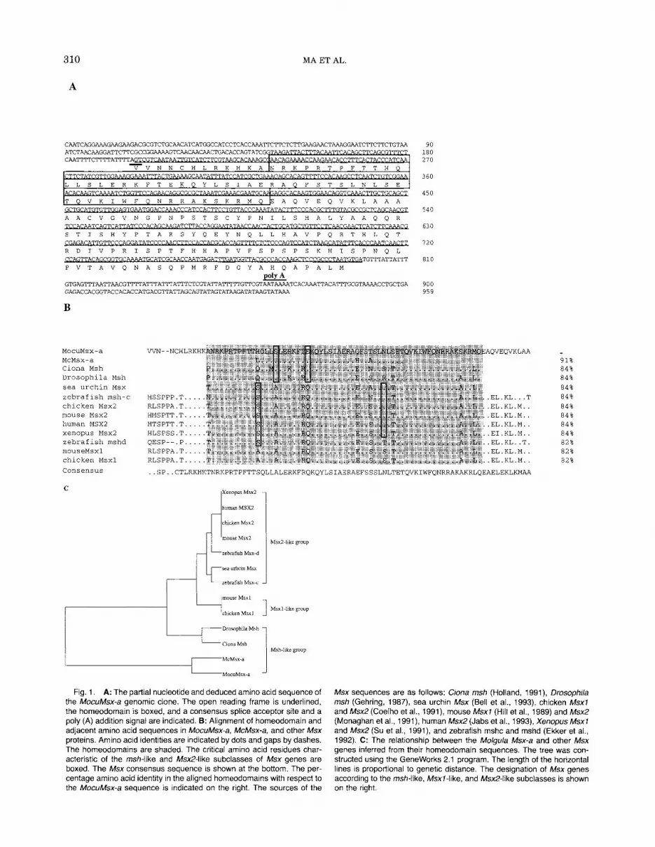
A multistep PCR protocol with primers correspond- ing to Msx homeobox and neighboring sequences (Bell et al., 1993) was used to amplify Msx genes from M . oculata and M. citrina. DNA fragments of the expected size were obtained, and upon sequencing, were found to contain part of an Msx-type homeodomain. The corre- sponding M. oculata and M . citrina genes were named MocuMsx-a and McMsx-a, respectively. A probe gener- ated from the MocuMsx-a homeobox was used to screen an M . oculata genomic DNA library for Msx clones. Thc partial sequence of an MocuMsx-a genomic clone is shown in Figure 1A. The sequenced region contains an open reading frame (ORF) including the MocuMsx-a homeodomain. A putative splice acceptor site was iden- tified just upstream and a polyadenylation site was lo- cated 67 bases downstream of the ORF, suggesting that the sequenced region corresponds to the second exon of an Msx gene. Alignment of the predicted MocuMsx-a amino acid sequence with the corresponding regions of other Msx proteins (Fig. 1B) confirmed its identifica- tion as an ascidian Msx gene.
Based on specific differences in the conserved homeodomain and adjacent regions, the Msx gene family is divided into msh-like, Msxl -like, and Msx2-like subclasses (Bell et al., 1993). The Mocu- Msx-a and McMsx-a homeodomains share consensus amino acids with the Drosophila and Ciona intestinalis msh genes (Fig. 1B). The alignment also shows significant homology in regions immediately adjacent to the MocuMsx-a homeodomain (Fig. lB), but no similarity with other Msx genes outside these regions. However, only the homeobox sequence is known in the Ciona msh gene (Holland, 1991), which is most similar to the Molgula Msx-a gene. The Molgula Msx homeodomains show sequence divergence with respect to each other and Ciona msh in three positions that are conserved in most other Msx proteins (Fig. 1B). The relationship between the Msx-a and other mem- bers of the Msx family is shown in Figure 1C. This comparison, which is based on a few specific differ- ences in the otherwise conserved homeodomain, suggests that the Msx-a genes are members of the
evolved by neoteny (Garstang, 1928). msh-like subclass of Msx genes.
310
A
MA ET AL
- V V N N C H L R K H K A I N R K P R T P F T T H Q
TATCGTTGGABBGEBBBmACTG&3&EA&%mATCCATCKTGWGTT'TTCCACA&XCTC.WTCTGTCC@,
CTTCCTGTTACCCPAATATACTTTCCCCACKTTTGTACGCCGCTCA~ A A C V G V N G P N P S T S C Y P N I L S H A L Y A A Q Q R <CTC.A.TCTTCACG A?. A A T A T S T I S H Y P T A R S Y Q E Y N Q L L H A V P Q R T H L Q T
R D I V P R I S P T F H H A P V F S P S P S K H I S P N O L
C ! & i G T T A C A G C G G T G C P A T C K . W C C M l ' G G % E E U S l 7 ' A C G C ~ G C X C C G C C C T A A T G n ; A TG'MTATTA'ITT P V T A V Q N A S Q P M R P D G Y A H Q A P A L M e G T G A G m A A T T A A C G T m A T T T A T T T A T T T C T C T C G T A m T T T G A G A G A C C A C G G T A C C A C A C C A T G A C G T T A T T A G C A G T T A G
B
MocuMsx-a MCMSX-a Ciona Msh Drosophila Msh sea urchin MSX zebrafish msh-c chicken Msx2 mouse Msx2 human MSXZ xenopus Msx2 zebrafish mshd mouseMsxl chicken Msxl Consensus
C
90 180 270
360
450
540
630
720
810
900 959
AQVEOVKLAA
84% 84%
..EL.KL ..T 84%
. EL.KL.M.. 84%
..EL.KL.M.. 84%
..EL.KL.M.. 84%
..EI.KL.M.. 84%
..EL.KL..T. 82%
..EL.KL.M.. 82%
..EL.KL.M. 82%
Xenopus Msx2
human MSXZ
chicken Max2
mouse Msx2
rebrafish Msx-d
MsxZ-like group
1 I T D r o s o p h i l a Msh
-Ciona Msh 1 Msh-like group
Fig. 1. A: The partial nucleotide and deduced amino acid sequence of the MocuMsx-a genomic clone. The open reading frame is underlined, the horneodomain is boxed, and a consensus splice acceptor site and a poly (A) addition signal are indicated. B: Alignment of homeodomain and adjacent amino acid sequences in MocuMsx-a, McMsx-a, and other Msx proteins. Amino acid identities are indicated by dots and gaps by dashes. The homeodomains are shaded. The critical amino acid residues char- acteristic of the msh-like and Msx2-like subclasses of Msx genes are boxed. The Msx consensus sequence is shown at the bottom. The per- centage amino acid identity in the aligned homeodomains with respect to the MocuMsx-a sequence is indicated on the right. The sources of the
Msx sequences are as follows: Ciona msh (Holland, 1991), Drosophila msh (Gehring, 1987), sea urchin Msx (Bell et al., 1993), chicken Msxl and Msx2 (Coelho et al., 1991), mouse Msxf (Hill et al., 1989) and Msx2 (Monaghan et al., 1991), human MsxZ(Jabs et al., 1993), Xenopus Msxf and Msx2 (Su et al., 1991), and zebrafish mshc and rnshd (Ekker et al., 1992). C: The relationship between the Molgula Msx-a and other Msx genes inferred from their horneodomain sequences. The tree was con- structed using the Geneworks 2.1 program. The length of the horizontal lines is proportional to genetic distance. The designation of Msx genes according to the msh-like, Msxf-like, and Msx2-like subclasses is shown on the right.

EXPRESSION OF AN ASCIDIAN Msx GENE 311
Molgula May Contain Multiple Msx Genes H1 digest showed a single band, the Eco RI and Hind111 digests contained two major bands of lower intensity
showed similar restriction enzyme cleavage patterns (data not shown). These results are consistent with the
Msx-a gene, although two alternative interpretations are that the Msx-a gene is single copy and exhibits a high degree of polymorphism, or that the MocuMsx-a probe reacts with a related gene. Exhaustive PCR screening did not yield other Molgula Msx genes, con- sistent with the view that one or two Msx-a genes are present in the Molgula genome.
we carried out Southern blots to estimate the num-
icDNA isolated from a single M. oculata individual was digested with Barn HI, Eco RI, and Hind 111 and the
The results are shown in Figure 2. Although the Barn
ber of Ms3c-a genes in the Molgula genome. Genom- and minor bands. Other Molgula
blots were hybridized with the M0cuMsx-a RNA probe. possibility that M o k u l a contains two copies of the
Temporal Expression of Msx-a mRNA The timing of Msx-a expression during early devel-
opment was determined by Northern blot and RNase protection analysis. A Northern blot containing RNA isolated from eggs and developing embryos was hybrid- ized with the MocuMsx-a antisense RNA probe. We detected a single 0.9 kb band, which first appeared at the gastrula stage, peaked in the neurula, and de- creased during the tailbud stages (Fig. 3). Transcripts were not detected in oocytes or early cleaving embryos (Fig. 3), indicating that Msx-a gene expression is strictly zygotic, as is the case with Msx genes in other organisms (Davidson and Hill, 1991). To determine whether Msx-a transcripts are also expressed in adults, we hybridized gastrula and adult RNA to kinetic ter- mination with the MocuMsx-a probe, treated the reac- tions with RNase, and resolved the protected products on polyacrylamide gels. As shown in Figure 4, a band of the expected size (290 bp) was present in embryonic but not adult RNA. A lower molecular weight band (about
Fig. 2. Southern blots of M. oculata genomic DNA digested with Barn HI (a), fco RI (b), or Hind 111 (c). Each lane was loaded with a digest of 10 pg of DNA from a single individual. The blot was hybridized with the MocuMsx-a probe.
3
Fig. 3. Temporal accumulation of Msx-a mRNA in M. oculafaembryos determined by Northern blot hybridization. The arrowhead indicates the position of the 0.9 kb MocuMsx-a transcript. From left to right the lanes were loaded with 10 pg of total RNA isolated from oocytes (a), 8-16 cell embryos (b), 64 cell embryos (c), mid-gastrulae (d), neurulae (e), mid- tailbud embryos (f), and late tailbud embryos (9). Equal loading of RNA was confirmed by quantification of 185 rRNA. The blot was hybridized with the MocuMsx-a antisense RNA probe.
Fig. 4. RNase protection analysis of MocuMsx-a expression in em- bryos and adults. The MocuMsx-a antisense RNA probe was hybridized with 20 pg of a tRNA control (a), total M. oculata gastrula RNA (b), or total M. oculafa adult RNA (c). The 290 bp fragment in b is indicated by the arrowhead.

312 MA ET AL.
Fig. 5. Spatial expression of the Msx-a gene during M. oculata em- bryogenesis determined by whole mount in situ hybridization. A, B and D, G were hybridized with the MocuMsx-a antisense RNA probe. A: A 32- cell embryo lacking Msx-a mRNA. 8: A 64-cell embryo showing Msx-a mRNA in the anterior (top) and posterior primary muscle lineage (bottom) cells of the vegetal hemisphere. C: A 64-cell embryo hybridized with the MocuMsx-a sense RNA probe. D: An early gastrula viewed from a lateral side showing Msx-a mRNA accumulation in presumptive notochord and
270 bp) was also protected. This band may reflect the presence of a related gene, or, since the RNA used in this analysis was obtained from several individuals, could be the result of sequence polymorphism. The re- sults suggest that Msx-a gene expression is restricted to the developmental period of the life cycle.
Spatial Expression of Msx-a mRNA The spatial pattern of Msx-a expression was deter-
mined by whole mount in situ hybridization. Mocu- Msx-a and McMsx-a antisense RNA probes were used to compare Msx-a expression during M. oculata and M. citrina development, respectively. This comparison was carried out because these species exhibit different lar- val forms (Berrill, 1935). M. oculata develops into a conventional tadpole larva, and adult tissues and or- gans do not differentiate until after metamorphosis (Swalla and Jeffery, 1990). In contrast, M. citrina forms a modified tadpole larva in which adult organs begin to differentiate before metamorphosis (Swalla et al., 1994), a heterochronic modification known as adulta- tion (Jagersten, 1972; Jeffery and Swalla, 1992).
The distribution of Msx-a mRNA during M . oculata embryogenesis is shown in Figure 5. Msx-a transcripts were first detected just before gastrulation (Fig. 5A-B). Hybridization with an MocuMsx-a sense RNA probe showed virtually undetectable levels of staining (Fig.
neural plate cells at the anterior lip (top left) and presumptive muscle cells at the posterior lip (top right of the blastopore, b). The dorsal (vegetal) side of the embryo is facing the top. E: A neurula showing Msx-a mRNA in the neural folds (nf) and posterior ectoderm (e). F: An early tailbud embryo showing Msx-a mRNA in the posterior ectoderm (e). G: A mid- tailbud embryo showing Msx-a expression in a subset of muscle cells (m) at the posterior tip of the tail. Scale bar = 20 pm.
5C). Comparison of embryos hybridized with antisense and sense probes suggests that there is a low level of Msx-a transcripts in all cells, although transcript lev- els were greatly enriched in specific cell types during development. In gastrulae, Msx-a mRNA was in- creased in the presumptive neural plate and notochord cells, which migrate over the anterior lip, and the pre- sumptive muscle cells, which migrate over the poste- rior lip of the blastopore (Fig. 5D). Msx-a transcripts declined in the mesoderm cells following their migra- tion into the embryo, but were increased in the neural plate, as it rolls inward to form the neural tube (Fig. 5E). During neurulation and continuing into the early tailbud stage (Fig. 5E-F), Msx-a mRNA accumulated in the posterior ectoderm, which will form the tail epi- dermis. At the mid-tailbud stage, Msx-a mRNA was confined to a few of the muscle cells a t the posterior tip of the tail (Fig. 5G). No further Msx-a expression was detected until after larval hatching, when transcripts appeared in epidermal regions of the swimming larva (data not shown; see Fig. 6E-F for M. citrina), which will develop into ampullae during metamorphosis.
The distribution of Msx-a mRNA during M. citrina embryogenesis is shown in Figure 6. The pattern of Msx-a expression was the same as that observed in M. oculatu with the following exceptions. First, Msx-a transcript levels increased in the muscle cells during

EXPRESSION OF AN ASCIDIAN Msr GENE 313
Fig. 6. Spatial expression of the Msx-a gene during M. cifrina em- bryogenesis determined by whole mount in situ hybridization. Msx-a mRNA distribution was examined using the McMsx-a antisense RNA probe. A: An early gastrula viewed from the dorsal (vegetal) side showing Msx-a mRNA in the presumptive muscle cells (rn) at the posterior lip of the blastopore (b). B: A late gastrula viewed from the dorsal side showing Msx-a mRNA in the notochord (no) and neural plate (np) cells at the anterior lip of the blastopore (b), which has almost closed. The scale bar in B represents 20 pn; magnification is the same in A and B. C,D: Mid and late tailbud embryos viewed from the lateral side showing Msx-a mRNA in the trunk (head) endoderm but not in the tail. E: A recently hatched larva viewed from the lateral side showing Msx-a mRNA in the anterior (left) and posterior (right) regions of the trunk. F: A swimming larva undergoing adultation viewed from the lateral side showing Msx-a mRNA in the anterior region (left), heart primordium (h), and several lateral sites in the trunk (arrowheads) where ampullae are forming. The black spots in the center of the trunk in E and F are melanized sensory cells in the brain. The scale bar in C represents 50 pn; magnification is the same in C-F.
gastrulation before they were elevated in the notochord and neural plate cells (Fig. 6A,B). Second, Msx-a ex- pression occurred throughout the trunk endoderm dur- ing the mid and late tailbud stages, but was lacking in the tail (Fig. 6C,D). The expression of the Msx-a gene in the endoderm may be related to heterochronic de- velopment of adult structures, which is typical of M . citrina and initiated in the trunk during the tailbud stages (Swalla et al., 1994). Third, Msx-a mRNA ap- peared in the heart primordium, as well as in the an- terior, lateral, and posterior regions of the trunk epi- dermis, which will later develop into ampullae (Fig. 6E,F). Msx-a expression was also examined by section- ing in situ hybridized larvae, and transcripts were ob-
Fig. 7. Spatial expression of the Msx-a gene in sections of in situ hybridized M. citrina larvae. A: A section through the larval trunk showing Msx-a rnRNA in the heart primordium (h) and in anterior (left) and pos- terior (right) epidermal epithelia. B: A section through the anterior portion of the larval trunk showing Msx-a mRNA expression in the heart primor- dium (h) and anterior epidermis. p: pericardial cavity. C: A section through the posterior region of the trunk showing Msx-a mRNA in pos- terior epidermal epithelia. Transcripts are also present in mesenchyme cells associated with the underlying endoderm (arrowheads). Scale bar: 15 pm.
served in the developing heart primordium and in ru- diments of the epidermal ampullae (Fig. 7). The heart and ampullar primordia are regions where mesen- chyme cells and epidermal or endodermal epithelia ap- pear to interact (Swalla et al., 1994). Thus, Msx-a is expressed in regions of mesenchymal-epithelial inter- actions during organogenesis. The embryonic and ju- venile expression patterns of the Msx-a gene appear to be superimposed during adultation in M . citrina.
DISCUSSION In vertebrates, Msx gene expression commences soon
after gastrulation, and proceeds in a complex pattern that correlates with inductive tissue interactions un- derlying organogenesis (Davidson and Hill, 1991). Here we describe Msx gene expression in ascidians, chordates with a simple larval body plan. The Msx-a gene is expressed in a relatively simple pattern in as- cidians, and the sites of expression are consistent with a role in morphogenetic cell movements and tissue in-

314 MA ET AL.
teractions in the embryo and developing juvenile. We suggest, moreover, that this expression pattern pro- vides insights into the archetypal role of the Msx genes in chordates.
Msx-a Genes Comparison of the ascidian Msx-a with other Msx
genes revealed a high degree of identity within the homeodomain and neighboring regions, but no signifi- cant homology outside these regions. The Msx-a home- odomain is more similar to that of Ciona and Drosoph- ila msh than to vertebrate Msx-1 and Msx-2, suggesting that the Msx-a genes are members of the msh-like subclass (Bell et al., 1993). However, the M. oculata Msx-a and Ciona msh homeodomains are diver- gent in 9 amino acid positions, some of which are also changed in the partially sequenced homeodomain of the M. citrina Msx-a gene. PCR analysis did not iden- tify additional Msx genes in either Molgula species. Although these negative results must be interpreted with caution, they suggest that the Molgula and Ciona msh-like genes are orthologous. We attribute the se- quence divergence of these genes to two factors: (1) the large phylogenetic distance known to separate the as- cidians Molgula and Ciona and (2) a fast molecular clock documented in the molgulid ascidians. Ciona in- testinalis and the Molgula species are members of the ascidian orders Enterogona and Pleurogona, respec- tively (Satoh, 1994). Comparison of 18s rDNA se- quences has shown that the genetic distance between these orders is greater than that between the amphib- ians and humans (Wada et al., 1992). Other compari- sons, based on both 18s and 28s rDNA sequences, sug- gest that the molgulids have a rapid rate of sequence divergence relative to other ascidian families (Had field et al., 1995). Thus, the Molgula Msx-a genes are likely to be divergent homologs of the Ciona and Drosophila msh genes.
The invertebrates and protochordates examined thus far possess a single Msx gene (Gehring, 1987; Holland, 1991; Holland et al., 1994; Dobias et al., manuscript in preparation), whereas vertebrates have two or more Msx genes (Davidson and Hill, 1991; Davidson, 1995). This has lead to the speculation that an msh-like gene duplicated and diverged to form the Msxl- and Msx2- like genes early during vertebrate evolution (Holland et al., 1994). As described above, the Molgula Msx-a genes, although divergent, are probably homologous to the msh genes of Ciona and other invertebrates, ap- pearing to support the idea that the Msx-1 and Msx-2 genes duplicated within the vertebrate lineage. How- ever, Southern blots showed multiple DNA bands hy- bridizing with the MocuMsx-a probe. There are several possible explanations for these results. First, there may be two copies of the Msx-a gene in the Molgula genome. Since a single copy msh gene has been de- scribed in Ciona, which is regarded to be a member of a more ancient ascidian group than Molgula (Berrill, 1936), the proposed duplication event may have oc-
curred during the evolution of molgulid ascidians. Sec- ond, although DNA from a single individual was used in the analysis, the degree of polymorphism may be extensive at the Molgula Msx-a locus. This interpreta- tion is consistent with a fast molecular clock in the family Molgulidae (Hadfield et al., 1995). It is possible that there is a related Msx-like gene present in ascid- ians that was not isolated by PCR analysis. Along with the recent identification in sea urchins of an Msx gene with an Msx2-like homeobox (Dobias et al., manuscript in preparation), this suggests that the Msx sub-family may have diverged earlier than previously appreci- ated, possibly during the emergence of the Deu- terostomes.
Maz Expression During Embryogenesis As in other animals, expression of the Msx-a gene in
ascidians is confined to the developmental period. In addition, Msx-a transcripts are absent from the gonads, which contain oocytes at all stages of development, in- dicating that the Msx-a gene does not contribute to the pool of maternal mRNAs. This is an important point, because early ascidian development is programmed to a large extent by maternal factors (Satoh, 1994). Our results imply that the Msx-a gene does not function in early developmental events, such as autonomous deter- mination of the primary muscle lineage (Nishida, 1992), although it is transiently expressed in muscle cells later in embryogenesis.
The Msx-a gene is expressed primarily in mesoderm (presumptive notochord and tail muscle cells) and ec- toderm (presumptive neural plate cells) during gastru- lation. Msx-a transcripts decline in the muscle and no- tochord cells shortly after these cells enter the blastopore, but are enhanced in the neural plate as it folds into the neural tube. The mesoderm cells showing enhanced Msx-a expression undergo specific changes in shape and migratory behavior known as involution (Trinkaus, 1984). The presumptive notochord cells in- volute over the anterior lip of the blastopore and the presumptive muscle cells involute over the posterior lip of the blastopore. In striking contrast, the endoderm cells, which participate in invagination rather than in- volution (Jeffery, 1992), do not accumulate appreciable levels of Msx-a mRNA. These results suggest that the Msx-a gene may function specifically in the involuting mesoderm. After the notochord and muscle cells have been internalized, the neural plate cells, which contain the highest levels of Msx-a transcripts a t the neurula stage, undergo changes in shape and roll inward to form the neural tube. The initial folding of the neural plate resembles involution of the mesoderm cells dur- ing gastrulation (see Satoh, 1978). Thus, the Msx-a gene may regulate similar morphogenetic events in the mesoderm and neural plate cells during the gastrula- tion and neurulation.
Msx-a transcripts are expressed in two additional lo- cations later in embryogenesis: the posterior ectoderm and a subset of muscle cells a t the tip of the tail. Ex-

EXPRESSION OF AN ASCIDIAN Msx GENE 315
pression in the posterior ectoderm is noteworthy be- cause these cells undergo morphogenetic movements in concert with the underlying notochord, neural tube, and muscle cells during tail formation. The muscle cells at the tip of the tail are also remarkable. While most of the tail muscle cells are generated by the pri- mary muscle lineage, which expresses Msx-a as it in- volutes over the posterior lip of the blastopore (see above), muscle cells at the tip of the tail are generated by the secondary muscle lineages (Satoh, 1994). The secondary muscle lineage cells are derived from cells in the anterior region of the fate map (Nishida, 1987)) which migrate extensively between gastrulation and the tailbud stage to reach the tip of the tail. Thus, Msx-a expression is also correlated with cell move- ments during later embryogenesis.
Msx Expression During Organogenesis The precocious differentiation of adult tissues and
organs during the larval phase in M. citrina has pro- vided information on the expression of the Msx-a gene during adult development. The results show that Msx-a mRNA accumulates in the developing heart. The heart develops from a primordium containing mes- enchyme cells and endoderm (De Seys-Longchamps, 1939). Msx-a expression in the developing ascidian heart is consistent with other studies in which Msx or Msx-related genes have been shown to be involved in cardiac development in Drosophila (Bodmer et al., 1990; Bodmer, 1993; Azpiazu and Frasch, 1993) and ver- tebrates (Chan-Thomas et al., 1993). These results sug- gest that aspects of Msx gene function underlying heart development may be conserved across wide phyloge- netic boundaries in the invertebrates and vertebrates. The Msx-a gene is also expressed at the sites of inter- action between mesenchyme and epidermis which will develop into ampullae. The ampullae are appendages that may function in adhesion to the substrate, respi- ration, and secretion of the adult tunic (Berrill, 1929; Cloney, 1982; Bates, 1991). Therefore, a common theme during adult development appears to be Msx-a expression in organ primordia generated by mesenchy- ma1 epithelial interactions.
Archetypal Role of Msx Genes in Chordate Development
The expression pattern of the Msx gene in ascidians is relatively simple compared to the complex patterns characteristic of Msx genes in higher vertebrates (Da- vidson and Hill, 1991; Davidson, 1995). During ascid- ian embryogenesis, Msx expression is elevated in the ectoderm and mesoderm cells that are undergoing mor- phogenetic movements. The expression pattern during ascidian gastrulation and neurulation is strikingly similar to aspects of Msx expression during vertebrate embryogenesis. For example, Msx genes are expressed in cells at the dorsal lip of the blastopore, in the invo- luting dorsal mesoderm, and neural folds in Xenopus embryos (Su et al., 1991) and in the primitive streak
and neural folds in chick embryos (Suzuki et al., 1991). Expression ceases in these tissues after they have com- pleted their migration. In the developing adult ascid- ian, Msx expression occurs in the heart and ampullar primordia, regions characterized by mesenchymal epi- thelial interaction. As described above, Msx genes are also expressed during vertebrate heart development. Although vertebrates have no organs resembling am- pullae, they do express Msx genes in the limb, mandib- ular process, tooth, and other locations in which organ primordia are developing based on epithelial mesen- chymal interactions (Davidson et al., 1991; Takahashi et al., 1991; Jowett et al., 1991). These results suggest that the archetypal function of Msx genes in chordates may be related to cell movements during gastrulation and neurulation and tissue interactions during organ- ogenesis. Thus, the complex pattern of Msx expression in vertebrates, particularly in derivatives of the cra- nial neural crest, probably evolved in the vertebrate lineage. Recent studies have shown that an Msx gene is also expressed during gastrulation and at sites of tissue interaction during sea urchin development (Dobias et al., manuscript in preparation). Although the Msx ex- pression pattern is remarkably similar in sea urchins, ascidians, and vertebrates, it remains to be determined whether Msx genes are functionally conserved in deli- terostomes.
The ascidian larva is sometimes considered as the prototype of the ancestral chordate from which the ver- tebrates may have evolved by neoteny (Garstang, 1928). Adultation, the process in which tissues begin to develop precociously in the larva, is thought to even- tually lead to neoteny (Jagersten, 1972). We suggest that our analysis of Msx gene expression in the ascid- ian M. citrina has provided insights into the origin of the vertebrates. We have shown that distinct patterns of Msx gene expression in the ascidian embryo and ju- venile are superimposed during adultation. A similar heterochronic alteration of Msx expression periods, along with the advent of new Msx functions in the cra- nial region (Gans and Northcutt, 1983), may have oc- curred during the evolution of vertebrates. The Msx gene may have a direct role in heterochrony. This pos- sibility is supported by the recent finding that a hyper- morphic mutation in the mouse Msx2 gene causes pre- mature fusion of the calvarial bones, a change in the relative timing of developmental events involved in patterning of the skull (Liu et al., 1995). Functional studies of the Msx genes may shed further light on the role of heterochrony in the evolution of the vertebrates.
EXPERIMENTAL PROCEDURES Ascidians
The ovoviviparous ascidian M. oculata was collected by dredging sand flats at Roscoff, France. The vivipa- rous ascidian M. citrina was obtained from the Marine Biological Laboratory (Woods Hole, MA). Adult ascid- ians were maintained in running sea water. M. oculata embryos were obtained by inseminating oocytes dis-

316 MA ET AL.
TABLE 1. Msx Oligonucleotide Primers and Corresponding Amino Acid Sequences
Primer Oligonucleotide sequence Amino acid sequence Position in Msx protein Mx 127 5'-AARCCNMGNACNCCNTTYAC-3' KPRTPFT N-terminal portion of homeodomain Mx 126 5'-TTYTCNARYTCNGCYTCYTG-3' KELEAEQ C-terminal just outside homeodomain Mx 220 5'-GNAARCAYAARRCNAAY MG-3' RKHKTNR N-terminal just outside homeodomain Mx 72 5'-GNACNRCNTTYACNNWC-3' RTTFTT C-terminal adjacent to Mx 127 Mx 221 5'-TGRAACCADATYTTNASYTG-3' QVKIWFQ C-terminal portion of homeodomain
sected from the gonads of gravid animals (Swalla and Jeffery, 1990). M. citrina embryos and hatched tadpole larvae were dissected from the brood chambers of gravid adults (Swalla et al., 1994). Embryos were cul- tured at 17°C in plastic Petri dishes containing Milli- pore (Bedford, MA) filtered sea water.
Isolation of DNA and RNA and Genomic Library Construction
DNA was isolated from dissected gonads according to the procedure of Davis et al. (1986). The M . oculata genomic library was constructed in the lambda FIX I1 vector (Stratagene, La Jolla, CA). Total RNA was iso- lated from M. citrina embryos and M. oculata gonads, embryos, and adults by the procedure of March et al. (1985).
Cloning and Sequencing the Molguln M m Genes The polymerase chain reaction (PCR) was performed
on M . oculata and M . citrina genomic DNA using de- generate oligodeoxynucleotide primers designed to hy- bridize to conserved regions within and adjacent to the homeodomains of Msx family genes (Bell et al., 1993) (Table 1). The first round of PCR was performed with 1 pg of genomic DNA, 0.5 pM of the primers Mx 220 and Mx 126 (Table l ) , 50 mM KC1, 1.5 mM MgCl,, 0.001% gelatin, 200 pM NTPs, 10 mM Tris-HC1 (pH 8.6>, and 1 U of Taq DNA polymerease (Boehringer Mannheim Biochemicals, Indianapolis, IN) in a final volume of 50 pl. We carried out 35 cycles of denaturation a t 94°C for 1 min, annealing at 50°C for 1 min, and extension at 72°C for 1 min. To increase the specificity of PCR am- plification, the first round PCR product was diluted 100-fold and used as a template in a second round of PCR using the Mx 127 and Mx 221 primers (Table 1) and the conditions described above. Subsequently, a third round of PCR was performed on the diluted sec- ond round PCR products using the primers Mx 72 and Mx 221 (Table 1). The third round PCR products were analyzed on a 0.7% agarose gel. As a result of the three rounds of PCR, a single band of about 130 bp was gen- erated from both the M . oculata and M . citrina DNA samples. The 130 bp DNA fragment was cloned into the pCRII vector (Invitrogen, San Diego, CA) according to the protocol provided. The nucleotide sequence of the inserts was determined by the dideoxy chain termina- tion method. The identity of the Molgula clones was confirmed by aligning the nucleotide and deduced pro- tein sequences with those of the Msx gene family.
An RNA probe was generated by linearizing the
plasmid containing the M . oculata subclone with Hin- dIII and transcription with T7 RNA polymerase. Screening the M . oculata genomic library with this RNA probe resulted in the isolation of three overlap- ping genomic clones. Restriction enzyme digestion and Southern blotting (see below) yielded a 6 kb HindIIIl Barn HI fragment that hybridized to the M . oculata insert. This DNA fragment was subsequently direc- tionally cloned into the Bluescript KS vector (Strata- gene) and partially sequenced using oligonucleotide primers corresponding to sequences within the known homeobox region. Computer analysis of sequence data and comparisons was carried out using the Geneworks 2.1 program (Intelligenetics, Mountain View, CA).
RNA Probes for Hybridizations Two oligonucleotides, Mx 246 (5'-GTGGATGGGTT-
TGGTS') and Mx 247 (5 '-AAAGTCAACAACAAC-3') were designed to generate an M . oculata Msx probe for the hybridization analyses. These oligonucleotides were used in a PCR reaction conducted as described above using the 6 kb M . oculata genomic clone as a template to generate a 374 bp DNA fragment. The fragment was cloned in pCRII and the identity and orientation of the insert was confirmed by sequencing. An Msx antisense RNA probe (MocuMsx-a) was gener- ated by linearizing the M . oculata subclone with Not I and transcription with Sp6 RNA polymerase using the Megascript kit (Ambion, Austin, TX). An M . citrina Msx antisense RNA probe (McMsx-a) was generated by linearizing the pCRII subclone containing the Msx ho- meobox with Not I and transcription with Sp6 poly- merase as described above. For some experiments, the corresponding sense RNA probes were also generated. Radioactive RNA probes were made using [32P]-UTP (800-3,000 Ci/mol). The radioactive nucleotides were purchased from New England Nuclear (Boston, MA). Digoxygenin (DIG)-labeled RNA probes were synthe- sized according to the instructions supplied with the Boehringer Mannheim kit.
RNase Protection RNase protection analysis was carried out by react-
ing 20 pg of total RNA with the radiolabeled Mocu- Msx-a antisense RNA probe. The RNase protection as- say was carried out using the RPAII kit (Ambion) according to protocol provided. The protected frag- ments were resolved on a denaturing 8% polyacryl- amide gel and visualized by autoradiography.

EXPRESSION OF AN ASCIDIAN Msx GENE 317
Filter Hybridizations Southern blot analysis was conducted with genomic
DNA exhaustively digested with Eco RI, Hzrz dIII, and Barn HI. The Southern blots were washed in 2 x SSC at 50°C. Northern hybridizations were carried out as described by Swalla et al. (1993). The Northern blots were washed in decreasing concentrations of SET (1 x SET: 30 mM Tris-HC1,2 mM EDTA, 150 mM NaC1) at increasing temperatures to obtain an appropriate sig- nal-to-noise ratio.
In Situ Hybridization Whole mount in situ hybridization was carried out
with DIG labeled antisense or sense RNA probes as described by Holland et al. (1991) with the following modifications. The fixed embryos were manually de- chorionated using tungsten needles. Treatment with proteinase K treatment was done at a concentration of 20 pg/ml for 30 min at 37°C. The hybridization and washing was done at 47”C, and the final wash was done in 0.5 x SSC, 50% formamide at 47°C. The whole mounts were cleared and photographed in 1:2 benzyl alcohol: benzyl benzoate. In some cases, the whole mounts were post-fixed in 4% paraformaldehyde, dehy- drated through an ethanol series, cleared in toluene, and embedded in Paraplast (Polysciences, Inc., War- rington, PA). The blocks were sectioned at 8 pm and viewed unstained.
ACKNOWLEDGMENTS We thank J. Machula, D. Martasian, and J. Reardon
for technical assistance. This research was supported by NIH grants HD-18582 (R.M.), HD-22416 (R.M.), and HD-07493 (W.R.J.) and NSF grants IBN-9304948 (B.J.S.) and DCB-9115543 (W.R.J.).
REFERENCES Azpiazu, N., and Frasch, M. (1993) tinman and bagpipe; Two homeo
box genes that determine cell fates in the dorsal mesoderm of Dro- sophila. Genes Dev. 7:1325-1340.
Bates, W.R. (1991) Ampullar morphogenesis in anural and urodele molgulid ascidians. Dev. Growth Differ. 33:401-411.
Bell, J.R., Noveen, A., Liu, Y.-H., Ma, L., Dobias, S., Kundu, R., Luo, W., Xia, Y., Lusis, A.J., Snead, M.L., and Maxson, R. (1993) Ge- nomic structure, chromosomal location, and evolution of the mouse Hox 8 gene. Genomics 16:123-131.
Berrill, N.J. (1929) Studies in tunicate development. Part I. General physiology of development of simple ascidians. Philos. Trans. R. SOC. Lond. [Biol.] 218:37-78.
Berrill, N.J. (1935) Studies in tunicate development. 111. Differential retardation and acceleration. Philos. Trans. R. SOC. Lond. [Biol.] 225255326.
Berrill, N.J. (1936) Studies in tunicate development. V. Evolution and classification. Philos. Trans. R. SOC. Lond. [Biol.] 226:43-70.
Bodmer, R. (1993) The gene tinman is required for specification of the heart and visceral muscles in Drosophila. Development 118:719- 729.
Bodmer, R., Jan, L.Y., and Jan, Y.N. (1990) A new homeobox-con- taining gene, msh-2, is transiently expressed early during meso- derm formation in Drosophila. Development 110:661-669.
Chan-Thomas, P.S., Thompson, R.P., Robert, B., Yacoub, M.H., and Barton, P.J.R. (1993) Expression of homeobox genes Msx-1 (Hex-7)
and Msx-2 (Hox-8) during cardiac development in the chick. Dev. Dyn. 197:203-216.
Cloney, R.A. (1982) Ascidian larvae and the events of metamorphosis. Am. Zool. 22:817-826.
Coelho, C.N.D., Sumoy, L., Rodger, B.J., Davidson, D.R., Hill, R.E., Upholt, W.B., and Kosher, R.A. (1991) Expression of a chicken ho- meobox-containing gene Ghox-8 during embryonic chick limb de- velopment. Mech. Dev. 34:143-154.
Davidson, D. (1995) The function and evolution of Msx genes: Pointers and paradoxes. Trends Genet. 11:375-422.
Davidson, D.R., and Hill, R.E. (1991) Msh-like genes: A family of homeobox genes with wide ranging expression during vertebrate development. Sem. Dev. Biol. 2:405-412.
Davidson, D.R., Crawley, A,, Hill, R.E., and Tickle, C. (1991) Position- dependent expression of two related homeobox genes in developing vertebrate limbs. Nature 352:429-431.
Davis, L.G., Dibner, M.D., and Batty, J.F. (1986) “Basic Methods in Molecular Biology.” New York Elsevier.
De Seys-Longchamps, M. (1939) Origine des premiere ebauches car- diaques chez le tuniciers. Tx. Stat. Zool. Wimereux 13529-634.
Ekker, M., Akimenko, M.-A,, Bremiller, R., and Westerfield, M. (1992) Regional expression of three homeobox transcripts in the inner ear of zebrafish embryos. Neuron 9:27-35.
Gans, C., and Northcutt, R.G. (1983) Neural crest and the origin of the vertebrates: A new head. Science 220:268-274.
Garstang, W. (1928) The morphology of the Tunicata, and its bearing on the phylogeny of the Chordata. Q. J. Microsc. SOC. 7551-187.
Gehring, W.J. (1987) The homeobox: Structural and evolutionary as- pects. In: “Molecular Approaches to Developmental Biology,” New York: A. R. Liss, pp. 115-129.
Grave, C. (1926) Molgula citrina (Alder and Hancock). Activities and structure of the free-swimming larva. J. Morphol. 42:453-471.
Gurdon, J.B. (1992) The generation of diversity and pattern in animal development. Cell 68:185-199.
Hadfield, K.A., Swalla, B.J., and Jeffery, W.R. (1995) Multiple origins of anural development in ascidians inferred from rDNA sequences. J. Mol. Evol. 40:413-427.
Hill, R.E., Jones, P.F., Rees, A.R., Sime, C.M., Justice, M.J., Copeland, N.G., Jenkins, N.A., Graham, E., and Davidson, D.R. (1989) A new family of mouse homeobox containing genes: Molecular structure, chromosomal localization and developmental expression of Hox 7.1. Genes Dev. 3:26-37.
Holland, P.W.H. (1991) Cloning and evolutionary analysis of the msh- like genes from the mouse, zebrafish and ascidian. Gene 98253- 257.
Holland, P.W.H., Holland, L.Z., Williams, N.A., and Holland, N.A. (1992) An amphioxus homeobox gene: Sequence conservation, spa- tial expression during development, and insights into vertebrate evolution. Development 116:653-661.
Holland, P.W.H., Garcia-Fernandez, J., Williams, N.A., and Sidow, A. (1994) Gene duplications and origins of vertebrate development. Development Suppl 1994125-133.
Jabs, E.W., Miiller, U., Li, X., Ma, L., Luo, W., Haworth, IS., Klisak, I., Sparks, R., Warman, M.L., Mulliken, J.B., Snead, M.L., and Max- son, R. (1993) A mutation in the homeodomain of the human MSX2 gene in a family affected with autosomal dominant craniosynosto- sis. Cell 75443-450.
Jagersten, C. (1972) “Evolution of the Metazoan Life Cycle. A Com- prehensive Theory.” New York: Academic.
Jeffery, W.R. (1992) A gastrulation center in the ascidian egg. Devel- opment Suppl 1992:53-63.
Jeffery, W.R., and Swalla, B.J. (1992) Evolution of alternate modes of development in ascidians. Bioessays 14919-226.
Jowett, A.K., Vainio, S., Ferguson, M.W.J., Sharpe, P.T. and Thesleff, I. (1993) Epithelial-mesenchymal interactions are required for Msx 1 and Msx 2 gene expression in the developing murine molar tooth. Development 117:461-470.
Liu, Y.-H., Kundu, R., Wu, L. Wen, L., Ignelzi, M.A. Jr., Snead, M.L., and Maxson, R.E. (1995) Premature suture closure and ectopic cra- nial bone in mice expressing Msx2 transgenes in the developing skull. Proc. Natl. Acad. Sci. U.S.A. (in press).
March, C.J., Mosley, B., Larsen, A., Cerreti, D.P., Braedt, G., Price,

318 MA ET AL.
V., Gillis, S., Henney, C.S., Kronheim, S.R., Grabstein, K., Conlon, P.J., Hopp, T.P., and Cosman, D. (1985) Cloning, sequence, and expression of two distinct human interleukin-1 complementary DNAs. Nature 315:641-647.
McGinnis, W., and Krumlauf, R. (1992) Homeobox genes and axial patterning. Cell 68:283-302.
Monaghan, A.P., Davidson, D.R., Sime, C., Graham, E., Baldock, R., Bhattacharya, S.S., and Hill, R.E. (1991) The msh-like genes define domains in the eye. Development 112:1053-1061.
Nishida, H. (1987) Cell lineage analysis in ascidian embryos by in- tracellular injection of a tracer enzyme. 111. Up to the tissue re- stricted stage. Dev. Biol. 121526-541.
Nishida, H. (1992) Regionality of egg cytoplasm that promotes muscle differentiation in embryo of the ascidian, Hulocynthiu roretzi. De- velopment 116:521-529.
Robert, B., Sassoon, D., Jacq, B., Gehring, W., and Buckingham, M. (1989) Hox-7, a mouse homeobox gene with a novel pattern of ex- pression during embryogenesis. EMBO J . 8:91-100.
Satoh, N. (1978) Cellular morphology and architecture during early morphogenesis of the ascidian egg: An SEM study. Biol. Bull. 155: 608-614.
Satoh, N. (1994) “Developmental Biology of Ascidians.” New York Cambridge.
Satokata, I., and Maas, R. (1994) Msxl deficient mice exhibit cleft palate and abnormalities of craniofacial and tooth development. Nature Genet. 6:348-355.
Schummer, M., Scheurlen, I., Schaller, C., and Galliot, B. (1992) HOM HOX homeobox genes are present in hydra (Chlorohydru uiridis- sima) and are differentially expressed during regeneration. EMBO J . 11:1815-1823.
Scott, M.P. (1994) Intimations of a creature. Cell 791121-1124. Su, M.-W., Suzuki, H.R., Solursh, M., and Ramirez, F. (1991) Progres-
sively restricted expression of a new homeobox-containing gene during Xenopus lueuis embryogenesis. Development 111:1179- 1187.
Suzuki, H.R., Padanilam, B.J., Vitale, E., Ramirez, F., and Solursh,
M. (1991) Repeating developmental expression of G-Hox-7, a novel homeobox containing gene in the chicken. Dev. Biol. 148:375-388.
Swalla, B.J., and Jeffery, W.R. (1990) Interspecific hybridization be- tween an anural and urodele ascidian: Differential expression of urodele features suggests multiple mechanisms control anural de- velopment. Dev. Biol. 142:319-334.
Swalla, B.J., Makabe, K.W., Satoh, N., and Jeffery, W.R. (1993) Novel genes expressed differentially in ascidians with alternate modes of development. Development 119307-318.
Swalla, B.J., White, M.E., Zhou, Z., and Jeffery, W.R. (1994) Hetero- chronic expression of an adult muscle actin gene during ascidian larval development. Dev. Genet. 15:51-63.
Takahashi, Y., and LeDouarin, N.M. (1990) cDNA cloning of a quail homeobox gene and its expression in neural crest-derived mesen- chyme and lateral plate mesoderm. Proc. Natl. Acad. Sci. U.S.A. 87:7482-7486.
Takahashi, Y., Bontoux, M., and LeDouarin, N.M. (1991) Epithelial mesenchymal interactions are critical for Quoz 7 expression and membrane bone differentiation in the neural crest derived mandib- ular mesenchyme. EMBO J. 9:2387-2393.
Takahashi, Y., Monsoroburq, A.H., Bontous, M., and Le Douarin, N.M. (1992) A role for Quoz 8 in the establishment of the dorsoven- tral pattern during vertebrate development. Proc. Natl. Acad. Sci. U.S.A. 8910237-10241.
Trinkaus, J.P. (1984) “Cells Into Organs. The Forces That Shape the Embryo.” Englewood Cliffs, NJ: Prentice-Hall.
Wada, H., Makabe, K.W., Nakauchi, M., and Satoh, N. (1992) Phylo- genetic relationships between solitary and colonial ascidians, as inferred from the central region of their respective 18s rDNAs. Biol. Bull. 183:448-455.
Walldorf, U., Fleig, R., and Gehring, W.J. (1989) Comparison of ho- meobox-containing genes of the honeybee and Drosophilu. Proc. Natl. Acad. Sci. U.S.A. 869971-9957.
Yokouchi, Y., Oshugi, K., Sasaki, H., and Kuroiwa, A. (1991) Chicken homeobox gene Msz-1: Structure, expression in limb buds and effect of retinoic acid. Development 113:431-444.


