Embed Size (px)
Citation preview

330
Expression of Heat Shock Proteins HSP70 and HSPSO in Endometrial Carcinomas Correlation with Clinicopathology, Sex Steroid Receptor Status, and p53 Protein Expression
Kanako Nanbu, M.D.
lkuo Konishi, M.D. Takayuki Komatsu, M.D.
Masaki Mandai, M.D.
Shinichi Yamamoto, M.D.
Hideki Kuroda, M.D.
Masafumi Koshiyama, M.D.
Takahide Mori, M.D.
Department of Gynecology and Obstetrics, Fac- ulty of Medicine, Kyoto University, Sakyo-ku, Kyoto, 606, Japan.
This study was supported by Grant-in-Aid for Scientific Research from the Ministry of Educa- tion 05454447, Japan.
Address for reprints: lkuo Konsihi, M.D., De- partment of Gynecology and Obstetrics, Faculty of Medicine, Kyoto University, Sakyo-ku, Kyoto, 606, Japan.
Received May 22, 1995; revision received Sep- tember 18,1995; accepted September 18,1995.
BACKGROUND. It has been suggested that heat shock proteins HSP70 and HSPSO are involved in the functional modulation of sex steroid receptors and are expressed in normal endometrium. However, little is known about the expression of HSP70 and HSPSO in endometrial carcinomas. METHODS. The immunohistochemical reactivity of monoclonal antibodies against HSP70 and HSPSO was examined in 42 endometrial carcinomas, and the presence or absence of correlation with the clinicopathologic features, sex steroid receptor status, and p53 protein expression was analyzed. RESULTS. Expression of HSP70 was found in 52% of endometrial carcinomas and was correlated with nonendometrioid histology ( P < 0.05), a poorly differentiated state ( P < 0.01), p53 protein expression ( P < 0.01), and absence of sex steroid receptors ( P < 0,001) in the tumor. By contrast, strong expression of HSPSO was observed in 29% of endometrial carcinomas, and occurred more frequently in well- differentiated carcinomas that were positive for sex steroid receptors. CONCLUSIONS. Both HSP70 and HSPSO are significantly correlated with the histol- ogy and the sex steroid receptor status of endometrial carcinomas. Expression of HSP70 may be associated with a loss of sex steroid receptors in either nonendome- trioid or poorly differentiated carcinoma of the endometrium, which frequently exhibits p53 protein expression. Conversely, strong expression of HSPSO may indi- cate high levels of sex steroid receptors in the tumor cells. Cancer 1996; 77:330- 8. 0 1996 American Cancer Society.
KEYWORDS heat shock protein, HSPM, HSPW, immunohistochemistry, endometrial carcinoma.
eat shock proteins (HSPs) are highly conserved proteins found in H nearly all organisms and are known to be induced by various kinds of stress including the exposure to nonphysiologic temperature.’ However, recent studies have demonstrated that several HSPs are expressed even under physiologic conditions and that they play an important role in the specific function of the cell.’ A 90-kilodalton (kD) heat shock protein (HSPSO) has been shown to modulate the function of sex steroid receptor proteins,‘ and a 72 kD heat shock protein (HSP70) has also been claimed to be associated with sex steroid receptors.”’ Our previous study has demonstrated that high levels of HSP70 expression could be induced in normal glandular cells of the endometrium during the secretory phase of the menstrual cycle, which may be related either to hormonal regulation of cell proliferation or to down-regulation of sex steroid receptors.“
Endometrial carcinomas are believed to be “hormone dependent” neoplasms, and approximately 70% of the cases have been shown to
0 1996 American Cancer Society

HSP7O and HSPSO in Endometrial CarcinomaslNanbu et al. 331
express sex steroid receptors, namely, estrogen receptors (ER) or progesterone receptors (PR).'.' Absence of these hormone receptors is related either to nonendometrioid- type histolod, '" or to advanced stage disease." However, little is known about the expression of HSPs during the development or progression of endometrial carcinomas. lhus, we examined immunohistochemically the expres- sion of HSP70 and HSPSO, as well as the localization of EIR and PR in endometrial cancer. In addition, HSP70 family proteins are known to be induced by p53 protein, which is overexpressed when there is a mutation in the tumor suppressor p53 Therefore, immunohis- tochemical expression of p53 protein has also been exam- ined in this series.
MATERIALS AND METHODS Fresh surgical specimens of endometrial carcinoma were obtained from 44 women who underwent hysterectomy and bilateral salpingo-oophorectomy with pelvic and para-aortic lymphadenectomy. From 10 of the 44 pa- tients, endometrial biopsy specimens were also obtained at preoperative hysteroscopy. Informed consent was ob- tained from each patient according to the guidelines (no. SO) of the Ethical Committee of Kyoto University Faculty of Medicine. Of the 44 patients, two premenopausal pa- tients were given medroxyprogesterone acetate 300 mgl day for 3 weeks before operation. Since the hormonal treatment may alter the results, statistical analyses in this study were performed in the remaining 42 patients. Seven of the 42 patients were premenopausal, and the re- maining 35 patients were postmenopausal, including 5 p,atients under 3 years of postmenopause, and 30 with 3 years or more of postmenopause. According to the Inter- national Federation of Gynecology and Obstetrics classi- fication (FIGO, 1988), the 42 cases of endometrial carci- noma consisted of 27 stage I, 7 stage 11, and 8 stage I11 patients. Histologically, 37 of the 42 cases were endome- trioid-type carcinoma, 2 were papillary serous carcinoma, and 3 were clear cell carcinoma. Of the 37 endometrioid- type carcinomas, 18 were well differentiated (Gl) , 14 were moderately differentiated (G2), and 5 were poorly differ- entiated (G3). Cryostat sections obtained from premeno- pausal patients revealed the presence of an area of histo- logically normal endometrium (three cases) in the vicinity uf carcinomatous glands. The materials, obtained imme- diately after the surgical procedure, were snap-frozen in OCT compound (Ames, Elkhart, IN) and stored at -80 "C. Serial cryostat sections were stained with hematoxylin and eosin for light microscopy.
Immunostaining of the cryostat sections for HSP70 and HSPSO was performed by the avidin-biotin peroxi- dase complex method using a Vectastain Elite ABC kit (Vector Laboratories, Burlingame, CA). Briefly, the sec- tions were fixed in 1% paraformaldehyde containing 8%
sucrose for 20 minutes, washed with 0.01 M phosphate- buffered saline (PBS) containing 8% sucrose for 30 mi- nutes, treated with 0.3% hydrogen peroxide, and incu- bated with normal horse serum for blocking nonspecific binding. The sections were then incubated with mouse anti-72-kD HSP monoclonal antibody (diluted 1:500; StressGen Biotechnologies, Victoria, BC, Canada), or mouse anti-90-kD HSP monoclonal antibody (diluted 1:500; StressGen Biotechnologies), or control normal mouse serum, at 4 "C overnight. They were then treated with biotinylated horse anti-mouse IgG, followed by treatment with avidin-biotin-peroxidase complex, and stained with diaminobenzidine with 0.15% hydrogen per- oxide. Counterstaining was performed with methyl green. Sections of ovarian carcinoma tissue that overexpressed p53 protein in addition to HSP70 and HSPSO were used as positive controls. Immunoreactivity for HSP70 and HSPSO was observed both in the nuclei and in the cytoplasm of the cancer cells. The staining intensity was graded as (-) for no immunostaining, (+) for weak staining, and (+ +) for strong staining independently by two observers.
Monoclonal anti-72-kD HSP antibody (SPA-810), specific for the inducible form of HSP70, which has also been referred to as clone C92F3A-5, was produced by StressGen following immunization of BALBlc mice with purified HSP72l73 isolated from human HeLa cells. Monoclonal antibody specific for HSPSO (SPA-830) has been referred to as AC88, and was isolated from the water mold Achlya ambisexualis. The SPA-830 antibody has been shown to be specific for HSPSO and to exhibit cross- reactivity with HSPSO from all vertebrates including hu- man, monkey, rabbit, rodent, and chicken. SPA-810 and SPA830 have been used previously for immunohisto- chemical investigation of the location of HSP70 and HSPSO in normal and abnormal human tissues."'""
Immunostaining for ER and PR was performed on serial cryostat sections by the peroxidase-antiperoxidase method, using ER-ICA and PgR-ICA monoclonal kits (Ab- bott, North Chicago, IL). Briefly, the sections were fixed in 3.7% formaldehyde in PBS for 10 minutes, and the slides were then treated with 0.3% hydrogen peroxide to block endogenous peroxidase activity and incubated with normal goat serum to reduce nonspecific binding of the primary antibody. They were then incubated with anti- ER monoclonal antibody (H222), anti-PR monoclonal an- tibody (KD68), or control rat IgG for 30 minutes at room temperature, followed by treatment with goat anti-rat IgG antiserum, and with peroxidase-antiperoxidase complex. Finally, diaminobenzidine and 0.06% hydrogen peroxide diluted in PBS were applied. Counterstaining was per- formed with methyl green. For positive controls, we used cryostat sections of breast carcinoma and commercially available slides of ER-positive and PR-positive cells. In the endometrial carcinomas, localization of ER and PR

332 CANCER January 15,1996 / Volume 77 / Number 2
was heterogenous, and therefore the percentage of posi- tive cells was graded as (-) when 0% of the nuclei were stained, (+) when less than 50% of the nuclei were stained, and (++) when 50% or more of the nuclei were stained.
Immunostaining of the cryostat sections for p53 pro- tein was performed by the avidin-biotin-peroxidase com- plex method, using a Vectastain Elite ABC kit. In brief, the sections were fixed in cold acetone for 10 minutes, treated with 0.3% hydrogen peroxide, and incubated with normal horse serum. The sections were then incubated with mouse monoclonal antibody for a denaturation-re- sistant epitope of p53 protein, PAb 1801 (p53 Ab-2, di- luted 1:lOO; Oncogene Science, Uniondale, NY), or con- trol normal mouse serum, at 4 "C overnight. They were then treated with biotinylated horse anti-mouse IgG, fol- lowed by treatment with avidin-biotin-peroxidase com- plex, and stained with diaminobenzidine with 0.15% hy- drogen peroxide. Counterstaining was performed with methyl green. For positive controls, we used cryostat sec- tions of ovarian carcinomas with p53 gene mutations.
Statistical analyses were performed using the chi- square test and Fisher's two-tailed exact test, to assess the correlation between HSP70 or HSP9O immunohisto- chemical positivity and conditions such as menstrual state of the patient, International Federation of Gynecol- ogy and Obstetrics (FIGO) stage, histologic type, grade of differentiation, and p53 protein and sex steroid receptor expression in endometrial carcinomas.
RESULTS Immunoreactivity specific to HSP70 and HSP9O was de- tected both in the nuclei and in the cytoplasm of the positive cells. Specific staining for ER and PR was exclu- sively confined to the nuclei of the positive cells. In the 10 specimens obtained both at preoperative curettage and at hysterectomy, there were no differences between the curettage and hysterectomy tissues in respect to expres- sion of HSP70, HSPSO, ER, PR and p53. Patient age, men- strual state, FIGO stage, histologic type, grade of differen- tiation, and immunohistocheniical expression of HSP70, HSPSO, ER, PR, and p53 in endometrial carcinomas are given in Table 1. Table 2 summarizes the relationship between HSP70 or HSP9O expression and the other vari- ables studied.
Expression of HSP70 Immunoreactivity for HSP70 was strongly positive in 9 (21%), weakly positive in 13 (31%), and negative in the remaining 20 (48%) of the 42 cases of endometrial carci- noma (Figs. lA, 2A). The clinical and histologic features of the HSP70-positive and HSP70-negative carcinomas were compared. With respect to the menstrual state, HSP70 positivity in the tumor was observed in 1 of the 7
(14%) premenopausal women, 2 of the 5 (40%) women at less than 3 years after menopause, and 19 of the 30 (63%) women who were at 3 years or more of postmeno- pause. As regards the FIGO stage, HSP70-positive tumors were found in 15 of the 27 (56%) Stage I patients, in 2 of the 7 (29%) Stage I1 patients, and in 5 of the 8 (63%) Stage 111 patients. There was no significant relationship between HSP70 expression and menstrual state or FIGO stages of the disease. Histologically, all the 5 nonendome- trioid-type carcinomas were HSP7O-positive, whereas 17 of the 37 (46%) endometrioid-type carcinomas were HSP70-positive ( P < 0.05). As regards grade of differentia- tion, HSP70 positivity was observed in 3 of 18 (17%) G1 cases, 10 of 14 (71%) G2 cases, and 4 of 5 (80%) G3 cases ( P < 0.01) (Table 2).
Expression of HSPSO HSPSO expression was observed in all 42 cases of endo- metrial carcinoma; 12 of them (29%) were strongly posi- tive, and the remaining 30 were weakly positive (Figs. lB, 2B). With respect to the menstrual state, strong expres- sion of HSP9O in the tumor was present in 4 of the 7 (57%) premenopausal women, 3 of the 5 (60%) women at less than 3 years after menopause, and in 5 of the 30 (17%) who were 3 years or more after menopause ( P < 0.05). As regards the FIGO stage, strong HSP9O positivity was observed in 9 of the 27 (33%) Stage I patients, in 2 of the 7 (29%) Stage I1 patients, and in 1 of the 8 (13%) Stage 111 patients. There was no significant relationship between strong expression of HSP9O and FIGO stage. His- tologically, 12 of the 37 (32%) endometrioid-type tumors were strongly positive for HSPSO, whereas all the nonen- dometrioid-type carcinomas were weakly positive for HSPSO; however, the difference between the two groups was not significant. Among the 37 endometrioid-type tu- mors, strong immunostaining for HSP9O was observed in 10 of the 18 (56%) G1 cases and in 2 of the 14 (14%) G2 cases, but in none of the 5 G3 cases ( P < 0.05).
Expression of Estrogen and Progesterone Receptors Estrogen receptor positivity was found in 23 (55%) and PR positivity in 28 of the 42 (67%) endometrial carcino- mas (Fig. 1C). Fourteen of the 42 carcinomas (33%) were negative for both ER and PR (Fig. 2C). Positivity for ER or PR in the tumor was found in all 7 premenopausal patients, in 4 of the 5 (80%) patients at less than 3 years after menopause, and in 17 of the 30 (57%) who were 3 years or more after menopause. With respect to FIGO stage, positivity for ER or PR was detected in 16 of the 27 (59%) Stage I patients, 5 of the 7 (71%) Stage I1 patients, and 7 of the 8 (88%) Stage I11 patients. There was no significant correlation between sex steroid receptor status and menstrual state or FIGO stage. With respect to histo- logic type, positivity for ER or PR was observed in 28 of

HSP70 and HSPSO in Endometrial CarcinomaslNanbu et al. 333
TABLE 1 Age, Menstrual State, FIG0 Stage, Histologic Type, Grade of Differentiation, and Immunohistochemical Expression of HSP70, HSPSO, ER, PR, and p53 in Endometrial Carcinomas
Patient no.
lmmunohistochemical expression" FIG0 Histologic Grade of
Age stage type differ. HSP70 HSPSO ER PI( P53
Premenopausal patients MPAl MPA2
1
3 4 5 6
7
Patienrs < 3 years after menopause
8 9
10 I I 12
Patients 2 3 years after menopause
13 14 15 16 17 18 19 20 21 22 23 24 25 26 27 28 29 30 31 32 33 34 35 36 37 38 39 40 41 42
31 39 41 48 43 47 43 32 48
49 53 55 51 53
43 55 57 62 62 64 69 69 73 54 GO 60 63 65 67 58 68 76 83 57 59 66 69 71 78 57 59 63 64 70
la la la la Ib Ib IIa l l la lllc
la la la Ic Ilk
la la la la l a Ia la la la Ib Ib Ib Ib Ib Ib IC
IC
Ic IC Ilb Ilb Ilb Ilb IIb Ilb IIla I l k Ilk Ilk lllc
E E E E E E E E E
E E E E E
E E Clear E E E E E Clear E E Serous E Clear E E E E E E E E E E Serous E E E E E
GI GI G I G2 G1 GI GI G1 GI
G1 G1 GI G2 G2
G3 G3 / G I G2 GI G1 GI / G3 G I / G? / G1 G2 G2 G2 G3 GI G3 GI Gl G2 / G2 G2 G2 G2 G2
+ t t t t
t
t t t t
t t
t
t
+t t t
t t
t t
t
t
t t t
t
t
t t t
t
t
t t
t t t
t
+ +t
t tt t
t
t
t t i t
t t t t
i t
t tt t i t t
t
i t
t t
+t
t t i
t
t t
f f
tt
-
-
-
t i
t
t -
-
-
+ + -
t t -
t t -
t i t -
tt -
t
t t
t -
t
+ t t tt -
t +i t -
ER: estrogen receptor; PR: progesterone receptor: HSP70: heat shock protein 7 2 kD: HSP90: heat shock protein 90 kD; p53: p53 proLein; E: endometrioid type; Clear: clear cell carcinoma: Serous: papillary serous carcinoma; MPA: patients were given medroxyprogesterone acetate 300 ingiday for 3 weeks before operation.
- : no expression; t: weak expression: tt: strong expression.

334 CANCER January 15,1996 I Volume 77 I Number 2
TABLE 2 Summary of the Relationship between HSP70 or HSPSO Expression and the Menstrual State, FIGO Stage, Histologic Type, Grade of Differentiation, Sex Steroid Receptor Status, and p53 Protein Expression in Endometrial Carcinomas
~~
lmmunohistochemical expression
Total no. HSP70 positive HSPSO strongly oi cases [no. (7011 P value positive [no. (%)I P value
Menstrual state
< 3 years Postmenopause 5 2 (401 NS 3 1601 <0.05 2 3 years Postmenopause 30 19 (63) 5 (171
FlGO stage I 27 15 (56) 9 (33) I1 7 2 (291 NS 2 (291 111 8 5 (63) 1 (13
Premenopausal 7 1 I l l ) 4 1571
Histologic type Endometrioid 37 17 (46) <0.05 12 (32) Noiieiidoinrtrioid 5 5 (100) 0 (0)
Grade of differentiation G1 18 3 (17) 10 (56) G2 14 10 (711 a 0 1 2 (141 ~0.05 G3 5 4 (80) 0 10)
ER or PR positive 28 8 (291 <O.OOl 12 (43) co.01 ER and PR negative 14 14 (100) 0 (0)
Sex steroid receptor status
p53 protein expression Positive 8 8 (100) <0.01 0 (01 ~ 0 . 0 5 Negative 34 I 4 (41) 12 135)
Total 42 22 1521 12 (29)
NS
NS
the 37 (76%) endometrioid tumors, but in none of the 5 nonendometrioid tumors ( P < 0.01). With regard to grade of differentiation in the 37 endometrioid-type carcino- mas, positivity for ER or PR was seen in 16 of the 18 (89%) G1 cases, 11 of the 14 (79%) G2 cases, and 1 of the 5 (20%) G3 cases ( P < 0.01).
Expression of p53 Protein Immunohistochemical expression of p53 protein was de- tected in 8 of the 42 (19%) endometrial carcinomas (Fig. 2D). All eight patients with p53-positive tumors were at 3 years or more after menopause. Of the eight p53-positive cases, six were in FIG0 Stage I, one in Stage 11, and one in Stage 111. There was no significant relationship between p53 expression and menstrual state or FIGO stage of dis- ease. With regard to the histologic features, however, p53 expression was detected in 5 of the 37 (14%) endometri- oid-type carcinomas but in 3 of the 5 (60%) nonendome- trioid-type tumors (P < 0.05). With respect to grade of differentiation in the 37 endometrioid tumors, p53 positi-
vity was found in 1 of the 18 (6%) G1 carcinomas, 1 of the 14 (7%) G2 tumors, and 3 of the 5 (60%) G3 tumors ( P < 0.01).
Relationships Among HSP70, HSPSO, Sex Steroid Receptors, and p53 Expression in Endometrial Carcinomas Among the 42 endometrial carcinomas, the expression of HSP70 was found in 8 of the 28 (29%) tumors positive for ER or PR, and in all 14 cases negative for ER and PR. There was an inverse relationship between HSP70 expres- sion and sex steroid receptor status ( P < 0.001). On the other hand, strong expression of HSP90 was found in 12 of the 28 (43%) cases positive for ER or PR, but in none of the 14 cases negative for ER and PR (P < 0.01). In addition, strong expression of HSP90 was well correlated with the dominance of PR-positive cells in the tumor; 10 of the 12 cases with strong HSP90 expression showed PR positivity in 50% or more of the tumor cells. Accordingly, endometrial carcinomas positive for sex steroid receptors

HSP7O and HSPSO in Endometrial Carcinomas/Nanbu et al. 335
FIGURE 1. lrnrnunohistochemical expression of HSP70 (A), HSPSO (B), PR (C), and p53 protein (D) in well differentiated carcinoma of the endometrium (Case 32) (original magnification x200).
were more commonly of the endometrioid type in histol- ogy, the well differentiated grade being associated with HSP70-negative and HSP90-positive tumors (Fig. 1).
As regards the relationship between p53 expression and sex steroid receptor status, all the 8 p53-positive car- cinomas were negative for ER and PR, whereas 28 of the 34 (82%) p53-negative carcinomas were positive for ER and PR ( P < 0.001). HSP70 expression was observed in all 8 p53-positive cases, and in 14 of the 34 (41%) p53- negative cases ( P < 0.01). Strong expression of HSP90 was observed in 12 of the 34 (35%) p53-negative tumors, but in none of the 8 p53-positive ones ( P < 0.05). Accord- ingly, endometrial carcinomas with p53 protein expres- sion were characteristically ER and PR negative, HSP70 positive, and weakly positive for HSP9O (Fig. 2).
Topologic Expression of HSP70, HSPSO, and the Sex Steroid Receptors in the Carcinomas and Adjacent Normal Endometrium of Premenopausal Women Of the nine cases of endometrial carcinoma in premeno- pausal women, three had histologically normal endome-
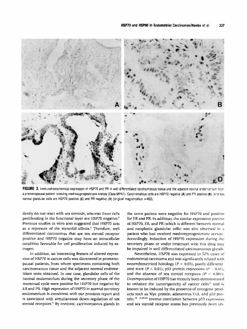
trium in the vicinity of carcinomatous glands. In one pa- tient (Case 2) who showed irregular cycles, both normal and carcinomatous glands were ER negative, PR positive, HSP70 negative, and weakly positive for HSP90. Another patient was in the secretory phase of the menstrual cycle at the time of operation (Case 3). In this case, carcino- matous glands were positive for both ER and PR, HSP70 negative, and strongly positive for HSP90, whereas the normal endometrial glands were negative for both ER and PR, strongly positive for HSP70, and weakly positive for HSP90. In addition, in one premenopausal patient who had received medroxyprogesterone acetate before opera- tion (Case MPAl), neoplastic glands were ER and PR posi- tive, HSP70 negative, and strongly positive for HSP90 (Fig. 3A,B); by contrast, the adjacent normal glands were ER and PR negative, HSP70 positive, and weakly positive for HSP90 (Fig. 3C,D).
DISCUSSION This study showed the expression of heat shock proteins HSP70 and HSP90 in endometrial carcinomas. Strong ex-

336 CANCER January 15,1996 / Volume 77 / Number 2
FIGURE 2. lrnmunohistochemical expression of HSP70 (A), HSPSO (B). PR (C), and p53 protein (D) in poorly differentiated carcinoma of the endometrium (Case 14) (original magnification x200)
pression of HSPSO was detected in 29% cases and corre- lated with well differentiated state of the tumor ( P < 0.05) and with sex steroid receptor status; intense immuno- staining for HSPSO was detected in 43% of the cases posi- tive for ER or PR, but in none of the tumors negative for EH and PR ( P < 0.01). This is consistent with a previous report that HSPSO mRNA levels have been correlated with ER levels of various breast cancer cell lines.'" Change of expression of HSPSO in normal endometrial glands has also been correlated with the fluctuation of levels of ste- roid receptor expression during the menstrual cycle.6 The heat shock protein HSPSO has been demonstrated to bind steroid receptor proteins and to modulate their func- tion."' Based on in vitro experiments, HSPSO has been proposed to bind with the hormone binding domain of unoccupied soluble receptors and to maintain them in an inactive state.' Subsequent binding of the ligand to the receptor triggers the release of HSPSO from the receptor complex and reverses the repression.2 However, another study has shown that HSPSO is necessary for maintenance
of the appropriate conformation required for hormone binding activity of the receptor." Therefore, HSPSO acts as a repressor of receptor function in the absence of li- gands, but as a necessary component of the receptor- activation pathway. In addition, HSPSO mRNA has been shown to be induced by estradiol in mouse uterus2" and in breast cancer cells.'8 These data suggest that high ex- pression of HSPSO may represent its association with high levels of sex steroid receptors or the response of the tumor cells to estrogen in endometrial carcinomas.
The heat shock protein HSP70 has also been demon- strated to bind the sex steroid receptor^,^-^ although its exact role in the receptor-activation pathway remains to be determined." This study showed that immunohisto- chemical expression of HSP70 was observed in 52% cases, and an absence of HSP70 expression was correlated with well differentiated state ( P < 0.01), and positivity of sex steroid receptors ( P < 0.001). In normal endometrium during the follicular phase, HSP70 is expressed in the glandular and stromal cells of the basal layer, which evi-
HSP7O and HSPSO in Endometrial CarcinomadNanbu et al. 337
FIGURE 3. lmmunohistochemical expression of HSP70 and PR in well differentiated carcinomatous tissue and the adjacent normal endometrium troiri a premenopausal patient receiving medroxyprogesterone acetate (Case MPAl). Carcinomatous cells are HSP70 negative (A) and PR positive (6). whereas normal glandular cells are HSP70 positive (C) and PR negative (0) (original magnification x400).
dently do not react with sex steroids, whereas those cells priiliferating in the functional layer are HSP70 negative." Previous studies in vitro also suggested that HSP70 acts as a repressor of the steroidal effects.5 Therefore, well differentiated carcinomas that are sex steroid receptor positive and HSP70 negative may have an intracellular condition favorable for cell proliferation induced by es- trogen.
In addition, an interesting feature of altered expres- sion of HSP70 in cancer cells was discovered in premeno- pausal patients, from whom specimens containing both carcinomatous tissue and the adjacent normal endome- trium were obtained. In one case, glandular cells of the normal endometrium during the secretory phase of the menstrual cycle were positive for HSP7O but negative for ER and PR. High expression of HSP70 in normal secretory endometrium is consistent with our previous report and is associated with simultaneous down-regulation of sex steroid receptors." By contrast, carcinomatous glands in
the same patient were negative for HSP70 and posi~ivt. for ER and PR. In addition, the similar expi-cssioii p i t v r i i
of HSP70, ER, and PR (which is different hetwrcn 1ioriii;iI
and neoplastic glandular cells) was also ohstwcd iii ;I
patient who had received medroxyprogesteronc acciaic. Accordingly, induction of HSP70 expression duririg ilic secretory phase or under treatment with this drug ilia!' be impaired in well differentiated carcinomatoiis glarids.
Nevertheless, HSP70 was expressed in 52% ciisi's (11' endometrial carcinoma and was significantly related \+i i l i
nonendometrioid histology ( P < 0.05), pocirlp diffiwriri- ated state ( P < 0.011, p53 protein expression ( I J ' (1.01 1, and the absence of sex steroid receptors (I' c: O . O ( i 1 ) . Overexpression of HSP70 has recently been dernoiistriiicd to enhance the tumorigenicity of cancer cells-' and is known to be induced by the presence of oncogcne p r [ i d - ucts such as Myc protein, adenovirus EIA, and 1~53 11rw
Inverse correlation between p5.7 cxprussio~i and sex steroid receptor status has previously Iiecri 011-
tein. 12- 14.23.24

338 CANCER January 15,1996 / Volume 77 / Number 2
served in endometrial,"' ovarian,I5 and breast carcino- mas.2s In this series, all eight p53-positive tumors were positive for HSP70 and negative for sex steroid receptors. In endometrial carcinomas, mutation or overexpression of p53 tumor suppressor gene have been shown to occur in nonendometrioid-type carcinomas'" or advanced stage of the tumor,'6 both of which are frequently devoid of sex steroid receptor^.'^^^^" Edwards et a12' reported that overexpression of HSP70 by heat shock results in a marked reduction in the cytoplasmic concentrations of PR in breast cancer cells. These data suggest that overex- pression of HSP70 may be related to the mechanism of down-regulation of sex steroid receptors and may play an important role in the loss of sex steroid receptors in nonendometrioid carcinomas or advanced tumors, espe- cially those expressing the p53 protein.G
REFERENCES 1.
2.
3.
4.
5.
6.
7.
8.
9.
10
11.
Lindquist S, Craig FA. The heat shock proteins. Annu Rev Genet 1988;22:631-77. Pratt WB. Transformation of glucocorticoid and progester- one receptors to the DNA-binding state. / Cell Biockem 1987;35:51-68. Kost SL, Smith DF, Sullivan WP, Welch WJ, Toft DO. Binding of heat shock proteins to the avian progesterone receptor. Mol Cell Biol 1989;9:3829-38. Schowalter DB, Sullivan WP, Maihle NJ, Dobson ADW, Con- neely OM, O'Malley BW, et al. Characterization of progester- one receptor binding to the 90- and 70-kDa heat shock pro- teins. / Biol Ckem 1991;266:21165-73. Bagchi MK, Tsai SY, Tsai MI, O'Malley BW. Progesterone enhances target gene transcription by receptor free of heat shock proteins hsp90, hsp56, and hsp70. Mol Cell Biol
Koshiyama M, Konishi I, Nanbu K, Nanbu Y. Mandai M , Komatsu T, et al. Immunohistochemical localization of heat shock proteins HSP70 and HSP9O in the human endome- trium: Correlation with sex steroid receptors and Ki-67 anti- gen expression. / Clin Endocrinol Metab 1995;80:1106-12. Gurpide E. Endometrial cancer: Biochemical and clinical correlates. J Nut1 Cancer Inst 1991;83:405-16. Wang DP, Konishi I, Koshiyama M, Mandai M, Nanbu Y, Ishikawa Y, et al. Expression of c-erbB-2 protein and epider- mal growth factor receptor in endometrial carcinomas: Cor- relation with clinicopathology and sex steroid receptor sta- tus. Cancer 1993;72:2628-37. Deligdish L, Holinka CF. Progesterone receptors in two groups of endometrial carcinoma. Cancer 1985; 57: 1385-8. Koshiyama M, Konishi I , Wang DP, Mandai M, Komatsu T, Yamamoto S, et al. lmmunohistochemical analysis of p53 protein over-expression in endometrial carcinomas: Inverse correlation with sex steroid receptor status. Virckoius Arch
Runowicz CD, Nuchtern LM, Braustein JD, Jones JG. Hetero-
1991; 11:4998-5004.
[A] 1993; 4231265-71.
12.
13.
14.
15.
16.
17.
18.
19.
20.
21.
22.
23.
24.
25.
26.
27.
geneity in hormone receptor status in primary and meta- static endometrial cancer. Gynecol Oncol 1990;38:437-41. Pinhasi-Kimhi 0, Michalovitz D, Ben-Zeev A, Oren M. Spe- cific interaction between the p53 cellular tumour antigen and major heat shock proteins. Nuture 1986;320:182-5. Sturzbecher HW, Chumakov P, Welch WJ, Jenkins JR. Mu- tant p53 proteins bind hsp72/73 cellular heat shock-related proteins in SV40-transformed monkey cells. Oncogene
Lehman TA, Bennett WP, Metcalf RA, Welsh JA, Ecker J, Modali RV, et al. p53 mutations, ras mutations, and p53- heat shock 70 protein complexes in human lung carcinoma cell lines. Cancer Res 1991;51:4090-6. Koshiyama M, Konishi I, Mandai M, Komatsu T, Yamamoto S, Nanbu K, et al. Immunohistocheniical analysis of p53 pro- tein and 72kDa heat shock protein (HSP72) expression in ovarian carcinomas: Correlation with clinicopathology and sex steroid receptor status. Virchows Arch 1995;425:603-9. Ferrarini M, Heltai S, Zocchi MR, Rugarli C. Unusual expres- sion and localization of heat-shock proteins in human tumor cells. Int J Cancer 1992;51:613-9. Heufelder AE, Goellner JR, Wenzel BE, Bahn RS. Immunohis- tochemical detection and localization of a 72-kilodalton heat shock protein in autoimmune thyroid disease. J Clin Endo- crinol Metab 1992;74:724-31. Jameel A, Skilton RA, Campbell TA, Chander SK, Coombes RC, Luqmani YA. Clinical and biological significance of HSP89 alpha in human breast cancer. Int / Cancer
Bresnick EH, Dalman FC, Sanchez ER, Pratt WB. Evidence that the 90-kDa heat shock protein is necessary for the ste- roid binding conformation of the L cell glucocorticoid recep- tor. / Biol Ckenz 1989;264:4992-7. Shyamala G, Gauthier Y, Moore SK, Catelli MG, Ullrich SJ. Estrogenic regulation of murine uterine 90-kilodalton heat shock protein gene expression. Mol Cell Endocrinol
Onate SA, Estes PA, Welch WJ, Nordeen SK, Edwards DP. Evidence that heat shock protein-70 associated with proges- terone receptor is not involved in receptor-DNA binding. Mol Endocrinol 1991; 5: 1993 -2004. Jaattela M. Over-expression of hsp7O confers tumorigenicity to mouse fibrosarcoma cells. Int / Cancer 1995; 60:689-93. Koskinen PI, Sistonen L, Evan G, Morirnoto R, Alitalo K. Nuclear colocalization of cellular and viral myc proteins with hsp70 in myc-overexpressing cells. / Virol 1991;65:842-51. Nevins JR. Induction of the synthesis of a 7,000 dalton mam- malian heat shock protein by the adenovirus E1A gene prod- uct. Cell 1982;29:913-9. Caleffi M, Teague MW, Jensen RA, Vnencak-Jones CL, Du- pont WD, Par1 FF. p53 gene mutations and steroid receptor status in breast cancer. Cancer 1994;73:2147-56. Kohler MF, Berchuck A, Davidoff AM, Humphrey PA, Dodge RK. Iglehart ID, et al. Overexpression and mutation of p53 in endometrial carcinoma. Cancer Res 1992;52:1622-7. Edwards DP, Estes PA, FadokVA, Bona BJ, Onate S, Nordeen SK, et al. Heat shock alters the composition of heteromeric steroid receptor complexes and enhances receptor activity i n v i i o Biockernistry 1992;31:2482-91.
1987; 1:201-11.
1992;50:409- 15.
1989;9:3567-70.


