Embed Size (px)
Citation preview

External carotid endarterectomy: Indications, technique, and late results Antonio V. Sterpetti, M.D., Richard D. Schultz, M.D., F.A.C.S., mad Richard J. Feldhaus, M.D., F.A.C.S., Omaha, Neb.
In the past 14 years, 22 patients (25 operated sides), with occlusion of the internal carotid artery (ICA), underwent ipsilateral external carotid artery (ECA) endarterectomy at our institution. Operative indications were amaurosis fugax in 13 sides and nonlateralizing transient isehemic attacks in the remaining 12. There were no operative deaths. One patient suffered a minor stroke after operation. Follow-up ranged from 6 to 110 months (median 36 months). In 16 cases, simple endarterectomy with or without vein patch closure was performed (type I). In two cases the ostium of the ICA was occluded with interrupted sutures after endarterectomy (type II). In the remaining seven cases the ICA was transposed as a patch over the endarterectomized ECA after endarterectomy (type III). All but six patients (six sides) underwent duplex scanning or angiography during follow-up. Four of nine patients with previous nonlateralizing symptoms had persistent symptoms after operation, whereas none of those with previous amaurosis fugax did. Recurrent occlusive disease was more common in type I reconstructions (p < 0.05). Proper ECA reconstruction results in long-term patency. In the patient with ipsilateral ICA occlusion, transposition of the ICA as a patch over the endarterectomized ECA offers a valid hemodynamic solution. Objective parameters are needed to identify patients with nonlateralizing symptoms who will benefit from operation. (1 VASC SURG 1988;7:31-9.)
The optimal management of patients with symp- tomatic chronic occlusion of the internal carotid ar- tery (ICA) remains controversial. More than 70% of the patients will initially sustain occlusion of the ICA with minimal or no neurologic deficits. I However, new symptoms can occur some time later, 2 indicating that ICA occlusion is not a stable condition, as might have been suggested? In these circumstances, the external carotid artery (ECA) becomes an important source of collateral circulation and a possible route for the passage ofemboli from the carotid bifurcation to the ipsilateral cerebral and retinal circulation.
The pathophysiologic mechanisms underlying re- current ischemic events after ICA occlusion have not been well elucidated. EC endarterectomy offers the theoretic possibility of increasing cerebral blood flow and removing sources of emboli localized at the ca- rotid bifurcation.
From the Department of Surgery, Creighton University School of Medicine.
Presented at the Thirty-fifth Scientific Meeting of the North Amer- ican Chapter, The International Society for Cardiovascular Sur- gery, Toronto, Ontario, Canada, June 8-9, 1987.
Reprint requests: Richard D. Schultz, M.D., Professor of Surgery, Chief Cardiovascular Section, Department of Surgery, Creigh- ton University School of Medicine, 601 No. 30th St., Omaha, NE 68131.
The purposes of our study were (1) to elucidate the mechanisms leading to cerebral symptoms in pa- tients with ICA occlusion and (2) to determine the functional results after EC endarterectomy.
PATIENTS AND METHODS
From May 1971 to December 1986, 33 patients underwent EC endarterectomy at our institution. The ipsilateral ICA was occluded in 22 patients; these patients form the basis of our report, a subset that represents only 2.2% of more than 1000 standard carotid endarterectomies performed during the same period.
The study group was made up of 15 men and seven womcn, who ranged in age from 51 to 76 years (mean 61.2 years). Prcoperative symptoms included amaurosis fugax in 13 patients and nonhemispheric symptoms in nine patients. Eight of the 13 patients with symptoms of amaurosis fugax had evidence of retinal emboli. Ten patients had previously under- gone endarterectomy of the contralateral ICA with- out relief of the cerebral symptoms. One patient with bilateral symptoms of amaurosis fugax and two pa- tients with nonlateralizing symptoms underwent bi- lateral ECA reconstruction. Before operation, all pa- tients underwent retrograde femoral angiography with visualization of both the intra- and extracranial
31

32 Sterpetti, Schultz, and Feldhaus Journal (#
VASCULAR SURGERY
\ Significant / n=l " / Significant StenosiSEcA [ ccASten°sis
n=4 / \ / ~ n = l
n=4 \ / n= l
n=2 Presence of ICA Stump
Fig. 1. Bmgiographic findings before 25 operations. CCA = common carotid artery;/CA = internal carotid artery; ECA = external carotid artery.
circulation mad noninvasive studies. Reduction of the lumen diameter of the ECA was calculated as the ratio between the minimal diameter of the vessel in the diseased portion and the diameter of the nearest portion of the artery, distal to the stenosis, without apparent disease (Fig. 1). For the purpose of the study, neurologic events that occurred within 12 months from operation were considered "persistent" symptoms, and the operation was considered a "func- tional" failure. Symptoms that occurred after 12 months from operation were considered "recurrent" symptoms, and the operation was considered, at least at the beginning, as a "functional success." Follow- up ranged from 6 to 110 months (median 36 months). Differences between groups were deter- mined by the Student t test, analysis of variance, and Fisher exact test where appropriate. 4
OPERATIVE PROCEDURES ECA reconstruction was performed with the pa-
tients under general ancsthesia with full cardiovas- cular monitoring. All operations wcre performed with the patients under normovolemic, normoten- sive, and normocarbic anesthesia. All patients had systemic heparinization. An intcrual shunt was never used. In eight patients, ECA reconstruction was per- formed after attempts to restore circulation in the occluded ICA failed. Endarterectomy was performed with the incision extending into the ECA orifice in all but six patients.
i!:
Fig. 2. Type I EC endarterectomy; after endarterectomy the stump of the ICA was not obliterated. Arteriotomy was closed with (four patients) or without (12 patients) vein patch grafting.
In 16 patients, the stump of the ICA was not obliterated after endarterectomy (including removal of thrombi or ulcerated plaque from the remnant of the ICA) (type I) (Fig. 2). Closure of the arteriotomy was achieved by means of 7-0 polypropylene (Pro- lene) running sutures. This closure was satisfactory in 12 patients; vein patch grafting was used in the remaining four. In two patients, interrupted sutures were inserted to obliterate the ostium of the occluded ICA after endarterectomy had been completed (type II) (Fig. 3). In seven patients, a segment of the occluded ICA was transected an appropriate length from its origin and was roiled over for angioplasty closure of the arteriotomy (type III) (Fig. 4).
RESULTS
Operative and histopathologic findings. De- tailed information about the operative findings was available in 20 patients (22 operated sides). In all patients, the atherosclerotic plaque involved the com- mon carotid artery (CCA) and extended toward the orifice of the ECA. The plaque in the ECA varied considerably in extent; in some patients the plaque had an abrupt transition with nondiscased intima, whereas in other paticnts, the passage from diseased and nondiseased intima was more gradual. Gcncraily, the atherosderotic plaque was localized mainly in the inner wall of the ECA (Fig. 5). The proximal remnant

Volume 7 Number 1 January. 1988
External carotid endarterectomy 33
i Fig. 3. Type 1I EC endarterectomy; after removal of the plaque, the internal carotid orifice was obliterated with interrupted sutures. (Modified with permission from O'Mara CS. In: Bergan lJ, Yao JST, eds. Cerebrovascular insufficiency. New York: Grune & Stratton, Inc., 1980:251-73.)
of the ICA was completely obliterated in six patients (6 of 25 = 24%). Gross ulceration was evident in 12 patients. The most common sites for ulceration were the CCA and the stump of the ICA. A striking finding was the presence of gross thrombi found in 12 (55%) of the 22 operated sites. In eight patients, the thrombus was present at the level of the stump of the ICA. In four patients, there was a thrombotic apposition over an ulcerated plaque at the level o f the CCA and ECA. A possible embolic source was present in 10 of the 12 patients with symptoms of amaurosis fugax and in two of the eight patients with nonhemispheric symptoms (p < 0.01).
Early complications. There were no operative deaths. One patient sustained a minor stroke (de- creased hand dexterity that did not prevent the pa- tient from functioning in his previous capacity). In this patient, an attempt was made to disobliterate the occluded ICA, and this could represent the direct cause of the stroke. One patient had a wound he- matoma that required surgical evacuation.
Functional results and late neurologic deficits. The indication for surgery was a major factor in in- fluencing the outcome. None of the 13 patients with previous symptoms of amaurosis fugax had persistent symptoms after surgery. Functional failure of the re- construction was more common among patients who underwent surgery because of nonlateralizing symp- toms; in four patients (five operated sides) symptoms persisted despite surgery (p < 0.01). None of the
;.!.:'-
/
>,
i
9-
-<
Fig. 4. Type [II EC endarterectomy; after removal of the plaque, a segment of endarterectomized internal carotid occlusion was used for patch angioplasty. (Modified with permission from O'Mara CS. In: Bergan JJ, Yao JST, eds. Cerebrovascular insufficiency. New York: Grune & Strat- ton, Inc., 1980:251-73.)
factors analyzed as possible prcdictors of outcome had prognostic significance. Patients in whom op- eration failed to relieve symptoms had a lower mean preoperative ophthalmic artery-to-brachial artery

ECA ICA
)ournaiof VASCULAR
SURGERY 34 Sterpetti, Schultz, and Feldhaus
Plaque Ulceration Thrombus Fig. 5. Operative findings in 22 operated sides (20 patients). Generally, the atherosclerotic plaque was mainly localized in the inner wall of the ECA (see text).
1.0
m
0,,
0
0.8
0.6
0.4
0.2 0.51 +_ 0.09 0.634_ 0.13
Preoperative Postoperative
Fig. 6. Preoperative and postoperative ophthalmic artery- brachial artery pressure index (OBPI) was determined in eight patients. There was a significant increase after external carotid endarterectomy (p < 0.01).
pressure index; however, this difference was not sta- tistically significant (p > 0.1).
Overall, ophthalmic artery-brachial artery pres- sure index was measured before and after opera- tion in eight patients, and in all but one patient, it increased significantly after operation (Fig. 6; p < 0.01). Of the 13 patients who were operated on because of symptoms of amaurosis fugax, three had
recurrent symptoms beyond 1 year from operation. In all patients, angiography revealed the presence of recurrent disease at the level of the endarterectomized site. Of the five patients who were operated on be- cause of previous nonlateralizing symptoms and in whom operation resulted in a functional success, one suffered a minor stroke in the hemisphere of the ICA occlusion 20 months after surgery. Angiography showed occlusion at the site of the endarterectomy. In all four patients with recurrent symptoms, the ostium of the ICA was not obliterated (p < 0.05).
Recurrent occlusive disease. Sixteen patients (19 operated sides) underwent angiography or du- plex scanning 8 to 80 months from operation (me- dian 36 months). Angiography was performed in 10 patients (11 operated sides) and duplex scanning was used in 15 patients (18 operated sides). In nine pa- tients, the studies showed the presence of recurrcnt disease. There was a close correlation between the type of ECA reconstruction performed and recur- rence of occlusive disease; recurrent disease was evident in eight of the 12 patients in whom the os- tium of the ICA was not obliterated (p < 0.05) (Ta- ble I). The importance of the ~,pe of ECA recon- struction per se in the prevention of recurrent disease is confirmed by the fact that the interval between control studies and operation, and the incidence of associated disorders was similar in the three groups (p > 0.2). The most common site of recurrence was the inner wall of the ECA.
Correlation between recurrent symptoms and recurrent disease. Symptoms of cerebral ischemia

Volume 7 Number 1 Januaq~ 1988 ExternM carotid endarterectomy 35
Table I. Recurrent occlusive disease
Method of EC endarterectomy
Type I Type H Type III
No recurrent disease 4/12 1/2 5/5 Recurrent stenosis 7/12 1/2 0/5 Occlusion i / I2 0/2 0/5
were present in only four of the nine patients with recurrent disease (44%). In two of the three patients with recurrent symptoms of amaurosis fugax, angio- graphic findings suggested the presence of ulceration. In the patient with a minor stroke, there was occlu- sion of the CCA. Another operation was performed and consisted of thromboendarterectomy and flap angioplasry with a segment of the endarterectomized ICA transposed over the arteriotomy. The lumen of the CCA was occluded by thrombotic apposition over an area of exuberant myointimal hyperplasia. In the five asymptomatic patients with recurrences, there were no angiographic findings of ulceration or ultrasonographic patterns ofheterogenous recurrent plaque.
DISCUSSION
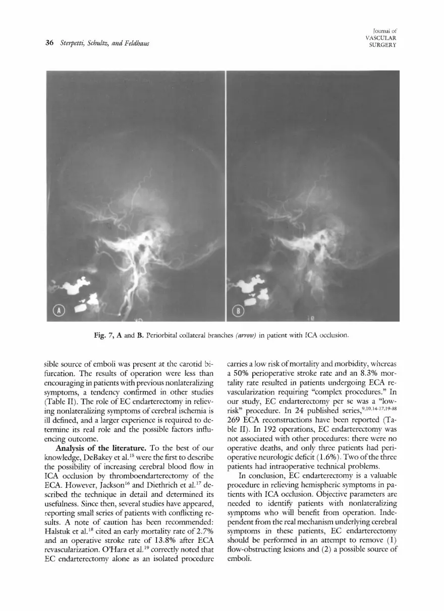
Anatomic and pathologic considerations. The existence of collateral pathways between the ECA branches and the intracranial cerebral circulation has been extensively documented by anatomic and ar- teriographic studies, s,~ In the early stages of human embryogenesis (20 to 24 mm embryonic size), the ophthalmic artery and the branches of the ECA, orig- inally separate, become thoroughly connected by a rich anastomotic network. 7 As the ophthalmic artery develops further, there is partial regression of the ECA branches supplying blood to the orbital cir- culation; in adults, the ECA ocular branches are rarely seen. The orbital channels can become ex- tremely developed and play an important functional role in special circumstances, such as in patients with orbital neoplasms or ICA occlusion (Fig. 7). Much of the current thinking concerning the pathophysi- ologic mechanism of cerebral ischemia in patients with carotid bifurcation occlusive disease has evolved in the past 20 years around two concepts: reduction of flow by the occlusive disease or distal embolization of plaque material.
In patients with ICA occlusion, both mechanisms could be responsible for the onset of symptoms of cerebral ischemia and can explain why symptoms might disappear after ECA endarterectomy. It has been suggested that anastomotic channels through
Table II. Summary of the literatureS: isolated EC endarterectomy (n = 192) (168 patients)
Perioperative mortality 0 Perioperative neurologic deficit 3/192 (1.6%) Relief from Symptoms~
Amaurosia fugax 46/50 (92%) Hemispheric TIA 46/53 (87%) Nonlateralizing symptoms 13/24 (54%)
Total 105/123 (85%)~
TIA = transient ischemic attacks. ~References 9, 10, 14-i7, and I9 through 38. ?Detailed information was available for only 118 patients. :~Some patients had more than one type of symptom (i.e., amau- rosis fugax and nonlateralizing symptoms) and were considered separately.
the branches of the ECA may account for 30% o]: the cerebral blood flow in patients with bilateral ICA occlusion.6 Machleder and Baker 8 have demonstrated that the ECA provides as much as 38% of the ICA back pressure at the time of operation. After ECA revascularization, cerebral blood flow, as measured by the xenon inhalation technique, can increase by 15% to 39% 9 The ophthalmic artery-brachial artery pressure index rose by 0.13 in a series of l0 patients after operation, x° The results of our study seem to support the observation that EC endarterectomy can significantly increase ipsilateral cerebral perfusion. Whether this augmented flow is responsible for relief of symptoms remains a matter of speculation. The embolic theory seems to explain better the nature and characteristics of episodes of transient ischemic attack or amaurosis fugax. The central retinal artery is 0.3 mm in diameter. ECA collaterals are 0.1 to 0.3 mm in diameter and enlarge significantly with increased flow. They are large enough to allow pas- sage of ophthalmoscopically observed retinal emboli. Finklestein et al.a~ reported a patient who suffered a lethal hemispheric infarction 3 days after angiograph- ically documented ICA occlusion; at autopsy the middle cerebral artery was occluded by thromboem- boli, with pathologic features similar to those of the thrombotic material present in the stump of the ICA. The following indirect observations support the em- bolization theory: (1) the simultaneous presence of a possible source of emboli in the stump of the ICA or at the level of the CCA and ECA and rethaal plaques 12as and (2) relief of symptoms after ECA reconstruction and closure of the stump of the ICA even in the absence of significant stenosis of the ECA.14
In our study symptoms of amaurosis fugax dis- appeared after operation, and in most patients a pos-
36 Ste,petti, Schultz, and Feldhaus
{ournai of VASCULAR
SURGERY
Fig. 7, A and B. Periorbital collateral branches (arrow) in patient with [CA occlusion.
sine source of emboli was present at the carotid bi- furcation. The results of operation werc less than encouraging in patients with previous nonlatcralizing symptoms, a tendency confirmed in other studies (Table II). The role of EC cndarterectomy in reliev- ing nonlateralizing symptoms of cerebral ischemia is ill defined, and a larger experience is requircd to de- termine its real role and the possible factors influ- encing outcome.
Analysis of the literature. To the best of our knowledge, DeBakey et a1.i5 werc the first to describe the possibility of increasing cerebral blood flow in ICA occlusion by thromboendarterectomy of the EGA. However, Jackson I~ and Diethrich et al. ~7 de- scribed the technique in detail and determined its usefulness. Since then, several studies have appeared, reporting small series of patients with conflicting re- suits. A note of caution has been recommended: Halstuk et al. is cited an early mortaliw rate of 2.7% and an operative stroke rate of 13.8% after ECA rcvascularization. O'Hara et ai. 19 correctly noted that EC endarterectomy alone as an isolated procedure
carries a low risk of mortality and morbidity, whereas a 50% perioperative stroke rate and an 8.3% mo> tality rate resulted in patients undergoing ECA re- vascularization requiring "complex procedures." In our study, EC endarterectomy per se was a "low- risk" procedure. In 24 published series, 9,I°'~4-~rag"s8 269 ECA reconstructions have been reported (Ta- ble II). In 192 operations, EC endarterectomy was not associated with other procedures: there were no operative deaths, and only three patients had peri- operative neurologic deficit (1.6%). Two of the three patients had intraoperative technical problems.
In conclusion, EC endarterectomy is a valuable procedure in relieving hemispheric symptoms in pa- tients with ICA occlusion. Objective parameters are needed to identify patients with nonlateralizing symptoms who will benefit from operation. Inde- pendent from the real mechanism underlying cerebral symptoms in these patients, EC endarterectomy should be performed in an attempt to remove (1) flow-obstructing lesions and (2) a possible source of emboli.

Volume 7 Number 1 January 1988 External carotid endarterecromy 37
Endarterectomy and patching with a segment of the ICA solve both problems and seem to offer a good hemodynamic solution. The patch is composed of autogenous tissue and has the advantage of being frequently available for use and easily prepared. Type II reconstruction can offer a valid solution if a smooth passage from the CCA to the ECA is guar- anteed by satisfactory vein patching. We studied boundary layer separation in in vitro glass models scaled from postoperative angiograms of the three types of reconstruction. Large areas of boundary layer separation were observed in glass models rep- resenting type I reconstructions, and this might the- oretically explain the high incidence of recurrent dis- ease when the stump of the ICA was not occluded.
REFERENCES 1. Fritz VU, Voll CL, Levien LJ. Internal carotid artery occlu-
sion: clinical and therapeutic implications. Stroke 1985; 16:940-4.
2. Hennerici M, Hulsbomer HB, Rautenberg W, Heftcr H. Spontaneous history of asymptomatic internal carotid occlu- sion. Stroke 1986;17:718-22.
3. Gomensoro JB. Joint study of extracranial arterial occlusion. VIII. Clinical radiographic correlation of carotid bifurcation lesions in 177 patients with transient cerebral ischemic attacks. JAMA 1973;224:985-91.
4. Rimm AA, Hartz AJ, Kalbfleisch JH, Anderson AJ, Hoffman RG. Basic biostatistics in medicine and epidemiology. Nor- walk, Corm: Appleton-Century-Crofts, 1980.
5. Taveras JM, Vount LA, Friedenberg RM. Angiographic dem- onstration of external-internal carotid anastomosis through the ophthalmic artery. Radiology 1964;83:525-30.
6. Fields WS, Brustrnan ME, Wribel J. Collateral circulation of the brain. Monogr Surg Sci 1965;2:183-259.
7. Vignaud J, Hasso AN, Lasjaunias P, Clay C. Orbital vascular anatomy and embryology. Radiology 1974; 111:617-26.
8. Machleder HI, Baker WF. External carotid artery shunting during carotid endarterectomy. Arch Surg 1974;108:785-8.
9. Zarins CK, Del Beccaro EJ, Johns L, Turcotte JK, Dohrmarm GJ. Increased cerebral blood flow after external carotid artery rcvascularization. Surgery 1981;89:730-4.
10. Schuler JJ, Flanigan DP, DeBord JR, Ryan TI, Castronuovo JJ, Lira LT, The treatment of cerebral ischemia by external carotid artery revascularization. Arch Surg 1983; 118:567-72.
11. Finklestein S, Kleinman GM, Cuneo R, Baringer JR. Delayed stroke following carotid occlusion. Neurology 1980;30: 84-8.
12. Countee RW, Vijayanathan T. External carotid artery in in- ternal carotid artery occlusion. Angiographic, therapeutic and prognostic considerations. Stroke 1979;10:450-60.
13. Torvik A, Jorgensen L. Thrombotic and embolic occlusions of the carotid arteries in an autopsy material. Part 1. Preva- lence, location and associated diseases. J Neurol Sci 1964; 1:24-39.
14. Barnett HJM, Peerless SJ, Kaufmann JCE. "Stump" of in- ternal carotid artery--a source for further cerebral embolic ischemia. Stroke 1978;9:448-56.
15. DeBakey ME, Crawford ES, Cooley DA, Morris GC, Garrett
HE, Fields wS. Cerebral arterial insufficiency: one to 1 i-year results following arterial reconstructive operation. Ann Surg 1965;161:921-45.
16. Jackson BB. The external carotid as a brain collateral. Am J Surg 1967;113:375-8.
17. Diethrich EB, Liddicoat JE, McCutchen JJ, DeBakey ME. Surgical significance of the external carotid artery in the treat- ment of cerebrovascular insufficiency. J Cardiovasc Surg 1968;15:213-23.
18. Halstuk KS, Baker WH, Littooy FN. External carotid end- arterectomy. J VASC SURG 1984;3:398-402.
19. O'Hara PJ, Hertzer NR, Beven EG. External carotid revas- culafization: review of a ten-year experience. J VAsc SURG 1985;5:709-14.
20. Ehrenfeld WK, Lord RSA. Transient monocular blindness through collateral pathways. Surgery 1969;65:911-5.
21. Lord RSA. Monocular transient ischemic attacks and the ex- ternal carotid artery. Med J Aust 1973;1:742-5.
22. Connolly JE, Stemmer EA. Endarterectomy of the external carotid artery. Arch Surg 1973;106:799-802.
23. Clayson KR, Edwards WH. Importance of the external ca- rotid artery in extracranial cerebrovascular occlusive disease. South Med J 1977;70:904-9.
24. Burnbaum MD, Selhorst JB, Harbison JW, Brush JJ. Amau- rosis fugax from disease of the external carotid artery. Arch Neurol 1977;34:532-5.
25. Karmody AM, Shah DM, Monaco VJ, Leather RP. On sur- gical reconstruction of the external carotid artery. Am J Surg 1978;136:176-80.
26. Jacques S, Garner JT, Tager R, Rosenstock J, Fields T. Im- proved cognition after external carotid endarterectomy, Surg Neurol 1978;10:223-5.
27. Berguer R, McCaffrey JF, Bauer RB. Bilateral internal carotid artery occlusion. Arch Surg 1980;115:840-3.
28. Hunt DG, Wheeler CG, Bukhari HI, Hempel GK. Surgical management of total carotid artery occlusion. Am J Surg 1980;139:700-3.
29. Hertzer NR. External carotid endarterectomy. Surg Gynecol Obstet 1981;153:186-90.
30. Countee RW, Vijayanathan T, Chavis P. Recurrent retinal ischemia beyond cervical carotid occlusions. J Neurosurg 1981;55:532-42.
31. Rogers LA. Carotid angioplasty in the management of ex- ternal carotid artery stenosis associated with an occluded in- ternal carotid artery. Neurosurgery 1982;11:20-4.
32. Levien L}, Voll C, Fritz VU. External carotid endarterec- tomy. S Mr Med J 1983;64:541-4.
33. O'Mara CS. Surgical significance of the external carotid artery. In: Bergan JJ, Yao JST, eds. Cerebrovascular insufficiency. New York: Grune & Stratton, Inc, 1980:251-73.
34. Lamberth WC. External carotid endarterectomy: indications, operative technique and results. Surgery 1983;93:57-63.
35. Bradac GB, Kaden B, Oppel F, Hirner A. Occlusion of in- ternal carotid artery. Further clinical angiographic, and ther- apeutic considerations. Neuroradiology 1984;26:445-50.
36. Fisher DF, Valentine RJ, Patterson CB, et al. Is external carotid endarterectomy a durable procedure. Am J Surg 1986;152:700-3.
37. Satiani B, Das BM, Vasko JS. Reconstruction of the external carotid artery. Surg Gynecol Obstet 1987;164:105-10.
38. Friedman SG, Lamparello PJ, Riles TS, Imparato AM, Sakwa MP. Surgical management of the patient with bilateral inter- nal carotid artery occlusion. J VASC Si:RG 1987;5:715-8.

38 Sterpetti, Schultz, and Feldhaus
~ournai o~" VASCULAR
SURGERY
DISCUSSION
Dr. Joseph Archie, Jr. (Raleigh~ N.C.). The authors are to be congratulated for extracting much information from a few operations. The major contribution of this paper, from my point of view, is that it tells us how to technically perform an external carotid endarterectomy, and perhaps more important, how not to do it.
Of the several indications for external carotid endar- tcrectomy, few will quibble with removing the source of the symptomatic emboli. However, operations to improve collateral cerebral blood flow fall into another category. Although this study and others show improved postop- erative ocular plethysmographic pressures or an increase in regional cerebral blood flow, the value of external carotid endarterectomy in decreasing the risk of stroke is not clear. However, some groups have shown a decreased incidence ofipsilateral stroke after this operation. One wonders how increased collateral flow via external carotid reconstruction will lower the incidence of stroke, if external cartotid- internal carotid bypass does not. I am sure, however, that we will continue to advise and do this operation to improve collateral cerebral blood flow.
Regarding geometry and hemodynamics of external carotid reconstruction, the clinical and model results of type I repairs, that is, endarterectomy and either patch or primary closure without internal carotid stump manage- ment, dearly deem it an unacceptable method. Common sense might tell us to close the orifice of the internal carotid artery. On the other hand, arteriograms of patients with occluded internal carotid arteries without significant ex- ternal stenosis frequently have an "out-pouching" or patent stump of the internal carotid artery, which is defined in this study as a precursor of trouble.
Dr. Sterpetti, do you believe such a type I arteriogram is an indicator of future progression of discase? More im- portant, should patients with this type I anatomy have the carotid bulb prophylactically streamlined to prevent occlu- sion or stenosis of the external carotid artery?
The optimal reconstruction advised is a smooth taper- ing of the common carotid artery into the external carotid artery, a profile that a fluid mechanics engineer might rec- ommend to minimize local boundary layer separation and turbulence. This is clearly a much simpler design problem and solution than the unanswered larger question of op- timal reconstruction of a standard carotid endarterectomy. My final question concerns the type III technique, in which an internal carotid artery flap is used to reconstruct the external carotid artery. Specifically, how does one avoid a pocket of type I effect with this technique? Would it not be better to reconstruct each artery in an individual manner, with the goal o f producing a gradual conic tapering from the common carotid artery into the manifold of the branches of the external carotid artery?
Dr. Sterpetti. At the present time, we have no data to answer your question about the role of prophylactic external carotid endarterectomy in preventing disease pro-
gression at this level in patients with occlusion of the in- ternal carotid artery. Indeed, the natural history of patients with internal carotid artery occlusion is ill defined. In 1983 Furlan et al. reported a stroke rate of 3% per year in 138 patients followed up for a mean period of 5 years. On the other hand, Cote et al. and Hennerici et al. reported a higher incidence, a stroke rate of about 10% to 11% per year.
I personally do not believe in prophylactic external carotid endarterectomy. However, I believe that this op- eration might be indicated in an asymptomatic patient with some evidence of distal cmbolization such as retinal emboli or cerebral infarct. Concerning the second question, what we really proved is that it is imperative to occlude the stump of the internal carotid artery. We had not enough data to make a meaningful comparison between what we called type II and III reconstruction, and I believe it remains a matter of personal preference. However, in spedmens from cadavers, I simulate what we called type II reconstruction, and I then performed control angiography. I found it very diNcult to create a smooth passage from the common ca- rotid to the external carotid artery with this technique.
Dr. Angelo Riberi (Youngstown, Ohio). Inour series of many carotid endarterectomies, we have come across a fair number of patients with external carotid stenosis. Twenty-five patients underwent successful endarterectomy in different fashions. It became apparent, however, that because of its size, the external carotid artery cannot usually be closed directly without narrowing the lumen of the vessel. We have gotten away from saphenous vein patches not because they did not work, but because they demand an additional incision of the anlde of the patient, who returns to the once month after month complaining of pain and swelling where the pain in the neck has already been resolved for many weeks.
Borrowing from our experience in the reconstruction of the deep femoral artery, by using the endarterectomized superficial femoral artery, we transposed this technique to the neck and have now performed 14 successful and con- secutive left or right external carotid endarterectomies by using the occluded internal carotid artery. First, the pro- cedure eliminates the stump problem, and the embolism is removed. Second, if you perform an endarterectomy on the internal carotid artery high in the neck, ligate the distal artery, and reverse the internal carotid to the external ca- rotid artery, you have achieved a complete reconstruction by using autogenous tissue, and the results are usually ex- cellent.
I assure you that both the preoperative and postop- erative arteriograms arc excellent. We have had no major problems after operation. A small hematoma was drained. There was no morbidity, mortality, or operative strokes.
Dr. William Gee (Allentown, Pa.). In the patient with a unilateral internal carotid occlusion, various branches of the ipsilateral external carotid artery provide collateral flow to the intracranial patent segment of the occluded vessel.

Volume 7 Number 1 Januaq, 1988 External carotid enda~¢erectomy 39
The final channel in most of these collateral pathways is the ipsilateral ophthalmic artery. Therefore it is not sur- prising that the authors observed modest improvement in the ophthalmic systolic pressures, as related to the respec- tive brachial systolic pressure, after repair of severe external carotid stenoses. Our experience with 10 similar patients demonstrated a 7% pressure improvement. However, ocu- lar blood flow is also calculated from the OPG-Gee test, and 51% improvement was observed on the side of op- eration in the 10 patients. Although duplex scanning can bc used to document the anatomic patency of these repairs, oculovascular hemodynamics are a useful means of assess- ing the quantitative improvement in the physiology asso- ciated with carotid reconstructions.
Dr. Sterpetti. 1 agree completely with you that after external carotid endarterectomy, you can expect an increase in ophthalmic pressure. I do not know whether this is a real increase in intracranial cerebral blood flow and what this increase means in terms of relief of nonhemispheric symptoms. For this reason, we did not analyze this type of data. Interestingly, there was no difference in the ophthalmic-brachial pressure index increase after operation in patients who were relieved of their symptoms and those who were not. The considerable increase in blood flow, as detected by the OPG-Gee you observed, might have im- portant implications either intraoperatively (as a method to monitor the efficiency of the reconstruction) or as a prognostic factor, and it deserves further analysis.
Dr. Victor Bernhard (Tucson, Ariz.). Dr. Stcrpctti, your paper emphasizes the necessity for meticulous oper- ative care and the need for intraoperative evaluation of the quality of the carotid repair by continuous-wave or pulsed Doppler examination, operative arteriography, or ultra- sonography. Have you applied these, or do you intend to in your patients?
Is there any real difference in the technique that you have identified using the flap, which has the shortcomings identified by Dr. Archie, or simply some form of patch that accomplishes the same thing and may give you a smoother outflow tract? I believe that is the critical issue and highlights the importance of operative angiography.
Dr. Sterpetti. This is a review of a ]5-year experience, and different surgeons performed the operations we re- ported. Thus it is a heterogeneous experience. For this reason, intraoperative angiography was performed in only five patients, and I agree with you that it should be per- formed routinely.
Concerning your second question, what we really dem- onstrated is the importance of occluding the stump of the internal carotid artery. Unfortunately, a meaningful com- parison between what we called type I1 and Il i reconstruc- tion was not feasible. However, it is very difficult to have a smooth passage from the common carotid to the external carotid artery with type 1I reconstruction as I saw in my experiments in cadavers.
Dr. Phillip Bendick (Royal Oak, Mich.). My question relates to function. You classified your patients into two groups according to symptoms: amaurosis fugax and non- hemispheric symptoms. It is not surprising that you would resolve an embolic phenomenon such as amaurosis fugax by removing the embolic source. Equally, however, it is not surprising that you may not resolve nonhemispheric symptoms by simply addressing one offbur cerebrovascular vessels without any attention noted, at least in your pre- sentation, to the contralateral carotid artery, the vertebro- basilar system, and perhaps even cardiac function and car- diac performance.
Therefore, what was the status of the contralateral ca- rotid artery in those patients with nonhemispheric symp- toms and the status of their vertebrobasilar system, and how was the decision made to operate on an external ca- rotid artery in those patients?
Dr. Sterpetti. Thank you very much for your question, which allows me to explain the details of our study pop- ulation. Ten patients had undergone previous contralateral carotid endarterectomy. Three patients with carotid occlu- sion in both sides underwent bilateral operation. Thus there was no surgical correctable disease in the contralateral carotid artery. In other words, this operation was usually the last option when everything was done. As you correctly pointed out, the real question that remains unanswered is, which patient with nonhemispheric symptoms will benefit from the operation? We addressed this problem in the article, and we were not able to find a convincing answer. Even if, as you can expect, 50% of these patients had evidence of coronary artery disease, there was no evidence of any possible correlation between cardiac events and neu- rologic symptoms. The indication for such an operation in the patient with nonhemispheric symptoms remains to be correctly defined, and for this purpose a larger experience is needed.