Embed Size (px)
Citation preview

Molecular Biology of the CellVol. 14, 2181–2191, May 2003
Ezrin Regulates E-Cadherin-dependent AdherensJunction Assembly through Rac1 ActivationPhilippe Pujuguet, Laurence Del Maestro, Alexis Gautreau, Daniel Louvardand Monique Arpin*
Unite Mixte de Recherche 144 Centre National de la Recherche Scientifique/Institut Curie, 75248Paris, France
Submitted July 18, 2002; Revised December 4, 2002; Accepted January 16, 2003Monitoring Editor: Keith Mostov
Ezrin, a membrane cytoskeleton linker, is involved in cellular functions, including epithelial cellmorphogenesis and adhesion. A mutant form of ezrin, ezrin T567D, maintains the protein in anopen conformation, which when expressed in Madin-Darby canine kidney cells causes extensiveformation of lamellipodia and altered cell-cell contacts at low cell density. Furthermore, these cellsdo not form tubules when grown in a collagen type I matrix. While measuring the activity of Rhofamily GTPases, we found that Rac1, but not RhoA or Cdc 42, is activated in ezrin T567D-expressing cells, compared with cells expressing wild-type ezrin. Together with Rac1 activation,we observed an accumulation of E-cadherin in intracellular compartments and a concomitantdecrease in the level of E-cadherin present at the plasma membrane. This effect could be reversedwith a dominant negative form of Rac1, N17Rac1. We show that after a calcium switch, thedelivery of E-cadherin from an internalized pool to the plasma membrane is greatly delayed inezrin T567D-producing cells. In confluent cells, ezrin T567D production decreases the rate ofE-cadherin internalization. Our results identify a new role for ezrin in cell adhesion through theactivation of the GTPase Rac1 and the trafficking of E-cadherin to the plasma membrane.
INTRODUCTION
The ezrin, radixin, and moesin (ERM) proteins act as linkersbetween the plasma membrane and the actin cytoskeleton.They have been implicated in the organization of specificdomains of the plasma membrane, such as the apical do-main of epithelial cells, the immunological synapse, or theformation of uropods (Berryman et al., 1995; Crepaldi et al.,1997; Serrador et al., 1997; Allenspach et al., 2001; Delon et al.,2001; Roumier et al., 2001). Their functions are regulated bychanges in their conformation. ERM proteins exist in thecytoplasm as dormant monomers in which the F-actin cy-toskeleton and the plasma membrane binding sites aremasked. This closed conformation is due to an intramolec-ular N- to C-ERM association domain (ERMAD) interaction(Bretscher et al., 1995; Gary and Bretscher, 1995). Thus, ab-rogation of the N/C-ERMAD interaction is required to“open up” the molecules and to expose their cryptic bindingsites (Berryman et al., 1995). Two factors have been impli-cated in the activation of ERM proteins. The binding tophosphatidylinositol 4,5-biphosphate is required for their
interaction with actin in vitro and with membrane proteinsin vivo (Nakamura et al., 1999; Barret et al., 2000). In addi-tion, phosphorylation of a conserved threonine residue inthe C-ERMAD, T567, inhibits the N/C-ERMAD interactionin vitro (Matsui et al., 1998; Nakamura et al., 1999) and invivo, converts inactive oligomers to active monomers (Gau-treau et al., 2000). Expression of an ezrin mutant, ezrinT567D, which mimics its phosphorylation, induces lamelli-podia formation in nonconfluent cells and tufts of microvilliin confluent cells. Moreover, production of ezrin T567Dperturbs the organization of epithelial cell monolayers(Yonemura et al., 1999; Gautreau et al., 2000).
A number of studies have implicated ERM proteins in theregulation of cell-cell and cell-matrix adhesion. Suppressionof all three ERM proteins with antisense oligonucleotidesdisrupts cell-cell and cell-matrix adhesion (Takeuchi et al.,1994). Overexpression of ezrin or of its N-terminal domainincreased the adhesion of insect cells (Martin et al., 1995). Itwas further shown that ERM proteins control cell adhesionthrough different means. For instance, interaction of ezrinwith intercellular adhesion molecule (ICAM)-2 is importantfor the activation of natural killer cells (Helander et al., 1996),the positioning of ICAM-3 in the uropod of T lymphocytesdepends on moesin (Serrador et al., 1997), and exclusion ofCD43 from the cell-cell contact area during formation of theimmunological synapse requires its interaction with ERM
Article published online ahead of print. Mol. Biol. Cell 10.1091/mbc.E02–07–0410. Article and publication date are at www.molbi-olcell.org/cgi/doi/10.1091/mbc.E02–07–0410.
* Corresponding author. E-mail address: [email protected].
© 2003 by The American Society for Cell Biology 2181

proteins (Allenspach et al., 2001; Delon et al., 2001; Roumieret al., 2001). ERM proteins have also been shown to controladhesion through the Rho GTPase pathway. ERM proteinsare involved in assembly of stress fibers and in the formationof focal adhesions upon activation of the small GTPases Rhoand Rac in permeabilized fibroblasts (Mackay et al., 1997).The binding of the TSC1 tumor suppressor hamartin toactivated ezrin has been implicated in the activation of RhoAand formation of focal contacts (Lamb et al., 2000). Phosphor-ylation of ezrin by the Rho kinase ROCK is required forRho-induced focal adhesion assembly (Tran Quang et al.,2000).
Herein, we report that ezrin T567D expression inducesmorphological changes in MDCK epithelial cells, leading tolamellipodia formation and perturbed cell-cell contacts. Wefound that expression of ezrin T567D leads to the activationof the GTPase Rac1, but neither that of RhoA, nor of Cdc 42.This activation of Rac1 perturbed the localization of E-cad-herin to the plasma membrane. E-cadherin accumulated inintracellular compartments and its delivery to the plasmamembrane was delayed during junction assembly. Our ob-servations provide the first cue on the role of ezrin in E-cadherin–dependent cell-cell junction assembly.
MATERIALS AND METHODS
Cell CultureThe T23 clone of the parental dog kidney-derived epithelial Madin-Darby canine kidney (MDCK) cell line (BD Biosciences Clontech,Palo Alto, CA), which expresses the tetracycline-repressible trans-activator (Barth et al., 1997), was used. Cells were grown in DMEM(Invitrogen, Carlsbad, CA) containing 10% fetal bovine serum, at37°C in 10% CO2. Geneticin (0.4 mg/ml) (Sigma-Aldrich, St. Louis,MO), indicated amounts of doxycyclin (Sigma-Aldrich) and hygro-mycin B (0.2 mg/ml) (Calbiochem, La Jolla, CA) were added to thegrowth medium of the transfected MDCK cells. To obtain polarizedmonolayers, 3 � 106 MDCK cells were plated on 24-mm Transwellfilters (Corning Glassworks, Corning, NY) and grown for 4 d.
Plasmid Constructs and TransfectionVSVG (vesicular stomatitis virus glycoprotein)-tagged-ezrin T567Dand VSVG-tagged-ezrin wild-type (Gautreau et al., 2000) were sub-cloned into the NotI site of the tetracycline-repressible pTRE vector(Tet-Off; BD Biosciences Clontech). Stable and transient transfec-tions were performed by electroporating cells by using an electricalpulse of 0.240 kV and 950 �F (Bio-Rad, Hercules, CA). The plasmidconferring the resistance to hygromycin was from BD BiosciencesClontech. The myc epitope-tagged dominant negative N17Rac1, anddominant active V12Rac1 plasmids were obtained from Dr. GaryBokoch (Scripps Research Institute, La Jolla, CA). The rhotekinRho-binding domain (amino acids 7–89) fused with glutathioneS-transferase (GST) was from Dr. Schwartz (Ren et al., 1999). ThePAK CRIB-encompassing domain (amino acids 70–118) fused withGST was from Dr. Lowe (Thompson et al., 1998).
AntibodiesMouse monoclonal anti-human E-cadherin, anti-human Rac1, andanti-human Cdc42 antibodies were obtained from TransductionLaboratories (Lexington, KY). Mouse monoclonal anti-human RhoAantibody was from Santa Cruz Biotechnology (Santa Cruz, CA). Ratmonoclonal anti-human E-cadherin (clone DECMA-1) and rabbitpolyclonal anti-human �-catenin were obtained from Sigma-Al-drich. Rabbit polyclonal anti-�-catenin was from Chemicon Inter-national (Temecula, CA). Mouse monoclonal anti-tubulin was from
Amersham Biosciences UK (Little Chalfont, Buckinghamshire,United Kingdom). Mouse monoclonal anti-transferrin receptor wasfrom Zymed Laboratories (South San Francisco, CA). Mouse mono-clonal anti-Na�/K� ATPase was from Upstate Biotechnology (LakePlacid, NY). Rabbit polyclonal anti-ezrin antibodies were describedpreviously (Algrain et al., 1993). Myc-tagged proteins were detectedwith a mouse monoclonal anti-human myc antibody (clone 9E10).The mouse monoclonal anti-VSVG antibody (clone P5D4) was de-scribed previously (Kreis, 1986).
ImmunofluorescenceCells were cultured on glass slides, washed with cold phosphate-buffered saline (PBS), fixed, and permeabilized with methanol/acetone (1:1, vol/vol) for 5 min at �20°C, and then incubated with15% fetal bovine serum in PBS for 1 h at room temperature. Alter-natively, cells were fixed with 3% paraformaldehyde and perme-abilized with 0.2% Triton X 100. Primary and secondary antibodieswere incubated for 1 h each. The nuclei were labeled using 4,6-diamidino-2-phenylindole (Sigma-Aldrich). Cells were mounted inPBS/glycerol (1:1 vol/vol) and viewed by epifluorescence (LeicaDMRA). Imaging was performed using MetaView.
GTPase Activity AssaysThe effectors rhotekin and PAK were used to affinity-precipitateendogenous cellular GTP-Rho, and GTP-Rac and Cdc 42, respec-tively (Ren et al., 1999). For affinity-precipitation of GTP-boundGTPases, cells were first washed with ice-cold PBS and incubatedwith the lysis buffer (50 mM Tris, pH 7.2, 1% Triton X-100, 0.5% NaDOC, 0.1% SDS, 500 mM NaCl, 10 mM MgCl2, containing a cocktailof protease inhibitors). The cleared lysates were incubated withGST-effector binding domain (20 �g) on beads for 1 h on ice. Thebeads were washed four times with 50 mM Tris, pH 7.2, 1% TritonX-100, 150 mM NaCl, 10 mM MgCl2, containing a cocktail of pro-tease inhibitors. Beads were resuspended in reduced SDS samplebuffer and heated at 95°C for 10 min. Samples were run on 13%SDS-PAGE gels and transferred to membranes. Immunodetectionwas performed with anti-Rho, -Rac, or -Cdc 42 antibodies. Theamount of GTP-bound GTPases was normalized to the total amountof GTPases present in whole cell lysates. Scanning and densitomet-ric analyses were performed with the ImageQuant image analysissystem (Amersham Biosciences UK).
Immunoprecipitation and ImmunoblottingCell lysates extracted in cold radioimmunoprecipitation assay(RIPA) buffer (1% NP-40, 0.5% deoxycholate, 0.2% SDS, 150 mMsodium chloride, 50 mM Tris-HCl, pH 7.4, containing a cocktail ofproteases inhibitors) were resolved by 10% or 7.5% SDS-PAGE.Proteins were transferred to nitrocellulose (Millipore, Bedford, MA),immunoprobed, and detected by enhanced chemiluminescence(Pierce Chemical, Rockford, IL). Scanning and densitometric analy-ses were performed with the ImageQuant image analysis system.For immunoprecipitations, cleared lysates were incubated with ei-ther 2 �g of the antibody of interest or an irrelevant antibody for 2 hat 4°C. After incubation, protein G beads (Sigma-Aldrich) werewashed four times with 1 ml of lysis buffer, suspended in Laemmlibuffer and the proteins resolved by 10% or 7.5% SDS-PAGE.
Metabolic Labeling and Pulse ChaseMetabolic labeling was performed in DMEM without Met and Cyscomplemented with 250 Ci/ml 35S-labeled Met and Cys (RedivuePromix, Amersham Biosciences, Amersham, United Kingdom).Cells were radiolabeled for 20 min and chased for the indicated timein standard DMEM containing 10% fetal bovine serum. After im-munoprecipitation, lysates were resuspended in reducing samplebuffer, boiled for 5 min, and resolved by 7.5% SDS-PAGE. The 35Ssignal was enhanced by incubating gels in 1 M salicylate for 20 min.
P. Pujuguet et al.
Molecular Biology of the Cell2182

Dried gels were exposed to a phosphoscreen for 3 d. Signals werequantified using a STORM 860 PhosphorImager and ImageQuantsoftware.
Cell Surface Biotinylation and Transport AssayCell surface proteins were biotinylated by incubating the cells with1.5 mg/ml sulfo-NHS-SS-biotin (Pierce Chemical) for 1 h at 4°C andfree biotin was quenched with a blocking solution (50 mM NH4Cl inPBS containing 1 mM MgCl2 and 0.1 mM CaCl2). Cells were theneither directly extracted in a RIPA buffer, or stripped to remove theextracellular bound biotin with 50 mM glutathione, 75 mM NaCl, 75mM NaOH and 2% bovine serum albumin, at 4°C, and RIPA ex-tracted. To measure the intracellular pool of biotinylated proteins,biotinylated cells were incubated at 37°C for 1 h to allow internal-ization of biotinylated proteins, and then stripped and extracted (Leet al., 1999). Cell extracts were centrifuged and incubated withstreptavidin magnetic beads (Dynal, Olson, Norway) to collect bio-tinylated proteins. These samples were then analyzed by SDS-PAGEand immunoblotting was performed using antibodies against E-cadherin, Na�/K� ATPase or transferrin receptor.
Three-dimensional CulturesThe tubulogenesis assay used was described previously (Crepaldi etal., 1997). Collagen type I gels were prepared as follows: 1 partDMEM 10�, 1 part NaHCO3 (37 g/l), 1 part fetal bovine serum weremixed with 3.5 parts of a suspension of 3 � 105 cells/ml and 3.5parts of type I collagen at 5 mg/ml (BD Biosciences, Franklin Lakes,NJ) at room temperature. The gels were covered with culture me-dium supplemented with 100 U/ml hepatocyte growth factor(HGF), and the cells grown for 6 d. Photographs were taken with anepifluorescence microscope (Leica) after paraformaldehyde fixation.
Cell ProliferationCell proliferation was measured with a cell proliferation kit (Cell-Titer 96; Promega, Madison, WI). Cells (5 � 103) were plated astriplicates in 96-well plates and cultured for the indicated times.Then, cells were incubated for 4 h in the presence of the substrate,lysed, and absorbance was recorded at 570 nm (SpectraMax; Mo-lecular Devices, Sunnyvale, CA).
RESULTS
Ezrin T567D Induces Morphological Changes WhenExpressed in MDCK CellsWe have previously shown that phosphorylation of thre-onine 567 regulates the transition from inactive ezrin oli-gomers to active monomers and that expression of a pseu-dophosphorylated variant of ezrin, ezrin T567D, induceddramatic morphological changes and impaired cell-celladhesion in LLC-PK1 cells (Gautreau et al., 2000). Toinvestigate the effects of ezrin T567D on cell adhesion wecotransfected MDCK cells expressing the tetracyclinetransactivator with either the plasmids encoding ezrinT567D, or wild-type ezrin and the plasmid conferring theresistance to hygromycin. The exogenous proteins weretagged at the COOH terminus with a VSVG epitope. Foreach construct, several independent clones were isolated.Three of them were characterized and used in the subse-quent experiments. Results shown are for one represen-tative clone of MDCK cells expressing either ezrin T567Dor wild-type ezrin. Immunoblotting with an anti-VSVGantibody showed that these clones expressed ezrin uponomission of doxycyclin from the culture medium, whereas
no expression was detected when cells were grown inpresence of doxycyclin (Figure 1A). Expression of ezrinT567D and wild-type ezrin was detected after 12 h ofinduction, and increased up to 4 d (Figure 1B). Immuno-
Figure 1. Inducible expression of ezrin T567D in MDCK cells. (A)MDCK cells transfected with cDNAs encoding ezrin T567D or wild-type ezrin were grown in the presence (� dox) or absence (� dox) ofdoxycyclin for 4 d. Total cell lysates were analyzed by immunoblottingwith an anti-VSVG antibody. (B) MDCK cells expressing ezrin T567Dor wild-type ezrin were grown for the indicated times in the absence ofdoxycyclin. Total cell lysates were analyzed by immunoblotting withan anti-VSVG antibody. Equal loading of the samples was checked byimmunoblotting with a tubulin antibody. (C) Immunofluorescencestaining of MDCK cells grown in the absence of doxycyclin for 4 d, byusing an anti-VSVG antibody. Bar, 10 �m.
Role of Ezrin in Cell-Cell Adhesion
Vol. 14, May, 2003 2183
fluorescence stainings indicated that wild-type ezrin wasconcentrated in the small microvilli of the dorsal cellsurface. However, ezrin T567D was also concentrated atcell-cell contacts. Ezrin T567D induced the formation ofirregular microvilli at the cell surface (Figure 1C).
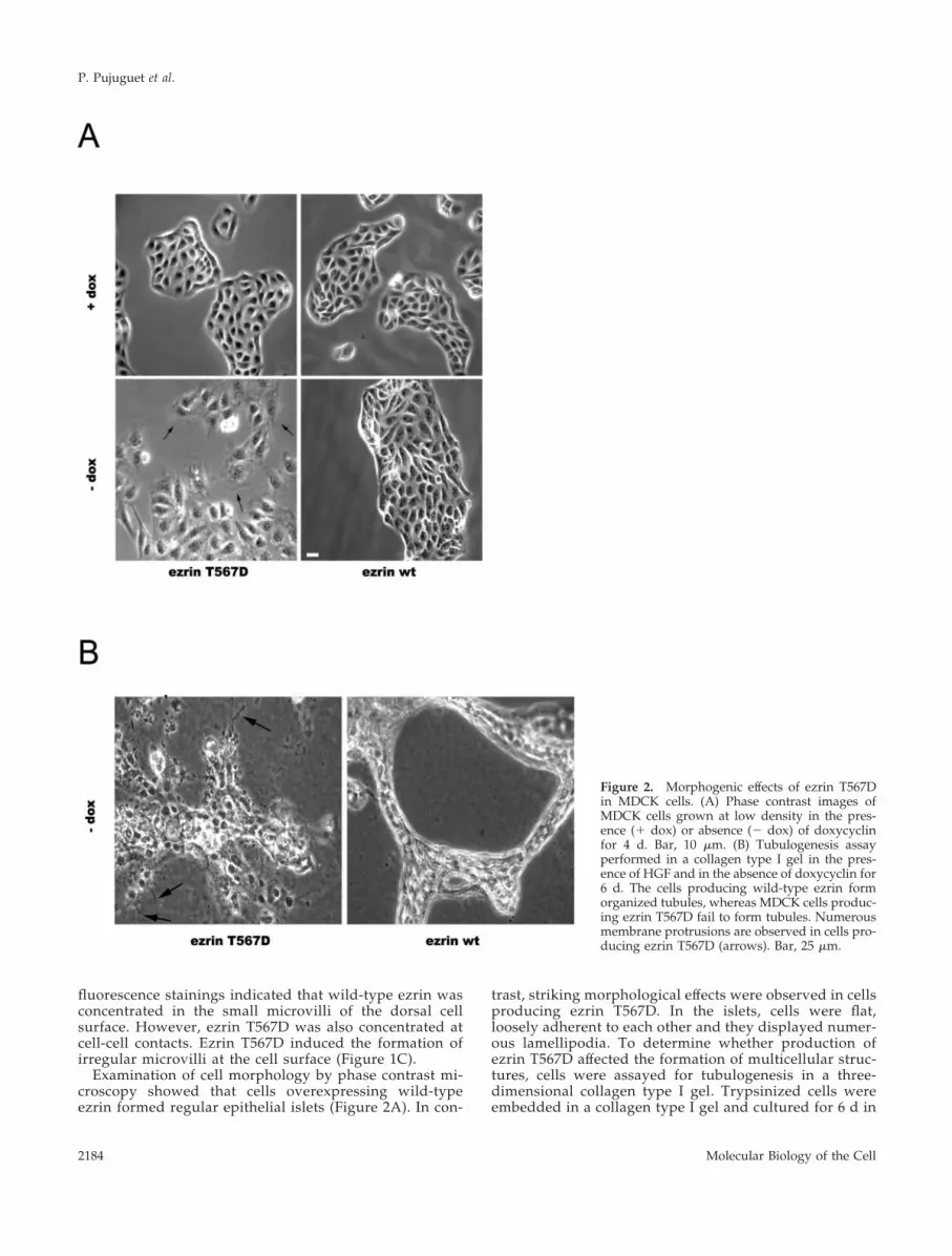
Examination of cell morphology by phase contrast mi-croscopy showed that cells overexpressing wild-typeezrin formed regular epithelial islets (Figure 2A). In con-
trast, striking morphological effects were observed in cellsproducing ezrin T567D. In the islets, cells were flat,loosely adherent to each other and they displayed numer-ous lamellipodia. To determine whether production ofezrin T567D affected the formation of multicellular struc-tures, cells were assayed for tubulogenesis in a three-dimensional collagen type I gel. Trypsinized cells wereembedded in a collagen type I gel and cultured for 6 d in
Figure 2. Morphogenic effects of ezrin T567Din MDCK cells. (A) Phase contrast images ofMDCK cells grown at low density in the pres-ence (� dox) or absence (� dox) of doxycyclinfor 4 d. Bar, 10 �m. (B) Tubulogenesis assayperformed in a collagen type I gel in the pres-ence of HGF and in the absence of doxycyclin for6 d. The cells producing wild-type ezrin formorganized tubules, whereas MDCK cells produc-ing ezrin T567D fail to form tubules. Numerousmembrane protrusions are observed in cells pro-ducing ezrin T567D (arrows). Bar, 25 �m.
P. Pujuguet et al.
Molecular Biology of the Cell2184

the presence of HGF and in the absence of doxycyclin.Unlike cells producing wild-type ezrin, ezrin T567D-pro-ducing cells did not form organized tubules but formedcellular aggregates with randomly distributed membraneprotrusions (Figure 2B). Thus, production of ezrin T567Din MDCK cells altered their ability to establish functionalcell-cell contacts.
Expression of Ezrin T567D in MDCK Cells Activatesthe Small GTPase Rac1Because small GTPases of the Rho family are regulators oflamellipodia membrane protrusions (Hall, 1998), weasked whether the membrane extensions observed inMDCK cells producing ezrin T567D were due to an acti-vation of these GTPases. GST-PAK and GST-rhotekin fu-sion proteins were used to measure the GTP loading ofendogenous Rac1-, Cdc42-, and RhoA-GTPases, from celllysates of cells expressing either ezrin T567D or wild-typeezrin and grown with or without doxycyclin for 4 d(Figure 3A). Based on four independent experiments, wefound that the level of Rac1-GTP was induced �2.5 fold incells producing ezrin T567D, compared with cells produc-ing wild-type ezrin (Figure 3, A and B). In contrast, no
change in the level of the RhoA- or Cdc42-GTP was de-tected in the cells expressing ezrin T567D compared withcells expressing wild-type ezrin (Figure 3A). This changein Rac1 activity was readily observed after 1-d omission ofdoxycyclin and increased up to 4 d (Figure 3C), as did theexpression level of ezrin T567D (Figure 1B). This indicatedthat Rac1 activity correlated with the expression of ezrinT567D. To rule out the possibility that the Rac1 activationobserved in MDCK cells producing ezrin T567D was dueto a difference of growth rate with cells expressing wild-type ezrin, we measured the cell growth of the differentclones up to day 4. No significant differences in thegrowth rate were observed between cells producing ezrinT567D and wild-type ezrin (data not shown).
Because lamellipodia were mainly observed in nonconflu-ent MDCK cells producing ezrin T567D, we asked whetheractivation of the GTPase Rac1 was sustained in confluentcells producing ezrin T567D. MDCK cells were grown atconfluence on filters in the absence of doxycyclin in themedium for 4 d. In these culture conditions, the level ofRac1-GTP was similar to that of control cells (Figure 3D).Together, these data indicated that production of constitu-tively activated ezrin leads to the activation of the GTPaseRac1 in subconfluent cells.
Figure 3. Ezrin T567D activatesthe GTPase Rac1. Lysates fromcells grown in the presence (�dox) or absence (� dox) of doxy-cyclin for 4 d were incubatedwith a GST-PAK fusion proteinfor the Rac1 and Cdc 42 activityassays, or with a GST-rothekinfusion protein for the RhoA activ-ity assay. (A) The amount of ac-tive GTP-bound Rac1, Cdc 42, orRhoA was analyzed by immuno-blotting with anti-Rac1, -Cdc 42,or -RhoA antibodies after the af-finity precipitation step. Totalamounts of Rac1, Cdc 42, orRhoA were determined by immu-noblotting of the cell lysates. (B)The densitometric quantificationof Rac1 activity shown in A isrepresentative of four indepen-dent experiments. Relative Rac1activity was calculated from theamount of GTP-bound Rac1 nor-malized to the amount of totalRac1 in cell lysates. An arbitraryunit of 1 was given for Rac1 ac-tivity in control cells. Rac activityis induced 2.5-fold in cells pro-ducing ezrin T567D comparedwith cells producing wt ezrin. (C)Immunoblotting with anti-Rac1antibodies of GTP-bound Rac1and total amount of Rac1 in ly-sates prepared from cells grownin absence of doxycyclin for theindicated time. (D) MDCK cellswere grown for 4 d after confluence with (� dox) or without (� dox) doxycyclin. Immunoblotting with an anti-Rac1 antibody shows a similaramount of GTP-bound Rac1 in both culture conditions.
Role of Ezrin in Cell-Cell Adhesion
Vol. 14, May, 2003 2185
Production of ezrin T567D in MDCK Cells PerturbsE-Cadherin Localization in a Rac1-dependentMannerBecause ezrin T567D production led to loosening of cell-cellcontacts and impaired tubulogenesis (Figure 2), we exam-ined whether it affected the E-cadherin–dependent cell-celladhesion. Two pools of E-cadherin were observed in theezrin T567D repressed cells and in cells producing wild-typeezrin. E-cadherin was detected at the cell-cell contacts and inintracellular compartments (Figure 4A). In contrast, in cellsproducing ezrin T567D we observed a strong accumulationof E-cadherin in intracellular compartments and a weakstaining at the plasma membrane.
To determine whether this decrease in E-cadherin at theplasma membrane of MDCK cells producing ezrin T567Dwas due to the activation of the GTPase Rac1, we overex-pressed in these cells the dominant negative form of Rac1,Rac1N17. On average, 150 RacN17-positive cells were exam-ined in three independent experiments. We observed thatexpression of Rac1N17 restored the localization of E-cad-herin at the cell-cell contacts in 92% of the Rac1N17-positivecells (Figure 4B). Conversely, Rac1V12, a constitutively ac-tive mutant of Rac1, induced the rounding of cells (Figure4B). Furthermore, 87% of Rac1V12 positive cells showed astrong intracellular accumulation of E-cadherin. Thus, acti-vation of the GTPase Rac1 in cells producing ezrin T567Dimpaired the localization of E-cadherin to the plasma mem-brane.
E-Cadherin Accumulates in IntracellularCompartments of MDCK Cells Producing EzrinT567DTo confirm the immunofluorescence data indicating a de-crease in E-cadherin at the plasma membrane, we measuredbiochemically the level of E-cadherin present at the surfaceof the cells producing ezrin T567D, or wild-type ezrin. Cellswere incubated at 4°C with biotin, and the amount of bio-tinylated E-cadherin was determined after immunoprecipi-tation with anti-E-cadherin antibodies and Western blotanalysis with streptavidin coupled to peroxidase. The levelof E-cadherin at the cell surface was 2.0-fold lower in cellsproducing ezrin T567D, compared with control cells (Figure5A). To determine whether this reduced level correspondedto a decrease in the total amount of E-cadherin, we evalu-ated the amount of E-cadherin in the different clones tested.As shown in Figure 5B, comparable levels of E-cadherinwere present in the cells expressing either ezrin T567D orwild-type ezrin. To confirm that the reduced amount ofE-cadherin at the plasma membrane was due to an accumu-lation of E-cadherin in intracellular compartments and not toan increased instability, we measured the half-life of E-cadherin. Pulse-chase analysis performed with MDCK cellsproducing ezrin T567D grown in presence or absence ofdoxycyclin in the medium indicated that E-cadherin half-lifewas similar in both culture conditions, 4.2 and 3.8 h, respec-tively (data not shown). Together, these results indicatedthat production of ezrin T567D in MDCK cells impaired thedelivery of E-cadherin to the plasma membrane.
E-Cadherin Delivery to the Plasma Membrane IsDelayed in Cells Producing Ezrin T567DBecause adherens junctions are dynamic structures contin-uously assembling and disassembling, MDCK cells inducedto express ezrin T567D, or wild-type ezrin were challengedby a calcium switch (Figure 6, A and B). Depletion of extra-cellular calcium disrupted epithelial cell-cell contacts andinduced E-cadherin internalization in both cell lines (Figure6A, time 0). On restoration of physiological level of extra-cellular calcium, adherens junctions progressively reestab-lished, with a concomitant increase in E-cadherin localiza-tion at the plasma membrane. After 2 h in high calciummedium, E-cadherin was significantly recruited at the cellsurface of cells producing wild-type ezrin, whereas thisdelivery was significantly inhibited in ezrin T567D-produc-ing cells (Figure 6, A and B). After 5 h in the high calciummedium, E-cadherin at the cell contacts further increased incells expressing wild-type ezrin, whereas it remained lowerat the cell surface of the cells producing ezrin T567D (Figure6, A and B). A neosynthetized pool of E-cadherin was notnecessary for reestablishment of cell-cell contacts becausethis occurred even in the presence of cycloheximide (datanot shown). These data confirmed that the expression ofezrin T567D delayed E-cadherin-based junction assemblyand E-cadherin delivery to the plasma membrane.
Ezrin T567D Slows Down E-CadherinInternalization in a Polarized MonolayerMDCK cells producing ezrin T567D were not able to form afunctional epithelium when assayed for tubulogenesis, indi-cating that the cells were not able to establish functionalcontacts. We thus examined whether production of ezrinT567D had any effects on E-cadherin present at the cell-cellcontacts of epithelial cells grown on filters. In these condi-tions, ezrin T567D-producing cells formed a confluentmonolayer with E-cadherin localized at the plasma mem-brane (Figure 7A). As assessed by biotinylation of cell sur-face proteins, a similar amount of E-cadherin was present atthe cell surface of cells producing ezrin T567D or wild-typeezrin (Figure 7A). By selective biotinylation of the apical orbasolateral side of the cells grown on filters, we observedthat E-cadherin localized correctly at the basolateral pole(Figure 7B). Na�/K� ATPase was used as a control of ba-solateral membrane marker excluded from the apical mem-brane domain. Thus, ezrin T567D production did not alterthe membrane-targeting events required for a polarized E-cadherin distribution. We therefore determined the ability ofE-cadherin to be internalized in cells grown on filters byusing a quantitative cell surface biotinylation assay (Figure7C). The amount of internalized E-cadherin was reduced incells expressing ezrin T567D compared with control cells.This down-regulation of internalization was also observedwith the transferrin receptor. As a control, no internalizedpool of the basolateral membrane protein Na�/K� ATPasewas detected.
DISCUSSION
We found herein that production of an ezrin mutant mim-icking its constitutive phosphorylation, leads to formation ofcell lamellipodia and disruption of cell-cell contacts in kid-
P. Pujuguet et al.
Molecular Biology of the Cell2186

Figure 4. Activation of the GTPase Rac1 inMDCK cells producing ezrin T567D per-turbs E-cadherin localization. (A) Immuno-fluorescence analysis with anti-E-cadherinantibodies of MDCK cells grown in thepresence (� dox) or absence (� dox) ofdoxycyclin for 4 d. An intracellular pool ofE-cadherin accumulates in cells producingezrin T567D with a concomitant decrease ofE-cadherin at the plasma membrane. (B)MDCK cells producing ezrin T567D weretransiently transfected with cDNAs codingfor Rac1N17 (top) or Rac1V12 (bottom).Double immunofluorescence staining of thecells was performed with anti-E-cadherinantibodies (right) and anti-myc antibodies(left). Bar, 10 �m.
Role of Ezrin in Cell-Cell Adhesion
Vol. 14, May, 2003 2187

ney epithelial cells. We propose these effects are due to theability of ezrin to activate the small GTPase Rac1. We reportherein that activation of the GTPase Rac1 pathway impairsthe delivery of E-cadherin to adherens junctions duringcell-cell contact assembly.
Production of ezrin T567D in nonconfluent MDCK cellsincreases the amount of GTP-bound Rac1, whereas the levelof GTP-bound RhoA or Cdc 42 does not change. This sug-gests that ezrin functions upstream of Rac1 activation. Howdoes ezrin activate Rac1? One possible mechanism isthrough an interaction between ezrin and the regulators ofthe GTPases. Indeed, a direct interaction between the FERMdomain of ERM proteins and RhoGDI has been observed. Ithas been proposed that, by sequestering RhoGDI, ERM pro-teins allow the activation of the Rho GTPase by the exchangefactors (Takahashi et al., 1997). Hence, in MDCK cells pro-ducing ezrin T567D, activation of the GTPase Rac1 could beincreased because the RhoGDI binding site on ezrin, nor-mally cryptic in wild-type ezrin, would be unmasked inezrin T567D. Another possibility by which ezrin may acti-vate the small GTPase Rac1 is through the regulation of theexchange factors themselves. Indeed, an association of theFERM domain of radixin with the exchange factor Dbl hasbeen observed in vitro (Takahashi et al., 1998). In addition,ezrin might act as a scaffold by recruiting activated Rac1, orits exchange factor, to the membrane at the sites where actinremodeling takes place. ERM proteins have been foundassociated with the plasma membrane of MDCK cells, andthis association was dependent on active Rho (Takaishi et al.,
1995; Kotani et al., 1997). It is indeed important to stress thatbeside being present at the dorsal membrane of the epithe-lial cells, ezrin T567D is also present at the lateral membrane,whereas wild-type ezrin is localized mostly to the apicalmembrane. Alternatively, ezrin T567D might interact with aprotein that is involved in the activation of Rac1. Interactionof hamartin with ERM proteins is required for the activationof the Rho pathway by serum or lysophosphatidic acid,which leads to cell adhesion to the substrate (Lamb et al.,2000).
Concomitant to the activation of Rac1, we observed exten-sive formation of lamellipodia and loosening of the cellcontacts. Our results show that the formation of lamellipo-dia in MDCK cells producing ezrin T567D is due to theactivation of Rac1 because dominant negative Rac1N17 re-inforced cell contacts and inhibited lamellipodia formation,whereas Rac1V12 induced membrane protusions and im-paired E-cadherin localization at cell-cell contacts. This effectof Rac1 was prominent with cells grown at low density.Similar morphological changes were observed when usingcontrolled expression of dominant active Rac1 in MDCKcells (Jou and Nelson, 1998) or in normal human keratino-cytes (Braga et al., 2000; Akhtar and Hotchin, 2001) grown atlow density. Scattering of epithelial cells after treatment withstimuli such as growth factors, integrin engagement, or li-gand binding to CD44 also involves Rac1 activation (Gi-mond et al., 1999; Oliferenko et al., 2000; Royal et al., 2000).Interestingly, we have previously shown that ezrin is adownstream target of the HGF receptor and that it potenti-ates the effect of HGF on cell scattering (Crepaldi et al., 1997).Thus, it is possible that the HGF-induced cell scattering ismediated by ezrin via the activation of Rac1.
However, in some experimental systems, activation of Rac1was correlated with establishment of E-cadherin–dependentcell-cell adhesion (Braga et al., 1997; Takaishi et al., 1997; Ehrlichet al., 2002). Expression of the Rac exchange factor Tiam 1restored E-cadherin mediated cell-cell adhesion in MDCK cellstreated with HGF or in Ras-transformed MDCK cells (Hordijket al., 1997; Sander et al., 1998). Using a calcium switch methodto follow junction assembly, it was observed that cadherin-dependent cell-cell contacts increased Rac1 activity (Nakagawaet al., 2001; Noren et al., 2001). One explanation to reconcilethese results is that the effect of Rac1 on the junctional com-plexes might depend on the state of cell-cell contacts, i.e., theirdegree of maturity. This was recently demonstrated by follow-ing the distribution of a GFP-tagged active Rac1 mutant, at lowcell density. Rac1 accumulated only at the newly formed cell-cell contacts, and not at older contacts (Ehrlich et al., 2002). Inline with this proposal, we have observed that when the cellsexpressing ezrin T567D were grown on filters, the level ofGTP-bound Rac1 and the amount of cell surface E-cadherinwere similar to those of control cells. Additionally, we ob-served that although cells expressing ezrin T567D failed toform tubules in collagen, cells grown on filters maintained anormal apical/basolateral polarity. These observations are cor-roborated by the observation that endogenous GTP-boundRac1 is required for cyst formation in collagen, but not formaintenance of polarity on filters (O’Brien et al., 2001).
The mechanism by which the small GTPase Rac1 inter-feres with assembly of E-cadherin is not known. Our resultsindicate that activated Rac1 inhibits the delivery of E-cad-herin to the plasma membrane during cell-cell junction as-
Figure 5. Ezrin T567D decreases the amount of E-cadherin at theplasma membrane. (A) MDCK cells were biotinylated at 0°C forlabeling of cell surface proteins. Biotinylated proteins were recov-ered on streptavidin beads and analyzed by immunoblotting withanti-E-cadherin antibodies. Immunoblotting with an anti-tubulinantibody served as loading control. (B) Total amounts of E-cadherinand tubulin were determined by immunoblotting. The total amountof E-cadherin was similar in MDCK cells producing ezrin T567D orwild-type ezrin.
P. Pujuguet et al.
Molecular Biology of the Cell2188

sembly of MDCK cells producing ezrin T567D. Because nei-ther the amount nor the half-life of E-cadherin changed inMDCK cells producing ezrin T567D compared with controlcells, the decrease in the amount of E-cadherin at the plasmamembrane results from the accumulation of this protein inintracellular compartments. These observations are in agree-ment with previous reports implicating activated Rac1 in theregulation of membrane protein traffic in MDCK cells andkeratinocytes (Lamaze et al., 1996; Jou et al., 2000; Akhtar andHotchin, 2001).
Our results indicate that activated Rac1 regulates E-cad-herin trafficking in subconfluent cells expressing ezrinT567D because in confluent cells the level of active Rac1 wassimilar to that of control cells. Yet, we have observed adecrease in the rate of E-cadherin endocytosis in confluentcells. This suggests that ezrin T567D might control somesteps of protein transport in addition to its role in Rac1activation. In line with this hypothesis, it has been proposedthat the interaction of ezrin with EBP50 is involved in therecycling of the �2-adrenergic receptor (Cao et al., 1999).
In conclusion, our observations indicate that ezrin plays arole in the transition from polarized epithelial cells to more“spread out” cells by regulating the transport of E-cadherinto the plasma membrane. This function implicates the acti-vation of the small GTPase Rac1 by ezrin and might involvea direct effect of ezrin on the membrane transport machin-ery. Elucidating the mechanisms by which ezrin controlsE-cadherin delivery to the plasma membrane may be rele-vant to understand its emerging role in tumor progression(Gautreau et al., 2002).
ACKNOWLEDGMENTS
We thank Drs. Philippe Chavrier and Thierry Galli for helpfulsuggestions and discussion. We thank Dr. Martin Schwartz and Dr.Peter N. Lowe for the generous gifts of plasmids. We thank Drs.Jyoti Srivastava and Ahmed Zahraoui for critically reading themanuscript. Dr. Philippe Pujuguet was supported by a postdoctoralfellowship from the Association pour la Recherche sur le Cancer.This work was supported by grants from La Ligue Nationale contre
Figure 6. Ezrin T567D delays E-cadherin delivery to the plasmamembrane. (A) MDCK cells cul-tured without doxycyclin for 4 dwere plated on coverslips andgrown in a low calcium mediumfor 4 h. Cells were then incubatedin a calcium-containing mediumfor different periods of time. Cellswere fixed and analyzed by im-munofluorescence with an anti-E-cadherin antibody. Bar, 10 �m.(B) Biotinylated cell surface pro-teins from cells expressing wild-type ezrin or ezrin T567D wererecovered on streptavidin beadsand analyzed by immunoblottingwith an anti-E-cadherin antibody.Immunoblotting with an anti-tu-bulin antibody served as loadingcontrol.
Role of Ezrin in Cell-Cell Adhesion
Vol. 14, May, 2003 2189

le Cancer and from the Association pour la Recherche contre leCancer (ARC 5599).
REFERENCES
Akhtar, N., and Hotchin, N.A. (2001). Rac1 regulates adherensjunctions through endocytosis of E-cadherin. Mol. Biol. Cell 12,847–862.
Algrain, M., Turunen, O., Vaheri, A., Louvard, D., and Arpin, M.(1993). Ezrin contains cytoskeleton and membrane binding domainsaccounting for its proposed role as a membrane-cytoskeletal linker.J. Cell Biol. 120, 129–139.
Allenspach, E.J., Cullinan, P., Tong, J., Tang, Q., Tesciuba, A.G.,Cannon, J.L., Takahashi, S.M., Morgan, R., Burkhardt, J.K., andSperling, A.I. (2001). ERM-dependent movement of CD43 defines anovel protein complex distal to the immunological synapse. Immu-nity 15, 739–750.
Barret, C., Roy, C., Montcourrier, P., Mangeat, P., and Niggli, V.(2000). Mutagenesis of the phosphatidylinositol 4,5-biphosphate(PIP2) binding site in the NH2-terminal domain of ezrin correlateswith its altered cellular distribution. J. Cell Biol. 151, 1067–1079.
Barth, A.I., Pollack, A.L., Altschuler, Y., Mostov, K.E., and Nelson,W.J. (1997). NH2-terminal deletion of beta-catenin results in stablecolocalization of mutant �-catenin with adenomatous polyposis coliprotein and altered MDCK cell adhesion. J. Cell Biol. 136, 693–706.
Berryman, M., Gary, R., and Bretscher, A. (1995). Ezrin oligomersare major cytoskeletal components of placental microvilli: a pro-posal for their involvement in cortical morphogenesis. J. Cell Biol.131, 1231–1242.
Braga, V.M.M., Betson, M., Li, X., and Lamarche-Vane, N. (2000).Activation of the small GTPase Rac is sufficient to disrupt cadherin-dependent cell-cell adhesion in normal human keratinocytes. Mol.Biol. Cell 11, 3703–3721.
Braga, V.M.M., Machesky, L.M., Hall, A., and Hotchin, N.A. (1997).The small GTPases Rho and Rac are required for the establishmentof cadherin-dependent cell-cell contacts. J. Cell Biol. 137, 1421–1431.
Bretscher, A., Gary, R., and Berryman, M. (1995). Soluble ezrinpurified from placenta exists as stable monomers and elongateddimers with masked C-terminal ezrin-radixin-moesin associationdomains. Biochemistry 34, 16830–16837.
Cao, T.T., Deacon, H.W., Reczek, D., Bretscher, A., and von Zastrow,M. (1999). A kinase-regulated PDZ-domain interaction controls en-docytic sorting of the �2-adrenergic receptor. Nature 401, 286–290.
Crepaldi, T., Gautreau, A., Comoglio, P.M., Louvard, D., and Arpin,M. (1997). Ezrin is an effector of HGF-mediated migration andmorphogenesis in epithelial cells. J. Cell Biol. 138, 423–434.
Delon, J., Kaibuchi, K., and Germain, R.N. (2001). Exclusion of CD43from the immunological synapse is mediated by phosphorylation-regulated relocation of the cytoskeletal adaptor moesin. Immunity15, 691–701.
Ehrlich, J.S., Hansen, M.D.H., and Nelson, W.J. (2002). Spatio-tem-poral regulation of Rac1 localization and lamellipodia dynamicsduring epithelial cell-cell adhesion. Dev. Cell 3, 259–270.
Gary, R., and Bretscher, A. (1995). Ezrin self-association involvesbinding of an N-terminal domain to a normally masked C-terminaldomain that includes the F-actin binding site. Mol. Biol. Cell 6,1061–1075.
Gautreau, A., Louvard, D., and Arpin, M. (2000). Morphogeniceffects of ezrin require a phosphorylation-induced transition fromoligomers to monomers at the plasma membrane. J. Cell Biol. 150,193–203.
Figure 7. Ezrin T567D slows down E-cadherin internalization inpolarized monolayer. (A) Confluent MDCK cells grown on filters inthe presence (� dox) or absence (� dox) of doxycyclin were surface-biotinylated at 0°C from the basal pole. Surface-biotinylated pro-teins were recovered from cell extracts on streptavidin beadsand analyzed by immunoblotting with anti-E-cadherin or -Na�/K�
ATPase antibodies. Ezrin T567D expression was detected in theabsence of doxycyclin by immunoblotting with a VSVG antibody.(B) Confluent MDCK cells grown on filters in the presence orabsence of doxycyclin were surface biotinylated at 0°C either fromthe apical or basal pole. Surface-biotinylated proteins were recov-ered from cell extracts on streptavidin beads and analyzed byimmunoblotting with anti-E-cadherin or -Na�/K� ATPase antibod-ies. (C) Internalization of surface-biotinylated E-cadherin. ConfluentMDCK cells grown on filters in the presence or absence of doxycy-clin were surface biotinylated at 0°C (surface) and either strippedwith glutathione immediately after biotinylation (reduced) or al-lowed to be internalized at 37°C for 1 h and stripped (intracellular).Biotinylated proteins were recovered from cell extracts on strepta-vidin beads and analyzed by immunoblotting with anti-E-cadherin,anti-Na�/K� ATPase, or anti-transferrin receptor.
P. Pujuguet et al.
Molecular Biology of the Cell2190

Gautreau, A., Louvard, D., and Arpin, M. (2002). ERM proteins andNF2 tumor suppressor: the Yin and Yang of cortical actin organiza-tion and cell growth signaling. Curr. Opin. Cell Biol. 14, 104–109.
Gimond, C., van der Flier, A., van Delft, S., Brakebusch, C., Kuik-man, I., Collard, J.G., Fassler, R., and Sonnenberg, A. (1999). Induc-tion of cell scattering by expression of �1 integrins in �1-deficientepithelial cells requires activation of members of the Rho family ofGTPases and downregulation of cadherin and catenin function.J. Cell Biol. 147, 1325–1340.
Hall, A. (1998). Rho GTPases and the actin cytoskeleton. Science 279,509–514.
Helander, T.S., Carpen, O., Turunen, O., Kovanen, P.E., Vaheri, A.,and Timonen, T. (1996). ICAM-2 redistributed by ezrin as a targetfor killer cells. Nature 382, 265–268.
Hordijk, P.L., ten Klooster, J.P., van der Kammen, R.A., Michiels, F.,Oomen, L.C.J.M., and Collard, J.G. (1997). Inhibition of invasion ofepithelial cells by Tiam1-Rac signaling. Science 278, 1464–1466.
Jou, T.-S., Leung, S.-M., Fung, L.M., Ruiz, W.G., Nelson, W.J., andApodaca, G. (2000). Selective alterations in biosynthetic and endo-cytic protein traffic in Madin-Darby canine kidney epithelial cellsexpressing mutants of the small GTPase Rac1. Mol. Biol. Cell 11,287–304.
Jou, T.-S., and Nelson, W.J. (1998). Effects of regulated expression ofmutant RhoA and Rac1 small GTPases on the development ofepithelial (MDCK) cell polarity. J. Cell Biol. 142, 85–100.
Kotani, H., Takaishi, K., Sasaki, T., and Takai, Y. (1997). Rho regu-lates association of both the ERM family and vinculin with theplasma membrane in MDCK cells. Oncogene 14, 1705–1713.
Kreis, T.E. (1986). Microinjected antibodies against the cytoplasmicdomain of vesicular stomatitis virus glycoprotein block its transportto the cell surface. EMBO J. 5, 931–941.
Lamaze, C., Chuang, T.-H., Terlecky, L.J., Bokoch, G.M., andSchmid, S.L. (1996). Regulation of receptor-mediated endocytosis byRho and Rac. Nature 382, 177–179.
Lamb, R.F., Roy, C., Diefenbach, T.J., Vinters, H.V., Johnson, M.W.,Jay, D.G., and Hall, A. (2000). The TSC1 tumor suppressor hamartinregulates cell adhesion through ERM proteins and the GTPase Rho.Nat. Cell Biol. 2, 281–287.
Le, T.L., Yap, A.S., and Stow, J.L. (1999). Recycling of E-cadherin: apotential mechanism for regulating cadherin dynamics. J. Cell Biol.146, 219–232.
Mackay, D.J.G., Esch, F., Furthmayr, H., and Hall, A. (1997). Rho-and Rac-dependent assembly of focal adhesion complexes and actinfilaments in permeabilized fibroblasts: an essential role for ezrin/radixin/moesin proteins. J. Cell Biol. 138, 927–938.
Martin, M., Andreoli, C., Sahuquet, A., Montcourrier, P., Algrain,M., and Mangeat, P. (1995). Ezrin NH2-terminal domain inhibits thecell extension activity of the COOH-terminal domain. J. Cell Biol.128, 1081–1093.
Matsui, T., Maeda, M., Doi, Y., Yonemura, S., Amano, M., Kaibuchi,K., Tsukita, S., and Tsukita, S. (1998). Rho-kinase phosphorylatesCOOH-terminal threonines of ezrin/radixin/moesin (ERM) pro-teins and regulates their head-to-tail association. J. Cell Biol. 140,647–657.
Nakagawa, M., Fukata, M., Yamaga, M., Itoh, N., and Kaibuchi, K.(2001). Recruitment and activation of Rac1 by the formation ofE-cadherin-mediated cell-cell adhesion sites. J. Cell Sci. 114, 1829–1838.
Nakamura, F., Huang, L., Pestonjamasp, K., Luna, E.J., and Furth-mayr, H. (1999). Regulation of F-actin binding to platelet moesin invitro by both phosphorylation of threonine 558 and polyphosphati-dylinositides. Mol. Biol. Cell 10, 2669–2685.
Noren, N.K., Niessen, C.M., Gumbiner, B.M., and Burridge, K.(2001). Cadherin engagement regulates Rho family GTPases. J. Biol.Chem. 276, 33305–33308.
O’Brien, L.E., Jou, T.-S., Pollack, A.L., Zhang, Q., Hansen, S.H.,Yurchenco, P., and Mostov, K.E. (2001). Rac1 orientates epithelialapical polarity through effects on basolateral laminin assembly. Nat.Cell Biol. 3, 831–838.
Oliferenko, S., Kaverina, I., Small, J.V., and Huber, L.A. (2000).Hyaluronic acid (HA) binding to CD44 activates Rac1 and induceslamellipodia outgrowth. J. Cell Biol. 148, 1159–1164.
Ren, X.D., Kiosses, W.B., and Schwartz, M.A. (1999). Regulation ofthe small GTP-binding protein Rho by cell adhesion and the cy-toskeleton. EMBO J 18, 578–585.
Roumier, A., Olivo-Marin, J.C., Arpin, M., Michel, F., Martin, M.,Mangeat, P., Acuto, O., Dautry-Varsat, A., and Alcover, A. (2001).The membrane-microfilament linker ezrin is involved in the forma-tion of the immunological synapse and in T cell activation. Immu-nity 15, 715–728.
Royal, I., Lamarche-Vane, N., Lamorte, L., Kaibuchi, K., and Park,M. (2000). Activation of Cdc42, Rac, PAK, and Rho-kinase in re-sponse to hepatocyte growth factor differentially regulates epithelialcell colony spreading and dissociation. Mol. Biol. Cell 11.
Sander, E.E., van Delft, S., ten Klooster, J.P., Reid, T., van derKammen, R.A., Michiels, F., and Collard, J.G. (1998). Matrix-depen-dent Tiam/Rac signaling in epithelial cells promotes either cell-celladhesion or cell migration and is regulated by phosphatidylinositol3-kinase. J. Cell Biol. 143, 1385–1398.
Serrador, J.M., Alonso-Lebrero, J.L., del Pozo, M.A., Furthmayr, H.,Schwartz-Albiez, R., Calvo, J., Lozano, F., and Sanchez-Madrid, F.(1997). Moesin interacts with the cytoplasmic region of intercellularadhesion molecule-3 and is redistributed to the uropod of T. lym-phocytes during cell polarization. J. Cell Biol. 138, 1409–1423.
Takahashi, K., Sasaki, T., Mammoto, A., Hotta, I., Takaishi, K.,Imamura, M., Nakano, K., Kodama, A., and Takai, Y. (1998). Inter-action of radixin with Rho small G protein GDP/GTP exchangeprotein Dbl. Oncogene 16, 3279–3284.
Takahashi, K., Sasaki, T., Mammoto, A., Takaishi, K., Kameyama, T.,Tsukita, S., Tsukita, S., and Takai, Y. (1997). Direct interaction of theRho GDP dissociation inhibitor with ezrin/radixin/moesin initiatesthe activation of the Rho small G protein. J. Biol. Chem. 272, 23371–23375.
Takaishi, K., Sasaki, T., Kameyama, T., Tsukita, S., Tsukita, S., andTakai, Y. (1995). Translocation of activated Rho from the cytoplasmto membrane ruffling area, cell-cell adhesion sites and cleavagefurrows. Oncogene 11, 39–48.
Takaishi, K., Sasaki, T., Kotani, H., Nishioka, H., and Takai, Y.(1997). Regulation of cell-cell adhesion by Rac and Rho small Gproteins in MDCK cells. J. Cell Biol. 139, 1047–1059.
Takeuchi, K., Sato, N., Kasahara, H., Funayama, N., Nagafuchi, A.,Yonemura, S., Tsukita, S., and Tsukita, S. (1994). Perturbation of celladhesion and microvilli formation by antisense oligonucleotides toERM family members. J. Cell Biol. 125, 1371–1384.
Thompson, G., Owen, D., Chalk, P.A., and Lowe, P.N. (1998). De-lineation of the Cdc42/Rac-binding domain of p21-activated kinase.Biochemistry 37, 7885–7891.
Tran Quang, C., Gautreau, A., Arpin, M., and Treisman, R. (2000).Ezrin function is required for ROCK-mediated fibroblast transfor-mation by the Net and Dbl oncogenes. EMBO J. 19, 4565–4576.
Yonemura, S., Tsukita, S., and Tsukita, S. (1999). Direct involvementof ezrin/radixin/moesin (ERM)-binding membrane proteins in theorganization of microvilli in collaboration with activated proteins.J. Cell Biol. 145, 1497–1509.
Role of Ezrin in Cell-Cell Adhesion
Vol. 14, May, 2003 2191


