Embed Size (px)
Citation preview

FACULTY OF NURSINGFACULTY OF NURSING
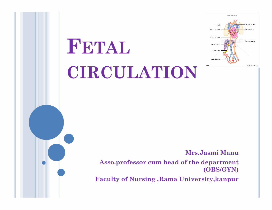
FETAL
CIRCULATION
Mrs.Jasmi Manu
Asso.professor cum head of the department (OBS/GYN)
Faculty of Nursing ,Rama University,kanpur
INTRODUCTION In the fully developed human, the heart serves
two main purposes. The right heart pumps blood to the lungs for
oxygenation and the left heart pumps oxygenated blood to rest of the body.
In the embryo and fetus, the lungs do not oxygenate the blood.
Fetal circulation is consequently quite different than that of a breathing baby or adult.
When a baby is born and takes its first breathes, the ducts close and blood is rerouted to the lungs.
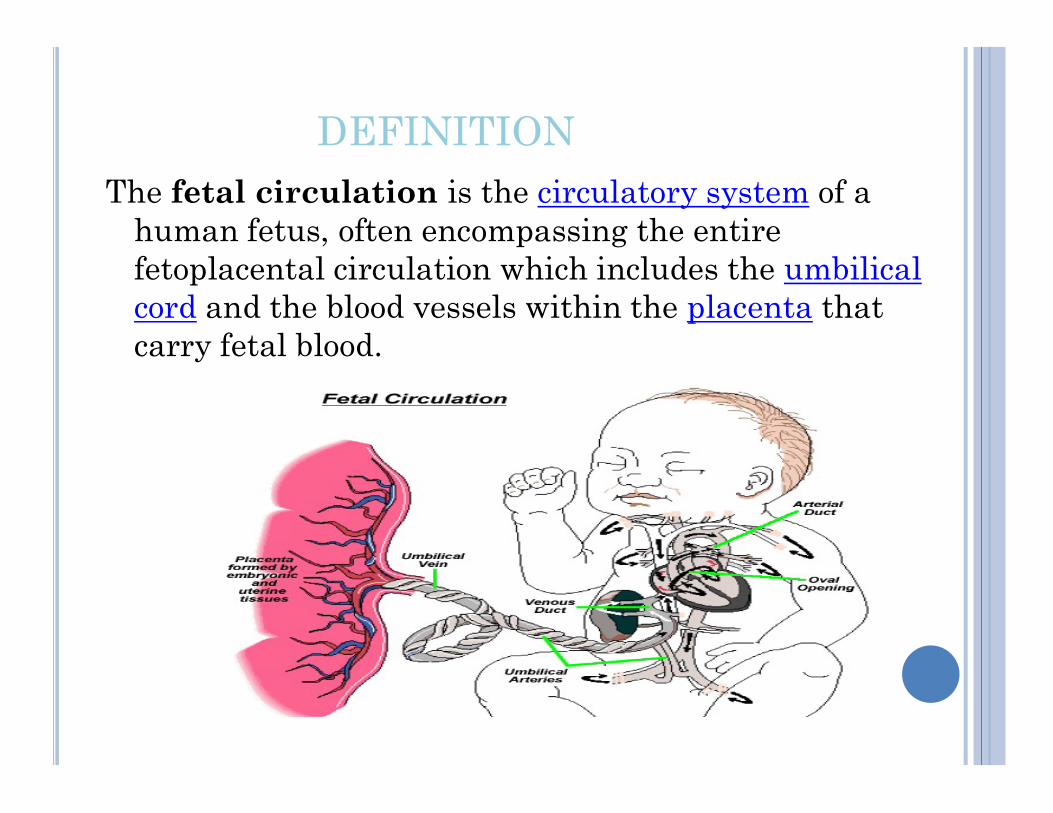
DEFINITIONThe fetal circulation is the circulatory system of a
human fetus, often encompassing the entire fetoplacental circulation which includes the umbilical cord and the blood vessels within the placenta that carry fetal blood.
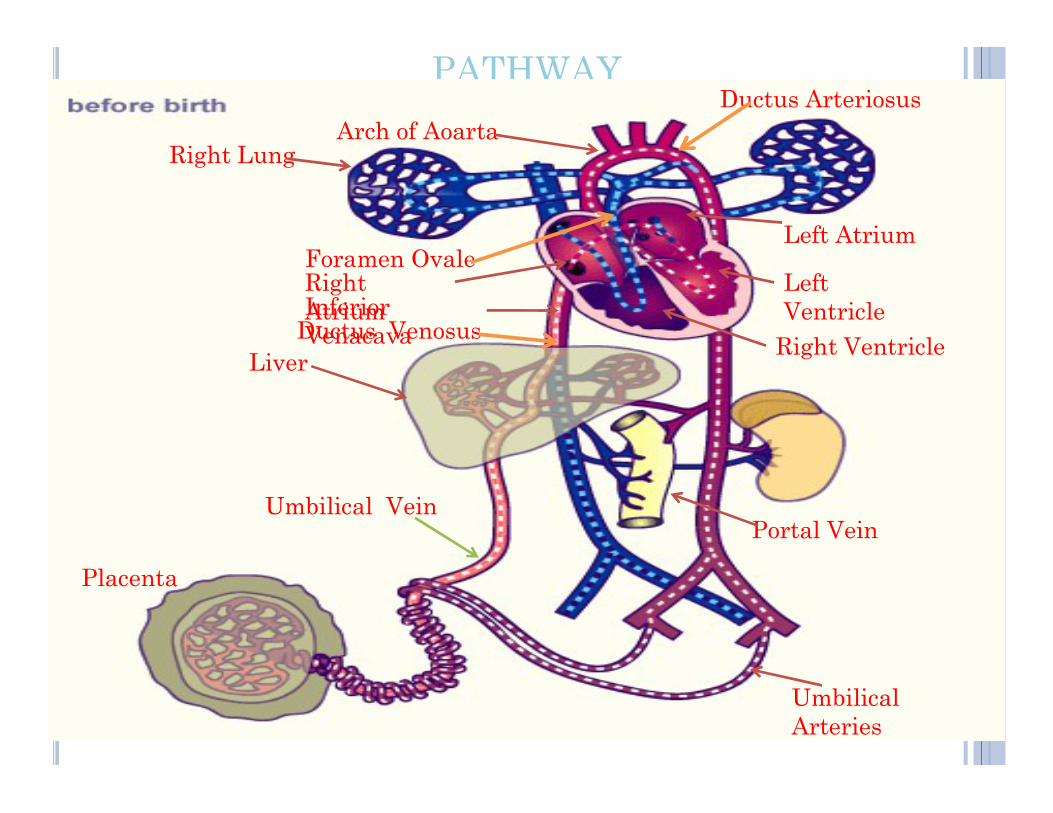
PATHWAY
LiverDuctus VenosusInferior Venacava
Right Atrium
Foramen Ovale
Right LungArch of Aoarta
Ductus Arteriosus
Left Atrium
Left Ventricle
Right Ventricle
Placenta
Umbilical Vein
Umbilical Arteries
Portal Vein
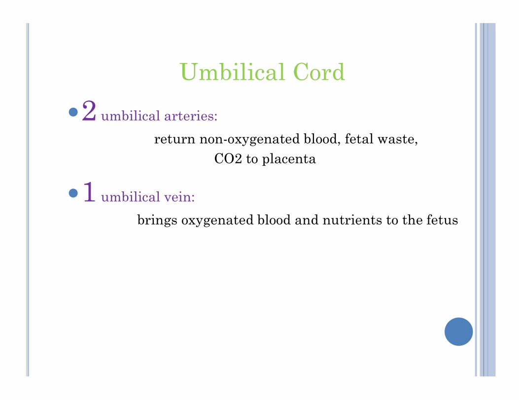
Umbilical Cord
2 umbilical arteries:
return non-oxygenated blood, fetal waste, CO2 to placenta
1 umbilical vein: 1 umbilical vein:
brings oxygenated blood and nutrients to the fetus
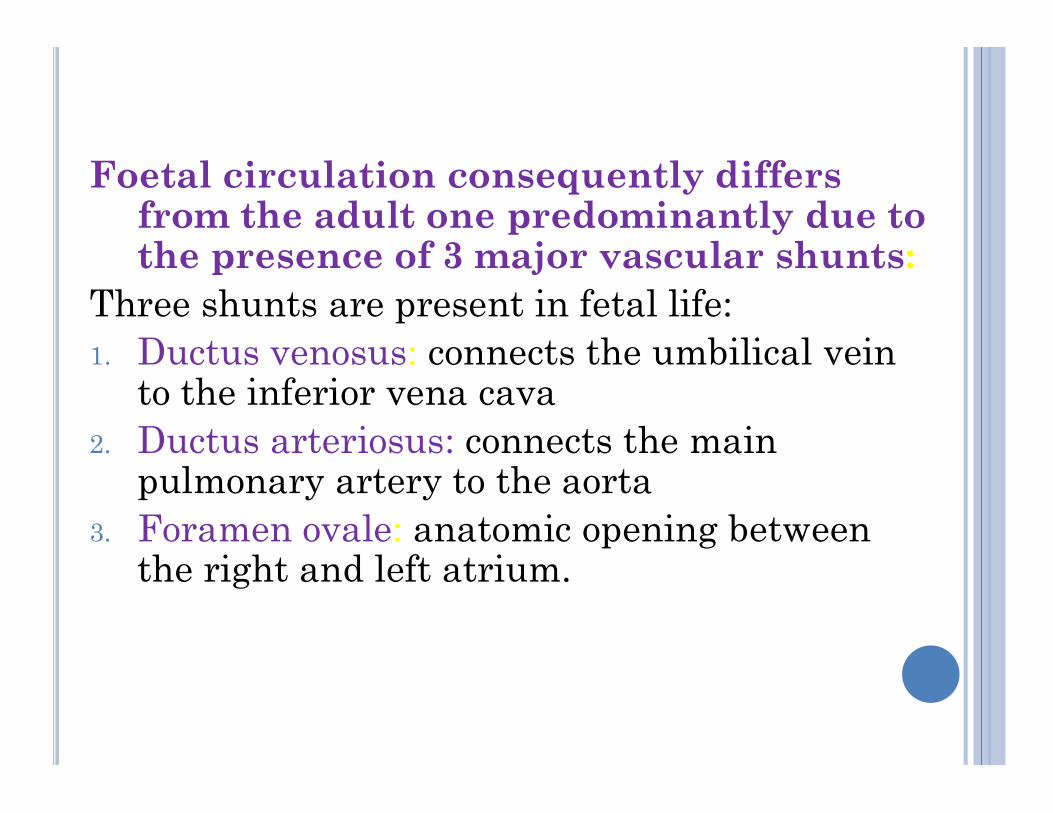
Foetal circulation consequently differs from the adult one predominantly due to the presence of 3 major vascular shunts:
Three shunts are present in fetal life:1. Ductus venosus: connects the umbilical vein
to the inferior vena cavato the inferior vena cava2. Ductus arteriosus: connects the main
pulmonary artery to the aorta3. Foramen ovale: anatomic opening between
the right and left atrium.
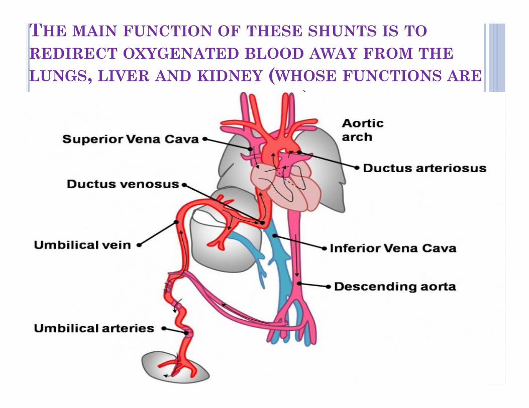
THE MAIN FUNCTION OF THESE SHUNTS IS TO
REDIRECT OXYGENATED BLOOD AWAY FROM THE
LUNGS, LIVER AND KIDNEY (WHOSE FUNCTIONS ARE
PERFORMED BY THE PLACENTA).
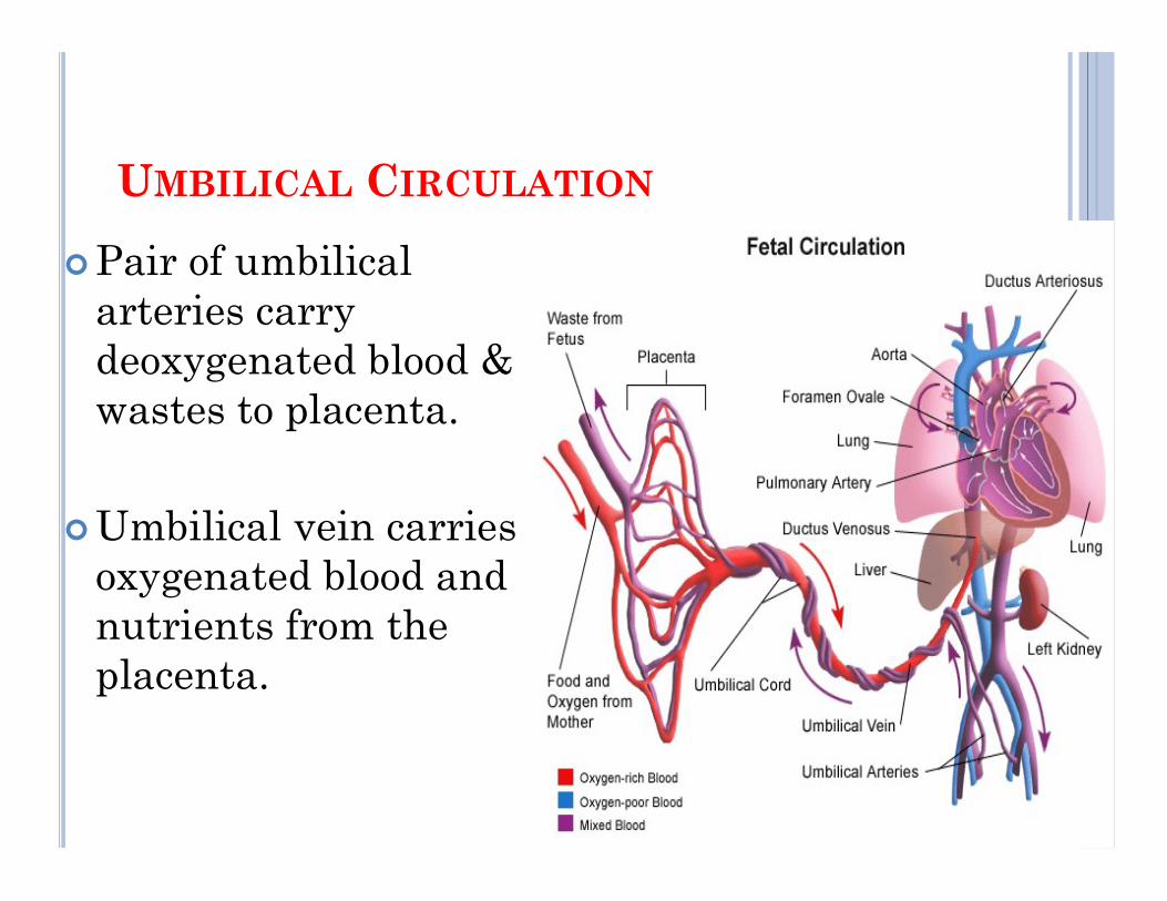
UMBILICAL CIRCULATION
Pair of umbilical arteries carry deoxygenated blood & wastes to placenta.
Umbilical vein carries oxygenated blood and nutrients from the placenta.
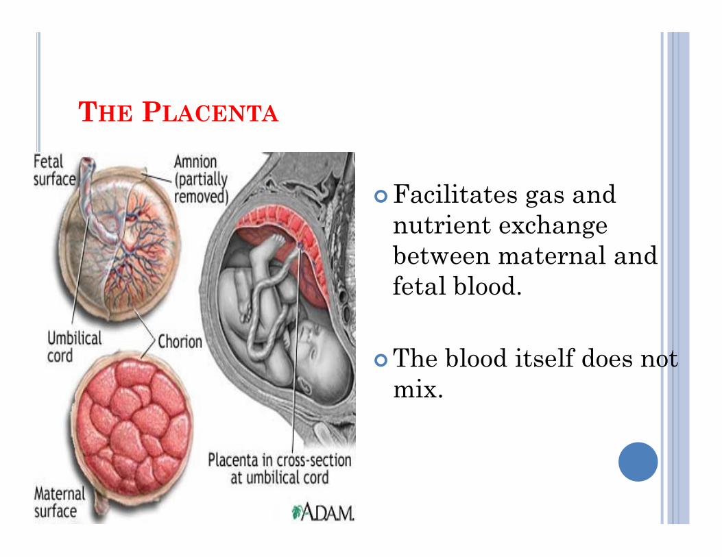
THE PLACENTA
Facilitates gas and nutrient exchange between maternal and fetal blood.fetal blood.
The blood itself does not mix.
PLACENTAL ROLE
The core concept behind fetal circulation is that fetal hemoglobin has a higher affinity for oxygen
than does adult hemoglobin, which allows a diffusion of oxygen from the mother's circulatory
system to the fetus. system to the fetus.
The circulatory system of the mother is not directly connected to that of the fetus, so the placenta functions as the respiratory center for the fetus as well as a site of filtration for
plasma nutrients and wastes.
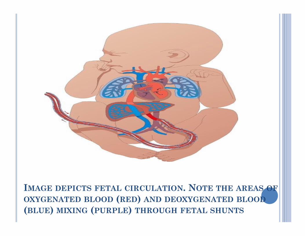
IMAGE DEPICTS FETAL CIRCULATION. NOTE THE AREAS OF
OXYGENATED BLOOD (RED) AND DEOXYGENATED BLOOD
(BLUE) MIXING (PURPLE) THROUGH FETAL SHUNTS
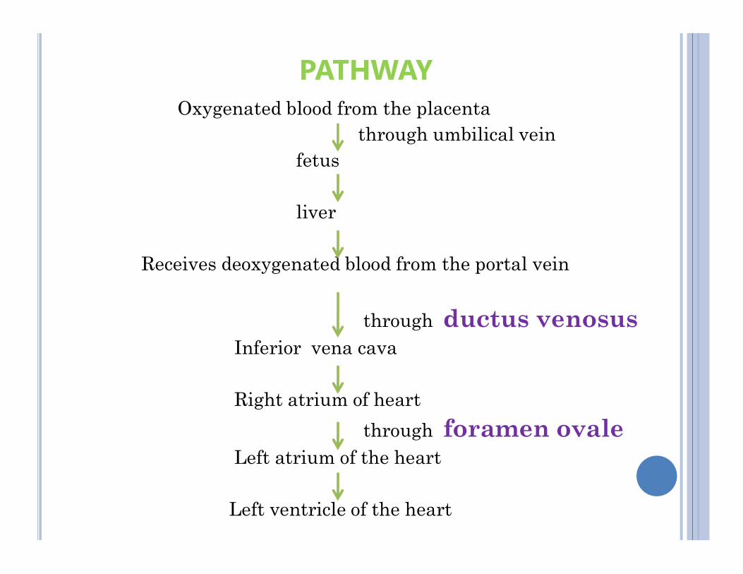
PATHWAYOxygenated blood from the placenta
through umbilical veinfetus
liver
Receives deoxygenated blood from the portal vein
through ductus venosusInferior vena cava
Right atrium of heart
through foramen ovaleLeft atrium of the heart
Left ventricle of the heart
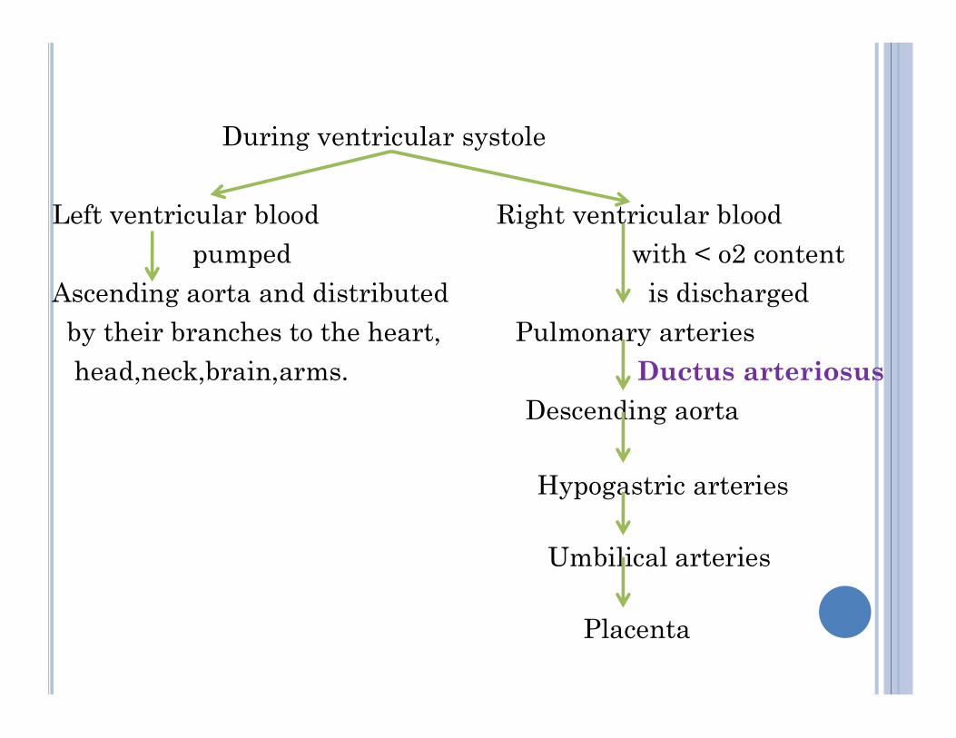
During ventricular systole
Left ventricular blood Right ventricular bloodpumped with < o2 content
Ascending aorta and distributed is dischargedby their branches to the heart, Pulmonary arterieshead,neck,brain,arms. Ductus arteriosushead,neck,brain,arms. Ductus arteriosus
Descending aorta
Hypogastric arteries
Umbilical arteries
Placenta
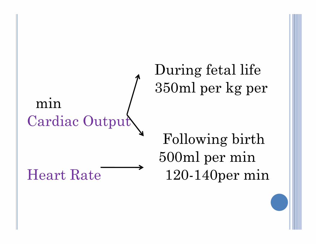
During fetal life350ml per kg per
minCardiac OutputCardiac Output
Following birth500ml per min
Heart Rate 120-140per min
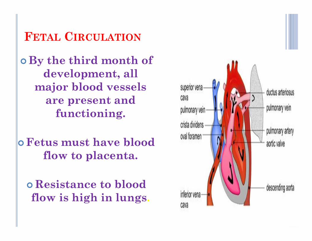
FETAL CIRCULATION
By the third month of development, all
major blood vessels are present and
functioning.
Fetus must have blood flow to placenta.
Resistance to blood flow is high in lungs.
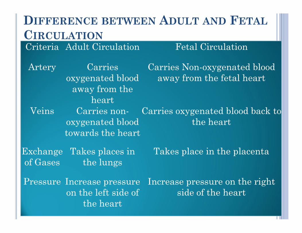
DIFFERENCE BETWEEN ADULT AND FETAL
CIRCULATIONCriteria Adult Circulation Fetal Circulation
Artery Carries oxygenated blood
away from the heart
Carries Non-oxygenated blood away from the fetal heart
Veins Carries non-oxygenated blood
Carries oxygenated blood back to the heartoxygenated blood
towards the heartthe heart
Exchange of Gases
Takes places in the lungs
Takes place in the placenta
Pressure Increase pressure on the left side of
the heart
Increase pressure on the right side of the heart
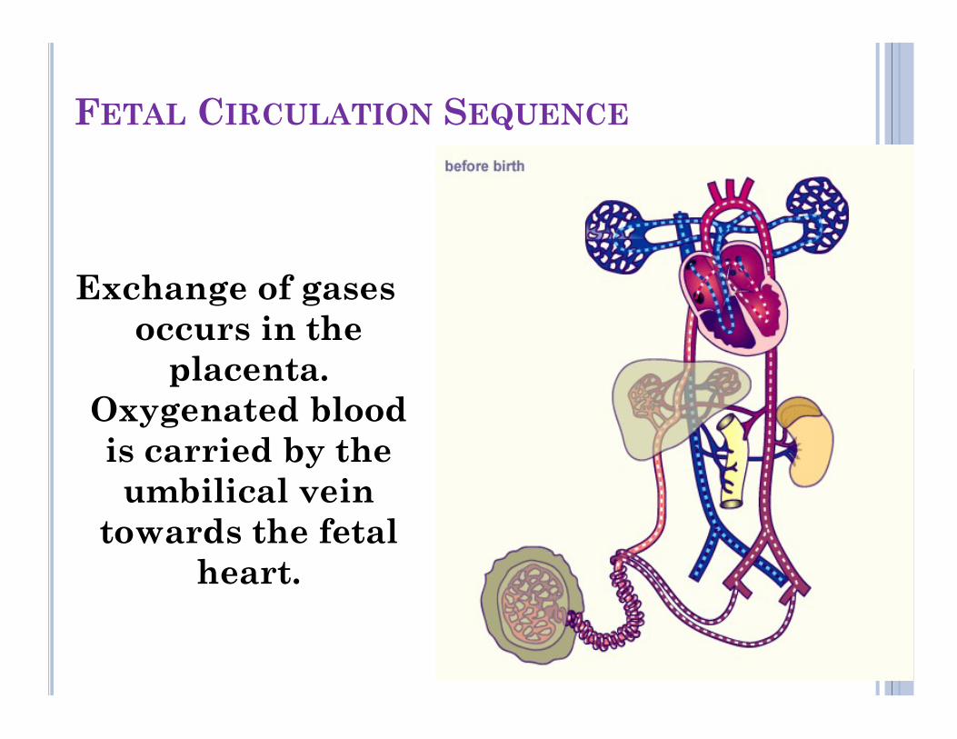
FETAL CIRCULATION SEQUENCE
Exchange of gases occurs in the
placenta. placenta. Oxygenated blood is carried by the umbilical vein
towards the fetal heart.
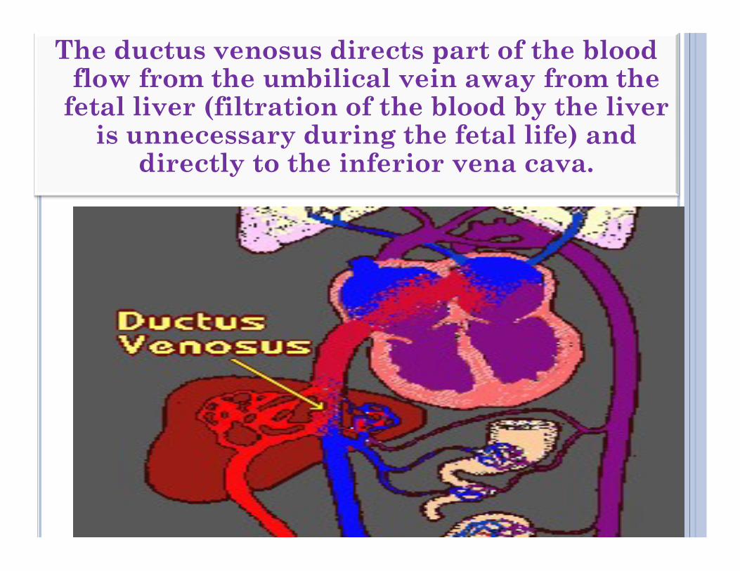
The ductus venosus directs part of the blood flow from the umbilical vein away from the
fetal liver (filtration of the blood by the liver is unnecessary during the fetal life) and
directly to the inferior vena cava.
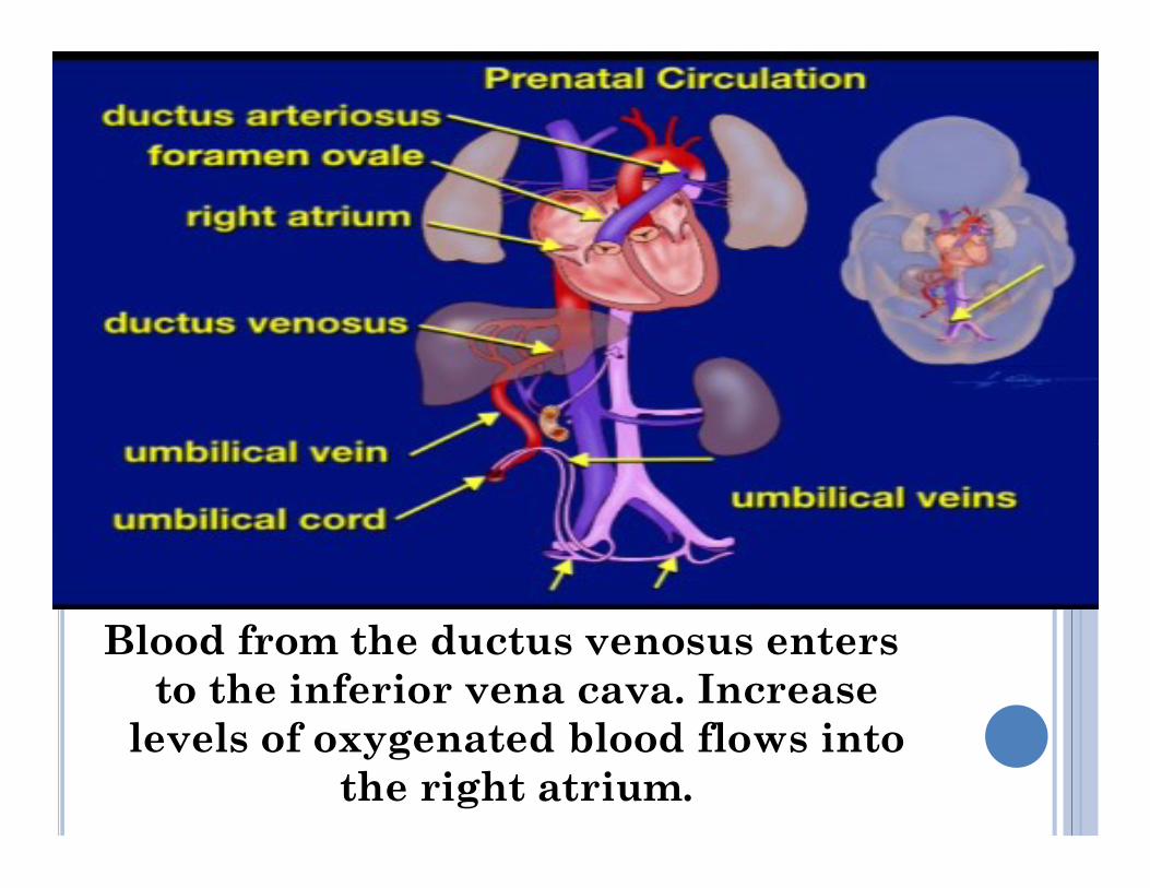
Blood from the ductus venosus enters to the inferior vena cava. Increase
levels of oxygenated blood flows into the right atrium.
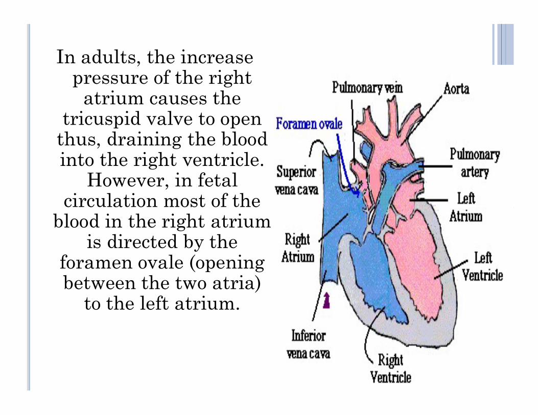
In adults, the increase pressure of the right
atrium causes the tricuspid valve to open
thus, draining the blood into the right ventricle.
However, in fetal circulation most of the circulation most of the
blood in the right atrium is directed by the
foramen ovale (opening between the two atria)
to the left atrium.
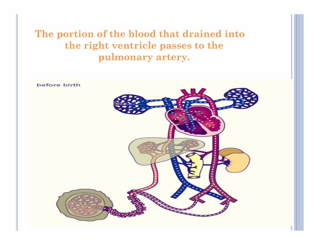
The portion of the blood that drained into the right ventricle passes to the
pulmonary artery.
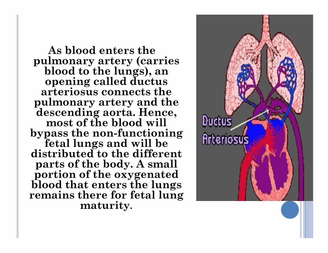
As blood enters the pulmonary artery (carries
blood to the lungs), an opening called ductus
arteriosus connects the pulmonary artery and the descending aorta. Hence,
most of the blood will bypass the non-functioning
most of the blood will bypass the non-functioning
fetal lungs and will be distributed to the different parts of the body. A small portion of the oxygenated blood that enters the lungs remains there for fetal lung
maturity.
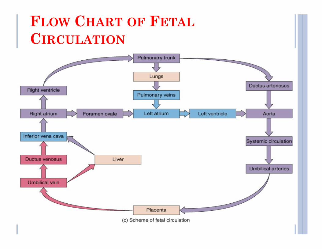
FLOW CHART OF FETAL
CIRCULATION
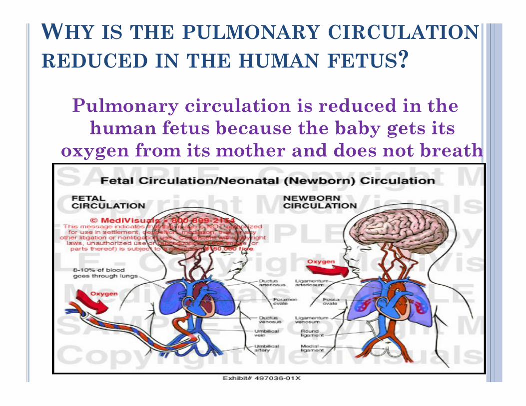
WHY IS THE PULMONARY CIRCULATION
REDUCED IN THE HUMAN FETUS?
Pulmonary circulation is reduced in the human fetus because the baby gets its
oxygen from its mother and does not breath on its own.
POSTNATAL CHANGES IN
CIRCULATION
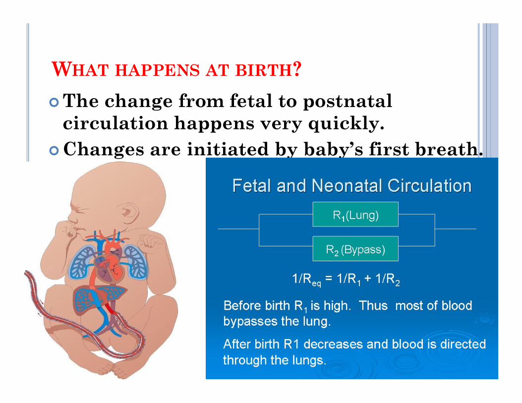
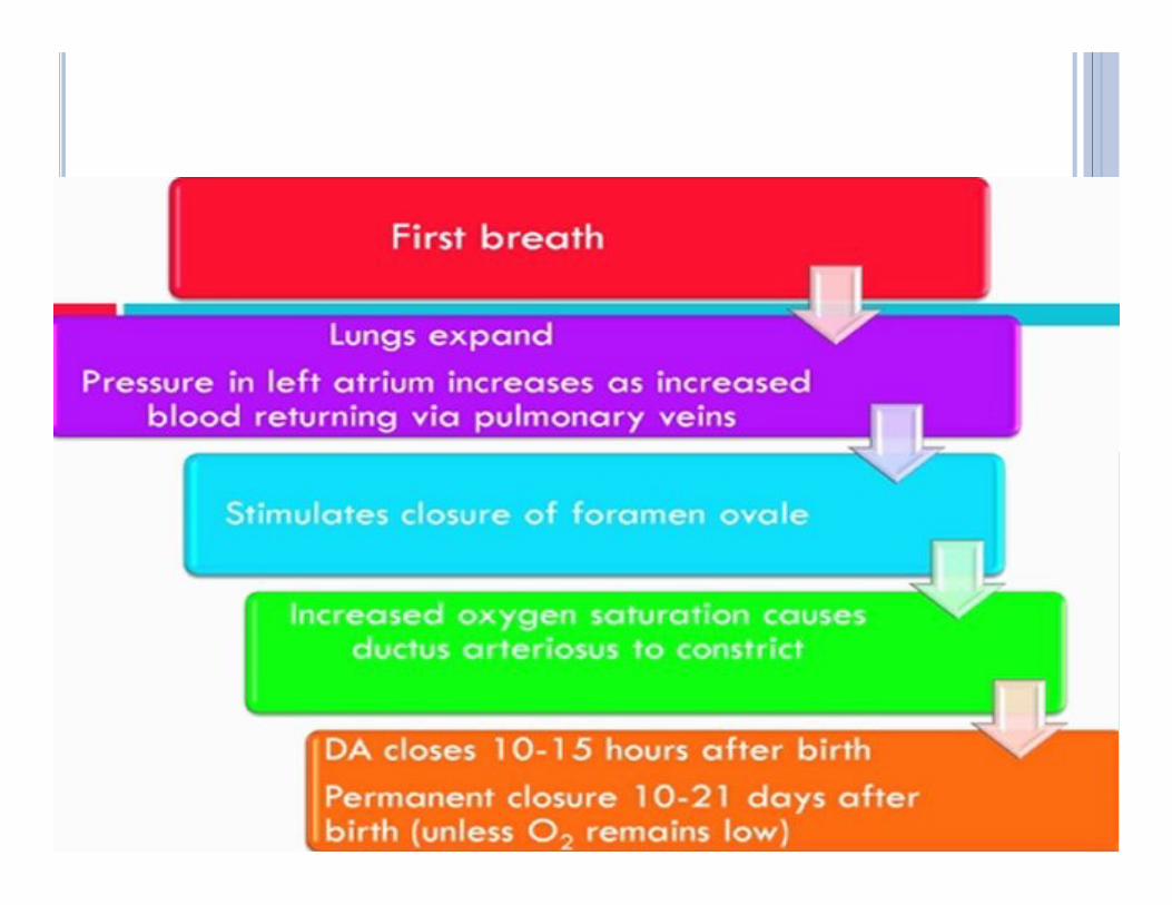
WHAT HAPPENS AT BIRTH?
The change from fetal to postnatal circulation happens very quickly.
Changes are initiated by baby’s first breath.
POST NATAL CHANGES
Gas exchange function is transferred from placenta to the lungs.
Separation of systemic and pulmonary circulations Increased metabolism to maintain body temperature and
hence increased cardiac output.hence increased cardiac output.
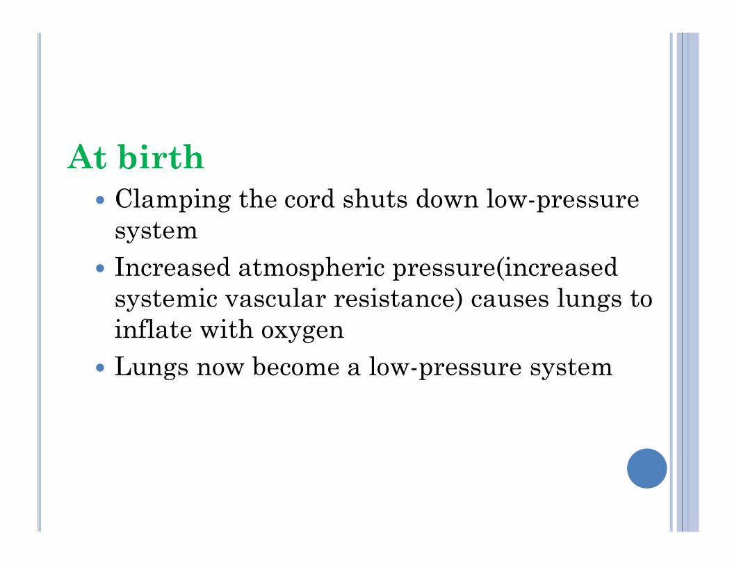
At birth Clamping the cord shuts down low-pressure
system Increased atmospheric pressure(increased
systemic vascular resistance) causes lungs to systemic vascular resistance) causes lungs to inflate with oxygen
Lungs now become a low-pressure system
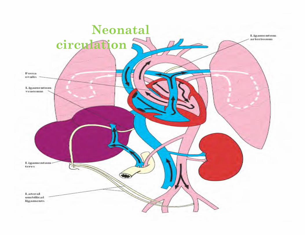
Neonatal circulation
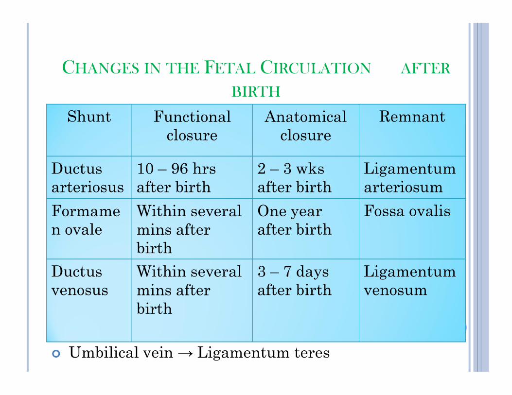
CHANGES IN THE FETAL CIRCULATION AFTER
BIRTH
Shunt Functional closure
Anatomical closure
Remnant
Ductus arteriosus
10 – 96 hrs after birth
2 – 3 wks after birth
Ligamentum arteriosum
Umbilical arteries → Umbilical ligaments
Umbilical vein → Ligamentum teres
Formamen ovale
Within several mins after birth
One year after birth
Fossa ovalis
Ductusvenosus
Within several mins after birth
3 – 7 days after birth
Ligamentumvenosum
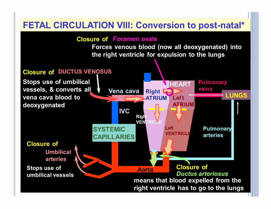
FETAL CIRCULATION VIII: Conversion to post-natal*
Pulmonary veinsVena cava Right
ATRIUM LUNGS
HEART
OLef t ATRIUM
Closure of Foramen ovale
DUCTUS VENOSUSClosure of
Stops use of umbilical vessels, & converts all vena cava blood to deoxygenated
Forces venous blood (now all deoxygenated) into the right ventricle for expulsion to the lungs
Pulmonary arteries
Right VENTRICLE
Left VENTRICLE
Aorta
SYSTEMIC CAPILLARIES
Umbilical arteries
Ductus arteriosus
IVCATRIUM
means that blood expelled from the right ventricle has to go to the lungs
Closure of
deoxygenated
Closure of
Stops use of umbilical vessels
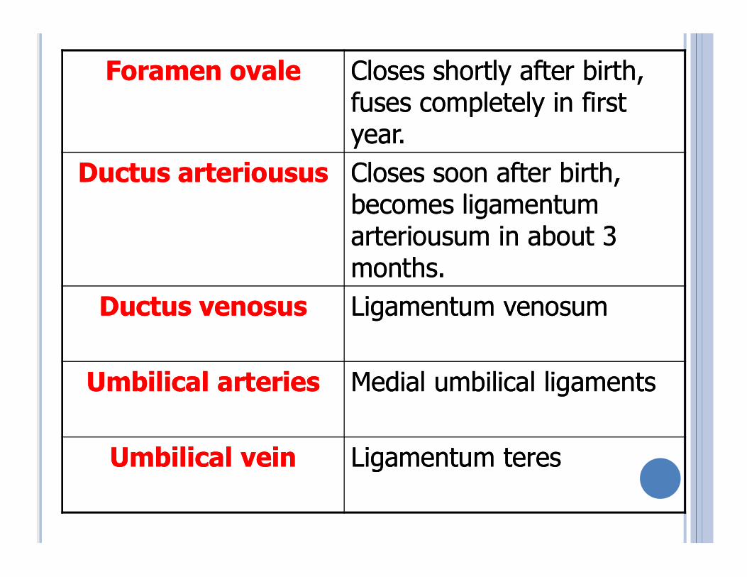
Foramen Foramen ovaleovale Closes shortly after birth, Closes shortly after birth, fuses completely in first fuses completely in first year.year.
DuctusDuctus arterioususarteriousus Closes soon after birth, Closes soon after birth, becomes ligamentum becomes ligamentum arteriousum in about 3 arteriousum in about 3 months.months.
DuctusDuctus venosusvenosus Ligamentum venosumLigamentum venosum
Umbilical arteriesUmbilical arteries Medial umbilical ligamentsMedial umbilical ligaments
Umbilical vein Umbilical vein Ligamentum teresLigamentum teres
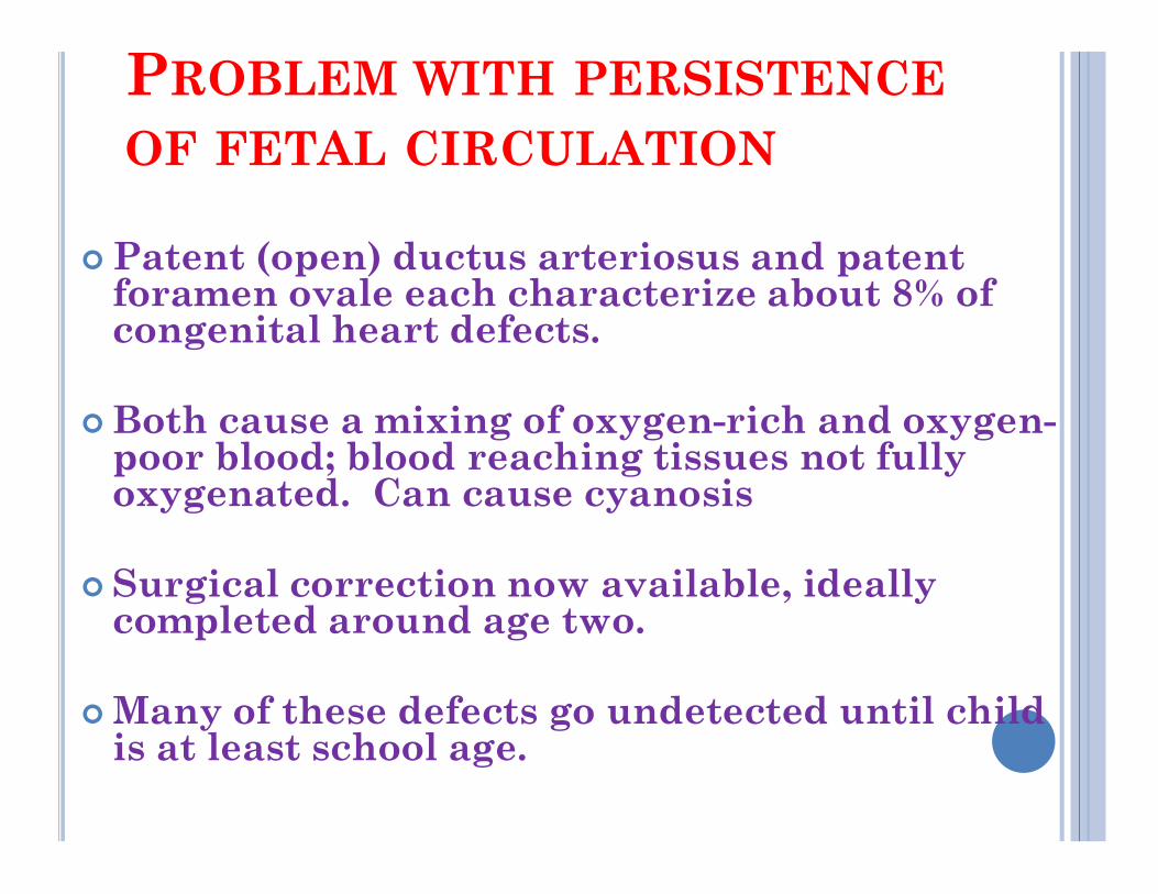
PROBLEM WITH PERSISTENCE
OF FETAL CIRCULATION
Patent (open) ductus arteriosus and patent foramen ovale each characterize about 8% of congenital heart defects.
Both cause a mixing of oxygen-rich and oxygen-poor blood; blood reaching tissues not fully poor blood; blood reaching tissues not fully oxygenated. Can cause cyanosis
Surgical correction now available, ideally completed around age two.
Many of these defects go undetected until child is at least school age.
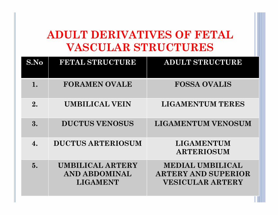
ADULT DERIVATIVES OF FETAL VASCULAR STRUCTURES
S.No FETAL STRUCTURE ADULT STRUCTURE
1. FORAMEN OVALE FOSSA OVALIS
2. UMBILICAL VEIN LIGAMENTUM TERES2. UMBILICAL VEIN LIGAMENTUM TERES
3. DUCTUS VENOSUS LIGAMENTUM VENOSUM
4. DUCTUS ARTERIOSUM LIGAMENTUMARTERIOSUM
5. UMBILICAL ARTERY AND ABDOMINAL
LIGAMENT
MEDIAL UMBILICAL ARTERY AND SUPERIOR
VESICULAR ARTERY
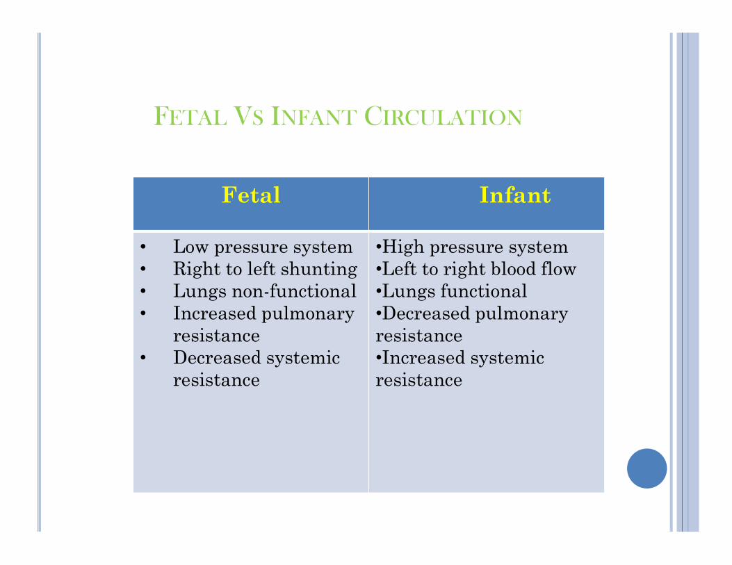
FETAL VS INFANT CIRCULATION
Fetal Infant
• Low pressure system• Right to left shunting• Lungs non-functional
•High pressure system•Left to right blood flow•Lungs functional• Lungs non-functional
• Increased pulmonary resistance
• Decreased systemic resistance
•Lungs functional•Decreased pulmonary resistance•Increased systemic resistance


BIBLIOGRAPHY
Daftary,N.Shirish. (2002).Manual Of Obstetrics2 nd Edition. New Delhi: Elsevier Publisher Page No:39-45
Dutta,D.C.(2004).Textbook Of Obstetric 6thEdition.CalcuttaDutta,D.C.(2004).Textbook Of Obstetric 6 Edition.Calcutta:New Central Book Agency Page No:41-45Gary, Cunningham and Leveno,Kanneth.(2004).
Williams Textbook Of Obstetrics22th Edition.Mc Graw Hill:Lippincott Williams & Wilkins Page No:91-104
•Singh,Inderbir.(1996).Human Embryology, 6th Edition.NewDelhi:Macmillan India Limited Page No.257-259