Embed Size (px)
Citation preview

Fas (CD95) Induces Alveolar Epithelial CellApoptosis in Vivo
Implications for Acute Pulmonary Inflammation
Gustavo Matute-Bello,* Robert K. Winn,†
Mechthild Jonas,§ Emil Y. Chi,§
Thomas R. Martin,* and W. Conrad Liles‡
From the Divisions of Pulmonary and Critical Care Medicine *
and Allergy and Infectious Diseases ‡ of the Department of
Medicine, the Department of Surgery,† and the Department of
Pathology,§ University of Washington, Seattle, Washington
Activation of the Fas/FasL system induces apoptosisof susceptible cells, but may also lead to nuclear fac-tor kB activation. Our goal was to determine whetherlocal Fas activation produces acute lung injury byinducing alveolar epithelial cell apoptosis and by gen-erating local inflammatory responses. Normal mice(C57BL/6) and mice deficient in Fas (lpr) were treatedby intranasal instillation of the Fas-activating mono-clonal antibody (mAb) Jo2 or an irrelevant controlmAb, and studied 6 or 24 hours later using bronchoal-veolar lavage (BAL), histopathology, DNA nick-end-labeling assays, and electron microscopy. Normalmice treated with mAb Jo2 had significant increasesin BAL protein at 6 hours, and BAL neutrophils at 24hours, as compared to lpr mice and to mice treatedwith the irrelevant mAb. Neutrophil recruitment waspreceded by increased mRNA expression for tumornecrosis factor-a , macrophage inflammatory protein-1a , macrophage inflammatory protein-2, macro-phage chemotactic protein-1, and interleukin-6, butnot interferon-g , transforming growth factor-b ,RANTES, eotaxin, or IP-10. Lung sections from Jo2-treated normal mice showed neutrophilic infiltrates,alveolar septal thickening, hemorrhage, and terminaldUTP nick-end-labeling-positive cells in the alveolarseptae and airspaces. Type II pneumocyte apoptosiswas confirmed by electron microscopy. Fas activationin vivo results in acute alveolar epithelial injury andlung inflammation, and may be important in thepathogenesis of acute lung injury. (Am J Pathol2001, 158:153–161)
The Fas/Fas ligand system plays a significant role in theregulation of apoptosis in many types of cells.1 This sys-tem is comprised of the cell membrane surface receptorFas (CD95) and its natural ligand, Fas-ligand (FasL).1 Fas
is a 45-kd type I membrane protein that is a member ofthe tumor necrosis factor family of surface receptors.2,3
Fas is expressed on many cells, including lymphocytes,neutrophils, monocytes, and alveolar epithelial cells.4–7
Binding of FasL to Fas results in apoptosis of susceptiblecells.8 Fas ligand is a 37-kd type II protein9,10 that existsas membrane-bound and soluble forms.8 Both forms arecapable of inducing apoptosis when engaging Fas,2,11
although the membrane-bound form seems to be moreefficient than the soluble form in vitro.11
Neutrophil apoptosis is generally regarded as an anti-inflammatory process.12,13 However, recent studies haveshown that binding of Fas to an agonist, in addition totriggering apoptosis, may also lead to activation of thenuclear factor kB under certain circumstances.14,15 Thiscellular activation is in addition to its pro-apoptotic func-tion and is probably independent of apoptosis.16 Nuclearfactor kB partially controls the expression of multipleinflammatory cytokines and cell surface receptors, in-cluding the chemokines interleukin-8 (IL-8) and GRO inhumans, as well as the murine GRO analogs KC andmacrophage inflammatory protein (MIP)-2.17–21
The potential role of the Fas/FasL system as a dualpro-apoptotic/pro-inflammatory system becomes rele-vant with the finding that circulating levels of sFasL arepresent in certain human diseases.22–28 Furthermore,sFasL is found in bronchoalveolar lavage (BAL) fluid frompatients with the acute respiratory distress syndrome(ARDS) at concentrations capable of inducing apoptosisof primary human small airway epithelial cells in vitro.29
Destruction of the alveolar epithelium is one of the hall-marks of ARDS,30,31 but the mechanisms that account forthe epithelial injury that occurs in ARDS remain unclear.Although it has been reported that repeated activation ofFas in the lungs of mice results in pulmonary fibrosis after2 weeks,32 the acute effects of Fas activation in the lungs
Supported in part by grants GR 42686 (to R. K. W.), HL 30542 (to R. K. W.,T. R. M.), AI 29103 (to T. R. M.), HL62995 (to W. C. L.), and GM 37696 (toT. R. M.) from the National Institutes of Health, the Medical ResearchService of the Department of Veterans Affairs (to T. R. M.), and a researchgrant from the Amgen Corporation (to G. M. B., T. R. M.).
Accepted for publication September 18, 2000.
Address reprint requests to W. Conrad Liles, M.D., Ph.D., Box 357185,Department of Medicine, University of Washington, Seattle, WA 98195.E-mail: [email protected].
American Journal of Pathology, Vol. 158, No. 1, January 2001
Copyright © American Society for Investigative Pathology
153

are unclear. We hypothesized that sFasL in the airspacesmay contribute to the pathogenesis of acute lung injuryby inducing apoptosis of epithelial cells, as well as bystimulating the release of inflammatory cytokines via nu-clear factor kB activation.
The major goal of this study was to determine whetheractivation of Fas in the alveoli of the lungs would induceapoptosis of type II pneumocytes in the alveolar wall, andinitiate an inflammatory response in the lungs. We treatednormal mice or naturally occurring mutant mice deficientin Fas expression (lpr mice) with the monoclonal antibody(mAb) Jo2, which activates Fas on the surface of cells invitro and in vivo.32,33 We identified apoptotic cells in thealveolar walls by DNA nick-end-labeling and electronmicroscopy, and the alveolar inflammatory response byquantitative histology, BAL, and cytokine ribonucleaseprotection assays (RPA).
Materials and Methods
Antibodies
The activating anti-Fas mAb Jo2 (Armenian hamster IgG)and a control mAb (Armenian hamster anti-TNP IgG)were purchased from PharMingen (San Diego, CA). Bothantibodies were free of azide and endotoxin, as deter-mined by the manufacturer. In addition, the antibodieswere confirmed to contain ,0.01 endotoxin U/ml by thelimulus amebocyte assay (ECL-1000; Biowhittaker, Walk-ersville, MD).
Animal Preparation
Male mice, weighing 20 to 30 g, were briefly anesthetizedwith inhaled halothane. Either mAb Jo2 or control mAb ata dose of 2.5 mg/g was administered to each mouse byintranasal instillation in a solution containing 1 mg/ml ofmAb in sterile phosphate-buffered saline (PBS), as pre-viously described.34 The animals were allowed to recoverfrom anesthesia, returned to their cages, and given freeaccess to water and food. At the end of the experimentthe animals were euthanized with ketamine and xylazine,the thorax was rapidly opened and the animal was ex-sanguinated by direct cardiac puncture. The lungs weredissected free and the trachea was cannulated to per-form BAL, or to fix the lungs, or to extract mRNA. Theprotocol was approved by the animal care committee ofthe University of Washington.
Experimental Protocols
We used male mice weighing 20 to 30 g. The mice wereeither C57BL/6 mice, (B&K Universal, Seattle, WA), ornaturally occurring mutant mice lacking the Fas receptor(lpr mice) (Jackson Laboratory, Bar Harbor, ME). The lprmice are derived from the C57BL/6 mouse strain. Themice were treated with either the Fas-activating mAb Jo2or an irrelevant mAb (hamster anti-TNP IgG) as de-scribed above, and euthanized at either 6 or 24 hoursafter the administration of the antibody. Studies with lpr
mice were performed to confirm that effects observedwith mAb Jo2 in C57BL/6 mice were specific for Fasactivation. As an additional comparison, C57BL/6 weretreated with an irrelevant mAb (hamster anti-TNP IgG)and euthanized at either 6 or 24 hours.
BAL Protocol
BAL was performed by instilling 0.9% NaCl containing0.6 mmol/L ethylenediaminetetraacetic acid in two sepa-rate 0.5 ml aliquots. The fluid was recovered by gentlesuction and placed on ice for immediate processing. Analiquot of the BAL fluid was processed immediately fortotal and differential cell counts. Total cell counts wereperformed with a hemocytometer, whereas differentialcell counts were performed on cytospin preparationsstained with modified Wright-Giemsa stain (Diff-Quik;American Scientific Products, McGaw Park, IL). The re-mainder of the lavage fluid was spun at 200 3 g for 30minutes, and the supernatant was removed asepticallyand stored in individual aliquots at 270°C.
The total protein concentration in BAL fluid was mea-sured using the bicinchoninic acid method (BCA assay;Pierce Co., Rockford, IL).
mRNA Extraction and RPA Protocol
Lung cytokine mRNA expression (RANTES, eotaxin, MIP-1a, MIP-2, IP-10, macrophage chemotactic protein-1(MCP-1), tumor necrosis factor-a, interleukin-6, interfer-on-g, transforming growth factor-b) was measured byRPA. Three mice were treated by intranasal instillation ofmAb Jo2 (2.5 mg/g), and three control mice were treatedwith an irrelevant mAb. After 6 hours, the mice wereeuthanized and their lungs excised. Lung RNA was ex-tracted with Triazol (Biotecx Laboratories, Inc., Houston,TX) according to the vendor’s instructions. RPA was per-formed using an RPAII kit (Ambion Inc., Austin, TX), withMCK-3 and MCK-5 template sets (Pharmingen, San Di-ego, CA) and [a-32P]UTP according to the manufactur-er’s instructions. Samples were run under denaturing elec-trophoresis on 5% polyacrylamide gel, then imaged andanalyzed in a Packard Cyclone Phosphorimager (Amer-sham Pharmacia Biotech, Piscataway, NJ). To control forrelative differences in RNA loading between samples, thespecific cytokine mRNA signals were normalized to theintensity of the respective glyceraldehyde-3-phosphate de-hydrogenase signal. Results are expressed as relativemRNA expression (mean 6 SEM), using the following equa-tion: normalized Jo2 induced expression (n 5 3)/normalizedcontrol expression (n 5 3).
Histopathology Protocols
The lungs were fixed by inflation with 10% neutral-buff-ered formalin at a transpulmonary pressure of 15 cm H2Oand embedded in paraffin. Within 24 hours of fixation,lung sections were stained with hematoxylin and eosin forlight microscopy, or by the DNA nick-end-labeling assayto evaluate apoptotic cells, or processed for transmissionelectron microscopy as described below.
154 Matute-Bello et alAJP January 2001, Vol. 158, No. 1

DNA Nick-End-Labeling Assay
The slides were submerged in 10% neutral-buffered for-malin for 10 minutes, followed by 70% ethanol for 5minutes. The slides were rehydrated for 10 minutes in PBSand treated with 0.002% proteinase K (Sigma, St. Louis,MO.) in double-distilled water for 5 to 15 minutes at roomtemperature. Endogenous peroxidase was quenched byplacing the slides in 2% hydrogen peroxide for 5 minutes.For equilibration, the slides were treated in Klenow labelingbuffer (TACS In situ Apoptosis Detection Kit; TrevigenInc., Gaithersburg, MD) for at least 1 minute and thenincubated for 60 minutes at 37°C with Klenow enzymeand Klenow dNTP mix in Klenow labeling buffer (all re-agents from Trevigen, Inc.) prepared according to in-structions from the manufacturer. Negative control slideswere incubated with the labeling mixture without the Kle-now enzyme. After incubation the slides were completelysubmerged in Klenow Stop buffer (Trevigen Inc.) for 5minutes at room temperature and rinsed in PBS for 2minutes. The samples were then treated for 15 minuteswith streptavidin-horseradish peroxidase detection solu-tion (Trevigen Inc.), washed twice for 2 minutes in PBSand incubated in diaminobenzidine (Trevigen Inc.) for 7minutes at room temperature. The samples were thenrinsed twice in distilled water and stained with 1% methylgreen in 0.1 mol/L sodium acetate (pH 4.0) for 5 minutes,quickly dehydrated in 95% and 100% ethanol, cleared inxylene, and mounted with Permount (Fisher Scientific,Pittsburgh, PA).
Electron Microscopy
Lung tissue was fixed for electron microscopy by immer-sion in 6.25% glutaraldehyde, 2% paraformaldehyde in0.1 mol/L sodium cacodylate buffer for 2 hours. Thetissue was postfixed in 2% potassium-ferrocyanide indistilled water for 4 hours at room temperature, rinsedwith distilled water, stained with 0.5% uranyl acetate for20 minutes, and then rinsed in distilled water. The sam-ples were dehydrated in a graded series of ethanol so-lutions and embedded in Eponate 12 (Ted Pella Inc.,Redding, CA). Thin sections were cut from two randomlyselected blocks with a diamond knife using an LKB Novaultramicrotome and collected on parlodion-coated 200
mesh copper grids (Electron Microscopy Sciences, FortWashington, PA.). The sections were stained with uranylacetate and lead citrate and examined with a JEOL TEM1200 EX at a magnification of 33000 or higher. Only cellswith a recognizable nucleus were included in the analy-sis. For each sample several sections from at least twoelectron microscopic blocks were used for evaluation.
Lung Injury Score
Each slide was evaluated by two separate investigators(GMB and WCL) in a blinded manner. To generate thelung injury score, a total of 300 alveoli were counted oneach slide at 3400 magnification. Within each field,points were assigned according to predetermined crite-ria (Table 1). All of the points for each category wereadded and weighted according to their relative impor-tance. The injury score was calculated according to thefollowing formula: injury score 5 [(alveolar hemorrhagepoints/no. of fields) 1 2 3 (alveolar infiltrate points/no. offields) 1 3 3 (fibrin points/no. of fields) 1 (alveolar septalcongestion/no. of fields)]/total number of alveoli counted.
Statistical Analysis
Comparisons between two groups were made with thetwo-tailed Fisher’s exact t-test. Comparisons betweenmultiple groups were made with the Kruskall-Wallis anal-ysis of variance and with factorial analysis of variance.35
For post hoc analysis, the Fisher’s test was used. A Pvalue ,0.05 was considered significant.
Results
Effect of Activation of the Fas System on BALFluid Total Protein Concentration,Polymorphonuclear Leukocytes (PMN)Recruitment, and Cytokine Production
Six hours after intranasal instillation of mAb Jo2, the BALtotal protein concentration was significantly increased inC57BL/6 mice (n 5 5) as compared to the lpr mice (n 55), or to the C57BL/6 mice treated with an irrelevant
Table 1. Quantitative Histopathology Score of Lung Injury
Tissue 0 1 2 3
Alveolar septae All septae are thinand delicate
Congested alveolarseptae in less than1/3 of the field
Congested alveolarseptae in 1/3 to 2/3of the field
Congested alveolarseptae in greaterthan 2/3 of the field
Alveolarhemorrhage
No hemorrhage At least 5erythrocytes peralveolus in 1 to 5alveoli
At least 5erythrocytes peralveolus in 5 to 10alveoli
At least 5erythrocytes peralveolus in morethan 10 alveoli
Intra-alveolar fibrin No intra-alveolarfibrin
Fibrin strands in lessthan 1/3 of the field
Fibrin strands in 1/3to 2/3 of the field
Fibrin strands ingreater than 2/3 ofthe field
Intra-alveolarinfiltrates
Less than 5 intra-alveolar cells perfield
5 to 10 intra-alveolarcells per field
10 to 20 intra-alveolar cells perfield
More than 20 intra-alveolar cells perfield
Fas/FasL Mediates Lung Injury 155AJP January 2001, Vol. 158, No. 1

control mAb (n 5 5) (Figure 1A). At 24 hours, all animalgroups had similar concentrations of total protein in theBAL fluid (Figure 1B).
In contrast to the early increase in BAL fluid total pro-tein, the BAL fluid neutrophil response was delayed. Sixhours after Jo2 instillation, there was trend toward moreBAL fluid PMN in the C57BL/6 mice treated with the mAbJo2, but this did not reach statistical significance (Figure2A). At 24 hours, the number of BAL fluid PMN wassignificantly higher in C57BL/6 mice treated with mAbJo2 (n 5 5), as compared to the lpr group (n 5 5), and toC57BL/6 animals treated with the control mAb (n 5 5)(Figure 2B).
To assess cytokine message expression we performedRPAs on mRNA extracted from the lungs of C57BL/6mice 6 hours after instillation of either Jo2 mAb (n 5 3) orcontrol mAb (n 5 3) (Figure 3). The cytokine expressionwas normalized to the GADPH signal to control for load-ing differences. Jo2-treated mice showed a sixfold in-crease in MIP-2 mRNA, a twofold increase in MIP-1amRNA, a fourfold increase in MCP-1 mRNA, a threefoldincrease in tumor necrosis factor-a mRNA, and a 60-foldincrease in interleukin-6 mRNA. In contrast, there wasno increase in message for transforming growth factor-b,
eotaxin, or IP-10. The relative expression of RANTES andinterferon-g were significantly decreased.
Histopathological Evidence of Lung Injury
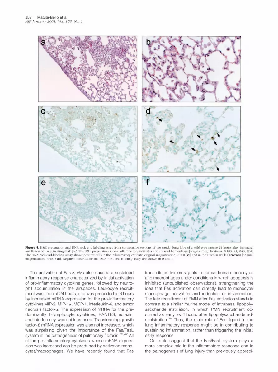
At 24 hours, C57BL/6 mice treated with mAb Jo2 (n 5 6)had a significantly higher lung injury score than animalstreated with the control mAb (n 5 7; P , 0.01) (Figure 4).The lung injury was characterized by patchy areas ofneutrophilic infiltrates with thickening of the alveolar sep-tae and areas of hemorrhage (Figure 5a). The majority ofthe cells infiltrating the airspaces, as well as cells in thealveolar septae, showed densely condensed nuclei sug-gesting apoptosis in areas of injury (Figure 5b). No his-topathological abnormalities were present in animals thatwere treated with the control mAb (Figure 6, a and b).
DNA nick-end-labeling assays confirmed the presenceof apoptosis in cells infiltrating the airspaces (Figure 5, cand d). In addition, positive cells were present in thealveolar septae of areas of injury. No positive cells wereseen in the animals treated with the control mAb (Figure6, c and d).
By electron microscopy, the lungs of a mouse treatedwith mAb Jo2 showed features characteristic of apopto-sis in alveolar type II cells. These features included in-creased electron density of the cytoplasm, condensation
Figure 1. Total BAL protein at 6 hours (A) or 24 hours (B) after the intranasaladministration of either an irrelevant control mAb or the Fas-activating mAb Jo2.C57, wild-type mice; lpr, Fas-deficient mice. Data are shown as means 6 SEM.
Figure 2. Total BAL PMN at 6 hours (A) or 24 hours (B) after the intranasaladministration of either an irrelevant control mAb or the Fas-activating mAb Jo2.C57, wild-type mice; lpr, Fas-deficient mice. Data are shown as means 6 SEM.
156 Matute-Bello et alAJP January 2001, Vol. 158, No. 1

of the chromatin, and vacuolization of the nuclear enve-lope (Figure 7).36–38 Interestingly, there were also mito-chondrial abnormalities. Changes consistent with apo-ptosis were not seen in any of the endothelial cells thatwere identified.
Discussion
The primary goal of this study was to determine whetheractivation of the Fas system in vivo results in damage tocells in the alveolar walls, and whether this event initiatesan acute inflammatory response. We found that C57BL/6mice developed patchy neutrophilic infiltrates, areas ofalveolar hemorrhage, thickening of the alveolar septae,and apoptosis of type II pneumocytes within 24 hoursafter treatment with Fas-activating mAb Jo2. These his-topathological changes were associated with an earlyincrease in BAL fluid protein and early induction of cyto-kine gene expression, followed by a later increase in BALfluid neutrophils. Similar changes did not occur in micelacking Fas (lpr), or in mice treated with an irrelevantantibody. These findings provide clear evidence that theinduction of apoptosis in cells of the alveolar wall initiatesa sustained inflammatory response in the lungs.
In humans, acute lung injury is characterized by epi-thelial and endothelial injury, neutrophilic alveolitis, andhyaline membrane formation. The primary event leadingto lung injury in ARDS has not been established. A prev-alent hypothesis is that the neutrophil mediates lung in-jury in ARDS.39 In this paradigm, uncontrolled neutrophilactivation leads to the accumulation of oxidants and pro-teases in the lungs, which cause damage to the cells ofthe alveolar environment. However, neutrophils can mi-grate into the lungs of humans without causing injury, andlarge numbers of neutrophils can migrate into the lungs ofsheep without causing injury to the tight epithelial barri-er.40,41 Blockade of PMN chemoattractants or systemicPMN depletion blocks lung injury in some, but not all
animal models.42–44 Although PMN may contribute tolung injury in some circumstances, it is not clear whetherthe first event leading to lung injury involves PMN migra-tion, or whether epithelial injury occurs before leukocytesare involved.45 Answering this question is critical to de-signing specific therapeutic strategies for ARDS.
The present study shows that activation of the Fassystem in vivo in the lungs of mice leads to a primaryepithelial injury. We found that intranasal administration ofthe Fas-activating mAb Jo2 resulted in an early increasein the BAL fluid total protein concentration, followed laterby PMN migration into the airspaces. The cells of thealveolar wall became apoptotic, as detected by DNAend-nick-labeling assays and electron microscopy. Thusin this model, damage to the alveolar epithelial barrieroccurred first, before PMN recruitment. The possibilitythat the Fas system might be involved in epithelial dam-age during acute lung injury was raised by our previousfindings that soluble FasL is elevated in BAL fluid frompatients with ARDS at concentrations capable of induc-ing apoptosis of normal human distal lung epithelial cellsin vitro, and that higher concentrations of soluble FasL inBAL fluid from patients on day 1 of ARDS are associatedwith increased mortality.29 An earlier study had demon-strated that a trypsin-sensitive pro-apoptotic factor (orfactors) was present in BAL fluid obtained from patientsduring the late stage of ARDS, and that this factor wasactive for fibroblasts and endothelial cells.46
Several lines of evidence suggest that the Fas systemcauses apoptosis of alveolar epithelial cells. The humanneoplastic alveolar epithelial cell line A549 expresses Fason its surface and undergoes apoptosis when exposed toactivating anti-Fas IgM.7 Furthermore, we have found thatnormal human distal lung epithelial cells express Fas andbecome apoptotic when exposed to recombinant humansoluble FasL.29 These results suggest that Fas-mediatedapoptosis of the alveolar epithelium could be an initialevent in the development of some forms of lung injury.
Figure 3. RPA analysis of cytokine mRNA expression in the lungs of mice, 6hours after the intranasal administration of the mAb Jo2. RPA analysis wasperformed as described in the Methods section. Results are expressed asrelative mRNA expression (mean 6 SEM), which represents normalizedJo2-induced expression (n 5 3 mice)/normalized control expression (n 5 3mice) for each cytokine. *, Value significantly greater than control (P , 0.05).**, Value significantly less than control (P , 0.05).
Figure 4. Histopathological lung injury score for C57 mice 24 hours afterreceiving either an irrelevant control mAb (n 5 7) or the Fas-activating mAbJo2 (n 5 6). The score represents the average of two independent investi-gators who read each H&E-stained slide in a blinded manner. The categoriesused to generate the score were alveolar septal congestion, alveolar hemor-rhage, intra-alveolar fibrin deposition, and intra-alveolar infiltrates (see textfor further explanation). *, P , 0.01.
Fas/FasL Mediates Lung Injury 157AJP January 2001, Vol. 158, No. 1
The activation of Fas in vivo also caused a sustainedinflammatory response characterized by initial activationof pro-inflammatory cytokine genes, followed by neutro-phil accumulation in the airspaces. Leukocyte recruit-ment was seen at 24 hours, and was preceded at 6 hoursby increased mRNA expression for the pro-inflammatorycytokines MIP-2, MIP-1a, MCP-1, interleukin-6, and tumornecrosis factor-a. The expression of mRNA for the pre-dominantly T-lymphocyte cytokines, RANTES, eotaxin,and interferon-g, was not increased. Transforming growthfactor-b mRNA expression was also not increased, whichwas surprising given the importance of the Fas/FasLsystem in the pathogenesis of pulmonary fibrosis.32,47 Allof the pro-inflammatory cytokines whose mRNA expres-sion was increased can be produced by activated mono-cytes/macrophages. We have recently found that Fas
transmits activation signals in normal human monocytesand macrophages under conditions in which apoptosis isinhibited (unpublished observations), strengthening theidea that Fas activation can directly lead to monocyte/macrophage activation and induction of inflammation.The late recruitment of PMN after Fas activation stands incontrast to a similar murine model of intranasal lipopoly-saccharide instillation, in which PMN recruitment oc-curred as early as 4 hours after lipopolysaccharide ad-ministration.34 Thus, the main role of Fas ligand in thelung inflammatory response might be in contributing tosustaining inflammation, rather than triggering the initial,early response.
Our data suggest that the Fas/FasL system plays amore complex role in the inflammatory response and inthe pathogenesis of lung injury than previously appreci-
Figure 5. H&E preparation and DNA nick-end-labeling assay from consecutive sections of the caudal lung lobe of a wild-type mouse 24 hours after intranasalinstillation of Fas activating mAb Jo2. The H&E preparation shows inflammatory infiltrates and areas of hemorrhage [original magnifications: 3100 (a); 3400 (b)].The DNA nick-end-labeling assay shows positive cells in the inflammatory exudate [original magnification, 3100 (c)] and in the alveolar walls (arrows) [originalmagnification, 3400 (d)]. Negative controls for the DNA nick-end-labeling assay are shown in e and f.
158 Matute-Bello et alAJP January 2001, Vol. 158, No. 1

ated. Apoptosis has traditionally been considered as amechanism promoting resolution of inflammation.13 How-ever, recent findings suggest that Fas activation canresult in an inflammatory response because of the re-lease of inflammatory cytokines.14,15 Studies by Miwaand colleagues16 demonstrated that membrane-boundFasL can induce PMN infiltration in mice. Furthermore,soluble FasL itself has been reported to serve as a che-motactic factor for human and murine PMN, but themechanism of this effect remains uncertain.48
The relative contribution of pro-apoptotic and pro-in-flammatory activities is likely to depend on other media-tors present in the microenvironment, as well as localconcentrations of soluble FasL. Fas-induced PMN apo-ptosis is suppressed by granulocyte colony-stimulatingfactor (G-CSF) and granulocyte-macrophage colony-
stimulating factor (GM-CSF).6 During the early phase ofARDS, PMN apop-tosis is inhibited by G-CSF and GM-CSF present in the airspaces.49 During acute inflamma-tion, the pro-apoptotic activity of FasL on PMN may becounteracted by G-CSF and GM-CSF, whereas its pro-inflammatory properties may contribute to PMN recruit-ment. However, epithelial cells, which express Fas butmay lack corresponding anti-apoptotic receptors, un-dergo apoptosis which results in injury to the alveolar-epithelial barrier. Thus, the Fas/FasL system may serve adual role in the pathogenesis of acute inflammatory injuryby causing direct damage to the alveolar-epithelial bar-rier and by recruiting PMN to the site of injury.
In summary, the data show that Fas activation in vivocan lead to apoptosis of alveolar epithelial cells withchanges in epithelial permeability, expression of inflam-
Figure 6. H&E preparation and DNA end-nick-labeling assay from consecutive sections of the caudal lung lobe of a wild-type mouse 24 hours after intranasalinstillation of an irrelevant control mAb. The H&E preparation shows normal lung architecture [original magnifications, 3100 (a); 3400 (b)], whereas the DNAnick-end-labeling assay shows no positive cells [original magnifications, 3100 (c); 3 160 (d)]. Negative controls for the DNA nick-end-labeling assay are shownin e (original magnification, 3100) and f (original magnification, 3160).
Fas/FasL Mediates Lung Injury 159AJP January 2001, Vol. 158, No. 1

matory cytokines, and recruitment of PMN into the alveoli.The data also show that epithelial injury precedes leuko-cyte infiltration when Fas is activated. These findings areconsistent with a dual role for the Fas/FasL system, asboth a pro-inflammatory and a pro-apoptotic system inthe lungs. The findings suggest a new paradigm for someforms of acute lung injury in which alveolar epithelialdeath is the critical initial factor that initiates inflammatoryresponses in the lungs.
Acknowledgments
We thank Joe Stalder, John Ruzinski, and Frank RadellaII, for their expert technical assistance.
References
1. Nagata S, Golstein P: The Fas death factor. Science 1995, 267:1449–1456
2. Itoh N, Yonehara S, Ishii A, Yonehara M, Mizushima S, Sameshima M,Hase A, Seto Y, Nagata S: The polypeptide encoded by the cDNA forhuman cell surface antigen Fas can mediate apoptosis. Cell 1991,66:233–243
3. Itoh N, Nagata S: A novel protein domain required for apoptosis.Mutational analysis of human Fas antigen. J Biol Chem 1993, 268:10932–10937
4. Kagi D, Vignaux F, Ledermann B, Burki K, Depraetere V, Nagata S,Hengartner H, Golstein P: Fas and perforin pathways as major mech-anisms of T-cell mediated cytotoxicity. Science 1994, 265:528–530
5. Liles WC, Klebanoff SJ: Regulation of apoptosis in neutrophils—Fastrack to death? J Immunol 1995, 155:3289–3291
6. Liles WC, Kiener PA, Ledbetter JA, Aruffo A, Klebanoff SJ: Differentialexpression of Fas (CD95) and Fas ligand on normal humanphagocytes: implications for the regulation of apoptosis in neutro-phils. J Exp Med 1996, 184:429–440
7. Fine A, Anderson NL, Rothstein TL, Williams MC, Gochuico BR: Fasexpression in pulmonary alveolar type II cells. Am J Physiol 1997,273:L64–L71
8. Tanaka M, Suda T, Takahashi T, Nagata S: Expression of the func-tional soluble form of human Fas ligand in activated lymphocytes.EMBO (Eur Mol Biol Organ) J 1995, 14:1129–1135
9. Suda T, Takahashi T, Goldstein P, Nagata S: Molecular cloning andexpression of the Fas ligand, a novel member of the tumor necrosisfactor family. Cell 1993, 75:1169–1178
10. Suda T, Nagata S: Purification and characterization of the Fas-ligandthat induces apoptosis. J Exp Med 1994, 179:873–879
11. Tanaka M, Itai T, Adachi M, Nagata S: Downregation of Fas ligand byshedding. Nat Med 1998, 4:31–36
12. Savill J: Apoptosis in resolution of inflammation. J Leukoc Biol 1997,61:375–380
13. Haslett C, Savill JS, Whyte MK, Stern M, Dransfield I, Meagher LC:Granulocyte apoptosis and the control of inflammation. Philos Trans RSoc Lond B Biol Sci 1994, 345:327–333
14. Ponton A, Clement MV, Stamenkovic I: The CD95 (APO-1/Fas) recep-tor activates NF-kB independently of its cytotoxic function. J BiolChem 1996, 271:8991–8995
15. Rensing-Ehl A, Hess S, Ziegler-Heitbrock HW, Riethmuller G, En-gelmann H: Fas/Apo-1 activates nuclear factor kB and induces inter-leukin-6 production. J Inflamm 1995, 45:161–174
16. Miwa K, Asano M, Horai R, Iwakura Y, Nagata S, Suda T: Caspase-1independent IL-1b release and inflammation induced by the apopto-sis inducer Fas ligand. Nat Med 1998, 4:1287–1292
17. Mukaida N, Okamoto S, Ishikawa Y, Matsushima K: Molecular mech-anism of interleukin-8 gene expression. J Leukoc Biol 1994, 56:554–558
18. Shattuck RL, Wood LD, Jaffe GJ, Richmond A: MCSA/GRO transcrip-tion is differentially regulated in normal retinal pigment epithelial andmelanoma cells. Mol Cell Biol 1994, 14:791–802
19. Xia Y, Pauza ME, Feng L, Lo D: RelB regulation of chemokine expres-sion modulates local inflammation. Am J Pathol 1997, 151:375–387
20. Frevert CW, Huang S, Danace H, Paulauskis JD, Kobzik L: Functionalcharacterization of the rat chemokine KC and its importance in neu-trophil recruitment in a rat model of pulmonary inflammation. J Immu-nol 1995, 154:335–344
21. Widmer U, Manogue KR, Cerami A, Sherry B: Genomic cloning andpromoter analysis of macrophage inflammatory protein (MIP)-2,MIP-1 alpha, and MIP-1 beta, members of the chemokine superfamilyof proinflammatory cytokines. J Immunol 1993, 150:4996–5012
22. Tanaka M, Suda T, Haze K, Nakamura N, Sato K, Kimura F, MotoyoshiK, Mizuki M, Tagawa S, Ohga S, Hatake K, Drummond AH, Nagata S:Fas ligand in human serum. Nat Med 1996, 2:317–322
23. Viard I, Wehrli P, Bullani R, Salomon D, Saurat JH, French LE: Inhibi-tion of toxic epidermal necrolysis by Nockade of CD95 with humanintravenous immunoglobulin. Science 1988, 282:490–503
24. Hashimoto H, Tanaka M, Suda T, Tomita T, Hayashida K, Takeuchi E,Kanako M, Takano H, Nagata S, Ochi T: Soluble Fas ligand in thejoints of patients with rheumatoid arthritis and osteochondritis. Arthri-tis Rheum 1998, 41:657–662
25. Nozawa K, Kayagaki N, Tokano Y, Yagita H, Okumura K, Hasimoto H:Soluble Fas (APO-1, CD95) and soluble Fas ligand in rheumaticdiseases. Arthritis Rheum 1997, 40:1126–1129
26. Taieb J, Mathurin P, Paynard Y, Gougerot-Pocidalo MA, Chollet-Martin S: Raised plasma soluble Fas and Fas ligand in alcoholic liverdisease. Lancet 1998, 351:1930–1931
27. Hasegawa D, Kojima S, Tatsumi F, Hayakawa A, Kosaka Y, Naka-mura H, Sako M, Osugi Y, Nagata S, Sano K: Elevation of the serumFas ligand in patients with hemophagocytic syndrome and Diamond-Blackfan anemia. Blood 1998, 91:2793–2799
28. Toyozaki T, Hiroe M, Tanaka M, Nagata S, Ohwada H, Marumo F:
Figure 7. Electron micrographs from the lungs of a wild-type mouse 24hours after intranasal instillation of Fas activating mAb Jo2, showing alveolartype II cells with very electron dense cytoplasm, and swollen mitochondria(M) with irregular cristae. In b the nuclear chromatin is condensed at theperiphery of the nucleus. Some of the lamellar bodies (LB) are degranulating(arrows). N nucleus. Original magnification, 313,500.
160 Matute-Bello et alAJP January 2001, Vol. 158, No. 1

Levels of soluble Fas ligand in myocarditis. Am J Cardiol 1998,82:246–248
29. Matute-Bello G, Liles WC, Steinberg KP, Kiener PA, Mongovin S, ChiEY, Jonas M, Martin TR: Soluble Fas ligand induces epithelial cellapoptosis in humans with acute lung injury (ARDS). J Immunol 1999,163:2217–2225
30. Bachofen M, Weibel ER: Structural alterations of lung parenchyma inthe adult respiratory distress syndrome. Clin Chest Med 1982,3:35–56
31. Matthay MA, Folkesson HG, Campagna A, Kheradmand F: Alveolarepithelial barrier and acute lung injury. New Horiz 1993, 1:613–622
32. Hagimoto N, Kuwano K, Miyazaki H, Kunitake R, Fujita M, KawasakiM, Kaneko Y, Hara N: Induction of apoptosis and pulmonary fibrosisin mice in response to ligation of Fas antigen. Am J Respir Cell MolBiol 1997, 17:272–278
33. De Leon M, Jackson KM, Cavanaugh JR, Mbangkollo D, Verret CR:Arrest of the cell cycle reduces susceptibility of target cells to per-form-mediated lysis. J Cell Biochem 1998, 69:425–435
34. Szarka RJ, Wang N, Gordon L, Nation PN, Smith RH: A murine modelof pulmonary damage induced by lipopolysaccharide via intranasalinstillation. J Immunol Methods 1997, 202:49–57
35. Rosner B: Fundamentals of Biostatistics. Boston, Duxbury Press,1982
36. Anglade P, Tsuji S, Vyas S: Morphological diversity of programmedcell death: deciphering of triggering signals needs comprehensiveclassification. Biomed Res 1997, 18:1–6
37. Kerr JFR, Gobe GC, Winterford CM, Harmon BV: Anatomical methodsin cell death. Methods in Cell Biology. Edited by Schwartz LM, Os-borne BA. San Diego, CA, Academic Press, 1995, pp 2–27
38. Lockshin RA, Zakeri Z: The biology of cell death and its relationshipto aging. Modern Cell Biology. Edited by Holbrook NJ, Martin GR,Lockshin RA. New York, Wiley-Liss, 1996, pp 167–180
39. Boxer LA, Axtell R, Suchard S: The role of the neutrophil in inflamma-tory diseases of the lung. Blood Cells 1990, 16:25–42
40. Wiener-Kronish JP, Albertine KH, Matthay MA: Differential responses
of the endothelial and epithelial barriers of the lung in sheep toEscherichia coli endotoxin. J Clin Invest 1991, 88:864–875
41. Martin TR, Pistorese BP, Chi EY, Goodman RB, Matthay MA: Effectsof leukotriene B4 in the human lung. Recruitment of neutrophils intothe alveolar spaces without a change in protein permeability. J ClinInvest 1989, 84:1609–1619
42. Carraway MS, Welty-Wolf KE, Kantrow SP, Huang YC, Simonson SG,Que LG, Kishimoto TK, Piantadosi CA: Antibody to E- and L-selectindoes not prevent lung injury or mortality in septic baboons. Am JRespir Crit Care Med 1998, 157:938–949
43. Steimle CN, Guynn TP, Morganroth ML, Bolling SF, Carr K, Deeb GM:Neutrophils are not necessary for ischemia-reperfusion lung injury.Ann Thorac Surg 1992, 53:64–73
44. Friedman M, Wang SY, Seilke FW, Cohn WE, Weintraub RM, JohnsonRG: Neutrophil adhesion blockade with NPC 15669 decreases pul-monary injury after total cardiopulmonary bypass. J Thorac Cardio-vasc Surg 1996, 111:460–468
45. Prescott SM, McIntyre TM, Zimmerman G: Two of the usual suspects,platelet-activating factor and its receptor, implicated in acute lunginjury [comment]. J Clin Invest 1999, 104:1019–1020
46. Polunovsky VA, Chen B, Henke C, Snover D, Wendt C, Iugbar DH,Bitterman PB: Role of mesenchymal cell death in lung remodelingafter injury. J Clin Invest 1993, 92:388–397
47. Kuwano K, Hagimoto N, Kawasaki M, Yatumi T, Nakamura N, NagataS, Suda T, Kunitake R, Macyama T, Miyazoki H, Hara N: Essentialroles of the Fas-Fas ligand pathway in the development of pulmonaryfibrosis. J Clin Invest 1999, 104:13–19
48. Seino K, Iwahuchi K, Kayagaki N, Miyata R, Nagaoka I, Matsuzawa A,Fukao K, Yagita H, Okamura K: Chemotactic activity of soluble Fasligand against phagocytes. J Immunol 1998, 161:4484–4488
49. Matute-Bello G, Liles WC, Radella F, Steinberg KP, Ruzinski JT, JonasM, Chi FY, Hudson LD, Martin TR: Neutrophil apoptosis in the acuterespiratory distress syndrome. Am J Respir Crit Care Med 1997,156:1969–1977
Fas/FasL Mediates Lung Injury 161AJP January 2001, Vol. 158, No. 1


