Embed Size (px)
Citation preview

Fatty acid-inducible ANGPTL4 governs lipid metabolicresponse to exerciseMilène Catoirea,1, Sheril Alexa,1, Nicolas Paraskevopulosa, Frits Mattijssena, Inkie Evers-van Goghb, Gert Schaartc,Jacob Jeppesend, Anita Kneppersa, Marco Mensinka, Peter J. Voshole, Gunilla Olivecronaf, Nguan Soon Tang,Matthijs K. C. Hesselinke, Jimmy F. Berbéeh,i, Patrick C. N. Rensenh,i, Eric Kalkhovenb, Patrick Schrauwenj,and Sander Kerstena,2
aNutrition, Metabolism and Genomics Group, Division of Human Nutrition, Wageningen University, 6700 EV, Wageningen, The Netherlands; bDepartment ofMetabolic Diseases, University Medical Centre Utrecht, 3584 EA, Utrecht, The Netherlands; Departments of cHuman Movement Sciences and jHuman Biology,Maastricht University Medical Centre, 6200 MD, Maastricht, The Netherlands; dSection of Molecular Physiology, Department of Nutrition, Exercise and Sports,University of Copenhagen, DK-2200 Copenhagen, Denmark; eMetabolic Research Laboratories, Institute of Metabolic Science, University of Cambridge,Cambridge CB2 0QQ, United Kingdom; fDepartment of Medical Biosciences/Physiological Chemistry, Umeå University, SE-90187 Umeå, Sweden; gSchool ofBiological Sciences, Nanyang Technological University, Singapore 637551; and hDepartment of Endocrinology and Metabolic Diseases and iEinthovenLaboratory for Experimental Vascular Medicine, Leiden University Medical Center, 2300 RC, Leiden, The Netherlands
Edited by David W. Russell, University of Texas Southwestern Medical Center, Dallas, TX, and approved February 4, 2014 (received for review January 16, 2014)
Physical activity increases energy metabolism in exercisingmuscle. Whether acute exercise elicits metabolic changes innonexercising muscles remains unclear. We show that one ofthe few genes that is more highly induced in nonexercisingmuscle than in exercising human muscle during acute exerciseencodes angiopoietin-like 4 (ANGPTL4), an inhibitor of lipopro-tein lipase-mediated plasma triglyceride clearance. Using a com-bination of human, animal, and in vitro data, we show thatinduction of ANGPTL4 in nonexercising muscle is mediated byelevated plasma free fatty acids via peroxisome proliferator-acti-vated receptor-δ, presumably leading to reduced local uptake ofplasma triglyceride-derived fatty acids and their sparing for use byexercising muscle. In contrast, the induction of ANGPTL4 in exercis-ing muscle likely is counteracted via AMP-activated protein ki-nase (AMPK)-mediated down-regulation, promoting the use ofplasma triglycerides as fuel for active muscles. Our data suggestthat nonexercising muscle and the local regulation of ANGPTL4 viaAMPK and free fatty acids have key roles in governing lipid ho-meostasis during exercise.
Acute exercise greatly increases the cellular demand for ATP,oxygen, glucose, and fatty acids. To meet these demands,
acute exercise is associated with marked changes in skeletalmuscle activity of key transporters and enzymes involved inglucose and fatty acid transport and oxidation (1). Much of theregulation occurs via allosteric regulation and covalent modifi-cation of rate-limiting enzymes. In addition, alterations at thelevel of mRNA increasingly are believed to represent an im-portant regulatory mechanism in the acute response to exercise(2). Indeed, acute exercise induces mRNA expression of manygenes involved in a variety of processes, including energy me-tabolism, hypertrophy, and signaling (3–6). Not surprisingly,most studies have focused on the events occurring in exercisingmuscle. In contrast, much less is known about the exercise-inducedchanges in nonexercising muscle. Studies have shown that restingskeletal muscle is crucial in the removal of lactate from the cir-culation during high-intensity exercise (7) and also plays a role inadrenaline and noradrenaline production during exercise (8). Inaddition, similar to exercising muscle, resting muscle exhibits en-hanced phosphorylation of mTOR following resistance exercise(9). Overall, however, the impact of exercise on metabolic pro-cesses and gene expression in nonexercising muscles remains ill-defined. It can be envisioned that exercise may elicit changes ingene expression in nonexercising muscle via circulating mediatorsincluding muscle-derived myokines and metabolites (10). Thepresent study was undertaken to try to elucidate the role of in-active muscle in the metabolic response to acute exercise.
ResultsTo investigate the molecular events occurring during exercise innonexercising muscle, we carried out an acute exercise trial inwhich 12 human subjects performed moderate- to high-intensitycycling exercise with one leg, and muscle biopsies were takenbefore and after exercise from the exercising and nonexercising(resting) leg. One-legged cycling allows the direct analysis of theeffects of acute exercise in exercising muscle, with the nonexercisingleg serving as control leg. Microarray analysis was performed on allfour muscle biopsies of nine subjects (4). Microarrays from twosubjects failed to meet quality control criteria and were excludedfrom analysis, and one subject refused to have biopsies taken.Surprisingly, the most significantly induced gene in the non-
exercising leg was angiopoietin-like 4 (ANGPTL4) (Fig. 1A),a sensitive target of the peroxisome proliferator-activated re-ceptor (PPAR) transcription factors that encodes a secreted in-hibitor of the enzyme lipoprotein lipase (LPL) (11–13). LPLcatalyzes hydrolysis of circulating triglycerides (TG) and thereforeplays a key role in uptake of fatty acids in skeletal muscle (14).Paired individual gene-expression profiles in muscle biopsies fromboth legs clearly showed that ANGPTL4 was induced much morestrongly in the nonexercising leg than in the exercising leg (Fig.
Significance
Physical exercise causes profound changes in energy metabo-lism in humans. In this study we show that resting skeletalmuscle has a crucial role in the metabolic response to acuteexercise. During endurance exercise, selective induction of theprotein angiopoietin-like 4 (ANGPTL4) in nonexercising musclereduces local fatty acid uptake, presumably to prevent fat over-load, while directing fatty acids to the active skeletal muscle asfuel. Our data thus suggest that nonexercising muscle has a keyrole in governing lipid homeostasis during exercise.
Author contributions: M.C., S.A., M.M., P.J.V., M.K.C.H., P.C.N.R., E.K., P.S., and S.K. de-signed research; M.C., S.A., N.P., F.M., I.E.-v.G., J.J., A.K., M.M., J.F.B., and S.K. performedresearch; N.S.T. contributed new reagents/analytic tools; M.C., S.A., N.P., F.M., I.E.-v.G.,G.S., G.O., N.S.T., J.F.B., P.C.N.R., and S.K. analyzed data; and M.C., S.A., and S.K. wrotethe paper.
The authors declare no conflict of interest.
This article is a PNAS Direct Submission.
Data deposition: The data reported in this paper have been deposited in the Gene Ex-pression Omnibus (GEO) database, www.ncbi.nlm.nih.gov/geo (accession nos. GSE41769,GSE18589, and GSE38590).1M.C. and S.A. contributed equally to this work.2To whom correspondence should be addressed. E-mail: [email protected].
This article contains supporting information online at www.pnas.org/lookup/suppl/doi:10.1073/pnas.1400889111/-/DCSupplemental.
www.pnas.org/cgi/doi/10.1073/pnas.1400889111 PNAS Early Edition | 1 of 10
PHYS
IOLO
GY
PNASPL
US

1B), and this finding was confirmed by quantitative PCR (qPCR)analysis (Fig. 1C). In fact, the microarray analysis indicated thatANGPTL4 was one of the few genes that was induced more highlyin the nonexercising leg than in the exercising leg: Other PPARtargets such as PDK4, SLC22A5, and KLF10 were induced to thesame extent in both legs (Fig. 1D). The same was true for LPLmRNA levels (Fig. 1D). In parallel with changes in ANGPTL4mRNA, the content of ANGPTL4 protein in muscle after exercisewas significantly higher in the nonexercising leg than in the exer-cising leg, as determined by ELISA (Fig. 1E).To localize ANGPTL4 protein in human muscle, we performed
immunofluorescence staining, which revealed substantial stainingof ANGPTL4 in human muscle fibers, with a slight preference fortype 1 slow oxidative fibers (Fig. 2). Consistent with the function ofANGPTL4 as inhibitor of endothelial-bound LPL, most intenseANGPTL4 protein staining was observed in the capillaries (Fig. 2).Induction of ANGPTL4 mRNA in skeletal muscle during one-
legged exercise was associated with a significant increase in theconcentration of plasma ANGPTL4 (Fig. 3A). Cycling with twolegs for a more extended period (3 h) caused an even more sig-nificant increase in the concentration of plasma ANGPTL4 (Fig.
3B). In contrast, the concentration of plasma ANGPTL4 was notaltered by 2 wk of intense endurance exercise training (Fig. 3C), orby a 12-wk endurance exercise program (Fig. 3D), indicating thatANGPTL4 is induced specifically by acute exercise.Because the nonexercising leg was inactive, changes in gene ex-
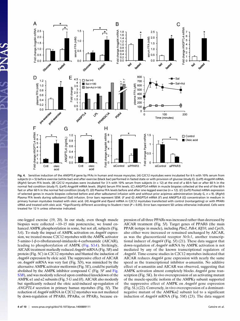
pression in the resting leg muscle cannot be caused by local con-tractile activity but must be related to systemic factors, includingcirculating metabolites. To verify this notion, serum from fastedhuman subjects collected before and after 2 h of cycling exercisewas added to mouse C2C12 myotubes, and Angptl4 mRNA ex-pression was determined by qPCR. Angptl4 mRNA was markedlyincreased in cells treated with postexercise serum as comparedwith preexercise serum (Fig. 4A). Intriguingly, no such effect wasobserved using pre- and postexercise plasma from subjects thatreceived repeated glucose drinks during the cycling exercise (Fig.4A). Angptl4 expression in C2C12 myotubes also was inducedmore strongly by plasma collected from subjects in the fasted statethan by plasma from subjects in the fed state (Fig. 4B). In thosesubjects, ANGPTL4 expression in skeletal muscle also was markedlyhigher in the fasted state than in the fed state (Fig. 4C). Together,these data suggest that ANGPTL4 is induced by a circulating factor
Fig. 1. Exercise induces ANGPTL4 gene expression in nonexercising human muscle. (A) Heatmap showing genes altered by exercise in the nonexercising legmuscle ranked by statistical significance. Values are displayed per subject (P1 to P9). Fold-change (FC) in gene expression is indicated. (B) mRNA expressionprofile of ANGPTL4 in the exercising and nonexercising legs according to microarray. (C) qPCR analysis of ANGPTL4 mRNA expression. (D) mRNA expression ofPPARδ targets KLF10, PDK4, and SLC22A5, as well as LPL. (E) ANGPTL4 protein levels in postexercise muscle biopsies as determined by ELISA. Error barsrepresent SEM. *Significantly different according to paired Student t test (P < 0.01).
2 of 10 | www.pnas.org/cgi/doi/10.1073/pnas.1400889111 Catoire et al.

that is specifically induced by exercise in the fasted state but is notelevated when glucose is consumed during exercise and that also isenriched in the fasted state as compared with fed state. One suchparameter is plasma free fatty acids (FFA) (Fig. 4 A and B, Right),levels of which also increased during one-legged cycling (Fig. 4D)and which previously had been shown to induce Angptl4 mRNApotently in various cell types, including cultured myocytes (15–18).In support of a role of plasma FFA in induction of ANGPTL4 geneexpression in human muscle, raising plasma FFA levels by salbu-tamol treatment markedly increased muscle ANGPTL4 expression,and this increase was largely blunted when salbutamol was coad-ministered with the lipolysis inhibitor acipimox (Fig. 4E). This ex-pression pattern was not found for other relevant genes such asPPARGC1A and PDK4.To study ANGPTL4 gene regulation by fatty acids further, we
used cultured myocytes. Oleic acid markedly induced ANGPTL4mRNA in human primary myotubes (Fig. 4F), accompanied bya similar increase in the secretion of ANGPTL4 protein (Fig.4G). Oleic acid also markedly induced Angptl4 expression inC2C12 myotubes, and this expression was significantly bluntedupon siRNA-mediated knockdown of PPARδ (Fig. 4H). Con-sistent with the high sensitivity of ANGPTL4 gene regulation to
stimulation by fatty acids, microarray analysis indicated thatANGPTL4 was the second most highly induced gene by oleicacid in human primary myotubes and mouse C2C12 myotubes(Fig. S1). Taken together, these data strongly suggest that themarked induction of ANGPTL4 mRNA in the nonexercisinghuman muscle is caused by increased plasma FFA levels asso-ciated with exercise.Despite being exposed to elevated plasma FFA, exercising
human muscle shows only a very minor increase in ANGPTL4expression. Accordingly, we hypothesized that induction ofANGPTL4 mRNA in exercising muscle by FFA is mitigated by acontraction-related factor. If so, expression of ANGPTL4 shouldincrease strongly after cessation of exercise when performed inthe fasted state, because plasma FFA levels remain high forhours after such exercise. Indeed, in the continued fasted state,ANGPTL4 mRNA in exercising human muscle is increased 20-fold at 4 h postexercise as compared with baseline postexercise,and the increase in ANGPTL4 mRNA is concurrent with sus-tained elevation of plasma FFA (Fig. S2).A candidate factor that may suppress ANGPTL4 in exercising
muscle is AMP-activated kinase (AMPK), which is activated spe-cifically in exercising muscle but not in nonexercising muscle during
Fig. 2. ANGPTL4 protein is detected in human muscle in myocytes and endothelial cells. Representative images of immunofluorescent staining of ANGPTL4in a biopsy from human vastus lateralis muscle show ANGPTL4 in green, myosin heavy chain 1 (MHC1), a marker type I fiber, in red, and caveolin in blue.
Fig. 3. Plasma ANGPTL4 levels are increased by acute exercise but not by exercise training. (A) Plasma ANGPTL4 levels before and after 1 h of one-leggedcycling exercise at 50% of the one-legged Wmax (study A, n = 12). (B) Plasma ANGPTL4 levels before and after 3 h of cycling exercise at 40% Wmax (Study B,n = 8). (C) Fasting plasma ANGPTL4 levels before and after an intense 2-wk endurance training program on a cycling ergometer (study C, n = 8). (D) Fastingplasma ANGPTL4 levels before and after a moderate-intensity, 12-wk endurance training program on a cycling ergometer (study D, n = 6).
Catoire et al. PNAS Early Edition | 3 of 10
PHYS
IOLO
GY
PNASPL
US
one-legged exercise (19, 20). In our study, even though musclebiopsies were collected ∼10–15 min postexercise, we found en-hanced AMPK phosphorylation in some, but not all, subjects (Fig.5A). To study the impact of AMPK activation on Angptl4 expres-sion, we treated mouse C2C12 myotubes with the AMPK activator5-amino-1-β-D-ribofuranosyl-imidazole-4-carboxamide (AICAR),leading to phosphorylation of AMPK (Fig. S3A). Strikingly,AICAR treatment markedly reducedAngptl4mRNA (Fig. 5B) andprotein (Fig. 5C) in C2C12 myotubes and blunted the induction ofAngptl4 expression by oleic acid. The suppressive effect of AICARon Angptl4 mRNA was very fast (Fig. 5D), was mimicked by thealternative AMPK activator metformin (Fig. 5E), could be partiallyabolished by the AMPK inhibitor compound C (Fig. 5F and Fig.S3B), and was modestly relieved upon combined knockdown of theAMPK α1 and α2 subunits (Fig. 5G andH). AICAR also modestlybut significantly reduced the oleic acid-induced up-regulation ofANGPTL4 secretion in primary human myotubes (Fig. 5I). Thereduction of Angptl4mRNA in C2C12 myotubes was not mediatedby down-regulation of PPARδ, PPARα, or PPARγ, because ex-
pression of all three PPARs was increased rather than decreased byAICAR treatment (Fig. 5J). Target genes of PPARδ (the mainPPAR isotype in muscle), including Plin2, Pdk4, Klf10, and Cpt1b,also either were increased or remained unchanged by AICAR,as was the glucocorticoid receptor Nr3c1, another transcrip-tional inducer of Angptl4 (Fig. 5J) (21). These data suggest thatdown-regulation of Angptl4 mRNA by AMPK activation is notmediated by any of the known transcriptional regulators ofAngptl4. Time-course studies in C2C12 myotubes indicated thatAICAR reduces Angptl4 gene expression with nearly the samespeed as the transcriptional inhibitor α-amanitin. No additiveeffect of α-amanitin and AICAR was observed, suggesting thatAMPK activation almost completely blocks Angptl4 gene tran-scription (Fig. 5K). In vivo overexpression of an activating mutantof the muscle-specific isoform of the AMPKγ subunit supportedthe suppressive effect of AMPK on Angptl4 gene expression(Fig. 5L) (22). Conversely, in vivo overexpression of a dominant-negative mutant of the AMPKα2 subunit led to a significantinduction of Angptl4 mRNA (Fig. 5M) (23). The data suggest
Fig. 4. Sensitive induction of the ANGPTL4 gene by FFAs in human and mouse myocytes. (A) C2C12 myotubes were incubated for 6 h with 10% serum fromsubjects (n = 5) before exercise (white bar) and after exercise (black bar) performed in fasted state or with provision of glucose (study E). (Left) Angptl4mRNA.(Right) Serum FFA levels. (B) C2C12 myotubes were incubated for 3 h with 10% serum from subjects (n = 12) at the end of a 60-h fast or after 60 h in thenormal fed condition (study F). (Left) Angptl4 mRNA levels. (Right) Serum FFA levels. (C) ANGPTL4 mRNA in muscle biopsies collected at the end of the 60-hfast or after 60 h in the normal fed condition (study F). (D) Plasma FFA levels before and after one-legged exercise (n = 12). (E) (Left) Pooled mRNA expressionof selected genes in muscle biopsies collected before and after salbutamol infusion with and without prior acipimox administration (study G, n = 9). (Right)Plasma FFA levels during salbutamol (Sal) infusion. Error bars represent SEM. (F and G) ANGPTL4 mRNA (F) and ANGPTL4 (G) concentration in medium inprimary human myotubes treated with oleic acid. (H) Angptl4 and Ppard mRNA in C2C12 myotubes transfected with control (nontargeting) or with PPARδsiRNA and treated with oleic acid. *Significantly different according to Student t test (P < 0.05). Error bars represent SD unless otherwise indicated. Cells weretreated for 12 h unless otherwise indicated.
4 of 10 | www.pnas.org/cgi/doi/10.1073/pnas.1400889111 Catoire et al.

that the stimulatory effect of plasma FFA on skeletal muscleANGPTL4 mRNA is counteracted by AMPK activation inexercising muscle.As previously observed for the PPARδ agonist GW501516 (17),
induction of Angptl4 mRNA in C2C12 myotubes by oleic acid wasassociated with a pronounced decrease in heparin-releasable LPLactivity (Fig. 6A) but induced Lpl mRNA (Fig. 6B). To study theimpact of Angptl4 up-regulation on skeletal muscle lipid uptake invivo, we used Angptl4-transgenic mice characterized by overex-pression of Angptl4 mRNA and protein in a variety of tissues,including skeletal muscle (Fig. 6C) (24). Transgenic Angptl4overexpression did not affect muscle weights or lean body masspercentage (Fig. S4). To assess the functional effect of Angptl4overexpression during exercise, we subjected WT and Angptl4-transgenic (Angptl4-Tg) mice to an acute moderate exercise bouton a motorized treadmill. Total LPL protein levels in skeletalmuscle (gastrocnemius) after exercise were not different in theWT and Angptl4-Tg mice (Fig. 6D). However, Angptl4-Tg miceshowed markedly reduced plasma clearance of [3H]triolein-
labeled very-low-density lipoprotein (VLDL)-like particles duringexercise (Fig. 6E) and reduced fatty acid uptake from the labeledVLDL-like particles into skeletal muscle but not into sub-cutaneous adipose tissue (Fig. 6F). Plasma clearance of [14C]-oleic acid and uptake into skeletal muscle and subcutaneousadipose tissue was unaffected in Angptl4-Tg mice (Fig. 6 G andH). Plasma levels of FFA, glucose, glycerol, and β-hydroxybu-tyrate were not different between the WT and Angptl4-Tg micein the exercised or resting state (Fig. S5A). To assess whetherreduced muscle uptake of plasma TG-derived fatty acids inAngptl4-Tg mice had any influence on muscle performance,we determined the maximal endurance capacity using an in-cremental treadmill protocol characterized by a gradual in-crease in speed and slope of the treadmill until exhaustion.Strikingly, Angptl4-Tg mice ran significantly less far than WTmice (Fig. 6I). Strength, as determined using the horizontalwire test, was not different in the two sets of animals (Fig. S5B).Depletion of muscle glycogen stores during exhaustive exer-cise was comparable in WT and Angptl4-Tg mice (Fig. 6J),
Fig. 5. AMPK activation suppresses Angptl4 mRNA. (A) Immunoblot for AMPK and phospho-AMPK in skeletal muscle biopsies from two selected subjectsbefore (t0) and after (t1) exercise. (B) Expression of Angptl4 mRNA in C2C12 myotubes treated with oleic acid (200 μM) and/or AICAR (1 mM) for 3 h. (C)Immunoblot for ANGPTL4 in C2C12 myotubes treated with oleic acid and/or AICAR. (D) Time-course of the effect of AICAR on Angptl4 mRNA in C2C12myotubes. (E) Comparison of the effect of AICAR (1 mM) and metformin (0.5 mM) on Angptl4 mRNA in C2C12 myotubes. (F) Effect of AICAR (1 mM) andcompound C cotreatment on Angptl4 mRNA in C2C12 myotubes. Concentrations are indicated in millimolars. (G) Angptl4 mRNA in C2C12 myotubestransfected with control (nontargeting) or AMPKα1/AMPKα2 siRNA and treated with AICAR. (H) Effective knockdown of AMPKα1 and AMPKα2 by AMPKα1/AMPKα2 siRNA. (I) ANGPTL4 levels in medium of human primary myotubes treated with oleic acid and AICAR. (J) Expression of PPARs and PPAR targets inC2C12 myotubes treated with AICAR. (K) Angptl4mRNA in C2C12 myotubes preincubated with 50 μg/mL α-Amanitin for 1 h and treated with AICAR for 3 h or6 h. (L) Angptl4 mRNA in the gastrocnemius of mice that overexpress an activating mutant of the muscle-specific isoform of the AMPKγ subunit. Error barsrepresent SEM. Data were extracted from GSE4065 (22). (M) Angptl4mRNA in the gastrocnemius of mice that overexpress a dominant-negative mutant of theAMPKα2 subunit. Cells were treated for 12 h unless otherwise indicated. Error bars represent SEM. *Significantly different according to Student t test (P <0.05). Error bars represent SD unless otherwise indicated.
Catoire et al. PNAS Early Edition | 5 of 10
PHYS
IOLO
GY
PNASPL
US

whereas liver glycogen levels remained higher in Angptl4-Tgmice after exhaustive exercise, a difference that reached statis-tical significance (Fig. 6K). Intramuscular TGs were lower inAngptl4-Tg mice, and this difference also reached significanceafter exhaustive exercise (Fig. 6L). No differences in baselineexpression of markers of oxidative capacity were observed in thetwo genotypes, suggesting that Angptl4 does not influenceoxidative capacity (Fig. 6M). Finally, we determined the relativedecrease in plasma TG in the exercised state as compared withresting state in WT, Angptl4-Tg, and Angptl4−/− mice, as a
measure of relative plasma TG utilization. The relative de-crease in plasma TG in the exercised vs. the resting state wasmuch more pronounced in Angptl4−/− mice than in WT andespecially Angptl4-Tg mice (Fig. 6N), likely because of thepreferential use of plasma TG in Angptl4−/− mice. Conversely,the relative increase in plasma FFA in the exercised vs. re-sting state was more pronounced in Angptl4−/− mice, likelybecause of the sparing of plasma FFA in favor of the use ofTG-derived fatty acids. Overall, these data indicate that up-regulation of Angptl4 impairs skeletal muscle uptake of fatty
Fig. 6. Angptl4 up-regulation impairs LPL activity and uptake of plasma TG-derived fatty acids in muscle. (A and B) Heparin-releasable LPL activity (A) and LplmRNA (B) in mouse C2C12 myotubes treated with oleic acid (400 μM) for 24 h. (C) ANGPTL4 protein abundance and Angptl4 mRNA expression in mouseskeletal muscle. (D) Total LPL activity in skeletal muscle (gastrocnemius) of WT and Angptl4-Tg mice at rest and after 90 min of moderate running exercise (12m/min). (E) Serum 3H activity after 15 min of running in WT and Angptl4-Tg mice injected with [14C]-oleate together with glycerol tri[3H]oleate-labeled VLDL-like particles. (F) 3H-activity in subcutaneous adipose tissue and gastrocnemius after 15 min of running. (G) Serum 14C activity after 15 min of running in WTand Angptl4-Tg mice injected with [14C]-oleate together with glycerol tri[3H]oleate-labeled VLDL-like particles. (H) 14C activity in subcutaneous adipose tissueand gastrocnemius after 15 min of running. (I) Distance covered, excluding warm-up, by WT and Angptl4-Tg mice subjected to an incremental exercise test toexhaustion. (J–L) Muscle glycogen (J), liver glycogen (K), and muscle TG (L) levels in WT and Angptl4-Tg mice at rest or after exhaustive running exercise. (M)mRNA expression of selected genes in skeletal muscle (gastrocnemius) of WT and Angptl4-Tg mice at rest. (N) The relative levels of plasma TG (Left) and FFA(Right) in the exercised state (90 min of moderate running exercise) compared with the resting state (90 min rest) in WT, Angptl4-Tg, and Angptl4−/− mice.*Significantly different from WT mice according to Student t test (P < 0.05). #Significantly different from resting mice according to Student t test (P < 0.05).Error bars represent SEM; n = 6–10 mice per group.
6 of 10 | www.pnas.org/cgi/doi/10.1073/pnas.1400889111 Catoire et al.

acids from circulating TG-rich lipoproteins and reduces liverglycogen utilization, leading to decreased performance ofexhaustive exercise.
DiscussionThe activity of LPL and the associated uptake in tissue of fattyacids derived from TG in plasma is under the control of differentphysiological stimuli in different tissues (14). In white adiposetissue LPL activity is decreased by fasting, which has been de-monstrated unequivocally to be mediated by up-regulation ofAngptl4 (25). In brown adipose tissue LPL activity is increased byexposure to cold; such exposure is associated with a decrease inAngptl4 mRNA (26), hinting at a potential role of ANGPTL4.Our data suggest that ANGPTL4 also plays an important role inLPL-dependent plasma clearance of TG in skeletal muscle,particularly during acute exercise, by coordinating lipid uptake inexercising and nonexercising muscles.Acute exercise increases adipose tissue lipolysis and raises
levels of FFA in plasma. Although the abundant plasma FFA areoxidized efficiently in exercising muscle, their increase leadsto elevated intramuscular fat storage in nonexercising muscle,possibly leading to lipotoxicity (27). Previously, we found thatANGPTL4 functions as a fatty acid-inducible antilipotoxic factorin cardiomyocytes and macrophages (15, 28). The present worksuggests that the exercise-induced increase in plasma FFAstimulates ANGPTL4 synthesis in nonexercising human muscle,leading to local inhibition of LPL activity and diminished uptake
of fatty acids derived from plasma TG in compensation for el-evated uptake of plasma FFA, presumably to mitigate lipidoverload and associated lipotoxicity in nonexercising muscleduring prolonged exercise. In contrast, in exercising muscle thestimulatory effect of FFA on ANGPTL4 is counteracted by AMPK-mediated suppression of ANGPTL4 mRNA, thereby maintainingLPL activity and supporting the use of plasma TG as fuel for theexercising muscle (Fig. 7).Previously, in vivo AMPK activation by AICAR was found to
lower plasma TG levels (29, 30). Moreover, AMPK activation byAICAR and metformin increased intralipid clearance and increasedheparin-releasable LPL activity in rat hearts without causingany change in LPL mRNA (31, 32). Furthermore, AICAR andmetformin enhanced LPL activity in rat L6 muscle cells (33).These findings suggest a role of LPL in the lowering of plasmaTG by AMPK. Based on the data presented here, it is plausiblethat the stimulatory effect of AICAR and metformin on LPLactivity and the concomitant lowering of plasma TG is mediatedby suppression of ANGPTL4 in muscle and possibly other tis-sues. Future studies are needed to address this question inmore detail.Our data indicate that overexpression of Angptl4 reduces max-
imal performance in endurance exercise, most likely by limitingthe provision of fatty acids derived from plasma TG to the muscleand by limiting the utilization of liver glycogen, which is an im-portant fuel for exercising mice. Until recently, because of anumber of methodological issues, the role of plasma TG as source
Fig. 7. Schematic representation of the proposed role of ANGPTL4 in providing lipid to exercising muscle. During exercise, circulating FFAs and VLDL particlesare directed to exercising and nonexercising muscle. In the nonexercising leg, increased FFA levels provoke an increase in ANGPTL4 expression via PPARδ,leading to inhibition of LPL activity and consequent reduction in uptake of fatty acids derived from VLDL, which likely is aimed at preventing lipid overload. Incontrast, in the exercising leg the stimulatory effect of FFA on ANGPTL4 mRNA is counteracted by AMPK-mediated suppression of ANGPTL4 mRNA. Asa result, LPL activity remains high, allowing full exploitation of fatty acids derived from VLDL as the substrate for fatty acid oxidation to meet the energeticneeds of exercising muscle.
Catoire et al. PNAS Early Edition | 7 of 10
PHYS
IOLO
GY
PNASPL
US

of fatty acids for oxidation in exercising muscle and thereby theoverall importance of plasma TG as fuel during endurance exer-cise likely have been underestimated (reviewed in ref. 1). Indeed,plasma TG uptake and clearance are increased many fold duringleg exercise as compared with the resting situation (34). The rel-evance of plasma TG during exercise is suggested further by theadaptive increase in muscle LPL mRNA and activity in responseto an acute exercise bout and after exercise training (35, 36).Unlike ANGPTL4, the expression of LPL is induced to the sameextent in exercising and nonexercising muscle. The enhanced ac-tivity of LPL in skeletal muscle is believed to account for the lowplasma TG concentrations in trained individuals (37, 38).ANGPTL4 adds to a growing list of secreted proteins whose
production in muscle is increased by acute exercise (39, 40). Formany of these proteins it is unclear whether the change in pro-duction mainly impacts the muscle locally or whether the proteinalso exerts an endocrine effect and thus functions as a myokine.Even though acute exercise leads to elevated ANGPTL4 levels inthe circulation, it is unclear whether this increase stems mainlyfrom the increased mRNA and production of ANGPTL4 in skel-etal muscle or whether other tissues contribute as well. Overall, thecoexpression of ANGPTL4 with LPL in tissues such as muscle andadipose tissue and the tissue-specific regulation of ANGPTL4 ex-pression suggest that ANGPTL4 may act mainly via local inhibitionof LPL (17, 41).In conclusion, our data indicate that, in addition to the re-
sponses in exercising muscle, molecular changes in nonexercisingmuscle likely play a key role in regulating the fuel supply duringexercise. It can be speculated that the beneficial effects of exerciseon various health parameters are conveyed by adaptive changes innonexercising muscles.
Experimental ProceduresHuman Intervention Studies. Twelve healthy men (age 51.5 ± 5.1 y, bodyweight 88 ± 17 kg, body mass index 26 ± 4) participated in study A (4).Subjects were asked to follow healthy eating guidelines the day before theexperiment and to refrain from alcohol consumption. Subjects fasted from10:00 PM the evening before the study began until the end of the in-tervention. During the exercise session subjects were allowed to drink waterad libitum. All subjects performed a single endurance exercise bout, con-sisting of 60 min of one-legged cycling at 50% of their one-legged maximalwork load (Wmax) (determined by a graded one-legged cycling test). One-legged cycling was performed on a cycle ergometer (Excalibur Sport)adapted with a custom-made leg support. Skeletal muscle biopsies weretaken before and after exercise from both legs of 11 subjects, with the av-
erage time of collection at ∼15 min pre- or postexercise. One subject becameapprehensive about the needle biopsy immediately before the first biopsycollection. Two subjects later were excluded from the microarray analysisbecause the microarrays failed to meet quality control criteria. In addition,a venous blood sample was taken before and shortly after exercise. Otherhuman intervention studies included in this paper have been publishedpreviously and are outlined briefly below.
In study B, eight young, untrained, healthymale subjects (age: 23.3 ± 3.2 y)performed 3 h of cycling on an electromagnetically braked ergometer at anintensity of 40% of the predetermined Wmax (42). To facilitate completionof the exercise test, subjects received two 125-mL servings of a maltodextrindrink during the second half of the test. Subjects were allowed to drinkwater ad libitum during the entire test. Plasma was collected before andafter 3 h of cycling exercise and was used for measurement of ANGPTL4.
In study C, eight young, untrained, healthymale subjects (age: 23.3 ± 3.2 y)followed a 2-wk exercise training program on a cycling ergometer (42).Training consisted of alternating days of interval and endurance trainingand always started with 7.5 min of warming up at 40% Wmax and endedwith 7.5 min of cooling down at 40% Wmax. Fasting plasma was collectedbefore and after the 2- wk training program and was used for the measure-ment of ANGPTL4. Plasma was collected 3 d after the last training session.
In study D, six healthy, nonobese male subjects (age: 42.7 ± 2.0 y) followeda 12-wk exercise training program on a cycling ergometer (43). Subjects trainedthree times per week for 12 wk for 47.5 ± 2.5 min at 40% of predeterminedmaximal oxygen consumption (VO2 max). Fasting plasma was collected be-fore and after the 12-wk training program and was used for measurement ofANGPTL4. Plasma was collected 3 d after the last training session.
Seven healthy, untrained male volunteers (age: 22.7 ± 0.6 y) participatedin study E. After the subjects fasted overnight, a Teflon cannula was insertedin an antecubital vein for sampling of blood. Subjects rested on a bed, and abaseline blood sample was taken. Immediately thereafter, subjects ingested1.4 g/kg bodyweight glucose or water. Subjects exercised at 50% VO2
max for 2 h and then rested for 4 h. At regular intervals subjects ingested0.35 g/kg bodyweight glucose or water. Muscle biopsies were collectedbefore exercise and after the 4-h rest period. All subjects underwent theexperimental protocol twice, once with glucose ingestion and once whilefasting (44).
Twelve healthy, leanmale volunteers (age: 23.6± 1.0 y) participated in studyF. Subjects stayed in the respiration chamber for 60 h in the normal fed con-dition or while being completely fasted, according to a randomized crossoverdesign with a 2-wk washout period. Blood samples were collected at the endof the study after an overnight fast (fed condition) or after a cumulative 60-hfast. Around the same time, a muscle biopsy was taken (45).
Nine healthy, lean male volunteers (age: 24.4 ± 1.3 y) participated in studyG. After subjects fasted overnight, two Teflon cannulas were inserted intoan antecubital vein of each arm. One cannula was used for the infusion ofsalbutamol, and one was used for sampling blood. A first blood sample andmuscle biopsy were taken, followed by a continuous infusion of salbutamolfor 3 h. In addition, two doses (250 mg) of acipimox or placebo were given
Table 1. Primer sequences
Forward primer Reverse primer
m36B4 ATGGGTACAAGCGCGTCCTG GCCTTGACCTTTTCAGTAAG
Cyclophilin CAGACGCCACTGTCGCTTT TGTCTTTGGAACTTTGTCTGCAA
PGC1-a AGACGGATTGCCCTCATTTGA TGTAGCTGAGCTGAGTGTTGG
Cpt1b ATCATGTATCGCCGCAAACT CCATCTGGTAGGAGCACATGG
Lpl CAGCTGGGCCTAACTTTGAG GACCCCCTGGTAAATGTGTG
Angptl4 GTTTGCAGACTCAGCTCAAGG CCAAGAGGTCTATCTGGCTCTG
Ppard TTGAGCCCAAGTTCGAGTTTG CGGTCTCCACACAGAATGATG
Cd36 TCCAGCCAATGCCTTTGC TGGAGATTACTTTTCAGTGCAGAA
Plin2 GGATGTGGTGACGACTACCAT ACAGACTTGGTCCTTTCCACG
Pdk4 TCTACAAACTCTGACAGGGCTTT CCGCTTAGTGAACACTCCTTC
Ppara TATTCGGCTGAAGCTGGTGTAC CTGGCATTTGTTCCGGTTCT
Pparg CACAATGCCATCAGGTTTGG GCTGGTCGATATCACTGGAGATC
Prkaa1 TTCGGGAAAGTGAAGGTGGG TCTTCTGCCGGTTGAGTATCT
Prkaa2 CAGGCCATAAAGTGGCAGTTA AAAAGTCTGTCGGAGTGCTGA
Cs GGACAATTTTCCAACCAATCTGC TCGGTTCATTCCCTCTGCATA
Klf10 ATGCTCAACTTCGGCGCTT CGCTTCCACCGCTTCAAAG
ANGPTL4 CACAGCCTGCAGACACAACTC GGAGGCCAAACTGGCTTTGC
GAPDH CATGTTCCAGTATGACTCCACTC GGCCTCACCCCATTTGATGT
8 of 10 | www.pnas.org/cgi/doi/10.1073/pnas.1400889111 Catoire et al.
orally at time −120 min and time 0. Blood samples were taken at regularintervals throughout the study (46). A second muscle biopsy was taken afterthe 3-h infusion.
Informed consent was obtained from all subjects. The studies were ap-proved by the Medical Ethics Committee of the institute involved (Wage-ningen University or Maastricht University).
Blood Samples. Blood was collected in tubes containing EDTA and wascentrifuged immediately for 10 min (1,000 × g, 4 °C). Blood samples wereanalyzed for FFA levels (Centre for Medical Diagnostics, Velp, The Netherlands)and ANGPTL4 levels (see below).
Skeletal Muscle Biopsies. Percutaneous needle biopsies were taken before andshortly after exercise from the vastus lateralis muscle from both legs, usingthe Bergström technique with suction. All biopsies were taken from separateincisions; the second biopsy in the leg was located 2 cm proximal to the firstbiopsy. Biopsies were taken on average 10–15 min before and after theexercise bout. All biopsies were collected, processed, and frozen within 30min postexercise. After each biopsy, the collected tissue sample was clearedcarefully of adipose tissue and blood and was frozen directly in liquid ni-trogen or was embedded into Tissue-Tek O.C.T. compound (Sakura TissueTek) and frozen in liquid nitrogen-cooled isopentane.
Muscle biopsy lysates were prepared using a lysis buffer consisting of50 mM Tris·HCl (pH 7.5), 150 mM NaCL, 1 mM EGTA, 1 mM EDTA, 0.27 Msucrose, and 2% Triton X-100. Protease and phosphatase inhibitors (Com-plete and PhosSTOP; Roch Diagnostics GmbH) were added to the buffer. Thebuffer was added to tissue in a 15:1 (buffer: tissue) ratio, and the tissue waslysed using TissueLyser II (Qiagen).
Immunofluorescence. Frozen muscle sections (5-μm thick) were treated with0.1% Triton X-100 in PBS and were incubated for 45 min at room temper-ature with the primary antibody mix [a polyclonal rabbit hANGPTL4,a mouse monoclonal IgM antibody directed to MHC1 (type 1 muscle fibers;Developmental Studies Hybridoma Bank), and a mouse monoclonal IgG1
antibody to caveolin-3 (BD Biosciences)] diluted in 0.05% Tween20 in PBS.After three washing steps with PBS, sections were incubated for 45 min atroom temperature with the appropriate fluorescent-labeled secondaryantibodies. The specificity of the antibody for ANGPTL4 was demonstratedpreviously via immunoblot of human plasma using appropriate peptidecontrols and was corroborated by immunochemical and immunofluores-cence staining of ANGPTL4 in human heart, intestine, and skin wounds,using appropriate negative controls (15, 47, 48).
Animal Experiments. Exercise studies were carried out using the TSE Pheno-Master treadmill module (TSE Systems) that allowed four mice to run si-multaneously. Four- to five-month-old littermate male WT and Angptl4-Tgmice bred on the C57BL/6 background for 15 generations were used for theexercise experiments. The number of mice per group varied from six to ninemice. All mice were acclimatized to the treadmill (10 m/min for 10 min) onthree consecutive days. On the day of exercise, mice were fasted (4 h) andeither were subjected to a moderate exercise protocol (12 m/min at 0% in-cline for 90 min) or ran until exhaustion (30 min at 12 m/min followed by anincrease of 2.5 m/min and 2° of incline every 5 min until exhaustion). Amouse was considered exhausted if it failed to run after it was prompted bya slider with plastic bristles three times within 1 min. The moment of ex-haustion was determined by an investigator unaware of the genotype of themice. A control group of nonexercising mice from each genotype remainedsedentary inside the treadmill. Upon cessation of exercise mice were anes-
thetized immediately using isoflurane, and blood was collected by orbitalpuncture. Mice then were killed by cervical dislocation, and tissues wereexcised quickly and were frozen immediately in liquid nitrogen.
The horizontal wire test was performed by placing a steel wire betweena gray PVC stand at a height of 30 cm. Mice hung on the wire with theirforepaws. Time measurements started at the moment that the researcherjudged the mouse had a good grip on the wire. Time measurement endedwhen the animal grasped the wire with at least one hind paw. If the animalfell, it was placed back in its cage for a 3-min rest, and measurementswere repeated.
Animal experiments were approved by the Animal Ethics Committeeof Wageningen University.
Plasma TG and FFA Clearance.After a 6-h fast, 6-mo-old maleWT andAngptl4-Tg mice were put on a treadmill at a speed of 14m/min. After 45 min ofrunning, mice were injected with a single bolus of [14C]-oleate and glyceroltri[3H]oleate-labeled VLDL-like particles and continued to run for another 15min. After 15 min, blood was collected; then the animal was killed imme-diately and tissues were collected. Tissues and serum were processed todetermine 3H and 14C activity. Preparation of VLDL-like particles, complexingof [14C]-oleate with BSA, the infusion protocol, and calculations were carriedout as described previously (49–51).
Cell Culture and Treatment. C2C12 myoblasts were maintained in DMEM(Lonza) supplemented with 10% (vol/vol) FCS and 1% penicillin/streptomycin/amphotericin (PSA) under 5% CO2 at 37 °C. At ∼70–90% confluency, myoblastswere treated or full-growth medium was replaced with alphaMEM (Lonza)supplemented with 2% (vol/vol) FCS and 1% PSA to promote differentiationinto myotubes. The differentiation medium was changed every 2–3 d. C2C12myotubes were used for treatment after 1 wk of differentiation. All experi-ments with C2C12 myotubes were performed in differentiation medium.
C2C12 myotubes were treated with human serum by replacing FCS withhuman serum and were incubated for 3–6 h. For siRNA-mediated knockdown,C2C12 cells were transfected with siRNA sequences using Lipofectamine RNAi-Max the day before differentiation was started. The ON-TARGETplus SMART-pool for PPARδ, AMPKα1, AMPKα2, and nontargeting was used (Dharmacon/Thermo-Fisher Scientific) at 100 pmol/mL The medium was replaced by differ-entiation medium (DMEM supplemented with 2% (vol/vol) horse serum, 100 μg/mL penicillin, and 100 μg/mL streptomycin) after 24 h. After 4 d of differenti-ation, cells were treated with oleic acid and AICAR as indicated below. Differ-entiated human primary myotubes were prepared as described previously (52).
Oleic acid (200 μM) was added to cells complexed with BSA (2.5:1). AICARand metformin were used at 1 mM. The AMPK inhibitor compound C wasused at concentrations indicated in the Fig. 5 legend. Cells were treated for12 h unless otherwise indicated.
RNA Isolation and qPCR. Total RNA was isolated using TRIzol reagent(Invitrogen) and was purified for microarray analysis using the Qiagen RNeasyMicro kit (Qiagen).
RNA was reverse transcribed using a First-Strand cDNA Synthesis Kit(Fermentas). Real-time PCR was carried out using SensiMiX (Bioline) on a CFX384 Bio-Rad thermal cycler (Bio-Rad). Cyclophilin, GADPH, and/or 36B4 wereused as housekeeping genes. Primers used are listed in Table 1.
ACKNOWLEDGMENTS. This work was supported by Dutch Diabetes Foun-dation Grant 2009.60.003, the Netherlands Nutrigenomics Centre, and theNetherlands Organization of Scientific Research.
1. Kiens B (2006) Skeletal muscle lipid metabolism in exercise and insulin resistance.
Physiol Rev 86(1):205–243.2. Keller P, et al. (2007) Using systems biology to define the essential biological networks
responsible for adaptation to endurance exercise training. Biochem Soc Trans 35(Pt 5):
1306–1309.3. Buford TW, Cooke MB, Willoughby DS (2009) Resistance exercise-induced changes of
inflammatory gene expression within human skeletal muscle. Eur J Appl Physiol
107(4):463–471.4. Catoire M, et al. (2012) Pronounced effects of acute endurance exercise on gene
expression in resting and exercising human skeletal muscle. PLoS ONE 7(11):
e51066.5. Schmutz S, et al. (2006) Endurance training modulates the muscular transcriptome
response to acute exercise. Pflugers Arch 451(5):678–687.6. Yang Y, Creer A, Jemiolo B, Trappe S (2005) Time course of myogenic and metabolic
gene expression in response to acute exercise in human skeletal muscle. J Appl Physiol
(1985) 98(5):1745–1752.
7. Buckley JD, Scroop GC, Catcheside PG (1993) Lactate disposal in resting trained and
untrained forearm skeletal muscle during high intensity leg exercise. Eur J Appl
Physiol Occup Physiol 67(4):360–366.8. Savard G, et al. (1987) Noradrenaline spillover during exercise in active versus resting
skeletal muscle in man. Acta Physiol Scand 131(4):507–515.9. Apró W, Blomstrand E (2010) Influence of supplementation with branched-chain
amino acids in combination with resistance exercise on p70S6 kinase phosphorylation
in resting and exercising human skeletal muscle. Acta Physiol (Oxf) 200(3):237–248.10. Pedersen BK, Febbraio MA (2008) Muscle as an endocrine organ: Focus on muscle-
derived interleukin-6. Physiol Rev 88(4):1379–1406.11. Lichtenstein L, et al. (2007) Angptl4 upregulates cholesterol synthesis in liver via in-
hibition of LPL- and HL-dependent hepatic cholesterol uptake. Arterioscler Thromb
Vasc Biol 27(11):2420–2427.12. Sukonina V, Lookene A, Olivecrona T, Olivecrona G (2006) Angiopoietin-like protein 4
converts lipoprotein lipase to inactive monomers and modulates lipase activity in
adipose tissue. Proc Natl Acad Sci USA 103(46):17450–17455.
Catoire et al. PNAS Early Edition | 9 of 10
PHYS
IOLO
GY
PNASPL
US

13. Yoshida K, Shimizugawa T, Ono M, Furukawa H (2002) Angiopoietin-like protein 4 isa potent hyperlipidemia-inducing factor in mice and inhibitor of lipoprotein lipase.J Lipid Res 43(11):1770–1772.
14. Wang H, Eckel RH (2009) Lipoprotein lipase: From gene to obesity. Am J PhysiolEndocrinol Metab 297(2):E271–E288.
15. Georgiadi A, et al. (2010) Induction of cardiac Angptl4 by dietary fatty acids is me-diated by peroxisome proliferator-activated receptor beta/delta and protects againstfatty acid-induced oxidative stress. Circ Res 106(11):1712–1721.
16. Kersten S, et al. (2009) Caloric restriction and exercise increase plasma ANGPTL4 levelsin humans via elevated free fatty acids. Arterioscler Thromb Vasc Biol 29(6):969–974.
17. Robciuc MR, et al. (2012) Angiopoietin-like 4 mediates PPAR delta effect on lipo-protein lipase-dependent fatty acid uptake but not on beta-oxidation in myotubes.PLoS ONE 7(10):e46212.
18. Staiger H, et al. (2009) Muscle-derived angiopoietin-like protein 4 is induced by fattyacids via peroxisome proliferator-activated receptor (PPAR)-delta and is of metabolicrelevance in humans. Diabetes 58(3):579–589.
19. Kristensen JM, et al. (2007) Absence of humoral mediated 5’AMP-activated proteinkinase activation in human skeletal muscle and adipose tissue during exercise.J Physiol 585(Pt 3):897–909.
20. Mascher H, Ekblom B, Rooyackers O, Blomstrand E (2011) Enhanced rates of muscleprotein synthesis and elevated mTOR signalling following endurance exercise in hu-man subjects. Acta Physiol (Oxf) 202(2):175–184.
21. Koliwad SK, et al. (2009) Angiopoietin-like 4 (ANGPTL4, fasting-induced adiposefactor) is a direct glucocorticoid receptor target and participates in glucocorticoid-regulated triglyceride metabolism. J Biol Chem 284(38):25593–25601.
22. Nilsson EC, et al. (2006) Opposite transcriptional regulation in skeletal muscle of AMP-activated protein kinase gamma3 R225Q transgenic versus knock-out mice. J BiolChem 281(11):7244–7252.
23. Mu J, Brozinick JT, Jr., Valladares O, Bucan M, Birnbaum MJ (2001) A role for AMP-activated protein kinase in contraction- and hypoxia-regulated glucose transport inskeletal muscle. Mol Cell 7(5):1085–1094.
24. Mandard S, et al. (2006) The fasting-induced adipose factor/angiopoietin-like protein4 is physically associated with lipoproteins and governs plasma lipid levels and adi-posity. J Biol Chem 281(2):934–944.
25. Kroupa O, et al. (2012) Linking nutritional regulation of Angptl4, Gpihbp1, and Lmf1to lipoprotein lipase activity in rodent adipose tissue. BMC Physiol 12:13.
26. Bartelt A, et al. (2011) Brown adipose tissue activity controls triglyceride clearance.Nat Med 17(2):200–205.
27. Schrauwen-Hinderling VB, et al. (2003) Intramyocellular lipid content is increasedafter exercise in nonexercising human skeletal muscle. J Appl Physiol (1985) 95(6):2328–2332.
28. Lichtenstein L, et al. (2010) Angptl4 protects against severe proinflammatory effectsof saturated fat by inhibiting fatty acid uptake into mesenteric lymph node macro-phages. Cell Metab 12(6):580–592.
29. Bergeron R, et al. (2001) Effect of 5-aminoimidazole-4-carboxamide-1-beta-D-ribo-furanoside infusion on in vivo glucose and lipid metabolism in lean and obese Zuckerrats. Diabetes 50(5):1076–1082.
30. Buhl ES, et al. (2002) Long-term AICAR administration reduces metabolic disturbancesand lowers blood pressure in rats displaying features of the insulin resistance syn-drome. Diabetes 51(7):2199–2206.
31. An D, et al. (2005) The metabolic “switch” AMPK regulates cardiac heparin-releasablelipoprotein lipase. Am J Physiol Endocrinol Metab 288(1):E246–E253.
32. Hauton D (2011) Does long-term metformin treatment increase cardiac lipoproteinlipase? Metabolism 60(1):32–42.
33. Ohira M, Miyashita Y, Murano T, Watanabe F, Shirai K (2009) Metformin promotesinduction of lipoprotein lipase in skeletal muscle through activation of adenosinemonophosphate-activated protein kinase. Metabolism 58(10):1408–1414.
34. Enevoldsen LH, Simonsen L, Bülow J (2005) Postprandial triacylglycerol uptake in thelegs is increased during exercise and post-exercise recovery. J Physiol 568(Pt 3):941–950.
35. Hamilton MT, Etienne J, McClure WC, Pavey BS, Holloway AK (1998) Role of localcontractile activity and muscle fiber type on LPL regulation during exercise. Am JPhysiol 275(6 Pt 1):E1016–E1022.
36. Vissing K, Andersen JL, Schjerling P (2005) Are exercise-induced genes induced byexercise? FASEB J 19(1):94–96.
37. Hardman AE (1998) The influence of exercise on postprandial triacylglycerol metab-olism. Atherosclerosis 141(Suppl 1):S93–S100.
38. Seip RL, Semenkovich CF (1998) Skeletal muscle lipoprotein lipase: Molecular regu-lation and physiological effects in relation to exercise. Exerc Sport Sci Rev 26:191–218.
39. Pedersen BK, Febbraio MA (2012) Muscles, exercise and obesity: Skeletal muscle asa secretory organ. Nat Rev Endocrinol 8(8):457–465.
40. Pillon NJ, Bilan PJ, Fink LN, Klip A (2013) Cross-talk between skeletal muscle andimmune cells: Muscle-derived mediators and metabolic implications. Am J PhysiolEndocrinol Metab 304(5):E453–E465.
41. Nilsson SK, et al. (2012) Triacylglycerol-rich lipoproteins protect lipoprotein lipase frominactivation by ANGPTL3 and ANGPTL4. Biochim Biophys Acta 1821(10):1370–1378.
42. Schrauwen-Hinderling VB, et al. (2003) The increase in intramyocellular lipid contentis a very early response to training. J Clin Endocrinol Metab 88(4):1610–1616.
43. Schrauwen P, et al. (2002) The effect of a 3-month low-intensity endurance trainingprogram on fat oxidation and acetyl-CoA carboxylase-2 expression. Diabetes 51(7):2220–2226.
44. Schrauwen P, et al. (2002) Effect of acute exercise on uncoupling protein 3 is a fatmetabolism-mediated effect. Am J Physiol Endocrinol Metab 282(1):E11–E17.
45. Hoeks J, et al. (2010) Prolonged fasting identifies skeletal muscle mitochondrial dys-function as consequence rather than cause of human insulin resistance. Diabetes59(9):2117–2125.
46. Hoeks J, et al. (2003) Effect of beta1- and beta2-adrenergic stimulation on energyexpenditure, substrate oxidation, and UCP3 expression in humans. Am J PhysiolEndocrinol Metab 285(4):E775–E782.
47. Alex S, et al. (2013) ANGPTL4 is produced by entero-endocrine cells in the humanintestinal tract. Histochem Cell Biol, 10.1007/s00418-013-1157-y.
48. Goh YY, et al. (2010) Angiopoietin-like 4 interacts with integrins beta1 and beta5 tomodulate keratinocyte migration. Am J Pathol 177(6):2791–2803.
49. Jong MC, et al. (1997) Reduced very-low-density lipoprotein fractional catabolic ratein apolipoprotein C1-deficient mice. Biochem J 321(Pt 2):445–450.
50. Rensen PC, et al. (1997) Particle size determines the specificity of apolipoproteinE-containing triglyceride-rich emulsions for the LDL receptor versus hepatic remnantreceptor in vivo. J Lipid Res 38(6):1070–1084.
51. Teusink B, et al. (2003) Contribution of fatty acids released from lipolysis of plasmatriglycerides to total plasma fatty acid flux and tissue-specific fatty acid uptake. Di-abetes 52(3):614–620.
52. Sparks LM, et al. (2011) Remodeling lipid metabolism and improving insulin re-sponsiveness in human primary myotubes. PLoS ONE 6(7):e21068.
10 of 10 | www.pnas.org/cgi/doi/10.1073/pnas.1400889111 Catoire et al.


