Embed Size (px)
Citation preview

IntroductionFetal calcium metabolism differs from that of adults inseveral ways, reflecting the unique needs of the devel-oping fetus (1). Calcium is actively transported acrossthe placenta, and the fetus normally has a higher bloodcalcium than does the mother, resulting in a maternal-fetal calcium gradient. In the adult, parathyroid hor-mone (PTH), acting through the PTH/PTHrP receptor,is the major regulator of blood calcium. In the fetus,however, PTH and parathyroid hormone–related pro-tein (PTHrP) share in the regulation of the fetus’sunique calcium metabolism, acting through thePTH/PTHrP receptor and other receptors. PTHrP mayplay a dominant role in regulating fetal-placental cal-cium homeostasis, while PTH is normally suppressedin late gestation and its importance to fetal calciumhomeostasis has been less clearly defined.
We have previously shown that absence of PTHrP (inthe Pthrp-null fetus; ref. 2) reduced the blood calciumto the maternal level (zero maternal-fetal gradient), andreduced the placental transfer of 45Ca (3). Further, wefound that administration of PTHrP 1-86 or PTHrP 67-86 (but not PTH 1-84 or PTH 1-34) increased the rateof placental calcium transfer in Pthrp-null fetuses.These experiments were consistent with the previousfinding that mid-molecular fragments of PTHrP (butnot intact PTH) could stimulate placental calcium
transfer in fetal lambs that had been thyroparathy-roidectomized (4, 5). In contrast to the findings inPthrp-null fetuses, we had noted in PTH/PTHrP receptor1–null (Pthr1-null) fetuses (6) that the fetal blood calci-um was reduced below that of the mother (reversedmaternal-fetal gradient) but that placental calciumtransfer was increased to about 150% of the value of theheterozygote littermates (3).
These experiments confirmed that PTHrP plays animportant role in normal fetal-placental calciumhomeostasis, given that in the absence of PTHrP, thefetus is hypocalcemic, hyperphosphatemic, and hasreduced placental calcium transfer. Pthrp-null fetuseshave markedly elevated serum PTH levels (7); evident-ly, however, PTH is unable to compensate completelyfor lack of PTHrP. In fact, when PTH is further stimu-lated in the Pthrp-null fetuses by simultaneously delet-ing the Calcium sensing receptor (Casr) gene (8), the bloodcalcium of such Pthrp–/–Casr–/– fetuses is higher thanthat of the pure Pthrp-null fetus, but the rate of placen-tal calcium transfer is still reduced in Pthrp–/–Casr–/–
fetuses (9).The observations in the Pthr1-null fetuses suggested
that PTH does have a role in maintaining the fetalblood calcium, as the blood calcium in these fetuseswas lower than that in Pthrp-null fetuses. The increasedplacental calcium transfer in Pthr1-null fetuses was
The Journal of Clinical Investigation | April 2001 | Volume 107 | Number 8 1007
Fetal parathyroids are not required to maintain placental calcium transport
Christopher S. Kovacs,1 Nancy R. Manley,2 Jane M. Moseley,3 T. John Martin,3
and Henry M. Kronenberg4
1Faculty of Medicine–Endocrinology, Memorial University of Newfoundland, St. John’s, Newfoundland, Canada2Institute of Molecular Medicine and Genetics, Department of Pediatrics, Medical College of Georgia, Augusta, Georgia, USA3St. Vincent’s Institute for Medical Research, University of Melbourne, Fitzroy, Victoria, Australia4Endocrine Unit, Massachusetts General Hospital and Harvard Medical School, Boston, Massachusetts, USA
Address correspondence to: Christopher S. Kovacs, Faculty of Medicine–Endocrinology, Memorial University of Newfoundland, 300 Prince Philip Drive, St. John’s, Newfoundland A1B 3V6, Canada. Phone: (709) 777-6881; Fax: (709) 777-8049; E-mail [email protected].
Received for publication September 13, 2000, and accepted in revised form March 13, 2001.
We used Hoxa3 knockout mice and other mouse models to study the role of the fetal parathyroidsin fetal calcium homeostasis. Hoxa3-null fetuses lack parathyroid glands, and absence of parathy-roid hormone (PTH) was confirmed with a rodent PTH immunoradiometric assay. The ionized cal-cium level of Hoxa3-null fetuses was significantly lower than that of wild-type or heterozygous lit-termates or of the mother. Both the rate of placental calcium transfer and the plasma PTHrP levelwere normal in Hoxa3 mutants and their heterozygous siblings. Because we had previously observedan increase in placental calcium transfer in PTH/PTHrP receptor 1–null (Pthr1-null) fetuses, weassayed plasma PTHrP in those mice. Pthr1-null fetuses had plasma PTHrP levels 11-fold higher thanthose of their littermates. Northern analysis, immunohistochemical, and in situ hybridization stud-ies of Pthr1-null fetuses indicated that liver and placenta had increased expression of PTHrP. Insummary, loss of fetal parathyroids in Hoxa3-null fetuses caused marked hypocalcemia but did notalter placental calcium transfer or the circulating PTHrP level. The findings in the Pthr1-null fetus-es indicate that several tissues may contribute to the circulating PTHrP level in fetal mice.
J. Clin. Invest: 107:1007–1015 (2001).

interpreted to indicate possible upregulation of PTHrP,and, in particular, the PTHrP mid-molecule that regu-lates placental calcium transfer (3).
We address the remaining important questions aboutthe relative roles of PTHrP and PTH in regulating fetalcalcium homeostasis: First, whether PTH plays animportant role in fetal calcium homeostasis, and sec-ond, what tissue source(s) of PTHrP is important forfetal calcium homeostasis. Regarding the latter ques-tion, indirect evidence suggests that the parathyroidsare a major source of fetal blood PTHrP in sheep (4, 5),but direct confirmation of this hypothesis is lacking. Infact, a detailed study of the parathyroids of fetal ratsfound abundant PTH mRNA but no detectable PTHrPmRNA (10), suggesting that the fetal parathyroids arenot the source of PTHrP in the circulation of thatspecies. PTHrP is expressed widely in fetal tissues(including the placenta) (1, 11), any number of whichmight contribute to the circulating PTHrP level andthe regulation of placental calcium transfer.
To address both questions, we utilized several mousemodels, including a model in which parathyroid glandsare missing as a consequence of a genetic deletion (12).In the latter model, Hoxa3-null fetuses die soon afterbirth, with abnormalities in tissues derived from thethird and fourth pharyngeal arches, including thyroidhypoplasia, deletion of the thymus, and (most relevantto our studies) absence of parathyroids. Thus, in addi-tion to lacking PTH, these null mice should lack PTHrPthat is derived from the parathyroids, but should retainPTHrP that is derived from the placenta and other fetaltissue sources. This model enabled us to examine thephysiological contribution of the parathyroids to fetalcalcium metabolism, and, in particular, to determinewhether that role is accomplished through PTH, PTHrP,or both. By comparing and contrasting the effects of lossof parathyroids (in Hoxa3-null) to loss of PTHrP (inPthrp-null) and resistance to PTH action (in Pthr1-null),both the role of PTH and the source(s) of PTHrP relevantto fetal calcium metabolism were examined.
We find that lack of parathyroids in Hoxa3-null fetus-es leads to absence of circulating PTH and clear dis-ruptions in mineral metabolism that establish a defi-nite role for PTH in normal fetal calcium homeostasis.However, absence of parathyroids did not alter serumPTHrP levels or the rate of placental calcium transfer,indicating that the parathyroids may not contributesignificantly to the circulating PTHrP level or the reg-ulation of placental calcium transfer. We then re-exam-ined the Pthr1 model (in which placental calcium trans-fer is upregulated) and found increased serum PTHrPlevels and evidence of upregulation of PTHrP by fetalliver and placenta. Altogether, the observations in thesecontrasted mouse models indicate that the parathy-roids do play a role in fetal calcium metabolismthrough the production of PTH but possibly notPTHrP, whereas placenta and liver (and possibly otherfetal tissues) may contribute to fetal calcium metabo-lism through the production of PTHrP.
MethodsKnockout mice and genotyping. Hoxa3, Pthrp, Pthr1, andCasr gene knockout mice were obtained by targeted dis-ruption of the murine genes in embryonic stem cells, asdescribed previously (2, 6, 8, 12, 13). Within-knockoutcomparisons were made among fetal littermates only.Each strain was back-crossed into Black Swiss (Tacon-ic Inc., Germantown, New York, USA) for at least threegenerations to provide a comparable genetic back-ground among the colonies. Mice were matedovernight; the presence of a vaginal mucus plug on themorning after mating marked gestational day 0.5. Nor-mal gestation in these mice is 19 days. All mice weregiven a standard chow diet and water. All studies wereperformed with the prior approval of the InstitutionalAnimal Care and Use Committee of the MassachusettsGeneral Hospital, and the Institutional Animal CareCommittee of Memorial University of Newfoundland.
Genomic DNA was obtained from fetal tails, andgenotyping was accomplished by PCR using primersthat were specific to the Hoxa3, Pthrp, Pthr1, and Casrgene sequences (3, 9, 12), in a single-tube, 36-cycle PCRreaction utilizing a PTC-200 Peltier Thermal Cycler (MJResearch, Cambridge, Massachusetts, USA). For thedouble-knockout mice, genotyping of the Casr allelesand Hoxa3 alleles was accomplished in two separatePCR reactions.
Whole blood and amniotic fluid collection. In general, fetalblood was drawn on day 18.5 of gestation to maximizethe volume of serum or whole blood obtained. Preg-nant mice were sacrificed by cervical dislocation toavoid the effects of anesthesia and hypoventilation onthe ionized calcium, pH, and PTH. The neck of eachindividual fetus was incised. Whole blood (∼40–75 µlobtainable per fetus) was collected into heparinizedcapillary tubes for ionized calcium measurements andinto plain capillary tubes (no anticoagulant) for serumcollection. For plasma PTHrP measurements, wholeblood was collected into plain capillary tubes that hadbeen flushed first with an EDTA-aprotinin buffer (0.5M Na2EDTA and 40 TI units per milliliter of apro-tinin). Heparinized samples were kept on ice until used.All other samples were separated by centrifugation; seraor plasma was then stored at –20°C until assayed.
Amniotic fluid was collected from fetuses on day 17.5 ofgestation, as the amniotic fluid was generally too scantyand viscous on day 18.5. Individual gestational sacs werelanced, and amniotic fluid was collected into heparinized100 µl capillary tubes and stored at –20°C until used.
Maternal blood was collected into capillary tubesfrom the tail vein, immediately before a caesarian sec-tion on day 18 ± 0.5 of pregnancy.
Placental calcium transport. This procedure has beendescribed in detail elsewhere (3). Briefly, pregnant damson day 17 ± 0.5 of gestation were given an intracardiacinjection of 50 µCi 45Ca and 50 µCi 51Cr-EDTA. Fiveminutes later, the dam was sacrificed, and each fetus wasremoved. The ratio of 45Ca/51Cr radioactivity was deter-mined for each fetususing a γ-counter and a liquid scin-
1008 The Journal of Clinical Investigation | April 2001 | Volume 107 | Number 8
tillation counter, respectively. The data were normalizedto the mean 45Ca/51Cr activity ratio of the Hoxa3+/– fetus-es in each litter, so that the results from different litterscould be compared. Owing to the effect of rounding ofthe individual normalized values, the overall mean for allHoxa3+/– fetuses was not exactly 100%.
Chemical assays. Ionized calcium was measured onwhole blood using a Chiron Diagnostics 634 Ca++/pHAnalyzer (Chiron Diagnostics, East Walpole, Massa-chusetts, USA). Total calcium and magnesium weredetermined on serum and/or amniotic fluid usingphotometric assays (Sigma-Aldrich, Oakville, Ontario,Canada). Serum PTH was measured using a rodentPTH 1-34 immunoradiometric assay (IRMA) kit(Nichols Institute, San Juan Capistrano, California,USA); the stated detection limit of the assay was 2.0pg/ml. Calcitonin was measured with an IRMA spe-cific for rat calcitonin (Immutopics, San Clemente,California, USA) (14). In lieu of mouse calcitoninstandards, values were expressed as pg-equivalents ratcalcitonin/ml; the stated detection limit of the assaywas 1.0 pg/ml. Plasma PTHrP was measured using asensitive RIA with an NH2-terminal epitope; thedetection limit of this assay in human plasma is nor-
mally 2 pmol/l (15). When applied to the assay ofmouse plasma, assay buffer was used instead ofhuman plasma as diluents for standards. Using thisassay, in Pthrp-null fetuses a reading of 3.8 ± 0.3pmol/l was obtained for plasma, significantly lower (P< 0.001) than the reading of 5.7 ± 0.2 pmol/l in the lit-termates. Assuming no circulating PTHrP in thePthrp-null fetuses, and given that the standard curvecontains no plasma, the level of 3.8 pmol/l reflectsnonspecific inhibition of antibody binding by plasmaand may approximate the detection limit of the assayunder these conditions. For the PTH, PTHrP, and cal-citonin assays, three or four samples of mouse serumwere pooled together to obtain sample volumes of 100µl. These pooled samples were then diluted (if neces-sary) with zero standard to meet the sample sizerequirements of the assay.
Tissue collection. For total RNA, the following fetal tissueswere taken from embryonic day (ED) 18.5 fetuses: head,neck (to represent the parathyroids), lung, liver, umbili-cal cord, and placenta. The tissues were harvested quick-ly after a rapid cervical dislocation and caesarean section,and the tissues were snap-frozen in liquid nitrogen beforebeing placed at –80°C for storage. For immunohisto-chemistry and in situ hybridization, fetal liver, placenta,and maternal liver (control) were harvested. The liver wasplaced directly into formalin for initial fixation, whereasthe placentas were obtained after placental perfusionwith paraformaldehyde (see later here).
Placental fixation by perfusion. Fixation by placentalperfusion with paraformaldehyde was carried out tominimize the degradation in mRNA or protein levelsthat might occur during fixation and processing of theRNase and protease-rich placental tissues (16). Subse-quently, the placentas were removed and placed in 10%formalin for standard processing, embedding in paraf-fin, and sectioning.
Riboprobe and DNA probe labeling. For in situ hybridiza-tion, the plasmids were linearized with appropriaterestriction enzymes, and labeled with 125 µCi of 35S-UTP using an SP6/T7 Transcription Kit (Promega/Fish-er Scientific Ltd., Burlington, Ontario, Canada), and theappropriate polymerase. Unincorporated nucleotideswere removed with the NucTrap columns (Stratagene,La Jolla, California, USA) per package instructions.
The Journal of Clinical Investigation | April 2001 | Volume 107 | Number 8 1009
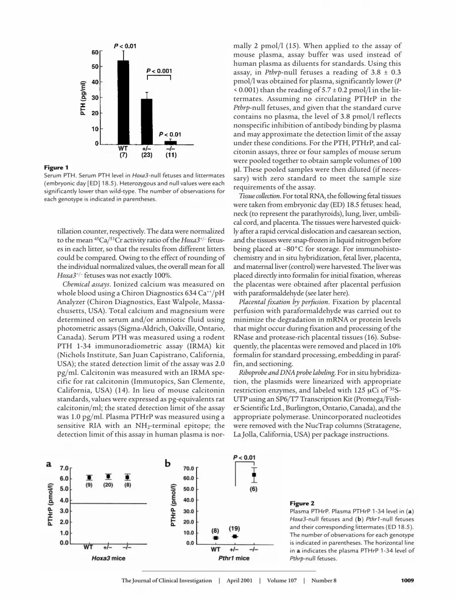
Figure 1Serum PTH. Serum PTH level in Hoxa3-null fetuses and littermates(embryonic day [ED] 18.5). Heterozygous and null values were eachsignificantly lower than wild-type. The number of observations foreach genotype is indicated in parentheses.
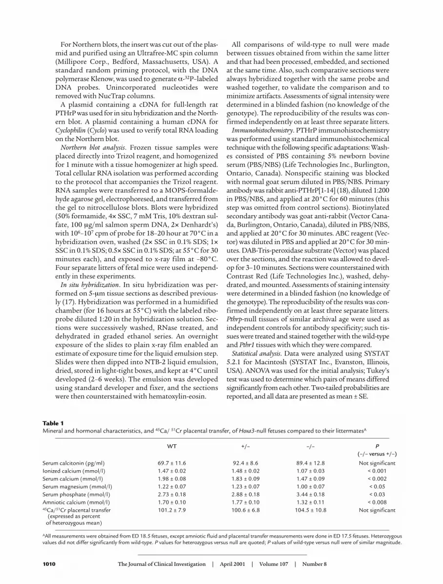
Figure 2Plasma PTHrP. Plasma PTHrP 1-34 level in (a)Hoxa3-null fetuses and (b) Pthr1-null fetusesand their corresponding littermates (ED 18.5).The number of observations for each genotypeis indicated in parentheses. The horizontal linein a indicates the plasma PTHrP 1-34 level ofPthrp-null fetuses.
For Northern blots, the insert was cut out of the plas-mid and purified using an Ultrafree-MC spin column(Millipore Corp., Bedford, Massachusetts, USA). Astandard random priming protocol, with the DNApolymerase Klenow, was used to generate α-32P–labeledDNA probes. Unincorporated nucleotides wereremoved with NucTrap columns.
A plasmid containing a cDNA for full-length ratPTHrP was used for in situ hybridization and the North-ern blot. A plasmid containing a human cDNA forCyclophilin (Cyclo) was used to verify total RNA loadingon the Northern blot.
Northern blot analysis. Frozen tissue samples wereplaced directly into Trizol reagent, and homogenizedfor 1 minute with a tissue homogenizer at high speed.Total cellular RNA isolation was performed accordingto the protocol that accompanies the Trizol reagent.RNA samples were transferred to a MOPS-formalde-hyde agarose gel, electrophoresed, and transferred fromthe gel to nitrocellulose blots. Blots were hybridized(50% formamide, 4× SSC, 7 mM Tris, 10% dextran sul-fate, 100 µg/ml salmon sperm DNA, 2× Denhardt’s)with 106–107 cpm of probe for 18–20 hour at 70°C in ahybridization oven, washed (2× SSC in 0.1% SDS; 1×SSC in 0.1% SDS; 0.5× SSC in 0.1% SDS; at 55°C for 30minutes each), and exposed to x-ray film at –80°C.Four separate litters of fetal mice were used independ-ently in these experiments.
In situ hybridization. In situ hybridization was per-formed on 5-µm tissue sections as described previous-ly (17). Hybridization was performed in a humidifiedchamber (for 16 hours at 55°C) with the labeled ribo-probe diluted 1:20 in the hybridization solution. Sec-tions were successively washed, RNase treated, anddehydrated in graded ethanol series. An overnightexposure of the slides to plain x-ray film enabled anestimate of exposure time for the liquid emulsion step.Slides were then dipped into NTB-2 liquid emulsion,dried, stored in light-tight boxes, and kept at 4°C untildeveloped (2–6 weeks). The emulsion was developedusing standard developer and fixer, and the sectionswere then counterstained with hematoxylin-eosin.
All comparisons of wild-type to null were madebetween tissues obtained from within the same litterand that had been processed, embedded, and sectionedat the same time. Also, such comparative sections werealways hybridized together with the same probe andwashed together, to validate the comparison and tominimize artifacts. Assessments of signal intensity weredetermined in a blinded fashion (no knowledge of thegenotype). The reproducibility of the results was con-firmed independently on at least three separate litters.
Immunohistochemistry. PTHrP immunohistochemistrywas performed using standard immunohistochemicaltechnique with the following specific adaptations: Wash-es consisted of PBS containing 5% newborn bovineserum (PBS/NBS) (Life Technologies Inc., Burlington,Ontario, Canada). Nonspecific staining was blockedwith normal goat serum diluted in PBS/NBS. Primaryantibody was rabbit anti-PTHrP[1-14] (18), diluted 1:200in PBS/NBS, and applied at 20°C for 60 minutes (thisstep was omitted from control sections). Biotinylatedsecondary antibody was goat anti-rabbit (Vector Cana-da, Burlington, Ontario, Canada), diluted in PBS/NBS,and applied at 20°C for 30 minutes. ABC reagent (Vec-tor) was diluted in PBS and applied at 20°C for 30 min-utes. DAB-Tris-peroxidase substrate (Vector) was placedover the sections, and the reaction was allowed to devel-op for 3–10 minutes. Sections were counterstained withContrast Red (Life Technologies Inc.), washed, dehy-drated, and mounted. Assessments of staining intensitywere determined in a blinded fashion (no knowledge ofthe genotype). The reproducibility of the results was con-firmed independently on at least three separate litters.Pthrp-null tissues of similar archival age were used asindependent controls for antibody specificity; such tis-sues were treated and stained together with the wild-typeand Pthr1 tissues with which they were compared.
Statistical analysis. Data were analyzed using SYSTAT5.2.1 for Macintosh (SYSTAT Inc., Evanston, Illinois,USA). ANOVA was used for the initial analysis; Tukey’stest was used to determine which pairs of means differedsignificantly from each other. Two-tailed probabilities arereported, and all data are presented as mean ± SE.
1010 The Journal of Clinical Investigation | April 2001 | Volume 107 | Number 8
Table 1Mineral and hormonal characteristics, and 45Ca/ 51Cr placental transfer, of Hoxa3-null fetuses compared to their littermatesA
WT +/– –/– P(–/– versus +/–)
Serum calcitonin (pg/ml) 69.7 ± 11.6 92.4 ± 8.6 89.4 ± 12.8 Not significantIonized calcium (mmol/l) 1.47 ± 0.02 1.48 ± 0.02 1.07 ± 0.03 < 0.001 Serum calcium (mmol/l) 1.98 ± 0.08 1.83 ± 0.09 1.47 ± 0.09 < 0.002Serum magnesium (mmol/l) 1.22 ± 0.07 1.23 ± 0.07 1.00 ± 0.07 < 0.05Serum phosphate (mmol/l) 2.73 ± 0.18 2.88 ± 0.18 3.44 ± 0.18 < 0.03Amniotic calcium (mmol/l) 1.70 ± 0.10 1.77 ± 0.10 1.32 ± 0.11 < 0.00845Ca/51Cr placental transfer 101.2 ± 7.9 100.6 ± 6.8 104.5 ± 10.8 Not significant
(expressed as percent of heterozygous mean)
AAll measurements were obtained from ED 18.5 fetuses, except amniotic fluid and placental transfer measurements were done in ED 17.5 fetuses. Heterozygousvalues did not differ significantly from wild-type. P values for heterozygous versus null are quoted; P values of wild-type versus null were of similar magnitude.
ResultsPTH and calcitonin secretion in Hoxa3 mutant fetuses. A pre-vious histological study of Hoxa3-null fetuses hadfailed to detect any recognizable parathyroid tissue andhad suggested that the thyroid tissue (including Ccells) was hypoplastic (12). To demonstrate that Hoxa3-null fetuses lack all parathyroid cells capable of secret-ing PTH, the serum PTH level was assayed. As seen inFigure 1, the serum PTH level was undetectable inHoxa3-null fetuses and was reduced in the heterozy-gotes compared with the wild-type. In contrast, assayof the serum calcitonin level showed no significant dif-ferences among the genotypes (Table 1). The normalcalcitonin levels in the Hoxa3-null fetuses make itunlikely that hypoplasia of the thyroidal C cells wouldhave a significant impact on calcium and bone metab-olism in these fetuses. These data demonstrated thatHoxa3-null fetuses lack serum PTH and, therefore,probably all parathyroid cells.
That the Hoxa3-null mice lack PTH is a distinguish-ing characteristic from the recently described Gcm2knockout mice, which also lack parathyroids but havenormal circulating PTH levels due to production ofPTH by the thymus (19). The absence of the parathy-roids and thymus in the Hoxa3-null mice explains thecomplete lack of serum PTH in this model. It is alsoapparent from these results that loss of one Hoxa3allele is sufficient to affect parathyroid hormone levels;this finding underscores the importance of Hoxa3 infetal parathyroid formation and function.
Fetal mineral metabolism in Hoxa3-null fetuses. Theaparathyroid Hoxa3-null fetuses had an ionized calci-um of 1.07 ± 0.03 mmol/l, which was significantlylower than the ionized calcium of the littermates (Table1). The maternal-fetal calcium gradient was obtainedby subtracting the maternal ionized calcium level (1.24± 0.02 mmol/l) from the fetal level. Although wild-typeand heterozygous fetuses had an average calcium gra-dient of +0.24 mmol/l, the gradient was reversed to–0.17 mmol/l in Hoxa3-null fetuses.
We previously reported that the ionized calcium ofPthrp-null fetuses was reduced to a level equal to thematernal ionized calcium, and that, as a consequence,the maternal-fetal calcium gradient was zero (3). In addi-
tion, Pthr1-null fetuses had an ionized calciumthat was lower than the maternal ionized cal-cium concentration, such that maternal-fetalcalcium gradient was reversed. Therefore, theionized calcium level of Hoxa3-null fetuses iscloser to that of Pthr1-null fetuses and sug-gests that the lower serum calcium cannot beexplained by loss of PTHrP alone.
The biochemical phenotype of the Hoxa3-null fetus-es was further characterized as shown in Table 1. Thetotal calcium level was also significantly lower inHoxa3-null fetuses, whereas the serum phosphate levelwas increased. The serum magnesium was reduced inparallel with the serum calcium level. Amniotic fluidwas collected as a surrogate for fetal urine, and the cal-cium content of amniotic fluid was found to be signif-icantly reduced in the Hoxa3-null fetuses. The lowamniotic fluid calcium content of the Hoxa3-null fetus-es is consistent with the reduced serum ionized calci-um and renal filtered load of calcium; in contrast,Pthrp-null fetuses (whose blood calcium is not as low asin Hoxa3-null) have unaltered amniotic calcium levels(data not shown). The raised serum phosphate is con-sistent with hypoparathyroidism and suggests that thefetal parathyroids may control the fetal serum phos-phate (although Pthrp-null mice also have similarlyraised serum phosphate levels).
The Journal of Clinical Investigation | April 2001 | Volume 107 | Number 8 1011
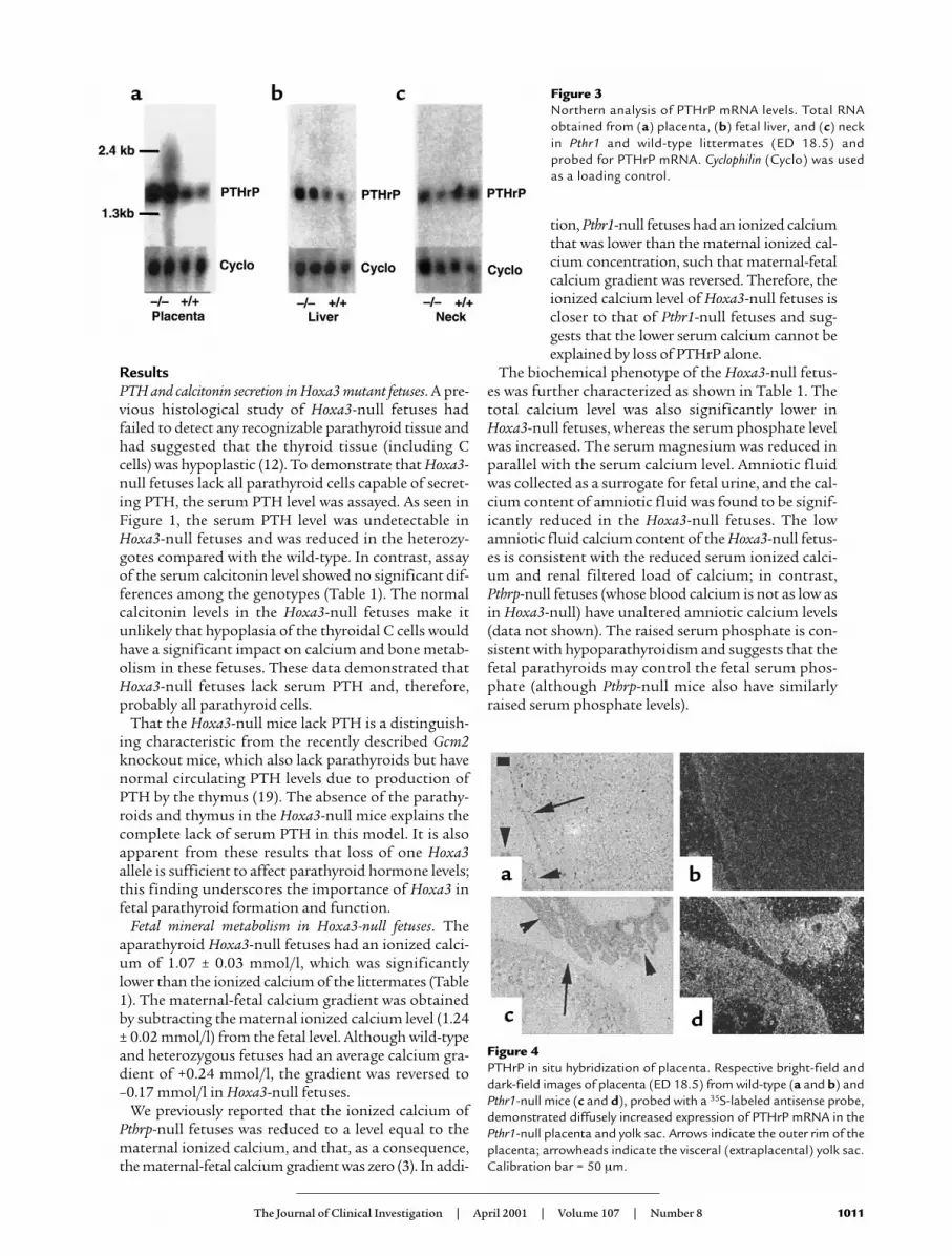
Figure 3Northern analysis of PTHrP mRNA levels. Total RNAobtained from (a) placenta, (b) fetal liver, and (c) neckin Pthr1 and wild-type littermates (ED 18.5) andprobed for PTHrP mRNA. Cyclophilin (Cyclo) was usedas a loading control.
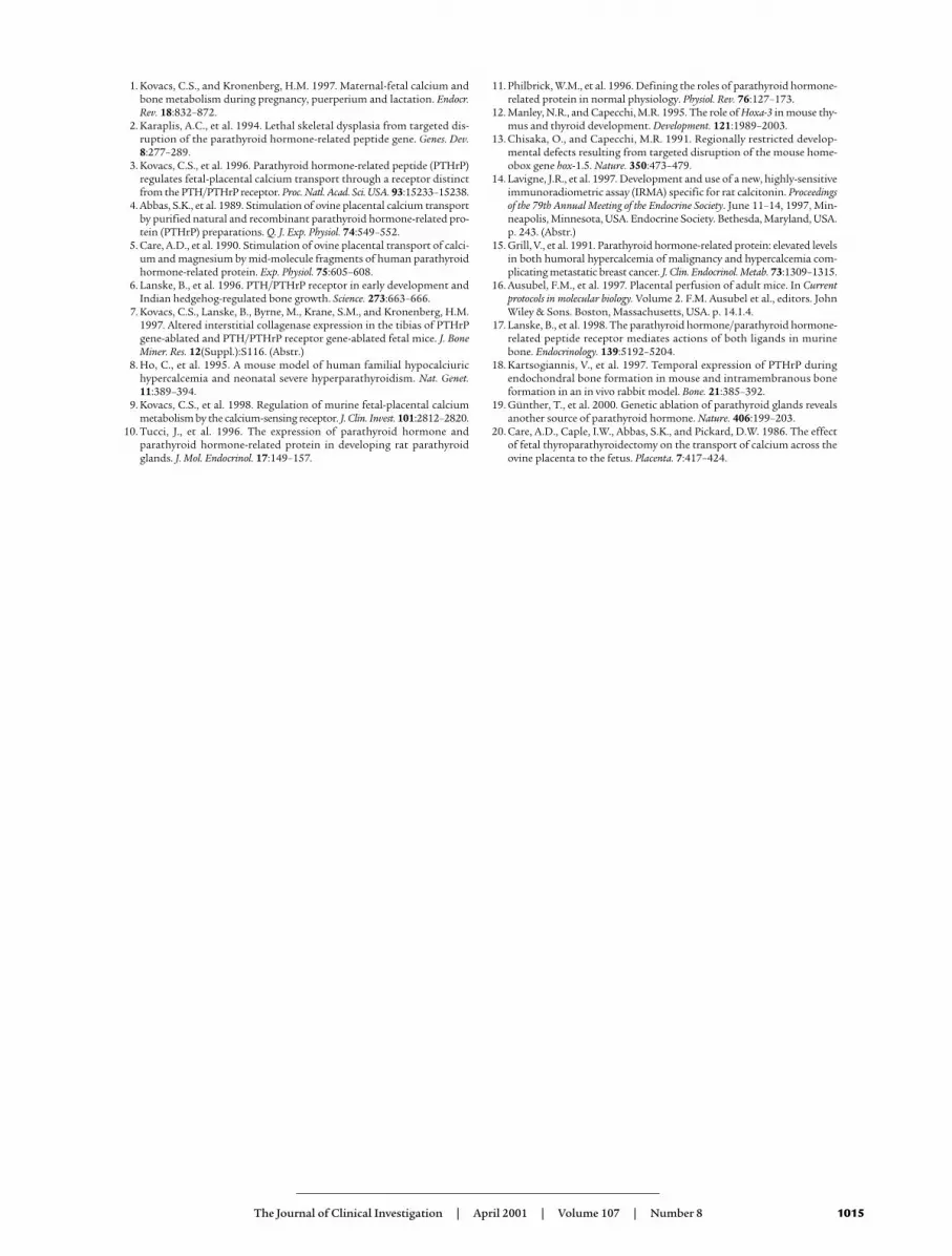
Figure 4PTHrP in situ hybridization of placenta. Respective bright-field anddark-field images of placenta (ED 18.5) from wild-type (a and b) andPthr1-null mice (c and d), probed with a 35S-labeled antisense probe,demonstrated diffusely increased expression of PTHrP mRNA in thePthr1-null placenta and yolk sac. Arrows indicate the outer rim of theplacenta; arrowheads indicate the visceral (extraplacental) yolk sac.Calibration bar = 50 µm.

Fetal mineral metabolism in Hoxa3-Casr double mutants.We previously reported that Casr-null fetuses are hyper-calcemic in utero with marked hyperparathyroidismand elevated PTH levels (9). Further, double-knockoutPthrp–/–Casr–/– fetuses had a higher ionized calciumthan the Pthrp-null alone (presumably due to hyper-parathyroidism as a consequence of the disruptedCasr). To validate further the hypothesis that Hoxa3-null fetuses lacked parathyroid cells and to demon-strate the role of PTH in mediating the phenotype ofthe Casr knockout mouse, double-knockoutHoxa3–/–Casr–/– fetuses were created. In this back-ground, wild-type fetuses had an ionized calcium of1.72 ± 0.18 mmol/l. By comparison, single mutantHoxa3–/– fetuses had an ionized calcium of 1.41 ± 0.08mmol/l, whereas that of Hoxa3–/–Casr+/– fetuses was1.43 ± 0.09 mmol/l, and of Hoxa3–/–Casr–/– doubleknockout fetuses was 1.40 ± 0.10 mmol/l (P = not sig-nificant). The ionized calcium of Hoxa3-null fetuseswas unaltered by a disrupted Casr, consistent with theabsence of parathyroids, and thereby the inability torespond to a Casr mutation.
Placental calcium transfer in Hoxa3 mutant. The lowerblood calcium of Hoxa3-null fetuses could result fromsuch diverse mechanisms as reduced placental calciumtransfer, decreased skeletal calcium resorption,increased mineral accretion by the skeleton, decreasedrenal tubular reabsorption of calcium, and absence of
the parathyroid calcium receptor. We examined the con-tribution of placental calcium transfer by intracardiacinjection of pregnant mice with 45Ca and 51Cr-EDTA (3).Using this technique, the placental calcium transfer ofHoxa3-null fetuses was no different from that of the het-erozygous and wt littermates (Table 1, bottom). There-fore, loss of fetal parathyroids did not affect placentalcalcium transfer in the Hoxa3-null fetuses.
Fetal plasma PTHrP in Hoxa3 and Pthr1 mutants.Because absence of the parathyroids in the Hoxa3-nullfetuses did not impair placental calcium transfer, theparathyroids, and PTH in particular, are not requiredfor maintenance of normal placental calcium transfer.If the parathyroids are an important source of PTHrPin the circulation, absence of the parathyroids mightalter the circulating level of PTHrP. To assess this, wemeasured PTHrP 1-34 in fetal plasma on day 18.5 ofgestation. Hoxa3-null fetuses had a plasma PTHrP levelthat was no different from that of the heterozygousand wild-type littermates (Figure 2a). Thus, loss of fetalparathyroids did not affect either the plasma PTHrP 1-34 level or the rate of placental calcium transfer.
To validate further the significance of the unalteredplasma PTHrP 1-34 level and the sensitivity of the assayunder these experimental conditions, we next turned tothe Pthr1 knockout model. We had previously observedthat Pthr1-null fetuses had a placental calcium transferrate that was upregulated to about 150% of the het-
1012 The Journal of Clinical Investigation | April 2001 | Volume 107 | Number 8
Figure 5PTHrP immunohistochemistry of placenta. Upper images (a–c) are from Pthrp-null, middle images (d–f) are from wild-type, and lower images(g–i) are from Pthr1-null placentas (ED 18.5). a, d, and g are control sections in which the primary antibody to PTHrP 1-34 was omitted.Pthrp-null sections are controls to demonstrate the antibody specificity. In h, PTHrP expression is higher in the labyrinthine (syncytial) tro-phoblasts (arrowheads) and spongiotrophoblasts (arrows) of Pthr1-null than in the wild-type (e); corresponding section of Pthrp-null (b)shows only nonspecific background. (i) Detail of the bilayered intraplacental yolk sac (IPYS) demonstrating PTHrP immunostaining in thecolumnar (visceral) cells of the IPYS (arrowheads), with less intense PTHrP immunostaining in the cuboidal (parietal) cells of the IPYS(arrows). Reichert’s membrane underlies the cuboidal cells in all sections. A corresponding section of wild-type IPYS (f) shows less intensePTHrP immunostaining. Calibration bars = 50 µm.

erozygous and wild-type value (3). We had speculatedthat this might reflect upregulation of PTHrP and itseffects on placental calcium transfer. Therefore, we nextmeasured plasma PTHrP 1-34 in Pthr1-null fetuses andfound the level to be 63.5 ± 2.9 pmol/l, a value 11-foldthat of heterozygous and wild-type littermates (Figure2b). Thus, increased placental calcium transfer in thePthr1-null fetus was associated with a marked increasein the plasma level of PTHrP; the decreased placentalcalcium transfer in the Pthrp-null was associated with areduction in plasma PTHrP; and the aparathyroid(PTH-less) Hoxa3-null had normal placental calciumtransfer and normal plasma PTHrP levels.
Tissue sources of PTHrP in the fetus. The striking increasein plasma PTHrP level of the Pthr1-null fetuses enableda focused search in this model to determine which tis-sue(s) might be overexpressing PTHrP and possibly con-tributing to the increased plasma level. The fetal parathy-roids were too small to be isolated, and thus a wholeneck section was used to represent the parathyroids. ByNorthern analysis of total RNA, a consistent increase inPTHrP mRNA was detected in placenta and liver of thePthr1-null fetuses (Figure 3), but not in neck (Figure 3)or in head, lung, and umbilical cord (data not shown).
Next, sections of liver and placenta were examined by insitu hybridization and immunohistochemistry to deter-mine which cell types were producing PTHrP. As seen inFigure 4, PTHrP mRNA was diffusely expressed in bothwild-type and Pthr1-null placenta, but the signal intensi-
ty was consistently increased in the Pthr1-null placenta.That this increase in mRNA resulted in increased pro-duction of protein was confirmed by immunohisto-chemistry, as seen in Figure 5. The peripheral and basalaspects of the placenta, which contains the spongiotro-phoblasts and giant trophoblasts, had more intensePTHrP immunoreactivity than the labyrinthine (syncy-tial) trophoblasts that make up the bulk of the murineplacenta (Figure 5e). This difference was maintained inthe Pthr1-null placenta, but with more intensive PTHrPimmunoreactivity in all three trophoblast cell types (com-pared with wild-type) (Figure 5, compare h with e).PTHrP immunoreactivity was also present in theintraplacental yolk sac (IPYS) of the wild-type and Pthr1-null placenta, more intensely in the columnar cells on thevisceral side of the IPYS, and most intensely in the Pthr1-null (Figure 5, compare i with f). In contrast to the find-ings in the Pthr1-null placenta, no difference in PTHrPexpression (mRNA or protein) was noted between wild-type and Hoxa3-null placentas (data not shown).
Examination of fetal liver showed the normal disarrayof proliferating hematopoietic cells and liver cells, withexpression of PTHrP in both lineages. PTHrP immunore-activity was markedly increased in hematopoietic andliver cells of the Pthr1-null liver sections (Figure 6, com-pare h and i with e and f). Thus, both placenta and liverwere demonstrated to have increased expression ofPTHrP (mRNA and protein) in the Pthr1-null fetus,which has increased circulating levels of PTHrP.
The Journal of Clinical Investigation | April 2001 | Volume 107 | Number 8 1013
Figure 6PTHrP immunohistochemistry of fetal liver. Upper images (a–c) are from Pthrp-null, middle images (d–f) are from wild-type, and lower images(g–i) are from Pthr1-null liver (ED 18.5). a, d, and g are control sections in which the primary antibody to PTHrP 1-34 was omitted. Pthrp-null sections are controls to demonstrate the antibody specificity. Normal extramedullary hematopoiesis and fetal liver cells are seen in allsections of fetal liver. Increased PTHrP immunoreactivity is demonstrated in liver and hematopoietic cells of Pthr1-null liver (h) comparedwith wild-type liver (e), especially in cells near central veins. Corresponding section of Pthrp-null (b) shows only nonspecific background. c,f, and i are higher power images of the same sections in b, e, and h, respectively. Calibration bars = 50 µm.

DiscussionThe present findings in aparathyroid Hoxa3-null fetus-es demonstrate that these mice are functionallyaparathyroid, as evidenced by absent PTH, reducedblood calcium, raised blood phosphate level, reducedserum magnesium, and reduced calcium in amnioticfluid. This confirms that the circulating level of PTHin fetal mice, though low, does have functional impor-tance in regulating fetal calcium and magnesiummetabolism. Also, the importance of Hoxa3 in parathy-roid gland development and function is underscoredby the finding that the circulating PTH level wasreduced in Hoxa3+/– fetuses. We have also found thatthe rate of placental calcium transfer and the plasmaPTHrP level are unaffected in the Hoxa3-null fetuses.This result shows that the parathyroids are notrequired for the normal regulation of placental calci-um transfer in mice, and that the parathyroids are nota major source of PTHrP in the circulation. Alterna-tively, it may be that other fetal tissue sources (liver, pla-centa) are compensating for the loss of PTHrP derivedfrom the parathyroids in these null mice.
That these aparathyroid Hoxa3-null fetuses havenormal placental calcium transfer contrasts with thepreviously reported finding of reduced placental cal-cium transfer in thyroparathyroidectomized fetallambs. This may reflect a species difference betweenmice and sheep, including differences in structureand function of the placentas (hemochorial versusepitheliochorial, respectively). Alternatively, it couldbe due to experimental design. The present studieswere carried out in intact fetal mice that lackedparathyroids from conception and whose motherswere heterozygous for the same genetic defect. Thestudies in fetal lambs involved a surgical procedureon the lambs, followed by later removal of the fetusand subsequent perfusion of the isolated placenta insitu (4, 20). Because plasma PTHrP measurementswere not carried out for these thyroparathyroidec-tomized lambs, it is not known whether the reduc-tion in placental calcium transfer was accompaniedby a reduction in plasma PTHrP.
A limitation of the PTHrP RIA is that it detects a por-tion of the NH2-terminal region of PTHrP (PTHrP 1-34). The size of circulating PTHrP could not be deter-mined, and yet it is likely that mid-molecular regionsof the molecule stimulate placental calcium transfer inboth mice and sheep. Given that the various fragmentsof PTHrP are thought to be derived from post-trans-lational processing of full-length PTHrP, an increaseor decrease in PTHrP 1-34 concentration might beaccompanied by a parallel change in the level of mid-molecular and COOH-terminal fragments of PTHrP.However, it is possible that the expression, metabolismand excretion of these fragments might be differen-tially regulated. Thus, a reduction in circulating mid-molecular PTHrP cannot be completely excluded inthe Hoxa3-null fetuses, as this was not specificallymeasured. The NH2-terminal assay was used because
it is sensitive and has been well-characterized. By con-trast, no well-characterized assays of mid-molecularPTHrP are available.
The in situ hybridization studies do not share this lim-itation since a cDNA for the entire PTHrP mRNA wasused. No downregulation of PTHrP mRNA expressionwas noted in the Hoxa3-null fetuses, whereas upregula-tion of PTHrP mRNA was found in specific tissues in thePthr1-null fetuses. Our findings indicate that PTHrPmRNA and protein expressed in placenta and liver areupregulated in response to loss of the PTH/PTHrPreceptor. These findings suggest that liver and placentamight be at least partly responsible for the increased cir-culating level of PTHrP in the Pthr1-null mice. It remainsto be determined whether these tissues contribute to thecirculating PTHrP level in normal circumstances.
In summary, our findings demonstrate that aparathy-roid Hoxa3-null fetuses have absent PTH, reduced ion-ized calcium and serum magnesium, and raised serumphosphate, but normal plasma PTHrP and normal rateof placental calcium transfer. In contrast, Pthr1-nullfetuses, which have an increased rate of placental calci-um transfer, also have an 11-fold increase in circulatingPTHrP level. Both liver and placenta (but not the neckor parathyroids) show increased expression of PTHrPmRNA and protein. The results indicate that the fetalparathyroids are required for maintenance of normalserum calcium, magnesium, and phosphate homeosta-sis, but are not required for maintenance of normal pla-cental calcium transfer. Presumably, PTH acts on fetalbone and perhaps kidney to raise blood calcium. Fur-thermore, the parathyroids do not appear to contributesignificantly to the circulating PTHrP level of fetuses.Instead, other fetal tissue sources, especially placentaand liver, may be more important for regulating fetalcalcium homeostasis than previously suspected.
It is now apparent that both PTHrP and PTH areimportant for normal fetal calcium homeostasis, andthat lack of either of these molecules will lead to dis-ruptions in calcium metabolism (such as hypocalcemiaand hyperphosphatemia), whereas only lack of PTHrPwill reduce the rate of placental calcium transfer.
AcknowledgmentsThe authors thank Anthony D. Care for helpful dis-cussions and suggestions. The technical assistance ofLinda L. Chafe, Mandy L. Woodland, Kirsten R.McDonald, and Neva Fudge (Memorial University ofNewfoundland), and Patricia Ho (University of Mel-bourne) is acknowledged. This work was supported byan NIH grant (DK-47038 to H.M. Kronenberg) and bya Fellowship award, a Scholarship award (MSH 35674),and an operating grant (MT-15439), all from the Med-ical Research Council of Canada to C.S. Kovacs. Addi-tional research support (to C.S. Kovacs) was obtainedfrom the Medical Research Foundation, the Researchand Development Committee, the Faculty of Medicine,and the Discipline of Medicine, all at Memorial Uni-versity of Newfoundland.
1014 The Journal of Clinical Investigation | April 2001 | Volume 107 | Number 8
1. Kovacs, C.S., and Kronenberg, H.M. 1997. Maternal-fetal calcium andbone metabolism during pregnancy, puerperium and lactation. Endocr.Rev. 18:832–872.
2. Karaplis, A.C., et al. 1994. Lethal skeletal dysplasia from targeted dis-ruption of the parathyroid hormone-related peptide gene. Genes. Dev.8:277–289.
3. Kovacs, C.S., et al. 1996. Parathyroid hormone-related peptide (PTHrP)regulates fetal-placental calcium transport through a receptor distinctfrom the PTH/PTHrP receptor. Proc. Natl. Acad. Sci. USA. 93:15233–15238.
4. Abbas, S.K., et al. 1989. Stimulation of ovine placental calcium transportby purified natural and recombinant parathyroid hormone-related pro-tein (PTHrP) preparations. Q. J. Exp. Physiol. 74:549–552.
5. Care, A.D., et al. 1990. Stimulation of ovine placental transport of calci-um and magnesium by mid-molecule fragments of human parathyroidhormone-related protein. Exp. Physiol. 75:605–608.
6. Lanske, B., et al. 1996. PTH/PTHrP receptor in early development andIndian hedgehog-regulated bone growth. Science. 273:663–666.
7. Kovacs, C.S., Lanske, B., Byrne, M., Krane, S.M., and Kronenberg, H.M.1997. Altered interstitial collagenase expression in the tibias of PTHrPgene-ablated and PTH/PTHrP receptor gene-ablated fetal mice. J. BoneMiner. Res. 12(Suppl.):S116. (Abstr.)
8. Ho, C., et al. 1995. A mouse model of human familial hypocalciurichypercalcemia and neonatal severe hyperparathyroidism. Nat. Genet.11:389–394.
9. Kovacs, C.S., et al. 1998. Regulation of murine fetal-placental calciummetabolism by the calcium-sensing receptor. J. Clin. Invest. 101:2812–2820.
10. Tucci, J., et al. 1996. The expression of parathyroid hormone andparathyroid hormone-related protein in developing rat parathyroidglands. J. Mol. Endocrinol. 17:149–157.
11. Philbrick, W.M., et al. 1996. Defining the roles of parathyroid hormone-related protein in normal physiology. Physiol. Rev. 76:127–173.
12. Manley, N.R., and Capecchi, M.R. 1995. The role of Hoxa-3 in mouse thy-mus and thyroid development. Development. 121:1989–2003.
13. Chisaka, O., and Capecchi, M.R. 1991. Regionally restricted develop-mental defects resulting from targeted disruption of the mouse home-obox gene hox-1.5. Nature. 350:473–479.
14. Lavigne, J.R., et al. 1997. Development and use of a new, highly-sensitiveimmunoradiometric assay (IRMA) specific for rat calcitonin. Proceedingsof the 79th Annual Meeting of the Endocrine Society. June 11–14, 1997, Min-neapolis, Minnesota, USA. Endocrine Society. Bethesda, Maryland, USA.p. 243. (Abstr.)
15. Grill, V., et al. 1991. Parathyroid hormone-related protein: elevated levelsin both humoral hypercalcemia of malignancy and hypercalcemia com-plicating metastatic breast cancer. J. Clin. Endocrinol. Metab. 73:1309–1315.
16. Ausubel, F.M., et al. 1997. Placental perfusion of adult mice. In Currentprotocols in molecular biology. Volume 2. F.M. Ausubel et al., editors. JohnWiley & Sons. Boston, Massachusetts, USA. p. 14.1.4.
17. Lanske, B., et al. 1998. The parathyroid hormone/parathyroid hormone-related peptide receptor mediates actions of both ligands in murinebone. Endocrinology. 139:5192–5204.
18. Kartsogiannis, V., et al. 1997. Temporal expression of PTHrP duringendochondral bone formation in mouse and intramembranous boneformation in an in vivo rabbit model. Bone. 21:385–392.
19. Günther, T., et al. 2000. Genetic ablation of parathyroid glands revealsanother source of parathyroid hormone. Nature. 406:199–203.
20. Care, A.D., Caple, I.W., Abbas, S.K., and Pickard, D.W. 1986. The effectof fetal thyroparathyroidectomy on the transport of calcium across theovine placenta to the fetus. Placenta. 7:417–424.
The Journal of Clinical Investigation | April 2001 | Volume 107 | Number 8 1015


