Embed Size (px)
Citation preview
FGF14 Regulates the Intrinsic Excitability of Cerebellar PurkinjeNeurons
Vikram G. Shakkottai1,*, Maolei Xiao2,*, Lin Xu1, Michael Wong1,3, Jeanne M. Nerbonne2,3,David M. Ornitz2,3, and Kelvin A. Yamada1,3
1 Department of Neurology, Washington University School of Medicine, St. Louis, MO 63110
2 Developmental Biology, Washington University School of Medicine, St. Louis, MO 63110
3 Hope Center for Neurological Disorders, Washington University School of Medicine, St. Louis, MO 63110
AbstractA missense mutation in the fibroblast growth factor 14 (FGF14) gene underlies SCA27, an autosomaldominant spinocerebellar ataxia in humans. Mice with a targeted disruption of the Fgf14 locus(Fgf14−/−) develop ataxia resembling human SCA27. We tested the hypothesis that loss of FGF14affects the firing properties of Purkinje neurons, which play an important role in motor control andcoordination. Current clamp recordings from Purkinje neurons in cerebellar slices revealedattenuated spontaneous firing in Fgf14−/− neurons. Unlike in the wild type animals, more than 80%of Fgf14−/− Purkinje neurons were quiescent and failed to fire repetitively in response to depolarizingcurrent injections. Immunohistochemical examination revealed reduced expression of Nav1.6protein in Fgf14−/− Purkinje neurons. Together, these observations suggest that FGF14 is requiredfor normal Nav1.6 expression in Purkinje neurons, and that the loss of FGF14 impairs spontaneousand repetitive firing in Purkinje neurons by altering the expression of Nav1.6 channels.
KeywordsSpinocerebellar ataxia; Intracellular fibroblast growth factor 14 (iFGF14); Purkinje neurons; Nav1.6; SCA27
IntroductionThe autosomal dominant spinocerebellar ataxias (SCAs) are heterogeneous neurologicaldisorders characterized by progressive cerebellar ataxia that also often present with paroxysmaldyskinesia, tremor, abnormal eye movements, and/or cognitive impairment (Schols et al.,2004; Taroni and DiDonato, 2004; Zoghbi, 2000; Zoghbi and Orr, 2000). The unexpectedfindings of ataxia, dystonia and tremor in mice with a targeted disruption in the Fgf14 locus(Wang et al., 2002) suggested a role for FGF14 in regulation of motor function, and led directlyto the identification of a mutation in FGF14 in a family with a new type of progressivespinocerebellar ataxia now referred to as SCA27 (Van Swieten et al., 2003). Affected
Correspondence should be addressed to Kelvin Yamada, Department of Neurology, Box 8111, Washington University School ofMedicine, 660 South Euclid Avenue, St. Louis, MO 63110. Email: [email protected].*Vikram Shakkottai and Maolei Xiao contributed equally to this work.Publisher's Disclaimer: This is a PDF file of an unedited manuscript that has been accepted for publication. As a service to our customerswe are providing this early version of the manuscript. The manuscript will undergo copyediting, typesetting, and review of the resultingproof before it is published in its final citable form. Please note that during the production process errors may be discovered which couldaffect the content, and all legal disclaimers that apply to the journal pertain.
NIH Public AccessAuthor ManuscriptNeurobiol Dis. Author manuscript; available in PMC 2010 January 1.
Published in final edited form as:Neurobiol Dis. 2009 January ; 33(1): 81–88. doi:10.1016/j.nbd.2008.09.019.
NIH
-PA Author Manuscript
NIH
-PA Author Manuscript
NIH
-PA Author Manuscript
individuals in this family possess a missense mutation (F145S) in the FGF14 gene onchromosome 13q34 (Brusse et al., 2005; Van Swieten et al., 2003). Interestingly, a single basepair deletion, leading to a frameshift mutation (D163fsX12) in FGF14 has also been identifiedin an individual patient presenting with ataxia and mild mental retardation (Dalski et al.,2005; Soong and Paulson, 2007).
FGF14 belongs to the intracellular fibroblast growth factor subfamily (iFGF) that also includesFGFs 11–14 (Itoh and Ornitz, 2008), proteins that are widely expressed in the nervous system(Smallwood et al., 1996; Wang et al., 2000; Yamamoto et al., 1998). Unlike the other membersof the FGF family, iFGFs are not secreted and do not interact with classical tyrosine kinaseFGF receptors (Itoh and Ornitz, 2008; Olsen et al., 2003; Ornitz and Itoh, 2001; Smallwoodet al., 1996). Members of the iFGF subfamily, however, have been shown to colocalize withvoltage-gated sodium (Nav) channels, to interact with the C-termini of Nav channel pore-forming (α) subunits (Goldfarb et al., 2007; Laezza et al., 2007; Liu et al., 2001; Liu et al.,2003; Lou et al., 2005; Wittmack et al., 2004), and to modulate hippocampal and granule cellexcitability (Goldfarb et al., 2007; Laezza et al., 2007; Wozniak et al., 2007; Xiao et al.,2007).
Purkinje neurons are the sole output of the cerebellum, critical for motor regulation andcoordination (Burgess et al., 1995; Grusser-Cornehls and Baurle, 2001; Koeppen, 2005;Kohrman et al., 1996; Levin et al., 2006; Sausbier et al., 2004; Trudeau et al., 2006). Adistinctive feature of mature Purkinje neurons is the robust expression of Nav1.6-encodedsodium channels (Afshari et al., 2004; Krzemien et al., 2000; Raman et al., 1997), the channelthat underlies the “resurgent” sodium current that is critical for sustaining the characteristichigh frequency firing of these cells (Raman et al., 1997). Mutations in human SCN8A, whichencodes Nav1.6, are associated with cerebellar atrophy and ataxia (Trudeau et al., 2006). Inaddition, mice in which Scn8a has been disrupted or that lack Nav1.6 specifically in Purkinjecells display cerebellar ataxia and impaired Purkinje cell firing (Kohrman et al., 1996; Levinet al., 2006; Raman et al., 1997). These observations suggested that the ataxia in SCA27individuals and in Fgf14−/− mice reflects impaired Purkinje cell firing due to alterations in Navchannel expression and/or functioning. The experiments here were designed to explore thishypothesis directly.
Materials and MethodsCerebellar slice recordings
Whole-cell recordings were obtained from Purkinje neurons in 300 μm parasagittal cerebellarslices prepared from 25–30 day old wild type (WT) and Fgf14−/−C57BL6 mice. Vibratomesections were cut in ice-cold solution containing (in mM): 87 NaCl, 2.5 KCl, 25 NaHCO3, 1NaH2PO4, 0.5 CaCl2, 7 MgCl2, 75 sucrose and 10 glucose, bubbled with 5% CO2/95% O2.Slices were incubated at room temperature in artificial CSF (ACSF), containing in mM: 125NaCl, 2.5 KCl, 26 NaHCO3, 1.25 NaH2PO4, 2 CaCl2, 1 MgCl2 and 10 glucose, bubbled with5% CO2/95% O2.
Purkinje neurons were visualized with infrared differential interference contrast (IR-DIC)optics on a Nikon upright microscope. Borosilicate glass patch pipettes (with resistances of 3–6 MΩ) were filled with internal recording solution containing (in mM): 140 K Gluconate, 2MgCl2, 1 CaCl2, 10 EGTA, 2 MgATP, 10 HEPES. Whole-cell recordings were made in ACSFat room temperature 1–5 hours after slice preparation using an Axopatch 200B amplifier,Digidata 1322A interface and pClamp-9 software (Molecular Devices, Union City, CA, USA).Although non-bridge amplifiers can distort action potentials (Magistretri et al., 1996), studiesutilizing the Axopatch amplifier showed no significant distortion of Purkinje neuron actionpotentials (Edgerton and Reinhart, 2003). Series resistance was monitored but not
Shakkottai et al. Page 2
Neurobiol Dis. Author manuscript; available in PMC 2010 January 1.
NIH
-PA Author Manuscript
NIH
-PA Author Manuscript
NIH
-PA Author Manuscript
compensated; cells were rejected if the series resistance exceeded 30 MΩ. Firing propertieswere examined in current clamp mode, and excitatory postsynaptic currents (EPSCs) wererecorded in voltage-clamp mode at a holding potential of −70 mV. EPSCs from selectedPurkinje neurons were evoked by applying square wave current pulses via a tungsten bipolarelectrode to the molecular layer ~100 μm from the Purkinje cell of interest. In someexperiments, 5 μM 6,7-dinitroquinoxaline-2,3-dione (DNQX), 10 μM ± 3,3-(2-carboxypiperazine-4-yl)-propyl-1-phosphate (CPP) and 100 μM picrotoxin were included inthe ACSF. Analog current and voltage traces were digitized at 10 kHz.
HistologyWT and Fgf14−/− C57BL6 mice were anesthetized with pentobarbital (60mg/kg,i.p.),transcardially perfused with a vascular rinse containing 0.9% NaCl, followed by ice-cold 4%paraformaldehyde in 0.1 M phosphate buffer (pH 7.4). Brains were dissected, postfixed in thesame solution overnight at 4°C, and cryoprotected in 30% sucrose in 0.1 M phosphate bufferedsaline (PBS). After embedding in O. C. T. (Canemco and Morivac, Quebec), 14 μm serialsagittal cryostat sections were collected in PBS.
For immunohistochemistry, free-floating tissue sections were washed in PBS and subsequentlyblocked in a solution containing 7.5% goat serum (Sigma) and 0.25% Triton X-100 (TX-100;Sigma) in PBS. Sections were then incubated at 4°C for 15 hr with a mouse anti-FGF14 oranti-Nav 1.6 (NeuroMab) monoclonal antibody diluted 1:1000, or with a polyclonal anti-calbindin (0–28K) antibody (Swant, Switzerland), diluted 1:5000, in PBS containing 1% goatserum and 0.25% TX-100. Sections were washed three times for 5 minutes at room temperaturein PBS and subsequently incubated with an Alexa 488-conjugated goat anti-mouse or an Alexa594 goat anti-rabbit secondary antibody (Molecular Probes), diluted 1:100, in PBS containing1% goat serum and 0.25% TX-100. After rinsing in PBS, sections were mounted and examinedusing a Zeiss Axioskop 40 or Apo Tome microscope.
For in situ hybridization, a 550 bp Fgf14 specific RNA probe (Wang et al., 2000) was labeledwith digoxigenin following the manufacturer’s protocol (Roche Diagnostics, IN). Free-floatingbrain sections (30 μm) were washed twice in PBS, and treated with freshly prepared 10μg/mlproteinase K (Invitrogen, Carlsbad, CA) at 37°C. After acetylation, sections were incubatedin hybridization buffer containing 0.2 μg/ml of the digoxigenin-labeled Fgf14 riboprobe at 43°C overnight. Hybridized sections were washed by successive immersions in 4x sodium chloridecitrate buffer (SCC; containing 150 mM NaCl, 15 mM sodium citrate; pH 7.0) at roomtemperature; 2x SCC containing 50% formamide at 50°C for 30 min); 2x SCC at 37°C for 10min; 2x SCC, containing 20 μg/ml RNase A, at 37°C for 30 min; 2x SCC at 37°C for 20 min;and 0.1x SCC at room temperature for 10 min. Controls hybridized without primary probeshowed no signal. The hybridization signals were detected with digoxigenin detection reagents(Roche Diagnostics, Indianapolis, IN). After rinsing in PBS, sections were mounted andanalyzed with a Zeiss Axioskop microscope.
Data Analyses and StatisticsCurrent and voltage-clamp data were compiled and analyzed using Clampfit Version 9 (Axon)and Origin 7 (Origin Lab, Northhampton, MA). All data are presented as means ± SEM.Statistical differences between WT and Fgf14−/− neurons were examined using the Studentst test; where appropriate, p values are reported in the text.
Shakkottai et al. Page 3
Neurobiol Dis. Author manuscript; available in PMC 2010 January 1.
NIH
-PA Author Manuscript
NIH
-PA Author Manuscript
NIH
-PA Author Manuscript
ResultsExpression of Fgf14 in Purkinje Neurons
Although it has previously been reported that Fgf14 expression is evident in the granule celllayer in the (mouse) cerebellum, the resolution was insufficient to identify expression inspecific cell types (Wang et al., 2002). To determine if Fgf14 is expressed in Purkinje neurons,high resolution in situ hybridization was performed on thick sections of wild type (WT) mousecerebellum. These experiments revealed robust expression of the Fgf14 transcript in Purkinjeneurons and moderate expression in granule cells, as well as in other interneurons of the granulecell layer (Figure 1A). In addition, immunohistochemical analysis revealed robust expressionof the FGF14 protein in WT Purkinje and granule cells (Figure 1B), whereas FGF14 isundetectable in Fgf14−/− cerebellum (Figure 1B).
Lack of Spontaneous and Tonic Firing in Fgf14−/− Purkinje NeuronsThe finding of robust expression of FGF14 in Purkinje neurons suggested that this proteinmight play a role in regulating the excitability of these cells and that the loss of FGF14 functionin these cells could contribute to the ataxic phenotype observed in Fgf14−/− mice and humanswith FGF14 mutations. To explore this hypothesis directly, whole-cell recordings wereobtained from Purkinje neurons in acute parasagittal cerebellar slices prepared from 25–30 dayold WT and Fgf14−/− mice. As reported previously (Raman et al., 1997), WT Purkinje neuronsare characterized by spontaneous repetitive firing (Figure 2). Most (10/14; 71%) of the WTPurkinje neurons examined displayed sustained tonic firing (Figure 2A). Three (of 14, 21%)of the cells fired episodic bursts of action potentials followed by quiescent periods (Figure 2B).One of the (14, 7 %) WT Purkinje neurons studied was quiescent and did not displayspontaneous tonic or burst firing. In marked contrast with WT Purkinje neurons, most (14 of17, 82%) of the Fgf14−/− Purkinje neurons did not fire spontaneously and had a restingmembrane potential of −60.1 + 1.1 mV (Figure 2C). Three (3 of 17, 18%)) of the Fgf14−/−
Purkinje neurons exhibited sustained tonic firing (Figure 2D), and the firing properties of thesecells were indistinguishable from tonic firing WT cells (Figure 2A). The main differencebetween the WT and Fgf14−/− Purkinje neurons, therefore, appeared to be the relativeproportions of spontaneously active and “silent” Purkinje neurons (Figure 2D).
In WT Purkinje neurons that display sustained tonic firing, small hyperpolarizing currentinjections stopped repetitive firing (Figure 3A), and injections of depolarizing currents inducedhigher frequency sustained repetitive firing (Figure 3B,C). In the WT Purkinje neurons withintermittent bursting and in the single “silent” WT cell, small depolarizing current injectionsconverted these cells to sustained tonic firing. All WT Purkinje neurons, therefore, eitherdisplayed tonic firing at rest or could be converted to a tonic firing pattern by depolarizingcurrent injections. In addition, the frequency of repetitive firing in WT cells increased whenthe amplitudes of the depolarizing current injections were increased (Figure 3B, C). The mean± SEM (n = 14) maximal firing frequency of WT Purkinje neurons recorded in response to 300pA current injections was 58 ± 6 Hz (Figure 3F).
In Fgf14−/− Purkinje neurons, the responses to depolarizing current injections were quitedistinct. Prolonged injections of depolarizing currents into “silent” Fgf14−/− Purkinje neuronsresulted in the firing of one or a few action potentials (Figure 3D, E), but these cells did notsustain tonic repetitive firing regardless of the amplitudes of the injected currents. The mean± SEM current threshold to evoke a spike in these “silent” Fgf14−/− cells was 160 ± 30 pA (n= 14). The properties of the small subset (3 of 17) of Fgf14−/− Purkinje neurons that displayedspontaneous tonic repetitive firing were similar to WT Purkinje neurons in that the firingfrequencies of these cells also increased when the amplitudes of the depolarizing currentinjections were increased, and the range of repetitive firing frequencies was similar to that
Shakkottai et al. Page 4
Neurobiol Dis. Author manuscript; available in PMC 2010 January 1.
NIH
-PA Author Manuscript
NIH
-PA Author Manuscript
NIH
-PA Author Manuscript
determined in WT cells (Figure 3F). The maximal firing frequency in the non-silentFgf14−/− Purkinje neurons was 47 ± 24 Hz (Figure 3F).
Individual Action Potential Waveforms are Similar in WT and Fgf14−/− Purkinje NeuronsExamination of the waveforms and the properties of individual action potentials in WT andFgf14−/− Purkinje neurons revealed no apparent differences (Figure 4 A, B). The voltagetrajectories (dV/dt) versus time (Figure 4 C, D), as well as phase plots of dV/dt versus voltage(Figure 4 E, F), of individual action potentials in WT and Fgf14−/− Purkinje neurons weresimilar. There were also no significant differences in the voltage thresholds for action potentialgeneration (Figure 4G) or in the durations of individual action potentials in WT andFgf14−/− Purkinje neurons (Figure 4H). In addition, the input resistances of WT andFgf14−/− Purkinje neurons were not significantly different (Figure 4H).
Evoked EPSCs are Indistinguishable in WT and Fgf14−/− Purkinje NeuronsDecreased excitability and impaired ability to sustain repetitive firing in response todepolarizing current injections was recently reported in Fgf14−/− cerebellar granule cells(Goldfarb et al., 2007). It was also reported in this study that the properties of voltage-gated(Nav) currents in Fgf14−/− cerebellar granule cells are altered. These observations might beinterpreted to suggest the possibility that the firing properties of Fgf14−/− Purkinje neurons arealtered indirectly due to reduced excitatory inputs from Fgf14−/− granule cells. To explore thishypothesis directly, paired excitatory postsynaptic currents (EPSCs) were evoked in WT andFgf14−/− Purkinje neurons by parallel fiber stimulation; stimuli were presented at an intervalof 50 ms. The amplitudes of the first and the second EPSCs evoked were examined and theextent of paired pulse facilitation was determined. As illustrated in Figure 5, the amplitudes ofthe evoked EPSCs, as well as the paired pulse facilitation ratios (PPFR), in WT (n = 7) andFgf14−/− (n = 14) Purkinje neurons were not significantly different (Figure 5C). To furtherexamine whether altered granule cell synaptic input could contribute to reduced Purkinje firing,recordings were obtained from Fgf14−/− Purkinje neurons in the presence of 100 μMPicrotoxin, 10 μM ± CPP, and 5 μM DNQX to block GABA, NMDA and AMPA/Kainatepostsynaptic receptors, respectively. The resting and active membrane properties of Fgf14−/−
Purkinje neurons (n = 3) examined in the presence and absence of these blockers of postsynapticreceptors were indistinguishable (data not shown). The findings here that (most) Fgf14−/−
Purkinje neurons lack the ability to sustain tonic repetitive firing, therefore, does not appear toreflect altered synaptic inputs but rather reflects alterations in the intrinsic membrane propertiesof these cells.
Nav1.6 Expression is Reduced in Fgf14−/− Purkinje NeuronsPrevious studies in heterologous cells and in isolated hippocampal neurons in vitro haverevealed that FGF14 affects the densities and the biophysical properties of Nav channels(Laezza et al., 2007; Liu et al., 2001; Liu et al., 2003; Lou et al., 2005; Wittmack et al.,2004). The observations here that repetitive firing is attenuated in Fgf14−/− Purkinje neurons(Figures 2 and 3), but that action potential thresholds and waveforms in Fgf14−/− and WTPurkinje neurons are similar (Figure 4), suggested that the loss of FGF14 likely reflects reducedexpression (rather than alterations in the properties) of Nav1.6 channels which underlie thehigh frequency firing characteristic of these cells (Afshari et al., 2004; Raman et al., 1997).Interestingly, it has been reported that the selective elimination of Nav 1.6 in Purkinje neuronsproduces a phenotype (Levin et al., 2006) that is remarkably similar to that observed here inFgf14−/− Purkinje neurons. To determine the functional consequences of the loss of FGF14 onNav1.6 expression, immunohistochemical experiments were performed with a specific anti-Nav1.6 antibody. In addition to morphological features, Purkinje cells were identified byCalbindin expression. As illustrated in Figure 6, although these experiments revealed robust
Shakkottai et al. Page 5
Neurobiol Dis. Author manuscript; available in PMC 2010 January 1.
NIH
-PA Author Manuscript
NIH
-PA Author Manuscript
NIH
-PA Author Manuscript
expression of Nav1.6 in the cell bodies and processes of calbindin-positive WT Purkinjeneurons, Nav 1.6 expression was reduced in Fgf14−/− Purkinje neurons. Calbindin staining inWT and Fgf14−/− Purkinje neurons, in contrast, was similar (Figure 6). These observationssuggest the interesting hypothesis that, in the absence of FGF14, Nav1.6 expression is reducedin Purkinje neurons, which results in impaired firing in these cells and underlies the ataxicphenotype that is prominent in Fgf14−/− mice.
DiscussionSpontaneous and Evoked Repetitive Firing are Attenuated in Fgf14−/− Purkinje Neurons
Purkinje neurons are the sole output of the cerebellar cortex, and Purkinje cell dysfunction ordegeneration has been linked to cerebellar ataxias in human and in animal models (Burgess etal., 1995; Grusser-Cornehls and Baurle, 2001; Koeppen, 2005; Kohrman et al., 1996; Levin etal., 2006; Sausbier et al., 2004; Trudeau et al., 2006). Mature Purkinje neurons expresspredominantly Nav 1.6-encoded Nav channels, which generate resurgent Nav currents duringaction potential repolarization, enabling sustained, high frequency repetitive firing (Afshari etal., 2004; Raman et al., 1997). Mice (Scn8atg) in which the gene (Scn8a) encoding Nav1.6 hasbeen disrupted throughout the nervous system develop early onset ataxia with tremor, andcellular electrophysiological studies revealed impaired repetitive firing in Purkinje neurons(Raman et al., 1997). In addition, mice (Scn8a-KO) harboring a Purkinje neuron specificdisruption of Scn8a display a mildly ataxic gait (Levin et al., 2006) that resembles that observedin mice with Purkinje cell degeneration (Grusser-Cornehls and Baurle, 2001). In contrast, nomotor impairment was evident in mice harboring granule cell specific deletion of Scn8a (Levinet al., 2006). Taken together, these observations suggest that the disruption of Nav1.6 functionin Purkinje neurons cells has greater impact on motor control and coordination compared toNav1.6 disruption in granule cells. Purkinje neurons of Scn8a-KO and Fgf14−/− mice exhibitimpaired spontaneous tonic repetitive firing even in response to depolarizing current injections.The loss of Nav1.6 in Purkinje neurons in combination with the previously described alterationin granule cell physiology (Goldfarb et al., 2007) probably accounts for the ataxic phenotypeof the Fgf14−/− mice similar to mice with a knockout of Scn8a in both Purkinje and granulecells. Although a majority of Fgf14−/− Purkinje neurons exhibited impaired firing, a smallsubset of neurons retained the pattern of sustained tonic firing seen in the WT neurons. It haspreviously been demonstrated that the expression of Nav channel isoforms changes duringdevelopment with Nav1.6 being the major Nav channel expressed in mature Purkinje neurons(Shah et al., 2001). The observations here may be related to having performed experimentsduring a phase of development in which some Purkinje neurons retained a more immaturepattern of Nav channel expression, Alternatively, the phenotypic differences in cell propertiesmay represent heterogeneity in patterns of Nav channel expression (Shah et al., 2001), orpossibly heterogeneities in FGF14 expression, in different areas of the cerebellum.
These combined observations also support the hypothesis that the attenuation of Purkinjeneuron repetitive firing contributes importantly to the ataxic phenotype seen in Fgf14−/− mice,as well as in SCA27 afflicted individuals, harboring the FGF14F145S mutation, which wasshown recently to act as a dominant negative that disrupts the function of wild type FGF14(Laezza et al., 2007). It is tempting to further speculate that the ataxia phenotype in humanswith a deletion mutation (D163fsx12) that produces a truncated FGF14 protein (Brusse et al.,2005), may also result from alterations in the firing properties of Purkinje neurons. Furtherexperiments aimed at exploring this hypothesis directly will be of considerable interest.
Nav1.6 Expression is Reduced in Fgf14−/− Purkinje NeuronsSeveral previous studies have demonstrated that FGF14 and the other intracellular FGFsinteract directly with the pore forming α subunits of neuronal voltage-gated sodium (Nav)
Shakkottai et al. Page 6
Neurobiol Dis. Author manuscript; available in PMC 2010 January 1.
NIH
-PA Author Manuscript
NIH
-PA Author Manuscript
NIH
-PA Author Manuscript
channels and regulate the cell surface expression of Nav channel α subunits (Goldfarb et al.,2007; Laezza et al., 2007; Lou et al., 2005). The results presented here demonstrate that theloss of FGF14 is associated with reduced expression of Nav1.6 in cerebellar Purkinje neurons.These observations, together with the phenotypic similarities between the Fgf14−/− mice andboth the Scn8atg and the Purkinje cell specific Scn8a-KO mice suggest that reduced expressionof Scn8a-encoded Nav1.6 channels may be a common pathogenic mechanism underlyingimpaired Purkinje neuron repetitive firing and cerebellar ataxia.
It seems reasonable to speculate that the direct modulation of the cell surface expression,distribution and/or localization of Nav1.6-encoded channels in Purkinje neurons, accounts forthe observed impairment of spontaneous and tonic repetitive firing in these cells. Furtherexperiments aimed at exploring the molecular mechanisms underlying the observed reductionin Nav1.6 expression will be needed to distinguish among these possibilities. Understandingthe etiology of the resulting disruption in physiology and patterns of Purkinje neuron firingmay lead to identification of novel therapeutic agents that can potentially restore normal firingas has been demonstrated in the Nav1.6 deficient Scn8atg mice (Grieco and Raman, 2004).Delineating the role of FGF14 in the regulation of cerebellar Nav channel expression couldprovide novel insights into the mechanisms underlying inherited ataxias, and may lead to newtherapeutic targets for treating these progressive disorders.
Relationship to Previous StudiesIt was recently reported that cerebellar granule cells in mice (Fgf12−/−/Fgf14−/−) lacking bothFGF12 and FGF14 express Nav channels that inactivate more rapidly and at more negativemembrane potentials, and that recover from inactivation more slowly than WT granule cellNav channels (Goldfarb et al., 2007). In addition, the Fgf12−/−/Fgf14−/− mice have moredramatic motor deficits than Fgf14−/− mice (Goldfarb et al., 2007). Electrophysiologicalexperiments revealed that Fgf14−/− granule cells were not able to sustain repetitive firing ofaction potentials in response to prolonged membrane depolarizations, in spite of the fact thatinput resistances, whole-cell membrane capacitances, resting membrane potentials, actionpotential thresholds and afterhyperpolarizations in Fgf14−/− granule cells were shown to bequite similar to WT granule cells (Goldfarb et al., 2007). The results presented here, however,suggest that impaired Purkinje cell firing will also be prominent and likely underlies the ataxicphenotype in Fgf12−/−/Fgf14−/−, as in Fgf14−/−, mice. The observed reduction in the firing ofFgf14−/− cerebellar granule neurons (Goldfarb et al., 2007), may also be a reflection of alteredNav channel expression in these cells. However, loss of Nav1.6 protein from axon initialsegments was not detected in granule cells from Fgf12−/−/Fgf14−/− mice. One possibleexplanation is that FGF14 deficiency causes a reduction in granule cell Nav1.6 protein levelssufficient to produce impaired granule cell firing, but that the Nav1.6 protein level reductionis not detectable by immunohistochemistry. Reduced excitability of cerebellar granule neurons,which project excitatory synaptic afferents to Purkinje cells, might be expected to furtherreduce the excitability of Purkinje neurons in Fgf14−/− mice. The experiments here, however,demonstrate that granule cell stimulated EPSC and paired pulse facilitation are similar in WTand Fgf14−/− Purkinje neurons. Nevertheless, further studies will be needed to determine if theselective loss of FGF14 in Purkinje neurons is sufficient to produce ataxia.
AcknowledgementsThis work was supported by the Hope Center for Neurological Disorders, the National Ataxia Foundation and by anNIH Neuroscience Blueprint Center Core P30 NS057105 grant to Washington University and 1R03NS62431-1. Someof this work was performed in a facility supported by NCRR grant C06 RR015502. The monoclonal antibody FGF14N56/21 was obtained from the UC Davis/NINDS/NIMH NeuroMab Facility, supported by NIH grant U24NS050606and maintained by the University of California at Davis.
Shakkottai et al. Page 7
Neurobiol Dis. Author manuscript; available in PMC 2010 January 1.
NIH
-PA Author Manuscript
NIH
-PA Author Manuscript
NIH
-PA Author Manuscript
ReferencesAfshari FS, Ptak K, Khaliq ZM, Grieco TM, Slater NT, McCrimmon DR, Raman IM. Resurgent Na
currents in four classes of neurons of the cerebellum. J Neurophysiol 2004;92:2831–2843. [PubMed:15212420]
Brusse E, de KI, Maat-Kievit A, Oostra BA, Heutink P, Van Swieten JC. Spinocerebellar ataxia associatedwith a mutation in the fibroblast growth factor 14 gene (SCA27): A new phenotype. Mov Disord2005;21:396–401. [PubMed: 16211615]
Burgess DL, Kohrman DC, Galt J, Plummer NW, Jones JM, Spear B, Meisler MH. Mutation of a newsodium channel gene, Scn8a, in the mouse mutant ‘motor endplate disease’. Nat Genet 1995;10:461–465. [PubMed: 7670495]
Dalski A, Atici J, Kreuz FR, Hellenbroich Y, Schwinger E, Zuhlke C. Mutation analysis in the fibroblastgrowth factor 14 gene: frameshift mutation and polymorphisms in patients with inherited ataxias. EurJ Hum Genet 2005;13:118–120. [PubMed: 15470364]
Edgerton JR, Reinhart PH. Distinct contributions of small and large conductance Ca2+-activated K+channels to rat Purkinje neuron function. J Physiol (Lond) 2003;548:53–69. [PubMed: 12576503]
Goldfarb M, Schoorlemmer J, Williams A, Diwakar S, Wang Q, Huang X, Giza J, Tchetchik D, KelleyK, Vega A, Matthews G, Rossi P, Ornitz DM, D’Angelo E. Fibroblast growth factor homologousfactors control neuronal excitability through modulation of voltage-gated sodium channels. Neuron2007;55:449–463. [PubMed: 17678857]
Grieco TM, Raman IM. Production of resurgent current in NaV1.6-null Purkinje neurons by slowingsodium channel inactivation with beta-pompilidotoxin. J Neurosci 2004;24:35–42. [PubMed:14715935]
Grusser-Cornehls U, Baurle J. Mutant mice as a model for cerebellar ataxia. Prog Neurobiol 2001;63:489–540. [PubMed: 11164620]
Itoh N, Ornitz DM. Functional evolutionary history of the mouse Fgf gene family. Dev Dyn 2008;237:18–27. [PubMed: 18058912]
Koeppen AH. The pathogenesis of spinocerebellar ataxia. Cerebellum 2005;4:62–73. [PubMed:15895563]
Kohrman DC, Smith MR, Goldin AL, Harris J, Meisler MH. A missense mutation in the sodium channelScn8a is responsible for cerebellar ataxia in the mouse mutant jolting. J Neurosci 1996;16:5993–5999. [PubMed: 8815882]
Krzemien DM, Schaller KL, Levinson SR, Caldwell JH. Immunolocalization of sodium channel isoformNaCh6 in the nervous system. J Comp Neurol 2000;420:70–83. [PubMed: 10745220]
Laezza F, Gerber BR, Lou JY, Kozel MA, Hartman H, Marie Craig A, Ornitz DM, Nerbonne JM. TheFGF14F145S Mutation Disrupts the Interaction of FGF14 with Voltage-Gated Na+ Channels andImpairs Neuronal Excitability. J Neurosci 2007;27:12033–12044. [PubMed: 17978045]
Levin SI, Khaliq ZM, Aman TK, Grieco TM, Kearney JA, Raman IM, Meisler MH. Impaired MotorFunction in Mice With Cell-Specific Knockout of Sodium Channel Scn8a (NaV1.6) in CerebellarPurkinje Neurons and Granule Cells. J Neurophysiol 2006;96:785–793. [PubMed: 16687615]
Liu C, Dib-Hajj SD, Waxman SG. Fibroblast growth factor homologous factor 1B binds to the C terminusof the tetrodotoxin-resistant sodium channel rNav1.9a (NaN). J Biol Chem 2001;276:18925–18933.[PubMed: 11376006]
Liu CJ, Dib-Hajj SD, Renganathan M, Cummins TR, Waxman SG. Modulation of the cardiac sodiumchannel Nav1.5 by fibroblast growth factor homologous factor 1B. J Biol Chem 2003;278:1029–1036. [PubMed: 12401812]
Lou JY, Laezza F, Gerber BR, Xiao M, Yamada KA, Hartmann H, Craig AM, Nerbonne JM, Ornitz DM.Fibroblast growth factor 14 is an intracellular modulator of voltage-gated sodium channels. J Physiol2005;569:179–193. [PubMed: 16166153]
Magistretri J, Mantegazza M, Guatteo E, Wanke E. Action potentials recorded with patch-clampamplifiers: are they genuine? Trends Neurosci 1996;19:530–534. [PubMed: 8961481]
Olsen SK, Garbi M, Zampieri N, Eliseenkova AV, Ornitz DM, Goldfarb M, Mohammadi M. Fibroblastgrowth factor (FGF) homologous factors share structural but not functional homology with FGFs. JBiol Chem 2003;278:34226–34236. [PubMed: 12815063]
Shakkottai et al. Page 8
Neurobiol Dis. Author manuscript; available in PMC 2010 January 1.
NIH
-PA Author Manuscript
NIH
-PA Author Manuscript
NIH
-PA Author Manuscript
Ornitz DM, Itoh N. Fibroblast growth factors. Genome Biol 2001;2:REVIEWS3005.1–REVIEWS3005.12. [PubMed: 11276432]
Raman IM, Sprunger LK, Meisler MH, Bean BP. Altered subthreshold sodium currents and disruptedfiring patterns in Purkinje neurons of Scn8a mutant mice. Neuron 1997;19:881–891. [PubMed:9354334]
Sausbier M, Hu H, Arntz C, Feil S, Kamm S, Adelsberger H, Sausbier U, Sailer CA, Feil R, HofmannF, Korth M, Shipston MJ, Knaus HG, Wolfer DP, Pedroarena CM, Storm JF, Ruth P. Cerebellarataxia and Purkinje cell dysfunction caused by Ca2+-activated K+ channel deficiency. Proc NatlAcad Sci U S A 2004;101:9474–9478. [PubMed: 15194823]
Schols L, Bauer P, Schmidt T, Schulte T, Riess O. Autosomal dominant cerebellar ataxias: clinicalfeatures, genetics, and pathogenesis. Lancet Neurol 2004;3:291–304. [PubMed: 15099544]
Shah BS, Stevens EB, Pinnock RD, Dixon AK, Lee K. Developmental expression of the novel voltage-gated sodium channel auxiliary subunit beta3, in rat CNS. J Physiol 2001;534:763–776. [PubMed:11483707]
Smallwood PM, Munoz-Sanjuan I, Tong P, Macke JP, Hendry SH, Gilbert DJ, Copeland NG, JenkinsNA, Nathans J. Fibroblast growth factor (FGF) homologous factors: new members of the FGF familyimplicated in nervous system development. Proc Natl Acad Sci USA 1996;93:9850–9857. [PubMed:8790420]
Soong BW, Paulson HL. Spinocerebellar ataxias: an update. Curr Opin Neurol 2007;20:438–446.[PubMed: 17620880]
Taroni F, DiDonato S. Pathways to motor incoordination: the inherited ataxias. Nat Rev Neurosci2004;5:641–655. [PubMed: 15263894]
Trudeau MM, Dalton JC, Day JW, Ranum LP, Meisler MH. Heterozygosity for a protein truncationmutation of sodium channel SCN8A in a patient with cerebellar atrophy, ataxia, and mentalretardation. J Med Genet 2006;43:527–530. [PubMed: 16236810]
Van Swieten JC, Brusse E, De Graaf BM, Krieger E, Van De Graaf R, De Koning I, Maat-Kievit A,Leegwater P, Dooijes D, Oostra BA, Heutink P. A mutation in the fibroblast growth factor 14 geneis associated with autosomal dominant cerebral ataxia. Am J Hum Genet 2003;72:191–199.[PubMed: 12489043]
Wang Q, Bardgett ME, Wong M, Wozniak DF, Lou J, McNeil1 BD, Chen C, Nardi A, Reid DC, YamadaK, Ornitz DM. Ataxia and paroxysmal dyskinesia in mice lacking axonally transported FGF14.Neuron 2002;35:25–38. [PubMed: 12123606]
Wang Q, McEwen DG, Ornitz DM. Subcellular and developmental expression of alternatively splicedforms of fibroblast growth factor 14. Mech Dev 2000;90:283–287. [PubMed: 10640713]
Wittmack EK, Rush AM, Craner MJ, Goldfarb M, Waxman SG, Dib-Hajj SD. Fibroblast growth factorhomologous factor 2B: association with Nav1.6 and selective colocalization at nodes of Ranvier ofdorsal root axons. J Neurosci 2004;24:6765–6775. [PubMed: 15282281]
Wozniak DF, Xiao M, Xu L, Yamada KA, Ornitz DM. Impaired spatial learning and defective theta burstinduced LTP in mice lacking fibroblast growth factor 14. Neurobiol Dis 2007;26:14–26. [PubMed:17236779]
Xiao M, Xu L, Laezza F, Yamada K, Feng S, Ornitz DM. Impaired hippocampal synaptic transmissionand plasticity in mice lacking fibroblast growth factor 14. Mol Cell Neurosci 2007;34:366–377.[PubMed: 17208450]
Yamamoto S, Mikami T, Ohbayashi N, Ohta M, Itoh N. Structure and expression of a novel isoform ofmouse FGF homologous factor (FHF)-4. Biochim Biophys Acta 1998;1398:38–41. [PubMed:9602045]
Zoghbi HY. Spinocerebellar ataxias. Neurobiol Dis 2000;7:523–527. [PubMed: 11042068]Zoghbi HY, Orr HT. Glutamine repeats and neurodegeneration. Annu Rev Neurosci 2000;23:217–247.
[PubMed: 10845064]
Shakkottai et al. Page 9
Neurobiol Dis. Author manuscript; available in PMC 2010 January 1.
NIH
-PA Author Manuscript
NIH
-PA Author Manuscript
NIH
-PA Author Manuscript
Figure 1.Fgf14/FGF14 expression in adult mouse Purkinje neurons. (A) High resolution thick sectionin situ hybridization revealed robust expression of Fgf14 mRNA in the Purkinje (p) cell layer,moderate expression in the granule (g) cell layer and low expression in the molecular (m) layerof the adult mouse cerebellum. At higher magnification (A′), Fgf14 mRNA is evident inPurkinje neuron cell bodies (arrow). Immunohistochemical experiments using a monoclonalanti-FGF14 antibody demonstrated FGF14 immunoreactivity in wild type (B), but not inFgf14−/− (C), adult mouse cerebellar Purkinje (arrows) and granule cells. Scale bar correspondsto 1 mm in (A), 125 μm in (A′) and 250 μm in (B) and (C).
Shakkottai et al. Page 10
Neurobiol Dis. Author manuscript; available in PMC 2010 January 1.
NIH
-PA Author Manuscript
NIH
-PA Author Manuscript
NIH
-PA Author Manuscript
Figure 2.Spontaneous firing is attenuated in Fgf14−/− Purkinje neurons. Whole-cell current clamprecordings were obtained from visually identified Purkinje neurons in acute cerebellar slicesprepared from 25–30 day old wild-type (WT) and Fgf14−/− mice as described in Materialsand Methods. Representative recordings from WT Purkinje neurons displaying (A) sustainedtonic firing (observed in ~70% of WT cells) and (B) episodic bursting (observed in ~20% ofWT cells) are shown; note the compressed time-base in (B) to illustrate several seconds ofquiescence preceding and following the burst of tonic firing. In contrast, most (~80%)Fgf14−/− Purkinje neurons were quiescent (C). The other (~20%; 3 of 17) Fgf14−/− Purkinjeneurons exhibited sustained tonic firing, and the repetitive firing properties of these cellsappeared indistinguishable from WT tonic firing Purkinje neurons. (D) Histogram showingpercentages of spontaneously firing and silent WT and Fgf14−/− Purkinje neurons.
Shakkottai et al. Page 11
Neurobiol Dis. Author manuscript; available in PMC 2010 January 1.
NIH
-PA Author Manuscript
NIH
-PA Author Manuscript
NIH
-PA Author Manuscript
Figure 3.Repetitive firing in response to depolarizing current injections is impaired in Fgf14−/− Purkinjeneurons. Current clamp recordings were obtained as from visually identified Purkinje neuronsin acute cerebellar slices from 25–30 day old WT and Fgf14−/− mice. (A) Spontaneous firing(gray) in WT Purkinje neurons stops in response to hyperpolarizing current injection (black).(B, C) In response to depolarizing current injection, repetitive firing is increased in WTPurkinje neurons (B), and the rate of firing is increased (C) with the amplitude of thedepolarizing current injection. (D) In contrast, depolarizing current injection in a quiescentFgf14−/− Purkinje neuron (gray) evoked only a single spike (black). (E) Multiple spikes couldbe elicited in quiescent Fgf14−/− Purkinje neurons by increasing the amplitude of thedepolarizing current injected, but sustained high frequency firing was not observed. (F) Mean± SEM spikes evoked in response to 1 sec depolarizing current injections in tonic firing WT(filled squares; n = 14), “silent” Fgf14−/− (filled triangles, n = 14) and tonic firing Fgf14−/−
(filled circles; n = 3) Purkinje neurons are plotted as a function of injected current amplitude.
Shakkottai et al. Page 12
Neurobiol Dis. Author manuscript; available in PMC 2010 January 1.
NIH
-PA Author Manuscript
NIH
-PA Author Manuscript
NIH
-PA Author Manuscript
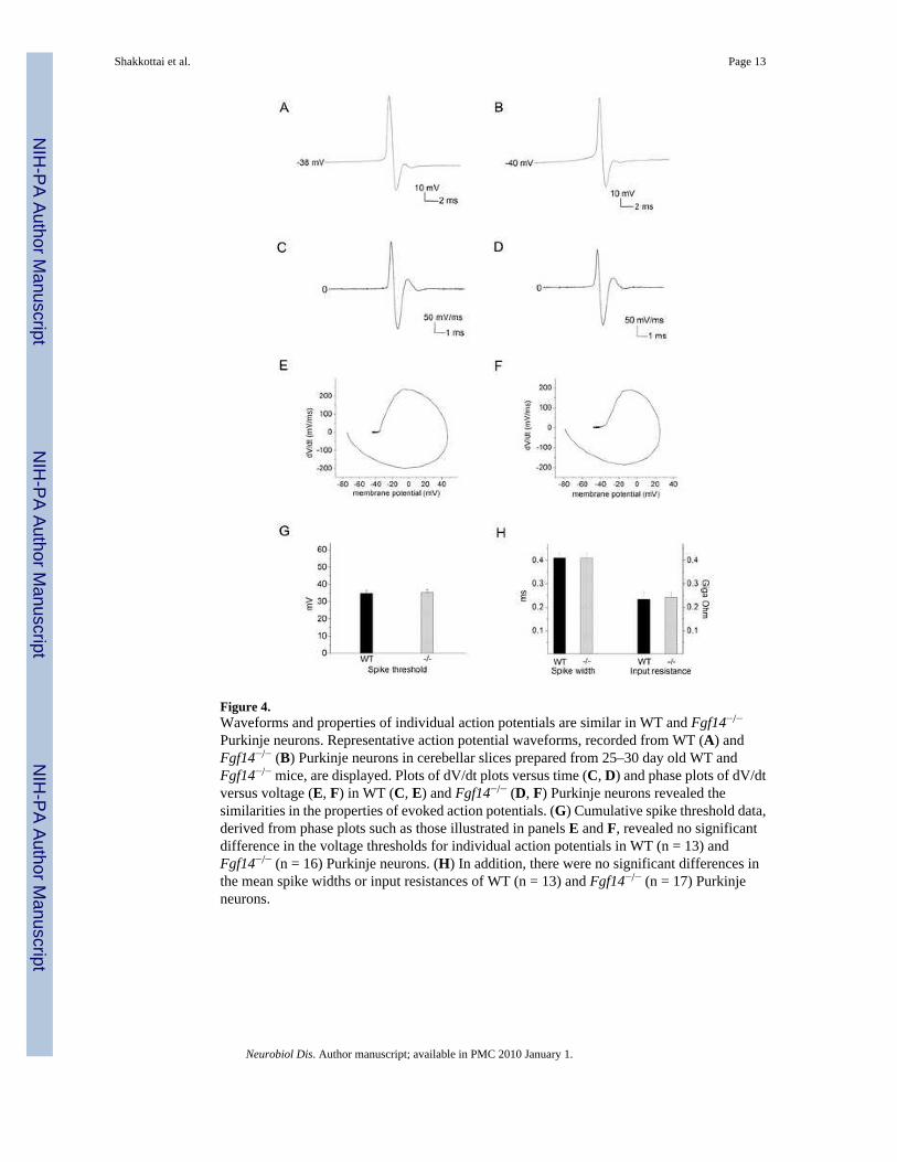
Figure 4.Waveforms and properties of individual action potentials are similar in WT and Fgf14−/−
Purkinje neurons. Representative action potential waveforms, recorded from WT (A) andFgf14−/− (B) Purkinje neurons in cerebellar slices prepared from 25–30 day old WT andFgf14−/− mice, are displayed. Plots of dV/dt plots versus time (C, D) and phase plots of dV/dtversus voltage (E, F) in WT (C, E) and Fgf14−/− (D, F) Purkinje neurons revealed thesimilarities in the properties of evoked action potentials. (G) Cumulative spike threshold data,derived from phase plots such as those illustrated in panels E and F, revealed no significantdifference in the voltage thresholds for individual action potentials in WT (n = 13) andFgf14−/− (n = 16) Purkinje neurons. (H) In addition, there were no significant differences inthe mean spike widths or input resistances of WT (n = 13) and Fgf14−/− (n = 17) Purkinjeneurons.
Shakkottai et al. Page 13
Neurobiol Dis. Author manuscript; available in PMC 2010 January 1.
NIH
-PA Author Manuscript
NIH
-PA Author Manuscript
NIH
-PA Author Manuscript
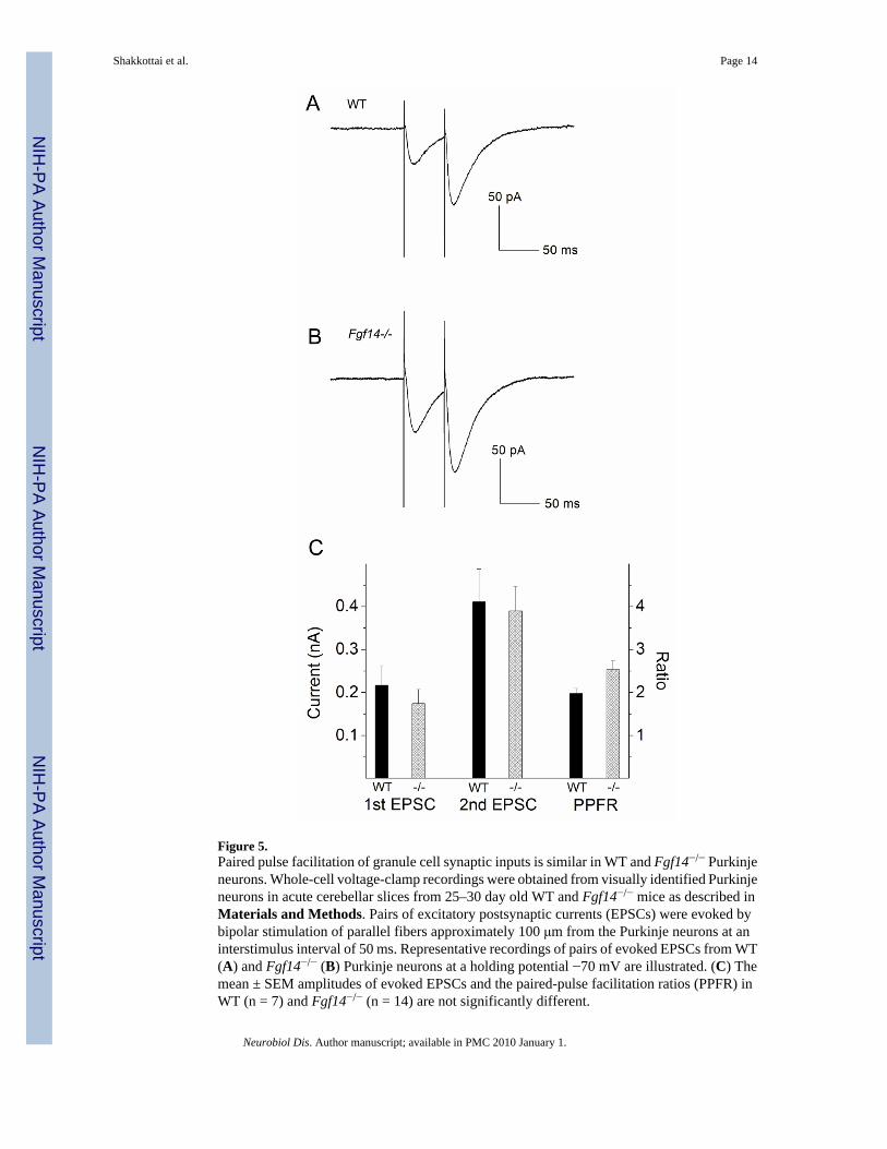
Figure 5.Paired pulse facilitation of granule cell synaptic inputs is similar in WT and Fgf14−/− Purkinjeneurons. Whole-cell voltage-clamp recordings were obtained from visually identified Purkinjeneurons in acute cerebellar slices from 25–30 day old WT and Fgf14−/− mice as described inMaterials and Methods. Pairs of excitatory postsynaptic currents (EPSCs) were evoked bybipolar stimulation of parallel fibers approximately 100 μm from the Purkinje neurons at aninterstimulus interval of 50 ms. Representative recordings of pairs of evoked EPSCs from WT(A) and Fgf14−/− (B) Purkinje neurons at a holding potential −70 mV are illustrated. (C) Themean ± SEM amplitudes of evoked EPSCs and the paired-pulse facilitation ratios (PPFR) inWT (n = 7) and Fgf14−/− (n = 14) are not significantly different.
Shakkottai et al. Page 14
Neurobiol Dis. Author manuscript; available in PMC 2010 January 1.
NIH
-PA Author Manuscript
NIH
-PA Author Manuscript
NIH
-PA Author Manuscript
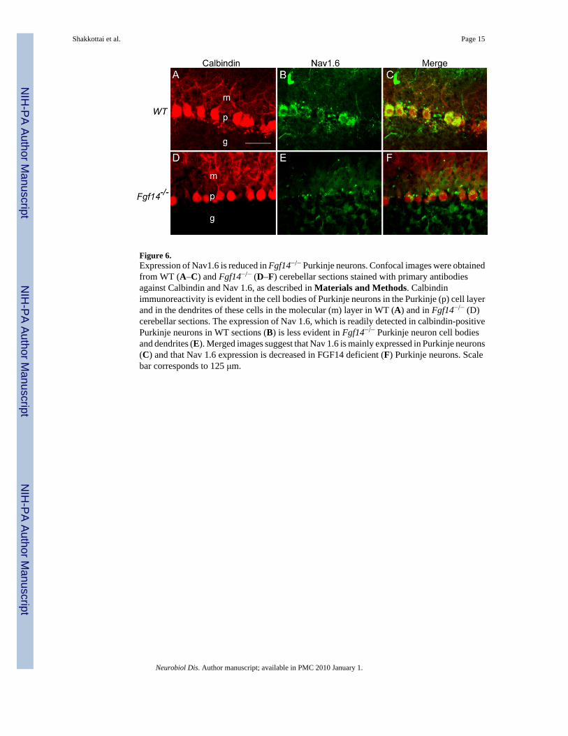
Figure 6.Expression of Nav1.6 is reduced in Fgf14−/− Purkinje neurons. Confocal images were obtainedfrom WT (A–C) and Fgf14−/− (D–F) cerebellar sections stained with primary antibodiesagainst Calbindin and Nav 1.6, as described in Materials and Methods. Calbindinimmunoreactivity is evident in the cell bodies of Purkinje neurons in the Purkinje (p) cell layerand in the dendrites of these cells in the molecular (m) layer in WT (A) and in Fgf14−/− (D)cerebellar sections. The expression of Nav 1.6, which is readily detected in calbindin-positivePurkinje neurons in WT sections (B) is less evident in Fgf14−/− Purkinje neuron cell bodiesand dendrites (E). Merged images suggest that Nav 1.6 is mainly expressed in Purkinje neurons(C) and that Nav 1.6 expression is decreased in FGF14 deficient (F) Purkinje neurons. Scalebar corresponds to 125 μm.
Shakkottai et al. Page 15
Neurobiol Dis. Author manuscript; available in PMC 2010 January 1.
NIH
-PA Author Manuscript
NIH
-PA Author Manuscript
NIH
-PA Author Manuscript


