Embed Size (px)
Citation preview

Flavodiiron Proteins in Oxygenic PhotosyntheticOrganisms: Photoprotection of Photosystem II by Flv2and Flv4 in Synechocystis sp. PCC 6803Pengpeng Zhang, Yagut Allahverdiyeva, Marion Eisenhut, Eva-Mari Aro*
Department of Biology, Plant Physiology and Molecular Biology, University of Turku, Turku, Finland
Abstract
Background: Flavodiiron proteins (FDPs) comprise a group of modular enzymes that function in oxygen and nitric oxidedetoxification in Bacteria and Archaea. The FDPs in cyanobacteria have an extra domain as compared to major prokaryoticenzymes. The physiological role of cyanobacteria FDPs is mostly unknown. Of the four putative flavodiiron proteins (Flv1–4)in the cyanobacterium Synechocystis sp. PCC 6803, a physiological function in Mehler reaction has been suggested for Flv1and Flv3.
Principal Findings: We demonstrate a novel and crucial function for Flv2 and Flv4 in photoprotection of photosystem II(PSII) in Synechocystis. It is shown that the expression of Flv2 and Flv4 is high under air level of CO2 and negligible atelevated CO2. Moreover, the rate of accumulation of flv2 and flv4 transcripts upon shift of cells from high to low CO2 isstrongly dependent on light intensity. Characterization of FDP inactivation mutants of Synechocystis revealed a specificdecline in PSII centers and impaired translation of the D1 protein in Dflv2 and Dflv4 when grown at air level CO2 whereas athigh CO2 the Flvs were dispensable. Dflv2 and Dflv4 were also more susceptible to high light induced inhibition of PSII thanWT or Dflv1 and Dflv3.
Significance: Analysis of published sequences revealed the presence of cyanobacteria-like FDPs also in some oxygenicphotosynthetic eukaryotes like green algae, mosses and lycophytes. Our data provide evidence that Flv2 and Flv4 have animportant role in photoprotection of water-splitting PSII against oxidative stress when the cells are acclimated to air levelCO2. It is conceivable that the function of FDPs has changed during evolution from protection against oxygen in anaerobicmicrobes to protection against reactive oxygen species thus making the sustainable function of oxygen evolving PSIIpossible. Higher plants lack FDPs and distinctly different mechanisms have evolved for photoprotection of PSII.
Citation: Zhang P, Allahverdiyeva Y, Eisenhut M, Aro E-M (2009) Flavodiiron Proteins in Oxygenic Photosynthetic Organisms: Photoprotection of Photosystem IIby Flv2 and Flv4 in Synechocystis sp. PCC 6803. PLoS ONE 4(4): e5331. doi:10.1371/journal.pone.0005331
Editor: Zoe Finkel, Mt. Alison University, Canada
Received December 24, 2008; Accepted March 18, 2009; Published April 24, 2009
Copyright: � 2009 Zhang et al. This is an open-access article distributed under the terms of the Creative Commons Attribution License, which permitsunrestricted use, distribution, and reproduction in any medium, provided the original author and source are credited.
Funding: This work was supported by the Academy of Finland (projects 121946, 118637, and 205361), Maj and Tor Nessling Foundation, EU/Energy Networkproject SOLAR-H2 (FP7 contract 212508) and Kone foundation. The funders had no role in study design, data collection and analysis, decision to publish, orpreparation of the manuscript.
Competing Interests: The authors have declared that no competing interests exist.
* E-mail: [email protected]
Introduction
The flavodiiron proteins (FDPs) are a large family of soluble
enzymes found in strict or facultative anaerobes among Bacteria
and Archaea as well as in some protozoan pathogens, reviewed in
[1]. By transferring electrons to oxygen or nitric oxide (NO), they
help avoiding accumulation of these toxic compounds. The FDP
family was originally grouped on the basis of sequence homology,
and previously named as A-type flavoprotein [2]. The typical
enzyme core contains two conserved structural modules: a b-
lactamase-like domain containing a diiron center, and a flavodoxin
domain with FMN binding site [3]. Despite the structural
similarity of FDPs, studies on these proteins from various
organisms have revealed significantly different redox potentials
[4,5], which indicate that the members of this family may interact
with distinct redox partners and participate in different cellular
processes. The oxygen reductase activity of FDPs functions in
preventing oxygen toxicity in some obligatory anaerobes when
cells are transiently exposed to oxygen [6,7]. Other FDPs like
Escherichia coli (E. coli) flavorubredoxin prefer NO as a substrate,
functioning in NO detoxification [8].
Amino acid alignments and subsequent in vitro studies led to a
discovery of an extra domain at the C terminus of FDPs in some
organisms. In case of cyanobacteria, the third module is a flavin
reductase domain, which can bind either FMN or FAD [2,9].
Analysis of sequenced cyanobacterial genomes reveals the
presence of several genes encoding distinct FDPs in one organism.
The genome of Synechocystis sp. PCC 6803 (hereafter Synechocystis)
comprises four putative FDP genes (CyanoBase: http://bacteria.
kazusa.or.jp/cyanobase/). Biochemical analysis provided evidence
that Synechocystis Flv3 is an NAD(P)H:oxygen oxidoreductase and
capable of reducing oxygen to water in vitro [9]. This result was
further confirmed by in vivo biophysical analysis indicating that
Synechocystis Flv1 and Flv3 are essential for Mehler reaction
transferring electrons to oxygen without formation of reactive
oxygen species (ROS) [10].
PLoS ONE | www.plosone.org 1 April 2009 | Volume 4 | Issue 4 | e5331

Although considerable progress towards overall understanding
of FDPs has been made during the past few years [1,11], the
physiological roles of cyanobacterial FDPs are far from being well
understood. Global gene expression profiles of Synechocystis have
shown that the transcription of some FDP genes is enhanced by
CO2 limitation [12,13], by high light [14] or UV-B light [15], and
by hydrogen peroxide [16]. Such DNA microarray data would
suggest that cyanobacterial FDPs are involved in coping with
photo-oxidative stress, but no experimental data is available to
support the assumption. In this work, we characterized the
inactivation mutants for the four different FDPs in Synechocystis to
address their physiological function under the conditions, which
promote photo-oxidative stress. Our results indicate a novel and
crucial role for the two FDPs, Flv2 and Flv4, in photoprotection of
Synechocystis cells and in the sustenance of the photosystem II (PSII)
complex.
Results
The genome of Synechocystis contains four genes encoding
putative flavodiiron proteins: sll1521, sll0219, sll0550 and
sll0217. Although flavodiiron proteins are abbreviated as FDPs
in recent reviews [1,11], we have here adapted nomenclature of
Synechocystis proteins according to Helman et al. [10] as Flv
proteins (Flv1, Flv2, Flv3 and Flv4).
Expression of flavodiiron protein genes under differentCO2 and light levels
As photoautotrophic inhabitants of aquatic environments,
cyanobacteria are challenged by fluctuation of light and deficiency
of inorganic carbon in their natural environments. Accordingly,
the effects of environmental CO2 conditions on the expression of
flv genes were investigated. The transcript levels of the four flv
genes (flv1, flv2, flv3 and flv4) from WT cells grown steadily at high
CO2 (air enriched with 3% CO2, HC) or low CO2 (air level, LC)
were studied first. Relative expression of the flv genes analyzed by
real-time quantitative RT-PCR (RT-Q-RT-PCR) is shown in
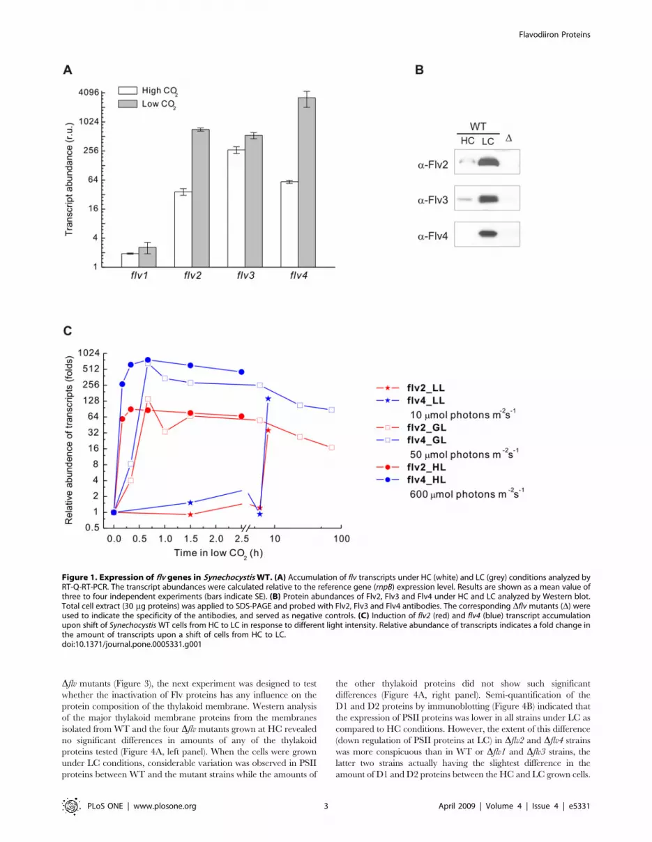
Figure 1A. Basically, the transcrips of the flv genes accumulated at
LC as compared to HC conditions, except for the flv1 transcripts,
which were at a very low level under both HC and LC conditions.
The transcripts of flv3 were the most abundant among the four flv
genes at HC, and roughly twice that amount was recorded in LC
grown cells. The transcripts of the flv2 and flv4 genes, on the
contrary, were strongly upregulated at LC, up to 20 and 54 fold,
respectively, as compared to HC grown cells (Figure 1A). This is in
line with previously published cDNA microarray data [12,13].
Differential expression of the flv genes at HC and LC was
studied at protein level by using specific antibodies prepared for
each of Synechocystis Flv proteins (Figure 1B). Under HC growth
conditions, Flv2 and Flv4 proteins were nearly undetectable and
Flv3 was present only in low amount in the immunoblots reflecting
low expression at protein level. In accordance with higher
transcript levels, WT cells grown at LC also accumulated
significant amounts of Flv2, Flv3 and Flv4 proteins. No Flv1
protein, however, was detected by immunoblotting, most probably
due to a low expression level of flv1 (see Materials and Methods). It
is interesting to note that the protein level of Flv3 was remarkably
higher under LC as compared to HC growth condition, despite
the fact that the transcript level showed only about two-fold
difference.
In order to get a more comprehensive view into the expression
of the flv genes, we checked the integrative effect of both the
carbon and light regimes. For this purpose, transcripts of the four
flv genes in Synechocystis WT were monitored by RT-Q-RT-PCR
upon a shift of cells from HC to LC, in combination with different
fluence rates (10, 50, 600 mmol photons m22 s21). As shown in
Figure 1C, a dramatic induction of the flv2 and flv4 transcripts
occurred after the shift. Moreover, the time course of induction
was highly dependent on light intensity. Under standard growth
light (50 mmol photons m22 s21), it took about 40 min to reach
the maximum induction of transcripts. Higher light intensity
(600 mmol photons m22 s21) accelerated the accumulation of
transcripts. The maximum induction appeared in about 20 min.
Conversely, the induction process was much slower under low
light illumination (10 mmol photons m22 s21). Indeed, the transcript
levels of flv2 and flv4 did not show any significant increase even after
transferring cells to LC for 6 h and only after 8 h at LC the transcript
amounts had reached the level recorded within 40 min under
standard growth light conditions. The flv1 and flv3 transcripts did not
show such remarkable induction (data not shown). Since the steady-
state level of flv2 and flv4 transcripts was nearly equal at high light
and growth light, we decided to perform further experiments mainly
under growth light conditions.
Cellular location of Flv2, Flv3 and Flv4 proteinsTo address the subcellular location of the Flv proteins in
Synechocystis, the total cell extract as well as the membrane and
soluble fractions of the WT cells grown at LC were isolated and
subjected to Western blot analysis (Figure 2). The fractions of WT
cells were also probed with Rubisco and D1 antibodies to indicate
the purity level of the subcellular fractions. The Flv2 protein was
found in both the membrane fraction and the soluble fraction. The
Flv3 protein was exclusively found in the soluble fraction, whereas
most of the Flv4 protein was found in the membrane fraction with
only trace amount in the soluble fraction. Total cell extracts of
corresponding mutant strains served as controls.
Expression of flv genes in WT and flv inactivation strainsDue to partially similar expression patterns, particularly
between flv2 and flv4 (Figure 1), and to a high homology between
the four Synechocystis Flv proteins [10], one could suggest functional
redundancy. To address this question, comparison of the flv gene
expression in WT and the four single flv gene inactivation mutants
was performed at both the transcript and protein levels. Figure 3A
shows the relative expression of flv genes as an induction of
transcripts at LC relative to that at HC. No significant difference
was recorded in the expression pattern of flv1 and flv3 between
WT and the Dflv mutants (except for the corresponding
inactivation strain). On the contrary, the expression level of the
flv2 and flv4 genes was dependent on the presence of other flv genes
and varied in different strains. Induction of flv2 and flv4 transcripts
was systematically higher in Dflv1 and Dflv3 than in WT. Most
interestingly, the expression of flv2 was hardly enhanced in Dflv4,
whereas strong induction of flv4 transcripts was observed in Dflv2.
In agreement with the transcript accumulation data, Western
blot analysis showed that the expression of the Flv3 protein is not
affected by inactivation of any other flv gene. However, slightly
higher amounts of Flv4 proteins were detected in the Dflv1 and
Dflv3 strains than in WT. The Flv2 protein was almost missing
from the Dflv4 strain under LC, whereas the expression of Flv4
protein was like in WT despite the inactivation of the flv2 gene
(Dflv2 mutant) (Figure 3B).
Protein content of the thylakoid membrane in WT andDflv strains
Since Flv2 and Flv4 proteins were found to be associated with
the membrane (Figure 2) and their expression varied among the
Flavodiiron Proteins
PLoS ONE | www.plosone.org 2 April 2009 | Volume 4 | Issue 4 | e5331
Dflv mutants (Figure 3), the next experiment was designed to test
whether the inactivation of Flv proteins has any influence on the
protein composition of the thylakoid membrane. Western analysis
of the major thylakoid membrane proteins from the membranes
isolated from WT and the four Dflv mutants grown at HC revealed
no significant differences in amounts of any of the thylakoid
proteins tested (Figure 4A, left panel). When the cells were grown
under LC conditions, considerable variation was observed in PSII
proteins between WT and the mutant strains while the amounts of
the other thylakoid proteins did not show such significant
differences (Figure 4A, right panel). Semi-quantification of the
D1 and D2 proteins by immunoblotting (Figure 4B) indicated that
the expression of PSII proteins was lower in all strains under LC as
compared to HC conditions. However, the extent of this difference
(down regulation of PSII proteins at LC) in Dflv2 and Dflv4 strains
was more conspicuous than in WT or Dflv1 and Dflv3 strains, the
latter two strains actually having the slightest difference in the
amount of D1 and D2 proteins between the HC and LC grown cells.
Figure 1. Expression of flv genes in Synechocystis WT. (A) Accumulation of flv transcripts under HC (white) and LC (grey) conditions analyzed byRT-Q-RT-PCR. The transcript abundances were calculated relative to the reference gene (rnpB) expression level. Results are shown as a mean value ofthree to four independent experiments (bars indicate SE). (B) Protein abundances of Flv2, Flv3 and Flv4 under HC and LC analyzed by Western blot.Total cell extract (30 mg proteins) was applied to SDS-PAGE and probed with Flv2, Flv3 and Flv4 antibodies. The corresponding Dflv mutants (D) wereused to indicate the specificity of the antibodies, and served as negative controls. (C) Induction of flv2 (red) and flv4 (blue) transcript accumulationupon shift of Synechocystis WT cells from HC to LC in response to different light intensity. Relative abundance of transcripts indicates a fold change inthe amount of transcripts upon a shift of cells from HC to LC.doi:10.1371/journal.pone.0005331.g001
Flavodiiron Proteins
PLoS ONE | www.plosone.org 3 April 2009 | Volume 4 | Issue 4 | e5331

PSII and the whole chain linear electron transfer activityin Dflv strains
LC-induced alteration of PSII protein level prompted us to
investigate in more detail the possible influence of Flv proteins on
photosynthetic electron transfer reactions. As a first approach, the
amount of PSII centers capable of primary charge separation and
reduction of QA was monitored as flash induced increase in
variable fluorescence (Fv) yield in the presence of 20 mM 3-(3,4-
dichlorophenyl)-1,1-dimethylurea (DCMU). As shown on the top
of Figure 5A, the fluorescence parameter Fv, a sensitive indicator
of active PSII centers, was very similar in WT and all Dflv mutant
strains when the cells were grown at HC and 50 mmol photons
m22 s21. It is also important to note that all strains grew equally
well (Figure 5B). At LC, on the contrary, significant differences in
PSII performance between WT and different Dflv strains were
observed. Under light intensity of 15 mmol photons m22 s21, the
difference in the Fv value between the strains was the smallest. The
higher the growth light intensity, the bigger were the differences in
the amount of active PSII centers between WT and the Dflv
mutants. Under light intensity of 50 mmol photons m22 s21
(standard growth light intensity), the amount of active PSII centers
in WT was 56% of the maximum obtained at HC. Dflv1 and Dflv3
had more functional PSII centers, about 70% of the maximum at
HC. On the contrary, the Dflv2 and Dflv4 strains had the poorest
PSII performance of all strains, having only 30% of the Fv at HC,
and the color of the cell cultures also turned more yellow
(Figure 5C) indicating changes in pigment composition as well.
Differences in the yield of Fv as described above are in line with
the amount of the D1 protein in the thylakoid membrane of
various strains as determined by immunoblot analysis (Figure 4).
Increase of light intensity to 200 mmol photons m22 s21 at LC
resulted in an even more yellow phenotype of the Dflv2 and Dflv4
mutants (Figure 5D) and further decreased the amount of
functional PSII centers in all strains. Only about one quarter of
the maximum PSII performance measured at HC was detected in
WT. The PSII function in Dflv1 and Dflv3 was somewhat more
efficient, about 40% of maximum measured at HC, whereas a
very low capacity for QA reduction (5%) was left in Dflv2 and
Dflv4.
Drastic differences in the amount of active PSII centers in the
Dflv mutants prompted us to measure the photosynthetic electron
transfer capacities by oxygen electrode (Table 1) and using 2,6-
dimethylbenzoquinone (DMBQ) as an artificial electron acceptor.
Again, the highest oxygen evolving activities were recorded from
HC grown cells and differences between the WT and the Dflv
mutants were only minor. The differences became evident when
cells were grown at LC, and these differences were the more
significant, the higher was the light intensity during growth. When
the cells were grown at 150 mmol photons m22 s21, the PSII
oxygen evolving capacity of the Dflv2 and Dflv4 mutants was only
about 35% of that of the WT and the Dflv1 and Dflv3 strains. It is
also interesting to note that the measured PSII oxygen evolution
rates were in general higher in Dflv1 and Dflv3 mutants than in
WT. When HCO32 was added as an electron acceptor (instead of
DMBQ), it became evident that in Dflv2 and Dflv4 mutants the
PSII capacity was a limiting factor for CO2 fixation upon
increasing the growth light intensity. On the other hand, the
WT, Dflv1 and Dflv3 strains showed clearly higher capacities for
PSII oxygen evolution at all light intensities (measured in the
presence of DMBQ) as compared to the oxygen evolution rates
recorded when HCO32 was added as a terminal electron
acceptor.
D1 turnover rates of WT and Dflv mutant strainsSince the PSII proteins were present in variable amounts in Dflv
mutants at LC growth conditions (Figure 4), it was next studied
whether this is related to the turnover of the D1 protein in mutant
strains. To this end, the in vivo 35S-Met pulse-chase labeling
experiments were applied using relatively high light (150 mmol
photons m22 s21) and LC conditions. Autoradiographs of the
accumulation of the label in the D1 protein and subsequent loss of
D1 during the chase are shown in Figure 6A. Quantification of
radioactivity in the D1 protein upon the pulse-chase experiment is
shown in Figure 6B and Figure 6C. Compared to WT, the
degradation of the D1 protein was faster in Dflv2, and slower in
Dflv3. Likewise, the synthesis of the D1 protein was not equivalent
in all strains. Dflv1 and Dflv3 synthesized D1 protein faster than
WT even though the degradation of the D1 protein was slowed
down in these strains as compared to WT. Importantly, in Dflv2,
the faster degradation of D1 as compared to WT or the Dflv1 and
Dflv3 mutant strains was not compensated with faster D1 synthesis
(Figure 6). The Dflv4 strain behaved very similar to the Dflv2
mutant (data not shown).
Susceptibility of Dflv mutant strains to short-term highlight stress
Pulse-chase experiments suggested that the Dflv2 and Dflv4
mutants cannot properly repair their damaged PSII centers when
CO2 level is low. Thus, the susceptibility of PSII to short-term high
light exposure at both HC and LC was examined in WT and the
Dflv2 and Dflv4 mutants. The cells grown under 50 mmol photons
m22 s21 at HC and LC were illuminated in a photobioreactor at
1500 mmol photons m22 s21 white light for up to 90 min, and the
PSII activity was measured during the course of illumination by
monitoring the oxygen evolving activity of the cells. Under LC,
Figure 2. Localization of Flv proteins in different cellularfractions. Protein samples (30 mg) of the total cell extract, membraneand soluble fractions of WT cells grown at LC were applied to SDS-PAGEand probed with Flv2, Flv3 and Flv4 antibodies. The antibodies againstD1 and Rubisco large subunit were used to indicate the purity of thefractions. The corresponding Dflv mutants (D) served as negativecontrols. The experimental molecular weight of each Flv was estimatedby comparing the migration of the respective proteins with themolecular weight markers in SDS-PAGE.doi:10.1371/journal.pone.0005331.g002
Flavodiiron Proteins
PLoS ONE | www.plosone.org 4 April 2009 | Volume 4 | Issue 4 | e5331

both Dflv2 and Dflv4 cells lost more of the PSII oxygen evolving
activity during the high light illumination than WT (Figure 7). On
the contrary, the photoinhibition of PSII in Dflv1 and Dflv3 was
slightly less severe than in WT (data not shown). After 90 min
treatment, more than 80% of PSII oxygen evolving activity still
remained in the WT, but only about 60% of PSII activity was left
in Dflv2 and Dflv4. On the other hand, when the HC grown cells
were bubbled with 3% CO2 in air, no apparent photoinhibition of
PSII was observed in WT or any of the flv mutants during the
90 min illumination period under 1500 mmol photons m22 s21
(Figure 7).
Discussion
Evolution of distinct flavodiiron proteins with oxygenicphotosynthesis
Flavodiiron proteins are known to function against oxygen and/
or nitric oxide toxicity in anaerobic microbes. In these organisms,
FDPs are single proteins and functionally assembled as homodi-
mers, reviewed in [1,11]. Discovery of FDPs in cyanobacteria
[1,2,9], which are oxygenic photosynthetic prokaryotes, suggested
that the functions of FDPs are probably more diverse than earlier
anticipated. Indeed, the genes encoding FDPs were originally
believed to be restricted to prokaryotes but more recently were
also discovered in the genomes of some anaerobic protists [17–20].
Moreover, we identified flavodiiron protein genes also in the
genomes of some ancient photosynthetic eukaryotes, such as green
algae, mosses and lycophytes (Figure 8), which further suggest a
role for FDPs during evolution of land plants. Referring to
Helman et al. [10], we abbreviate here the FDPs from oxygenic
photosynthetic organisms as Flv proteins. Interestingly, these
proteins share a common feature distinct from those in anaerobic
photosynthetic bacteria and non-photosynthetic microbes. They
all have a C-terminal flavin reductase domain, which makes it
possible to use NAD(P)H as an electron donor. Moreover, analysis
of all available cyanobacterial genomes revealed that there are at
Figure 3. Expression of flv genes in WT and Dflv mutants under HC and LC conditions. (A) Accumulation of flv transcripts analyzed by RT-Q-RT-PCR. The transcript level of rnpB is used as a reference. Bars represent the ratio of expression of individual flv genes at LC to that at HC6SE forthree independent experiments. (B) Expression of Flv proteins. Immunoblotting was performed by loading 40 mg of total cell extract in each well,and probed with Flv2, Flv3 and Flv4 antibodies.doi:10.1371/journal.pone.0005331.g003
Flavodiiron Proteins
PLoS ONE | www.plosone.org 5 April 2009 | Volume 4 | Issue 4 | e5331

Figure 4. Thylakoid protein content of WT and Dflv mutant cells grown at HC and LC. (A) Membrane proteins (25 mg in each well) wereseparated by SDS-PAGE and immunoblotting was performed using D1, D2, AtpA/B, Cytf, PsaA/B, NdhJ and NdhD3 specific antibodies. (B) Relativeamounts of the D1 and D2 proteins in WT and the Dflv mutant cells grown under HC and LC conditions. Protein amounts are indicated as apercentage of those measured in the WT cells grown at HC (100%). Values are the mean6SE from 3 independent experiments.doi:10.1371/journal.pone.0005331.g004
Flavodiiron Proteins
PLoS ONE | www.plosone.org 6 April 2009 | Volume 4 | Issue 4 | e5331

least two genes encoding distinct FDPs within the same organism,
making one or more pairs (Figure 8).
The phylogenetic tree (Figure 8) shows a clear separation of the
proteins between oxygenic photosynthetic organisms and those
from non-photosynthetic microorganisms (e.g. E. coli, Moorella
thermoacetica, Desulfovibrio gigas). Flv proteins from oxygenic
photosynthetic organisms group into two nearly symmetrical
clusters (named here as Cluster FlvA and Cluster FlvB), one
partner of the pair from each cluster. Both FlvA and FlvB clusters
can be further divided into four subclusters. Two of these
subclusters are made of Flv proteins from b-cyanobacteria (Flv1,
Flv2 in Cluster FlvA or Flv3, Flv4 in Cluster FlvB). Intriguingly,
Anabaena and Nostoc punctiforme posses as many as six and five Flvs,
respectively, some of these genes grouping together in subcluster
Flv1 (Flv1a–b) or Flv3 (Flv3a–b), which indicates multiple gene
duplication events. However, a-cyanobacteria like Synechococcus sp.
CC9902 or Prochlorococcus strains and ancient eukaryotic species,
such as Chlamydomonas reinhardtii, Physcomitrella patens and Selaginella
moellendorffii, have only two Flvs, and form their own subclusters
branching out from the b-cyanobacterial clusters. It appears that
Figure 5. PSII fluorescence and the phenotype of the Dflv mutants. Cells were grown at HC and 50 mmol photons m22 s21 or at LC under 15,50, 120 or 200 mmol photons m22 s21. (A) The amplitude of the flash-induced variable Chl fluorescence of the WT and Flv mutant cells wasmeasured in the presence of 20 mM DCMU and at a Chl concentration of 5 mg Chl/ml. The results are shown as a percentage of the amplitude of theflash-induced variable Chl fluorescence in the WT-cells grown at HC (set as 100%). 6SD for three independent experiments. (B) Photographs of cellcultures grown under HC and 50 mmol photons m22 s21, (C) under LC and 50 mmol photons m22 s21, (D) under LC and 200 mmol photons m22 s21.doi:10.1371/journal.pone.0005331.g005
Table 1. Net photosynthesis and PSII activities of WT and flv mutants.
Strain HC, GL LC, LL LC, GL LC, HL
Photosyn. PSII Photosyn. PSII Photosyn. PSII Photosyn. PSII
WT 263613 47769 228615 294612 232615 30069 32666 35663
Dflv1 303615 486630 226613 33268 21967 33265 285610 341610
Dflv2 23163 419625 26364 267616 21667 21068 15168 12867
Dflv3 189610 485627 210616 354618 20765 330616 30967 36863
Dflv4 227614 472613 266616 264612 22166 19163 15264 128611
Measurements were made with Synechocystis cells and the activities are presented as steady-state oxygen evolution rates (mmol O2 mg21 Chl h21) measured undersaturating light. The cells were grown at high (HC) or low CO2 (LC) under GL: growth light, 50 mmol photons m22 s21; LL: low light, 15 mmol photons m22 s21; HL: highlight, 150 mmol photons m22 s21. Net photosynthesis rate (Photosyn.) was measured as oxygen evolution (mmol O2 mg21 Chl h21) in the presence of 10 mM NaHCO3.The PSII oxygen evolving activity was measured in the presence of 2 mM DMBQ. The results are means6SD of 4 independent experiments.doi:10.1371/journal.pone.0005331.t001
Flavodiiron Proteins
PLoS ONE | www.plosone.org 7 April 2009 | Volume 4 | Issue 4 | e5331

the presence of FDPs in pairs is a common feature for oxygenic
photosynthetic organisms, which provides evidence that an ancient
gene duplication event occurred with appearance of oxygen-
evolving photosynthesis and thus before the primary endosymbi-
osis. Possibly, these pairs assemble into heterodimers to fulfill their
function [10]. Although the Flv proteins occur in all oxygenic
photosynthetic prokaryotes and in some oxygenic eukaryotes like
green algae or mosses, it is important to emphasize that we could
not identify any Flv homologs in higher plants.
Flavodiiron proteins are highly expressed in Synechocystisas a response to low CO2 environment
The presence of several Flv homologs in all cyanobacteria and
some photosynthetic eukaryotes suggests particular importance for
these proteins in ancient oxygenic photosynthetic organisms.
However, addressing the physiological function of Flv proteins has
so far been successful only for Synechocystis Flv1 and Flv3. They
were shown to function in Mehler reaction and donating electrons
from PSI directly to molecular oxygen [10]. Interestingly, this
reaction was suggested to avoid formation of ROS and in fact Flv1
and Flv3 were suggested to be essential for protection of the cells
against formation of ROS in vivo. The function of the other two Flv
proteins Flv2 and Flv4 in Synechocystis has remained completely
enigmatic. Search of published DNA microarray data revealed
that the transcription of flv2 and flv4 genes is induced by CO2
limitation [12,13] and transiently also by high light [14]. These
data were validated and extended here to show that the
Figure 6. D1 turnover rates of WT and Dflv mutants under LC conditions. Synechocystis cells were grown under LC and 50 mmol photonsm22 s21. A radioactive pulse was given to the cell suspension for 5, 10, 20, and 30 min under 150 mmol photons m22 s21 illumination. Chaseexperiments were performed under similar conditions for 0.5, 1, 2, and 3 h. (A) Autoradiogram of the membrane proteins separated by SDS-PAGE.The bands corresponding to the D1 and pre-D1 proteins are indicated by arrows. (B) Quantification of radioactivity in the D1 protein during pulse.(C) Quantification of radioactivity in the D1 protein during chase. Values are the mean of two independent experiments.doi:10.1371/journal.pone.0005331.g006
Figure 7. Photoinhibition of PSII in WT, Dflv2 and Dflv4 underHC and LC. The cells were grown under HC and LC conditions at50 mmol photons m22 s21, and then subjected to high light illuminationin a photobioreactor at 1500 mmol photons m22 s21 for 90 min withcontinuous bubbling of the cell cultures with 3% (HC) and air level (LC) ofCO2, respectively. Samples were withdrawn during the high lighttreatment, and the PSII activity was monitored by steady-state oxygenevolution measurements in the presence of 2 mM DMBQ as an artificialelectron acceptor. The results are shown as a percentage of oxygenevolution measured before the high light treatment, which is set as 100%(for absolute O2 evolution activities of the controls from HC and LCconditions, see Table 1). Mean6SD for three independent experiments.doi:10.1371/journal.pone.0005331.g007
Flavodiiron Proteins
PLoS ONE | www.plosone.org 8 April 2009 | Volume 4 | Issue 4 | e5331

expression of the flv2 and flv4 genes is strongly induced under
air level of CO2 (LC) at both the transcript (Figure 1A) and
protein levels (Figure 1B). Moreover, the induction pattern of
flv2 and flv4 depends on both the carbon and light regimes
(Figure 1C), the most rapid induction occurring at LC and high
irradiance i.e. under conditions that favor the production of
ROS in the photosynthetic electron transfer chain. These data
provided preliminary evidence that Flv2 and Flv4 might also be
involved in the reactions related to protection against oxidative
stress.
A distinct phenotype of the Dflv mutants appeared only under
LC (Figure 5C and 5D). Under HC environments, the Synechocystis
WT and all Dflv mutant cells appeared much greener and grew
much faster than under LC, and no obvious differences between
WT and any of the Dflv mutants were observed in the amount of
PSII centers (Figure 5A and 5B), even at irradiances as high as
Figure 8. Phylogenetic analysis of flavodiiron proteins in anaerobes, facultative aerobes and oxygenic photosynthetic organisms.Amino acid sequences of cyanobacterial proteins were obtained from CyanoBase (http://bacteria.kazusa.or.jp/cyanobase/), Selaginella amino acidsequences from JGI (http://genome.jgi-psf.org/Selmo1/Selmo1.home.html) and the other amino acid sequences from NCBI (http://www.ncbi.nlm.nih.gov/). Ana, Anabaena sp. PCC 7120 (Flv1a: All0177, Flv1b: All3891, Flv2: All4444, Flv3a: All0178, Flv3b: All3895, Flv4: All4446); Ama, Acaryochlorismarina MBIC11017 (Flv1: Am1_1384, Flv3: Am1_1386); Cya, Cyanothece sp. ATCC 51142 (Flv1: Cce_2580, Flv2: Cce_3835, Flv3: Cce_3635, Flv4:Cce_3833); Cre, Chlamydomonas reinhardtii (FlvA: XP_001692916, FlvB: XP_001699345); Dgi, Desulfovibrio gigas (ROO: AAG34792); Eco, Escherichia coli(FlRd: Q46877); Gvi, Gloeobacter violaceus PCC 7421 (Flv1: Glr1776, Flv3: Glr1775); Mae, Microcystis aeruginosa NIES-843 (Flv1: MAE61610, Flv2:MAE50820, Flv3: MAE01310, Flv4: MAE50840); Mth, Moorella thermoacetica (FDP: AAG00802); Npu, Nostoc punctiforme ATCC 29133 (Flv1a:Npun_F4867, Flv1b: Npun_F5838, Flv2: Npun_R0591, Flv3a: Npun_F4866, Flv3b: Npun_F5837); PMM, Prochlorococcus marinus MED4 (FlvA: PMM0042,FlvB: PMM0043); PMT, Prochlorococcus marinus MIT9313 (FlvA: PMT2165, FlvB: PMT2164); Ppa, Physcomitrella patens subsp. patens (FlvA:XP_001759251, FlvB: XP_001756079); Pro, Prochlorococcus marinus SS120 (FlvA: Pro0044, FlvB: Pro0045); Smo, Selaginella moellendorffii (FlvA:estExt_Genewise1.C_61201, FlvB: estExt_fgenesh2_pg.C_1160033); Sel, Synechococcus elongatus PCC 6301 (Flv1: Syc2283, Flv3: Syc2284); Syc,Synechococcus sp. CC 9902 (FlvA: Syncc9902_2183, FlvB: Syncc9902_2180); Syn, Synechocystis sp. PCC 6803 (Flv1: Sll1521, Flv2: Sll0219, Flv3: Sll0550,Flv4: Sll0217), Tel, Thermosynechococcus elongatus BP-1 (Flv2: Tll1373, Flv4: Tlr1088); Ter, Trichodesmium erythraeum IMS 101 (Flv1: Tery_0770, Flv3:Tery_0302).doi:10.1371/journal.pone.0005331.g008
Flavodiiron Proteins
PLoS ONE | www.plosone.org 9 April 2009 | Volume 4 | Issue 4 | e5331

200 mmol photons m22 s21 (data not shown). Thus the Flv
proteins seem to be dispensable at HC environments, and this is in
line with a relative low expression level of all the four Flv proteins
under HC (Figure 1B). On the contrary, the LC (air level CO2)
phenotype was particularly distinct in Dflv2 and Dflv4 cell cultures,
which under 200 mmol photons m22 s21 turned more yellow than
WT or Dflv1 and Dflv3 (Figure 5D). Such phenotype of the Dflv2
and Dflv4 mutants under LC, pointing to changes in the pigment
composition of the cells, is also strongly linked to downregulation
of PSII, and is the more severe the higher is the light intensity
upon growth (Figure 5).
Flavodiiron proteins Flv2 and Flv4 function inphotoprotection of photosystem II
A strong light dependent decline in functional PSII centers in
the Dflv2 and Dflv4 mutants, when connected with LC conditions,
suggests that Flv2 and Flv4 are crucial for protection of PSII
centers against photoinhibition. PSII activity in cells is a delicate
balance between constant photodamage and repair of the PSII
centers, which is reflected in a rapid turnover of the PSII reaction
center protein D1 [21]. Only when the repair of PSII centers
cannot keep up with the rate of photodamage to the PSII centers,
accumulation of damaged and malfunctional PSII centers takes
place. This situation typically occurs under conditions of high
production of ROS, which has been shown to inhibit the synthesis
of new D1 proteins and the repair of damaged PSII centers [22].
Testing the capacity of the Dflv mutants for rapid D1 protein
turnover revealed faster D1 degradation rate for Dflv2 (and Dflv4,
data not shown) than for WT or Dflv1 and Dflv3, whereas the
synthesis of D1 remained at slow rate (Figure 6). Retardation of
D1 protein synthesis and thus the repair of damaged PSII centers
in Dflv2 and Dflv4 mutants provided indirect evidence that the Flv2
and Flv4 proteins have a crucial role in preventing the production
of ROS under LC conditions.
Results above were obtained with cells after steady state growth
under LC or HC conditions whereas traditional PSII photoinhibi-
tion experiments are generally based on short-term exposure of
cell to high irradiance. Such a short-term high light exposure at
LC conditions, however, revealed similar trends in PSII photo-
inhibition among the Dflv mutants as observed upon long-term
high-light acclimation. Indeed, the mutants Dflv2 and Dflv4 were
clearly more susceptible to photoinhibition of PSII than the WT
(Figure 7) or the Dflv1 and Dflv3 mutants (data not shown) and thus
exhibited considerably lower PSII oxygen evolution activities after
reaching a balance between PSII damage and repair. It is thus
evident that under ambient low CO2 environments, the absence of
the Flv2 and/or the Flv4 proteins makes the Synechocystis cells face
chronic photoinhibition.
The protective role of CO2 against PSII photoinhibition was
likewise proven in these short-term illumination experiments by
continuously bubbling the cells with 3% CO2 (in air), and the
protection was found to be equally efficient in HC acclimated
Dflv2 and Dflv4 cells and in HC acclimated WT cells. Indeed, no
PSII photoinhibition could be detected in our high light and HC
illumination conditions (Figure 7). Since the HC acclimated WT
cells illuminated under similar high-light conditions but only air
level of CO2 showed distinct PSII photoinhibition (unpublished
data), and the similar phenomenon was reported also by Singh and
his colleagues [23], the importance of CO2 in protection against
PSII photoinhibition during the high-light exposure appears to be
an indisputable fact. The extent of such protection naturally
depends on experimental conditions including the intensity of light
and the length of the light path, among other things. Although the
molecular mechanism behind the photoprotection of PSII by
elevated CO2 is not fully understood, it is clear that the cells are
confronted with less oxidative stress under HC than under LC
conditions where the terminal electron acceptors (availability of
CO2) and thus the electron flow through PSII are limiting [24],
which in turn accelerates photoinhibition by downregulation of
PSII repair [25]. Indeed, the Flv2 and Flv4 proteins are
dispensable under HC conditions, at least when the stress
conditions induced by high light are not too severe (Figure 7).
Nevertheless, under natural growth environments the cyano-
bacterial cells are most frequently experiencing the limitation in
the availability of CO2. Thus a pertinent question is how the Flv2
and Flv4 proteins protect PSII against photoinhibition under such
conditions? The role of Flv1 and Flv3 has been demonstrated
earlier as an electron acceptor from PSI and in mediating electrons
further to molecular oxygen without formation of ROS [10]. Our
results, however, show that the Flv2 and Flv4 proteins have a
much more crucial function in photoprotection of Synechocystis cells
under LC conditions and this protection is particularly targeted
against photoinhibition of PSII. At the moment we can only
speculate about the molecular mechanism(s) behind such photo-
protection by the Flv2 and Flv4 proteins. While Flv3 protein was
found to be completely soluble, we found that the Flv2 and Flv4
proteins are tightly associated with the membrane and therefore
might potentially be involved in photosynthetic or respiratory
electron transfer routes. Since no transmembrane helices can be
predicted from the amino acid sequences of any of the four Flv
proteins, it would be interesting to clarify the interaction partners
of Flv2 and Flv4 for membrane docking. It remains to be shown
whether Flv2 and Flv4 can function in cyclic electron flow around
PSI or accept electrons directly from PSII or from the PQ pool to
alleviate the excitation pressure on PSII, which is known to lead to
a damage of PSII and production of ROS. Also a possibility that
the Flv2 and Flv4 proteins play a role in the CO2-concentrating
mechanism, thus alleviating the photoinhibition of PSII under LC
environments remains to be investigated.
Although the regulation pattern of the Flv2 and Flv4 proteins
closely resembles each other when the CO2 level changes, and also
the elimination of either the Flv2 or the Flv4 protein results in
similar LC sensitive phenotype, it is not yet clear whether the Flv2
and Flv4 proteins function in Synechocystis as a heterodimer. It is
also intriguing to note that the Dflv1 and Dflv3 mutants showed
enhanced expression of Flv4 proteins (Figure 3B), and they also
maintained more PSII centers (Figure 5A) as well as higher PSII
activity in cells than any other strains (Table 1). It is thus
conceivable that the expression of all four Flv proteins is mutually
regulated in Synechocystis cells but only the Flv2 and Flv4 proteins
have a direct control over the number of functional oxygen
evolving PSII centers.
Concluding remarksWe have demonstrated a vital role of flavodiiron proteins,
particularly Flv2 and Flv4, for oxygenic photosynthesis of
Synechocystis cells at ambient air levels of CO2. There seems to be
an evolutionary trend in the diversity and function of the
flavodiiron proteins, which in anaerobic microbes is directed
against O2/NO toxicity whereas in oxygenic photosynthetic
organisms the Flv2 and Flv4 proteins protect against photoinhibi-
tion of the oxygen evolving PSII complex. Development of
different photoprotective mechanisms in the course of evolution of
land plants has gradually made the function of flavodiiron proteins
dispensable and eventually resulted in elimination of respective
genes from the genomes of higher plants. It will be intriguing to
investigate whether the flavodiiron proteins in Chlamydomonas,
Flavodiiron Proteins
PLoS ONE | www.plosone.org 10 April 2009 | Volume 4 | Issue 4 | e5331

Physcomitrella and Selaginella have a similar protective role against
photoinhibition of PSII as the flavodiiron proteins in Synechocystis.
Materials and Methods
Cell culture conditionsSynechocystis glucose-tolerant strain (WT) and the single flavo-
diiron protein inactivation mutants Dflv1 (Dsll1521), Dflv2
(Dsll0219), Dflv3 (Dsll0550), and Dflv4 (Dsll0217) [10] were grown
in BG-11 medium [26] supplemented with 20 mM Hepes-NaOH
(pH 7.5) at 30uC under gentle agitation. The cells were routinely
cultivated under 50 mmol photons m22 s21 (white light) at high
CO2 (air enriched with 3% CO2, HC) or low CO2 (air level, LC)
conditions. For specific experiments, the cells were grown under
different light intensities: 15, 50, 120, or 200 mmol photons
m22 s21. For all experiments, the cells were harvested at the
logarithmic phase (for WT the O.D.750 was 1.0 and for the
mutants between 0.6 and 1.1, depending on the growth rate of the
mutants) and resuspended in fresh BG-11 medium.
Long- and short-term shifts of cells to differentenvironmental conditions
Synechocystis WT cells grown at 50 mmol photons m22 s21 and
HC were transferred to LC and the photon fluence rate of 10, 50
or 600 mmol photons m22 s21 for up to 96 h.
In another set of experiments, both LC and HC grown WT and
Dflv mutant cells were transferred to a photobioreactor (FMT 150,
Photon Systems Instruments, Czech Republic) and illuminated
under 1500 mmol photons m22 s21 (white light) up to 90 min.
During these short-term photoinhibition experiments, the LC
grown Synechocystis cells were bubbled with air whereas the HC
grown cells were bubbled with 3% CO2 in air. The length of the
light path in photobioreactor was 2.5 cm and the chl concentra-
tion was set at 5 mgChl/ml.
RNA isolation and RT-Q-RT-PCR assayTotal RNA was isolated by Trizol (Invitrogen) method [27],
and treated with 1 unit DNase (Ambion Turbo DNase kit, USA)
to remove genomic DNA. First strand cDNA was synthesized
from 1 mg purified RNA using the BioRad iScript cDNA
Synthesis kit (BioRad Laboratories Inc). The primers were
designed for generating amplicons of similar length (350–
450 bp). House-keeping gene rnpB encoding the RNase P
subunit B was used as a reference. The primer pairs used in
this study are summarized in Table 2. The RT-Q-RT-PCR was
performed as described before [28] on a BioRad IQ5 system.
The efficiency of each individual reaction and the relative
change in gene expression relative to the control were calculated
as described earlier [29]. Melting curve analysis was performed
for each run to ensure the specificity of the product amplification
(data not shown).
Protein isolation, electrophoresis and immunodetectionTotal cell extract as well as the membrane and soluble
fractions of Synechocystis cells were isolated according to Zhang et
al. [30]. Harvested cells were suspended in a buffer containing
50 mM Hepes-NaOH, pH 7.5, 30 mM CaCl2, 800 mM sorbi-
tol, 1 mM e-amino-n-caproic acid, and the cells were broken by
vortexing 661 min at 4uC in the presence of glass beads. The
total cell extract was obtained by centrifugation at 30006g for
5 min to remove the glass beads and unbroken cells. The
membranes were then pelletted at 180006g for 25 min, and
resuspended in 50 mM Hepes-NaOH, pH 7.5, 600 mM sucrose,
30 mM CaCl2, 1 M glycinbetaine. The soluble fraction was
obtained after centrifugation of the supernatant at 1100006g for
20 min.
Protein samples were solubilized in Laemmli SDS sample buffer
containing 5% b-mercaptoethanol and 6 M urea at room
temperature for at least 1 h, and separated by 12% or 14%
SDS-PAGE [31]. The proteins were transferred to a polyvinyli-
dene fluoride (PVDF) membrane (Immobilon-P, Millipore, USA)
using a semidry apparatus (Pharmacia), and immunodetected by
protein specific antibodies. The Flv1, Flv2, Flv3 and Flv4
antibodies were prepared against amino acids 145–159 of
Synechocystis Sll1521, against amino acids 521–535 of Synechocystis
Sll0219, against amino acids 377–391 of Sll0550, and against
amino acids 412–426 of Sll0217 proteins, respectively. The
specificities of the Flv antibodies were verified by analysis of
Synechocystis WT and Flv mutants and by using recombinant
proteins produced in E. coli. All Flv antibodies gave a positive
signal with respective recombinant E. coli strains, although no Flv1
signal was obtained from Synechocystis extracts. Antibodies against
NdhJ, NdhD3, D1, D2 and AtpA/B proteins were used as
described earlier [28,30,32,33].
In vivo pulse-chase labeling of membrane proteinsPulse-labeling of Synechocystis proteins was performed accord-
ing to [28]. Synechocystis cell suspension (chlorophyll a concen-
tration at 10 mg/ml) was supplemented with 5 mCi/ml 35S-L-
Met (Amersham Biosciences UK Ltd.) for 5, 10, 20 and 30 min
at 150 mmol photons m22 s21 of white light. Nonradioactive
Met (1 mM) was added to terminate the labeling. For chase
experiment, the 5 min pulse-labeled cells were pelletted, and
resuspended in BG-11 medium supplemented with 1 mM
nonradioactive Met and chased at HC and LC conditions at
150 mmol photons m22 s21. After chase of 0.5, 1, 2, and 3 h,
the samples were withdrawn, cooled down rapidly on ice, and
the cells were immediately harvested by centrifugation at 4uC.
Membranes were isolated and proteins separated by SDS-
PAGE, electrotransferred to a PVDF membrane and visualized
by autoradiography on X-ray films.
Oxygen evolution and fluorescence measurementsSteady-state oxygen evolution rates were measured with Clark
type oxygen electrode (Hansatech DW1, UK) at saturating light
intensity. To measure the net photosynthesis rate, the cells were
suspended in BG-11 medium supplemented with 10 mM
NaHCO3, and for the PSII activity measurements 2 mM
DMBQ was added as an artificial electron acceptor. Flash-
induced increase and subsequent decay of chlorophyll fluores-
cence yield were measured according to Allahverdiyeva et al.
[34].
Table 2. Oligonucleotide sequences used to perform RT-Q-RT-PCR.
Gene name Forward primer (59R39) Reverse primer (59R39)
flv1 (sll1521) GATAATTTTGTCGGCACCCTAA AGTCCAACCTTCATCGAACACT
flv2 (sll0219) AGCTTTGCATAGTCCGTCAGA CGCAGGACGAGAACAAATAAG
flv3 (sll0550) CGGCACCACTTACAATTCCTA GGTCATAGGTGAGCATGGTGT
flv4 (sll0217) CCAGTACCTCACCCAGAAACA AAGCTAGGGTTTCCAACAGGA
rnpB CCAATTTCCCAAGACTACGG GGCAGGAAAAAGACCAACCT
doi:10.1371/journal.pone.0005331.t002
Flavodiiron Proteins
PLoS ONE | www.plosone.org 11 April 2009 | Volume 4 | Issue 4 | e5331

Phylogenetic analysisThe tree was constructed from a ClustalX [35] alignment of
flavodiiron protein amino acid sequences with the program
TreeView 1.5.0.
Acknowledgments
Prof. Aaron Kaplan and Prof. Teruo Ogawa are thanked for kindly
providing us with the flavodiiron protein knockout mutants. The PsaA/B
antibody was a kind gift from Prof. Parag Chitnis, the Cytf antibody from
Prof. Lixin Zhang, and the Rubisco large subunit antibody from Dr.
Martin A.J. Parry.
Author Contributions
Conceived and designed the experiments: PZ EMA. Performed the
experiments: PZ. Analyzed the data: PZ. Contributed reagents/materials/
analysis tools: PZ ME EMA. Wrote the paper: PZ YA ME EMA.
Supervised the biophysical measurements: YA.
References
1. Vicente JB, Justino MC, Goncalves VL, Saraiva LM, Teixeira M (2008)
Biochemical, spectroscopic, and thermodynamic properties of flavodiiron
proteins. Methods Enzymol 437: 21–45.
2. Wasserfallen A, Ragettli S, Jouanneau Y, Leisinger T (1998) A family of
flavoproteins in the domains Archaea and Bacteria. Eur J Biochem 254:
325–332.
3. Frazao C, Silva G, Gomes CM, Matias P, Coelho R, et al. (2000) Structure of a
dioxygen reduction enzyme from Desulfovibrio gigas. Nat Struct Biol 7:
1041–1045.
4. Nolling J, Ishii M, Koch J, Pihl TD, Reeve JN, et al. (1995) Characterization of a
45-kDa flavoprotein and evidence for a rubredoxin, two proteins that could
participate in electron transport from H2 to CO2 in methanogenesis in
Methanobacterium thermoautotrophicum. Eur J Biochem 231: 628–638.
5. Gomes CM, Silva G, Oliveira S, LeGall J, Liu MY, et al. (1997) Studies on the
redox centers of the terminal oxidase from Desulfovibrio gigas and evidence for its
interaction with rubredoxin. J Biol Chem 272: 22502–22508.
6. Chen L, Liu MY, LeGall J, Fareleira P, Santos H, et al. (1993) Rubredoxin
oxidase, a new flavor-hemo-protein, is the site of oxygen reduction to water by
the ‘‘strict anaerobe’’ Desulfovibrio gigas. Biochem Biophys Res Commun 193:
100–105.
7. Kawasaki S, Ishikura J, Watamura Y, Niimura Y (2004) Identification of O2-
induced peptides in an obligatory anaerobe, Clostridium acetobutylicum. FEBS Lett
571: 21–25.
8. Gomes CM, Giuffre A, Forte E, Vicente JB, Saraiva LM, et al. (2002) A novel
type of nitric-oxide reductase. Escherichia coli flavorubredoxin. J Biol Chem 277:
25273–25276.
9. Vicente JB, Gomes CM, Wasserfallen A, Teixeira M (2002) Module fusion in an
A-type flavoprotein from the cyanobacterium Synechocystis condenses a multiple-
component pathway in a single polypeptide chain. Biochem Biophys Res
Commun 294: 82–87.
10. Helman Y, Tchernov D, Reinhold L, Shibata M, Ogawa T, et al. (2003) Genes
encoding A-type flavodiiron proteins are essential for photoreduction of O2 in
cyanobacteria. Curr Biol 13: 230–235.
11. Vicente JB, Carrondo MA, Teixeira M, Frazao C (2008) Structural studies on
flavodiiron proteins. Methods Enzymol 437: 3–19.
12. Wang HL, Postier BL, Burnap RL (2004) Alterations in global patterns of gene
expression in Synechocystis sp. PCC 6803 in response to inorganic carbon
limitation and the inactivation of ndhR, a LysR family regulator. J Biol Chem
279: 5739–5751.
13. Eisenhut M, von Wobeser EA, Jonas L, Schubert H, Ibelings BW, et al. (2007)
Long-term response toward inorganic carbon limitation in wild type and
glycolate turnover mutants of the cyanobacterium Synechocystis sp. strain PCC
6803. Plant Physiol 144: 1946–1959.
14. Hihara Y, Kamei A, Kanehisa M, Kaplan A, Ikeuchi M (2001) DNA microarray
analysis of cyanobacterial gene expression during acclimation to high light. Plant
Cell 13: 793–806.
15. Huang L, McCluskey MP, Ni H, LaRossa RA (2002) Global gene expression
profiles of the cyanobacterium Synechocystis sp. strain PCC 6803 in response to
irradiation with UV-B and white light. J Bacteriol 184: 6845–6858.
16. Li H, Singh AK, McIntyre LM, Sherman LA (2004) Differential gene expression
in response to hydrogen peroxide and the putative PerR regulon of Synechocystis
sp. strain PCC 6803. J Bacteriol 186: 3331–3345.
17. Andersson JO, Sjogren AM, Davis IA, Embley TM, Roger AJ (2003)
Phylogenetic analyses of diplomonad genes reveal frequent lateral gene transfers
affecting eukaryotes. Curr Biol 13: 94–104.
18. Sarti P, Fiori PL, Forte E, Rappelli P, Teixeira M, et al. (2004) Trichomonas
vaginalis degrades nitric oxide and expresses a flavorubredoxin-like protein: a newpathogenic mechanism? Cell Mol Life Sci 61: 618–623.
19. Loftus B, Anderson I, Davies R, Alsmark UC, Samuelson J, et al. (2005) Thegenome of the protist parasite Entamoeba histolytica. Nature 433: 865–868.
20. Andersson JO, Hirt RP, Foster PG, Roger AJ (2006) Evolution of four genefamilies with patchy phylogenetic distributions: influx of genes into protist
genomes. BMC Evol Biol 6: 27.
21. Aro E-M, Virgin I, Andersson B (1993) Photoinhibition of photosystem II.Inactivation, protein damage and turnover. Biochim Biophys Acta –
Bioenergetics 1143: 113–134.22. Nishiyama Y, Allakhverdiev SI, Murata N (2006) A new paradigm for the action
of reactive oxygen species in the photoinhibition of photosystem II. Biochim
Biophys Acta - Bioenergetics 1757: 742–749.23. Singh M, Satoh K, Yamamoto Y, Kanervo E, Aro E-M (2008) In vivo quality
control of photosystem II in cyanobacteria Synechocystis sp. PCC 6803: D1 proteindegradation and repair under the influence of light, heat and darkness.
Indian J Biochem Biophys 45: 237–243.
24. Baroli I, Melis A (1998) Photoinhibitory damage is modulated by the rate ofphotosynthesis and by the photosystem II light-harvesting chlorophyll antenna
size. Planta 205: 288–296.25. Takahashi S, Murata N (2008) How do environmental stresses accelerate
photoinhibition? Trends Plant Sci 13: 178–182.26. Williams JKG (1988) Construction of specific mutations in PSII photosynthetic
reaction center by genetic engineering. Methods Enzymol 167: 766–778.
27. McGinn PJ, Price GD, Maleszka R, Badger MR (2003) Inorganic carbonlimitation and light control the expression of transcripts related to the CO2-
concentrating mechanism in the cyanobacterium Synechocystis sp. strainPCC6803. Plant Physiol 132: 218–229.
28. Zhang P, Sicora CI, Vorontsova N, Allahverdiyeva Y, Battchikova N, et al.
(2007) FtsH protease is required for induction of inorganic carbon acquisitioncomplexes in Synechocystis sp. PCC 6803. Mol Microbiol 65: 728–740.
29. Sicora CI, Appleton SE, Brown CM, Chung J, Chandler J, et al. (2006)Cyanobacterial psbA families in Anabaena and Synechocystis encode trace,
constitutive and UVB-induced D1 isoforms. Biochim Biophys Acta -Bioenergetics 1757: 47–56.
30. Zhang P, Battchikova N, Jansen T, Appel J, Ogawa T, et al. (2004) Expression
and functional roles of the two distinct NDH-1 complexes and the carbonacquisition complex NdhD3/NdhF3/CupA/Sll1735 in Synechocystis sp PCC
6803. Plant Cell 16: 3326–3340.31. Laemmli UK (1970) Cleavage of structural proteins during the assembly of the
head of bacteriophage T4. Nature 227: 680–685.
32. Mulo P, Tyystjarvi T, Tyystjavi E, Govindjee, Maenpaa P, et al. (1997)Mutagenesis of the D-E loop of photosystem II reaction centre protein D1.
Function and assembly of photosystem II. Plant Mol Biol 33: 1059–1071.33. Folea IM, Zhang P, Nowaczyk MM, Ogawa T, Aro E-M, et al. (2008) Single
particle analysis of thylakoid proteins from Thermosynechococcus elongatus andSynechocystis 6803: localization of the CupA subunit of NDH-1. FEBS Lett 582:
249–254.
34. Allahverdiyeva Y, Deak Z, Szilard A, Diner BA, Nixon PJ, et al. (2004) Thefunction of D1-H332 in photosystem II electron transport studies by
thermoluminescence and chlorophyll fluorescence in site-directed mutants ofSynechocystis 6803. Eur J Biochem 271: 3523–3532.
35. Thompson JD, Gibson TJ, Plewniak F, Jeanmougin F, Higgins DG (1997) The
CLUSTAL_X windows interface: flexible strategies for multiple sequencealignment aided by quality analysis tools. Nucleic Acids Res 25: 4876–4882.
Flavodiiron Proteins
PLoS ONE | www.plosone.org 12 April 2009 | Volume 4 | Issue 4 | e5331


