Embed Size (px)
Citation preview
1
Fluorescence lifetime modification in Eu:Lu2O3
nanoparticles in the presence of silver nanoparticles
Haoyan Wei, Zachary Cleary, Sang Park, Keerthesinghe Senevirathne, Hergen Eilers
Applied Sciences Laboratory, Institute for Shock Physics, Washington State University
Spokane, WA 99210-1495
* Pre-print version
* Published in Journal of Alloys and Compounds
Haoyan Wei ([email protected])
2
ABSTRACT
Europium-doped lutetium-oxide (Eu:Lu2O3) nanoparticles were synthesized using a combustion
technique and a co-precipitation technique, and their properties were compared. Surface-modification
utilizing small silane molecules and long chain polymers were explored to de-agglomerate and disperse
the particles. Structural, morphological and optical properties were characterized with x-ray diffraction,
scanning and transmission electron microscopy, and laser spectroscopy respectively to evaluate these
materials. The luminescent behaviors were compared between the pristine and modified Eu:Lu2O3
nanoparticles to study the influence of surface ligands on emission properties. Subsequently, the
Eu:Lu2O3 nanoparticles were placed on top of a thin film consisting of silver nanoparticles and
combined with silver nanoparticles and dispersed in a polymer matrix. The presence of the silver
nanoparticles led to a reduction of the fluorescence lifetime of 12-14%.
KEYWORDS Scintillator; Eu:Lu2O3 nanoparticles; luminescence; surface functionalization; metal-
enhanced radiative decay rate
3
1. INTRODUCTION
The threat of nuclear and radiological attacks by terrorists has placed additional emphasis on the
development of new scintillator materials used for security screening in border and port controls [1-6].
In general, it is desired to have scintillator materials with a high density and a high light output (photons
per MeV). In recent years, there has been a growing interest in Eu:Lu2O3 materials because of their
merits of a very high density of 9.4 g/cm3 and resultant high stopping power for ionizing radiations. As
a result, they can be physically engineered into thinner screens with superior spatial resolution. The
primary emission band of Eu:Lu2O3 is ca. 610 nm, which makes this material highly attractive for use as
X-ray phosphors in digital radiography since most CCD detectors are most sensitive to the red light.
Counting applications in surveillance, as opposed to imaging applications, also require that the
scintillator material has a short luminescence lifetime, ideally in the nanosecond-range. However, the
luminescence lifetime of Eu:Lu2O3 is about 1.5~2 ms, relatively long for counting applications.
Different dopants with a shorter luminescence lifetime that are suitable for the Lu2O3 host have not been
identified, yet.
Alternatively, radiative-decay engineering could be a viable approach to overcome this limitation [7-
15]. In this approach, the luminescence rates are modified through the close proximity of metallic
nanostructures which alter the portion of energy into the radiative and non-radiative pathways. This
approach has been successfully applied to the fields of bio-medical imaging and bio-sensing [11-13].
The fluorescence lifetime (τ0) and quantum efficiency (Q0) of a material with the presence of a nearby
metal can be described as:
)(
10
nrm k (1)
)(0
nrm
m
kQ
(2)
Where Γ is the radiative decay rate, knr is the non-radiative decay rate and subscript m denotes metal. As
indicated in the above equations, it is possible to simultaneously decrease the decay time and increase
the luminescence intensity through the introduction of a metallic luminescent decay rate (Γm). This
4
manipulation is possible because the metallic luminescent decay rate and the metallic non-radiative
decay rate operate on different length scales and can thus be de-coupled.
The enhancement of luminescence from Eu3+ ions by metallic particles has been studied previously on
host materials of glass and gels [16-18]. Recently the use of different host matrix Lu2O3 containing
silver particles was also reported [19]. However, the effect on the fluorescence life time is less
investigated. In these systems, the metal particles are incorporated through the direct reduction of metal
cations from premixed solution with the host materials. The distance between metal particles and Eu3+
activator ions are not controlled. Since the fluorescence response is the competition between the
enhancement from the local field around metals and non-radiative relaxation due to damping of
fluorophore-oscillating dipoles, the spacing between metal particles and fluorophores is crucial for the
performance optimization.
Maximizing the interaction and fine tuning the distance between metal particles and fluorophores
require the synthesis of nanophase Eu:Lu2O3 and their surface functionalization to subsequently control
the placement of metallic nanostructures in close proximity. Since the luminescent emission is sensitive
to the boundary and interface conditions, the effect of surface ligands has yet to be investigated. In this
contribution, we compare two popular synthesis routes of Eu:Lu2O3 nanoparticles. Initial approaches for
surface modification against agglomeration were explored on the synthesized materials. The influence
of surface coupling agents and metal particles on optical properties, including reduction in fluorescence
lifetime of this material, is discussed.
2. EXPERIMENTAL
Eu:Lu2O3 nanocrystals were synthesized using two methods, a combustion method [20-22] and a co-
precipitation method [23]. Europium nitrate pentahydrate (Eu(NO3)3·5H2O 99.999%) and lutetium
nitrate hydrate (Lu(NO3)3·5H2O 99.999%) were obtained from MV Laboratories, Inc. in NJ, USA. For
the combustion method, these materials were dissolved in deionized (DI) H2O with a designated 5 at%
Eu concentration and stirred for 10 minutes at room temperature. Glycine (Sigma-Aldrich, USA) was
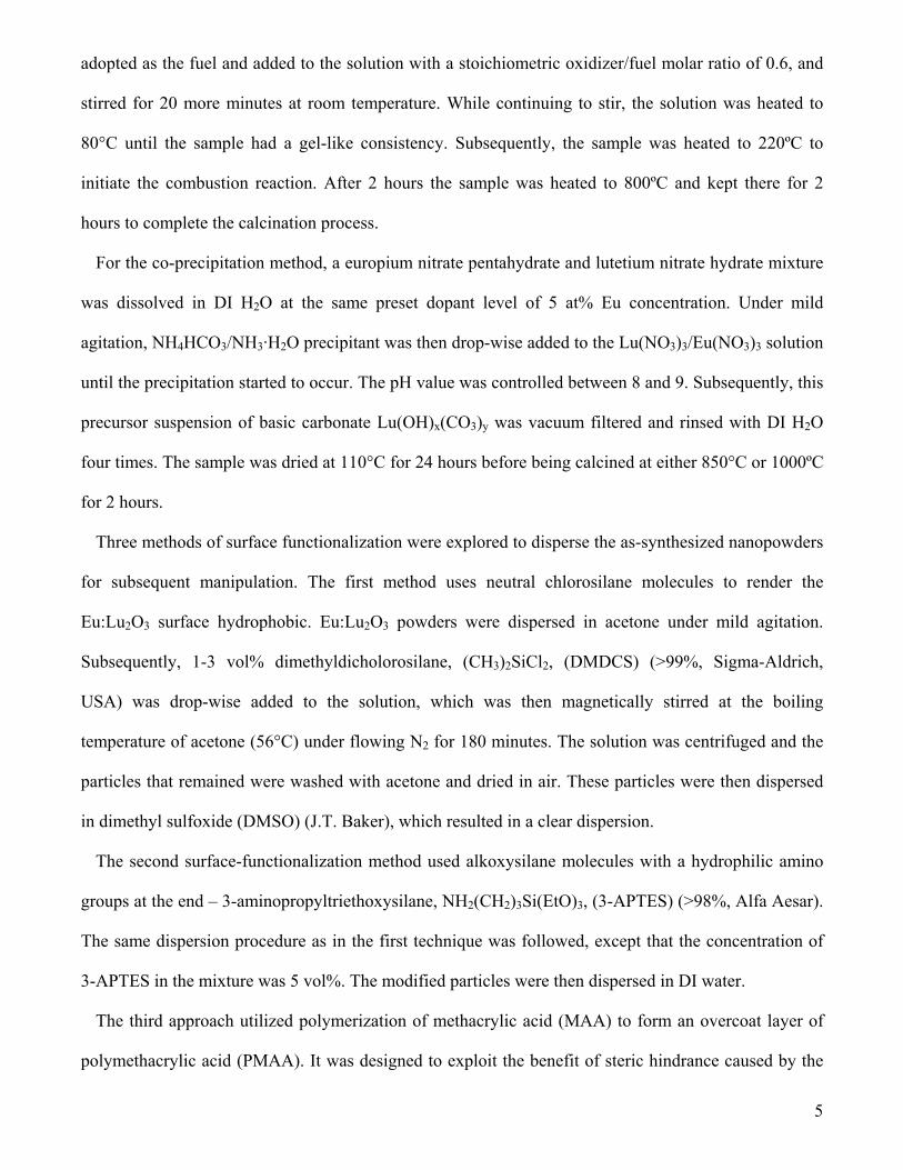
5
adopted as the fuel and added to the solution with a stoichiometric oxidizer/fuel molar ratio of 0.6, and
stirred for 20 more minutes at room temperature. While continuing to stir, the solution was heated to
80°C until the sample had a gel-like consistency. Subsequently, the sample was heated to 220ºC to
initiate the combustion reaction. After 2 hours the sample was heated to 800ºC and kept there for 2
hours to complete the calcination process.
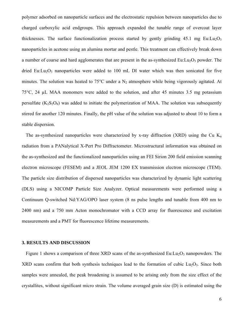
For the co-precipitation method, a europium nitrate pentahydrate and lutetium nitrate hydrate mixture
was dissolved in DI H2O at the same preset dopant level of 5 at% Eu concentration. Under mild
agitation, NH4HCO3/NH3·H2O precipitant was then drop-wise added to the Lu(NO3)3/Eu(NO3)3 solution
until the precipitation started to occur. The pH value was controlled between 8 and 9. Subsequently, this
precursor suspension of basic carbonate Lu(OH)x(CO3)y was vacuum filtered and rinsed with DI H2O
four times. The sample was dried at 110°C for 24 hours before being calcined at either 850°C or 1000ºC
for 2 hours.
Three methods of surface functionalization were explored to disperse the as-synthesized nanopowders
for subsequent manipulation. The first method uses neutral chlorosilane molecules to render the
Eu:Lu2O3 surface hydrophobic. Eu:Lu2O3 powders were dispersed in acetone under mild agitation.
Subsequently, 1-3 vol% dimethyldicholorosilane, (CH3)2SiCl2, (DMDCS) (>99%, Sigma-Aldrich,
USA) was drop-wise added to the solution, which was then magnetically stirred at the boiling
temperature of acetone (56°C) under flowing N2 for 180 minutes. The solution was centrifuged and the
particles that remained were washed with acetone and dried in air. These particles were then dispersed
in dimethyl sulfoxide (DMSO) (J.T. Baker), which resulted in a clear dispersion.
The second surface-functionalization method used alkoxysilane molecules with a hydrophilic amino
groups at the end – 3-aminopropyltriethoxysilane, NH2(CH2)3Si(EtO)3, (3-APTES) (>98%, Alfa Aesar).
The same dispersion procedure as in the first technique was followed, except that the concentration of
3-APTES in the mixture was 5 vol%. The modified particles were then dispersed in DI water.
The third approach utilized polymerization of methacrylic acid (MAA) to form an overcoat layer of
polymethacrylic acid (PMAA). It was designed to exploit the benefit of steric hindrance caused by the
6
polymer adsorbed on nanoparticle surfaces and the electrostatic repulsion between nanoparticles due to
charged carboxylic acid endgroups. This approach expanded the tunable range of overcoat layer
thicknesses. The surface functionalization process started by gently grinding 45.1 mg Eu:Lu2O3
nanoparticles in acetone using an alumina mortar and pestle. This treatment can effectively break down
a number of coarse and hard agglomerates that are present in the as-synthesized Eu:Lu2O3 powder. The
dried Eu:Lu2O3 nanoparticles were added to 100 mL DI water which was then sonicated for five
minutes. The solution was heated to 75°C under a N2 atmosphere while being vigorously agitated. At
75°C, 24 µL MAA monomers were added to the solution, and after 45 minutes 3.5 mg potassium
persulfate (K2S2O8) was added to initiate the polymerization of MAA. The solution was subsequently
stirred for another 120 minutes. Finally, the pH value of the solution was adjusted to about 10 to form a
stable dispersion.
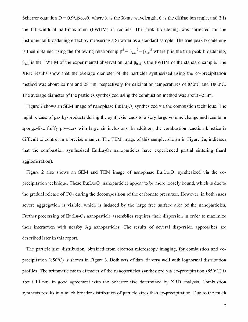
The as-synthesized nanoparticles were characterized by x-ray diffraction (XRD) using the Cu Kα
radiation from a PANalytical X-Pert Pro Diffractometer. Microstructural information was obtained on
the as-synthesized and the functionalized nanoparticles using an FEI Sirion 200 field emission scanning
electron microscope (FESEM) and a JEOL JEM 1200 EX transmission electron microscope (TEM).
The particle size distribution of dispersed nanoparticles was characterized by dynamic light scattering
(DLS) using a NICOMP Particle Size Analyzer. Optical measurements were performed using a
Continuum Q-switched Nd:YAG/OPO laser system (8 ns pulse lengths and tunable from 400 nm to
2400 nm) and a 750 mm Acton monochromator with a CCD array for fluorescence and excitation
measurements and a PMT for fluorescence lifetime measurements.
3. RESULTS AND DISCUSSION
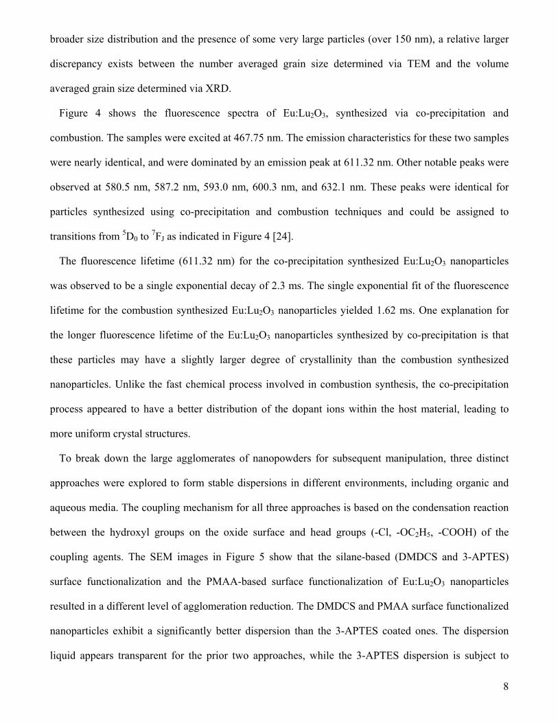
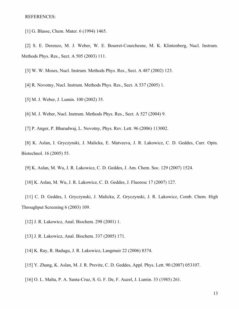
Figure 1 shows a comparison of three XRD scans of the as-synthesized Eu:Lu2O3 nanopowders. The
XRD scans confirm that both synthesis techniques lead to the formation of cubic Lu2O3. Since both
samples were annealed, the peak broadening is assumed to be arising only from the size effect of the
crystallites, without significant micro strain. The volume averaged grain size (D) is estimated using the
7
Scherrer equation D = 0.9/cos, where is the X-ray wavelength, is the diffraction angle, and is
the full-width at half-maximum (FWHM) in radians. The peak broadening was corrected for the
instrumental broadening effect by measuring a Si wafer as a standard sample. The true peak broadening
is then obtained using the following relationship β2 = βexp2 – βinst
2 where β is the true peak broadening,
βexp is the FWHM of the experimental observation, and βinst is the FWHM of the standard sample. The
XRD results show that the average diameter of the particles synthesized using the co-precipitation
method was about 20 nm and 28 nm, respectively for calcination temperatures of 850ºC and 1000ºC.
The average diameter of the particles synthesized using the combustion method was about 42 nm.
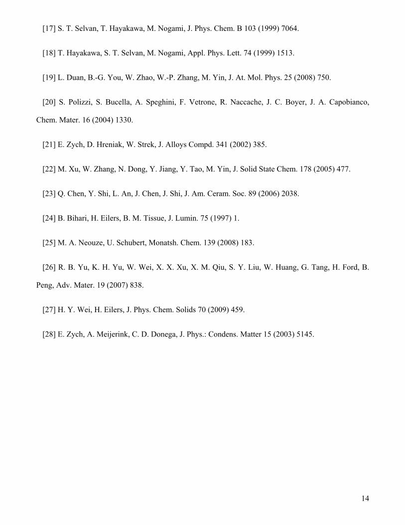
Figure 2 shows an SEM image of nanophase Eu:Lu2O3 synthesized via the combustion technique. The
rapid release of gas by-products during the synthesis leads to a very large volume change and results in
sponge-like fluffy powders with large air inclusions. In addition, the combustion reaction kinetics is
difficult to control in a precise manner. The TEM image of this sample, shown in Figure 2a, indicates
that the combustion synthesized Eu:Lu2O3 nanoparticles have experienced partial sintering (hard
agglomeration).
Figure 2 also shows an SEM and TEM image of nanophase Eu:Lu2O3 synthesized via the co-
precipitation technique. These Eu:Lu2O3 nanoparticles appear to be more loosely bound, which is due to
the gradual release of CO2 during the decomposition of the carbonate precursor. However, in both cases
severe aggregation is visible, which is induced by the large free surface area of the nanoparticles.
Further processing of Eu:Lu2O3 nanoparticle assemblies requires their dispersion in order to maximize
their interaction with nearby Ag nanoparticles. The results of several dispersion approaches are
described later in this report.
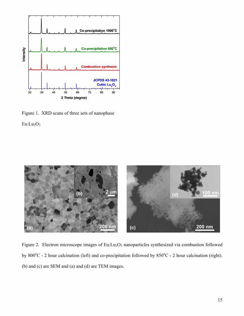
The particle size distribution, obtained from electron microscopy imaging, for combustion and co-
precipitation (850ºC) is shown in Figure 3. Both sets of data fit very well with lognormal distribution
profiles. The arithmetic mean diameter of the nanoparticles synthesized via co-precipitation (850ºC) is
about 19 nm, in good agreement with the Scherrer size determined by XRD analysis. Combustion
synthesis results in a much broader distribution of particle sizes than co-precipitation. Due to the much
8
broader size distribution and the presence of some very large particles (over 150 nm), a relative larger
discrepancy exists between the number averaged grain size determined via TEM and the volume
averaged grain size determined via XRD.
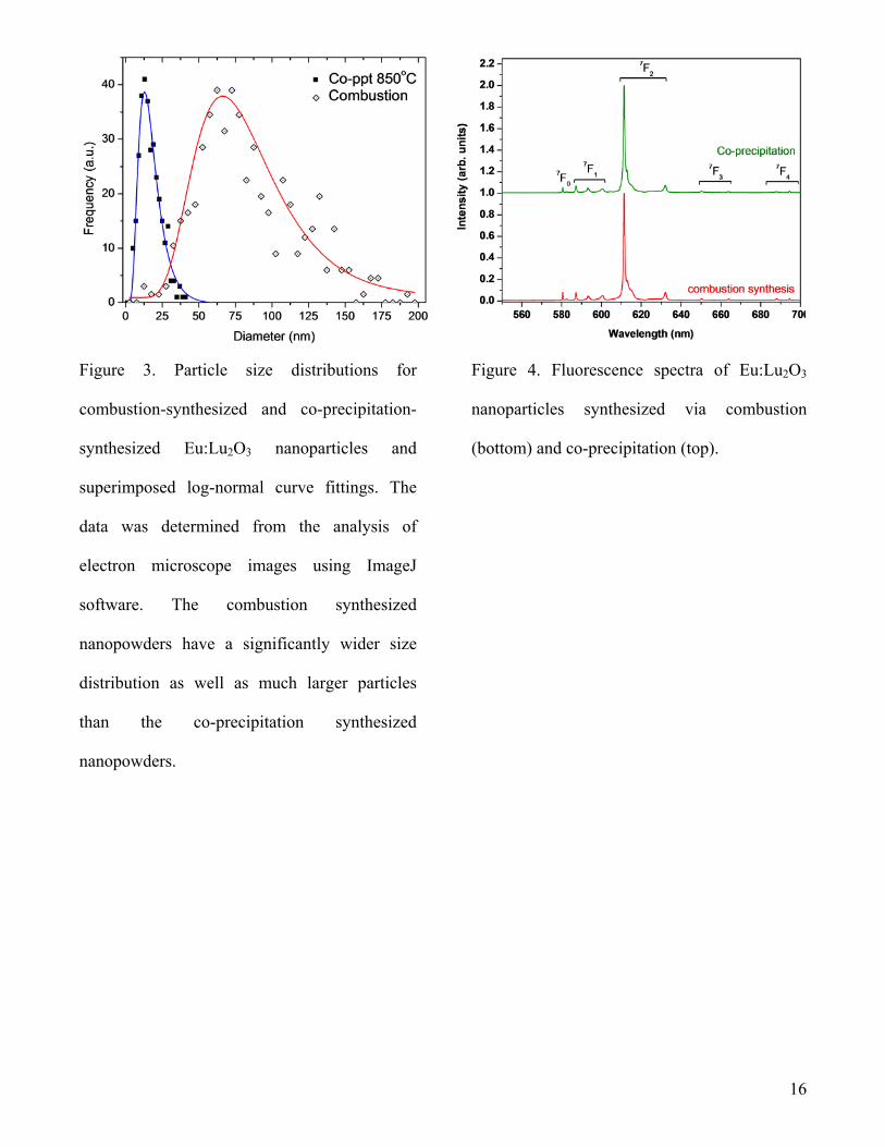
Figure 4 shows the fluorescence spectra of Eu:Lu2O3, synthesized via co-precipitation and
combustion. The samples were excited at 467.75 nm. The emission characteristics for these two samples
were nearly identical, and were dominated by an emission peak at 611.32 nm. Other notable peaks were
observed at 580.5 nm, 587.2 nm, 593.0 nm, 600.3 nm, and 632.1 nm. These peaks were identical for
particles synthesized using co-precipitation and combustion techniques and could be assigned to
transitions from 5D0 to 7FJ as indicated in Figure 4 [24].
The fluorescence lifetime (611.32 nm) for the co-precipitation synthesized Eu:Lu2O3 nanoparticles
was observed to be a single exponential decay of 2.3 ms. The single exponential fit of the fluorescence
lifetime for the combustion synthesized Eu:Lu2O3 nanoparticles yielded 1.62 ms. One explanation for
the longer fluorescence lifetime of the Eu:Lu2O3 nanoparticles synthesized by co-precipitation is that
these particles may have a slightly larger degree of crystallinity than the combustion synthesized
nanoparticles. Unlike the fast chemical process involved in combustion synthesis, the co-precipitation
process appeared to have a better distribution of the dopant ions within the host material, leading to
more uniform crystal structures.
To break down the large agglomerates of nanopowders for subsequent manipulation, three distinct
approaches were explored to form stable dispersions in different environments, including organic and
aqueous media. The coupling mechanism for all three approaches is based on the condensation reaction
between the hydroxyl groups on the oxide surface and head groups (-Cl, -OC2H5, -COOH) of the
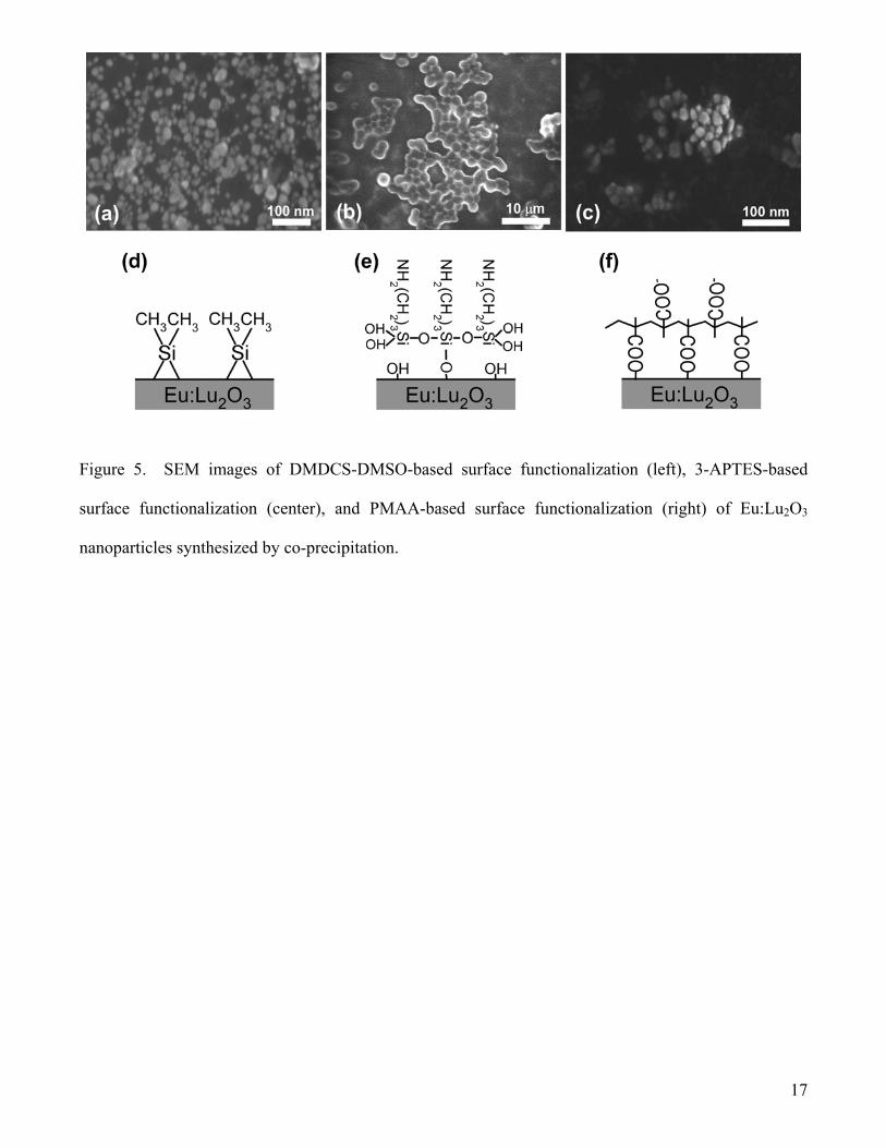
coupling agents. The SEM images in Figure 5 show that the silane-based (DMDCS and 3-APTES)
surface functionalization and the PMAA-based surface functionalization of Eu:Lu2O3 nanoparticles
resulted in a different level of agglomeration reduction. The DMDCS and PMAA surface functionalized
nanoparticles exhibit a significantly better dispersion than the 3-APTES coated ones. The dispersion
liquid appears transparent for the prior two approaches, while the 3-APTES dispersion is subject to
9
flocculation. The separation of the nanoparticles can be clearly observed in the images, with the
presence of a black gap between the particles indicating their dispersion. The 3-APTES-based surface
functionalization led to the formation of large particles (~1 µm) that formed agglomerates. Most likely,
these particles contain Eu:Lu2O3 nanoparticles.
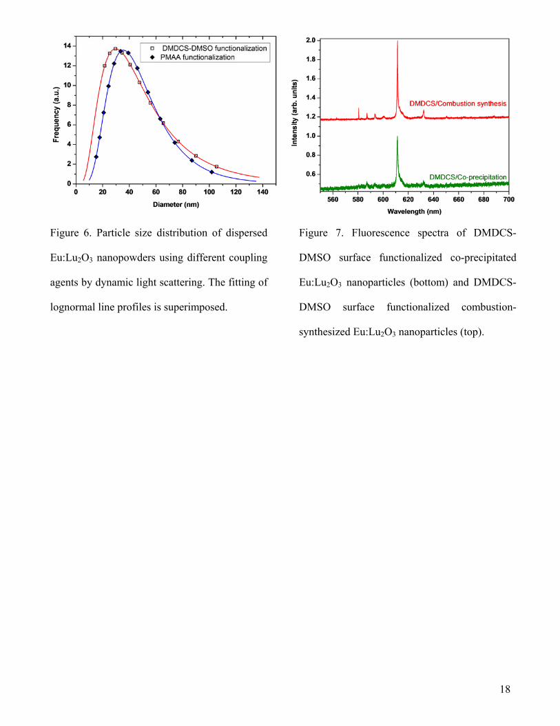
The size distribution of the DMDCS and PMAA modified Eu:Lu2O3 nanoparticles in dispersion were
measured with dynamic light scattering (DLS) as shown in Figure 6. It further confirms the important
role of DMDCS and PMAA in the separation and subsequent stabilization of Eu:Lu2O3 nanoparticles.
The proposed chemical grafting mechanisms are schematically illustrated in Figure 5. For organo-
silanes, their head groups (-Cl or –OC2H5) hydrolyze to hydroxyl moieties under the presence of water.
Although the amount of moisture present in solvent and particles was minute, they were effectively
concentrated to the oxide surface which behaves as drying agent. This water was required to activate the
silane condensation with the hydroxylized oxide surface. However, a strong side reaction (inter-silane
condensation) could occur in parallel.
The presence of three head groups in 3-APTES resulted in excess self-polymerization, forming a very
thick overcoat layer and severe crosslink between coated particles as indicated in Figure 5b. In contrast,
DMDCS has fewer head groups and stronger interactions with hydroxylized oxides [25]. That means
DMDCS reacts with not only a single hydroxyl group but also its neighbor (Figure 5d). This reaction
primarily forms a monolayer on the oxide surface with two chemical bonds between Si and oxides [26].
The coated particles are separated in the liquid due to the physical presence of a silane overcoat (steric
hindrance). A third type of coupling agent – a polymer – is used to form dispersions in aqueous media
and extend the potential tunable range of the coating thickness via controlled polymerization. The
PMAA anchored on Eu:Lu2O3 through carboxyl groups and the large polymer backbone fulfill the steric
hindrance. On the outermost layer, un-grafted carboxyl groups oriented outwards, lead to hydrophilic
behavior. This hydrophilicity enables a stable dispersion in an aqueous medium. In addition,
electrostatic repelling due to negatively charged carboxyl groups contributes further to the dispersion.
10
While in dispersion, the surface-functionalized samples showed very little or dramatically reduced
fluorescence, with the PMAA surface-functionalized material yielding the highest level of luminescence
amongst these dispersions. This reduction in fluorescence is most likely due to energy transfer from Eu-
ions near the Eu:Lu2O3 nanoparticle surface to solvent molecules. However, after centrifugation and
removal of the solvent, the fluorescence signal recovered again.
Figure 7 shows the fluorescence spectra of DMDCS surface-functionalized co-precipitated Eu:Lu2O3
nanoparticles and DMDCS surface-functionalized combustion-synthesized Eu:Lu2O3 nanoparticles. The
signal-to-noise ratio is greatly reduced in contrast to that of the as-synthesized nanopowders. The
fluorescence signal from DMDCS surface-functionalized co-precipitated Eu:Lu2O3 nanoparticles was so
weak that no fluorescence lifetime data acquisition was attainable. This lower signal may be due to the
smaller size of the nanoparticles synthesized via co-precipitation resulting in a larger surface/volume
ratio, thus affecting the optical properties of co-precipitation-synthesized materials in a larger degree
than combustion-synthesized materials. The short fluorescence lifetime (1.25 ms for combustion-
synthesized Eu:Lu2O3) and the weak fluorescence signals indicate that the interaction with DMDCS
results in significant non-radiative decay channels.
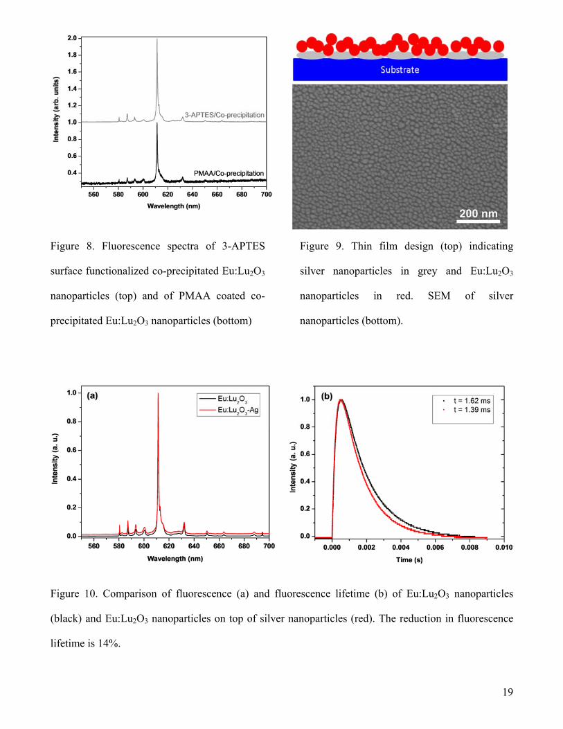
Figure 8 shows the fluorescence spectra of centrifuged 3-APTES surface functionalized co-
precipitated Eu:Lu2O3 nanoparticles (τ0=2.2 ms) and of PMAA coated co-precipitated Eu:Lu2O3
nanoparticles (in dispersion). The present measurement on the fluorescence lifetime of PMAA coated
co-precipitated Eu:Lu2O3 nanoparticles did not yield conclusive results. As the pH and the amount of
salt changes, so does the fluorescence lifetime. These fluorescence lifetime data require further
investigation before conclusions can be drawn regarding the PMAA surface functionalization.
However, it appears as though 3-APTES and PMAA affect the optical properties of Eu:Lu2O3 less
than the DMDCS surface functionalization process. The fluorescence signal from 3-APTES surface
functionalized co-precipitated Eu:Lu2O3 nanoparticles showed the best S/N ratio, while the fluorescence
signal from the PMAA coated co-precipitated Eu:Lu2O3 nanoparticles had a weak S/N ratio. Energy
transfer from Eu ions near the surface of the Eu:Lu2O3 nanoparticles appears to be the least affected for
11
3-APTES. This may be due to the lesser degree of reaction between 3-APTES and the Eu:Lu2O3
nanoparticles, as indicated in Figure 5.
To study the effectiveness of metal-enhanced fluorescence decay rate we used two different
approaches. The first approach is a thin-film structure, in which we deposited by thermal evaporation a
thin film consisting of silver nanoparticles onto a microscope slide [27]. Subsequently, we dispersed
combustion-synthesized Eu:Lu2O3 nanoparticles in a solvent and placed a drop of this dispersion on top
of the silver layer. Figure 9 shows a design schematic and SEM images of the Ag nanoparticles and
Eu:Lu2O3 nanoparticles after placement on top of the silver nanoparticles.
Figure 10 shows a comparison of the fluorescence spectra and fluorescence lifetimes for Eu:Lu2O3
nanoparticles and Eu:Lu2O3 nanoparticles on top of silver nanoparticles. The fluorescence spectra for
the two samples appear to be identical, with no apparent differences. However, the fluorescence lifetime
for Eu:Lu2O3 nanoparticles on top of silver nanoparticles is 14% shorter than the fluorescence lifetime
of Eu:Lu2O3 nanoparticles without silver. This effect was expected from equation 1 and is consistent
with the concept of metal-enhanced fluorescence or radiative decay engineering.
For the second approach, we mixed combustion-synthesized Eu:Lu2O3 nanoparticles along with silver
nanoparticles into a polystyrene matrix without the use of any surface modifiers, resulting in a brown-
colored bulk sample. Again, the fluorescence spectra appear to be identical, while the fluorescence
lifetime is shortened – by 12% in this case. Using equations 1 and 2, the measured lifetimes of 1.62 ms
and 1.39 ms in Figure 10, as well as a quantum efficiency of 85% [28], one can calculate Γ=525 s-1,
knr=92.6 s-1, Γm=102 s-1, and Q0=87.13%. Because Eu:Lu2O3 starts out with a relatively high quantum
efficiency of 85%, the observed changes are relatively small. However, the initial observations confirm
that it is possible to reduce the fluorescence lifetime of scintillator materials. Nevertheless, the
implementation of this approach requires further investigations on the improvement of surface
functionalization and dispersion of these materials, the influence of surface ligands on optical responses,
as well as optimization of interparticle spacing between colloidal metallic nanoparticles and Eu:Lu2O3
nanoparticles.
12
4. CONCLUSION
Nanoparticles of the scintillator material Eu:Lu2O3 were successfully synthesized using combustion
and co-precipitation. The co-precipitation method yielded particles with a diameter of about 25 nm and
the combustion technique yielded particles with a diameter of about 40 nm. Three surface modification
techniques were explored to improve the dispersion of the nanoparticles, and their effects on the
fluorescent emission were studied. DMDCS surface functionalization resulted in significantly better
dispersion than 3-APTES and PMAA based approaches. However, this approach significantly reduced
the fluorescence of the material. As expected, the combination of the Eu:Lu2O3 nanoparticles with silver
nanoparticles led to reductions of the fluorescence lifetime of 12-14% in the present design. Further
tailoring of the optical properties depends on the optimization of the separation between metal and
Eu:Lu2O3 nanoparticles through improved dispersion and surface functionalization.
ACKNOWLEDGEMENT
This work was supported by DHS/DNDO Grant 2008DN-077ARI004-02, NSF Grant CBET-
0735911, and ONR Grant N00014-03-1-0247.
13
REFERENCES:
[1] G. Blasse, Chem. Mater. 6 (1994) 1465.
[2] S. E. Derenzo, M. J. Weber, W. E. Bourret-Courchesne, M. K. Klintenberg, Nucl. Instrum.
Methods Phys. Res., Sect. A 505 (2003) 111.
[3] W. W. Moses, Nucl. Instrum. Methods Phys. Res., Sect. A 487 (2002) 123.
[4] R. Novotny, Nucl. Instrum. Methods Phys. Res., Sect. A 537 (2005) 1.
[5] M. J. Weber, J. Lumin. 100 (2002) 35.
[6] M. J. Weber, Nucl. Instrum. Methods Phys. Res., Sect. A 527 (2004) 9.
[7] P. Anger, P. Bharadwaj, L. Novotny, Phys. Rev. Lett. 96 (2006) 113002.
[8] K. Aslan, I. Gryczynski, J. Malicka, E. Matveeva, J. R. Lakowicz, C. D. Geddes, Curr. Opin.
Biotechnol. 16 (2005) 55.
[9] K. Aslan, M. Wu, J. R. Lakowicz, C. D. Geddes, J. Am. Chem. Soc. 129 (2007) 1524.
[10] K. Aslan, M. Wu, J. R. Lakowicz, C. D. Geddes, J. Fluoresc 17 (2007) 127.
[11] C. D. Geddes, I. Gryczynski, J. Malicka, Z. Gryczynski, J. R. Lakowicz, Comb. Chem. High
Throughput Screening 6 (2003) 109.
[12] J. R. Lakowicz, Anal. Biochem. 298 (2001) 1.
[13] J. R. Lakowicz, Anal. Biochem. 337 (2005) 171.
[14] K. Ray, R. Badugu, J. R. Lakowicz, Langmuir 22 (2006) 8374.
[15] Y. Zhang, K. Aslan, M. J. R. Previte, C. D. Geddes, Appl. Phys. Lett. 90 (2007) 053107.
[16] O. L. Malta, P. A. Santa-Cruz, S. G. F. De, F. Auzel, J. Lumin. 33 (1985) 261.
14
[17] S. T. Selvan, T. Hayakawa, M. Nogami, J. Phys. Chem. B 103 (1999) 7064.
[18] T. Hayakawa, S. T. Selvan, M. Nogami, Appl. Phys. Lett. 74 (1999) 1513.
[19] L. Duan, B.-G. You, W. Zhao, W.-P. Zhang, M. Yin, J. At. Mol. Phys. 25 (2008) 750.
[20] S. Polizzi, S. Bucella, A. Speghini, F. Vetrone, R. Naccache, J. C. Boyer, J. A. Capobianco,
Chem. Mater. 16 (2004) 1330.
[21] E. Zych, D. Hreniak, W. Strek, J. Alloys Compd. 341 (2002) 385.
[22] M. Xu, W. Zhang, N. Dong, Y. Jiang, Y. Tao, M. Yin, J. Solid State Chem. 178 (2005) 477.
[23] Q. Chen, Y. Shi, L. An, J. Chen, J. Shi, J. Am. Ceram. Soc. 89 (2006) 2038.
[24] B. Bihari, H. Eilers, B. M. Tissue, J. Lumin. 75 (1997) 1.
[25] M. A. Neouze, U. Schubert, Monatsh. Chem. 139 (2008) 183.
[26] R. B. Yu, K. H. Yu, W. Wei, X. X. Xu, X. M. Qiu, S. Y. Liu, W. Huang, G. Tang, H. Ford, B.
Peng, Adv. Mater. 19 (2007) 838.
[27] H. Y. Wei, H. Eilers, J. Phys. Chem. Solids 70 (2009) 459.
[28] E. Zych, A. Meijerink, C. D. Donega, J. Phys.: Condens. Matter 15 (2003) 5145.
15
Figure 1. XRD scans of three sets of nanophase
Eu:Lu2O3
Figure 2. Electron microscope images of Eu:Lu2O3 nanoparticles synthesized via combustion followed
by 800oC - 2 hour calcination (left) and co-precipitation followed by 850oC - 2 hour calcination (right).
(b) and (c) are SEM and (a) and (d) are TEM images.
16
Figure 3. Particle size distributions for
combustion-synthesized and co-precipitation-
synthesized Eu:Lu2O3 nanoparticles and
superimposed log-normal curve fittings. The
data was determined from the analysis of
electron microscope images using ImageJ
software. The combustion synthesized
nanopowders have a significantly wider size
distribution as well as much larger particles
than the co-precipitation synthesized
nanopowders.
Figure 4. Fluorescence spectra of Eu:Lu2O3
nanoparticles synthesized via combustion
(bottom) and co-precipitation (top).
17
Figure 5. SEM images of DMDCS-DMSO-based surface functionalization (left), 3-APTES-based
surface functionalization (center), and PMAA-based surface functionalization (right) of Eu:Lu2O3
nanoparticles synthesized by co-precipitation.
18
Figure 6. Particle size distribution of dispersed
Eu:Lu2O3 nanopowders using different coupling
agents by dynamic light scattering. The fitting of
lognormal line profiles is superimposed.
Figure 7. Fluorescence spectra of DMDCS-
DMSO surface functionalized co-precipitated
Eu:Lu2O3 nanoparticles (bottom) and DMDCS-
DMSO surface functionalized combustion-
synthesized Eu:Lu2O3 nanoparticles (top).
19
Figure 8. Fluorescence spectra of 3-APTES
surface functionalized co-precipitated Eu:Lu2O3
nanoparticles (top) and of PMAA coated co-
precipitated Eu:Lu2O3 nanoparticles (bottom)
Figure 9. Thin film design (top) indicating
silver nanoparticles in grey and Eu:Lu2O3
nanoparticles in red. SEM of silver
nanoparticles (bottom).
Figure 10. Comparison of fluorescence (a) and fluorescence lifetime (b) of Eu:Lu2O3 nanoparticles
(black) and Eu:Lu2O3 nanoparticles on top of silver nanoparticles (red). The reduction in fluorescence
lifetime is 14%.


