Embed Size (px)
Citation preview
Copyright 2008 by the American Chemical Society VOLUME 112, NUMBER 25, JUNE 26, 2008
ARTICLES
Fluorescence Spectroscopy and Amplified Spontaneous Emission (ASE) of Phenylimidazoles:Predicted Vibronic Coupling Along the Excited-State Intramolecular Proton Transfer in2-(2′-Hydroxyphenyl)imidazoles
Juan Carlos del Valle,*,† R. M. Claramunt,‡ and J. Catalan*,†
Department of Quımica Fısica Aplicada, UniVersidad Autonoma de Madrid, Cantoblanco,C-2-203, E-28049 Madrid, Spain and Department of Quımica Organica y Biologıa, Facultad de Ciencias,UNED, Senda del Rey 9, E-28040 Madrid, Spain
ReceiVed: July 5, 2007; ReVised Manuscript ReceiVed: April 14, 2008
Methylation at the 1N position of 2-phenylimidazole provides the shortest wavelength for a liquid-state laserdye reported to date; that is, the 1-methyl-2-phenylimidazole molecule in cyclohexane solution yields amplifiedspontaneous emission (ASE) with a peak wavelength at 314.5 nm and a constant laser gain value of 5 cm-1
from 310 to 317 nm. Methyl substitution in this case favors the appearance of laser action (owing to atorsion-vibrational mechanism) in cyclohexane as compared with the nonmethylated species which does notexhibit ASE in this solvent. The 2-(2′-hydroxyphenyl)imidazole molecules give rise to ASE with high gainvalues (ca. 9 cm-1) at 450 and 466 nm. The mechanism of population inversion is understood in terms of avibronic coupling between the hydroxyl stretching motion and the torsional vibration of the phenyl andimidazole rings. The proton-transfer spectroscopy of 2-(2′-hydroxyphenyl)imidazoles is studied in dioxane,cyclohexane, dimethyl sulfoxide, methanol, and water. The greater the acidity of the solvent the greater thedisruption of the intramolecular hydrogen bond; solvent acidity is the main parameter which favors formationof the open-form species in the ground electronic state. Methyl substitution at the 1N position favors formationof the open species for 2-hydroxyphenylimidazoles in the ground electronic state, which decreases their owncapacity to undergo ASE. Low-temperature absorption spectroscopy confirms aggregation processes for 2-(2′-hydroxyphenyl)imidazoles in solution. In accordance with X-ray analyses in the solid phase, these moleculesform associations through intermolecular chains of the type NsH · · ·O or OsH · · ·N.
I. Introduction
Laser spectroscopy has dealt with the research of themechanisms responsible for the laser action monitored in manymolecular,1–3 ion,2–5 and atomic systems2,6 with the aid of light
absorption and emission techniques with special emphasis inthe UV region in which obtaining molecular-dye systems withhigh laser gain has taken much effort.
Among the molecular-dye laser systems, those concernedwith the proton-transfer mechanism in the excited state guaranteepopulation inversion,7 thus presenting high laser gains. The firstproton-transfer laser was proposed in 1983 based on the3-hydroxyflavone molecular structure;7,8 this molecule presents
* Corresponding authors. Phone: +91-4974263. Fax: +91-4974785.E-mail: [email protected]; [email protected]; [email protected].
† Universidad Autonoma de Madrid.‡ UNED.
10.1021/jp7117604 CCC: $40.75 2008 American Chemical SocietyPublished on Web 05/30/2008
an excited-state intramolecular-proton-transfer (ESIPT) processwhich yields laser action. Other pioneer families of ESIPT laserswere reported in 1986, for instance, the salicylamide,9 sodiumsalicylate,10 and 2-(2-hydroxyphenyl) benzimidazole10,11 mo-lecular lasers with efficiencies up to 10%. However, all of themexhibit lasing action in the visible spectral region.
Another laser mechanism proposed to develop lasing actionin the UV spectrum consisted of a torsion-conformationalchange undergone on photoexcitation of 2-phenylbenzimidazoleand its derivatives.12,13 The inter-ring torsion turns out to beessential to develop the lasing action, which also possesses largegain coefficient values. In any case, as previously published7,12,13
the population-inversion requirement is supplied by a large orsignificant structural change on electronic excitation. Forinstance, if upon photoexcitation the structural change is notsignificant, the population inversion is not attained and themolecular system does not undergo laser or amplified spontane-ous emission (ASE), as occurs for the fluorene molecule.14
Interestingly, the amplified spontaneous emission (ASE, lasingaction) of a hydroxyl-substituted tetraphenylimidazole (i.e.,acetic acid 2-{4-[2-(2-hydroxyphenyl)-4,5-diphenylimidazol-1-yl]phenyl}-ethylester)15 has been recently reported from a largesingle crystal owing to an ESIPT mechanism.
We propose simple phenylimidazole structures, which reduceits heterocyclic size, to undergo laser emission with high gainvalues in the UV region, displaying a significant and sufficientstructural change upon photoexcitation and a comprehensivemolecular laser mechanism. The population-inversion mecha-nism involved in 2-(2′-hydroxyphenyl)imidazole (2HOPI) willbe explored from a theoretical and experimental point of view.This mechanism consists of an excited-state proton-transferprocess along with a vibronic coupling between the intramo-lecular hydroxy stretching and the inter-ring torsional vibration(vide infra). A methyl substitution has been undergone at thepyrrole nitrogen of 2-(2′-hydroxyphenyl)imidazole (1M2HOPI)in order to better visualize this vibronic coupling.
From an experimental point of view, nothing has beenpublished on the photophysics of the 2HOPI and 1M2HOPImolecules. Nevertheless, experimental evidence was reported16
on some derivatives of 2HOPI which exhibited large molarabsorption coefficients (εmax = 20 000 M-1 cm-1) in cyclohex-ane, thus featuring π,π* transitions, for instance, 4,5-diphenyl-2-(2′-hydroxyphenyl)imidazole (DPHPI), 4,5-dimethyl-2-(2′-hydroxyphenyl)imidazole (DMeHPI), and methylphenyl-2-(2′-hydroxyphenyl)imidazole (MePHPI). It is also reported16 thatthe shape and position of the first absorption bands changescarcely with solvent polarity, thus proving that the n,π* statesare shifted to higher energies, owing to the intramolecular hydro-gen interaction with the nonbonding electrons of the pyridinicnitrogen. A planar ground-state conformation is predicted onthe basis of the red shift found for the first absorption band forMePHPI as compared to that for DPHPI, from 30417 to 325nm, respectively.16 The fluorescence of DMeHPI in cyclohexane,acetonitrile, and p-dioxane showed only one emission band (withrespective quantum yields of 0.47, 0.48, and 0.55) largely shiftedfrom the corresponding absorption spectrum (8320, 7762, and7854 cm-1, respectively), thus assigned to ESIPT emission.16
The corresponding stimulated emission (obtained by using aresonant cavity)16 pumped with an XeCl laser at 308 nm showedlarge gain values in acetonitrile and p-dioxane, 8.9% and 12.1%,respectively.
DMeHPI (at ca. 350 nm) and MePHPI presented an extraemission band in methanol and dimethylformamide also ascribedto the solvated open form.16 In water at neutral pH, for instance,
DMeHPI showed a chemical equilibrium between the open formand the keto form in the ground state.18
The rotamerism and ESIPT processes for the S1, T1, and So
states of the 2-(2′-hydroxyphenyl)imidazole (2HOPI) were inves-tigated at the AM1 level using configurational interaction withsingle excitations (CIS)19 and at the HF/CIS/D95** level.20
These calculations have addressed both ESIPT and rotamerism,separate and uncoupled, processes.19,20
Basically, the same conclusions have been drawn in both ofthese studies.19,20 (I) Rotamerism in 2HOPI in the ground andthe first singlet π,π* excited state indicates that the cis (enolform, closed form) and trans rotamers (another closed form)are interconvertible in the ground state but not in the first excitedstate. (II) The two tautomers keto-enol of 2HOPI become closerin energy compared to the ground state (ca. 27 kcal/mol energydifference), thus giving rise to an exothermic process in the T1
state and S1 state. (III) The energy barrier for the proton-transferprocess decreases upon excitation to the T1 and S1 states. Theketo form is less destabilized than the enol form also for the1n,π* (S2 state) excitation.20
However, it is well known that the HF and CIS/HF treatmentsoverestimate the hydrogen-bond interactions (as well as the AM1method), and they usually predict too high proton-transferbarriers,21–23 owing to the neglect of electronic correlation (i.e.,∆Eenol-keto for 2HOPI in So, S1 - π,π*, S2 - n,π*, and T1 -π,π* results in 17.6, 9.3, 15.4, and 17.7 kcal/mol, respectively).20
The theoretical framework introduced in the following studyon 2HOPI and its 1N-methylated derivative is intended tocombine both processes, the inter-ring torsion and ESIPT, andshed light on the experimental evidence. As described in theTheoretical and Experimental Methods section, DFT methodol-ogy and the TD/CIS approach have been utilized for the sakeof taking into account a greater contribution for the electroniccorrelation.
II. Theoretical and Experimental Methods
A. Absorption Spectroscopy. Absorption measurementswere made for the various phenylimidazoles (Scheme 1) insolution at 298 K in the solvents given in the text and figurelegends. The absorption spectra were recorded with the aid ofa Shimadzu UV-2100 spectrophotometer.
Absorption from room temperature (RT) to 110 K (slowfreezing) was recorded with the aid of a Cary 5 spectrometer(Varian) and a cryostat Oxford Optistat DN, which uses liquidnitrogen to control temperature. The solvents were of spectro-metric quality and checked for fluorescence impurities.
SCHEME 1: Closed-Form Molecular Structures for2HOPI, 1M2HOPI, and 2MeOPIa
a The atom numbering is implemented.
5556 J. Phys. Chem. A, Vol. 112, No. 25, 2008 del Valle et al.
B. Emission Spectroscopy. Emission spectra were obtainedwith the aid of an AB2 Aminco-Bowman Series 2 luminescencespectrometer. All spectra were recorded at 298 and 77 K. Thelow-temperature (quick freezing) fluorescence in methylcyclo-hexane and cyclohexane was recorded using a liquid nitrogenDewar, which was built in a sample chamber purged with pureand dry N2 gas. Fluorescence spectra were corrected forinstrumental sensitivity when plotted together with the corre-sponding ASE spectra.
Emission spectra from RT to 113 K (slow freezing) wererecorded with the aid of AB2 Aminco-Bowman Series 2luminescence spectrometer and a cryostat Oxford Optistat DN,which uses liquid nitrogen to control temperature.
Fluorescence quantum yields were measured at 298 K onfreshly prepared samples with absorbances at the excitationwavelength of ca. 0.1. The fluorescence standard 2-aminopy-ridine (Φf ) 0.66)24 in 0.1 N H2SO4 was used for themeasurements, which were corrected by taking into account therefractive index of the solvent.
Fluorescence lifetimes were determined with a phase andmodulation spectrofluorimeter (SLM 48000S) at frequenciesbetween 1 and 250 MHz.25 Decisions on the suitability of thelifetimes rested on examination of the residual deviations withfrequency and the reduced �2 values, which were close to unity.The detection limit of our instrument is ca. 30 ps.
C. Amplified Spontaneous Emission (ASE) Spectroscopy.The ASE laser spike measurements12,26,27 were made by primaryexcitation with a Nd:YAG laser (Spectra-Physics model DCR-3G) using the fourth harmonic (266 nm) for 1-methyl-2-phenyl-imidazole (1M2PI), 2-(2′-hydroxyphenyl)imidazole (2HOPI),1-methyl-2-(2′-hydroxyphenyl)imidazole (1M2HOPI), and 2-(2′-methoxypheny)imidazole (2MeOPI). The output of the Nd:YAGlaser was focused to a narrow line in the dye cell in a transversegeometry. A DL 251 laser dye cell of 0.8 cm optical lengthwith oblique windows was used (NSG precision cells Inc.), thuspreventing optical feedback. The dye solution was stirred toprevent secondary processes, such as, for example, local heating,from interfering with the experiments. The ASE signal wasdispersed by a 300 lines/mm and 0.32 m polychromator(Instruments SA model HR320) detected by an optical multi-channel analyzer (OMA) system consisting of an intensifiedsilicon photodiode array (EG and G/PAR, model 1421) andanalyzed by a system processor (EG and G/PAR, model 1461/1463). To reduce the noise, the detector was operated in a gatedmode synchronously with the Nd:YAG laser. The ASE signalwas detected through a pinhole 0.05 cm in diameter placed ata distance of ca. 50 cm from the sample cell. To measure thegain coefficient values (R(λ)),28 an optically calibrated half-cellshutter was placed between the primary laser excitation beamand the cell in order to block one-half the intensity of theexcitation beam. The intensities (I) of the primary laserexcitation at the respective full-cell length (L) 0.8 cm) andhalf-cell length (L/2 ) 0.4 cm) of the shutter were opticallycalibrated with a volume absorbing disk calorimeter (Scientech,Inc. model 36-0001).
R(λ)) 2L
ln[ IL
IL⁄2- 1] (1)
The photostability of the molecules studied was demonstratedby monitoring the absorption spectra before and after the laserexperiments using a cuvette 0.01 cm in optical length.
D. Materials. The 2-phenylimidazole molecule was pur-chased from Aldrich Chemical Co. and used as supplied. 2-(2′-Hydroxyphenyl)imidazole (2HOPI),29 1-methyl-2-phenylimi-
dazole,30 2-(2′-methoxypheny)imidazole (2MeOPI), and 1-methyl-2-(2′-hydroxyphenyl)imidazole (1M2HOPI)29 were synthesizedas described elsewhere.31 The NMR and X-ray analyses as wellas the crystal and molecular structure were also publishedelsewhere.31
E. Theoretical Calculations. Ground-state (So) and first tri-plet-state (T1) geometries were optimized using density func-tional theory32,33 with the B3LYP hybrid functional and the6-31G** basis set.34 All calculations were executed by employ-ing the GAUSSIAN 03 package35 and the SPARTAN 4.1 pro-gram.36 The global minima were checked by means of vibra-tional frequency analyses. All the structures thus identified werefound to correspond to true minima as they had all realvibrational frequencies. Calculations on the first singlet excitedstate were executed at the time-dependent (TD) DFT level37,38
and also with the CIS39 methodology by utilizing the split-valence 6-31G** basis set. The proton-transfer potential-energycurves were calculated with full geometry optimization for thestationary points in the So and T1 states and for the intermediategeometry points by fixing the OH distances and allowing theremaining coordinates to be optimized without constraints.
The S1 state energy was estimated following two procedures:(i) with the aid of the So-optimized geometry at the DFT leveland TD-DFT calculations [TD/DFT(So)] and (ii) the TD/CISapproach, which is based upon using (a) CIS methodology bykeeping the hydrogen-bond coordinates of the fully optimizedSo structure at the DFT level fixed (i.e., N3sH2′, H2′sO2′,O2′sC2′, N3sH2′sO2′, Scheme 1) and allowing the remainingcoordinates to be fully optimized and (b) subsequently the finalgeometry is obtained in a TD-DFT calculation. Use of only theCIS methodology without constraints provides very unreliableresults for molecular systems with intramolecular hydrogenbonds.21–23
III. Results and Discussion
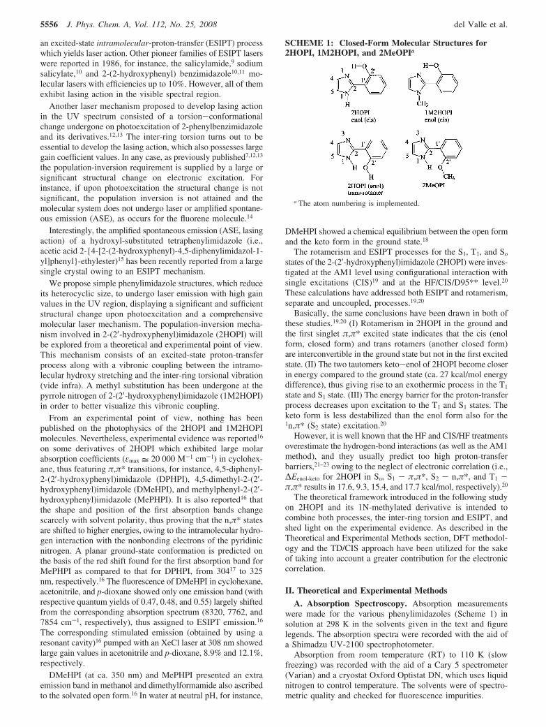
A. Absorption and Fluorescence Spectroscopy and ASEof 1-Methyl-2-phenylimidazole. The fluorescence spectroscopyand amplified spontaneous emission (ASE) of 2-phenylimida-zole, 2-phenylbenzimidazole, and 1-methyl-2-phenylbenzimi-dazole in dioxane were reported previously.12 They exhibit highASE spikes at 320, 341, and 345.5 nm, respectively, with highlaser gain values of ca. 10 cm-1. Also, the 1-methyl-2-phenyl-imidazole molecule presents ASE at 323.5 nm in dioxane(Figure 1). In the current work, the N-methyl substitution empha-sizes the inter-ring torsional-vibrational mechanism whichexplains how this molecule achieves population inversion.
Figure 1. Absorption, fluorescence (λexc ) 260 nm), and ASE spectraof 1-methyl-2-phenylimidazole in dioxane (bottom) and cyclohexane(top).
Fluorescence Spectroscopy and ASE of Phenylimidazoles J. Phys. Chem. A, Vol. 112, No. 25, 2008 5557
Indeed, in cyclohexane 2-phenylimidazole did not yield lasingaction, also owing to low solubility; its concentration could notbe larger than 4.3 × 10⁻³ M; however, the 1-methyl derivativeat 4.2 × 10⁻³ M gives rise to dual-peak ASE at 314.5 and 323.5nm (Figure 1), thus providing the shortest laser radiation reportedup to date. The shortest tunable laser was technically reported2,3,40
for 2,2′-dimethyl-p-terphenyl with a peak laser wavelength at336 nm. The laser-gain spectrum for the 1-methyl-2-phenylimi-dazole is implemented in the Supporting Information (SI, Sfig-1)and shows a constant gain value of 5 cm-1 from 310 to 317nm in cyclohexane and a gain value of ca. 8 cm-1 at 323.5nmin dioxane. The 1N-methylated molecules possess larger inter-ring dihedral angles in the ground electronic state than thosefor their analogues without methyl groups.12 Indeed, uponphotoexcitation they all tend to be coplanar, but the inter-ringdihedral angle changes to a greater extent for the methylatedones; for instance, for 2-phenylbenzimidazole it is 20° in theSo state and changes to 0° in the S1 state, but for its 1N-methyl-ated derivative it changes from 42° (So) to 2° (S1).12
The molecules 2HOPI and 1M2HOPI comprise one imidazolering covalently linked to a phenol group, thereby implementingboth feasible processes: ESIPT and inter-ring torsion. Thecoupling of both motions is investigated in the followingsections.
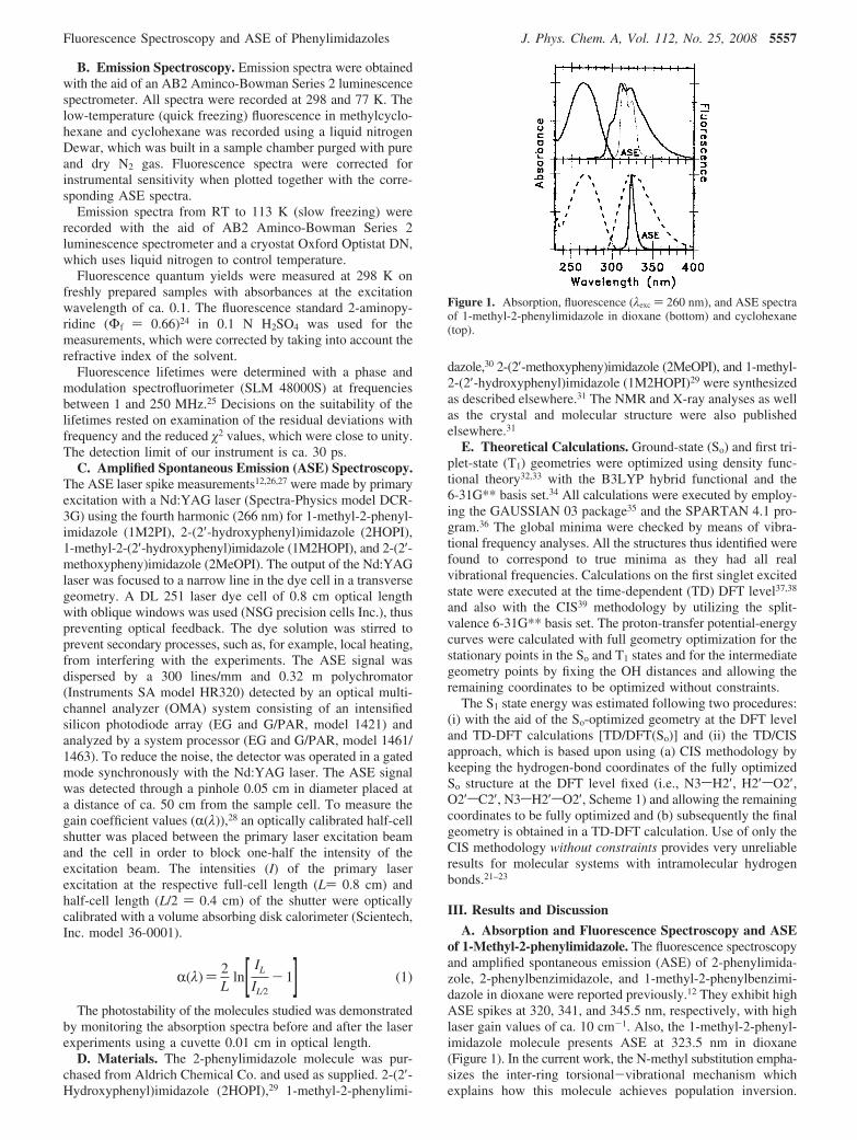
B. Absorption and Fluorescence Spectroscopy in 2-(2′-Hydroxyphenyl)imidazoles. The molecular structures of some2-(2′-hydroxyphenyl)imidazoles were determined by X-ray cry-stallography (Figure 2 and SI, Table S1).31 The 2HOPI moleculecontains a strong intramolecular hydrogen bond (IMHB) incrystal phase. The inter-ring torsional angle of -1.2° and theIMHB dihedral angle of -3.2° yield a quasi-planar molecularstructure. This structure differs from that of the 1-methylderivative, 1M2HOPI, which does not exhibit IMHB in thecrystal phase (Figure 2); indeed, the hydroxyl-phenyl group istwisted and thus facing the 1-methyl group, forming the trans
rotamer molecule (Scheme 2, another open form). The inter-ring dihedral angle for 1M2HOPI is -116.8° (+63.2°), and thehydroxy vs imidazole dihedral angle results in -153.2° (+26.8°).In the crystal phase 2HOPI and 1M2HOPI form chains throughN-H · · ·O and O-H · · ·N intermolecular hydrogen bonds,respectively.
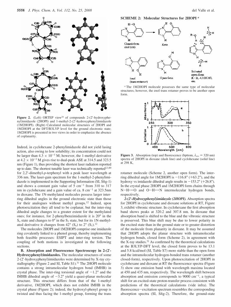
2-(2′-Hydroxyphenyl)imidazole (2HOPI). Absorption spectrafor 2HOPI in cyclohexane and dioxane solutions at RT, Figure3, exhibit vibronic structure. In cyclohexane the first absorptionband shows peaks at 320.2 and 307.8 nm. In dioxane thatabsorption band is shifted to the blue and the vibronic structureis preserved. This blue shift may be due to lower polarity inthe excited state than in the ground state or to greater distortionof the molecule from planarity in dioxane. It may be assumedthat 2HOPI adopts the planar structure with intramolecularhydrogen bonds, closed form (Scheme 2), in agreement withthe X-ray studies.31 As confirmed by the theoretical calculationsat the B3LYP-DFT level, the closed form proves to be 13.1and 5.8 kcal/mol (SI, Table S7) more stable than the open formand the intramolecular hydrogen-bonded trans rotamer (anotherclosed-form), respectively. Upon photoexcitation of 2HOPI incyclohexane and dioxane at RT the fluorescence spectra (Figure3) show one emission band with wavelength maxima locatedat 450 and 435 nm, respectively. The wavelength shift betweenabsorption and emission corresponds to 9000 cm-1, a typicalshift for an excited-state proton-transfer process consistent withpredictions of the theoretical calculations (vide infra). Thefluorescence-excitation spectrum resembles the correspondingabsorption spectra (SI, Sfig-2). Therefore, the ground-state
Figure 2. (Left) ORTEP view24 of compounds 2-(2′-hydroxyphe-nyl)imidazole (2HOPI) and 1-methyl-2-(2′-hydroxyphenyl)imidazole(1M2HOPI). (Right) Calculated molecular structures of 2HOPI and1M2HOPI at the DFT/B3LYP level for the ground electronic state;1M2HOPI is presented in two views in order to emphasize the absenceof coplanarity.
SCHEME 2: Molecular Structures for 2HOPI a
a The 1M2HOPI molecule possesses the same type of molecularstructures; however, the enol trans rotamer proves to be another openform.
Figure 3. Absorption (top) and fluorescence (bpttom, λexc ) 320 nm)spectra of 2HOPI in dioxane (dash line) and cyclohexane (solid line)at 298 K,
5558 J. Phys. Chem. A, Vol. 112, No. 25, 2008 del Valle et al.
molecular source of this emission is the closed form (enol cis,Scheme 2), the intramolecularly hydrogen-bonded 2HOPIspecies. The proton-transfer fluorescence quantum yield mea-sured in cyclohexane is 0.51 with a lifetime of 4.2 ns. Fromthese data, a radiative rate (kf) of 1.2 × 10⁸ s-1 is calculated.On the assumption that the proton-transfer tautomer forms withunit quantum yield in the first singlet excited state.
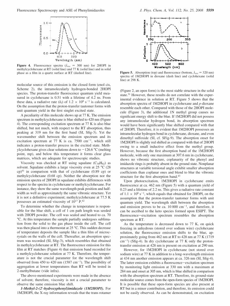
A peculiarity of this molecule shows up at 77 K. The emissionspectrum in methylcyclohexane is blue shifted to 420 nm (Figure4). The corresponding excitation spectrum at 77 K is also blueshifted, but not much, with respect to the RT absorption, thuspeaking at 319 nm for the first band (SI, Sfig-3). Yet thewavenumber shift between the emission spectrum and itsexcitation spectrum at 77 K is ca. 7500 cm-1, which stillindicates a proton-transfer process in the excited state. Meth-ylcyclohexane gives clear solutions down to -126.6 °C (meltingpoint, mp), and below this temperature it forms clear glassmatrices, which are adequate for spectroscopic studies.
Viscosity was checked at RT using squalane (C30H62) assolvent. Squalane exhibits a large viscosity even at 25 °C (28cp)41 in comparison with that of cyclohexane (0.89 cp) ormethylcyclohexane (0.68 cp). Neither the absorption nor theemission spectra of 2HOPI in squalane exhibits differences withrespect to the spectra in cyclohexane or methylcyclohexane. Forinstance, they show the same wavelength peak position and half-width as well as approximately the same vibronic structure. Yetit is not a definitive experiment; methylcyclohexane at 77.5 Kpossesses an estimated viscosity of 1017 P.42
To determine whether the change in temperature is respon-sible for the blue shift, a cell of 1 cm path length was seededwith 2HOPI powder. The cell was sealed and heated to ca. 70°C. At this temperature the sample partially undergoes sublima-tion from the solid to the gas phase inside the cell. The cellwas then placed into a thermostat at 25 °C. This sudden decreaseof temperature deposits the sample like a thin film of microc-rystals on the walls of the cell. Thereafter, an absorption spec-trum was recorded (SI, Sfig-3), which resembles that obtainedin methylcyclohexane at RT. The fluorescence emission for thisfilm at RT matches (Figure 4) that emission band recorded fora methylcyclohexane solution at 77 K. Therefore, the temper-ature is not the crucial parameter for the wavelength shiftobserved from 450 to 420 nm (1587 cm-1). The possibility ofaggregation at lower temperatures than RT will be tested in2-methylbutane (vide infra).
The above-mentioned experiments were made in the absenceof solvent; therefore, viscosity is not a crucial parameter toobserve the same emission blue shift.
1-Methyl-2-(2′-hydroxyphenyl)imidazole (1M2HOPI). For1M2HOPI, the X-ray information reveals that the trans rotamer
(Figure 2, an open form) is the most stable structure in the solidstate.31 However, these results do not correlate with the exper-imental evidence in solution at RT. Figure 5 shows that theabsorption spectra of 1M2HOPI in cyclohexane and p-dioxaneresemble each other. Compared with those of the 2HOPI mole-cule (Figure 3), the additional 1N methyl group causes nosignificant energy shift to the blue. If 1M2HOPI did not possessany intramolecular hydrogen bond, its absorption spectrumwould have been significantly blue shifted compared with thatof 2HOPI. Therefore, it is evident that 1M2HOPI possesses anintramolecular hydrogen bond in cyclohexane, dioxane, and evendimethyl sulfoxide (SI, cf. SFig-9). The absorption onset for1M2HOPI is slightly red shifted as compared with that of 2HOPIowing to a small inductive effect from the methyl group.However, because the first absorption band of the 1M2HOPImolecule, with only one maximum at 307.6 nm in cyclohexane,shows no vibronic structure, coplanarity of the phenyl andimidazole rings is probably absent in the ground state. Nonplanarstructures at variable torsional angle exhibit smaller absorptioncoefficients than coplanar ones and blend to blur the vibronicstructure for the first absorption band.43
Upon photoexcitation, 1M2HOPI in cyclohexane emitsfluorescence at ca. 462 nm (Figure 5) with a quantum yield of0.23 and a lifetime of 2.2 ns. This gives a radiative rate constantof 1.1 × 10⁸ s-1, which equals that obtained for 2HOPI, on theassumption that the proton-transfer tautomer forms with unitquantum yield. The wavelength shift between the absorptionand emission proves to be ca. 10 800 cm-1, and therefore, itcan be ascribed to the keto species formed upon ESIPT. Thefluorescence-excitation spectrum resembles the absorptionspectrum at RT.
As the temperature is decreased down to 77 K, on quickfreezing in anhydrous (stored over sodium wire) cyclohexanesolution, the fluorescence emission shifts to the blue, ap-proximately going from 462 nm at RT to 428 nm at 77 K (1719cm-1) (Sfig-4). In dry cyclohexane at 77 K only the proton-transfer emission at 428 nm is present on excitation at 290 nm.
However, for 1M2HOPI in cyclohexane (not stored oversodium wire) at 77 K in addition to a long-wavelength emissionat 434 nm another emission appears at ca. 326 nm (SI, Sfig-4).The latter emission exhibits a fluorescence-excitation spectrumby monitoring light emission at 330 nm with a maximum at284 nm and onset at 305 nm, which is blue shifted in comparisonwith the absorption spectrum at RT. Therefore, its ground-statemolecular source comes from the open-form species of 1M2HOPI.It is possible that these open-form species are also present atRT but in a minor contribution, and therefore, its emission couldnot be easily observed. As can be demonstrated, on excitation
Figure 4. Fluorescence spectra (λexc ) 300 nm) for 2HOPI inmethylcyclohexane at RT (solid line) and 77 K (solid line) and in solidphase as a film in a quartz surface at RT (dashed line). Figure 5. Absorption (top) and fluorescence (bottom, λexc ) 320 nm)
spectra of 1M2HOPI in dioxane (dash line) and cyclohexane (solidline) at 298 K.
Fluorescence Spectroscopy and ASE of Phenylimidazoles J. Phys. Chem. A, Vol. 112, No. 25, 2008 5559
at 270 nm in cyclohexane at RT, a fluorescence band appearsat ca. 326 nm which resembles that obtained at 77 K. Therefore,the absorption spectra for 1M2HOPI in cyclohexane (not storedover sodium wire) are multicomponent, thus consisting at leastof one open-form species and the IMHB species (closed form)in chemical equilibrium (Scheme 2). The open-form speciescould be one of two types: the open form in Scheme 2 or thetrans rotamer in Scheme 1 (another open form for 1M2HOPI).The main fluorescence band in cyclohexane at RT alwayscorresponds to the proton-transfer tautomer emission; at 77 Kin cyclohexane (not stored over sodium wire), however, theopen-form emission dominates (SI, Sfig-4). On the other hand,by monitoring light at 420 nm it shows an excitation spectrumwith peaks at 324 and 343 nm and with onset at ca. 370 nm,which is red shifted with respect to the absorption spectrumrecorded at RT, and it could be ascribed to aggregate formation(vide infra, cf. Figures 6 and 7). Therefore, the emission bandat 434 nm could be composed of both the keto tautomer andaggregate emission. Tiny water content plays an important role,since for the anhydrous cyclohexane 1M2HOPI shows neitherthe open-form emission nor the aggregate emission.
However, not only for 1M2HOPI in cyclohexane, dioxane,and dimethyl sulfoxide (DMSO, SI, Sfig-5) but also for 2HOPIin all the solvents the open-form emission at short wavelengthsis present, though in a minor contribution (Sfig-6, S1). The NMRspectrum31 indicates that the most stable structure in the ground
electronic state corresponds to the IMHB structure, closed form(Scheme 2) for 2HOPI and 1M2HOPI in CDCl3. However, inDMSO, a mixture of closed form and open form is feasible,thus indicating that the OH · · ·N intramolecular hydrogen bondsare partially broken. This could be due to the interaction of thesemolecules with traces of water (cf. Sfig-4). The main reasonlies in the intrinsic effects of imidazoles, the affinity of thenitrogenous lone pairs for water, based on the large basicity ofthe nitrogen and large solvent acidity of water. Imidazoleexhibits a larger basicity than pyridine; for instance, imidazoleshows a proton affinity (PA) of 223.6 kcal/mol44 in the gas phasevs that of 220.8 kcal/mol45 for pyridine and a pKb of 7.1144 inaqueous solution compared to 5.3545 for pyridine. On the otherhand, opposite of what is expected even in anhydrous cyclo-hexane, 2HOPI and 1M2HOPI may exhibit a very tiny emissionband at about 326 nm, unobservable unless one looks at itpurposely by increasing the voltage of the photodetector. It isright to state that the large basicity of the pyridinic nitrogenmay favor the intermolecular hydrogen-bond interaction asconcluded from X-ray data values.31
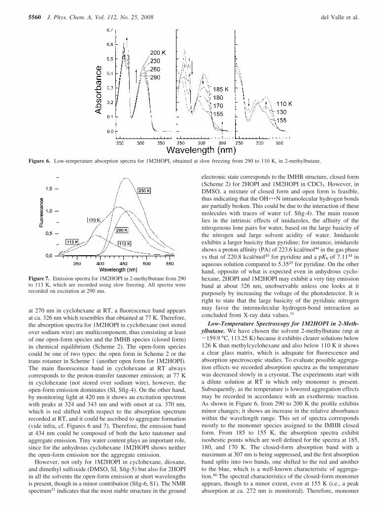
Low-Temperature Spectroscopy for 1M2HOPI in 2-Meth-ylbutane. We have chosen the solvent 2-methylbutane (mp at-159.9 °C, 113.25 K) because it exhibits clearer solutions below126 K than methylcyclohexane and also below 110 K it showsa clear glass matrix, which is adequate for fluorescence andabsorption spectroscopic studies. To evaluate possible aggrega-tion effects we recorded absorption spectra as the temperaturewas decreased slowly in a cryostat. The experiments start witha dilute solution at RT in which only monomer is present.Subsequently, as the temperature is lowered aggregation effectsmay be recorded in accordance with an exothermic reaction.As shown in Figure 6, from 290 to 200 K the profile exhibitsminor changes; it shows an increase in the relative absorbancewithin the wavelength range. This set of spectra correspondsmostly to the monomer species assigned to the IMHB closedform. From 185 to 155 K, the absorption spectra exhibitisosbestic points which are well defined for the spectra at 185,180, and 170 K. The closed-form absorption band with amaximum at 307 nm is being suppressed, and the first absorptionband splits into two bands, one shifted to the red and anotherto the blue, which is a well-known characteristic of aggrega-tion.46 The spectral characteristics of the closed-form monomerappears, though to a minor extent, even at 155 K (i.e., a peakabsorption at ca. 272 nm is monitored). Therefore, monomer
Figure 6. Low-temperature absorption spectra for 1M2HOPI, obtained at slow freezing from 290 to 110 K, in 2-methylbutane.
Figure 7. Emission spectra for 1M2HOPI in 2-methylbutane from 290to 113 K, which are recorded using slow freezing. All spectra wererecorded on excitation at 290 nm.
5560 J. Phys. Chem. A, Vol. 112, No. 25, 2008 del Valle et al.
and aggregate are present in chemical equilibrium. At 130 and110 K the closed form monomer seems to have disappeared asthe spectra differ significantly from that at 290 K. In addition,they show another isosbestic point that differs from the onewhich appears from 155 to 185 K. Therefore, it is concludedthat a different aggregation process has taken placed, the firstabsorption band shifting to the red of that found at 155 K.
The fluorescence spectra were recorded also from 290 to 113K with the same technique as above using a slow-freezingtechnique described in the Theoretical and Experimental Meth-ods section. Figure 7 shows four emission spectra at 290, 230,170, and 113 K on excitation at 290 nm. The fluorescence bandsat 290 and 230 K are centered at 460.5 and 454.5 nm, res-pectively, thus showing a blue shift as the temperature decreases.In contrast, at 170 and 113 K the spectrum changes and shiftsto the blue, thus showing, in accordance to the excitation spectra(not shown), a complex band of at least three components.
The absorption and emission spectra agree well with thatbelow 155 K: the IMHB closed-form species disappears bygenerating new aggregates.
Also, below 200 K the excitation spectra by monitoring at365 nm are characterized by a wavelength maximum at ca.290-300 nm and an onset at a shorter wavelength than that forthe closed-form monomer, which can be assigned to the open-form species. On excitation at 350 nm the emission spectrayields an emission band with a peak maximum at 460.5 nm for290 K, at 454.5 nm for 230 K, at 408 nm for 170 K, and at 424nm for 113 K. Spectra for the highest two temperatures revealan emission band from the closed-form monomer species,whereas spectra for the lowest two temperatures exhibit emissionbands that can be assigned to aggregates and open forms.
The emission spectrum at 113 K in Figure 7 differs greatlyfrom those at 77 K in cyclohexane (SI, SFig-4). This differenceis based on the quality of the matrices formed in cyclohexaneand 2-methylbutane and how they have been frozen. The2-methylbutane solvent forms a clear matrix at 113 K by slowfreezing; however, cyclohexane (mp at 4-5 C) does not forma clear matrix at 77 K because of cracking and formation of awhite solid. Conversely to cyclohexane at 77 K (SI, SFig-4), inwhich IMHB closed-form species of 1M2HOPI get trapped byquick freezing as well as the open-form species, the 2-meth-ylbutane matrix by slow freezing at 113 K does not trap IMHBclosed-form species (Figure 7).
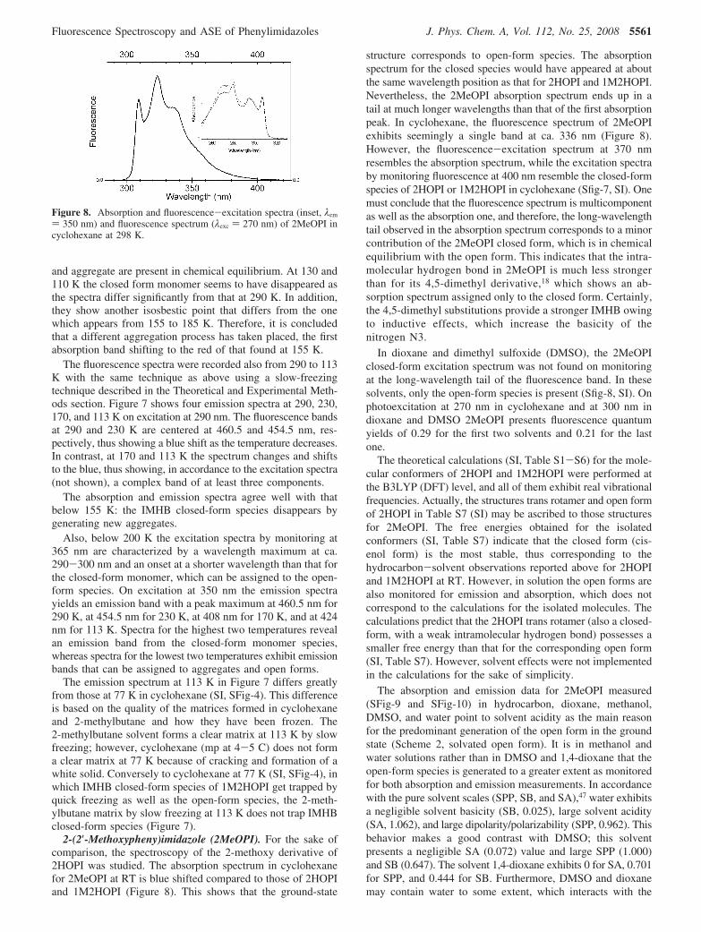
2-(2′-Methoxypheny)imidazole (2MeOPI). For the sake ofcomparison, the spectroscopy of the 2-methoxy derivative of2HOPI was studied. The absorption spectrum in cyclohexanefor 2MeOPI at RT is blue shifted compared to those of 2HOPIand 1M2HOPI (Figure 8). This shows that the ground-state
structure corresponds to open-form species. The absorptionspectrum for the closed species would have appeared at aboutthe same wavelength position as that for 2HOPI and 1M2HOPI.Nevertheless, the 2MeOPI absorption spectrum ends up in atail at much longer wavelengths than that of the first absorptionpeak. In cyclohexane, the fluorescence spectrum of 2MeOPIexhibits seemingly a single band at ca. 336 nm (Figure 8).However, the fluorescence-excitation spectrum at 370 nmresembles the absorption spectrum, while the excitation spectraby monitoring fluorescence at 400 nm resemble the closed-formspecies of 2HOPI or 1M2HOPI in cyclohexane (Sfig-7, SI). Onemust conclude that the fluorescence spectrum is multicomponentas well as the absorption one, and therefore, the long-wavelengthtail observed in the absorption spectrum corresponds to a minorcontribution of the 2MeOPI closed form, which is in chemicalequilibrium with the open form. This indicates that the intra-molecular hydrogen bond in 2MeOPI is much less strongerthan for its 4,5-dimethyl derivative,18 which shows an ab-sorption spectrum assigned only to the closed form. Certainly,the 4,5-dimethyl substitutions provide a stronger IMHB owingto inductive effects, which increase the basicity of thenitrogen N3.
In dioxane and dimethyl sulfoxide (DMSO), the 2MeOPIclosed-form excitation spectrum was not found on monitoringat the long-wavelength tail of the fluorescence band. In thesesolvents, only the open-form species is present (Sfig-8, SI). Onphotoexcitation at 270 nm in cyclohexane and at 300 nm indioxane and DMSO 2MeOPI presents fluorescence quantumyields of 0.29 for the first two solvents and 0.21 for the lastone.
The theoretical calculations (SI, Table S1-S6) for the mole-cular conformers of 2HOPI and 1M2HOPI were performed atthe B3LYP (DFT) level, and all of them exhibit real vibrationalfrequencies. Actually, the structures trans rotamer and open formof 2HOPI in Table S7 (SI) may be ascribed to those structuresfor 2MeOPI. The free energies obtained for the isolatedconformers (SI, Table S7) indicate that the closed form (cis-enol form) is the most stable, thus corresponding to thehydrocarbon-solvent observations reported above for 2HOPIand 1M2HOPI at RT. However, in solution the open forms arealso monitored for emission and absorption, which does notcorrespond to the calculations for the isolated molecules. Thecalculations predict that the 2HOPI trans rotamer (also a closed-form, with a weak intramolecular hydrogen bond) possesses asmaller free energy than that for the corresponding open form(SI, Table S7). However, solvent effects were not implementedin the calculations for the sake of simplicity.
The absorption and emission data for 2MeOPI measured(SFig-9 and SFig-10) in hydrocarbon, dioxane, methanol,DMSO, and water point to solvent acidity as the main reasonfor the predominant generation of the open form in the groundstate (Scheme 2, solvated open form). It is in methanol andwater solutions rather than in DMSO and 1,4-dioxane that theopen-form species is generated to a greater extent as monitoredfor both absorption and emission measurements. In accordancewith the pure solvent scales (SPP, SB, and SA),47 water exhibitsa negligible solvent basicity (SB, 0.025), large solvent acidity(SA, 1.062), and large dipolarity/polarizability (SPP, 0.962). Thisbehavior makes a good contrast with DMSO; this solventpresents a negligible SA (0.072) value and large SPP (1.000)and SB (0.647). The solvent 1,4-dioxane exhibits 0 for SA, 0.701for SPP, and 0.444 for SB. Furthermore, DMSO and dioxanemay contain water to some extent, which interacts with the
Figure 8. Absorption and fluorescence-excitation spectra (inset, λem
) 350 nm) and fluorescence spectrum (λexc ) 270 nm) of 2MeOPI incyclohexane at 298 K.
Fluorescence Spectroscopy and ASE of Phenylimidazoles J. Phys. Chem. A, Vol. 112, No. 25, 2008 5561
2MeOPI molecule, thereby generating solvated open-formstructures and shifting to the blue its fluorescence and absorptionspectra.
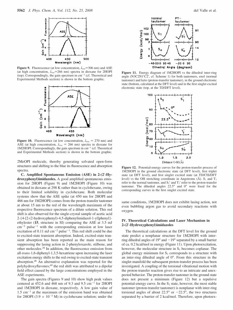
C. Amplified Spontaneous Emission (ASE) in 2-(2′-Hy-droxyphenyl)imidazoles. A good amplified spontaneous emis-sion for 2HOPI (Figure 9) and 1M2HOPI (Figure 10) wasobtained in dioxane at 298 K rather than in cyclohexane, owingto their limited solubility in cyclohexane. Both molecularsystems show that the ASE spike (at 450 nm for 2HOPI and466 nm for 1M2HOPI) comes from the proton-transfer tautomerat about 15 nm to the red of the wavelength maximum of therespective fluorescence spectrum of a dilute solution. This redshift is also observed for the single-crystal sample of acetic acid2-{4-[2-(2-hydroxyphenyl)-4,5-diphenylimidazol-1-yl]phenyl}-ethylester (15, structure in SI) comparing the ASE at 3.5 mJcm-2 pulse-1 with the corresponding emission at low laserexcitation of 0.11 mJ cm-2 pulse-1. This red shift could be dueto excited-state transient absorption. Indeed, excited-state tran-sient absorption has been reported as the main reason forsuppressing the lasing action in 2-phenyloxazole, stilbene, andother molecules.28 In addition, the fluorescence emission fromall-trans-1,6-diphenyl-1,3,5-hexatriene upon increasing the laserexcitation energy shifts to the red owing to excited-state transientabsorption.48 An alternative explanation was reported for thepolyhydroxyflavones;49 the red shift was attributed to a dipolarfield effect caused by the large concentrations employed in theASE experiments.
The gain spectra (Figures 9 and 10) show high peak valuescentered at 452.6 and 468 nm of 9.3 and 9.5 cm-1 for 2HOPIand 1M2HOPI in dioxane, respectively. A low gain value of1.5 cm-1 at the maximum of the emission band was obtainedfor 2HOPI (3.9 × 10⁻³ M) in cyclohexane solution; under the
same conditions, 1M2HOPI does not exhibit lasing action, noteven bubbling argon gas to avoid secondary reactions withoxygen.
IV. Theoretical Calculations and Laser Mechanism in2-(2′-Hydroxyphenyl)imidazoles
The theoretical calculations at the DFT level for the groundstate predict a nonplanar structure for 1M2HOPI with inter-ring dihedral angles of 19° and -19° separated by a small barrierof ca. 0.2 kcal/mol in energy (Figure 11). Upon photoexcitation,however, the molecular structure in S1 becomes coplanar. Theglobal energy minimum for S1 corresponds to a structure withan inter-ring dihedral angle of 0°. From this structure in thesinglet manifold the subsequent proton-transfer process has beeninvestigated. A coupling of the torsional vibrational motion withthe proton-transfer reaction gives rise to an intricate and unex-pected behavior. The proton-transfer tautomer in the ground statedoes not present a minimum (Figure 12) but a repulsivepotential-energy curve. In the S1 state, however, the most stabletautomer (proton-transfer tautomer) is nonplanar with inter-ringtorsional angles of about 22.5° or -22.5°, the two structuresseparated by a barrier of 2 kcal/mol. Therefore, upon photoex-
Figure 9. Fluorescence (at low concentration, λexc)306 nm) and ASE(at high concentration, λexc)266 nm) spectra in dioxane for 2HOPI(top). Correspondingly, the gain spectrum in cm-1 (cf. Theoretical andExperimental Methods section) is shown in the bottom graphic.
Figure 10. Fluorescence (at low concentration, λexc ) 270 nm) andASE (at high concentration, λexc ) 266 nm) spectra in dioxane for1M2HOPI. Correspondingly, the gain spectrum in cm-1 (cf. Theoreticaland Experimental Methods section) is shown in the bottom graphic.
Figure 11. Energy diagram of 1M2HOPI vs the dihedral inter-ringangle (N3C2N1′C2′, cf. Scheme 1) for both tautomers, enol (normaltautomer) and keto (proton-transfer tautomer), in the ground electronicstate (bottom, calculated at the DFT level) and in the first singlet excitedelectronic state (top, at the TD/DFT level).
Figure 12. Potential-energy curves for the proton-transfer process of1M2HOPI in the ground electronic state (at DFT level), first tripletstate (at DFT level), and first singlet excited state (at TD/CIS/DFTlevel) vs the OH stretching coordinate in Angstroms (Å). S1 and T1
refer to the normal tautomer, and S1′ and T1′ refer to the proton-transfertautomer. The dihedral angles 22.5° and 0° were fixed for thecorresponding curves in the first singlet excited state.
5562 J. Phys. Chem. A, Vol. 112, No. 25, 2008 del Valle et al.
citation of the normal tautomer the 1M2HOPI molecule becomescoplanar and undergoes an exothermic proton transfer in the S1
state, which corresponds to a nonplanar conformation.In contrast to the So state, the calculations predict an exother-
mic proton-transfer process over a small barrier in the lowesttriplet state (Figure 12). The T1 proton-transfer process isdictated by the change from a nonplanar structure (5.0°), normaltautomer, to another nonplanar structure (6.4°), the proton-transfer tautomer. An important feature of this proton-transferprocess relies on how the proton is transferred from oxygen tonitrogen; the inter-ring torsion precedes the proton transfer. Byplotting the inter-ring dihedral angle vs the OH distance (Sfig-11) it is observed that though the two tautomer minima possessnonplanar conformations, the proton is transferred from thecoplanar conformation with a dihedral angle of 0° for the OHdistances from 1.1 to 1.7 Å.
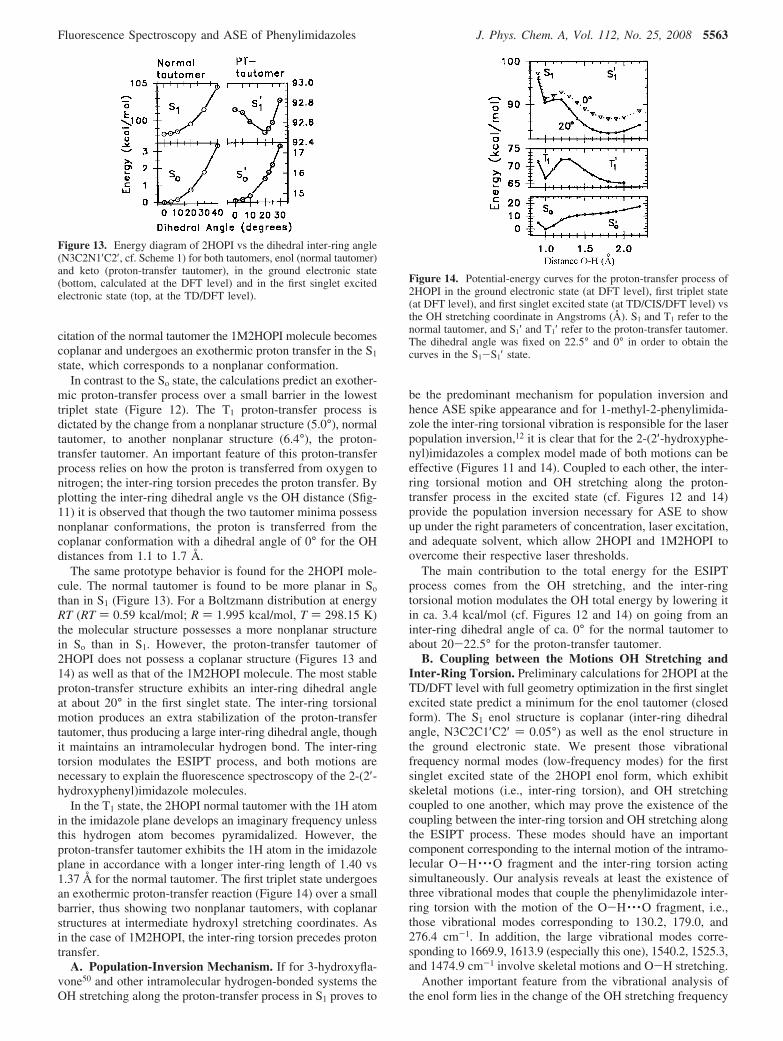
The same prototype behavior is found for the 2HOPI mole-cule. The normal tautomer is found to be more planar in So
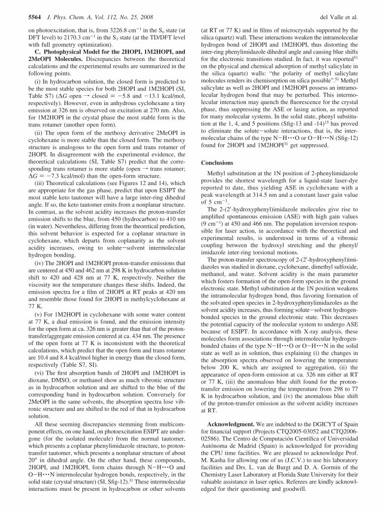
than in S1 (Figure 13). For a Boltzmann distribution at energyRT (RT ) 0.59 kcal/mol; R ) 1.995 kcal/mol, T ) 298.15 K)the molecular structure possesses a more nonplanar structurein So than in S1. However, the proton-transfer tautomer of2HOPI does not possess a coplanar structure (Figures 13 and14) as well as that of the 1M2HOPI molecule. The most stableproton-transfer structure exhibits an inter-ring dihedral angleat about 20° in the first singlet state. The inter-ring torsionalmotion produces an extra stabilization of the proton-transfertautomer, thus producing a large inter-ring dihedral angle, thoughit maintains an intramolecular hydrogen bond. The inter-ringtorsion modulates the ESIPT process, and both motions arenecessary to explain the fluorescence spectroscopy of the 2-(2′-hydroxyphenyl)imidazole molecules.
In the T1 state, the 2HOPI normal tautomer with the 1H atomin the imidazole plane develops an imaginary frequency unlessthis hydrogen atom becomes pyramidalized. However, theproton-transfer tautomer exhibits the 1H atom in the imidazoleplane in accordance with a longer inter-ring length of 1.40 vs1.37 Å for the normal tautomer. The first triplet state undergoesan exothermic proton-transfer reaction (Figure 14) over a smallbarrier, thus showing two nonplanar tautomers, with coplanarstructures at intermediate hydroxyl stretching coordinates. Asin the case of 1M2HOPI, the inter-ring torsion precedes protontransfer.
A. Population-Inversion Mechanism. If for 3-hydroxyfla-vone50 and other intramolecular hydrogen-bonded systems theOH stretching along the proton-transfer process in S1 proves to
be the predominant mechanism for population inversion andhence ASE spike appearance and for 1-methyl-2-phenylimida-zole the inter-ring torsional vibration is responsible for the laserpopulation inversion,12 it is clear that for the 2-(2′-hydroxyphe-nyl)imidazoles a complex model made of both motions can beeffective (Figures 11 and 14). Coupled to each other, the inter-ring torsional motion and OH stretching along the proton-transfer process in the excited state (cf. Figures 12 and 14)provide the population inversion necessary for ASE to showup under the right parameters of concentration, laser excitation,and adequate solvent, which allow 2HOPI and 1M2HOPI toovercome their respective laser thresholds.
The main contribution to the total energy for the ESIPTprocess comes from the OH stretching, and the inter-ringtorsional motion modulates the OH total energy by lowering itin ca. 3.4 kcal/mol (cf. Figures 12 and 14) on going from aninter-ring dihedral angle of ca. 0° for the normal tautomer toabout 20-22.5° for the proton-transfer tautomer.
B. Coupling between the Motions OH Stretching andInter-Ring Torsion. Preliminary calculations for 2HOPI at theTD/DFT level with full geometry optimization in the first singletexcited state predict a minimum for the enol tautomer (closedform). The S1 enol structure is coplanar (inter-ring dihedralangle, N3C2C1′C2′ ) 0.05°) as well as the enol structure inthe ground electronic state. We present those vibrationalfrequency normal modes (low-frequency modes) for the firstsinglet excited state of the 2HOPI enol form, which exhibitskeletal motions (i.e., inter-ring torsion), and OH stretchingcoupled to one another, which may prove the existence of thecoupling between the inter-ring torsion and OH stretching alongthe ESIPT process. These modes should have an importantcomponent corresponding to the internal motion of the intramo-lecular O-H · · ·O fragment and the inter-ring torsion actingsimultaneously. Our analysis reveals at least the existence ofthree vibrational modes that couple the phenylimidazole inter-ring torsion with the motion of the O-H · · ·O fragment, i.e.,those vibrational modes corresponding to 130.2, 179.0, and276.4 cm-1. In addition, the large vibrational modes corre-sponding to 1669.9, 1613.9 (especially this one), 1540.2, 1525.3,and 1474.9 cm-1 involve skeletal motions and O-H stretching.
Another important feature from the vibrational analysis ofthe enol form lies in the change of the OH stretching frequency
Figure 13. Energy diagram of 2HOPI vs the dihedral inter-ring angle(N3C2N1′C2′, cf. Scheme 1) for both tautomers, enol (normal tautomer)and keto (proton-transfer tautomer), in the ground electronic state(bottom, calculated at the DFT level) and in the first singlet excitedelectronic state (top, at the TD/DFT level).
Figure 14. Potential-energy curves for the proton-transfer process of2HOPI in the ground electronic state (at DFT level), first triplet state(at DFT level), and first singlet excited state (at TD/CIS/DFT level) vsthe OH stretching coordinate in Angstroms (Å). S1 and T1 refer to thenormal tautomer, and S1′ and T1′ refer to the proton-transfer tautomer.The dihedral angle was fixed on 22.5° and 0° in order to obtain thecurves in the S1-S1′ state.
Fluorescence Spectroscopy and ASE of Phenylimidazoles J. Phys. Chem. A, Vol. 112, No. 25, 2008 5563
on photoexcitation, that is, from 3226.8 cm-1 in the So state (atDFT level) to 2170.3 cm-1 in the S1 state (at the TD/DFT levelwith full geometry optimization).
C. Photophysical Model for the 2HOPI, 1M2HOPI, and2MeOPI Molecules. Discrepancies between the theoreticalcalculations and the experimental results are summarized in thefollowing points.
(i) In hydrocarbon solution, the closed form is predicted tobe the most stable species for both 2HOPI and 1M2HOPI (SI,Table S7) (∆G open f closed ) -5.8 and -13.1 kcal/mol,respectively). However, even in anhydrous cyclohexane a tinyemission at 326 nm is observed on excitation at 270 nm. Also,for 1M2HOPI in the crystal phase the most stable form is thetrans rotamer (another open form).
(ii) The open form of the methoxy derivative 2MeOPI incyclohexane is more stable than the closed form. The methoxystructure is analogous to the open form and trans rotamer of2HOPI. In disagreement with the experimental evidence, thetheoretical calculations (SI, Table S7) predict that the corre-sponding trans rotamer is more stable (open f trans rotamer;∆G ) -7.3 kcal/mol) than the open-form structure.
(iii) Theoretical calculations (see Figures 12 and 14), whichare appropriate for the gas phase, predict that upon ESIPT themost stable keto tautomer will have a large inter-ring dihedralangle. If so, the keto tautomer emits from a nonplanar structure.In contrast, as the solvent acidity increases the proton-transferemission shifts to the blue, from 450 (hydrocarbon) to 410 nm(in water). Nevertheless, differing from the theoretical prediction,this solvent behavior is expected for a coplanar structure incyclohexane, which departs from coplanarity as the solventacidity increases, owing to solute-solvent intermolecularhydrogen bonding.
(iv) The 2HOPI and 1M2HOPI proton-transfer emissions thatare centered at 450 and 462 nm at 298 K in hydrocarbon solutionshift to 420 and 428 nm at 77 K, respectively. Neither theviscosity nor the temperature changes these shifts. Indeed, theemission spectra for a film of 2HOPI at RT peaks at 420 nmand resemble those found for 2HOPI in methylcyclohexane at77 K.
(v) For 1M2HOPI in cyclohexane with some water contentat 77 K, a dual emission is found, and the emission intensityfor the open form at ca. 326 nm is greater than that of the proton-transfer/aggregate emission centered at ca. 434 nm. The presenceof the open form at 77 K is inconsistent with the theoreticalcalculations, which predict that the open form and trans rotamerare 10.4 and 8.4 kcal/mol higher in energy than the closed form,respectively (Table S7, SI).
(vi) The first absorption bands of 2HOPI and 1M2HOPI indioxane, DMSO, or methanol show as much vibronic structureas in hydrocarbon solution and are shifted to the blue of thecorresponding band in hydrocarbon solution. Conversely for2MeOPI in the same solvents, the absorption spectra lose vib-ronic structure and are shifted to the red of that in hydrocarbonsolution.
All these seeming discrepancies stemming from multicom-ponent effects, on one hand, on photoexcitation ESIPT are under-gone (for the isolated molecule) from the normal tautomer,which presents a coplanar phenylimidazole structure, to proton-transfer tautomer, which presents a nonplanar structure of about20° in dihedral angle. On the other hand, these compounds,2HOPI, and 1M2HOPI, form chains through N-H · · ·O andO-H · · ·N intermolecular hydrogen bonds, respectively, in thesolid state (crystal structure) (SI, Sfig-12).31 These intermolecularinteractions must be present in hydrocarbon or other solvents
(at RT or 77 K) and in films of microcrystals supported by thesilica (quartz) wall. These interactions weaken the intramolecularhydrogen bond of 2HOPI and 1M2HOPI, thus distorting theinter-ring phenylimidazole dihedral angle and causing blue shiftsfor the electronic transitions studied. In fact, it was reported51
on the physical and chemical adsorption of methyl salicylate inthe silica (quartz) walls: “the polarity of methyl salicylatemolecules renders its chemisorption on silica possible”.51 Methylsalicylate as well as 2HOPI and 1M2HOPI possess an intramo-lecular hydrogen bond that may be perturbed. This intermo-lecular interaction may quench the fluorescence for the crystalphase, thus suppressing the ASE or lasing action, as reportedfor many molecular systems. In the solid state, phenyl substitu-tion at the 1, 4, and 5 positions (Sfig-13 and -14)15 has provedto eliminate the solute-solute interactions, that is, the inter-molecular chains of the type N-H · · ·O or O-H · · ·N (Sfig-12)found for 2HOPI and 1M2HOPI31 get suppressed.
Conclusions
Methyl substitution at the 1N position of 2-phenylimidazoleprovides the shortest wavelength for a liquid-state laser-dyereported to date, thus yielding ASE in cyclohexane with apeak wavelength at 314.5 nm and a constant laser gain valueof 5 cm-1.
The 2-(2′-hydroxyphenyl)imidazole molecules give rise toamplified spontaneous emission (ASE) with high gain values(9 cm-1) at 450 and 466 nm. The population inversion respon-sible for laser action, in accordance with the theoretical andexperimental results, is understood in terms of a vibroniccoupling between the hydroxyl stretching and the phenyl/imidazole inter-ring torsional motions.
The proton-transfer spectroscopy of 2-(2′-hydroxyphenyl)imi-dazoles was studied in dioxane, cyclohexane, dimethyl sulfoxide,methanol, and water. Solvent acidity is the main parameterwhich fosters formation of the open-form species in the groundelectronic state. Methyl substitution at the 1N position weakensthe intramolecular hydrogen bond, thus favoring formation ofthe solvated open species in 2-hydroxyphenylimidazoles as thesolvent acidity increases, thus forming solute-solvent hydrogen-bonded species in the ground electronic state. This decreasesthe potential capacity of the molecular system to undergo ASEbecause of ESIPT. In accordance with X-ray analysis, thesemolecules form associations through intermolecular hydrogen-bonded chains of the type N-H · · ·O or O-H · · ·N in the solidstate as well as in solution, thus explaining (i) the changes inthe absorption spectra observed on lowering the temperaturebelow 200 K, which are assigned to aggregation, (ii) theappearance of open-form emission at ca. 326 nm either at RTor 77 K, (iii) the anomalous blue shift found for the proton-transfer emission on lowering the temperature from 298 to 77K in hydrocarbon solution, and (iv) the anomalous blue shiftof the proton-transfer emission as the solvent acidity increasesat RT.
Acknowledgment. We are indebted to the DGICYT of Spainfor financial support (Projects CTQ2005-03052 and CTQ2006-02586). The Centro de Computacion Cientıfica of UniversidadAutonoma de Madrid (Spain) is acknowledged for providingthe CPU time facilities. We are pleased to acknowledge Prof.M. Kasha for allowing one of us (J.C.V.) to use his laboratoryfacilities and Drs. L. van de Burgt and D. A. Gormin of theChemistry Laser Laboratory at Florida State University for theirvaluable assistance in laser optics. Referees are kindly acknowl-edged for their questioning and goodwill.
5564 J. Phys. Chem. A, Vol. 112, No. 25, 2008 del Valle et al.
Supporting Information Available: Experimental andtheoretical information. This material is available free of chargevia the Internet at http://pubs.acs.org.
References and Notes
(1) Drexhage, K. H. Structure and Properties of Laser Dyes. Top. Appl.Phys. 1973, 1. (Dye Lasers), 144.
(2) King, T. A. Comprehensive Series in Photochemistry & Photobi-ology. Lasers and Current Optical Techniques in Biology; Palumbo, G.,Protesi, R., Eds.; Royal Society of Chemistry: Cambridge, UK, 2004; Vol.4, p 3.
(3) King, T. A. Comprehensive Series in Photochemistry & Photobi-ology. Lasers and Current Optical Techniques in Biology; Palumbo, G.,Protesi, R., Eds.; Royal Society of Chemistry: Cambridge, UK, 2004; Vol.4, p 33.
(4) Okasabu, K.; Kawachi, T.; Oyama, H.; Hara, T.; Yamaguchi, N.;Ando, K. Jpn. J. Appl. Phys., Part 1 2000, 39, 70.
(5) Davis, C. C.; King, T. A. AdV. Quantum Electron. 1975, 3, 169.(6) Esslinger, T.; Bloch, I.; Hansch, T. W. Phys. Blaetter 2000, 56
(2), 47.(7) Khan, A.; Kasha, M. Proc. Natl. Acad. Sci. U.S.A. 1983, 80, 1767.(8) Chou, P. T.; McMorrow, D.; Aartsma, T. J.; Kasha, M. J. Phys.
Chem. 1984, 88, 4596.(9) Acuna, A. U.; Costela, A.; Munoz, J. M. J. Phys. Chem. 1986, 90,
2807.(10) Acuna, A. U.; Amat-Guerri, F.; Catalan, J.; Costela, A.; Figuera,
J. M.; Munoz, J. M. Chem. Phys. Lett. 1986, 132, 567.(11) Costela, A.; Amat-Guerri, F.; Catalan, J.; Douhal, A.; Figuera, J. M.;
Munoz, J. M.; Acuna, U. A. Opt. Commun. 1987, 64, 457.(12) Catalan, J.; de Paz, J. L. G.; del Valle, J. C.; Kasha, M. J. Phys.
Chem. A 1997, 101, 5284.(13) Catalan, J.; Mena, E.; Fabero, F.; Amat-Guerri, F. J. Chem. Phys.
1992, 96, 2005.(14) Fluorene (10-3 M) in cyclohexane solution on excitation at 266
nm does not yield lasing action. Several more concentrations and solventswere tried with identical results.
(15) Park, S.; Kwon, O.-H.; Kim, S.; Park, S.; Choi, M.-G.; Cha, M.;Park, S. Y.; Jang, D.-J. J. Am. Chem. Soc. 2005, 127, 10070.
(16) Douhal, A.; Amat-Guerri, F.; Lillo, M. P.; Acuna, A. U. J.Photochem. Photobiol. A: Chem. 1994, 78, 127.
(17) Testa, A. C. Chem. Phys. Lett. 1991, 50, 243.(18) Brauer, M.; Mosquera, M.; Perez-Lustres, J. L.; Rodrıguez-Prieto,
F. J. Phys. Chem. A 1998, 102, 10736.(19) (a) Purkayastha, P.; Chattopadhyay, N. J. Mol. Struct. 2002, 604,
87. (b) Int. J. Mol. Sci. 2003, 4, 335.(20) Fores, M.; Duran, M.; Sola, M.; Adamowicz, L. J. Phys. Chem. A
1999, 103, 4413.(21) Catalan, J.; de Paz, J. L. G. J. Phys. Chem. A 2001, 105 (30), 7315.(22) Sobolewski, A. L.; Domcke, W. The Reaction Path in Chemistry:
Current Approaches and Perspectives. In Understanding Chemical ReactiV-ity; Heidrich, D., Ed.; Kluwer: Dordrecht, The Netherlands, 1995; Vol. 16,p 257.
(23) Sobolewski, A. L.; Domcke, W. Chem. Phys. 1998, 232, 257.(24) Meech, S. R.; Phillips, D. J. Photochem. 1983, 23, 193.(25) Catalan, J.; del Valle, J. C. J. Am. Chem. Soc. 1993, 115, 4321.(26) del Valle, J. C.; Kasha, M.; Catalan, J. Chem. Phys. Lett. 1996,
263, 154.
(27) del Valle, J. C.; Kasha, M.; Catalan, J. J. Phys. Chem. A 1997,101, 3260.
(28) Shank, C. V. ReV. Mod. Phys. 1975, 47 (3), 649.(29) Rogers, G. A.; Bruice, T. C. J. Am. Chem. Soc. 1974, 96, 2463.(30) Mahmood, A.; Liu, H.; Joes, J. G.; Edwards, J. O.; Sweigart, D. A.
Inorg. Chem. 1988, 27, 2149.(31) Foces-Foces, C.; Llamas-Saiz, A. L.; Claramunt, R. M.; Cabildo,
P.; Elguero, J. J. Mol. Struct. 1998, 440, 193.(32) Becke, A. D. J. Chem. Phys. 1993, 98, 5648.(33) Lee, C.; Yang, W.; Parr, R. G. Phys. ReV. B 1988, 37, 785.(34) Hariharan, P. C.; Pople, J. A. Chem. Phys. Lett. 1972, 16, 217.(35) Frisch, M. J.; Trucks, G. W.; Schlegel, H. B.; Scuseria, G. E.; Robb
M. A.; Cheeseman, J. R.; Montgomery, J. A., Jr.; Vreven, T.; Kudin, K. N.;Burant, J. C.; Millam, J. M.; Iyengar, S. S.; Tomasi, J.; Barone, V.;Mennucci, B.; Cossi, M.; Scalmani, G.; Rega, N.; Petersson G. A.; Nakatsuji,H.; Hada, M.; Ehara, M.; Toyota, K.; Fukuda, R.; Hasegawa, J.; Ishida,M.; Nakajima, T.; Honda, Y.; Kitao, O.; Nakai, H.; Klene, M.; Li, X.; Knox,J. E.; Hratchian, H. P.; Cross, J. B.; Adamo, C.; Jaramillo, J.; Gomperts,R.; Stratmann, R. E.; Yazyev, O.; Austin, A. J.; Cammi, R.; Pomelli, C.;Ochterski, J. W.; Ayala, P. Y.; Morokuma, K.; Voth, G. A.; Salvador, P.;Dannenberg, J. J.; Zakrzewski, V. G.; Dapprich, S.; Daniels, A. D.; Strain,M. C.; Farkas, O.; Malick, D. K.; Rabuck, A. D.; Raghavachari, K.;Foresman, J. B.; Ortiz, J. V.; Cui, Q.; Baboul, A. G.; Clifford, S.; Cioslowski,J.; Stefanov, B. B.; Liu, G.; Liashenko, A.; Piskorz, P.; Komaromi, I.;Martin, R. L.; Fox, D. J.; Keith, T.; Al-Laham, M. A.; Peng, C. Y.;Nanayakkara, A.; Challacombe, M.; Gill, P. M.W.; Johnson, B.; Chen, W.;Wong, M. W.; Gonzalez, C.; Pople, J. A. Gaussian 03, Revision C.02;Gaussian, Inc.: Wallingford, CT, 2004.
(36) SPARTAN, Version 4.1; Wavefunction, Inc.: Irvine, CA, 1996.(37) Hehre, W. J.; Radom, L.; Schleyer, R. V.; Pople, J. A. Ab Initio
Molecular Orbital Theory; Wiley: New York, 1986.(38) Casida, M. E. In Recent AdVances in Density Functional Methods,
Part I; Chang, D. P., Ed.; World Scientific: Singapore, 1995; Vol. 155.(39) Foresman, J. B.; Head-Gordon, M.; Pople, J. A. J. Phys. Chem.
1992, 96, 135.(40) Brackmann, U., Ed.; Lambdachrome Laser Dye; Lambda Physik
GmbH: Göttingen, Germany, 1986.(41) De Smet, K. Phys. ReV. E 1998, 57, 1384.(42) Campbell, A.; Willard, J. E. J. Phys. Chem. A 1968, 72, 1918.(43) Catalan, J. Chem. Phys. Lett. 2006, 421, 134.(44) Catalan, J.; Claramunt, R. M.; Elguero, J.; Laynez, J.; Menendez,
M.; Anvıa, F.; Avıan, J. H.; Taagepera, M.; Taft, R. W. J. Am. Chem. Soc.1988, 110, 4105.
(45) Lias, S. G.; Liebman, J. F.; Levin, R. D. J. Phys. Chem. Ref. Data1984, 13 (3), 695.
(46) McRae, E. G.; Kasha, M. The Molecular Exciton Model. In PhysicalProcesses in Radiation Biology; Augenstein, L., Rosenberg, B., Mason,S. F., Eds.; Academic Press: New York, 1964; pp 23-42.
(47) Catalan, J. In Handbook of SolVents; Wypych, G., Ed.; W. A.Publishing: New York, ChemTec Publishing: Toronto, 2001.
(48) del Valle, J. C.; Tarkalanov, N.; Saltiel, J. J. Phys. Chem. B 1999,103, 9350.
(49) Gormin, D.; Sytnik, A.; Kasha, M. J. Phys. Chem. A 1997, 101,672.
(50) Del Valle, J. C. J. Chem. Phys. 2006, 124, 1.(51) Acuna, A. U.; Catalan, J.; Toribio, F. J. Phys. Chem. 1981, 85,
241.
JP7117604
Fluorescence Spectroscopy and ASE of Phenylimidazoles J. Phys. Chem. A, Vol. 112, No. 25, 2008 5565


![Synthesis and characterization of polyesters based on 1,1,1-[bis(4-hydroxyphenyl)-4′-pentadecylphenyl]ethane](https://img.pdfslide.net/doc/110x75/631c95a8d5372c006e04825b/synthesis-and-characterization-of-polyesters-based-on-111-bis4-hydroxyphenyl-4-pentadecylphenylethane.jpg)


![2-[7-(3,5-Dibromo-2-hydroxyphenyl)-6-ethoxycarbonyl-2-oxo-5 H -2,3,6,7-tetrahydrothiopyrano[2,3- d ][1,3]thiazol-6-yl]acetic acid ethanol monosolvate](https://img.pdfslide.net/doc/110x75/6336c1b22c3abbd36103c3de/2-7-35-dibromo-2-hydroxyphenyl-6-ethoxycarbonyl-2-oxo-5-h-2367-tetrahydrothiopyrano23-.jpg)
![Synthesis and characterisation of tin(IV) and organotin(IV) derivatives 2-{[(2-hydroxyphenyl)imino]methyl}phenol](https://img.pdfslide.net/doc/110x75/631651da3ed465f0570bfb1c/synthesis-and-characterisation-of-tiniv-and-organotiniv-derivatives-2-2-hydroxyphenyliminomethylphenol.jpg)


![9-[(2-Hydroxybenzylidene)amino]-11-(2-hydroxyphenyl)-10,13-diphenyl-8-oxa-12-azoniatricyclo[7.3.1.0 2,7 ]trideca-2(7),3,5-triene acetate ethanol disolvate](https://img.pdfslide.net/doc/110x75/634ec0ed0d6acb880b02f609/9-2-hydroxybenzylideneamino-11-2-hydroxyphenyl-1013-diphenyl-8-oxa-12-azoniatricyclo7310.jpg)

![Vibronic transitions from coupled-cluster response theory: Theory and application to HSiF and H[sub 2]O](https://img.pdfslide.net/doc/110x75/6352bbcbcb577625920ddd71/vibronic-transitions-from-coupled-cluster-response-theory-theory-and-application.jpg)


![Preparation and structure determination of 1-benzyl-, 1-methyl- and 1 H -5-[(2-nitro-2-phenyl)ethenyl]imidazoles](https://img.pdfslide.net/doc/110x75/63455d3038eecfb33a06868f/preparation-and-structure-determination-of-1-benzyl-1-methyl-and-1-h-5-2-nitro-2-phenylethenylimidazoles.jpg)