Embed Size (px)
Citation preview


Lactase expression in sheep 933
7a)c Xu
Ln N
eF vCDn w
> > > > 7 D , 4
00 0 < < < < z1 2 3 4 5 6 7 8 9
Vill in Aa.F
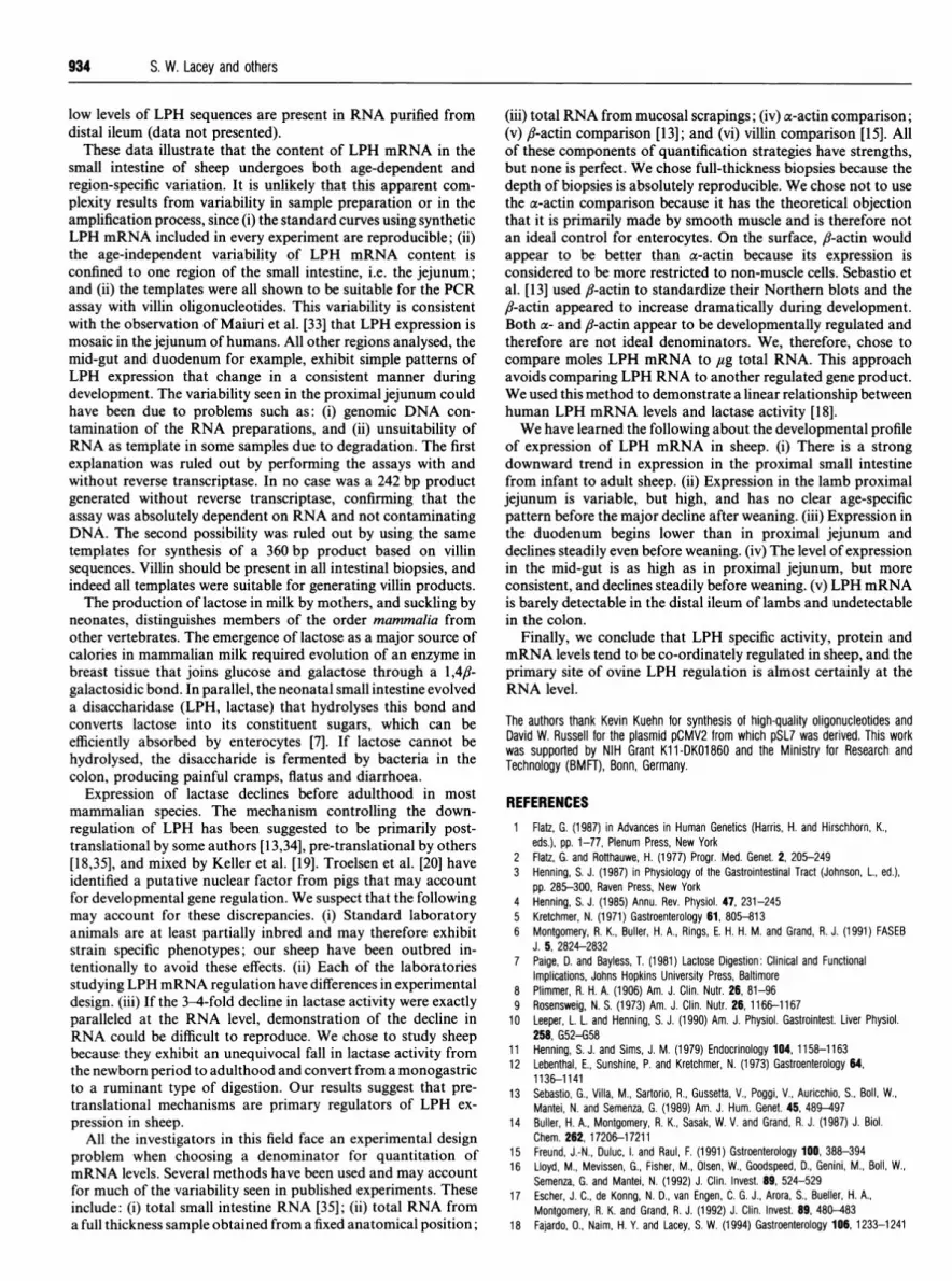
Figure 4 Villin control for template suitabilityTo test the suitability of each RNA sample in our LPH PCR assay, we performed a similar assayusing oligonucleotides directed to villin. Villin is a component of microvillar membranes. Thisis a typical assay using RNA from the proximal jejunum of four lambs (lanes 1-4), three adultsheep (lanes 5-7), adult sheep kidney (lane 8), and control lacking RNA (lane 9). All RNAtemplates reported in this manuscript were tested using the villin oligonucleotides and provedsuitable in the PCR assay.
spite of considerable variation between young animals. Tissuehomogenates from the same biopsies were examined by Westernblotting for the presence of immunoreactive LPH protein ex-pression. Figure 3 shows that mature (160 kDa) LPH protein isreadily detectable in each of the homogenates prepared fromfetal and lamb proximal jejunum, while no LPH protein can bedetected in homogenates prepared from the proximal jejunum ofadult sheep or from lamb colon.Another possible explanation for the variation in LPH mRNA
was that some of the RNA samples were not suitable foramplification. We therefore tested all RNA samples for suitabilityby amplifying a larger (400 bp) region of villin mRNA. Villinwas chosen because it is a structural component of microvilli and
Z~b ArA
Duodenum..
:: -- ;....
104 54 40 21
Jejunum
301 70 101 577
Mid small intestine
460 304 205 160Human LPH mRNA
attomole equivalents
is therefore present at all ages. A typical example of these controlexperiments is shown in Figure 4 including jejunum for some ofthe lambs, adult sheep, a positive control from adult kidney andan mRNA negative control. Clearly, all the template RNAs weresuitable for our assay.A second aim of this work was to investigate patterns of
expression of LPH mRNA in different regions of small intestinefrom lambs of various ages. We focused on lambs because LPHmRNA and lactase activity were too low in the stomach, smallintestine and colon in our adult animals to permit meaningfulcomparison (data not shown). Accordingly, the quantity ofLPHmRNA was measured in samples of RNA prepared fromduodenum, a second site in the jejunum 10 cm distal to the first,the mid-point of the small intestine, distal ileum and colon. Theresults obtained are shown in Figure 5 for duodenum, proximaljejunum and mid small intestine. Distal ileum and colon hadlevels that were too low to be quantified.LPH mRNA was detectable in the duodenum of all the lambs
studied. The levels were consistently lower than that seen injejunum and mid small intestine. Furthermore, there was a clearage-related decline in LPH mRNA extracted from the lambduodenum. The hallmark variability ofLPH mRNA levels in theproximaljejunum is reproduced here. The levels were consistentlyhigh but have no other age-related pattern like that seen in theduodenum and mid small intestine. This variability in expressionis reminiscent of the results obtained previously (Figure 2) withsamples taken from a more proximal site in the jejunum. LPHsequences in the mid gut are abundant in young lambs anddecline steadily, but slightly, with increasing age before weaningin lambs. Adult animals have neither lactase activity nor LPHmRNA detectable in the mid gut (data not shown). No LPHmRNA is detectable in RNA prepared from colon and extremely
Log (amol LPH RNA)0 2.0 2.5 3.0 3.5 4.0
.'..::.:o.
2.5 3.0 3.5 4.0Log (amol LPH mRNA)
Figure 5 Longiudinal profile of LPH mRNA expression in lamb small intestine
Relative levels of LPH mRNA from three additional sites in the ovine small intestine are presented for the lambs at the ages indicated and are expressed in human LPH RNA attomole equivalents.Duodenal samples were obtained halfway between the pylorus and ligament of Treitz. The jejunal samples were obtained 10 cm distal to the samples used in Figures 2 and 3, and the mid smallintestine was selected halfway between the ligament of Treitz and the ileocecal valve. All assays began with 1 ,ug total RNA. There is a marked age-related decline in LPH mRNA in the duodenum,variability in the proximal jejunum as was seen in Figure 2, and an age-related decline in the mid small intestine. As in Figure 2, the sheep LPH RNA determinations are relative amounts comparedwith PCR products generated in the standard assay using synthetic human LPH RNA as template. The experiment used for quantification had the cleanest standard curve of three similar assays.
934 S. W. Lacey and others
low levels of LPH sequences are present in RNA purified fromdistal ileum (data not presented).
These data illustrate that the content of LPH mRNA in thesmall intestine of sheep undergoes both age-dependent andregion-specific variation. It is unlikely that this apparent com-plexity results from variability in sample preparation or in theamplification process, since (i) the standard curves using syntheticLPH mRNA included in every experiment are reproducible; (ii)the age-independent variability of LPH mRNA content isconfined to one region of the small intestine, i.e. the jejunum;and (ii) the templates were all shown to be suitable for the PCRassay with villin oligonucleotides. This variability is consistentwith the observation of Maiuri et al. [33] that LPH expression ismosaic in the jejunum of humans. All other regions analysed, themid-gut and duodenum for example, exhibit simple patterns ofLPH expression that change in a consistent manner duringdevelopment. The variability seen in the proximal jejunum couldhave been due to problems such as: (i) genomic DNA con-tamination of the RNA preparations, and (ii) unsuitability ofRNA as template in some samples due to degradation. The firstexplanation was ruled out by performing the assays with andwithout reverse transcriptase. In no case was a 242 bp productgenerated without reverse transcriptase, confirming that theassay was absolutely dependent on RNA and not contaminatingDNA. The second possibility was ruled out by using the sametemplates for synthesis of a 360 bp product based on villinsequences. Villin should be present in all intestinal biopsies, andindeed all templates were suitable for generating villin products.The production of lactose in milk by mothers, and suckling by
neonates, distinguishes members of the order mammalia fromother vertebrates. The emergence of lactose as a major source ofcalories in mammalian milk required evolution of an enzyme inbreast tissue that joins glucose and galactose through a 1,4fi-galactosidic bond. In parallel, the neonatal small intestine evolveda disaccharidase (LPH, lactase) that hydrolyses this bond andconverts lactose into its constituent sugars, which can beefficiently absorbed by enterocytes [7]. If lactose cannot behydrolysed, the disaccharide is fermented by bacteria in thecolon, producing painful cramps, flatus and diarrhoea.
Expression of lactase declines before adulthood in mostmammalian species. The mechanism controlling the down-regulation of LPH has been suggested to be primarily post-translational by some authors [13,34], pre-translational by others[18,35], and mixed by Keller et al. [19]. Troelsen et al. [20] haveidentified a putative nuclear factor from pigs that may accountfor developmental gene regulation. We suspect that the followingmay account for these discrepancies. (i) Standard laboratoryanimals are at least partially inbred and may therefore exhibitstrain specific phenotypes; our sheep have been outbred in-tentionally to avoid these effects. (ii) Each of the laboratoriesstudying LPH mRNA regulation have differences in experimentaldesign. (iii) If the 3-4-fold decline in lactase activity were exactlyparalleled at the RNA level, demonstration of the decline inRNA could be difficult to reproduce. We chose to study sheepbecause they exhibit an unequivocal fall in lactase activity fromthe newborn period to adulthood and convert from a monogastricto a ruminant type of digestion. Our results suggest that pre-translational mechanisms are primary regulators of LPH ex-pression in sheep.
All the investigators in this field face an experimental designproblem when choosing a denominator for quantitation ofmRNA levels. Several methods have been used and may accountfor much of the variability seen in published experiments. Theseinclude: (i) total small intestine RNA [35], (ii) total RNA froma full thickness sample obtained from a fixed anatomical position;
(iii) total RNA from mucosal scrapings; (iv) a-actin comparison;(v) fl-actin comparison [13]; and (vi) villin comparison [15]. Allof these components of quantification strategies have strengths,but none is perfect. We chose full-thickness biopsies because thedepth of biopsies is absolutely reproducible. We chose not to usethe a-actin comparison because it has the theoretical objectionthat it is primarily made by smooth muscle and is therefore notan ideal control for enterocytes. On the surface, fl-actin wouldappear to be better than a-actin because its expression isconsidered to be more restricted to non-muscle cells. Sebastio etal. [13] used ,-actin to standardize their Northern blots and the,/-actin appeared to increase dramatically during development.Both a- and ,-actin appear to be developmentally regulated andtherefore are not ideal denominators. We, therefore, chose tocompare moles LPH mRNA to ,ug total RNA. This approachavoids comparing LPH RNA to another regulated gene product.We used this method to demonstrate a linear relationship betweenhuman LPH mRNA levels and lactase activity [18].We have learned the following about the developmental profile
of expression of LPH mRNA in sheep. (i) There is a strongdownward trend in expression in the proximal small intestinefrom infant to adult sheep. (ii) Expression in the lamb proximaljejunum is variable, but high, and has no clear age-specificpattern before the major decline after weaning. (iii) Expression inthe duodenum begins lower than in proximal jejunum anddeclines steadily even before weaning. (iv) The level of expressionin the mid-gut is as high as in proximal jejunum, but moreconsistent, and declines steadily before weaning. (v) LPH mRNAis barely detectable in the distal ileum of lambs and undetectablein the colon.
Finally, we conclude that LPH specific activity, protein andmRNA levels tend to be co-ordinately regulated in sheep, and theprimary site of ovine LPH regulation is almost certainly at theRNA level.
The authors thank Kevin Kuehn for synthesis of high-quality oligonucleotides andDavid W. Russell for the plasmid pCMV2 from which pSL7 was derived. This workwas supported by NIH Grant K11-DK01860 and the Ministry for Research andTechnology (BMFT), Bonn, Germany.
REFERENCES1 Flatz, G. (1987) in Advances in Human Genetics (Harris, H. and Hirschhorn, K.,
eds.), pp. 1-77, Plenum Press, New York2 Flatz, G. and Rotthauwe, H. (1977) Progr. Med. Genet. 2, 205-2493 Henning, S. J. (1987) in Physiology of the Gastrointestinal Tract (Johnson, L., ed.),
pp. 285-300, Raven Press, New York4 Henning, S. J. (1985) Annu. Rev. Physiol. 47, 231-2455 Kretchmer, N. (1971) Gastroenterology 61, 805-8136 Montgomery, R. K., Buller, H. A., Rings, E. H. H. M. and Grand, R. J. (1991) FASEB
J. 5, 2824-28327 Paige, D. and Bayless, T. (1981) Lactose Digestion: Clinical and Functional
Implications, Johns Hopkins University Press, Baltimore8 Plimmer, R. H. A. (1906) Am. J. Clin. Nutr. 26, 81-969 Rosensweig, N. S. (1973) Am. J. Clin. Nutr. 26, 1166-1167
10 Leeper, L. L. and Henning, S. J. (1990) Am. J. Physiol. Gastrointest. Liver Physiol.258, G52-G58
11 Henning, S. J. and Sims, J. M. (1979) Endocrinology 104, 1158-116312 Lebenthal, E., Sunshine, P. and Kretchmer, N. (1973) Gastroenterology 64,
1136-114113 Sebastio, G., Villa, M., Sartorio, R., Gussetta, V., Poggi, V., Auricchio, S., Boll, W.,
Mantei, N. and Semenza, G. (1989) Am. J. Hum. Genet. 45, 489-49714 Buller, H. A., Montgomery, R. K., Sasak, W. V. and Grand, R. J. (1987) J. Biol.
Chem. 262, 17206-1721115 Freund, J.-N., Duluc, I. and Raul, F. (1991) Gstroenterology 100, 388-39416 Lloyd, M., Mevissen, G., Fisher, M., Olsen, W., Goodspeed, D., Genini, M., Boll, W.,
Semenza, G. and Mantei, N. (1992) J. Clin. Invest. 89, 524-52917 Escher, J. C., de Konng, N. D., van Engen, C. G. J., Arora, S., Bueller, H. A.,
Montgomery, R. K. and Grand, R. J. (1992) J. Clin. Invest. 89, 480-48318 Fajardo, 0., Naim, H. Y. and Lacey, S. W. (1994) Gastroenterology 106, 1233-1241

Lactase expression in sheep
19 Keller, P., Zwicker, E., Mantei, N. and Semenza, G. (1992) FEBS Lett. 313, 265-26920 Troelsen, J. T., Olsen, J., Noren, 0. and Sjostrom, H. (1992) J. Biol. Chem. 267,
20407-2041121 Shirazi-Beechey, S. P., Kemp, R. B., Dyer, J. and Beechey, R. B. (1989) Comp.
Biochem. Physiol. 94, 801-80622 Shirazi-Beechey, S. P., Smith, M. W., Wang, Y. and James, P. S. (1991) J. Physiol.
(London) 437, 691-69823 Chirgwin, J. M., Przybyla, A. E., MacDonald, R. J. and Rutter, W. J. (1979)
Biochemistry 18, 5294-529924 Sambrook, J., Fritsch, E. F. and Maniatis, T. (1989) Molecular Cloning: A Laboratory
Manual, 2nd edn., vols. 1-3 (Ford, N., Nolan, C. and Ferguson, M., eds.), ColdSpring Harbor Laboratory Press, Cold Spring Harbor, NY
25 Mantei, N., Villa, M., Enzler, T., Wacker, H., Boll, W., James, P., Hunziker, W. andSemenza, G. (1988) EMBO J. 7, 2705-2713
26 Sanger, F., Nicklen, S. and Coulson, A. R. (1977) Proc. Natl. Acad. Sci. U.S.A. 74,5463-5467
27 Dahlqvist, A. and Asp, N.-G. (1971) Anal. Biochem. 44, 654-65728 Bradford, M. M. (1976) Anal. Biochem. 72, 248-25429 Laemmli, U. K. (1970) Nature (London) 227, 680-68530 Hauri, H.-P., Sterchi, E. E., Bienz, D., Fransen, J. A. M. and Marxer, A. (1985) J. Cell
Biol. 101, 838-85131 Naim, H. Y., Lacey, S. W., Sambrook, J. F. and Gething, M. J. H. (1991) J. Biol.
Chem. 266, 12313-1232032 Dulac, I., Boukamel, R., Mantei, N., Semenza, G., Raul, F. and Freund, J.-N. (1991)
Gene 103, 275-27633 Maiuri, L., Rossi, M., Raia, V., O'Auria, S., Swallow, D. and Quaroni, A. (1992)
Gastroenterology 103, 1739-174634 Freund, J.-N., Duluc, I., Foltzer-Jourdainne, C., Gosse, F. and Raul, F. (1990)
Biochem. J. 268, 99-10335 Buller, H. A., Kothe, M. J. C., Goldman, D. A., Grubman, S. A., Sasak, W. V.,
Matsudaira, P. T., Montgomery, R. K. and Grand, R. J. (1990) FEBS Lett. 248,39-42
935
Received 27 September 1993/19 April 1994; accepted 28 April 1994


